Preprint
Article

This version is not peer-reviewed.

Maternal Resveratrol Supplementation Attenuates Prenatal Stress Impacts on Anxiety- and Depressive-like Behaviors by Regulating Bdnf Transcripts Expression in the Brain of Adult Male Offspring Rats

A peer-reviewed article of this preprint also exists.

Submitted:

31 December 2024

Posted:

02 January 2025

You are already at the latest version

Abstract

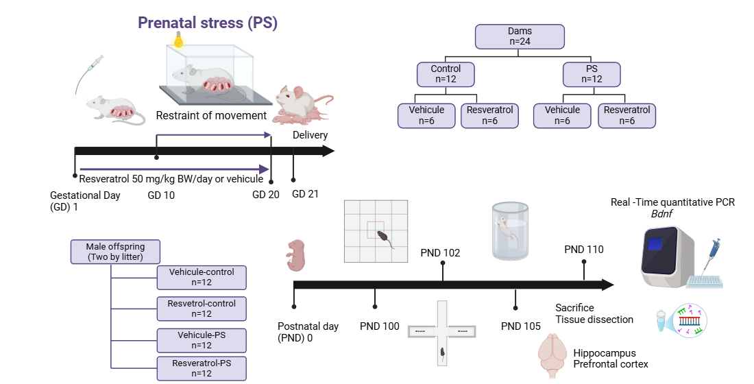
Prenatal stress has been reported to harm the physiological and biochemical functions of the brain of the offspring, potentially resulting in anxiety- and depression-like behaviors later in life. Trans- Resveratrol (RESV) is known for its anti-inflammatory, anxiolytic, and antidepressant properties. However, whether administering RESV during pregnancy can counteract the anxiety- and de-pression like behaviors induced by maternal stress is unknown. This study aimed to assess the protective potential of RESV against molecular and behavioral changes induced by prenatal stress. During pregnancy, the dams received 50 mg/kg BW/day of RESV orally. They underwent a movement restriction for forty-five minutes, three times a day, in addition to being exposed to artificial light 24 hours before delivery. The male offspring were left undisturbed until early adulthood, at which point they underwent behavioral assessments, including the open field test, elevated plus maze, and forced swim test. Subsequently, they were euthanized, and the hippo-campus and prefrontal cortex were extracted for RT-qPCR analysis to measure Bdnf mRNA ex-pression. By weaning, results showed that prenatal stress led to reduced weight gain and, in adulthood, increased anxiety- and depression-like behaviors and changes in Bdnf mRNA expres-sion. However, these effects were attenuated by maternal RESV supplementation. The findings suggest that RESV can prevent anxiety- and depression-like behaviors induced by prenatal stress by modulating Bdnf mRNA expression.

Keywords: 
;  ;  ;  ;  

1. Introduction

Stress plays a key role throughout the lifetime of a being, but its impact is significant in critical developmental phases [1]. A large amount of evidence emphasizes the impact of psychological stress during pregnancy on various aspects of offspring growth and development [2]. The prenatal period is crucial as it determines neural plasticity and regulates brain programming. Disruptions in these processes can lead to neurological disorders in offspring, including generalized anxiety disorder, depressive disorders, attention deficit hyperactivity disorder, autism spectrum disorders, and schizophrenia [3].
The underlying mechanisms by which prenatal stress (PS) affects the neurodevelopmental programming process remain unclear. However, altered hypothalamic-pituitary-adrenal (HPA) axis [4], reactive oxygen species [5], neuroinflammatory pathways [6], and immune system hyperactivation are believed to play a role [7]. Likewise, these effects are influenced by the type and severity of stress, the timing and duration of exposure, and the gender of the offspring [8]. These factors affect the transcription of essential genes involved in neurodevelopment, such as brain-derived neurotrophic factor (BDNF) [9]. BDNF is the most abundant neurotrophin in the brain. It is crucial for neuronal survival and growth, synaptic transmission, neurogenesis, modulation of neurotransmitters, as well as learning, memory, and stress resistance [10]. Studies suggest that stress during early developmental stages can alter the methylation patterns of the Bdnf gene on different brain regions of the offspring, causing a decrease in their expression in the hippocampus and prefrontal cortex [11], which has been implicated in various psychological and neurological disorders, such as anxiety, depression, schizophrenia, Parkinson’s, and Alzheimer’s disease [12,13]. Even though a reduction in total Bdnf mRNA and protein is the most often seen effect of PS on BDNF, contradictory results have also been found [9].
Resveratrol (trans-3,5,4′-trihydroxy-trans-stilbene, RESV), a polyphenol found in grapes and berries [14], has a variety of biological properties, including antioxidant, anti-inflammatory, anticancerogenic, and neuroprotective effects [15]. Studies have reported that RESV exerts anxiolytic- and antidepressant-like effects in rodents by downregulating hyperactivity of the HPA axis and upregulating BDNF levels in the hippocampus, amygdala, and prefrontal cortex [16,17]. Similarly, RESV can ameliorate neuroinflammation induced by maternal separation via activating the Sirt1/NF-κB pathway [18]. Considering that this information suggests that RESV reduces the effect of stress, it is intriguing that the effect of maternal RESV supplementation on anxiety-like and depression-like behaviors in the offspring has not been explored.
In the present study, the male offspring rat was used to investigate whether maternal resveratrol intake can prevent prenatal stress-induced anxiety and depression-like behaviors in adulthood and, if so, whether the mechanism involves the modulation of the Bdnf expression in the hippocampus and prefrontal cortex.

2. Materials and Methods

2.1. Drugs

RESV was purchased from Sigma Chemical Co. (St. Louis, MO, USA). The drugs were dissolved in an aqueous solution of 0.5% sodium carboxymethyl cellulose, and the vehicle (VEH) groups received only this solution.

2.2. Animals

Forty female Wistar rats (200 and 250 g) were provided from the Instituto Nacional de Perinatología Isidro Espinosa de Los Reyes (Mexico City). The rats were housed in a standard environment (temperature: 22 ± 1 ° C; relative humidity: 55 ± 5%) with ad libitum access to food and water. After two weeks of acclimatization, breeding was conducted by placing two females with a male for 24 hours. Gestational day (GD) 1 was marked by performing a vaginal smear to confirm the presence of sperm. The delivery day was appointed as postnatal day (PND) 0. The subjects of the study were two male offspring rats by litter. The experimental protocol was conducted following the Official Mexican Standard (NOM-062-ZOO-1999) and with the approval of the Institutional Committee for the Care and Use of Laboratory Animals of the Research (Mexico, Project 2017-3-138). We took special care to minimize animal suffering and the number of animals used. All experiments were conducted between 10:00 and 15:00, and independent groups were used.

2.3. Experimental Groups

In GD 1, the dams were divided into four groups of 5 as follows: (1) VEH-control group, (2) RSV-control group, (3) VEH-PS group, and (4) RESV-PS group. PS groups (VEH and RESV) were subjected to the method of restriction of movement. RSV groups were treated orally with 50 mg/kg BW/day of RESV, started on GD 1, and continued until GD 20 at 9 am daily. After birth, the offspring were sexed, and only males were selected for this study. On PND 21, two male offspring were taken from each litter, were weaned, and grouped into four groups according to their respective treatments. The male offspring were left undisturbed until early adulthood. They underwent behavioral assessments: an open field test at PND 100, an elevated plus maze at PND 102, and a forced swim test at PND 105.

2.4. Model of Restraint of Movement

The dams (PS groups) were submitted to the method of restriction of movement, which consists of placing the dams in a ventilated acrylic tube 7 cm wide and 19 cm long well ventilated; this is designed for pregnant rats cannot move and does not cause any harm, thus was done in periods of 45 minutes 3 times a day on the ten last days of gestation, additional of this stressor the dams were exposed to an artificial white light induced by a lamp during the last 24 hours until the birth [19,20]. The control group did not receive any intervention.

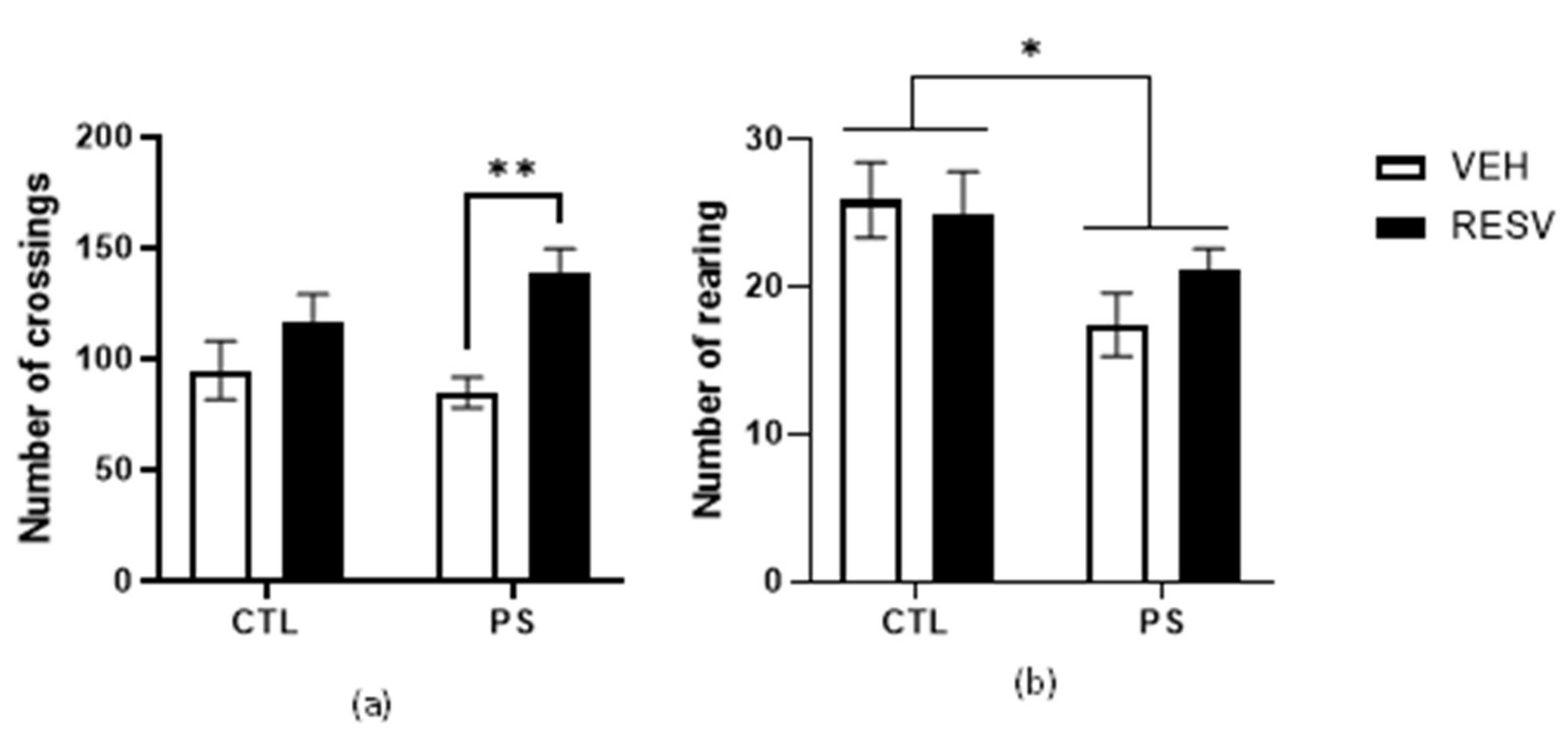
2.5. Open Field Test

The open field test was used to evaluate the locomotor activity of the male offspring rats at PND 100 [21,22]. The test was conducted in a black acrylic box (60 x 60 x 60 cm) with divisions on the floor (15 equal squares). The results were expressed as the number of crossings and rearing over 5 minutes. The session was video recorded, and at the end, 5% ethanol was used to clean the surface.

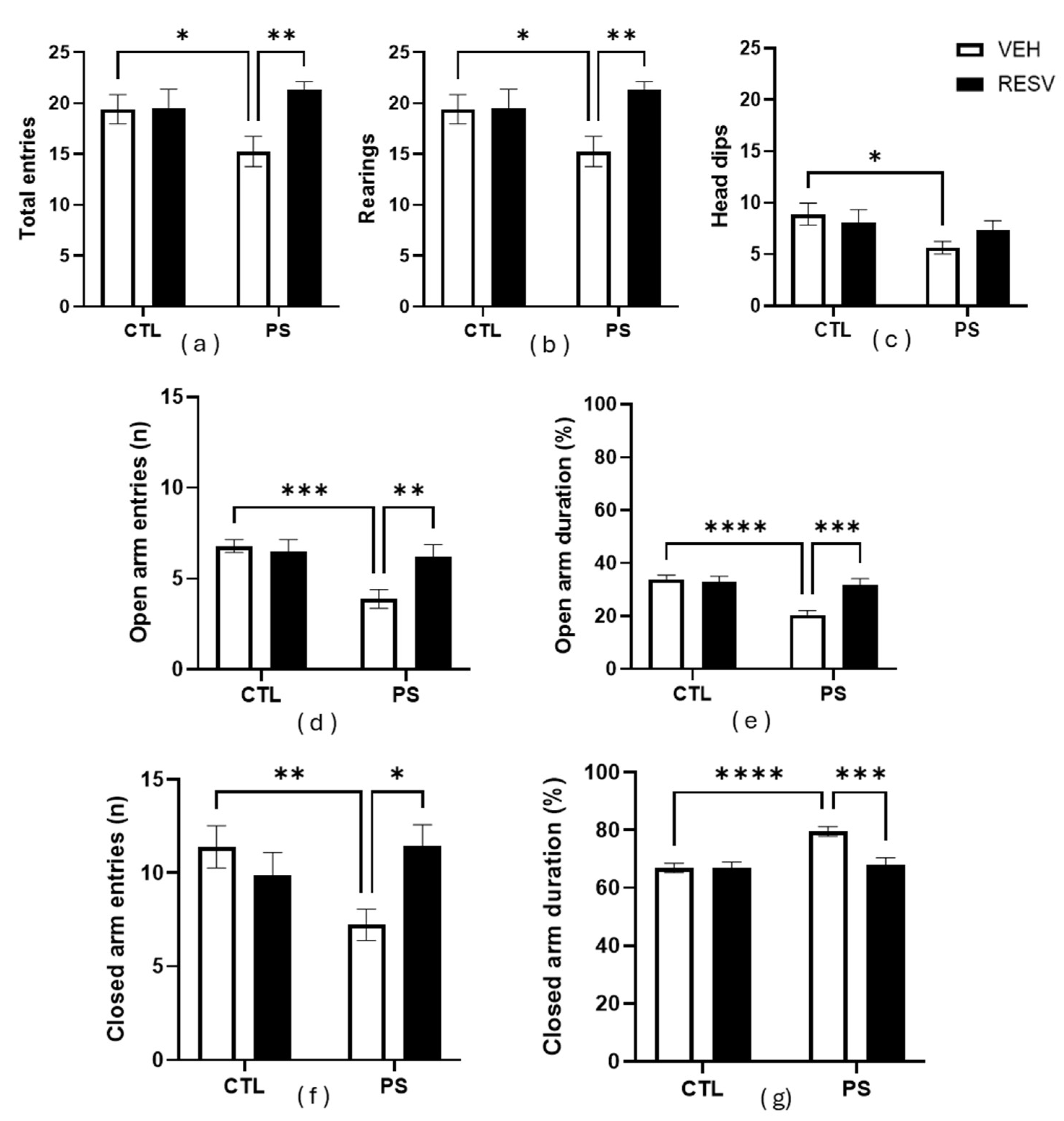
2.6. Elevate Plus Maze

The elevated plus maze test was performed at PND 102 days using a standard procedure [23]. A decrease in the time spent in, and the number of entries into, open arms is thought to be associated with increased anxiety-like behavior in rodents. The apparatus consisted of a cross-shaped platform having two opposing open arms (each 50 cm long × 10 cm wide), two opposing closed arms (each 50 cm long × 10 cm × 40 cm high), and a central arena (10 cm × 10 cm). The apparatus was supported by a platform that kept it 50 cm from the floor. Each rat was placed on the central platform facing one of the open arms and allowed to freely explore for 5 min. The apparatus was washed with 5% ethanol after each mouse to avoid odor interference. The tests were video recorded, and the following behaviors related to anxiety and stress were evaluated: time spent in open and closed arms (when more than 80% of the body is in one arm), rearing (rising to touch or not touch the wall of the maze), and head dipping (poke the head beyond the edge of the maze).

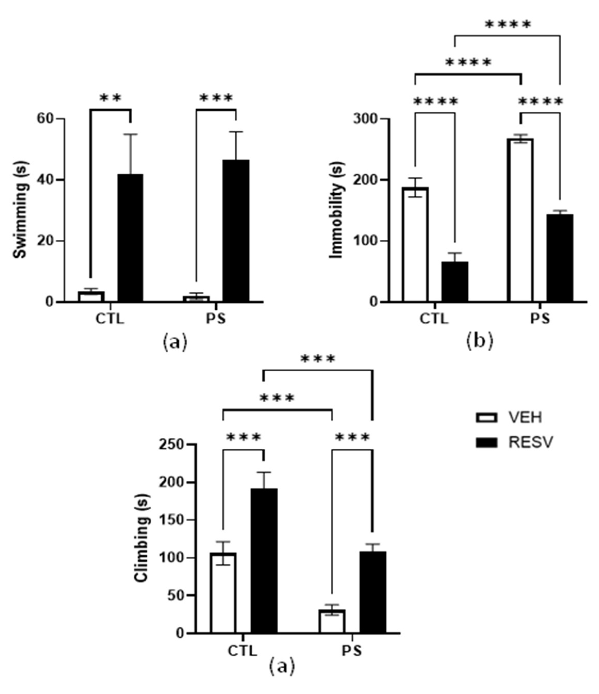
2.7. Forced Swimming Test

To further evaluate the depression-like behavior, a forced swimming test was used at PND 105 [24]. This test was performed in an acrylic cylinder (46 cm in height, 20 cm in diameter) filled with water at 25 ± 1° C to a depth of 30 cm, which ensures that the rat does not have the possibility of touching the bottom with its tail. Between every test, the water was changed to avoid any contamination. The test includes two sections: a 15-minute pretest and a 5-minute test 24 hours later. The data was analyzed by an experimented researcher on a screen focusing on the behaviors related to stress and depression: swimming (when rats made horizontal movements), immersions, floating (when only doing the necessary movements to stay afloat), and climbing (when the rats were in vertical motion).

2.8. Tissue Preparation

All rats were sacrificed on PND 110. The hippocampus and prefrontal cortex were quickly dissected from the brain and stored in a −80°C refrigerator. When needed, samples were taken out for biochemical analyses.

2.9. RNA Isolation

Total RNA was purified with an AllPrep DNA/RNA Mini kit (QIAGEN, USA), following manufacturer instructions. RNA integrity was analyzed by agarose gel electrophoresis, and its purity and quantity were obtained using a Multiskan spectrophotometer (Thermo Fisher Scientific, USA).

2.10. Real Time Quantitative PCR

cDNA synthesis was conducted using the M-MLV reverse transcriptase enzyme (Invitrogen, USA) and 10 mM of each deoxynucleotide (Invitrogen, USA), as specified by the supplier. cDNA was subjected to real-time quantitative PCR (RT-qPCR) using primers targeting Bdnf exon. Gapdh was used as an internal control of constitutive expression. The sequences of the specific primers are listed in Table 1. SYBR Green Master Mix (Thermo Fisher Scientific, USA) was used as the detection method in a CFX96 Touch Real-Time PCR thermocycler (Bio-Rad, USA) following cycling conditions specified by the manufacturer. Relative quantification was performed with the ΔΔCt method [25].

2.11. Statistical Analyses

All statistical analyses were performed using GraphPad Prism version 10.3.1 (GraphPad, San Diego, CA, USA). The data are expressed as the means ± SEM, and p-value < 0.05 was considered statistically significant. The normal distribution of the data was evaluated by the Shapiro-Wilk test. All data were analyzed using two-way analysis of variance (ANOVA) with PS and RESV as independent variables followed by Bonferroni post hoc multiple comparison tests.

3. Results

3.1. Litter Size and Body Weight of Pups

The litters had a similar number of pups and males and females across all experimental groups. As shown in Table 2, there was no significant difference in the body weight of newborn pups. However, by weaning, the PS-VEH group showed less weight gain than the CTL-VEH group (p < 0.001). This effect was mitigated in the CTL-RESV and PS-RESV groups (p < 0.0001).

3.2. Open Field Activity

As shown in Figure 1a, the number of crossings showed a main effect of RSV [F (1, 34) = 11.51; p = 0.002] but no effect of PS [F (1, 34) = 2.28; p = 0.61] nor an interaction between these factors [F (1, 34) = 1.98; p = 0.17]. The post hoc test showed that RSV increased the number of crossings (p = 0.01).
The main effect of PS on rearing activity was significant [F (1,34) = 6.95; p = 0.01], showing a decrease in the number of rearing. However, there was no significant effect of RSV [F (1,34) = 0.32; p = 0.57] nor an interaction between these factors [F (1,34) = 1.01; p = 0.32]. The post hoc test further confirmed the decrease in rearing activity due to PS (p = 0.01) (Figure 1b).

3.3. Elevated plus maze

The analysis of elevated plus maze data is presented in Figure 2. In total entries, there was a main effect of PS [F (1, 32 ) = 4.95; p = 0.033] and an interaction between factors [F(1, 32) = 6.62; p = 0.015] but no effect of RSV [F (1, 32) = 2.21; p = 0.147]). Post hoc testing revealed that PS decreased the total entries (p = 0.001), and this reduction was prevented by RSV (p = 0.009)(Figure 2a).
Rearing (Figure 2b): There was a main effect of RSV [F (1, 32 ) = 4.76; p = 0.037] and of the interaction between factors [F (1, 32) = 4.46; p = 0.043] but no effects of PS [F (1,32) = 0.678; p = 0.416]. The post hoc test showed that the PS decreased rearing activity compared to CTL groups (p = 0.040), and RSV prevented this effect (p = 0.005) (Figure 2).
Head dips (Figure 2c): There was a main effect of PS [F (1,32) = 4.17; p = 0.049] but no effect of RSV [F (1,32 ) = 0.205; p = 0.654] o in the interaction between factors [F (1,32 ) = 1.53; p = 0.224]. The Bonferroni test showed that PS decreased the number of head dips compared to CTL groups (p = 0.023).
Open arm entries (number [n]): there was a main effect of PS [F (1, 32 ) = 8.46; p = 0.007] and in the interaction between factors [F(1, 32 ) = 5.77; p = 0.023] but no effect of RSV [F (1, 32) = 3.44; p = 0.073]. The Bonferroni test showed that PS decreased the number of open-arm entries compared to CTL groups (p < 0.001), and this effect was prevented by RSV (p = 0.005) (Figure 2d).
Open arm duration (%): Analysis of data showed that there was a main effect in both PS [F (1, 32 )= 14.45; p < 0.001] and RSV [F (1, 32 ) = 7.31; p = 0.011] as well as in the interaction between factors [F(1,32 ) = 10.04; p = 0.003]. The post hoc test showed that the PS decreasing % open arm duration compared to CTL groups (p < 0.001) and RSV prevented this effect (p < 0.001) (Figure 2e),
Closed arm entries (n): There was interaction between factors [F (1, 32) = 6.89; p = 0.013] but no effect of PS [F (1, 32) = 1.42; p = 0.242] or RSV [F (1, 32) = 1.52; p = 0.227]. The Bonferroni test showed that PS decreased the number of closed-arm entries compared to CTL groups (p = 0.091), and this effect was prevented by RSV (p = 0.01) ) (Figure 2f).
Closed arm duration (%): Analysis of data showed that there was a main effect in both PS [F (1, 32 ) = 12.44; p = 0.001] and RSV [F (1, 32) = 8.79; p = 0.006] as well as in the interaction between factors [F (1, 32) = 8.71; p = 0.006]. The post hoc test showed that the PS increasing % closed arm duration compared to CTL groups (p < 0.001) and RSV prevented this effect (p < 0.001) ) (Figure 2g).

3.4. Forced Swimming Test

Figure 3 presents the results of the forced swimming test. Analysis of swimming behavior revealed main effects of RSV [F (1, 32) = 28.14; p < 0.001] but no effect of PS [F (1, 32) = 0.04, p = 0.845], or interaction [F (1, 32) = 0.156; p = 0.695]. Bonferroni test showed that RSV groups spent more time swimming than VEH groups (p < 0.01) (Figure 3a).
There was a significant effect of both the PS [F(1, 32) = 48.11; p < 0.001] and RSV [F(1, 32) = 117.22; p < 0.001] on immobility time. However, the two factors had no interaction effect [F(1, 32) = 0.009; p = 0.927]. The Bonferroni test revealed that PS rats exhibited significantly greater immobility time compared to CTL rats (p < 0.001), and RSV was effective in preventing this increase (p < 0.001) (Figure 3b).
The study found a significant main effect of PS [F (1, 32) = 29.59; p < 0.001] on climbing behavior. Similarly, there was a significant main effect of RSV [F (1, 32) = 31.62; p < 0.001]. However, there was no interaction between these factors [F (1, 32) = 0.07; p = 0.793]. The post hoc test showed that PS groups spent less time climbing than CTL groups (p < 0.001) and RSV prevented this effect (p < 0.001) (Figure 3c).

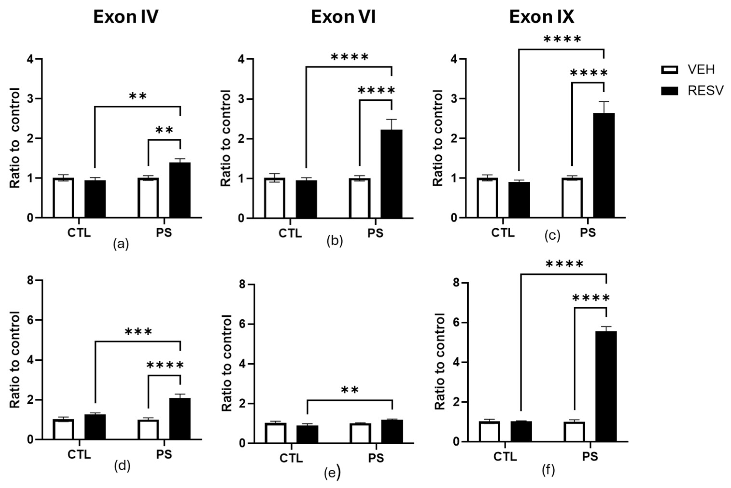
3.5. Gene Expression of Bdnf

We investigated the prefrontal cortex and hippocampal gene expression of main Bdnf transcripts (exon IV, exon VI, and exon IX) to find molecular changes underlying PS and RSV effects (Figure 4).
Gene expression of Bdnf transcripts was evaluated in the prefrontal cortex, which showed a main effect of PS, RSV, and the PS x RSV interaction in exons VI and IX. By exon IV, there was only a main effect in RESV and the PS x RSV interaction (Figure 4a-c). The post hoc test showed an increase in expression levels in PS-RSV groups compared to PS-VEH groups (p < 0.0001) and CTL-RESV groups (p < 0.001). Exon IV, main effect of PS: F (1, 12) = 4.57, p = 0.054; main effect of RSV: F (1, 12) = 7.62, p = 0.018; PS x RSV interaction: F (1, 12) = 13.06, p = 0.003]. Exon VI, main effect of PS: F (1, 12) = 15.63, p = 0.002; main effect of RSV: F(1, 12) = 18.63, p = 0.001 ; main effect of PS x RSV interaction : F (1, 12) = 23.02, p < 0.001. Exon IX, main effect of PS: F(1, 12) = 26.40, p < 0.001; main effect of RSV: F (1, 12) = 27.29, p < 0.001 ; PS x RSV interaction : F(1, 12) = 34.50, p < 0.001.
The gene expression profile in the hippocampus revealed that PS significantly affected the expression level of all Bdnf transcripts (Figure 4d-f). The main effect of RESV and the interaction of PS x RSV were only seen in exon IX. The Bonferroni test showed that expression levels of all Bndf transcripts were higher in PS-RSV groups compared to CTL-RSV groups (p < 0.001). Additionally, there was an increase in the expression levels of exon IV and a decrease in exon IX in the PS-VEH groups compared to the CTL-VEH groups. Exon IV, main effect of PS: F (1, 12) = 44.50, p < 0.0001; main effect of RSV: F (1, 12) = 0.178, p = 0.687; PS x RSV interaction: F (1, 12) = 1.57, p = 0.234. Exon VI, main effect of PS: F (1, 12) = 12.57, p = 0.004; main effect of RSV: F (1, 12) = 0.856, p = 0.373; PS x RSV interaction: F (1, 12) = 0.736, p = 0.407. Exon IX, main effect of PS: F(1, 12) = 208.5, p < 0.0001; main effect of RSV: F(1, 12) = 385.7, p < 0.0001 ; PS x RSV interaction : F(1, 12) = 281.6, p < 0.0001.

4. Discussion

In this study, we explored the effect of maternal RESV supplementation in prenatally stressed male offspring rats. The results suggest that PS throughout the gestational period led to reduced body weight gain and, in adulthood, increased anxiety- and depression-like behaviors and changes in Bdnf mRNA expression. Maternal RESV supplementation attenuated all these effects.
In recent years, a growing body of evidence suggests a connection between the early environment and later psychiatric disorders. Such research has been primarily inspired by the developmental origins of the health and disease (DOHaD) model, which proposes a link between fetal development and cardiovascular and metabolic disease in later life [26]. Barker and colleagues applied the DOHaD model early to mental health outcomes [27]. They investigated adult suicide rates of birth weight and growth during the first year of life. Their results showed that while birth weight alone was not a predictor, the average weight of 12-month-old infants was over 400 grams lower in those who later committed suicide. These observations suggested that altered programming could influence growth in infancy and mood throughout life.
PS caused by maternal movement restraint is a well-established model of early stress recognized for causing enduring physiological, neurobiological, and behavioral changes [28]. Low birth weight is one of the most reported effects of gestational stress in newborn humans, and it has been associated with the development of a variety of metabolic diseases in adulthood [26]. On the other hand, most studies examining the bodyweight effects in rat offspring have reported contradictory findings. Therefore, in the current study, male offspring did not display an effect of the gestational stress manipulation on weight at PD1. A difference in weight was seen until the weaning (male offspring from stressed mothers weighing less than those from control mothers). Consistent with our findings that weight differences in offspring rats become noticeable only later in their development, Baker et al. [29,30] reported in two separate studies that gestational stress did not affect the weight of offspring from stressed mothers between postnatal days 2 and 24. However, weight differences appeared in female offspring from stressed mothers starting around 36 days of age, and a similar pattern was observed in male offspring from stressed mothers, but only in adulthood. This weight effect could be explained by abnormal secretion of growth hormone and irregularities in the HPA, hypothalamic-adrenal, and hypothalamic-thyroid axes [31,32]. Nevertheless, due to variations in results, it is crucial to consider the timing, dosage, and duration of the gestational stress model used [8].
Early-life exposure to adverse environments is often linked to higher rates of mood disorders in adulthood. Maternal movement restraint can lead to hyperactivation of the HPA axis, increased anxiety- and depression-related behavior, and changes in the expression of serotoninergic, dopaminergic, and glutaminergic receptors in the brains of offspring [28,32,33,34,35]. Studies have demonstrated that maternal movement restraint can exacerbate depressive-like behaviors in juvenile and adult male Wistar or Sprague Dawley rats, as observed in the forced swimming test and anxiety-like behaviors in adolescent male Sprague-Dawley rats, as determined in the elevated plus maze [35,36,37]. Additionally, it is associated with a lower tendency to explore in the open field test [34,35]. Our results agree with these findings, as adult male rat offspring exposed to PS showed fewer rearings and crossings in the open field test compared to the CTL groups, which suggests that the PS group showed a lower tendency to explore. These rats also displayed anxiety-like behaviors, evidenced by reduced time spent in and number of entries into the open arms in the elevated plus maze test, and depression-like behaviors, indicated by increased immobility time and decreased climbing behavior in the forced swimming test. Several studies have shown that maternal stress can affect the development of central monoaminergic and glutamatergic neurons [38,39]. For instance, the offspring of stressed rats displayed hyperactive behavior in the central serotonin system, which could be linked to the anxiety-inducing effects of maternal movement restraint. Additionally, it has been proven that restraint stress exerted onto the pregnant dam can have long-term effects on the dopaminergic system development in their offspring [33]. According to these studies, PS increases dopamine (DA) D2 receptors in limbic areas, reduces DA-stimulated release in cortical areas, increases in the nucleus accumbens, and disrupts the DA-glutamate balance, which could lead to an increased susceptibility to depression in the offspring.
BDNF is a member of the family of neurotrophins that are needed for the proper development and survival of neurons, and the dysregulation of its expression is related to neurodegenerative and neuropsychiatric disorders [40]. Regarding affective disorders, it has been documented that the rodent Bdnf gene is regulated by stress and HPA axis activation [41]. Because Bdnf has a very complex gene structure and can produce a variety of mRNAs with different functions [42], we decided to evaluate the effect of PS on the expression of different exons of Bdnf in brain areas involved in anxiety and depression, like the prefrontal cortex and hippocampus. Our data showed that PS increases the expression of exon IV while it reduces the expression of exon IX selectively in the hippocampus. In the prefrontal cortex, the prenatal manipulation did not affect the expression level of any of the exons evaluated. Our findings support previously reported studies, such as Boersma et al. [43], which described altered Bdnf expression in the hippocampus due to PS exposure in adult offspring without altering expression in the prefrontal cortex, and the previously reported association between exon IV with depression and anxiety [44,45]. Considering the critical roles in the modulation level of total Bdnf mRNA and their exons by PS, our results emphasize the impact of stressful experiences in the prenatal stage on neurodevelopment and the origins of neuropsychiatric disorders.
Several beneficial effects of RESV have been reported, and many are particularly relevant to behavioral disorders. Its ability to regulate key molecular targets, including genes such as Bdnf, has been a subject of intense research [46]. Preclinical studies have shown that RESV plays an antidepressant and anxiolytic role in rodent models of anxiety and depression induced by estrogen deficiency, social isolation and maternal separation [18,47,48]. Our findings suggested that RESV improved anxiety-like behaviors caused by maternal movement restraint, as evidenced by the significant increase in the time spent and the number of entries into the open arms in the elevated plus maze. Furthermore, RESV reduced the immobility time and increased climbing behavior in the forced swimming tests, implying that RESV ameliorated depression-like behaviors caused by PS. In addition, our data showed that RESV increased the expression of Bdnf exons IV, VI, and IX in the prefrontal cortex and hippocampus of prenatally stressed adult male offspring. These results agree with the published data on the increasing effects of RSV on the expression of hippocampal levels of total Bdnf mRNA in male adult rats and Bdnf transcripts in pregnant rats [49,50].
In summary, the results of this study suggest that PS can lead to anxiety- and depression-like behavior in male offspring, effects being mediated by the regulation of Bdnf mRNA expression in the hippocampus. Additionally, RESV mitigated the anxiety- and depression-like behaviors induced by PS by increasing the expression of Bdnf exons IV, VI, and IX. This study is one of the first to evaluate the potential of pharmacological modulation of individual Bdnf transcripts by RSV for the treatment of anxiety and depression. However, a limitation of our study is that it does not provide information on the BDNF protein or whether changes in the expression of Bdnf transcripts are related to altered epigenetic mechanisms. This should be addressed in future experiments.

5. Conclusions

In conclusion, the results of this study suggested that PS can lead to anxiety- and depression-like behavior in male offspring, effects that are mediated by the regulation of Bdnf mRNA expression in the hippocampus. Additionally, RESV ameliorated anxiety- and depression-like behaviors induced by PS by increasing the expression of Bdnf exons IV, VI, and IX. While our study offers the possibility of novel and more specific treatments, future investigations are required to fully understand the mechanisms underlying the selective expression of different Bdnf transcripts in different brain areas and the temporal regulation of the expression of Bdnf.

Author Contributions

Conceptualization, L.-M.M. and E.-R.J.; methodology, V.-J.G., E.V.M. and G.-P.R.; formal analysis, V.-J.G., E.V.M.; writing—original draft preparation, L.-M.M., E.-R.J., V.-J.G.; writing—review and editing, L.-M.M., E.-R.J., V.-J.G., V.-M.E.R. and G.-P.R. All authors have read and agreed to the published version of the manuscript.

Funding

This research was funded by Instituto Nacional de Perinatología Isidro Espinosa de Los Reyes (grant number Project 2017-3-138), Instituto Politécnico Nacional, and COFAA-SIP/IPN (SIP 20221200).

Institutional Review Board Statement

The animal study protocol was approved by the Institutional Committee for the Care and Use of Laboratory Animals of the of Instituto Nacional de Perinatología Isidro Espinosa de Los Reyes (Protocol 2017-3-138; approved on 19 November 2019) .

Informed Consent Statement

Not applicable.

Data Availability Statement

The raw data of this article will be made available by the authors upon request .

Acknowledgments

We gratefully acknowledge Instituto Nacional de Perinatología, Instituto Politécnico Nacional, and COFAA-SIP/IPN.

Conflicts of Interest

The authors declare no conflicts of interest.

References

  1. Barker, D.J. A new model for the origins of chronic disease. Med. Health Care Philos. 2001, 4, 31–35. [Google Scholar] [CrossRef]
  2. Baker, S.L.; Mileva, G.; Huta, V.; Bielajew, C. In utero programming alters adult response to chronic mild stress: part 3 of a longitudinal study. Brain Res. 2014, 1588, 175–189. [Google Scholar] [CrossRef]
  3. Van den Bergh, B.R.H.; van den Heuvel, M.I.; Lahti, M.; Braeken, M.; de Rooij, S.R.; Entringer, S.; Hoyer, D.; Roseboom, T.; Räikkönen, K.; King, S.; et al. Prenatal developmental origins of behavior and mental health: The influence of maternal stress in pregnancy. Neurosci. Biobehav. Rev. 2020, 117, 26–64. [Google Scholar] [CrossRef] [PubMed]
  4. Krontira, A.C.; Cruceanu, C.; Binder, E.B. Glucocorticoids as Mediators of Adverse Outcomes of Prenatal Stress. Trends Neurosci. 2020, 43, 394–405. [Google Scholar] [CrossRef]
  5. Haq, S.U.; Bhat, U.A.; Kumar, A. Prenatal stress effects on offspring brain and behavior: Mediators, alterations and dysregulated epigenetic mechanisms. J. Biosci. 2021, 46, 34. [Google Scholar] [CrossRef]
  6. Han, V.X.; Patel, S.; Jones, H.F.; Dale, R.C. Maternal immune activation and neuroinflammation in human neurodevelopmental disorders. Nat. Rev. Neurol. 2021, 17, 564–579. [Google Scholar] [CrossRef] [PubMed]
  7. Nazzari, S.; Frigerio, A. The programming role of maternal antenatal inflammation on infants' early neurodevelopment: A review of human studies: Special Section on "Translational and Neuroscience Studies in Affective Disorders" Section Editor, Maria Nobile MD, PhD. J. Affect Disord. 2020, 263, 739–746. [Google Scholar] [CrossRef]
  8. Kim, D.R.; Bale, T.L.; Epperson, C.N. Prenatal programming of mental illness: current understanding of relationship and mechanisms. Curr. Psychiatry Rep. 2015, 17, 5. [Google Scholar] [CrossRef] [PubMed]
  9. Badihian, N.; Daniali, S.S.; Kelishadi, R. Transcriptional and epigenetic changes of brain derived neurotrophic factor following prenatal stress: A systematic review of animal studies. Neurosci. Biobehav. Rev. 2020, 117, 211–231. [Google Scholar] [CrossRef]
  10. Kowiański, P.; Lietzau, G.; Czuba, E.; Waśkow, M.; Steliga, A.; Moryś, J. BDNF: A Key Factor with Multipotent Impact on Brain Signaling and Synaptic Plasticity. Cell Mol. Neurobiol. 2018, 38, 579–593. [Google Scholar] [CrossRef] [PubMed]
  11. Miao, Z.; Wang, Y.; Sun, Z. The Relationships Between Stress, Mental Disorders, and Epigenetic Regulation of BDNF. Int. J. Mol. Sci. 2020, 21, 1375. [Google Scholar] [CrossRef] [PubMed]
  12. Balaratnasingam, S.; Janca, A. Brain Derived Neurotrophic Factor: a novel neurotrophin involved in psychiatric and neurological disorders. Pharmacol. Ther. 2012, 134, 116–124. [Google Scholar] [CrossRef] [PubMed]
  13. Miranda, M.; Morici, J.F.; Zanoni, M.B.; Bekinschtein, P. Brain-Derived Neurotrophic Factor: A Key Molecule for Memory in the Healthy and the Pathological Brain. Front. Cell Neurosci. 2019, 13, 363. [Google Scholar] [CrossRef] [PubMed]
  14. Farhan, M.; Rizvi, A. The Pharmacological Properties of Red Grape Polyphenol Resveratrol: Clinical Trials and Obstacles in Drug Development. Nutrients 2023, 15, 4486. [Google Scholar] [CrossRef]
  15. Faisal, Z.; Mazhar, A.; Batool, S.A.; Akram, N.; Hassan, M.; Khan, M.U.; Afzaal, M.; Hassan, U.U.; Shah, Y.A.; Desta, D.T. Exploring the multimodal health-promoting properties of resveratrol: A comprehensive review. Food Sci. Nutr. 2024, 12, 2240–2258. [Google Scholar] [CrossRef] [PubMed]
  16. Moore, A.; Beidler, J.; Hong, M.Y. Resveratrol and Depression in Animal Models: A Systematic Review of the Biological Mechanisms. Molecules 2018, 23, 2197. [Google Scholar] [CrossRef]
  17. Shayganfard, M. Molecular and biological functions of resveratrol in psychiatric disorders: a review of recent evidence. Cell Biosci. 2020, 10, 128. [Google Scholar] [CrossRef]
  18. Wei, R.M.; Zhang, Y.M.; Feng, Y.Z.; Zhang, K.X.; Zhang, J.Y.; Chen, J.; Luo, B.L.; Li, X.Y.; Chen, G.H. Resveratrol ameliorates maternal separation-induced anxiety- and depression-like behaviors and reduces Sirt1-NF-kB signaling-mediated neuroinflammation. Front Behav. Neurosci. 2023, 17, 1172091. [Google Scholar] [CrossRef] [PubMed]
  19. Molina, P.; Andero, R.; Armario, A. Restraint or immobilization: A comparison of methodologies for restricting free movement in rodents and their potential impact on physiology and behavior. Neurosci. Biobehav. Rev. 2023, 151, 105224. [Google Scholar] [CrossRef]
  20. Weinstock, M. Alterations induced by gestational stress in brain morphology and behaviour of the offspring. Prog. Neurobiol. 2001, 65, 427–451. [Google Scholar] [CrossRef]
  21. Seibenhener, M.L.; Wooten, M.C. Use of the Open Field Maze to measure locomotor and anxiety-like behavior in mice. J. Vis. Exp. 2015, 96, e52434. [Google Scholar] [CrossRef]
  22. Kraeuter, A.K.; Guest, P.C.; Sarnyai, Z. The Open Field Test for Measuring Locomotor Activity and Anxiety-Like Behavior. Methods Mol. Biol. 2019, 1916, 99–103. [Google Scholar] [CrossRef] [PubMed]
  23. Pellow, S.; Chopin, P.; File, S.E.; Briley, M. Validation of open: closed arm entries in an elevated plus-maze as a measure of anxiety in the rat. J. Neurosci. Methods. 1985, 14, 149–167. [Google Scholar] [CrossRef]
  24. Porsolt, R.D.; Le Pichon, M.; Jalfre, M. Depression: a new animal model sensitive to antidepressant treatments. Nature 1977, 266, 730–732. [Google Scholar] [CrossRef] [PubMed]
  25. Schmittgen, T.D.; Livak, K.J. Analyzing real-time PCR data by the comparative C(T) method. Nat. Protoc. 2008, 3, 1101–1108. [Google Scholar] [CrossRef] [PubMed]
  26. Barker, D.J. The developmental origins of chronic adult disease. Acta Paediatr. Suppl. 2004, 93, 26–33. [Google Scholar] [CrossRef]
  27. Barker, D.J.; Osmond, C.; Rodin, I.; Fall, C.H.; Winter, P.D. Low weight gain in infancy and suicide in adult life. B.M.J. 1995, 311, 1203. [Google Scholar] [CrossRef]
  28. Maccari, S.; Morley-Fletcher, S. Effects of prenatal restraint stress on the hypothalamus-pituitary-adrenal axis and related behavioural and neurobiological alterations. Psychoneuroendocrinology 2007, 32 (Suppl 1), S10–S15. [Google Scholar] [CrossRef]
  29. Baker, S.; Chebli, M.; Rees, S.; Lemarec, N.; Godbout, R.; Bielajew, C. Effects of gestational stress: 1. Evaluation of maternal and juvenile offspring behavior. Brain Res. 2008, 1213, 98–110. [Google Scholar] [CrossRef]
  30. Baker, S.; Rees, S.; Chebli, M.; Lemarec, N.; Godbout, R.; Huta, V.; Bielajew, C. Effects of gestational stress: 2. Evaluation of male and female adult offspring. Brain Res. 2009, 1302, 194–204. [Google Scholar] [CrossRef]
  31. Nicolaides, N.C.; Kanaka-Gantenbein, C.; Pervanidou, P. Developmental Neuroendocrinology of Early-Life Stress: Impact on Child Development and Behavior. Curr. Neuropharmacol. 2024, 22, 461–474. [Google Scholar] [CrossRef] [PubMed]
  32. Anifantaki, F.; Pervanidou, P.; Lambrinoudaki, I.; Panoulis, K.; Vlahos, N.; Eleftheriades, M. Maternal Prenatal Stress, Thyroid Function and Neurodevelopment of the Offspring: A Mini Review of the Literature. Front. Neurosci. 2021, 15, 692446. [Google Scholar] [CrossRef] [PubMed]
  33. Baier, C.J.; Katunar, M.R.; Adrover, E.; Pallarés, M.E.; Antonelli, M.C. Gestational restraint stress and the developing dopaminergic system: an overview. Neurotox. Res. 2012, 22, 16–32. [Google Scholar] [CrossRef]
  34. Sun, H.; Guan, L.; Zhu, Z.; Li, H. Reduced levels of NR1 and NR2A with depression-like behavior in different brain regions in prenatally stressed juvenile offspring. PLoS One 2013, 8, e81775. [Google Scholar] [CrossRef]
  35. Weinstock, M. Prenatal stressors in rodents: Effects on behavior. Neurobiol. Stress. 2016, 6, 3–13. [Google Scholar] [CrossRef] [PubMed]
  36. Zhou, J.; Ma, Y.; Chen, J.; Yao, D.; Feng, C.; Dong, Y.; Ren, Y.; Ma, H.; Wang, Z.; Li, G.; et al. Effects of RhoA on depression-like behavior in prenatally stressed offspring rats. Behav. Brain Res. 2022, 432, 113973. [Google Scholar] [CrossRef] [PubMed]
  37. Zohar, I.; Shoham, S.; Weinstock, M. Perinatal citalopram does not prevent the effect of prenatal stress on anxiety, depressive-like behaviour and serotonergic transmission in adult rat offspring. Eur. J. Neurosci. 2016, 43, 590–600. [Google Scholar] [CrossRef] [PubMed]
  38. Oosterhof, C.A.; El Mansari, M.; Merali, Z.; Blier, P. Altered monoamine system activities after prenatal and adult stress: A role for stress resilience? Brain Res. 2016, 1642, 409–418. [Google Scholar] [CrossRef]
  39. Roshan-Milani, S.; Seyyedabadi, B.; Saboory, E.; Parsamanesh, N.; Mehranfard, N. Prenatal stress and increased susceptibility to anxiety-like behaviors: role of neuroinflammation and balance between GABAergic and glutamatergic transmission. Stress 2021, 24, 481–495. [Google Scholar] [CrossRef]
  40. Colucci-D'Amato, L.; Speranza, L.; Volpicelli, F. Neurotrophic Factor BDNF, Physiological Functions and Therapeutic Potential in Depression, Neurodegeneration and Brain Cancer. Int. J. Mol. Sci. 2020, 21, 7777. [Google Scholar] [CrossRef] [PubMed]
  41. Naert, G.; Ixart, G.; Maurice, T.; Tapia-Arancibia, L.; Givalois, L. Brain-derived neurotrophic factor and hypothalamic-pituitary-adrenal axis adaptation processes in a depressive-like state induced by chronic restraint stress. Mol. Cell Neurosci. 2011, 46, 55–66. [Google Scholar] [CrossRef]
  42. You, H.; Lu, B. Diverse Functions of Multiple Bdnf Transcripts Driven by Distinct Bdnf Promoters. Biomolecules 2023, 13, 655. [Google Scholar] [CrossRef]
  43. Boersma, G.J.; Lee, R.S.; Cordner, Z.A.; Ewald, E.R.; Purcell, R.H.; Moghadam, A.A.; Tamashiro, K.L. Prenatal stress decreases Bdnf expression and increases methylation of Bdnf exon IV in rats. Epigenetics 2014, 9, 437–447. [Google Scholar] [CrossRef] [PubMed]
  44. Sakata, K.; Jin, L.; Jha, S. Lack of promoter IV-driven BDNF transcription results in depression-like behavior. Genes Brain Behav. 2010, 9, 712–721. [Google Scholar] [CrossRef]
  45. Sakata, K.; Mastin, J.R.; Duke, S.M.; Vail, M.G.; Overacre, A.E.; Dong, B.E.; Jha, S. Effects of antidepressant treatment on mice lacking brain-derived neurotrophic factor expression through promoter IV. Eur. J. Neurosci. 2013, 37, 1863–1874. [Google Scholar] [CrossRef] [PubMed]
  46. Moore, A.; Beidler, J.; Hong, M.Y. Resveratrol and Depression in Animal Models: A Systematic Review of the Biological Mechanisms. Molecules 2018, 23, 2197. [Google Scholar] [CrossRef] [PubMed]
  47. Liu, T.; Ma, Y.; Zhang, R.; Zhong, H.; Wang, L.; Zhao, J.; Yang, L.; Fan, X. Resveratrol ameliorates estrogen deficiency-induced depression- and anxiety-like behaviors and hippocampal inflammation in mice. Psychopharmacology (Berl) 2019, 236, 1385–1399. [Google Scholar] [CrossRef] [PubMed]
  48. Gu, Z.; Chu, L.; Han, Y. Therapeutic effect of resveratrol on mice with depression. Exp. Ther. Med. 2019, 17, 3061–3064. [Google Scholar] [CrossRef] [PubMed]
  49. Rahvar, M.; Nikseresht, M.; Shafiee, S.M.; Naghibalhossaini, F.; Rasti, M.; Panjehshahin, M.R.; Owji, A.A. Effect of oral resveratrol on the BDNF gene expression in the hippocampus of the rat brain. Neurochem. Res. 2011, 36, 761–765. [Google Scholar] [CrossRef] [PubMed]
  50. Shojaei, S.; Panjehshahin, M.R.; Shafiee, S.M.; Khoshdel, Z.; Borji, M.; Ghasempour, G.; Owji, A.A. Differential Effects of Resveratrol on the Expression of Brain-Derived Neurotrophic Factor Transcripts and Protein in the Hippocampus of Rat Brain. Iran J. Med. Sci. 2017, 42, 32–39. [Google Scholar]
Figure 1. Effects of maternal resveratrol supplementation in the open field test of prenatal stress adult male offspring. (a) Number of crossings; (b) Number of rearing. All data are mean ± SEM with 10-11 animals in each group. * p < 0.05; ** p < 0.01 by two-way ANOVA followed by Bonferroni post hoc tests. CTL= control; PS = prenatal stress; VEH = vehicule; RESV = resveratrol.
Preprints 144663 g001
Figure 2. Effects of maternal resveratrol supplementation in the elevated plus maze of prenatal stress adult male offspring. (a) Total entries; (b) Rearings; (c) Head dips; (d) Open arm entries; (e) Open arm duration; (f) Closed arm entries; (g) Closed arm duration. All data are mean ± SEM with 10 - 11 animals in each group.* p < 0.05; ** p < 0.01; *** p < 0.001; **** p < 0.0001 by two-way ANOVA followed by Bonferroni post hoc tests. CTL= control; PS = prenatal stress; VEH = vehicule; RESV = resveratrol.
Preprints 144663 g002
Figure 3. Effects of maternal resveratrol supplementation in the forced swim test of prenatal stress adult male offspring. (a) Swimming; (b) Immobility; (c) Climbing. All data are mean ± SEM with 10 - 11 animals in each group. ** p < 0.01; *** p < 0.001; **** p < 0.0001 by two-way ANOVA followed by Bonferroni post hoc tests. CTL= control; PS = prenatal stress; VEH = vehicule; RESV = resveratrol.
Preprints 144663 g003
Figure 4. Effects of maternal supplementation of resveratrol on the expression of exons IV, IV and IX of the Bdnf in the prefrontal cortex (a, b,c) and hippocampus (d, e, f) of prenatal stress adult male offspring. All data are mean SEM with four animals in each group. ** p < 0.01; *** p < 0.001; **** p < 0.0001 by two-way ANOVA followed by Bonferroni post hoc tests. CTL= control; PS = prenatal stress; VEH = vehicule; RESV = resveratrol.
Preprints 144663 g004
Table 1. Sequences of primers used for qRT-PCR amplification of transcripts of interest.
Gene name Primer forward (5’ to 3’) Primer reverse (5’ to 3’)
Bdnf exon IV TGGTGGCCGATATGTACTCC ACTGAAGGCGTGCGAGTATT
Bdnf exon VI TTGTTGTCACGCTCCTGGTC GATGAGACCGGGTTCCCTCA
Bdnf exon IX TTCCTCCAGCAGAAAGAGCA TCCCTGGCTGACACTTTTGA
Gapdh GGATGCAGGGATGATGTTC TGCACCACCAACTGCTTAG
Table 2. Summarized data on the litter size, number of males and females born, body weights and changes in body weight.
Groups of dams Litter size (mean ± SEM) No. of pups born No. of males and females Body weight of pups (g) Changes in body weight (g)
CTL-VEH 12.00 ± 0.16 11 to 13 M = 4.66 ± 0.40
F = 7.33± 0.12
PD1=7.00 ± 0.09
PD21= 48.50 ± 1.17
41.00 ± 1.20
PS-VEH 10.66 ± 0.10 10 to 11 M = 5.33 ± 0.14
F = 5.33 ± 0.14
PD1=6.60 ± 0.09
PD21=34.00 ± 0.57
27.50 ± 0.58*
CTL-RESV 10.33 ± 18.85 9 to 12 M = 4.66 ± 0.40
F= 5.66 ± 0.74
PD1=6.70 ± 0.11
PD21=47.40 ± 0.66
40.60 ± 0.67
PS-RESV 12.66 ± 2.05 12 to 13 M = 4.33 ± 0.43
F= 8.33 ± 0.41
PD1=7.08 ± 0.13
PD21=51.20 ± 0.94
44.30 ± 0.90
* p < 0.0001 vs. CTL-RESV and PS-RESV groups. CTL= control; RESV = resveratrol; M = male; F = female; PD = posnatal day.
Disclaimer/Publisher’s Note: The statements, opinions and data contained in all publications are solely those of the individual author(s) and contributor(s) and not of MDPI and/or the editor(s). MDPI and/or the editor(s) disclaim responsibility for any injury to people or property resulting from any ideas, methods, instructions or products referred to in the content.
Copyright: This open access article is published under a Creative Commons CC BY 4.0 license, which permit the free download, distribution, and reuse, provided that the author and preprint are cited in any reuse.
Prerpints.org logo

Preprints.org is a free preprint server supported by MDPI in Basel, Switzerland.

Subscribe

Disclaimer

Terms of Use

Privacy Policy

Privacy Settings

© 2025 MDPI (Basel, Switzerland) unless otherwise stated