
Submitted:
27 December 2024
Posted:
31 December 2024
You are already at the latest version
Abstract
A 70-year-old woman with no significant medical history presented to emergency room with abdominal pain and diarrhea. Routine stool tests for bacterial pathogens and fecal leukocytes were negative. However, microscopic examination revealed an unusual round structure with hyaline pseudopod-like extensions and a double membrane, devoid of visible internal structures or motility. Literature review suggested it might be Urbanorum sp, a controversial organism first described in the 1990s. While some studies propose Urbanorum sp. as a human pathogen, others suggest alternative explanations, such as plant-origin debris. The classification of this structure remains uncertain, highlighting the need for further research to determine its true nature and potential clinical significance.
Keywords:
Microbiology
; Microscope
; Parasitology
Introduction
Laboratory diagnosis plays a critical role in identifying the etiological agents of diarrheal diseases, particularly in distinguishing parasitic infections from bacterial, viral, or non-infectious causes. Parasitological evaluation typically involves stool microscopy, which remains the cornerstone for detecting eggs, cysts, and trophozoites of intestinal parasites. Advanced techniques, such as concentration methods (e.g., formalin-ethyl acetate sedimentation), staining (e.g., trichrome, modified acid-fast), and immunoassays, enhance sensitivity and specificity. Molecular diagnostics, including polymerase chain reaction (PCR), have further revolutionized parasite detection by identifying genetic material of protozoa and helminths with high accuracy. Despite these advancements, challenges persist, such as differentiating artifacts or contaminants from true pathogens, highlighting the need for well-trained personnel and rigorous methodological approaches. Accurate diagnosis is crucial to guide appropriate treatment, reduce complications, and improve patient outcomes, especially in regions where parasitic infections are endemic.
Urbanorum sp. is a debated entity reported in the context of diarrheal illnesses, particularly in South America (Figure 1). Its first mention dates back to the 1990s, attributed to findings by Professor Francisco Tirado Santamaría [1]. However, the original description lacks formal scientific validation, relying instead on anecdotal evidence and unverified microscopic observations. Subsequent case reports, such as one in Ecuador [2], have attempted to associate Urbanorum sp. with diarrheal episodes, but these studies often fail to meet rigorous diagnostic standards, including confirmatory staining or molecular analysis. Critics argue that these structures could be non-pathogenic artifacts, such as food debris or adipose cells, rather than a true parasitic organism. The absence of conclusive evidence underscores the importance of scientific rigor in establishing causality between microscopic findings and clinical syndromes.
Case presentation
A 70-year-old woman with no significant medical history presented to the emergency department of Clínica AMEBPBA in Ciudad Autónoma de Buenos Aires, Argentina, with abdominal pain and diarrhea. Following a thorough physical examination, a basic stool culture and fecal leukocyte test were ordered. However, no additional tests, such as those for gastrointestinal viruses or routine laboratory evaluations, were requested by the attending physician.
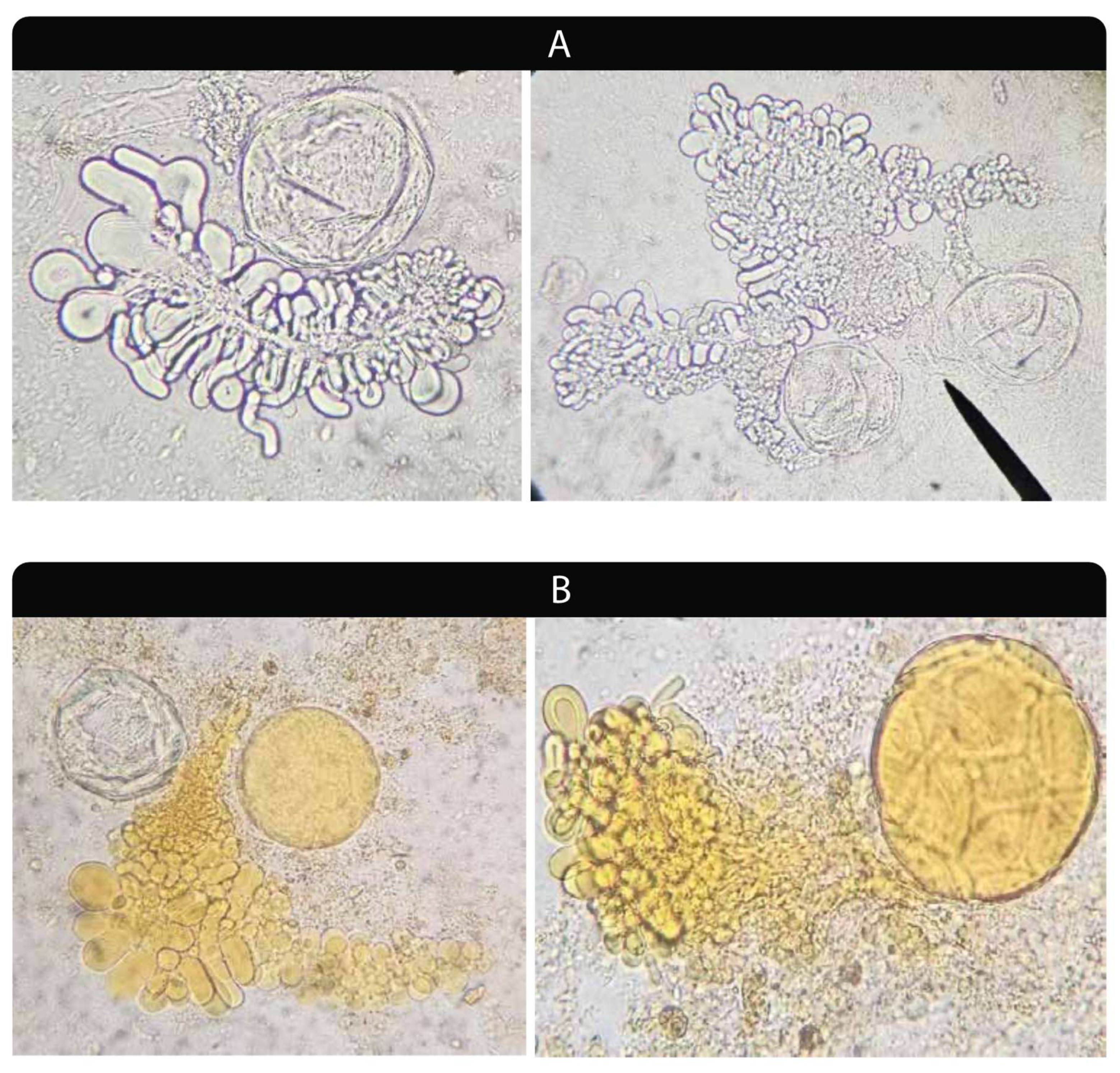
The stool culture was negative for Salmonella sp., Shigella sp., and Aeromonas sp., and no leukocytes were observed under direct microscopic examination. However, a distinctive structure was repeatedly observed in multiple smears. The round structure, approximately 100 µm in diameter, exhibited hyaline pseudopod-like extensions. It appeared to have a double membrane, lacking any visible internal structures, and showed no movement. The samples were examined using an optical microscope with and without Lugol's iodine (Figure 2).
Although staining techniques such as acridine orange to identify DNA material, and lipid-specific dyes, could have provided valuable insights into the nature of the observed structures, these methodologies were not available in our laboratory at the time of analysis. Future studies incorporating these techniques would be essential to further characterize these structures and clarify their composition.
Regarding our case, we recommended that the attending physician arrange a follow-up appointment for a thorough and detailed anamnesis, along with additional investigations. A few weeks later, the patient was re-admitted and reported that her diarrhea had resolved spontaneously without any therapeutic intervention. She also mentioned traveling to Colombia a year ago. However, as no other clinical symptoms were present, no further studies were conducted. Although a definitive etiological diagnosis was not made, a hypothesis was proposed based on the current scientific literature.
Discussion
Unfamiliar with this structure, we conducted a literature search to identify it. Some scientific publications suggested it might be a parasite named Urbanorum sp., first reported in the 1990s by Colombian researcher Francisco Tirado Santamaria [1] as mentioned above. Coello Peralta et al [2] described a clinical case in Ecuador, suggesting fecal-oral transmission through contaminated food or water. Additional cases have been reported in Peru, Colombia, Brazil, and Mexico [3,4,5,6].
However, the classification of Urbanorum sp. remains controversial. Silva-Ramos et al. [7] provided a well-founded analysis of Coello Peralta's work, highlighting that the diagnosis relied solely on microscopic examination without the support of rigorous confirmatory tests. Their commentary emphasizes the importance of robust methodologies in establishing the validity of new pathogenic entities. They also mentioned that the structures shown in the picture accompanying the description of the case are considered by experts to be adipose cells [8]. Even some researchers propose it could have a plant kingdom origin, specifically from the pulp of Persea Americana (avocado) [9]. Consequently, the validity of Urbanorum sp. as a new human pathogen is questionable.
Conclusion
Based on the current evidence, we agree with Silva-Ramos, there is insufficient scientific support to classify the observed structure as a living organism. Further research is required to elucidate the true nature of these microscopic structures, including a detailed analysis of their cellular structure, nuclei, and other characteristics.
Rigorous investigation is essential to establish whether this entity is a new human pathogen capable of causing clinical diseases.
We believe that this case could contribute to other scientists who encounter this structure and have access to more and better resources, allowing them to give it a true meaning.
Funding
This study was conducted without any external funding.
Conflicts of Interest
Authors have no conflict of interest.
References
- Francisco Tirado Santamaría. Urbanorum spp. [internet]. Santander: Catedra Libre UIS; 2013 [citado el 30 de julio de 2015]. Disponible en: http://www.buenastareas.com/ ensayos/Urbanorum-Spp/70918639. html.
- Coello Peralta, R.D.; Parra-Guayasamin, S.G.; Yancha Moreta, C.A.; et al. A Case of Urbanorum spp. in a Woman from an Urban-Marginal Sector of Ecuador. Am J Case Rep 2023, 24, e939583-1. [Google Scholar] [CrossRef] [PubMed]
- Mirano, R.I.; Zapata, L.A.; Náquira, C. [Urbanorum spp. in Perú.]. Rev Peru Med Exp Salud Publica [In Spanish]. 2016, 33, 593–95. [Google Scholar] [CrossRef]
- Sousa de Aguiar, R.P.; Alves, L.L. Urbanorum spp: First report in Brazil. Am J Case Rep. 2018, 19, 486–90. [Google Scholar] [CrossRef]
- Ayol Pérez LG, Diaz Gines KL, Perlaza Achanci KF. [Epidemiology of acute diarrheal syndrome caused by the protozoan Urbanorum spp. Degree project prior to obtaining the Bachelor’s Degree in Nursing]. State University of Milagro, Faculty of Health Sciences, October, 2017 [in Spanish]. Available from: URL: http://repositorio.unemi.edu.ec/handle/123456789/3636.
- Rivadeneira Alvarez, A. Enteroparasitosis y diagnóstico parasitológico de Fasciola hepática por el método de concentración formol - éter Ritchie en comparación con el método directo en comunidades de la Región Andina El Tejar Saquisilí Cotopaxi, Región Costa Pedro Vicente Maldonado y Región Amazónica Comunidades Waoranis diciembre 2015 junio 2016. [Internet]. Quito: UCE; 2017 [citado: 2024, septiembre] Available from: URL: http://www.dspace.uce.edu.ec/handle/25000/9823.
- Silva-Ramos, C.R.; Cuervo, C.; Faccini-Martínez, A.; Gómez-Marín, J.E. Urbanorum: a case of scientific negligence. Infectio 2024, 69–70. [Google Scholar] [CrossRef]
- Rivero de Rodríguez Zulbey. Es Urbanorum spp. un parásito?. Kasmera [Internet]. 2016 Jun [citado 2024 Dic 26] ; 44( 1 ): 5-6. Disponible en: http://ve.scielo.org/scielo.php?script=sci_arttext&pid=S0075-52222016000100001&lng=es.
- Guzman, Carmen & Nessi, Anaibeth & Galindo, Mónica & Pérez, María & Pérez, Eva. (2023). ALGUNAS EVIDENCIAS MORFOLÓGICAS DE LA NATURALEZA VEGETAL DE "URBANORUM SPP". 46. 24. https://www.researchgate.net/publication/375606645_ALGUNAS_EVIDENCIAS_MORFOLOGICAS_DE_LA_NATURALEZA_VEGETAL_DE_URBANORUM_SPP.
Figure 1.
Urbanorum in the literature: The map highlights a concentration of research in America. The sparse distribution of publications in other regions suggests a need for further investigation into this topic. (References are listed in Supplementary).
Figure 1.
Urbanorum in the literature: The map highlights a concentration of research in America. The sparse distribution of publications in other regions suggests a need for further investigation into this topic. (References are listed in Supplementary).
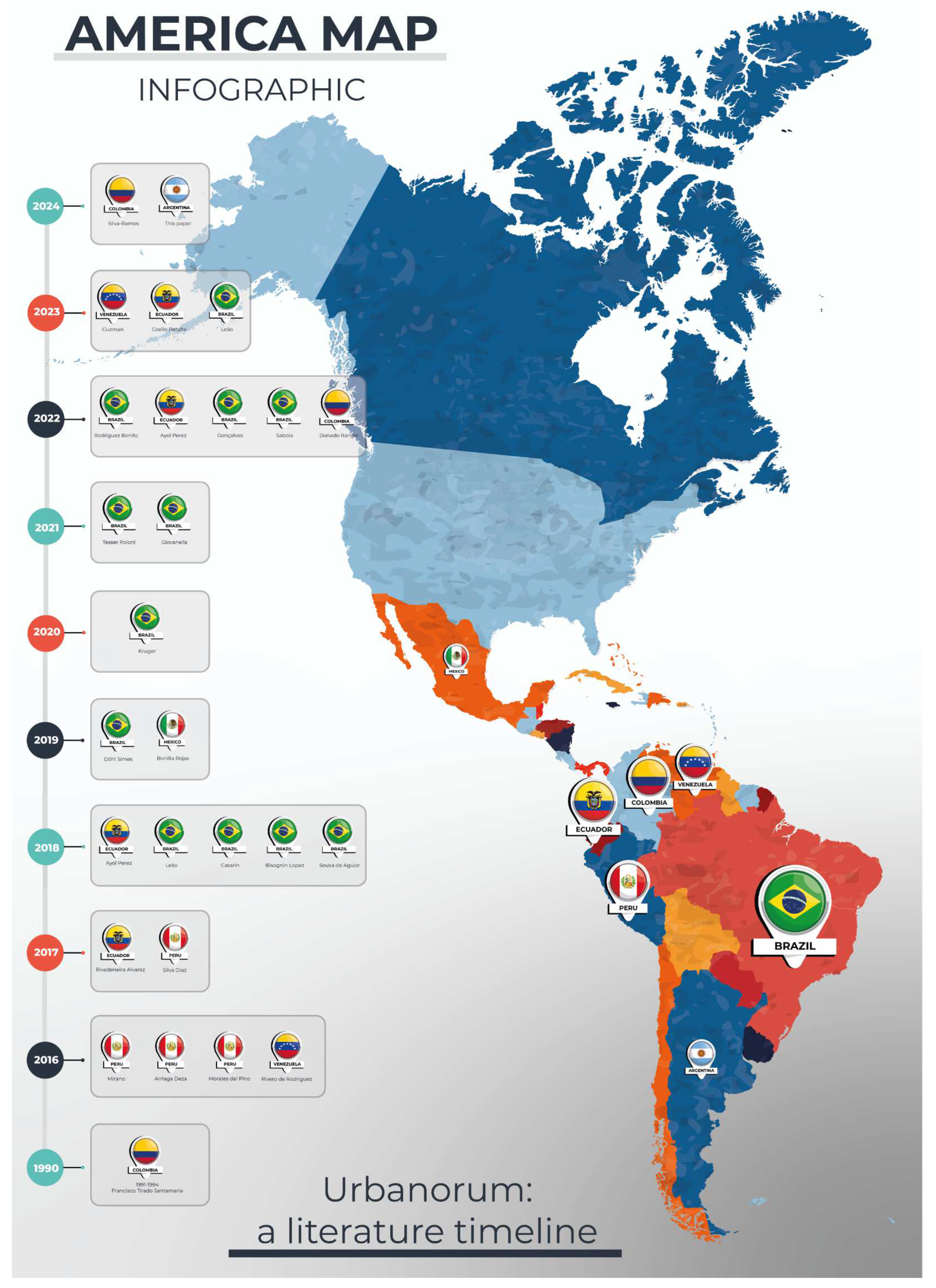
Figure 2.
Microscopic images of a fecal sample: A. Direct examination with 40x objective using saline solution. B. Examination with 40x objective using Lugol's iodine, highlighting the structure's shape, yellow color, and morphology.
Figure 2.
Microscopic images of a fecal sample: A. Direct examination with 40x objective using saline solution. B. Examination with 40x objective using Lugol's iodine, highlighting the structure's shape, yellow color, and morphology.
Disclaimer/Publisher’s Note: The statements, opinions and data contained in all publications are solely those of the individual author(s) and contributor(s) and not of MDPI and/or the editor(s). MDPI and/or the editor(s) disclaim responsibility for any injury to people or property resulting from any ideas, methods, instructions or products referred to in the content. |
© 2024 by the authors. Licensee MDPI, Basel, Switzerland. This article is an open access article distributed under the terms and conditions of the Creative Commons Attribution (CC BY) license (http://creativecommons.org/licenses/by/4.0/).
Copyright: This open access article is published under a Creative Commons CC BY 4.0 license, which permit the free download, distribution, and reuse, provided that the author and preprint are cited in any reuse.


