
Submitted:
24 December 2024
Posted:
25 December 2024
You are already at the latest version
Abstract
Species of the genus Eryngium L. of the Apiaceae family are successfully used in folk medicine in various countries worldwide, but they are hardly implemented in official medicinal and pharmaceutical practices. Therefore, it is advisable to conduct phytochemical and pharmacological research in E. planum L. herb extracts to develop and implement new phytomedicines based on this raw material. Purified water, 40% and 70% ethanol were used for obtaining soft extracts. Totally 7 hydroxycinnamic acids, 6 flavonoids, and 3 tannin metabolites were identified and quantified in the E. planum extracts by HPLC. These extracts were characterized as practically non-toxic medicines (V toxicity class, LD50> 5000 mg/kg). The E. planum extracts have shown a pronounced anti-inflammatory effect in experimental formalin oedema. The hepatoprotective activity of the E. planum extracts has been established. They were affected to reduce serum thiobarbituric acid (TBA) levels by 29.3%, 31.5% and 32.4%, respectively, compared to untreated animals and in liver homogenate by 59.5%, 65.4% and 66.8%, respectively. Alanine transaminase (ALT) activity decreased by 26.9%, 30.8% and 33.8%, respectively. Aspartate transaminase (AST) activity decreased by 23.9 %, 25.7 % and 30.5 %, respectively. The 70% ethanol extract has the most pronounced sedative effect due to a significant decrease in motor activity and a moderate antibacterial effect against gram-positive microorganisms. The most promising is the 70% ethanol extract of E. planum herb, which requires further preclinical and clinical studies.
Keywords:
Sea holly
; biologically active substances
; phenolic substances
; hepatoprotective activity
; anti-inflammatory activity
; sedative activity
1. Introduction
The species of the genus Eryngium L. (commonly known as sea holly) from the family Apiaceae are perennial, biennial, or annual herbs distributed in the temperate climatic zones of the northern and southern hemispheres. Many species of this genus are both edible and ornamental plants. While they are not officially recognized as medicinal raw materials in medical practice, some species are successfully used in traditional medicine in various countries worldwide [1,2,3]. Most species of the genus Eryngium are wild plants. They are distributed in Europe, North and South America, Southwest and Central Asia, Australia, and North Africa [4]. Eight species of the genus (E. fluminense, E. corniculatum, E. viviparum, E. atlanticum, E. alpinum, E. maroccanum, E. variifolium, and E. galioides) are included in the IUCN Red List of Threatened Species [5]. In Ukraine, three species of the genus Eryngium are distributed: Eryngium planum L. (flat sea holly), Eryngium campestre L. (field eryngo), and Eryngium maritimum L. (sea holly) (Figure 1) [8]. Raw materials cultivated in Ukraine have not been studied, although E. planum is the most common species.
The aerial and underground parts of species in the genus Eryngium contain a significant amount of biologically active substances. The flavonoids such as rutin, kaempferol, isoquercetin, luteolin, and apigenin, as well as caffeic, rosmarinic, ferulic, and chlorogenic acids, among other compounds capable of pharmacological activity, were identified in E. planum [6]. Polyphenols and pectic substances in the aerial parts of E. planum, E. campestre, and E. maritimum were studied [7]. Isoquercetin, quercetin, kaempferol, chlorogenic acid, caffeic acid, p-coumaric acid, and ferulic acid were identified in all Eryngium species. The pectin content extracted with hot water varied significantly among the species, with the highest level found in E. maritimum (8.8%), a moderate level in E. planum (4.65%), and the lowest in E. campestre (1.7%) [7]. The primary components of the leaves and stems of essential oils of E. planum were monocyclic monoterpenes (limonene, α-, β-pinene) and sesquiterpenes. The essential oil derived from the inflorescences of E. planum contained 43.2% chrysantenyl acetate and oxygenated sesquiterpenes [8].
The roots of E. planum contain carbohydrates such as fructose, glucose, and sucrose; organic acids, including malic, citric, glycolic, and oxalic acids; essential oil (0.13%); and triterpenoid saponins: eryngium saponins A, B, C, and D. Additionally, the roots contained polyacetylene compounds, such as falcarione, falcarinolone, and falcarinol, as well as phenolic carboxylic acids and their derivatives, including rosmarinic and chlorogenic acids [9,10]. The flowers of E. planum contained carbohydrates; organic acids (malic, citric, glycolic, and oxalic acids); and flavonoids, such as quercetin and kaempferol [8]. The fruits of E. planum are characterized by the following chemical composition: carbohydrates (fructose, glucose, sucrose), oxalic acid, flavonoids (quercetin, kaempferol), and fatty acids (27–29%) such as palmitic, stearic, oleic, linoleic, and petroselinic acids [11,12,13,14].
The Pakistani laboratory “Kamal Laboratories” produces homoeopathic remedies under the names “ERYNGIUM Ø” and “ERYNGIUM 30”, based on E. aquaticum. These are indicated for issues with the urinary system, such as difficulty and frequent urination, pain and spasms in the urinary tract, and renal colic [15]. E. aquaticum L. is listed in the American Homoeopathic Pharmacopoeia and the Homoeopathic Pharmacopoeia of India [16,17]. In 2021, the French company SEPPIC introduced a new active cosmetic ingredient based on E. maritimum. Their development, patented under “CELTOSOME™ Eryngium maritimum”, is a concentrated active ingredient derived from sea holly, containing a specific composition of phenolic acids and flavonols for skin restoration and elasticity [18]. In traditional medicine, the Eryngium species treats dropsy, sexual disorders, stomach and heart pain, insomnia, rheumatism, toothache, cavities, and bronchial asthma. The roots were considered an antidote for mushroom poisoning. Externally, Eryngium is applied in baths for phytodermatoses and arthritis [19]. Ethanolic extracts of E. planum from basal leaves have demonstrated anti-amoebic activity [20]. Extracts of E. planum and E. campestre are used as remedies for inflammatory dental diseases such as periodontitis, cavities, and toothache [21]. E. planum extract has an inhibitory effect on α-amylase and α-glucosidase, making it beneficial in treating type II diabetes mellitus [6]. Preclinical in vivo studies indicate that a 70% ethanolic extract of E. planum improved long-term memory in rats [22]. Extracts of E. billardieri induced apoptosis in cancer cells [23].
Numerous studies confirm that Eryngium species are promising medicinal plants. Therefore, further research is crucial to develop new medicinal products based on these plants and incorporate them into official medical practice.
The aim of the research was to carry out phytochemical research and screening of pharmacological activity in E. planym herb extracts to establish their potential perspective for use in official medical and pharmaceutical practice. The sedative and hepatoprotective activities of E. planum were studied for the first time
2. Materials and Methods
2.1. Plant Raw Materials
The raw materials were harvested during the peak flowering period in the village of Pidluzhzhya, Tysmenytsia District, Ivano-Frankivsk Region. The collection adhered to the fundamental rules for harvesting medicinal raw materials, with care taken to preserve the local flora [24]. Before harvesting, the plant was identified. The herb was cut using a knife in dry weather. Immediately after collection, the raw materials were dried in the shade outdoors, avoiding direct sunlight. A total of 2.0 kg of dry raw material was obtained. The plant’s identity was confirmed based on the botanical catalogue with the consulting assistance of Professor A.R. Grytsyk from Ivano-Frankivsk National Medical University (IFNMU) [25]. Voucher specimens No. 583–585 were deposited at the Department of Pharmaceutical Management, Drug Technology, and Pharmacognosy at Ivano-Frankivsk National Medical University.
2.2. Extracts Preparation
The objects of the study were soft extracts of E. planum L. herb, obtained using purified water, 40%, and 70% ethanol. The E. planum herb was pre-ground to a particle size of 0.5–2 mm. 500 g of raw material was extracted using the fractional maceration method with purified water, 40% ethanol, or 70% ethanol, yielding soft extracts labelled EP1, EP2, and EP3, respectively. Triple extraction was performed using the following sequential raw material-to-solvent ratios: 1:5, 1:3, and 1:2. The aqueous-alcoholic extracts were infused for 12 hours at each stage at room temperature, after which the extracts were drained and combined. For the aqueous extract, the mixture at each stage was boiled with a new portion of the solvent for 15 minutes and then infused for 3 hours. The combined extracts were filtered, and the solvent was evaporated under vacuum using a Buchi Rotavapor R-300 (Buchi Labortechnik AG, Flawil, Switzerland) under the following conditions: vacuum 150 mbar, rotation 80 rpm, and heating bath at 80°C, until the final moisture content did not exceed 25%. The yields of the thick extracts EP1, EP2, and EP3 were 24.6±0.9%, 23.1±1.1%, and 22.1±0.8%, respectively.
2.3. Phytochemical Research
HPLC Methods
Flavonoids were analyzed using high-performance liquid chromatography (HPLC) on an Agilent Technologies 1200 liquid chromatograph. The mobile phase comprised acetonitrile (A) and 0.1% formic acid solution in water (B). Gradient elution was performed under the following conditions: 0 min – A (30%): B (70%); 20 min – A (70%): B (30%); 22 min – A (100%): B (0%); 30 min – A (100%): B (0%). Separation was achieved on a Zorbax SB-C18 chromatographic column (3.5 µm, 150 × 4.6 mm) (Agilent Technologies, Santa Clara, USA) with a flow rate of 0.25 ml/min. The sample injection volume was 100 µl, and the column was maintained at 25°C. Detection was performed using a diode-array detector with signal recording at 280 and 365 nm, with spectra captured in the range of 210–270 nm [26,27].
The analysis of hydroxycinnamic acids was conducted on the same Agilent Technologies 1200 liquid chromatograph. Methanol (A) and 0.1% formic acid in water (B) were used as the mobile phase. Gradient elution parameters were as follows: 0 min – A (25%): B (75%); 25 min – A (75%): B (25%); 27 min – A (100%): B (0%); 35 min – A (100%): B (0%). Separation occurred on a Zorbax SB-Aq column (4.6 mm × 150 mm, 3.5 µm) (Agilent Technologies, USA) with a flow rate of 0.5 ml/min, a column temperature of 30°C, and an injection volume of 4 µl. Detection was carried out using a diode-array detector with signals monitored at 250 and 275 nm and absorption spectra recorded within the range of 210–270 nm [28,29,30].
Tannin metabolites were examined using an Agilent Technologies 1200 liquid chromatograph. Methanol (A) and 0.1% formic acid in water (B) were the mobile phase. Gradient elution conditions included: 0 min – A (20%): B (80%); 25 min – A (75%): B (25%); 27 min – A (100%): B (0%); 35 min – A (100%): B (0%). The separation process employed a Zorbax SB-C18 chromatographic column (3.5 µm, 150 × 4.6 mm) (Agilent Technologies, USA) with a flow rate of 0.25 ml/min, a thermostat setting of 35°C, and an injection volume of 4 µl. The diode-array detector recorded signals at 250 and 275 nm, while absorption spectra were captured across the range of 210–270 nm [31,32].
Substance identification and quantification were based on comparisons with standard reference materials from Sigma-Aldrich (Burlington, MA, USA). Quantitative analysis of total flavonoid content, expressed as rutin, was carried out spectrophotometrically according to the Ukrainian State Pharmacopoeia (SPHU) 2.0 [27,33]. The quantitative determination of tannins and total amounts of polyphenols in terms of pyrogallol was carried out using the spectrophotometric method in accordance with the pharmacopeial method SPHU 2.0 [33,34,35]. The quantitative content of polysaccharides was determined according to the methodology described in the monograph “Plantago major leaves” in the (SPHU 2.0, Vol. 3 [34].
2.4. Pharmacological Research
The pharmacological and toxicological properties of E. planum herb extracts were studied according to the “European Convention for the Protection of Vertebrate Animals Used for Experimental and Other Scientific Purposes” (Strasbourg, 1986) and other laws based on it [36,37,38]. The research was approved by the Bioethics Commission of the Ivano-Frankivsk National Medical University (protocol №125/22 from 25.05.2021).
2.4.1. Acute Toxicity
To determine acute toxicity, the methodology for preclinical safety studies of medicinal products was used. E. planum extracts were administered intragastrically to animals using a probe at a dose of 5000 mg/kg. The animals were divided into four groups, with six individuals in each group. Animals in the first three groups received EP1, EP2, and EP3 extracts of E. planum, respectively. The fourth group consisted of intact (control) animals. The animals were observed for 14 days. The following parameters were assessed: general condition, including mortality rate, breathing patterns, motor activity, skin, tail, ear, and mucous membrane colouration, salivation, and gastrointestinal state (characteristics and colouration of excretions). Body weight was monitored on days 3, 7, and 14. On the 15th day, the animals were decapitated under general anaesthesia, organs were weighed, and blood samples were collected for biochemical analysis. This study aimed to evaluate the potential toxic effects and systemic impact of the E. planum extracts on various physiological and biochemical parameters [39].
2.4.2. Antimicrobial Activity
The study of the antimicrobial activity of E. planum herb extracts on the growth of pure cultures of gram-positive bacteria and yeasts was conducted at the Department of Microbiology, Virology, and Immunology of IFNMU with the consultative assistance of the department head, Doctor of Medical Sciences, Professor R.V. Kutsyk. The research was performed on clinical isolates of antibiotic-sensitive and antibiotic-resistant microorganisms. Bacterial cultures were identified using the biochemical microtests “STAPHYtest 16”, “ENTEROtest 24”, and “NEFERMENTtest 24” (Lachema, Brno, Czech Republic), as well as based on a combination of morphological and cultural properties following the recommendations of the 9th edition of Bergey’s Manual of Determinative Bacteriology. Yeast-like fungal cultures were identified using 40 biochemical tests with the VITEK 2 system and VITEK 2 YST ID cards (bioMérieux, Crapon, France) [40].
Screening of the antimicrobial activity of plant extracts was performed using the agar diffusion micromethod. This method is characterized by high sensitivity and discriminatory power, allowing for reliable differentiation between active and inactive extracts. Petri dishes placed on a strictly horizontal and even surface were filled with 30 ml of agar. After the medium solidified, wells with a diameter of 4.0 mm were created using a specialized punch with uniform edges. The agar was evenly inoculated with a test-culture suspension (concentration of 1×10⁷ CFU/ml). Experimental wells were filled with 20 µl of plant extracts (100 mg/ml), while control wells received 20 µl of solvents (40% and 70% aqueous ethanol). After 24 hours of incubation, the diameters of growth inhibition zones for bacterial test cultures were measured. Fungistatic activity was recorded after 2 days of cultivation, and fungicidal activity was recorded after 4 days. Digital images of the agar plates were captured, and the data were processed using the UTHSCSA ImageTool 3.0 software (The University of Texas Health Science Center in San Antonio, ©1995-2002) [41,42].
The activity of E. planum herb extracts was established against the following microorganisms: Staphylococcus aureus, Staphylococcus haemolyticus, Enterococcus faecalis, α-hemolytic streptococci Streptococcus oralis, Streptococcus gordonii, β-hemolytic streptococci of group B Streptococcus agalactiae, β-hemolytic streptococci of group G Streptococcus dysgalactiae ssp. equisimilis, Bacillus subtilis, Escherichia coli, Escherichia fergusonii, Citrobacter freundii, Pseudomonas aeruginosa, Candida albicans, Candida tropicalis, Candida lipolytica, Candida lusitaniae, and Candida kefyr [40].
2.4.3. Sedative Activity
The study of sedative activity was conducted on 35 non-linear sexually mature white mice weighing 18–20 g. The animals were divided into 5 groups of 7 animals each. Animals in groups I, II, and III received intraperitoneal injections of E. planum herb extracts EP1, EP2, and EP3, respectively, at a dose of 50 mg/kg body weight. Group IV received a reference drug, motherwort extract, at an effective dose of 50 mg/kg body weight. Group V consisted of intact animals that received an equivalent volume of purified water. The administration of phytopreparations began 5 days before the study, with the final dose administered 30 minutes before the experiment on the fifth day. The tests used included the “Elevated Plus Maze,” “Cube,” “Black-and-White Box,” and “Open Field,” which are part of behavioural methods for studying the pharmacological activity of psychotropic agents. In the “Elevated Plus Maze” test, the apparatus consisted of two crossed arms in the shape of a cross, one enclosed by walls. The dark arm allowed the test animals to hide and avoid light and open spaces, exhibiting defensive behaviour. Entry into the open arm was considered an indication of exploratory behaviour and, correspondingly, a lower level of fear of the unknown [31,39].
2.4.4. Hepatoprotective Activity
The study of the hepatoprotective activity of E. planum herb extracts was conducted using a model of acute carbon tetrachloride-induced hepatitis [39,43]. The domestic hepatoprotective drug “Silibor” was used as a reference [44]. The intensity of pathological changes in the liver was assessed by measuring thiobarbituric acid reactive substances (TBA-RS) in blood serum and liver homogenates. The activity of alanine aminotransferase (ALT), aspartate aminotransferase (AST), and alkaline phosphatase (ALP) was measured in blood serum [39,43]. ALT and AST activities were determined using the unified dinitrophenylhydrazine method of Reitman and Frankel with reagent kits from “Philisit-Diagnostics” (Ukraine) [45,46]. ALP activity was determined photometrically by the intensity of the red dye formed by the oxidative coupling of phenol with 4-aminoantipyrine. The level of lipid peroxidation product (TBA-RS) was assessed through its reaction with 2-thiobarbituric acid spectrophotometrically, using the method of E.N. Korobeynikova and biochemical kits from the domestic manufacturer “Philisit-Diagnostics” (Dnipro, Ukraine) [47].
2.5. Statistical Analysis
3. Results
Extracts EP1, EP2, and EP3 from E. planum herb are soft, viscous masses of dark brown to dark brown-green colour with a characteristic odour.
3.1. Phytochemical analysis of extracts
The qualitative composition of biologically active compounds (BACs) in the soft extracts of E. planum was studied using identification reactions and thin-layer chromatography (TLC). The soft extracts of E. planum herb contain amino acids, simple phenols, coumarins, flavonoids, hydroxycinnamic acids, tannins, and saponins.
The HPLC method was used to identify and quantify phenolic compounds in the soft extracts of E. planum herb (Table 1).
In total, 16 phenolic compounds were identified in the soft extracts of E. planum herb. The dominant hydroxycinnamic acids in the extracts were chlorogenic acid, sinapic acid, and trans-cinnamic acid. Six flavonoids were identified and quantified in the soft extracts, with naringenin being the most abundant. Among the tannin metabolites, the dominant compounds were epicatechin and gallocatechin.
3.2. Pharmacological Research
3.2.1. Acute Toxicity
To determine acute toxicity, the methodology for preclinical safety assessment of medicinal products was used [39]. E. planum extracts were administered intragastrically to animals using a probe at a dose of 5000 mg/kg body weight. Immediately after the administration of the researched extracts and throughout the entire experimental period, the general condition of the animals remained normal. Salivation was normal in the group of animals that consumed EP1. For the groups, EP2 and EP3, moistening of fur around the mouth was noticed during the first 24 hours. Breathing was calm, not laboured for animals in all the groups. All the animals were active, coordination preserved. Seizures were not observed. Fur was dry, white, smooth; tail and paws were light pink. During the study, an increase in body weight was observed in both the intact and experimental groups, which didn’t differ significantly. The effect of E. planum extracts on the biochemical parameters of blood serum on day 14 was assessed using AST (0.48±0.01 µmol/hour·ml) and ALT (0.29±0.01 µmol/hour·ml) levels. These data significantly (p ≤ 0.05) differed from the intact animal group 0.42±0.02 and 0.24±0.01 respectively. Thus, the biochemical parameters of mice blood under the influence of E. planum herb extracts fluctuated within normal limits.
3.2.2. Antimicrobial Activity
The aqueous extract EP1 and the hydroalcoholic extract EP2 demonstrated negligible antimicrobial activity, whereas the E. planum herb extract EP3 exhibited moderate antimicrobial activity. The results are presented in Figure 2.
3.2.3. Sedative Activity
The results of the behavioural activity studies of animals in the “Elevated Plus Maze” test are presented in Table 2.
In the “Open Field” test, the effects of the extracts on the central nervous system were examined to evaluate the sedative effect (Figure 3).
The data presented in Table 2 indicate that animals in groups III and IV spent more time in the open arms and the center of the maze compared to the closed arms, suggesting high exploratory activity and low fear levels. The indicators for group III animals, which received E. planum extract EP3, were comparable to those of the control group, which received motherwort extract, a proven sedative. The time spent by animals in the maze’s centre was minimal across all groups and did not differ significantly. An important indicator of stress resilience is the number of downward glances by the animals. This indicator for groups I–III was nearly double that of the intact group. The significant reduction in activity indicators for the experimental animals (Figure 3) over five minutes indicates pronounced sedative activity of the tested extracts. The locomotor activity of animals in group III decreased significantly by 2.4 times compared to the control group.
The “Cube” test, designed to study cognitive responses and fear levels toward a new object, provided further insights. The results of these studies are presented in Table 3.
The data presented in Table 3 indicate that E. planum extracts and motherwort extract influenced the locomotor activity of mice toward a novel object. The first approach to the cube was performed the fastest by mice in groups III and IV (19.0 seconds and 14.3 seconds, respectively). The total duration of approaches by mice in group III was nearly four times greater than that of intact animals, indicating increased exploratory activity due to the EP3 extract and a lack of anxiety. For the “Black-and-White Box” test, a two-chamber apparatus was used, one chamber shielded from light and the other illuminated. Mice were placed in the dark compartment, and the latency of the first exit to the light chamber, the number of exits, and the duration of time spent in the light compartment were recorded. The results of these studies are presented in Table 4.
The time of the first exit from the dark compartment (Table 4) is a critical indicator characterizing the animals’ fear level. The first exit times for animals in groups II and III were 12.7 seconds and 10.3 seconds, respectively, close to the times observed in the control group (8.3 seconds) that received motherwort extract. The number of exits from the dark chamber to the light chamber varied between 3 and 11 across the groups; however, this parameter did not significantly affect the total duration of time spent in the light chamber. The longest duration in the light chamber was observed in group III animals (63.6 seconds). These results suggest that E. planum extracts EP2 and EP3 reduced feelings of fear and anxiety in the experimental animals.
3.2.4. Hepatoprotective Activity
The results of the effects of E. planum extracts on the biochemical parameters of blood serum and liver homogenates in the model of acute carbon tetrachloride-induced hepatitis are presented in Figure 4.
The results (Figure 4) demonstrated that the administration of carbon tetrachloride caused liver damage, as evidenced by altered ALT, AST, and ALP activities and an accumulation of TBA-reactive substances (TBA-RS) compared to intact animals. ALT and AST levels in untreated animals approximately doubled compared to those in intact animals. ALP and TBA-RS levels in the blood serum of untreated animals increased by 20.7% and 75.3%, respectively, relative to intact animals. TBA-RS levels in liver homogenates increased approximately 13-fold. The use of E. planum herb extracts and the reference drug “Silibor” in experimental hepatitis was accompanied by a noticeable reduction in pathological manifestations. This was reflected in decreased TBA-RS levels in blood serum and liver homogenates, as well as reduced ALT and AST activities in the blood serum, compared to the untreated group of animals.
4. Discussion
The phytochemical analysis of the thick extracts of E. planum herb identified the qualitative composition and quantitative content of the main groups of biologically active substances, including flavonoids, hydroxycinnamic acids, tannins, and polysaccharides. Using HPLC, 16 phenolic compounds were identified and quantified in the dense extracts (Table 1). Among all identified flavonoids, naringenin was dominant (33.97%), while chlorogenic acid (34.05%), sinapic acid (24.37%), and trans-cinnamic acid (19.62%) were the most abundant hydroxycinnamic acids. Previously, it was reported that the extracts of E. planum contain mainly flavonoids, especially rutin and isoquercitrin [6,7], but in our samples, naringenin was the dominant compound.
The study results showed that intragastric administration of E. planum extracts at a dose of 5000 mg/kg does not lead to the death of animals. No changes in behaviour or vital signs were observed, indicating the absence of toxic effects at this dose. This characterizes the extracts as practically non-toxic (toxicity class V, LD50 > 5000 mg/kg). As shown in the data from Figure 2, body weight increased in all experimental animals. Macroscopic examination of the internal organs of all groups revealed no pathological signs. The colour, shape, size, and position of the organs were anatomically normal. The heart muscle had the correct shape, with a dark-red cut surface, and the lungs were unchanged. The surfaces of the liver, pancreas, and kidneys were uniform and smooth. The spleen was elastic and blood-filled.
The E. planum extract EP3 (70% ethanol as the solvent) exhibits moderate antimicrobial activity (Figure 2) against gram-positive microorganisms, including staphylococci, streptococci, and bacilli. Previously the E. amethystinum and E. alpinum showed a broad spectrum of antibacterial and antifungal activity with minimum inhibitory concentrations (MIC) less than or equal to 1.944 and 1.11 mg/mL, respectively [11]. The antibacterial activity of E. caeruleum remedies against six bacterial strains (Staphylococcus aureus, Bacillus subtilis, Streptomyces scabies, Erwinia amylovora, Xanthomonas axonopodis pv. citri, and Klebsiella sp.) was confirmed in vitro [49]. So, the data of E. planum extracts’ anti-microbial activity is novel.
The sedative activity of E. planum was studied for the first time. It was found that EP3 extract (extractant - 70% ethanol) has the most pronounced sedative effect due to a significant decrease in motor activity in the «Open Field test» and reduction of concern in «Cube tests», «Raised Cross Maze» and “Black and White Camera”.
The results of the study (Figure 4) showed that the administration of E. planum extracts EP1, EP2, and EP3 to animals reduced ALT activity by 26.9%, 30.8%, and 33.8%, respectively, and AST activity by 23.9%, 25.7%, and 30.5%, respectively. The extracts also reduced TBA-reactive substances (TBA-RS) levels in blood serum by 29.3%, 31.5%, and 32.4%, respectively, and in liver homogenates by 59.5%, 65.4%, and 66.8%, respectively, compared to untreated animals. These findings indicate that using E. planum herb extracts in acute toxic liver damage significantly reduces the toxic effects of carbon tetrachloride, providing hepatoprotective activity that is nearly comparable to the reference drug. Among the extracts, EP3 at a dose of 25 mg/kg body weight demonstrated the most intensive and effective action in acute hepatitis compared to EP1 and EP2. The hepatoprotective activity of E. planum extracts has also been studied for the first time.
5. Conclusions
The phytochemical and pharmacological screening studies demonstrated the potential use of E. planum L. herb extracts to develop new phytomedicines with hepatoprotective and sedative activities. Using HPLC, 16 phenolic compounds were identified and quantified in the extracts, including 6 flavonoids, 7 hydroxycinnamic acids, and 3 tannin metabolites. The quantitative content of the main groups of phenolic compounds was determined spectrophotometrically. The soft extracts of E. planum were practically non-toxic substances (LD50 > 5000 mg/kg) with pronounced sedative and hepatoprotective effects. The most promising substance was the extract obtained using a 70% aqueous ethanol solution. The conducted studies showed the promising potential of the obtained extracts, but further pre-clinical and clinical studies need to be carried out before suggesting applications in clinical medicine.
Author Contributions
Conceptualization. A.G, O.K. and A.R.; methodology. A.G. and R.H.; software. K.M.; validation. K.M., R.H. and O.K.; formal analysis. K.M. and R.H.; investigation. K.M. and R.H.; resources. A.G, O.K. and A.R.; data curation. K.M. and R.H.; writing—original draft preparation. A.G, O.K. and A.R.; writing—review and editing. A.G, O.K. and A.R.; visualization. K.M.; supervision. A.G, O.K. and A.R.; project administration. A.G, O.K. and A.R.; funding acquisition. A.G, O.K. and A.R. All authors have read and agreed to the published version of the manuscript.
Funding
This work was supported by the European Union in the MSCA4Ukraine project “Design and development of 3D-printed medicines for bioactive materials of Ukrainian and Estonian medicinal plants origin” (ID number 1232466).
Institutional Review Board Statement
The pharmacological and toxicological properties of E. planum herb extracts were studied according to the “European Convention for the Protection of Vertebrate Animals Used for Experimental and Other Scientific Purposes” (Strasbourg, 1986). The research was approved by the Bioethics Commission of the Ivano-Frankivsk National Medical University (protocol №125/22 from 25.05.2021).
Informed Consent Statement
Not applicable.
Data Availability Statement
The data supporting the results of this study can be obtained from the corresponding authors upon reasonable request.
Conflicts of Interest
The authors declare no conflicts of interest.
References
- Eryngium. The Plant List. Royal Botanic Gardens 2024. http://www.theplantlist.org/tpl1.1/search?q=Eryngium.
- Eryngium. International Plant Names Index, The Royal Botanic Gardens, Kew, Harvard University Herbaria & Libraries and Australian National Botanic Gardens; 2024. https://www.ipni.org/n/30001853-2.
- BHL. Biodiversity Heritage Library. 2024. https://www.biodiversitylibrary.org/.
- Royal Botanic Gardens, Kew. https://www.kew.org/.
- IUCN. Red List of Threatened Species 2024. https://www.iucnredlist.org/.
- Paun, G.; Neagu, E.; Moroeanu, V.; Albu, C.; Savin, S.; Lucian Radu, G. Chemical and Bioactivity Evaluation of Eryngium Planum and Cnicus Benedictus Polyphenolic-Rich Extracts. BioMed Research International 2019, 2019, 1–10. [Google Scholar] [CrossRef] [PubMed]
- Conea, S.; Vlase, L.; Chirila, I. Comparative Study on the Polyphenols and Pectin of Three Eryngium Species and Their Antimicrobial Activity. Cellulose Chemistry and Technology 2016, 50, 473–481. [Google Scholar]
- Thiem, B.; Kikowska, M.; Kurowska, A.; Kalemba, D. Essential Oil Composition of the Different Parts and In Vitro Shoot Culture of Eryngium Planum L. Molecules 2011, 16, 7115–7124. [Google Scholar] [CrossRef] [PubMed]
- Kowalczyk, M.; Masullo, M.; Thiem, B.; Piacente, S.; Stochmal, A.; Oleszek, W. Three New Triterpene Saponins from Roots of Eryngium Planum. Natural Product Research 2014, 28, 653–660. [Google Scholar] [CrossRef] [PubMed]
- Rodrigues, T.L.M.; Silva, M.E.P.; Gurgel, E.S.C.; Oliveira, M.S.; Lucas, F.C.A. Eryngium Foetidum L. (Apiaceae): A Literature Review of Traditional Uses, Chemical Composition, and Pharmacological Activities. Evidence-Based Complementary and Alternative Medicine 2022, 2022, 1–15. [Google Scholar] [CrossRef]
- Kremer, D.; Zovko Končić, M.; Kosalec, I.; Košir, I.J.; Potočnik, T.; Čerenak, A.; Srečec, S.; Dunkić, V.; Vuko, E. Phytochemical Traits and Biological Activity of Eryngium Amethystinum and E. Alpinum (Apiaceae). Horticulturae 2021, 7, 364. [Google Scholar] [CrossRef]
- Calviño, C.I.; Levin, G.A. A New Species of Eryngium (Apiaceae, Saniculoideae) from the USA. Systematic Botany 2019, 44, 446–450. [Google Scholar] [CrossRef]
- Bährle-Rapp, M. Eryngium campestre. In Springer Lexikon Kosmetik und Körperpflege; Springer Berlin Heidelberg: Berlin, Heidelberg, 2007; pp. 189–189 ISBN 978-3-540-71094-3.
- Rehman, T.; Khan, Y.; Zeb, M.; Izhar, S. Phytochemical Analysis, Green Synthesis of Silver and Gold Nanoparticles, and Antibacterial Activity of Eryngium Amethystinum. Bioscience Research 2021, 18, 2788–2794. [Google Scholar]
- Kamal Laboratories. https://kamal.pk/.
- Homeopathic Pharmacopeia Convention of the United States (HPCUS); https://www.hpus.com/.
- Homeopathic Pharamacopeia of India (HPI); https://www.nhp.gov.in/.
- SEPPIC. CELTOSOMETM ERYNGIUM MARITIMUM. https://www.seppic.com/en/wesource/celtosome-eryngium-maritimum.
- Grodzinsky, A.M. Medicinal Plants: Encyclopedic Guide; Ukrainian encyclopedia named after M. P. Bazhana.; 1990.
- Derda, M.; Thiem, B.; Budzianowski, R. The Evaluation of the Amebicidal Activity of Eryngium Planum Extracts. Acta Poloniae Pharmaceutica 2013, 70, 1027–1034. [Google Scholar]
- Сonea, S.; Parvu, A.; Taulescu, M.; Vlase, L. Effects of Eryngium Planum and Eryngium Campestre Extracts on Ligatureinduced Rat Periodontitis Article. Digest Journal of Nanomaterials and Biostructures 2015, 693–704. [Google Scholar]
- Ozarowski, M.; Thiem, B.; Mikolajczak, P.L.; Piasecka, A.; Kachlicki, P.; Szulc, M.; Kaminska, E.; Bogacz, A.; Kujawski, R.; Bartkowiak-Wieczorek, J.; et al. Improvement in Long-Term Memory Following Chronic Administration of Eryngium Planum Root Extract in Scopolamine Model: Behavioral and Molecular Study. Evidence-Based Complementary and Alternative Medicine 2015, 2015, 1–13. [Google Scholar] [CrossRef] [PubMed]
- Hasanbeiglu, S.; Hosseini, K.; Molavi, O.; Asgharian, P.; Tarhriz, V. Eryngium Billardieri Extract and Fractions Induce Apoptosis in Cancerous Cells. ACAMC 2022, 22, 2189–2201. [Google Scholar] [CrossRef]
- Convention on the Conservation of European Wild Flora and Fauna and Natural Habitats.; 1979.
- Dobrochaeva, D.N.; Kotov, M.I.; Prokudin, Y.N.; Barbarich, A.I. Key to Higher Plants of Ukraine; Naukova Dumka: Kyiv, Ukraine, 1999. [Google Scholar]
- Melnyk, N.; Pawłowska, K.A.; Ziaja, M.; Wojnowski, W.; Koshovyi, O.; Granica, S.; Bazylko, A. Characterization of Herbal Teas Containing Lime Flowers – Tiliae Flos by HPTLC Method with Chemometric Analysis. Food Chemistry 2021, 346, 128929. [Google Scholar] [CrossRef] [PubMed]
- Kukhtenko, H.; Bevz, N.; Konechnyi, Y.; Kukhtenko, O.; Jasicka-Misiak, I. Spectrophotometric and Chromatographic Assessment of Total Polyphenol and Flavonoid Content in Rhododendron Tomentosum Extracts and Their Antioxidant and Antimicrobial Activity. Molecules 2024, 29, 1095. [Google Scholar] [CrossRef]
- Vlasova, I.; Gontova, T.; Grytsyk, L.; Zhumashova, G.; Sayakova, G.; Boshkayeva, A.; Shanaida, M.; Koshovyi, O. Determination of Standardization Parameters of Oxycoccus Macrocarpus (Ait.) Pursh and Oxycoccus Palustris Pers. Leaves. SR: PS 2022, 48–57. [CrossRef]
- Vronska, L.V. Chromatographic Profile of Hydroxycinnamic Acids of Dry Extract of Blueberry Shoots. Pharmaceutical journal 2019, 5–18. [Google Scholar]
- Hordiei, K.; Gontova, T.; Trumbeckaite, S.; Yaremenko, M.; Raudone, L. Phenolic Composition and Antioxidant Activity of Tanacetum Parthenium Cultivated in Different Regions of Ukraine: Insights into the Flavonoids and Hydroxycinnamic Acids Profile. Plants 2023, 12, 2940. [Google Scholar] [CrossRef]
- Starchenko, G.; Hrytsyk, A.; Raal, A.; Koshovyi, O. Phytochemical Profile and Pharmacological Activities of Water and Hydroethanolic Dry Extracts of Calluna Vulgaris (L.) Hull. Herb. Plants 2020, 9, 751. [Google Scholar] [CrossRef]
- Kovaleva, A.M.; Abdulkafarova, E.R. Phenolic Compounds from Potentilla Anserina. Chem Nat Compd 2011, 47, 446–447. [Google Scholar] [CrossRef]
- European Pharmacopoeia; 11th ed.; Council of Europe: Strasbourg, 2022.
- State Pharmacopoeia of Ukraine; 2nd ed.; Ukrainian Scientific Pharmacopoeial Center of Drugs Quality: Kharkiv, Ukraine, 2015.
- Hrytsyk, Y.; Koshovyi, O.; Hrytsyk, R.; Raal, A. Extracts of the Canadian Goldenrod (Solidago Canadensis L.) – Promising Agents with Antimicrobial, Anti-Inflammatory and Hepatoprotective Activity. ScienceRise: Pharmaceutical Science 2024, 78–87. [CrossRef]
- European Convention for the Protection of Vertebrate Animals Used for Experimental and Other Scientific Purposes 1999.
- Council Directive 2010/63/EU on the Protection of Animals Used for Scientific Purposes 2010.
- On Approval of the Procedure for Preclinical Study of Medicinal Products and Examination of Materials of Preclinical Study of Medicinal Products; 2009.
- Stefanov, O.V. Preclinical Studies of Drugs; Avicenna: Kyiv, Ukraine, 2001.
- Bergey, D.H. Bergey’s Manual of Determinative Bacteriology; Baltimore, Williams & Wilkins Co, 1957.
- Kutsyk, R.V. Screening Study of the Antimicrobial Activity of Medicinal Plants of the Carpathian Region against Polyantibiotic-Resistant Clinical Strains of Staphylococci. Message 1. Galician Medical Bulletin 2004, 11, 44–48. [Google Scholar]
- Kutsyk, R. Study of Antimicrobial Activity of Medicinal Plants of the Carpathian Region against Antibiotic-Resistant Clinical Strains of Staphylococci. Galician Medical Herald 2005, 12, 52–58. [Google Scholar]
- Huzio, N.; Grytsyk, A.; Raal, A.; Grytsyk, L.; Koshovyi, O. Phytochemical and Pharmacological Research in Agrimonia Eupatoria L. Herb Extract with Anti-Inflammatory and Hepatoprotective Properties. Plants 2022, 11, 2371. [Google Scholar] [CrossRef] [PubMed]
- Kovalenko, V.N. Compendium 2020. Medicines; MORION: Kyiv, Ukraine, 2020.
- Shanaida, M.; Oleschuk, O.; Lykhatskyi, P.; Kernychna, I. Study of the Hepatoprotective Activity of the Liquid Extract of the Garden Thyme Herb in Tetrachloromethane Hepatitis. Pharmaceutical journal 2017, 42, 92–97. [Google Scholar]
- Yezerska, O.; Gavrilyuk, M.; Gavrilyuk, O.; Nektyegaev, I.; Kalynyuk, T. Study of Hepatoprotective Activity of Chicory Extract (Cichorium Intybus L.). Current issues of pharmaceutical and medical science and practice 2014, 2023.
- Korobeinikova, E.N. Modification of Determination of Lipid Peroxidation Products in the Reaction with Thiobarbituric Acid. Laboratory work 1989, 8–10. [Google Scholar]
- Lapach, S.N.; Chubenko, A.V.; Babich, P.N. Statistical Methods in Biomedical Research Using Excel; MORION: Kyiv, 2000.
- Konovalov, D.A.; Cáceres, E.A.; Shcherbakova, E.A.; Herrera-Bravo, J.; Chandran, D.; Martorell, M.; Hasan, M.; Kumar, M.; Bakrim, S.; Bouyahya, A.; et al. Eryngium Caeruleum: An Update on Ethnobotany, Phytochemistry and Biomedical Applications. Chin Med 2022, 17, 114. [Google Scholar] [CrossRef]
Figure 1.
Species of the genus Eryngium growing in Ukraine (A – flat sea holly (Eryngium planum), B – field eryngo (Eryngium campestre), C – sea holly (Eryngium maritimum)).
Figure 1.
Species of the genus Eryngium growing in Ukraine (A – flat sea holly (Eryngium planum), B – field eryngo (Eryngium campestre), C – sea holly (Eryngium maritimum)).
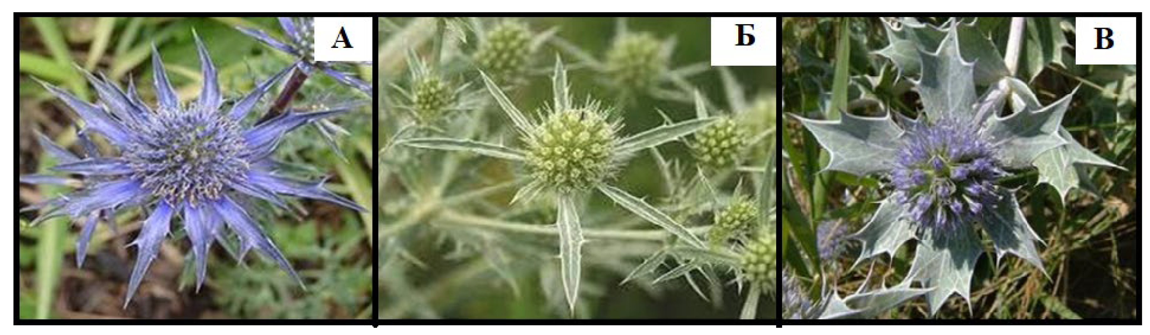
Figure 2.
Antimicrobial activity of Eryngium planum herb extract EP3.
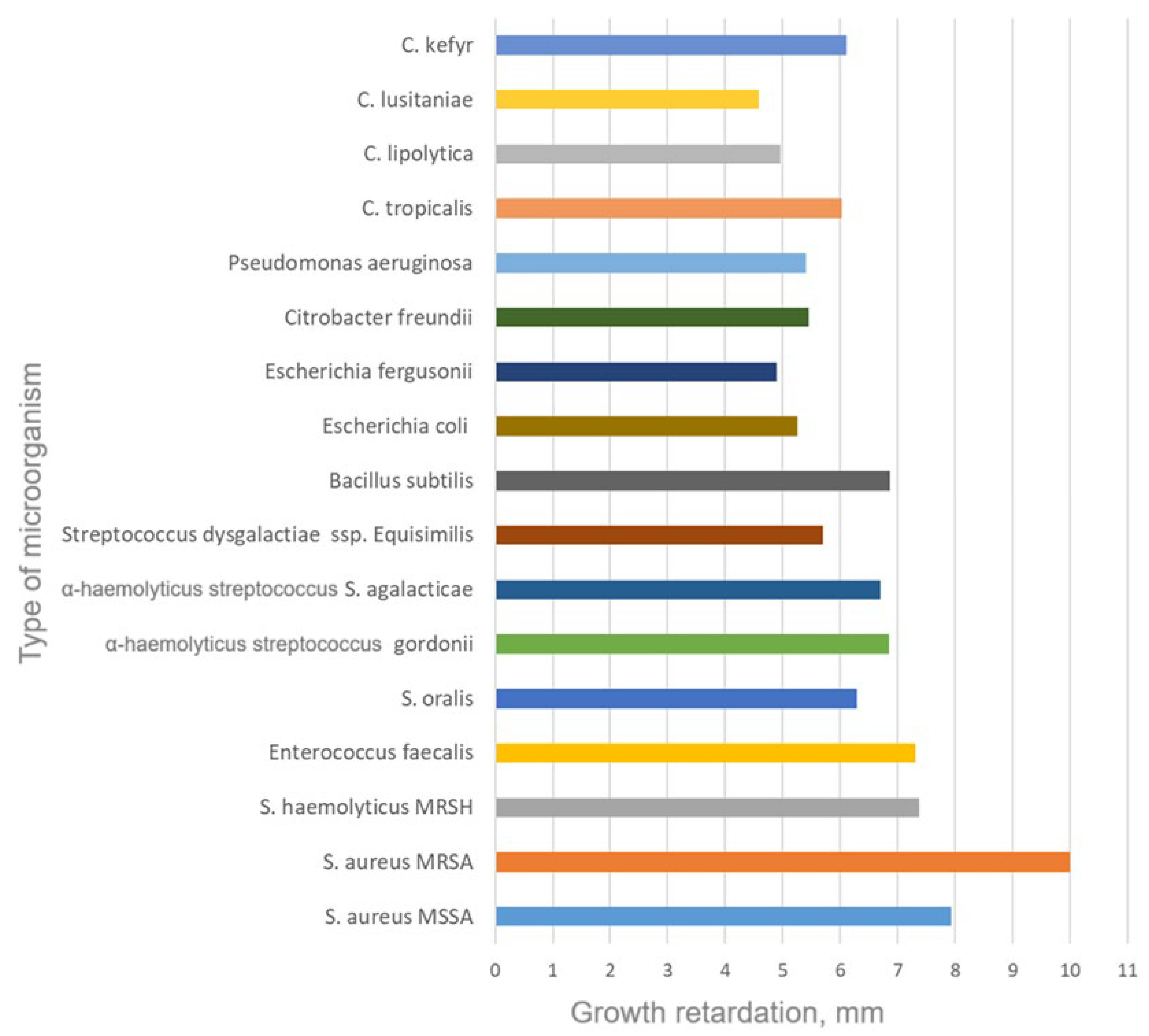
Figure 3.
Locomotor activity of animals in the “Open Field” test.
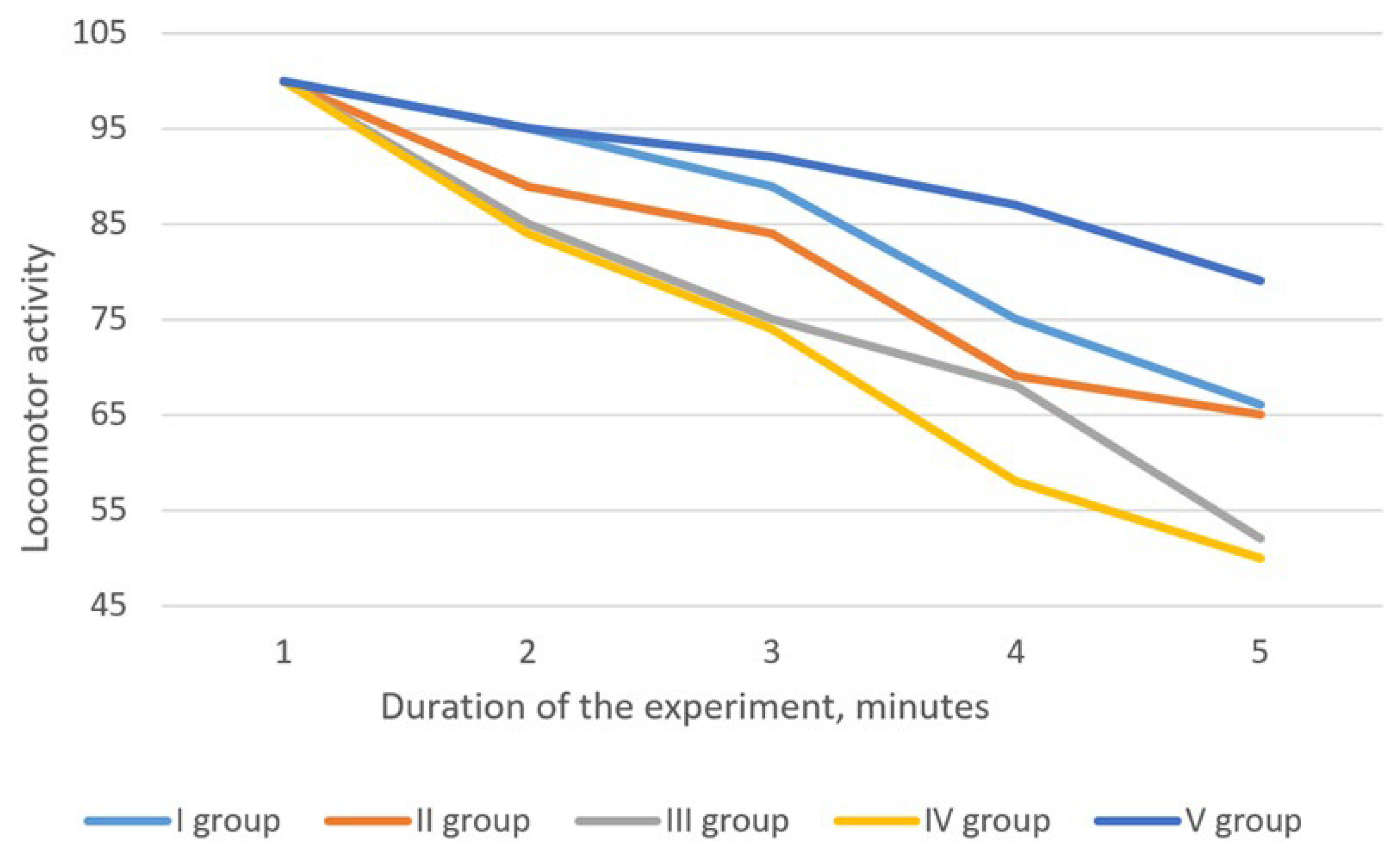
Figure 4.
Effects of Eryngium planum extracts on biochemical parameters of blood serum and liver condition in acute hepatitis. EP1 – water extract, EP2 – 40% ethanolic extract, EP3 – 70% ethanolic extract.
Figure 4.
Effects of Eryngium planum extracts on biochemical parameters of blood serum and liver condition in acute hepatitis. EP1 – water extract, EP2 – 40% ethanolic extract, EP3 – 70% ethanolic extract.
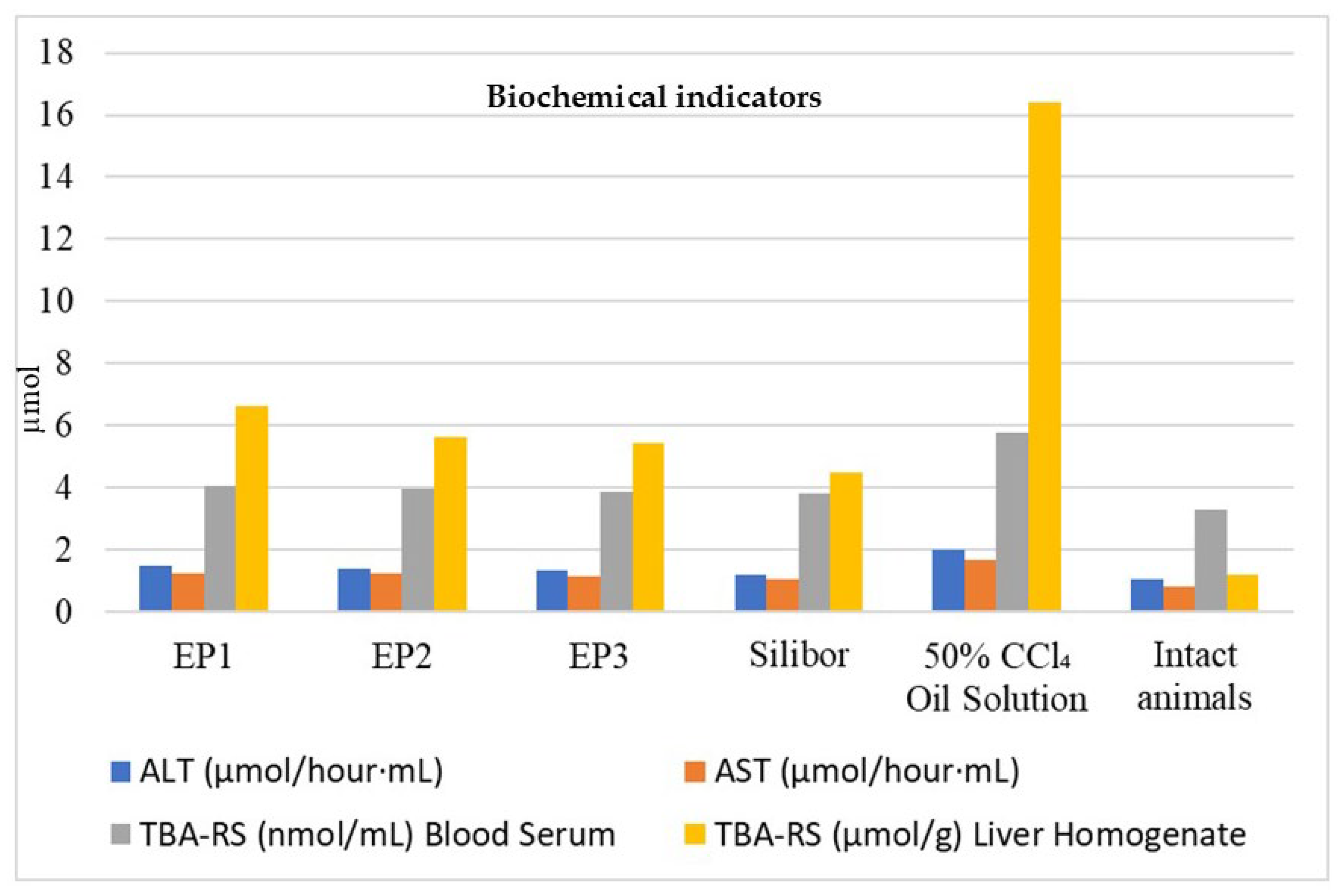

Table 1.
Content of phenolic compounds in the soft extracts of Eryngium planum herb.
| № | Compound | Content in dense extract, µg/g | ||
|---|---|---|---|---|
| EP1 | EP2 | EP3 | ||
| Hydroxycinnamic acids | ||||
| 1 | Chlorogenic acid | 3945.81±174.12 | 4931.72±215.32 | 4843.06±92.07 |
| 2 | Caffeic acid | 1268.87±94.14 | 1492.33±87.12 | 1319.78±79.16 |
| 3 | Syringic acid | 176.09±13.07 | 234.98±19.06 | 312.86±15.37 |
| 4 | p-Coumaric acid | 418.61±21.16 | 531.85±16.43 | 591.77±28.92 |
| 5 | trans-Ferulic acid | 729.98±31.88 | 910.11±29.19 | 937.32±51.06 |
| 6 | Sinapic acid | 3011.84±156.18 | 3532.33±141.82 | 3319.96±138.46 |
| 7 | trans-Cinnamic acid | 2358.07±93.61 | 2841.49±105.04 | 2909.52±135.47 |
| Flavonoids | ||||
| 8 | Rutin | 1382.762±102.04 | 2412.37±92.08 | 2631.62±108.37 |
| 9 | Quercetin-3-O-glucoside | 509.71±25.07 | 917.74±37.05 | 993.52±47.08 |
| 10 | Naringenin | 2944.71±172.73 | 4430.62±201.42 | 4873.07±212.92 |
| 11 | Neohesperidin | 989.72±39.09 | 2091.52±87.41 | 2313.43±93.36 |
| 12 | Quercetin | 697.92±29.93 | 1393.55±65.12 | 1493.72±72.17 |
| 13 | Kaempferol | 919.23±58.39 | 1801.17±11.03 | 1945.91±74.32 |
| Tannin metabolites | ||||
| 14 | Epicatechin | 1507.72±82.06 | 1420.08±73.93 | 1274.58±59.37 |
| 15 | Epicatechin gallate | 431.25±15.03 | 368.42±10.96 | 331.09±21.12 |
| 16 | Gallocatechin | 1607.71±57.86 | 1525.03±65.41 | 1318.62±45.16 |
| Content of compound groups, % | ||||
| Total polyphenols (%) | 14.92±0.48 | 17.52±0.61 | 21.84±0.97 | |
| Tannins (%) | 9.13±0.29 | 8.72±0.21 | 8.17±0.23 | |
| Flavonoids (%) | 3.11±0.14 | 5.71±0.26 | 6.19±0.13 | |
| Polysaccharides (%) | 21.14±0.95 | 7.73±0.36 | - | |
Notes: EP1 – water extract, EP2 – 40% ethanolic extract, EP3 – 70% ethanolic extract.
Table 2.
Behaviour of animals in the “Elevated Plus Maze” test.
| Animal Group | Time spent, % | Number of downward glances | ||
|---|---|---|---|---|
| Closed arm | Open arm | In center | ||
| I group (EP1) | 60.9±3.10* | 34.2±1.71* | 4.9±0.22* | 5.7±0.29* |
| II group (EP2) | 53.1±2.85* | 40.8±1.25* | 6.1±0.29* | 6.8±0.30* |
| III group (EP3) | 48.3±2.41* | 44.5±1.21* | 7.2±0.31* | 6.5±0.35* |
| IV group (control) | 44.5±2.40 | 49.7±1.20 | 5.8±0.28 | 7.8±0.35 |
| V group (intact) | 78.8±2.89* | 16.2±0.82* | 5.0±0.19* | 3.5±0.12* |
Note: * - the deviation of the indicator is significant compared to the data of the control group (p<0.05). EP1 – water extract, EP2 – 40% ethanolic extract, EP3 – 70% ethanolic extract.
Table 3.
Behaviour of animals in the “Cube” test.
| Group | Time of first exit (s) | Number of exits | Total duration of exits (s) |
|---|---|---|---|
| I group (EP1) | 53.1±2.15* | 2.5±0.01* | 37.2±1.18* |
| II group (EP2) | 28.6±1.41* | 2.7±0.01* | 36.2±1.41* |
| III group (EP3) | 19.0±1.12* | 3.2±0.02* | 63.8±2.09* |
| IV group (control) | 14.3±0.92 | 5.0±0.02 | 78.0±2.14 |
| V group (intact) | 75.0±3.16* | 1.8±0.01* | 16.1±0.98* |
Note: * - the deviation of the indicator is significant compared to the data of the control pathology group (p<0.05). EP1 – water extract, EP2 – 40% ethanolic extract, EP3 – 70% ethanolic extract.
Table 4.
Behaviour of animals in the “Black-and-White Box” test.
| Group | Time of first exit (s) | Number of exits | Total duration of exits (s) |
|---|---|---|---|
| I group (EP1) | 23.0±0.98* | 6.5±0.15 | 33.2±1.22 |
| II group (EP2) | 12.7±0.51 | 10.2±0.20 | 48.2±1.25 |
| III group (EP3) | 10.3±0.12 | 7.5±0.15 | 63.6±1.65 |
| IV group (control) | 8.3±0.10 | 9.8±0.15 | 55.3±1.50 |
| V group (intact) | 28.2±0.26 | 3.8±0.15 | 13.2±0.25 |
Note: * - the deviation of the indicator is significant compared to the data of the control pathology group (p<0.05).
Disclaimer/Publisher’s Note: The statements, opinions and data contained in all publications are solely those of the individual author(s) and contributor(s) and not of MDPI and/or the editor(s). MDPI and/or the editor(s) disclaim responsibility for any injury to people or property resulting from any ideas, methods, instructions or products referred to in the content. |
© 2024 by the authors. Licensee MDPI, Basel, Switzerland. This article is an open access article distributed under the terms and conditions of the Creative Commons Attribution (CC BY) license (http://creativecommons.org/licenses/by/4.0/).
Copyright: This open access article is published under a Creative Commons CC BY 4.0 license, which permit the free download, distribution, and reuse, provided that the author and preprint are cited in any reuse.


