
Submitted:
24 June 2025
Posted:
25 June 2025
You are already at the latest version
Abstract
Tulbaghia violacea, a plant native to Africa, is popular for its uses in traditional medicine, as well as in food and for household purposes. This study aimed to determine phytochemicals present in T. violacea, investigate the antioxidant and the enzyme-based antidiabetic activities in vitro, and identify potential differences in the activities depending on the growing environment of the plant. T. violacea leaves samples from two provinces in South Africa, viz. Gauteng (GP), an inland province and the Eastern Cape (EC), a coastal province were compared. The samples were extracted using maceration with hexane, acetone and methanol. Antioxidant activity was determined qualitatively and quantitatively with the DPPH radical scavenging assay, while the antidiabetic effects were determined using the α- amylase and α- glucosidase inhibition assays. The qualitative phytochemical screening revealed variable presence of saponins, flavonoids, tannins, alkaloids, steroids, cardiac glycosides and phenolics from both samples. The EC hexane extract showed the highest antioxidant activity (41,3%, IC50 68,3ug/ml). The GP and EC hexane extracts exhibited α-amylase inhibition of 85,5% (IC50= 9,02ug/ml ) and 85,7% (IC50= 15,03ug/ml) respectively, while the GP acetone extract showed α-glucosidase inhibition of 60,0% (IC50 = 42,2ug/ml). The findings elucidate on the antidiabetic mechanisms of T. violacea, showing in vitro α- amylase and α- glucosidase inhibitory activities and validate its ethnomedicinal use for diabetes.
Keywords:
Tulbaghia violacea
; antidiabetic
; α-amylase inhibition
; α-glucosidase inhibition
; antioxidant
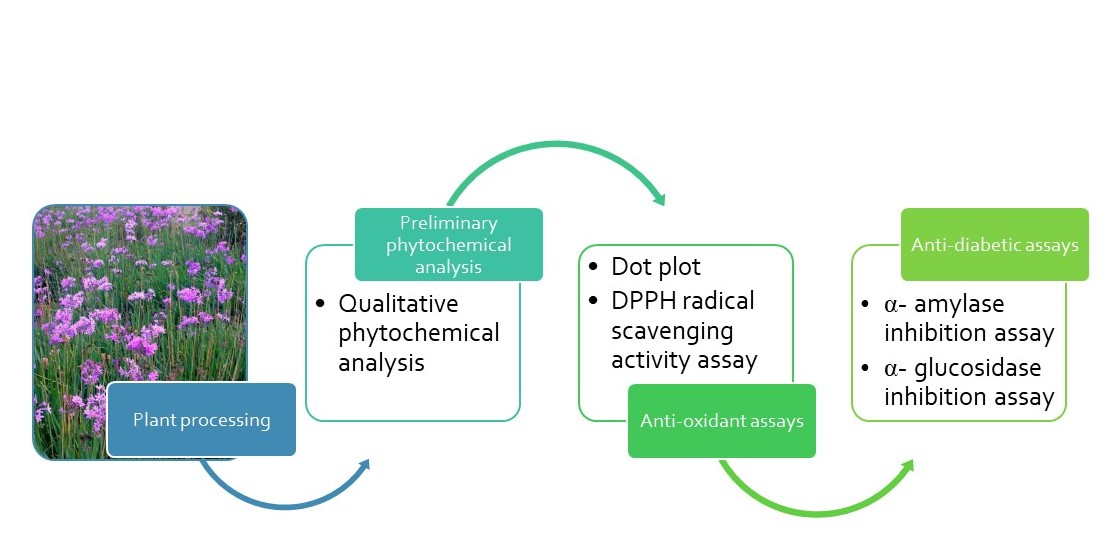
1. Introduction
The use of natural products as medicine has been described as traditional medicine (TM), and according to the World Health Organisation (WHO), TM refers to the knowledge, skills and practices which are based on the beliefs and experiences indigenous to cultures and is used to maintain health [1]. For centuries, people in many parts of the world, have relied on TM for their health needs and the practice continues to this day. When TM is adopted outside of its traditional culture, it is often referred to as complementary or alternative medicine and the most widely used TM systems used today include those from China, India and Africa among others [2]. The WHO states that trends in the use of complementary, alternative and traditional medicine have been increasing. It has been stated that in Africa, Asia, Latin America and the Middle east, 75-90% of the population still use traditional medicine [3]. It has been shown that people seek traditional and complementary medicine services and treatments for various purposes, including noncommunicable diseases, disease prevention and , health promotion [4,5]. It has also been purported that nearly one quarter of all modern medicine is derived from natural products of which many were originally used in traditional medicine [6].
One of the commonly used plants in TM is Tulbaghia violacea, a fast-growing, bulbous plant with narrow, long, strap-like fleshy leaves which releases a strong garlic smell when bruised. It is a geophyte belonging to the Alliaceae family and is commonly known as wild garlic, wildeknoffel or wildeknoflok (Afrikaans), utswelane or itswele lomlambo (isiXhosa) and isihaqa (isiZulu). It is a drought-resistant plant which can be found in the Eastern Cape, KwaZulu-Natal and Limpopo provinces in South Africa (SA) and thrives well in both dry and fertile areas [7,8]. T. violacea has many uses both in TM as well as in food and for household purposes. The leaves and flowers are used in salads and some Zulu-speaking people use it in the same way as spinach due to its peppery-garlic taste. When crushed onto the skin, the strong smell is used to repel fleas, ticks and mosquitoes. In a medicated bath, T. violacea is used to treat paralysis and rheumatism as well as to reduce the fever of a patient. A boiled water decoction of its bulbs is taken orally to clear coughs and colds. It is also used as a remedy for pulmonary tuberculosis and intestinal worms. The leaves are used to treat oesophageal cancer and the rhizomes or bulbs are made into a tea for the treatment of fever or high blood pressure [7,8]. T. violacea is one of the forty-eight plant species used by communities of the Eastern Cape province, SA, for the treatment of diabetes mellitus [9,10]. These different uses highlight the importance of this plant towards managing communicable and non-communicable diseases. Various solvent extracts of the whole plant has been reported to have a wide range of pharmacological activities, including antimicrobial, antiparasitic, anticancer, antioxidant, anticardiovascular and antithrombogenic activities [11,12,13]. Of importance to this study is the ethnomedicinal use of the plant for diabetes mellitus and its antidiabetic activity.
According to the International Diabetes Federation, 537 million people in the world and 4.2 million people in South Africa were found to have diabetes mellitus (DM) in 2021 [14]. DM is a chronic, metabolic disorder resulting in hyperglycaemia. In Type 1 diabetes mellitus (T1DM), there is an absolute deficiency in insulin secretion and this form of diabetes is observed in approximately 10% of patients diagnosed with DM. Type 2 diabetes mellitus (T2DM) is caused by a combination of factors such as defective insulin secretion by the beta cells of the pancreas or when skeletal muscle cells develop insulin resistance. T2DM accounts for approximately 90-95 % of all DM. Uncontrolled DM can result in serious complications and health problems including loss of vision, kidney problems, heart disease and issues with the nervous system [14,15,16,17].
Currently, there is no cure for diabetes mellitus, however there are drugs available to help control the hyperglycaemia associated with the condition; with the goal of therapy being to control blood glucose levels within the normal range and to prevent the long term complications associated with uncontrolled diabetes.
For the management of T1DM, exogenous insulin is used, with multiple dosage regimens. For T2DM, lifestyle modifications are combined with oral antidiabetic drugs. Classes of oral antidiabetics include sulfonylureas, biguanides, thiazolidinediones, alpha- glucosidase inhibitors, dipeptidyl peptidase IV (DPP-IV) inhibitors and sodium-glucose co-transporter 2 inhibitors [18,19].
Although many classes of drugs exist for the management of T2DM, each class is associated with multiple side effects. For this reason, the screening of medicinal plants for their antidiabetic activity is important, as they are more cost efficient, widely available and generally associated with a lower incidence of side effects [20]. T. violacea has been reported to contain phytochemicals sucha as flavonoids, terpenoids, saponins as well as sulphur-containing compounds [11,12]. The rhizome extracts of T. violacea reduced blood glucose and attenuated diabetes associated physiological complications in streptozotocin-induced diabetic rats [12]. Given its rich phytochemical profile, it is rational to expect that the plant may exert its effects in the various conditions through diverse mechanisms, most of which have not been explored. It is therefore hypothesized that T. violacea exerts anti-diabetic effects through inhibition of key carbohydrate-digesting enzymes, similar to the known a-glucosidase inhibitors such as acarbose.
Many authors have stated that environmental factors such as temperature and humidity may influence certain phytochemicals present in plants such as T. violacea, and that the class, content and quantity of the compounds may vary depending on the ecological factors in the area where the plant is cultivated [21]. For this reason, this study explored the enzyme-based antidiabetic mechanisms of T. violacea leaf extracts, while comparing samples of T. violacea grown in the Eastern Cape and Gauteng provinces in South Africa; to observe the impact of environmental and ecological factors on the pharmacological activity of the plant.
2. Materials and Methods
2.1. Plant Material
2.1.1. Sample Collection, Preparation and Handling
T. violacea plants were collected from two nurseries, one in Gauteng (GP), Pretoria (25.6713° S, 28.0742° E) and the other in the Eastern Cape (EC), Makhanda (33 18.535° S, 26 31.291° E), South Africa. Fresh samples were authenticated at the Department of Botany in RU. The leaves were separated from the flowers and stems and the roots were replanted and allowed to reshoot to prevent destructive harvesting. The leaves were rinsed under running water to remove any sand or dust particles and were air-dried in the laboratory, out of direct sunlight to avoid potential degradation of their phytochemicals. Once completely dry, the leaves were ground into a fine powder using a Powteq knife mill (Powteq Knife Mill, HM100, Huilongguan, Changping District, Beijing, China) and then stored in closed containers at room temperature for further use.
2.1.2. Extraction of the Plant Material
The powdered plant material were extracted using serial maceration extraction method with slight modifications, using hexane, acetone and methanol. Organic solvents were selected to maximise compound diversity, and their choice was based on their polarity differences and their ability to extract similar polarity compounds. Hexane (relative polarity 0.009) was intended to extract non-polar compounds as the lowest polarity (non-polar solvent), acetone (relative polarity 0.355) for intermediate polarity compounds as a mid-polar solvent and methanol (relative polarity 0.762) for polar compounds as the highest polarity solvent among the 3 solvents.
The powdered material (around 100 g) for each province sample, were placed in beakers, covered with hexane solvent and placed in a sonicator (Scientech ultrasonic cleaner, Model 703, Labotec, Midrand, South Africa) at 37°C for one hour. After the hour, the supernatants were filtered using filter discs (Filter Discs Qual, 3 hw, 125 mm. BOECO, Hamburg Germany). New hexane solvent was added and the process was repeated. The resulting supernatant was filtered and combined with the first one. The plant samples were once again covered in hexane and placed on an orbital platform shaker (model 261, 8kg, 120w, Labotec, Midrand, South Africa) overnight, set at 120 revolution per minute (rpm). The next morning, the supernatant was filtered and combined with the previous two batches. The filtrate was then concentrated using a Stuart rotary evaporator (RE 400, COLE-PARMER LTD. Stone, ST15 OSA, Staffordshire, United Kingdom) at a temperature of 37°C with a speed of 120 rpm. The entire extraction process was repeated for both acetone and methanol. The resulting extracts were then stored at room temperature for further use. For the quantitative assays, stock solutions of 500ug/ml of each extract were prepared, in their solvents of extraction.
2.2. Qualitative Phytochemical Analysis
Qualitative phytochemical analysis detects the presence of various compounds based on their selective reaction with specific reagents and the identification is based on the development of a specific colour. All of the extracts were subjected to preliminary phytochemical analysis following standard colorimetric methods as described by Madike et al. [21,22] with slight modifications, where the extracts were re-dissolved in the solvents used to extract them (ie. hexane, acetone and methanol) to a final concentration of 1 mg/ml each. Different tests were conducted for the following phytochemicals:
Saponins
A volume of 2 millilitres (ml) of crude plant extract was added to 2 ml of distilled water and then shaken vigorously. The formation of foam and a persistent froth was an indication for the presence of saponins.
Anthraquinones
A volume of 2 ml of plant extract was treated with approximately 20 drops of 10% ammonia solution. The appearance of a pink precipitate indicated the presence of anthraquinones in the extract.
Flavonoids
The plant extract (3 ml) was treated with 1 ml of dilute NaOH followed by the addition of 1 ml dilute HCl. A yellow colour that appeared after the addition of NaOH indicated the presence of flavonoids. A colourless solution that appeared after the addition of HCl, confirmed their presence.
Tannins
One ml of ferric chloride was added to 2 ml of the plant extract. The occurrence of black or blue-green colour indicated the presence of tannins.
Alkaloids
Approximately 2 ml of extract was added to a test tube and 15 drops of Dragendorff’s reagent was added. The formation of a reddish brown to orange precipitate indicated the presence of alkaloids.
Steroids
One ml of chloroform and 1 ml of concentrated sulfuric acid were added to 2 ml of plant extract. The appearance of a red colour in the lower chloroform layer indicated the presence of steroids.
Cardiac glycosides
Two ml of extract was treated with 1 ml glacial acetic acid, 1 ml of ferric chloride and 1 ml concentrated sulfuric acid. The presence of a green- blue colour of the solution indicated the presence of cardiac glycosides.
Phenols
Ferric chloride was added to 1 ml crude plant extract. A black or blue-green colour indicated the presence of phenols.
2.3. Antioxidant Assay
2.3.1. Qualitative Antioxidant Activity (Dot-Plot Assay)
The method described by Gupta et al. [23] was followed with slight modifications. . The molecule, 2,2-diphenyl-1-picrylhydrazyl (DPPH) is a stable, free radical with an intense, purple colour in solution. Antioxidants are able to transfer an electron to DPPH and cause a discolouration of the DPPH solution [24].
The solvent extracts of T. violacea were redissolved in the solvents that were used to extract them. A pre-coated silica gel thin layer chromatography (TLC) plate (TLC Silica gel 60 F₂₅₄, Sigma Aldrich) was prepared to accommodate all the 6 crude extracts. Each extract was spotted in a dot form on the TLC plate and left to dry. Once dry, the TLC plate was sprayed with the prepared 0.01 mM DPPH solution. The white to pale yellow discoloration of the dots against the purple background of the sprayed TLC plate was taken as a positive indication of antioxidant activity.
2.3.2. Quantitative DPPH Radical Scavenging Assay
The DPPH radical scavenging assay was conducted according to a method described by Kwon et al. [25] with slight modifications. The DPPH method is a common radical scavenging methods used, as it is extremely fast, requires no expensive reagents or sophisticated instruments, allows quicker preparation and analysis of samples, has high sensitivity, and is easy to use [24].
In a 96-well plate, 100 µl of freshly prepared DPPH (0.01 mM) was added to 200 µl of plant extracts of varying concentrations (10, 20, 30, 40 and 50 µg/ml). The reaction mixture was allowed to stand at room temperature in the dark for 30 minutes. Thereafter the absorbance was measured at 517 nm using a Spectramax M3- multimode microplate reader (Molecular Devices, Beijing, China). For the assay, ascorbic acid at varying concentrations (10, 20, 30, 40 and 50 µg/ml) was used as the standard compound and distilled water was used as a negative control. The assay was conducted in triplicate. The percentage of DPPH radical scavenging activity was calculated using the following equation:
where:
Abs sample is the absorbance of the sample (plant extracts) measured at 517 nm,
Abs control is the absorbance of the control measured at 517 nm
2.4. Antidiabetic Assays
2.4.1. The α- amylase Inhibition Assay
The α-amylase inhibitory activity was determined by the method described in the Worthington Manual [26] with slight modifications. For the reaction, 500 µl plant extract (10, 20, 30, 40 and 50 µg/ml) was added to 500 µl of a 1% α-amylase solution (1 U/ml) and the mixture was incubated at 37°C for 15 minutes. Then 500 µl of a 1% starch solution in 0.02 M phosphate buffer (pH= 6.9) was added and the mixture was incubated at 37°C for a further 10 minutes. This was followed by addition of 1 ml of 3,5- dinitrosalicylic acid reagent to each test tube and the mixture was boiled for 10 minutes. The reaction mixture was then diluted by adding 2 ml distilled water. Absorbance was read at 540 nm using a Spectramax M3- multimode microplate reader (Molecular Devices, Beijing, China). Acarbose (10, 20, 30, 40 and 50 µg/ml) was used as the standard. The experiment was carried out in triplicate and the α- amylase inhibitory activity was calculated using the following equation:
where:
Abs sample is the absorbance of the sample (plant extracts) measured at 540 nm,
Abs control is the absorbance of the control measured at 540 nm
2.4.2. The α-glucosidase Inhibition Assay
The α-glucosidase inhibitory activity was determined by the method described in the Worthington Manual [27] with slight modifications. In a 96-well plate, the reaction mixture containing 100 µl phosphate buffer (0.1 M, pH= 6.9), 40 µl α- glucosidase (1 U/ml) and 80 µl plant extracts (10, 20, 30, 40 and 50 µg/ml) were incubated at 37°C for 10 minutes. Then 20 µl para-nitrophenyl-α-D-glucopyranoside (5 mM) was added and the reaction mixture was incubated for 10 minutes at 37°C. The reaction was stopped by the addition of 100 µl of 0.1 M Na2CO3. Absorbance was read at 405 nm using a Spectramax M3- multimode microplate reader (Molecular Devices, Beijing, China). Acarbose (10, 20, 30, 40 and 50 µg/ml) was used as the standard. The experiment was carried out in triplicate and the α- glucosidase inhibitory activity was calculated using the following equation:
where:
Abs sample is the absorbance of the sample (plant extracts) measured at 405 nm,
Abs control is the absorbance of the control measured at 405 nm
2.5. Statistical Analysis
Statistical analysis was carried out on the results of the assays to establish if there was a significant difference between the activity of T. violacea plant extracts and the standards (controls). The experiments were carried out in triplicates and statistical analysis was done using GraphPad Prism 10 and one-way ANOVA trailed by Dunnett’s post-hoc test. Significant statistical difference was noted where p < 0.05. Microsoft Excel and GraphPad Prism 10 were used to plot the graphs and calculate the IC50 values using the linear regression method where the linear equation was drawn to best represent the data points obtained from each of the concentrations for each extract.
3. Results
3.1. Qualitative Phytochemical Analysis
The qualitative phytochemical profile of T. violacea extracts is presented in Table 1.
The results revealed variable presence of phytochemicals in the EC and GP samples of T. violacea including saponins, flavonoids, alkaloids, steroids, cardiac glycosides and phenolic compound. Saponins, alkaloids, steroids and phenolic compounds had a greater presence in the EC sample while cardiac glycosides had a greater presence in the GP sample of T. violacea compared across the different solvent extracts. Interestingly, no anthraquinones were detected in any of the T. violacea extracts.
3.2. Antioxidant assay
3.2.1. Qualitative Antioxidant Screening: Dot-Plot Method
The results of the dot-plot assay , shown in Figure 1, showed that the hexane, acetone and methanol crude extracts of T. violacea from EC and GP had antioxidant activity as all of the extracts changed the purple DPPH free radical to yellow. The hexane extract from the EC showed the brightest yellow spot while the acetone extracts showed the lowest colour change.
3.2.2. DPPH Radical Scavenging Assay
The quantitative antioxidant assay results of the extracts are shown in Figure 2, indicating where there are statistically significant differences as compared to the control, ascorbic acid. Table 2 and Table 3 highlight the results of each solvent extract, showing the EC and GP samples, indicating where statistically significant differences exist between the 2 samples.
The results of the radical scavenging assay, as depicted in Figure 2, confirmed the qualitative dot-plot results, that all extracts (hexane, acetone and methanol) of T. violacea from EC and GP had antioxidant activities. The results also indicated clearly that as the concentration of the extracts increased, their % DPPH radical scavenging increased. The hexane extract from EC had the highest radical scavenging activity at a concentration of 50 µg/ml, followed by the methanol extract of T. violacea from GP. All extracts had radical scavenging activity with statistically significant differences to the radical scavenging activity of ascorbic acid at the same concentrations (Figure 2).
Comparison of the EC samples and the GP samples (Table 2) showed that the extracts from the EC had radical scavenging activity that was significantly different to the radical scavenging activity of the GP samples. The EC hexane and acetone extracts as well as the GP methanol extract had higher radical scavenging activities at the highest tested concentrations.
3.3. Antidiabetic Assays
The α-amylase inhibitory activity and the α-glucosidase inhibitory activity of the different concentrations of T. violacea extracts were tested, using acarbose as the positive control. All the extracts of T. violacea revealed variable inhibition of the two enzymes.
3.3.1. The α- amylase Inhibition Assay
The results of the α- amylase inhibition assay, as depicted in Figure 3, indicated that all the tested extracts of T. violacea had α- amylase inhibitory activity, that was concentration dependent, similar to the acarbose (control). All the extracts had good inhibition of α- amylase with the hexane extract from GP having the highest inhibitory activity of 85. 7% at 50 µg/ml, followed closely by the hexane extract from EC with 85.5% inhibition. The lowest inhibition was seen with the acetone extracts of T. violacea from EC and GP. Interestingly, all the EC acetone extracts showed % inhibition of α- amylase lower than the control. Most of the T. violacea extracts from EC and GP, had statistically significant α- amylase inhibitory activity when compared to that of acarbose.
Table 4 presents the compared results of the EC samples and the GP samples for hexane, acetone and methanol extracts of T. violacea. The EC hexane extracts, had a higher α- amylase inhibitory effect than the GP sample at all concentrations of 10, 20, 30 and 40 µg/ml and their activity differences were statistically significant, except at the 50 µg/ml. Similarly, the GP acetone extract had a higher α- amylase inhibitory activity than the EC sample at concentrations of 10, 20, 30 and 40 µg/ml however, at 50 µg/ml, the EC sample showed a greater inhibitory effect. With regard to the methanol extracts, at concentrations of 10 and 20 µg/ml, the EC sample showed a greater inhibitory effect while the GP sample showed a higher one at concentrations of 30, 40 and 50 µg/ml.
Overall, the results of the α- amylase inhibition assay showed good inhibitory activity exerted by both the EC and GP samples of T. violacea and at most concentrations the inhibitory activities were found to be statistically significant.
The IC50 values of all the T. violacea extracts from EC and GP were comparable to the IC50 value of acarbose (Table 5), except for the hexane extract.
3.3.2. The α- glucosidase Inhibition Assay
The results of the α- glucosidase inhibition assay, as depicted in Figure 4, indicated that all the tested extracts of T. violacea had α- glucosidase inhibitory activity, which was concentration dependent, similarly to their α- amylase inhibition. All the extracts had good inhibition of α- glucosidase with the EC acetone extract having the highest inhibitory activity of 60.0% at 50 µg/ml, and the lowest % inhibition exhibited by the hexane extracts. The majority of the T. violacea extracts from EC and GP, at all the tested concentrations, had α- glucosidase inhibitory activity that was statistically significant compared to that of acarbose.
Table 6 compared the results of the EC samples and the GP samples for hexane, acetone and methanol extracts of T. violacea. The GP hexane extract had a higher α- glucosidase inhibitory activity than that of the EC sample, with significant statistical differences at the concentrations of 10, 40 and 50 µg/ml. The EC acetone extract had a higher inhibitory effect than the GP sample at all concentrations, with statistically significant differences in all the results. The EC methanol extract had a higher inhibitory effect compared to the GP sample, with significant statistical differences for all the tested concentrations
The IC50 values of all the T. violacea extracts from EC and GP were better than the IC50 value of acarbose (Table 7).
4. Discussion
The phytochemical screening of T. violacea showed the presence of saponins, flavonoids, tannins, alkaloids, steroids, cardiac glycosides and phenolics in the various extracts. This is in correlation with the results published in other studies, where Tulbaghia species were reported to contain flavonoids, tannins, [11,28], glycosides, , terpenoids, saponins and steroids [28]. It is also in agreement with the findings by Makhuvele et al. [12], that methanolic extracts of T. violacea leaf contained flavonoids, terpenoids, tannins and saponins. This current study however, showed the presence of alkaloids in all extracts from EC and GP in contrast to Takaidza et al. [28] alkaloids were not detected from T. violacea. Their study used the leaves of T. violacea collected from GP which would be similar to the GP samples in this study. The dissimilarity could be attributed to the difference in the areas where the plants were located within the GP and differences in extraction methods. The absence of glycosides and anthraquinones in the present study was interestingly similar to those by Makhuvele et al. [12]. The differences in phytochemicals could be due to the different seasons the plants were harvested in, the environment which includes the soil type, the pH, or differences in the extract preparation methods.
The antioxidant activities of the extracts confirmed through the DPPH assay corroborate the concentration-dependent antioxidant activity of T. violacea reported in other studies [12,28,29,30,31]. All the extracts showed significantly lower DPPH radical scavenging activities in comparison with ascorbic acid, with the findings similar to those reported for the methanol extract [12] and the acetone extract [30,31]. However, the acetone extract of the GP sample had the lowest radical scavenging activity, in contrast with the findings by Takaidza et al. [28] that showed that T. violacea rhizomes extracts had the highest scavenging activity for DPPH among the acetone extracts of Tulbaghia species tested, and Makgati [31], who reported that the crude acetone extracts of T. violacea roots had higher free radicals scavenging activity than the crude leaf extracts. These may suggest that the rhizome and roots have more antioxidant activity than the leaves, highlighting the importance of appropriate plant part selection for medicinal use. Other studies on medicinal plants have reported that in general, leaves showed higher antioxidant activity compared to other plant parts [32,33]. The hexane extract had the highest DPPH radical scavenging activity among the extracts in the study, which is interesting since hexane as the non-polar and most lipophilic solvent had shown variable presence of saponins, flavonoids, alkaloids, glycosides, steroids and phenolic compounds. No reports on hexane extracts of T. violacea could be found in literature for comparison. Overall, the differences in the radical scavenging activities and antioxidant capacity of T. violacea samples from EC and GP could be due to the differences in the phytochemical composition and concentrations influenced by environmental and ecological factors.
The role of amylase in the body is to break down carbohydrates or starch into maltose and polysaccharides while glucosidase is responsible for breaking down starch and disaccharides into glucose. After carbohydrates are broken down they are absorbed and cause an increase in blood glucose levels [34,35]. Inhibiting carbohydrate digestion can be beneficial in controlling postprandial hyperglycaemia in diabetic individuals and this can be achieved by inhibiting α- amylase and α- glucosidase. It has been reported that T. violacea has a hypoglycaemic effect which could be due to its impact on multiple pathways of the hyperglycemic process [36]. The hexane, acetone and methanolic extracts of T. violacea in the current study had α- amylase and α- glucosidase inhibitory effects that were comparable to or greater than that of acarbose. The results suggest a potential mechanism for the antidiabetic effect of T. violacea leaf, and provide the rationale for the ethnobotanical use of the plant for treatment of DM. It is also not implausible to consider that those who take this plant in their diet may benefit from its nutritional value, as well as its antidiabetic effects, although the findings have to be confirmed through in vivo studies. Clinically, acarbose is used in combination with other antidiabetic medicines, and patients usually stop taking the drug due to its adverse effects such as dyspepsia, diarrhoea and flatulence [19]. The findings in this study suggest a positive outlook for the role of T. violacea as an adjunct to antidiabetic therapy. The antidiabetic properties of T. violacea have been reported in other studies with other mechanisms through which the effects are exerted. Methanolic extracts of the rhizome were reported to have significantly reduced blood glucose, including fasting blood glucose levels [36,37], increased plasma insulin and liver glycogen content and improved glucose tolerance in streptozotocin diabetes- induced rat models [11,36,37].
At the time of this study, there were no reports in literature on the α- amylase and α- glucosidase inhibitory activities of T. violacea leaf. One study reported minimal and absence of α- glucosidase inhibition by the tea extract and methanol extract respectively, of T. violacea whole plant [38]. While there are no reports on the antidiabetic effects of T. violacea hexane extracts, other biological activities have been reported, including pro-apoptotic effect in cancer cell lines [39]. The α- amylase and α- glucosidase inhibitory effects reported in this study may be attributed to the different phytochemicals detected in the hexane, acetone and methanol extracts. Flavonoids, glycosides, saponins, terpenoids and steroids in plants including T. violacea, have been shown to have antidiabetic effects through various mechanisms [12,37,40] and the hepatoprotective role of polyphenolic compounds in T2DM as antioxidants has been reported [37].
The one limitation in the study is the absence of a water extract, and water is the solvent in traditional medicine practice. However, this does not diminish the importance of the findings that showed the range of phytochemicals present in T. violacea leaf, and the antioxidant and antidiabetic activities of the plant.
5. Conclusions
The results of the current study demonstrated one of the mechanisms of the antidiabetic properties of T. violacea leaf, showing in vitro α- amylase and α- glucosidase inhibitory activities. The antioxidant activity and the groups of phytochemicals responsible for the effects were also confirmed. The findings explain the continued use of T. violacea in traditional medicine for diabetes mellitus and other conditions in South Africa. They provide the impetus for follow up research towards the development of new drugs derived from T. violacea for the management of DM, as well as the potential role of T. violacea as an adjunct to antidiabetic therapy. This would include isolation and identification of the active compounds from all the hexane and the acetone extracts which demonstrated the highest α- amylase and α- glucosidase inhibitory activities.
Author Contributions
Conceptualization, T.K and M.M.; methodology, T.K, S.M. N.S and M.M.; software, T.K.; validation, S.M., N.S and M.M.; formal analysis, T.K., S.M. and M.M.; investigation T.K.; resources, M.M. and N.S; data curation, S.M. and M.M.; writing—original draft preparation, T.K.; writing—review and editing, S.M. N.S and M.M.; visualization, T.K, S.M. and M.M.; supervision, N.S. and M.M.; project administration, M.M.; funding acquisition, M.M. All authors have read and agreed to the published version of the manuscript.
Funding
This research received no external funding, and the APC was funded by Rhodes University.
Institutional Review Board Statement
Not applicable.
Informed Consent Statement
Not applicable.
Data Availability Statement
All data for this study have been included in the manuscript, and any further inquiries can be directed to the corresponding author.
Acknowledgments
The authors would like to thank the Department of Pharmaceutical Sciences, School of Pharmacy, Sefako Makgatho Health Sciences University for the initial plant extraction work in the project.
Conflicts of Interest
The authors declare no conflicts of interest. The study had no external funders; as such, no funders had any role in the design of the study; in the data collection, analyses, or interpretation; in the writing of the manuscript; or in the decision to publish the results.
Abbreviations
The following abbreviations are used in this manuscript:
| DM | Diabetes mellitus |
| DPPH DPP-IV |
2,2-diphenyl-1-picrylhydrazyl Dipeptidyl peptidase 4 |
| EC | Eastern Cape Province |
| GP SA T1DM T2DM TLC |
Gauteng Province South Africa Type 1 diabetes mellitus Type 2 diabetes mellitus Thin layer chromatography |
References
- World Health Organisation. Traditional Medicine. Available online: https://www.afro.who.int/health-topics/traditional-medicine (accessed on 5 February 2025).
- Che, C.; George, V.; Ijinu, T.; Pushpangadan, P.; Marobela, K. Traditional Medicine. In Pharmacognosy: Fundamentals, Applications and Strategies, Badal, S.; Delgoda, R., Eds.; Elsevier: Academic Press, 2017, pp. 15-30. [CrossRef]
- Mothibe, M.E.; Sibanda, M. African Traditional Medicine: South African Perspective. In Traditional and Complementary Medicine, Mordeniz, C. IntechOpen, 2019, pp. 31-57. [CrossRef]
- World Health Organisation. Traditional Medicine. Draft traditional medicine strategy: 2025–2034. Available online: https://apps.who.int/gb/ebwha/pdf_files/EB156/B156_16-en.pdf (accessed on 5 February 2025).
- Shetty, P.; Rinaldi, A. Traditional medicine for modern times: Facts and figures. Available online: https://www.scidev.net/global/features/traditional-medicine-modern-times-facts-figures/ (accessed on 6 April 2021).
- Thomford, N.E.; Senthebane, D. A.; Rowe, A.; Munro, D.; Seele, P.; Maroyi, A.; Dzobo, K. Natural Products for Drug Discovery in the 21st Century: Innovations for Novel Drug Discovery. Int J Mol Sci 2018, 19, 6, 578. [Google Scholar] [CrossRef] [PubMed]
- South African National Biodiversity Institute. Tulbaghia violacea. Available online: https://pza.sanbi.org/tulbaghia-violacea (accessed on 6 April 2023).
- Styger, G.; Aboyade, O.M.; Gibson, D.; Hughes, G. Tulbaghia--A Southern African Phytomedicine. J Altern Complement Med 2016, 22, 4, 255–261. [Google Scholar] [CrossRef]
- Odeyemi, S.; Bradley, G. Medicinal Plants Used for the Traditional Management of Diabetes in the Eastern Cape, South Africa: Pharmacology and Toxicology. Molecules 2018, 23, 11, 2759. [Google Scholar] [CrossRef]
- Sagbo, I.J.; Hussein, A.A. Antidiabetic Medicinal Plants Used in the Eastern Cape Province of South Africa: An Updated Review. Processes 2022, 10, 1817. [Google Scholar] [CrossRef]
- Danquah, C.A.; Minkah, P.A.B.; Agana, T.A.; Moyo, P.; Ofori, M.; Doe, P.; Rali, S.; Osei Duah Junior, I.; Amankwah, K.B.; Somuah, S.O.; et al. The Phytochemistry and Pharmacology of Tulbaghia, Allium, Crinum and Cyrtanthus: ‘Talented’ Taxa from the Amaryllidaceae. Molecules 2022, 27, 4475. [Google Scholar] [CrossRef] [PubMed]
- Makhuvele, R.; Gbashi, S.; Njobeh, P.B. GC-HRTOF-MS Metabolite Profiling and Antioxidant Activity of Methanolic Extracts of Tulbaghia violacea Harv. J King Saud Univ Sci 2022, 34, 7, https://www.sciencedirect.com/science/article/pii/S1018364722004591. [Google Scholar] [CrossRef]
- Makhoahle, P.; Rampana, D.E. Antioxidant Activities, Total Polyphenol Profile and Anticancer Activity, of Leaf, Bulb and Root Extracts of Tulbaghia violacea from Bloemfontein. Phcog J 2023, 15, 5, 761–767. [Google Scholar] [CrossRef]
- International Diabetes Federation. South Africa, Key Information. Available online: https://idf.org/our-network/regions-and-members/africa/members/south-africa/ (accessed on 15 December 2023).
- World Health Organisation. Diabetes. Available online: https://www.who.int/news-room/fact-sheets/detail/diabetes (accessed on 15 December 2023).
- Centers for Disease Control and Prevention. What is Diabetes? Available online: https://www.cdc.gov/diabetes/basics/diabetes.html#:~:text=Diabetes%20is%20a%20chronic%20(long,your%20pancreas%20to%20release%20insulin (accessed on 15 December 2023).
- American Diabetes Association. Diagnosis and Classification of Diabetes Mellitus. Diabetes care 2009, 32, Suppl 1, S62–S67. [Google Scholar] [CrossRef]
- Galicia-Garcia, U.; Benito-Vicente, A.; Jebari, S.; Larrea-Sebal, A.; Siddiqi, H.; Uribe, K.B.; Ostolaza, H.; Martín, C. Pathophysiology of Type 2 Diabetes Mellitus. Int J Mol Sci 2020, 21, 17, 6275. [Google Scholar] [CrossRef]
- Brutsaert, E.F.; Medications for Diabetes Mellitus Treatment - Endocrine and Metabolic Disorders. MSD Manual Professional Edition. Available online: https://www.msdmanuals.com/professional/endocrine-and-metabolic-disorders/diabetes-mellitus-and-disorders-of-carbohydrate-metabolism/medications-for-diabetes-mellitus-treatment (accessed on 5 February 2022).
- Dias, D.A.; Urban, S.; Roessner, U. A Historical Overview of Natural Products in Drug Discovery. Metabolites 2012, 2, 2, 303–336. [Google Scholar] [CrossRef]
- Madike, L.; Takaidza, S.; Pillay, M. Preliminary Phytochemical Screening of Crude Extracts from the Leaves, Stems, and Roots of Tulbaghia violacea. Int J Pharmacogn Phytochem Res 2017, 9, 10. [Google Scholar] [CrossRef]
- Kumar, A.; Mathew, L. Comparative Account of the Preliminary Phytochemical Aspects of Helicanthes elastica (Desr) Danser growing on two Different Hosts. J Pharmacogn Phytochem 2014, 3, 1, 218–221, https://www.phytojournal.com/archives/2014/vol3issue1/PartD/47.1-184.pdf. [Google Scholar]
- Gupta, S.; Adak, S.; Rajak, R.C. , Banerjee, R. In Vitro Efficacy of Bryophyllum pinnatum Leaf Extracts as Potent Therapeutics. Prep Biochem Biotechnol 2016, 46, 5, 489–494. [Google Scholar] [CrossRef]
- Gulcin, İ.; Alwasel, S.H. DPPH Radical Scavenging Assay. Processes 2023, 11, 2248. [Google Scholar] [CrossRef]
- Kwon, S.H.; Wang, Z.; Hwang, S.H; Kang, Y.H.; Lee, J.Y.; Lim, S.S. Comprehensive Evaluation of the Antioxidant Capacity of Perilla frutescens Leaves Extract and Isolation of Free Radical Scavengers using Step-wise HSCCC Guided by DPPH-HPLC. Int J Food Prop 2017, 20, sup1, 921–34.
- Worthington, K.; Worthington, V. Amylase, Alpha- Assay, Worthington Enzyme Manual 2011. Available online: https://www.worthington-biochem.com/products/amylase-alpha/assay (accessed on 24 May 2022).
- Worthington, K.; Worthington, V. Maltase, Worthington Enzyme Manual 2011. Available online: https://www.worthington-biochem.com/products/maltase/manual (accessed on 24 May 2022).
- Takaidza, S.; Pillay, M.; Mtunzi, F. Analysis of the Phytochemical Contents and Antioxidant Activities of Crude Extracts from Tulbaghia species. J Tradit Chin Med 2018, 38, 2, 272–279. [Google Scholar] [CrossRef]
- Olorunnisola, O.; Bradley, G.; Afolayan, A. Chemical composition, antioxidant activity and toxicity evaluation of essential oil of Tulbaghia violacea Harv. J Med Plant Res 2012, 6(14). https://academicjournals.org/journal/JMPR/article-full-text-pdf/B46B01A27275.
- Takaidza, S.; Kumar, A.M.; Ssemakalu, C.C.; Natesh, N.S.; Karanam, G.; Pillay, M. Anticancer Activity of Crude Acetone and Water Extracts of Tulbaghia violacea on Human Oral Cancer Cells. Asian Pac J Trop Biomed 2018, 8, 9, 456–462. [Google Scholar] [CrossRef]
- Makgati, M.M.B. The Antifungal Activity and Bioanalysis of Fractions from Tulbaghia violacea (leaf and root) Acetone Extracts. Masters Dissertation. Vaal University of Technology, Vanderbijlpark, South Africa, 2022. https://digiresearch.vut.ac.za/server/api/core/bitstreams/3adf99d5-40d0-4e0e-8ae3-b174edfc2b42/content.
- Ben Jalloul, A.; Chaar, H.; Tounsi, M.S.; Abderrabba, M. Variations in Phenolic Composition and Antioxidant Activities of Scabiosa maritima (Scabiosa atropurpurea sub. maritima L.) Crude Extracts and Fractions According to Growth Stage and Plant Part. S Afr J Bot 2022, 146, 703–714. [Google Scholar] [CrossRef]
- Shai, K.; Hassan, Z.B.; Lebelo, S.; Ng’ambi, J.W.; Mabelebele, M.; Sebola, N.A. Chemical Analysis and Biological Activities of Various Parts of Securidaca longipedunculata from South Africa. Nat Prod Commun 2024, 19, 9. [Google Scholar] [CrossRef]
- Hurtado, C. W. Carbohydrate Digestion and Absorption. NASPGHAN Physiology Series. Available online: https://www.naspghan.org/files/documents/pdfs/training/curriculum-resources/physiology-series/Carbohydrate_digestion_NASPGHAN.pdf (accessed on 26 May 2022).
- Callahan, A.; Leonard, H.; Powell, T. Digestion and Absorption of Carbohydrates. Available online: https://med.libretexts.org/Bookshelves/Nutrition/Book%3A_Nutrition_Science_and_Everyday_Application_(Callahan_Leonard_and_Powell)/04%3A_Carbohydrates/4.04%3A_Digestion_and_Absorption_of_Carbohydrates (accessed on 26 May 2022).
- Moodley, K.; Mackraj, I. Metabolic effects of Tulbaghia violacea Harv. in a Diabetic Model. Afr J Tradit Complement Altern Med 2016, 13, 4, 113–122. [Google Scholar] [CrossRef]
- Moodley, K.; Joseph, K.; Naidoo, Y.; Islam, S.; Mackraj, I. Antioxidant, Antidiabetic and Hypolipidemic Effects of Tulbaghia violacea Harv. (Wild garlic) Rhizome Methanolic Extract in a Diabetic Rat Model. BMC Complement Altern Med 2015, 15, 408. [Google Scholar] [CrossRef] [PubMed]
- Stevens, M.R. Phytochemical Analysis and In Vitro Anti-diabetic Activity of Selected South African Medicinal Plants Traditionally Used to Treat Diabetes Mellitus. Masters thesis. Nort West University, Potchefstroom, South Africa, 2023. https://repository.nwu.ac.za/bitstream/handle/10394/42055/Stevens_MR.pdf?sequence=1&isAllowed=y.
- Motadi, L.R; Choene, M.S.; Mthembu, N.N. Anticancer Properties of Tulbaghia violacea Regulate the Expression of p53-Dependent Mechanisms in Cancer Cell Lines. Sci Rep 2020, 10, 1, 12924. [Google Scholar] [CrossRef]
- Emeka Aba, P.; Asuzu, I.U. Mechanisms of Actions of Some Bioactive Anti-diabetic Principles from Phytochemicals of Medicinal Plants: A Review. Indian J Nat Prod Resour 2018, 9, 2, 85–96. [Google Scholar] [CrossRef]
Figure 1.
Dot-plot of T. violacea extracts from EC and GP after spraying with DPPH. EC-Eastern Cape, GP-Gauteng Province.
Figure 1.
Dot-plot of T. violacea extracts from EC and GP after spraying with DPPH. EC-Eastern Cape, GP-Gauteng Province.
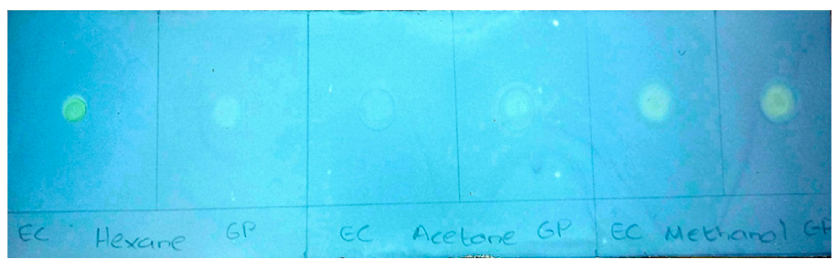
Figure 2.
DPPH radical scavenging activity (%) of T. violacea extracts (EC and GP) and ascorbic acid. * p < 0.05; H-hexane, A-acetone, M-methanol, EC-Eastern Cape, GP-Gauteng Province.
Figure 2.
DPPH radical scavenging activity (%) of T. violacea extracts (EC and GP) and ascorbic acid. * p < 0.05; H-hexane, A-acetone, M-methanol, EC-Eastern Cape, GP-Gauteng Province.
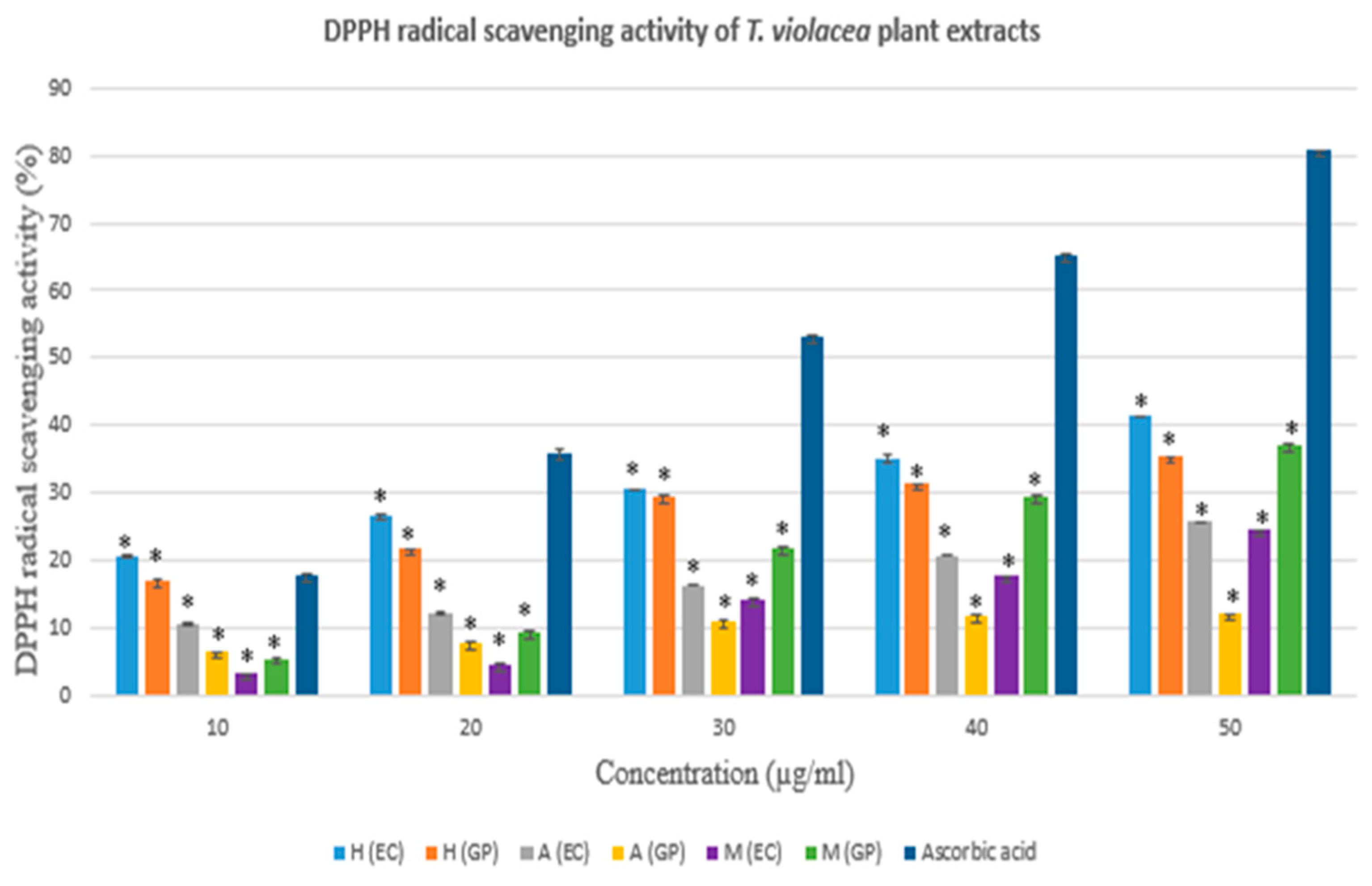
Figure 3.
The α- amylase inhibition (%) of T. violacea extracts (EC and GP) and acarbose. * p <0.05; H-hexane, A-acetone, M-methanol, EC-Eastern Cape, GP-Gauteng Province.
Figure 3.
The α- amylase inhibition (%) of T. violacea extracts (EC and GP) and acarbose. * p <0.05; H-hexane, A-acetone, M-methanol, EC-Eastern Cape, GP-Gauteng Province.
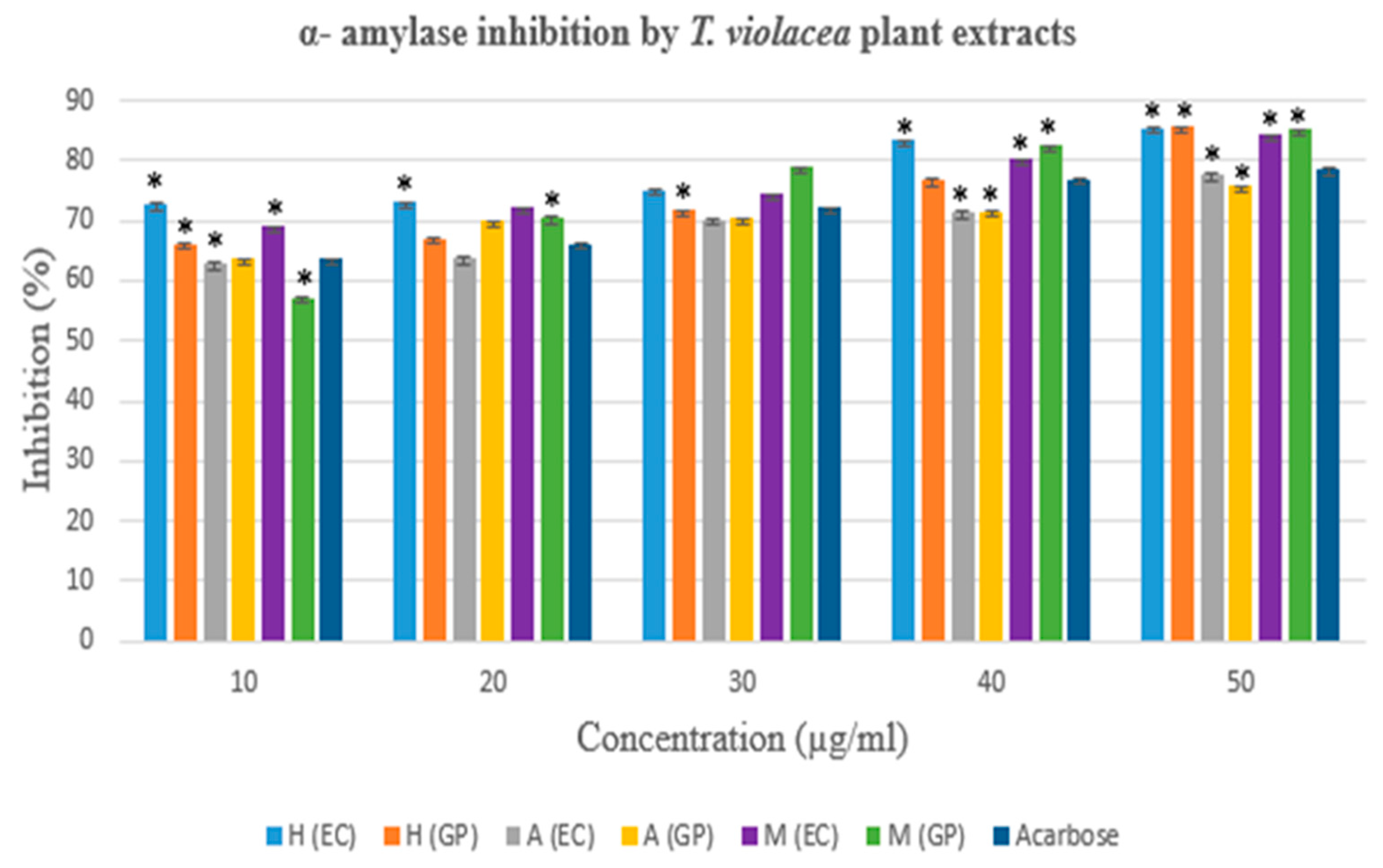
Figure 4.
The α- glucosidase inhibition (%) of T. violacea extracts (EC and GP) and acarbose. * p < 0.05; H-hexane, A-acetone, M-methanol, EC-Eastern Cape, GP-Gauteng Province.
Figure 4.
The α- glucosidase inhibition (%) of T. violacea extracts (EC and GP) and acarbose. * p < 0.05; H-hexane, A-acetone, M-methanol, EC-Eastern Cape, GP-Gauteng Province.
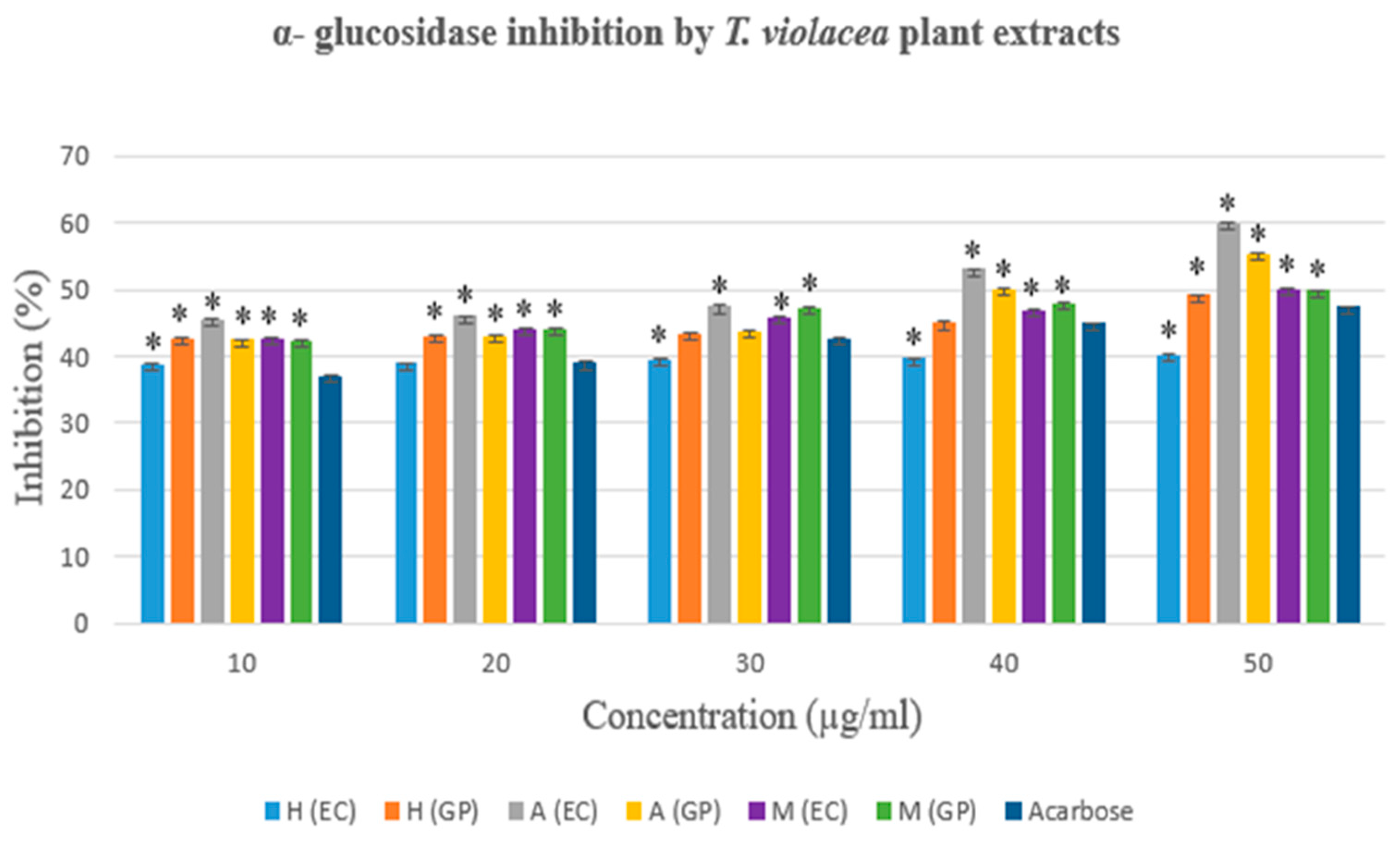

Table 1.
Qualitative phytochemical analysis of T. violacea extracts.
| Phytochemical | T. violacea extracts | |||||
|---|---|---|---|---|---|---|
| Hexane | Acetone | Methanol | ||||
| EC | GP | EC | GP | EC | GP | |
| Saponins | ++ | ++ | + | +/- | ++ | ++ |
| Anthraquinones | - | - | - | - | - | - |
| Flavonoids | + | + | - | - | - | - |
| Tannins | - | - | + | + | ++ | ++ |
| Alkaloids | + | + | ++ | + | + | +/- |
| Steroids | +++ | + | ++ | + | + | - |
| Cardiac glycosides | ++ | +++ | + | ++ | - | - |
| Phenols | +/- | +/- | + | + | +++ | ++ |
Not present (-); trace presence (+/-); moderate presence (+); present in appreciable quantity (++); impressive presence (+++), EC- Eastern Cape Province; GP- Gauteng Province.
Table 2.
Comparison of the DPPH radical scavenging activity (%) of T. violacea extracts.
| Concentration (µg/ml) | DPPH radical scavenging activity (%) | |||||
|---|---|---|---|---|---|---|
| Hexane Extract | Acetone Extract | Methanol Extract | ||||
| EC | GP | EC | GP | EC | GP | |
| 10 | 20,505* | 17,021* | 10,686* | 6,410* | 3,209* | 5,535* |
| 20 | 26,465* | 21,664* | 12,227* | 7,785* | 4,701* | 9,519* |
| 30 | 30,516* | 29,613* | 16,337* | 11,094* | 14,220* | 21,855* |
| 40 | 35,098* | 31,372* | 20,689* | 11,611* | 17,610* | 29,502* |
| 50 | 41,314* | 35,362* | 25,762* | 12,069* | 24,443* | 37,202* |
EC-Eastern Cape, GP-Gauteng Province. *p < 0.05.
Table 3.
IC50 values of various extracts of T. violacea (EC and GP) and the ascorbic acid standard.
| Extracts and standard | IC50 value (µg/ml) | ||
|---|---|---|---|
| T. violacea (EC) | T. violacea (GP) | Ascorbic acid | |
| Hexane | 68.259 | 79.565 | |
| Acetone | 115.11 | 295.568 | |
| Methanol | 97.105 | 65.138 | |
| Ascorbic acid | 29.595 | ||
EC-Eastern Cape, GP-Gauteng Province.
Table 4.
Comparison of the α- amylase inhibition (%) of T. violacea extracts.
| Concentration (µg/ml) | α- amylase inhibition activity (%) | ||||||
|---|---|---|---|---|---|---|---|
| Hexane Extract | Acetone Extract | Methanol Extract | |||||
| EC | GP | EC | GP | EC | GP | ||
| 10 | 72,778* | 66,167* | 62,891* | 63,754* | 69,094* | 57,228* | |
| 20 | 73,056* | 67,056* | 63,862* | 69,903* | 72,168* | 70,550* | |
| 30 | 75,111* | 71,833* | 70,168 | 70,496 | 74,434* | 78,846* | |
| 40 | 83,389* | 76,889* | 71,467 | 71,597 | 80,334* | 82,567* | |
| 50 | 85,500 | 85,667 | 77,724* | 75,750* | 84,218* | 85,194* | |
EC-Eastern Cape, GP-Gauteng Province. *p < 0.05.
Table 5.
IC50 values of various extracts of T. violacea (EC and GP) and the acarbose standard.
| Extracts and standard | IC50 value (µg/ml) | ||
|---|---|---|---|
| T. violacea (EC) | T. violacea (GP) | Acarbose | |
| Hexane | 9,022 | 15,083 | |
| Acetone | 16,189 | 16,504 | |
| Methanol | 16,146 | 16,201 | |
| Acarbose | 16,366 | ||
EC-Eastern Cape, GP-Gauteng Province.
Table 6.
Comparison of the α- glucosidase inhibition (%) of T. violacea extracts.
| Concentration (µg/ml) | α- glucosidase inhibition activity (%) | |||||
|---|---|---|---|---|---|---|
| Hexane Extract | Acetone Extract | Methanol Extract | ||||
| EC | GP | EC | GP | EC | GP | |
| 10 | 38,849* | 42,606* | 45,414* | 42,444* | 42,727* | 42,263* |
| 20 | 39,000 | 43,000* | 46,000* | 43,000* | 44,000* | 44,000* |
| 30 | 39,521 | 43,475 | 47,475* | 43,758 | 45,960* | 47,333* |
| 40 | 39,721* | 45,000 | 53,000* | 50,000* | 47,000* | 48,000* |
| 50 | 40,101* | 49,172* | 60,010* | 55,273* | 49,960* | 49,838* |
EC-Eastern Cape, GP-Gauteng Province. * p < 0.05.
Table 7.
IC50 values of various extracts of T. violacea (EC and GP) and the standard, acarbose.
| Extracts and standard | IC50 value (µg/ml) | ||
|---|---|---|---|
| T. violacea (EC) | T. violacea (GP) | Acarbose | |
| Hexane | 42,2 | 43,332 | |
| Acetone | 34,570 | 38,878 | |
| Methanol | 41,607 | 41,148 | |
| Acarbose | 45,609 | ||
EC-Eastern Cape, GP-Gauteng Province.
Disclaimer/Publisher’s Note: The statements, opinions and data contained in all publications are solely those of the individual author(s) and contributor(s) and not of MDPI and/or the editor(s). MDPI and/or the editor(s) disclaim responsibility for any injury to people or property resulting from any ideas, methods, instructions or products referred to in the content. |
© 2025 by the authors. Licensee MDPI, Basel, Switzerland. This article is an open access article distributed under the terms and conditions of the Creative Commons Attribution (CC BY) license (http://creativecommons.org/licenses/by/4.0/).
Copyright: This open access article is published under a Creative Commons CC BY 4.0 license, which permit the free download, distribution, and reuse, provided that the author and preprint are cited in any reuse.


