Preprint
Article

This version is not peer-reviewed.

Scientific Analysis of Contemporary Artworks Made from Food Ingredients: Forcusing on Lee Ungno’s Composition(1967)

Submitted:

23 December 2024

Posted:

24 December 2024

You are already at the latest version

Abstract

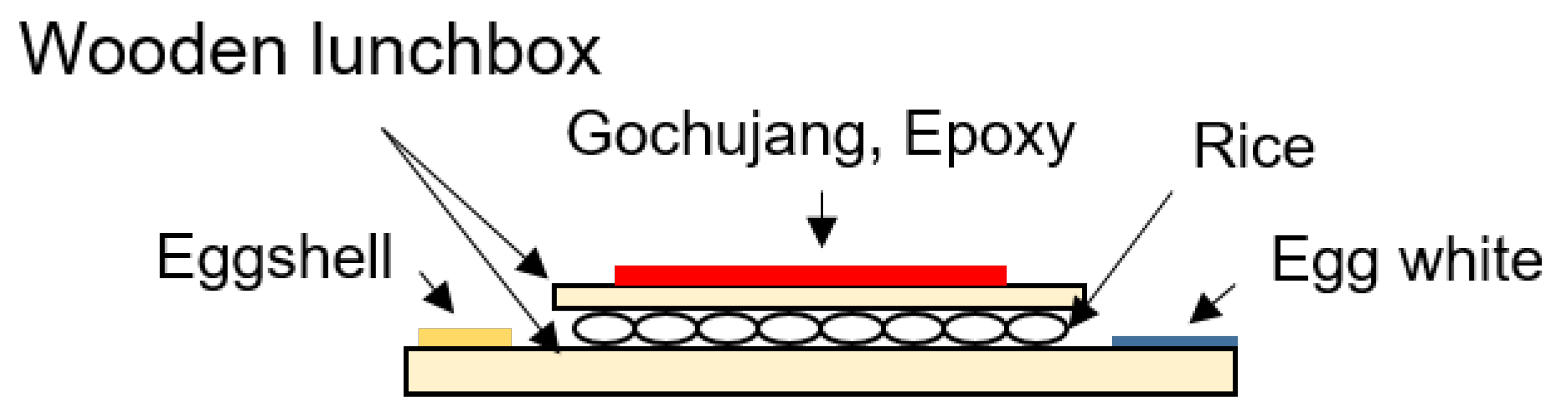
In this study, we used scientific analysis to estimate the materials used in Lee Ungno’s “Composition (1967)”, which is composed of ingredients such as gochujang and rice grains available in the prison, with the aim of establishing durable conservation materials for the artwork. The support material of the artwork was identified as hard pine, and the red-colored layers were identified as epoxy resin and gochujang. The adhesive applied to affix the wooden pieces to the support was determined to consist of rice grains, the translucent layer at the bottom of the artwork consisted of egg whites, and the yellow-colored layer contained eggshells. During the scientific analysis of the artwork, a combination of various other ingredients was identified as a result of the decomposition of ingredients, damage by microorganisms, and contamination. By utilizing control materials for comparison based on interviews and records of the artist, it was possible to estimate the specific type of food ingredients used. This approach provides fundamental information for the conservation of contemporary food-based artworks, and we anticipate that our findings will contribute to future conservation strategies for similar artworks.

Keywords: 
;  ;  ;  ;  

1. Introduction

Contemporary artwork uses a broad spectrum of ingredients without restrictions on the material [1]. One particularly notable example is EAT ART, which emerged in the 1960s the 70s, in which food or food ingredients are used as a medium for artistic expression. As one of the pioneers of this movement, Daniel Spoerri (1930–) launched his own restaurant to create culinary dishes and subsequently transformed leftover food, cutlery, and crockery into artistic displays, thus elevating commonplace occurrences such as meals to the status of art. Visitors can eat the food (artworks) or watch it decay; additionally, unlike previous installation artworks, these artworks require visitors to use various senses, including sight, smell, and taste [2,3] .
One of the essential features of EAT ART, which is also a limitation, is its transience as food materials are changed and destroyed over time. Various conservation methods have been proposed for this purpose. For example, Victor Grippo’s “Analogia (1970–1971)” is an artwork showing potatoes attached to electrical connectors. Instead of conserving the potatoes themselves, conservators decided to focus on the operational nature of the artwork and to conserve it by continually replacing the potatoes [4]. In Janine Antony’s “Lick and Lather (1993)”, the artist created identical busts of herself using soap and chocolate. Here, the objective was to observe changes in the artworks as the artist washed herself with the soap and licked the chocolate; therefore, no specific conservation measures were implemented [3]. Thus, unlike traditional conservation methods, EAT ART may be conserved using methods such as replacement or preservation, focusing on the artist’s intentions.
In South Korea as well, Lee Ungno (1904–1989) created artworks from food ingredients in prison after being implicated in the “Dongbaekrim” (East Berlin) incident in 1967. His best known works include “Composition(1967)”, which he created using gochujang and rice grains on the wooden lunch box provided by the courthouse, “Fan (1967)”, in which he stuck gochujang and eggshells to a fan, and “Crowds (1967)”, made using a mixture of paper and rice. In an interview, Lee revealed that he would work using whatever material he could find around him in the extreme, isolated environment of prison, including gochujang and rice grains [5,6,7,8].
Lee Ungno’s “Composition (1967)”, which is the focus of our study, requires conservation efforts as a result of splitting and bending of the support, biological deterioration due to pest damage and mold (Figure 1A), and spalling of the color layer (Figure 1B). The material properties of this work, created using gochujang and rice grains, indicate that damage over time is inevitable. In this study, we used scientific analysis to estimate the materials used in the work by comparing actual food materials and general materials and used these data to suggest suitable strategies for conservation. In addition, through a complementary approach using non-material data, such as artist interviews and records, alongside scientific analysis, we aimed to achieve a deeper understanding of Lee Ungno’s “Composition”, and to explore strategies that could contribute to the conservation of contemporary art. We anticipate that our findings could help solve issues related to the conservation of contemporary art based on food ingredients and be used as basic data in similar case studies in the future.

2. Analytical Methods

Before starting conservation, we divided the work into support and media and analyzed the tree species used in the wooden support and the constituent materials in the media. Before analysis, the medium was classified into rice adhesive (RA), red-colored layer 1 (R1), red-colored layer 2 (R2), translucent layer (T), and yellow-colored layer (Y; Figure 2).

2.1. Support Analysis

We analyzed the tree species from fragments of support that had become dislodged. For the test specimens, a stainless-steel razor blade (ST-300, Dorco, Korea) was used to prepare slices in three planes. The slices were placed on a glass slide in glycerin 50wt% (in water) and covered with a cover glass to finish the preparation. The tissues on the slides were examined under a light microscope (ECLIPSE LV100, Nikon, Japan), and images were captured of distinguishing characteristics that could aid in identification. For species identification, we referred to ‘Mokjaejojikgwa sikbyeol [Timber organization and identification] [9] and ‘Hanguksan mokjaeui seongjilgwa yongdo I [Wood properties and uses of the tree species grown in Korea I] [10].

2.2. Screen Material Analysis
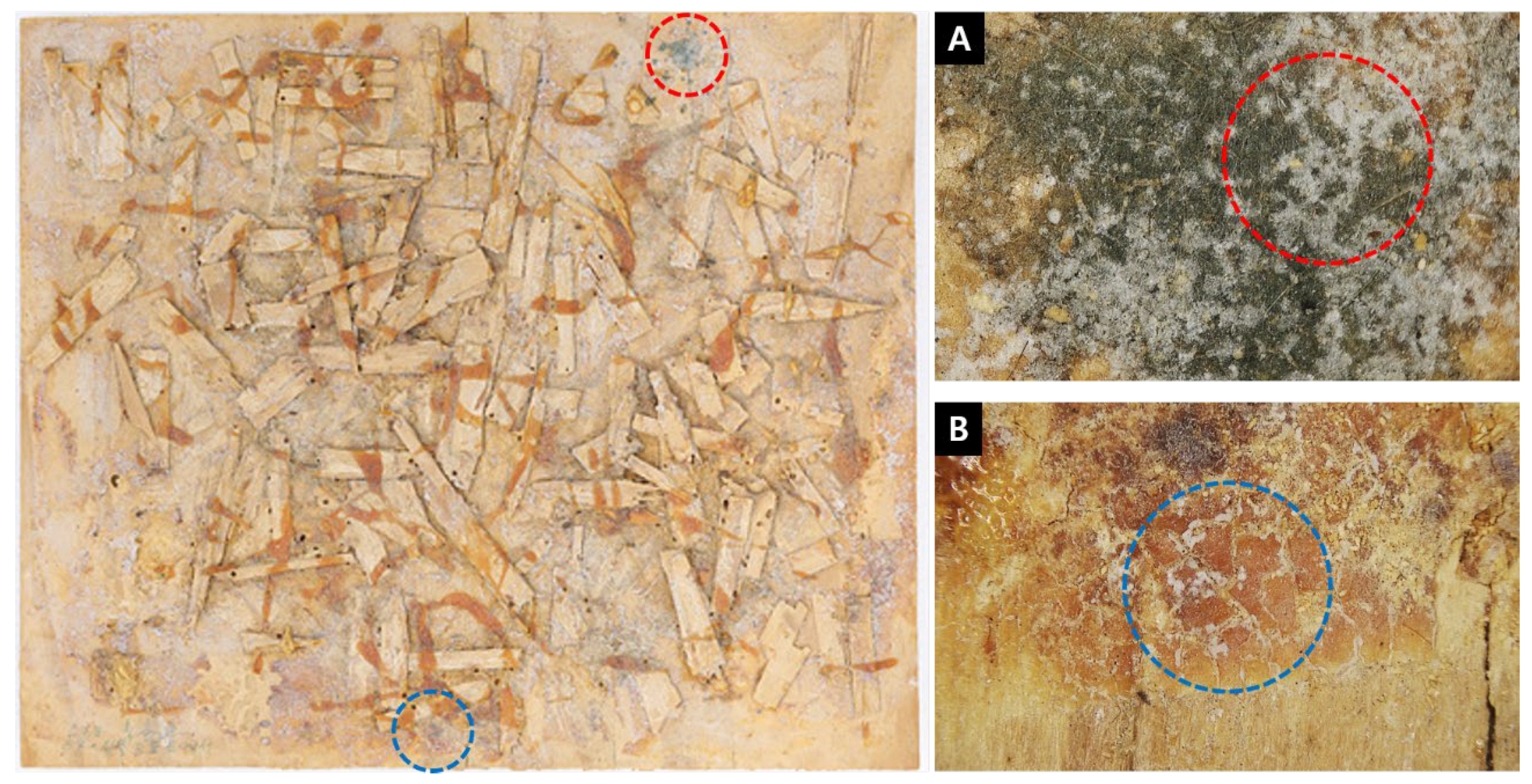
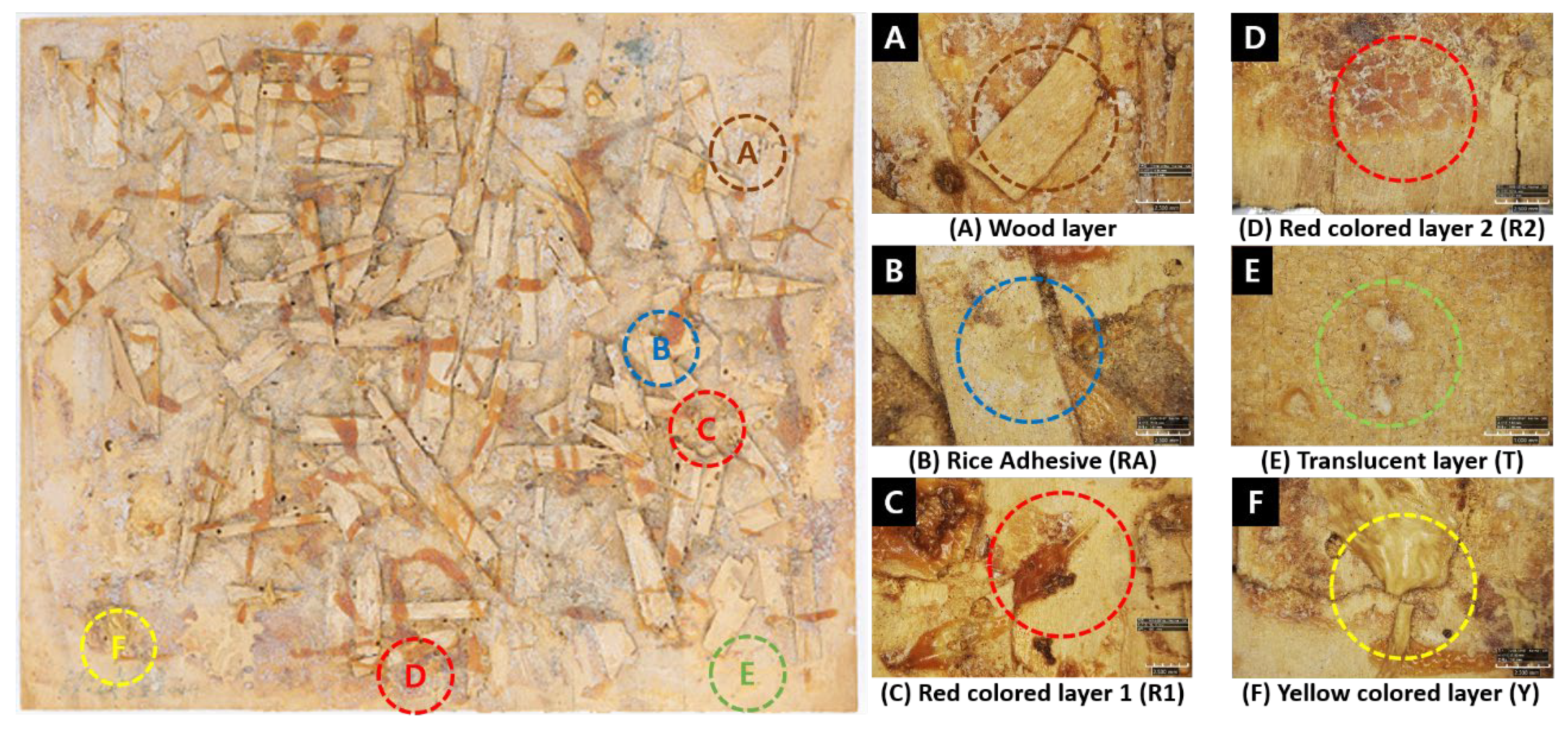
We performed FT-IR analysis to investigate the rice adhesive, red layers, translucent coating layer, and yellow-colored layer. An FT-IR device (Cary620 Microscope, Agilent, USA) was used in the ATR mode. After confirming the analysis location using the built-in microscope, we measured each specimen in triplicate with a resolution of 4 cm-1, scan range of 500–4,000 cm-1, and scan number of 32. To select the comparison materials, along with a library search, we focused on materials known to have been used in the artist’s works: egg whites, egg yolks, eggshells, gochujang, and rice grains, analyzed the substances under the same conditions, and compared the results with the screen materials.
The red-colored layers, translucent layer, rice adhesive layer, and four control specimens (gochujang, egg whites, egg yolks, and rice grains) were estimated to be organic substances based on the FT-IR results and were further analyzed using py-GC/MS. The samples were placed in an 8 mm× 4 mm sample cup and immediately inserted, without preprocessing, into the sampler of the pyrolyzer (JCI-55, JAI, Japan) attached to the gas chromatography–mass spectrometry device (8890, Agilent, USA/5977B, Agilent, USA) and mounted on a Pyrolyzer(JCI-55, JAI, Japan). We used a DB-5msUI column (5%-phenyl-methyl polysiloxane, 30 m × 250 µm × 0.25 µm, Agilent, USA), which was set to 330°C for plant-derived materials (gochujang, rice grains) and 550°C for animal-derived materials (egg whites, egg yolks). The gas was He 99.999%, the flow rate was 1.0 ml/min, and the inlet temperature was 250°C for plant-derived materials and 300°C for animal-derived materials. The liquid was injected into the inlet in split mode with a split ratio of 10:1, and the column oven temperature was gradually increased by 10°C/min from a starting temperature of 50°C to a final temperature of 250°C and maintained at 250°C for 10 min before stopping the analysis. The mass spectrometer was operated in E.I. mode (70 eV, m/z 30–800) with a transition temperature of 300°C and an ionization temperature of 230°C [11,12,13,14].
The yellow-colored layer and one of the control specimens (eggshells) were determined to be inorganic substances based on the FT-IR results; therefore, they were further analyzed using XRD. Using an XRD device (Smartlab, Rigaku, Japan), we analyzed the specimens at a scan speed/duration time of 5.08 m, 2-theta of 5–90 deg, voltage of 45 kV and current of 200 mA with a copper target.

3. Results

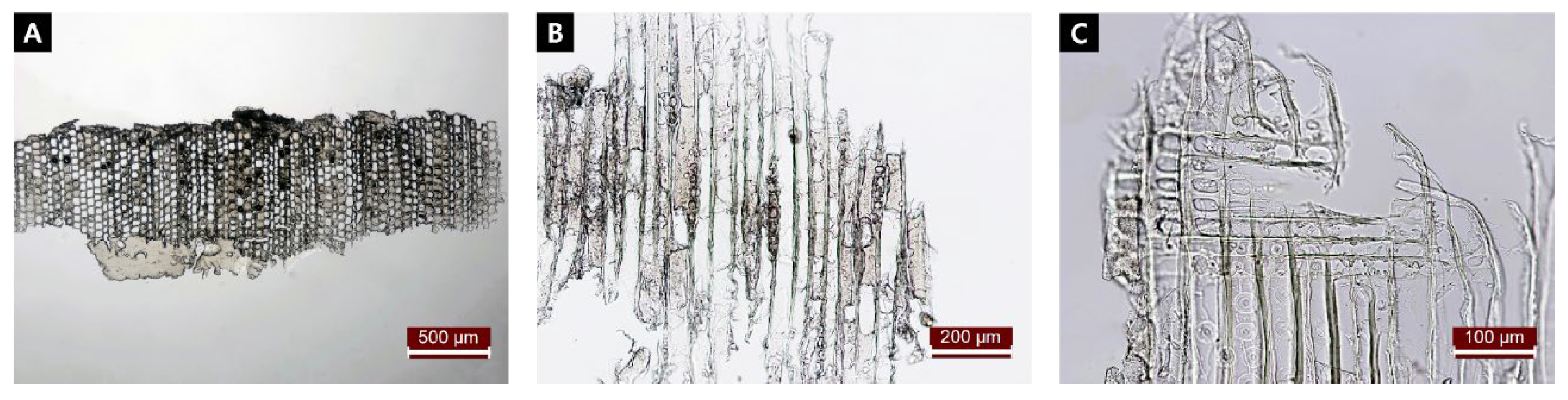
3.1. Species Identification in the Support

As a result of species identification in the accompanying materials, it was determined to be a hard pine belonging to the family Pinaceae and subgenus Pinus. The cross section was in the form of a very thin veneer of approximately 0.6–0.7 mm; therefore, we were unable to verify earlywood/latewood progression, and due to the limitations of the sample. Additionally, due to the constraints of the sample, we were also unable to detect resin canals in the tangential section (Figure 3A, B). However, in the radial section, the radial tissue was composed of radial tracheids and parenchyma, the cross-field pits were window-like, and serrated thickening of the radial tracheids was observed (Figure 3C). Within the genus Pinus, serrated thickening of radial tracheids is a feature of the subgenus Pinus that differentiates it from the subgenus Strobus ; thus, the material was finally identified as hard pine.

3.2. Analysis of Media Materials

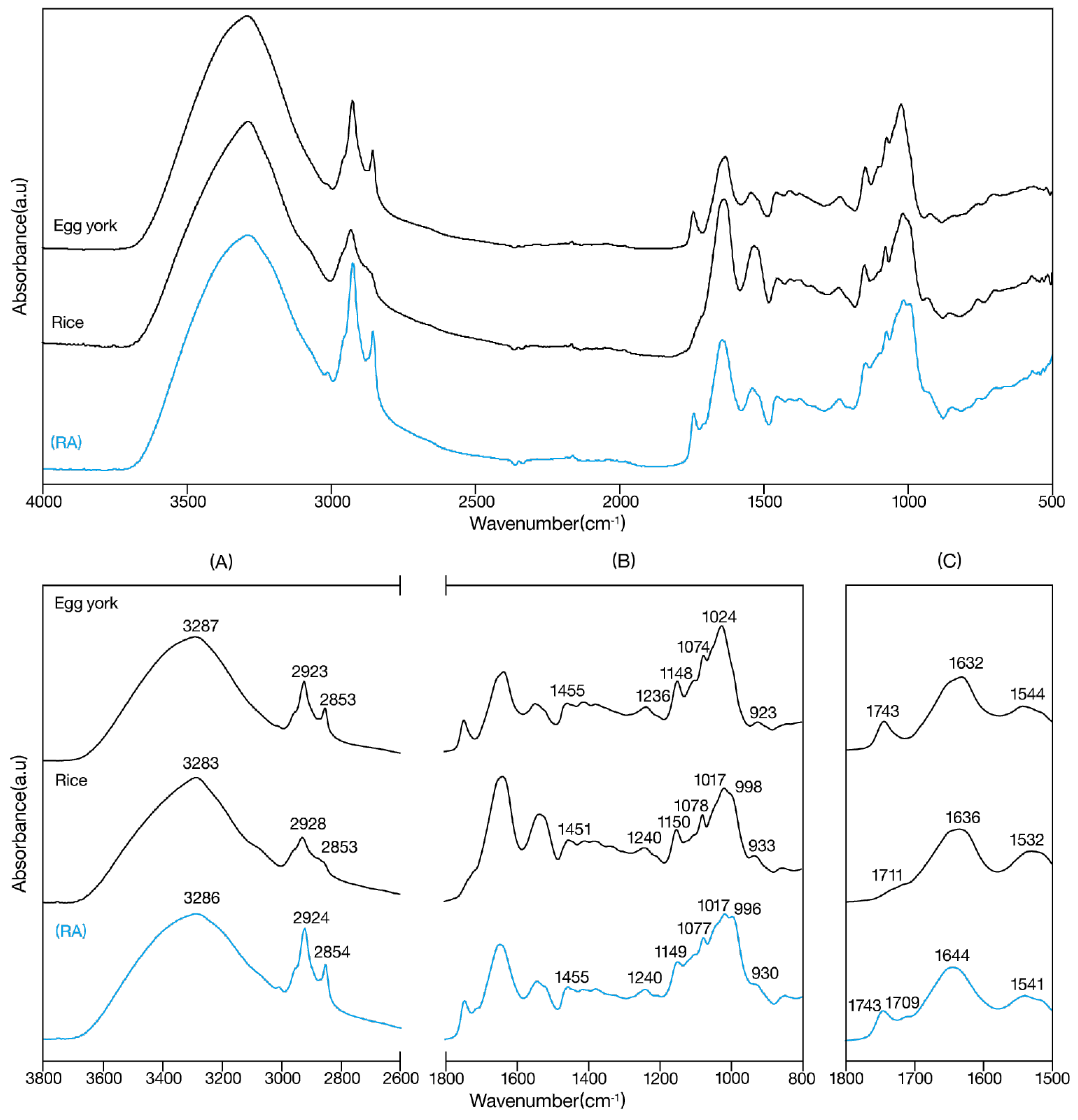
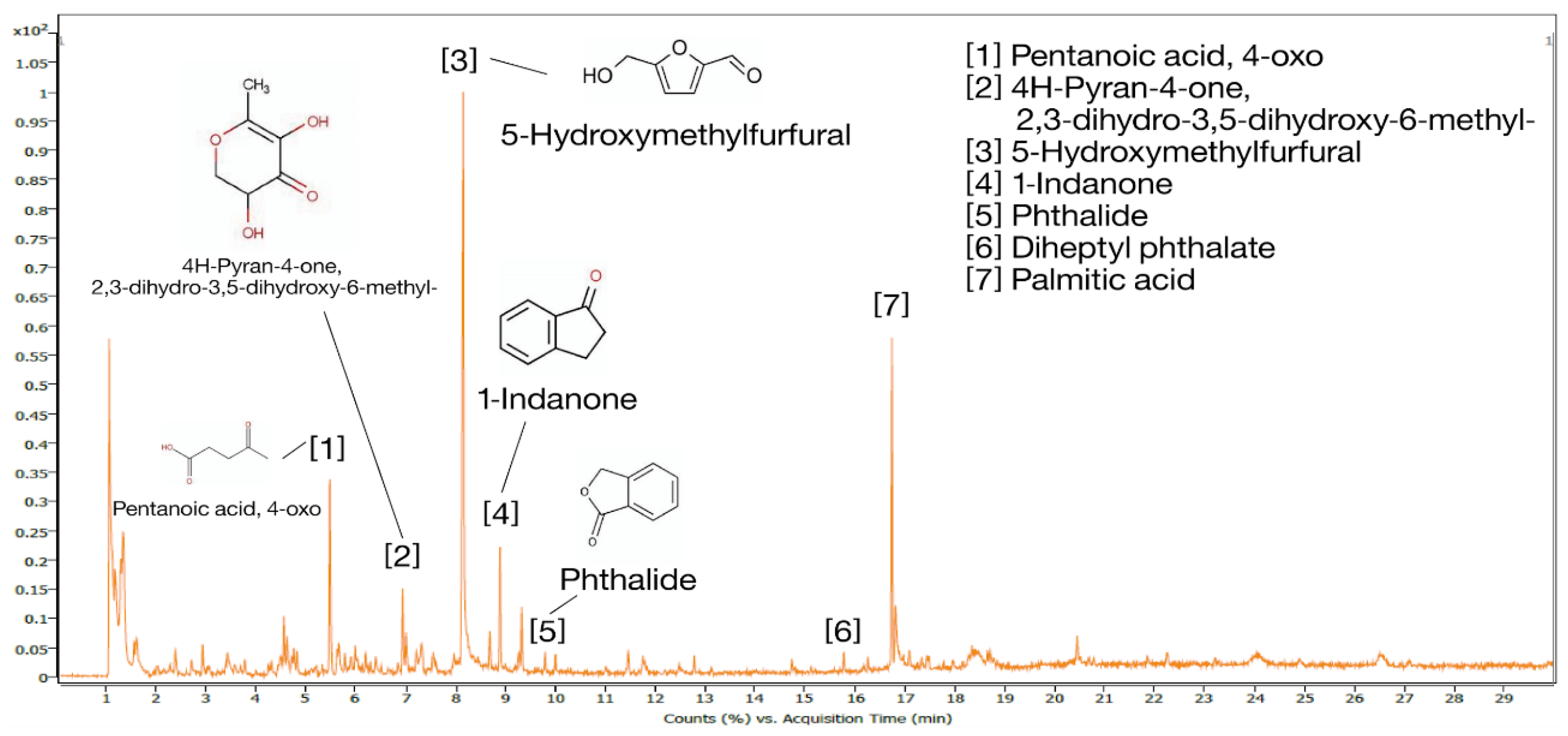
3.2.1. Rice Grain Adhesive (RA)

IR spectroscopy results for (RA) in Figure 4, we observed a broad absorption band at 3700–3000 cm-1 region due to –OH, and absorption peaks at 2924 cm-1 and 2850㎝-1 corresponding to symmetric and asymmetric stretching, respectively, of methyl and carbonyl groups. A –C=O carbonyl ester peak was observed at 1743 cm-1, a C=O amide Ⅰ peak was observed at 1644 cm-1, and a peak was observed at 1541㎝-1, indicating N-H amide II absorption by proteins. Scissoring vibrations due to C-H bending in CH3 groups were observed at 1455 cm-1, C-O tertiary alcohol was observed at 1236㎝-1 and 1148㎝-1, C-O primary alcohol was observed at 1077 cm-1, C-4-OH glucose residue peak was observed at 1017㎝-1, and CO-NH vibration peak was observed at 930 cm-1 [16,17,18].
When the above results were compared with the reference IR spectra, the –C=O carbonyl ester peak at 1743 ㎝-1 observed in egg yolks was also observed in (RA). In addition, (RA) and the rice grain sample shared C=O stretching absorption in the 1709–1711 ㎝-1 region, C-O tertiary alcohol, C-O primary alcohol, C-4-OH glucose residue, and CO-NH vibration peaks at 1149, 1074, 1017, and 933 cm-1, respectively. (RA), egg yolks, and rice grains all showed amide I absorption at 1632–1644㎝-1 and amide II absorption at 1532–1544 ㎝-1 [19]. Combining the above results, RA was judged to be most similar to the rice grain spectrum but could have been mixed with other food ingredients, such as egg yolks.
Pyrolysis-GC/MS results for (RA) in Figure 5, 'Ethanol', 'Furfural', 'Ethyl benzene', 'm-Xylene', '5-methylfurfural', 'Levoglucosenone', and '5-hydroxymethylfurfural' were detected at retention times of 1–15 min, and 'Dibutyl phthalate' and 'Palmitic acid' were detected at retention times of 16–30 min. Among the detected compounds, furfural, 5-methylfurfural, levoglucosenone, and 5-hydroxymethylfurfural originated from glucose, the basic monomer in starch. Furans are produced during the pyrolysis of sugars, and levoglucosenone, a pyrolysis product common to non-derivatized carbohydrates, results from the dehydration of glucose and has the structure of glucose after the loss of water molecules [11,20,21,22,23,24].
Based on the above results, we can surmise that (RA) is a rice grain. In contrast, aromatic amino acid compounds found in egg yolks, such as 'Toluene', 'Indolizine', 'Hexadecanenitrile', '9-octadecenamide, (Z)-' [25,26,27], were not detected. Thus, we concluded that the –C=O carbonyl ester peak observed at 1743 cm-1 in the IR spectroscopy was attributed to aromatic benzenes such as ethyl benzene, m-xylene, and dibutyl phthalate, rather than aromatic amino acids found in egg yolk.

3.2.2. Red Colored Layer 1 (R1)

IR spectroscopy analysis of (R1) in Figure 6, we observed a broad absorption band for –OH in the 3700–3000 cm-1 region and peaks at 2918 and 2849 cm-1, corresponding to symmetric and asymmetric stretching due to C-H bonds. We observed a peak at 1713 cm-1 due to C=O carbonyl stretching, at 1685㎝-1 due to α,β-unsaturated carbonyl compounds, and at 1607 cm-11 due to the stretching vibration of C=C bonds in aromatic compounds. There were also peaks at 1538 and 1509 cm-1 showing asymmetric and symmetric stretching of C=C in aromatic compounds, and at 1456, 1397, and 1363 cm-1 showing the scissoring vibration of methylene (CH2) groups. Finally, in the 1264–1012 cm-1 region we observed absorption due to the scissoring vibration of C-O bonds, and in the 970–747 cm-11 region we observed absorption due to the scissoring vibration of aromatic C-H bonds [28,29].
When the above results were compared with the reference IR spectra, (R1) was found to be most similar to the epoxy compounds. vibration at 1264 cm-1. However, the peaks at 1713, 1685, 1538, and 1105 cm-1 are not observed Most of the peaks in R1 were consistent with epoxy, including the stretching vibrations of the C=C bonds in aromatic compounds at 1607, 1583, and 1509 cm-11, CH2 scissoring vibration at 1456 cm-1, and C-O scissoring in epoxy, and of the peaks above, C=O carbonyl stretching and C-O scissoring vibration peaks around 1710 and 1100 cm-1 are detected in polyesters.
Pyrolysis-GC/MS analysis of (R1) in Figure 7, we detected 'Phenol', 'Indane', '2-methylstyrene', 'Naphthalene, 1,2-dihydro-', 'p-Cumenol', 'p-Isopropenylphenol', and '1-Naphthol, 6,7-dimethyl-' at retention times of 1–15 min. They are aromatic compounds with aromatic hydrocarbon. In particular, phenol, p-cumenol, and p-isopropenylphenol were detected in bisphenol A, which is a major constituent of the epoxy. The detection of naphthalene, 1,2-dihydro-, 1-naphthol, and 6,7-dimethyl- compounds suggests that they could have been mixed with a pigment such as Naphthol Red, although some may also be detected in epoxy [12,30,31]. At retention times of 16–30 min, 'Palmatic acid', 'Stigmasterol', and 'Squalene' were detected. These are all fatty acid compounds that have physiological roles in the metabolism of organisms and have been reported to be detected mostly in oils and fats [32,33]. Thus, the peaks at 1685, 1538, and 874 cm-1 in the IR spectrum were attributed to fatty acids and proteins rather than aromatic compounds.

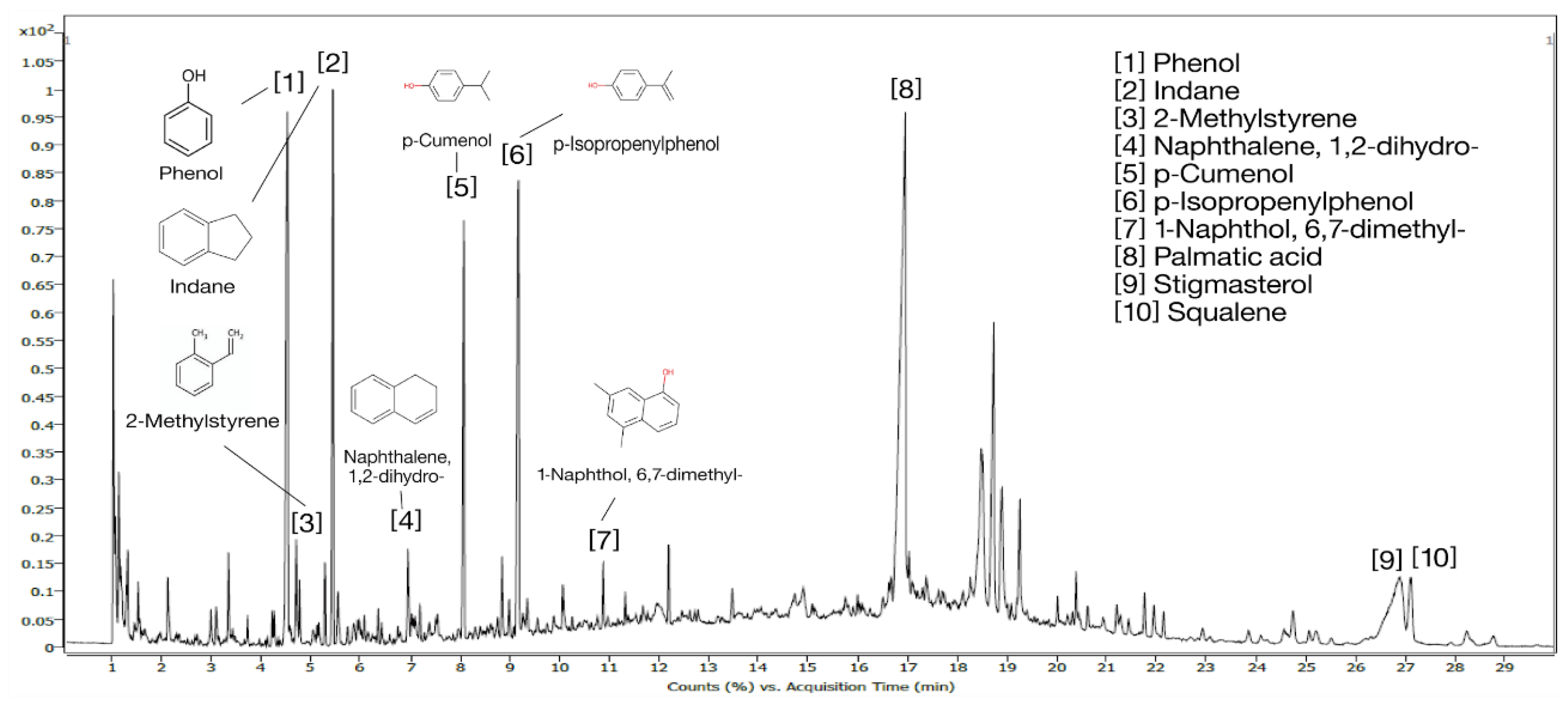
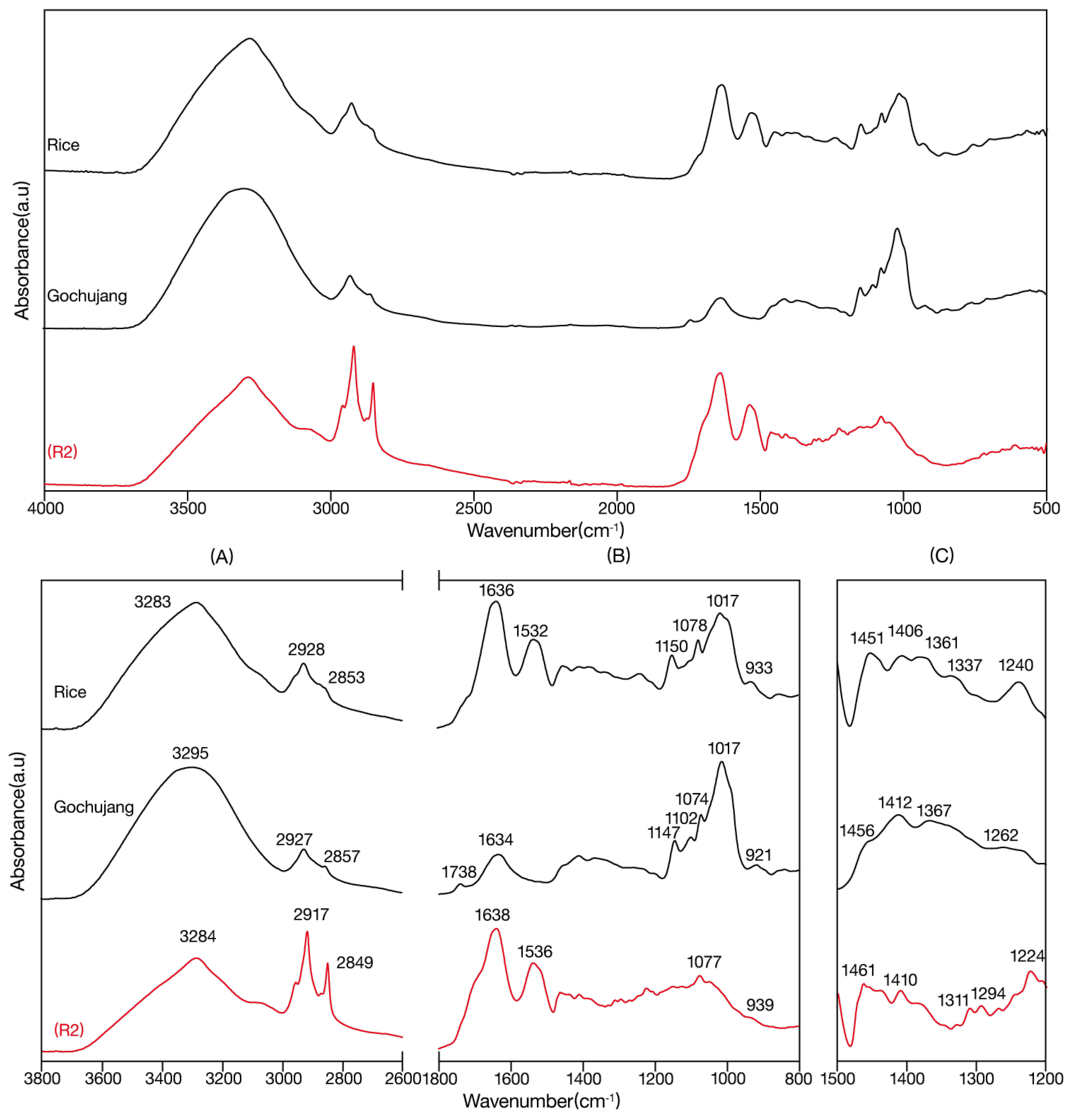
3.2.3. Red Colored Layer 2(R2)

IR spectrum of (R2) in Figure 8, we observed a broad absorption band for –OH at 3284 cm-1, and a symmetric and asymmetric stretch of C-H bonds at 2917 and 2849 cm-1. We observed an amide I peak at 1638 cm-1 due to C=O carbonyl stretching and an amide II peak at 1536 cm-1 due to N-H bending. At 1461 cm-1 we observed the bending vibration of CH2 or CH3, and at 1410 cm-1 we observed the bending vibration of O-H. At 1311 cm-11, we observed a peak due to amide III proteins. At 1294, 1224, and 1077 cm-1 we observed peaks due to the scissoring vibration of C-O-C glycosidic bonds [16,17,18].
When the above results were compared with the reference IR spectra, the patterns of R2 were most similar to those of gochujang. Among the peaks for (R2), the –OH band at 3284㎝-1, the peaks at 2917 and 2849 cm-1 due to symmetric and asymmetric stretching of C-H bonds, and the amide I peak at 1638 cm-1 were consistent with various compounds, including rice grains. However, given that rice is used in the manufacture of gochujang, some overlap is expected between the peaks. In contrast, the band at 1638 cm-1 was identified as containing the carboxyl ester peak in gochujang at 1743 cm-1, and so the material was considered to be similar to gochujang.
Pyrolysis-GC/MS results for (R2) in Figure 9, 'Pentanoic acid, 4-oxo', '4H-pyran-4-one, 2,3-dihydro-3,5-dihydroxy-6-methyl-', '5-hydroxymethylfurfural', '1-indanone', and 'Phthalide' compounds were detected at retention times of 1–15 min. Of these, 4H-pyran-4-one, 2,3-dihydro-3,5-dihydroxy-6-methyl-, and 5-hydroxymethylfurfural are produced during heating or fermentation of sugars [34], and Pentanoic acid, 4-oxo, is an ingredient that appears during the fermentation process of red pepper paste, and raw materials such as glutinous rice, beans, and red pepper powder have been reported as organic acids produced by metabolism such as Bacteria and Lactobacillus during aging [35]. At retention times of 16–30 min, 'Dibutyl phthalate', and 'Palmatic acid' were detected. Palmatic acid, a compound found in plant-derived oils and fats, was detected in gochujang. Thus, based on the results of IR spectroscopy and pyrolysis-GC/MS, we determined that the sample contained gochuajng.

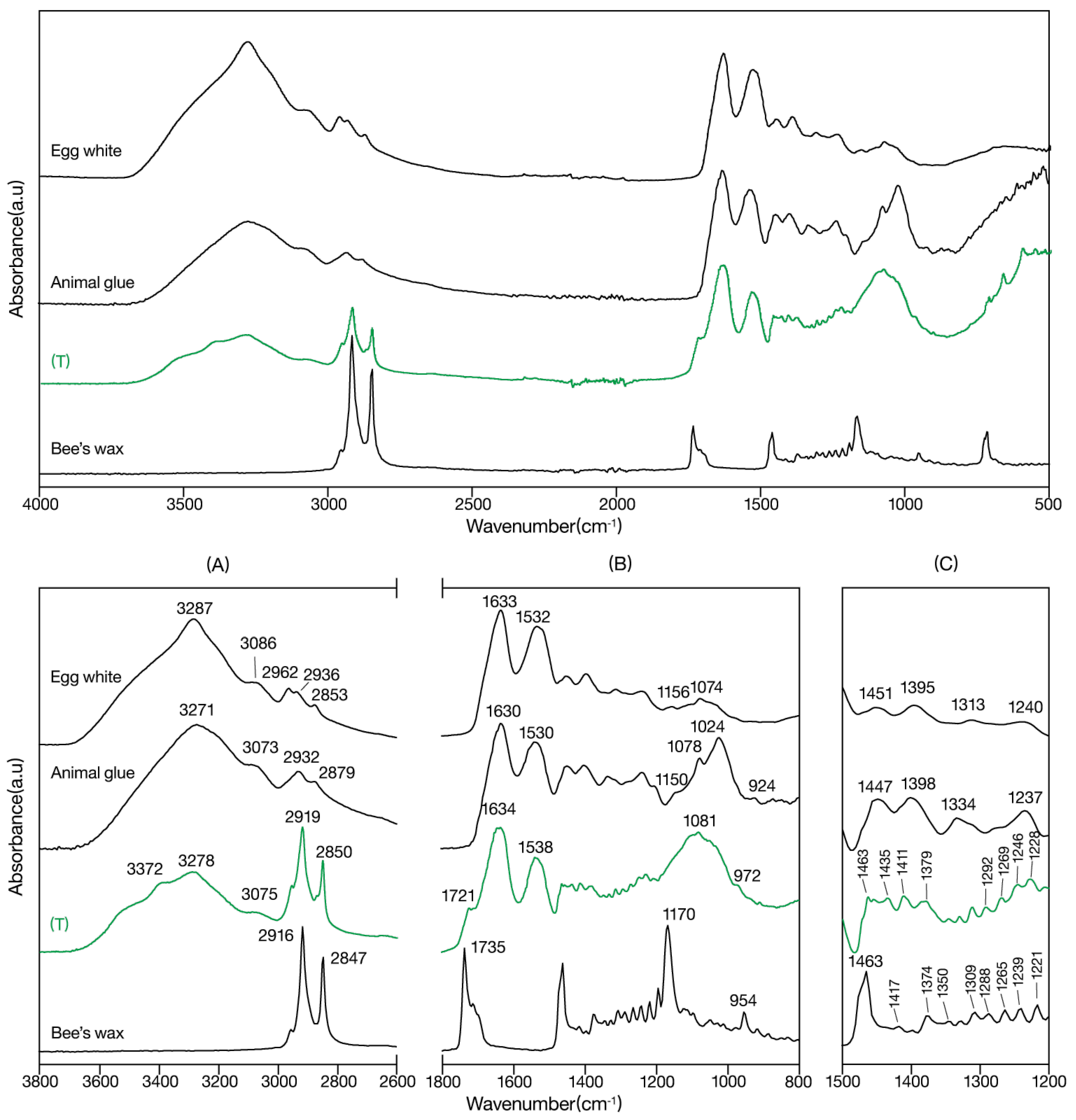
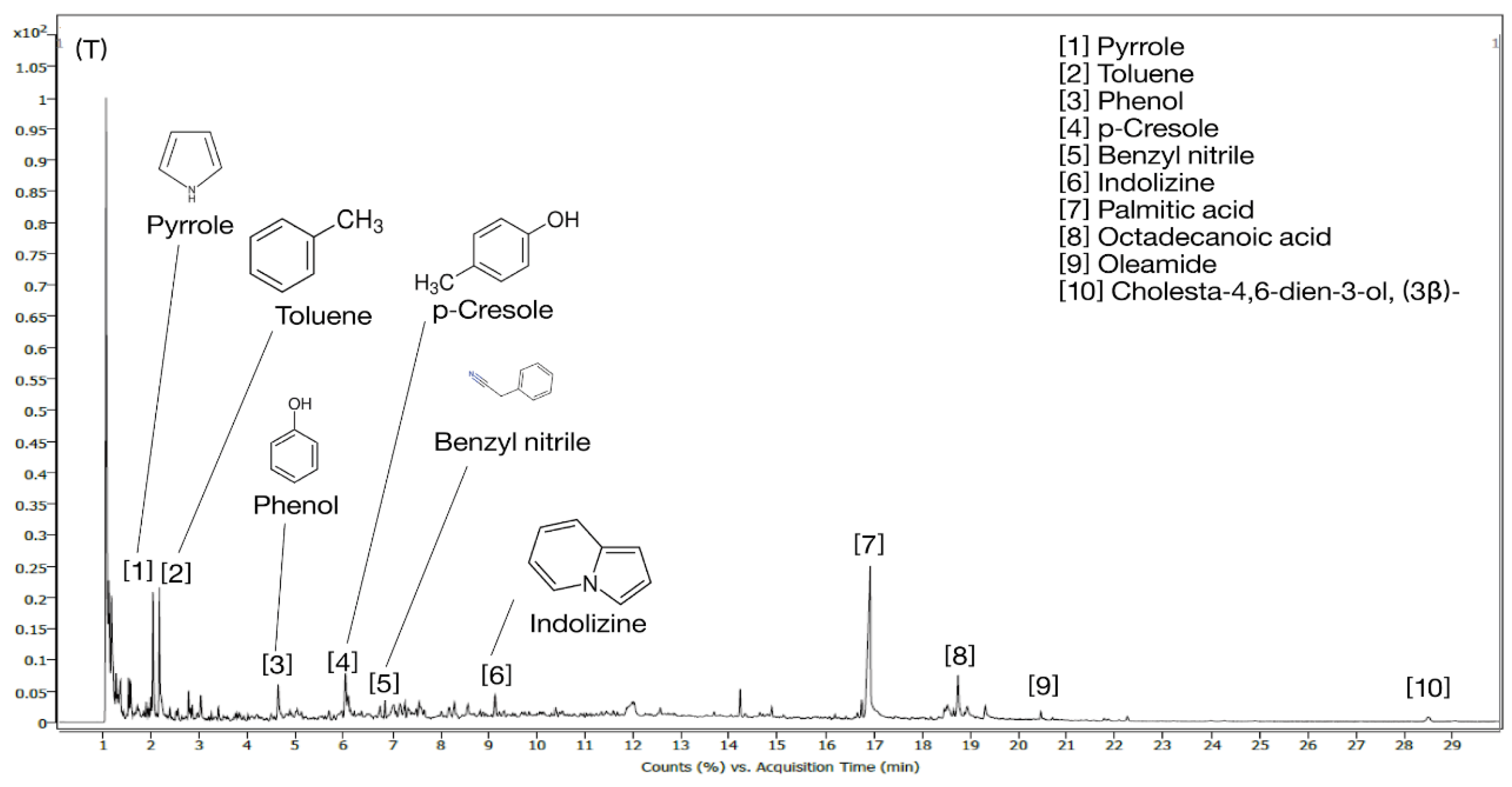
3.2.4. Translucent Layer (T)

IR spectra of (T) in Figure 10, there was a broad absorption band for –OH in the 3700–3000 cm-1 region, and split peaks at 3372, 3278, and 3075 cm-1for N-H. Symmetric and asymmetric stretching of methyl and carbonyl groups at 2919 and 2850 cm-1. We observed C=O carbonyl acid stretching at 1721 cm-1, C=O amide I at 1634 cm-1, and amide II proteins at 1538㎝-1. At 1463, 1435, and 1411 cm-1 we observed CH3 asymmetric bending and CH2 scissoring peaks were observed, and at 1379㎝-1 there was a peak due to the CH3 umbrella mode. At 1292, 1269, 1246, and 1228 cm-1 we observed C(O)-O stretching vibrations due to aromatic ethers, –OH plane vibrations, and amide III proteins. At 1081 cm-1, we observed a –CH peak, and at 972 cm-1, we observed Si-O stretching [36,37,38,39,40,41].
When these results were compared with the reference IR spectra, (T) showed similar peaks to the C=O acid stretch shoulder at 1721 cm-1 in beeswax, as well as absorption in the 1463–1447 and 1398–1379 cm-1 regions due to CH3 asymmetric bending and CH2 scissoring, as commonly seen in egg whites and gelatin. In addition, the amide III absorption in the 1500–1200 cm-was also consistent with that of beeswax. However, (T) also showed N-H absorbing peaks, unlike other samples, and this is thought to be due to basification, specifically ammonization resulting from the material's exposure to heat..
In pyrolysis-GC/MS for (T) in Figure 11, we detected 'Pyrrole', 'Toluene', 'Phenol', 'p-Cresol', 'Benzyl nitrile', and 'Indolizine' at retention times of 1–15 min, and 'Palmitic acid', 'Octadecanoic acid', 'Oleamide', and 'Cholesta-4,6-dien-3-ol (3β)-' at retention times of 16–30 min. Pyrrole is derived from the amino acid proline, which is one of the major amino acids in gelatin. These compounds have been detected in both gelatin and egg whites [42,43,44]. Meanwhile, Aromatic amino acids such as toluene and benzyl nitrile, tyrosine compounds such as phenol and p-cresol, tryptophan compounds such as indolizine are all detected in egg whites [45,46,47,48,49]. Moreover, palmitic acid (C16), octadecanoic acid (C28), and cholesta-4,6-dien-3-ol, (3β)- compounds detected at retention times of 16 min or longer reportedly originate from esters and fatty acids present in most natural waxes [50]. Therefore, (T) could be considered a mixture of natural macromolecular substances, such as egg whites, gelatin, and beeswax.

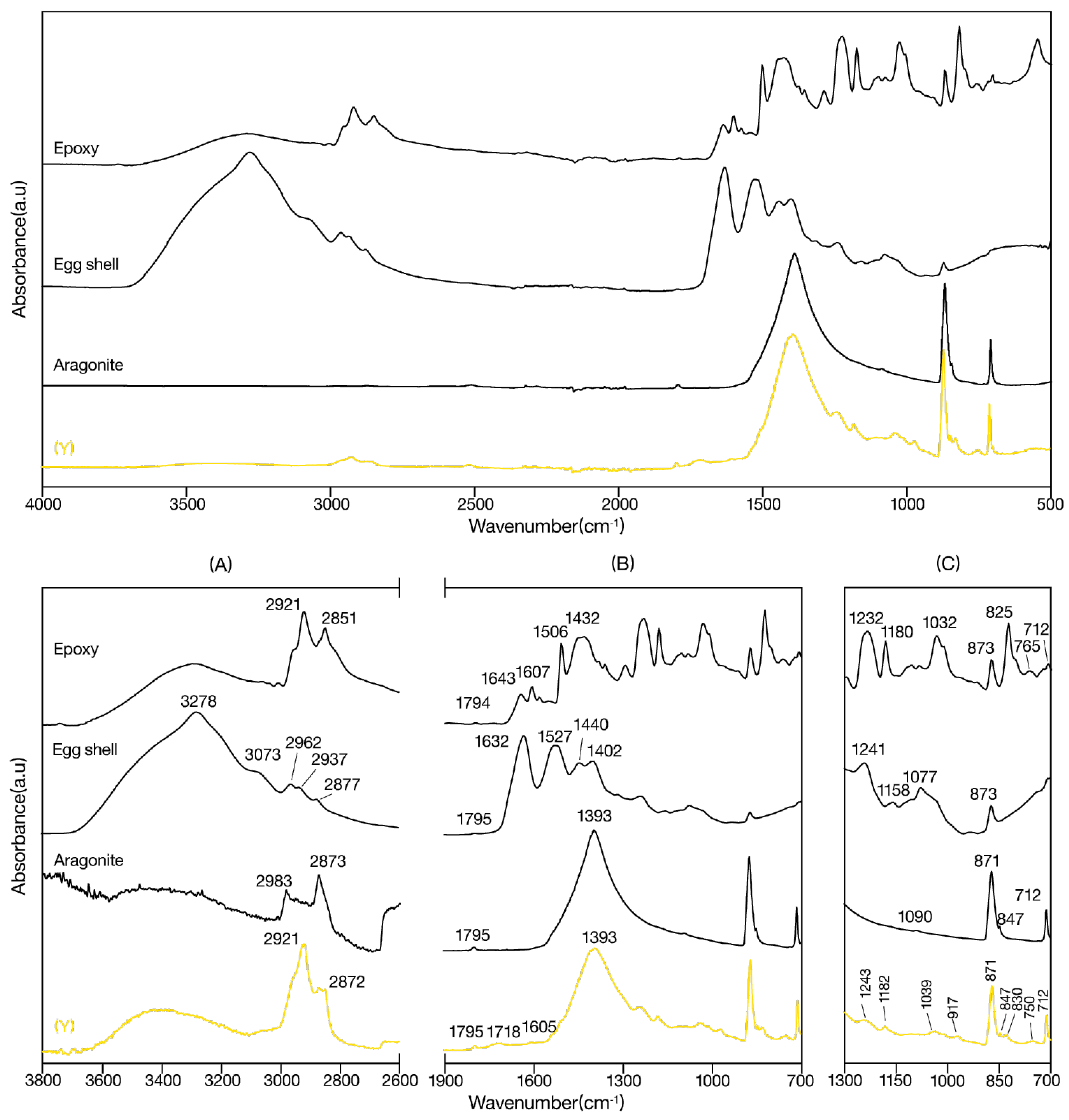
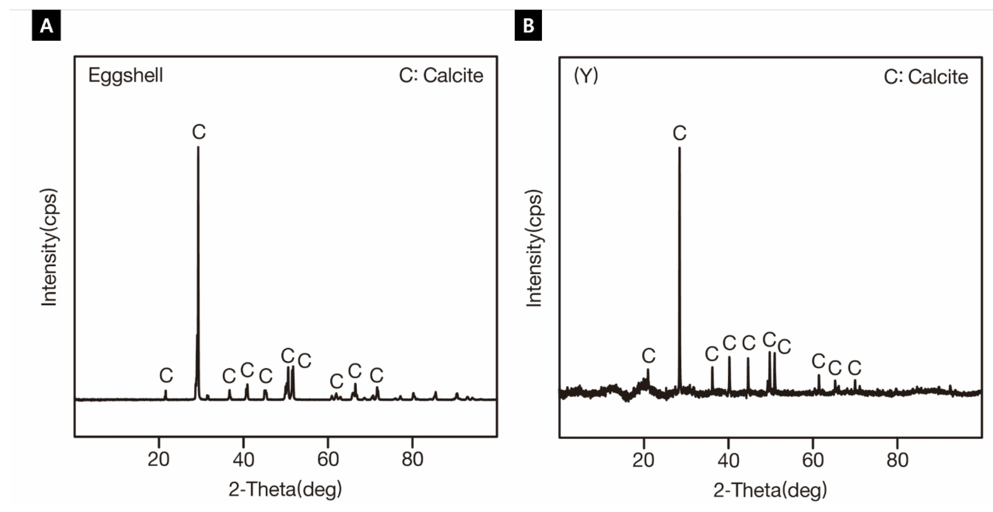
3.2.5. Yellow Colored Layer (Y)

IR spectrum of (Y) in Figure 12, we observed a broad absorption band for –OH in the 3700–3000 cm-1 region, and symmetric and asymmetric stretching of methyl and carbonyl groups at 2921 and 2872 cm-1. We observed a peak at 1795 cm-1 due to CaCO3, a peak at 1718 cm-1 due to C=O carbonyl acid stretching, a peak at 1605 cm-1 due to C=C stretching in aromatic carbons, and a CO3 asymmetric peak at 1500–1300 cm-1 (peak: 1393 cm-1). We observed an amide III peak at 1243 cm-1, a C-O-C asymmetric peak at 1182 cm-1, and a peak due to C-N groups at 1039 cm-1. Finally, the peak at 917 cm-1 showed =C-H bending, the peak at 871 cm-1 showed out-of-plane bending of carbonates, and the peak at 712 cm-1 showed in-plane bending of carbonates [40,51,52,53].
When we compared the above results with the reference IR spectra, the absorptions at 2921 and 2872 cm-1 (methyl, carbonyl group symmetric, asymmetric stretch) were similar to the peaks for epoxy, whereas the CaCO3 peak at 1795 cm-1, the CO3 asymmetric peak at 1393 cm-1, and the carbonate peaks at 871 and 712 cm-1 were also observed in epoxy, eggshells, and aragonite. The C=C stretching in aromatic carbons at 1605 cm-1 was observed in epoxy, whereas the amide III peak at 1243 cm-1 was observed in eggshells, and the C-O-C asymmetric peak at 1182 cm-1 was identical to that in epoxy. Based on the above results, it appears that the primary component of (Y) is calcium carbonate (CaCO3) and that (Y) is potentially a varnish supplemented with additives like epoxy..
When XRD analysis was conducted on (Y) in Figure 13, we detected calcite in both (Y) and eggshells, and no other compounds were detected. In addition to egg whites, we could consider the use of, for example, lime paint, which also contains calcite compounds. However, when we considered calcites that could be obtained at the time the work was created in prison, we concluded that the artist was likely to have used eggshells.

4. Discussion

4.1. Materials in Lee Ungno’s “Composition”

To estimate the materials used in Lee Ungno’s “Composition (1967)”, which is known to have been created using food ingredients such as gochujang and rice grains, we applied techniques such as wood identification, IR spectroscopy, pyrolysis-GC/MS, and XRD to the materials in the artwork, food materials, and other general materials.
Upon analyzing the tree species used in the work support, we found that the wooden lunch box that had been used was crafted from hard pine. Hard pine is one of the main tree species in Korean forests, and has been a favored material for many centuries due to its low weight, softness, and ease of processing [54]. Current single-use products, such as wooden lunch boxes and chopsticks, mostly use hard pine, cypress, poplar, or willow [55]. Based on the above results, we surmised that various tree species would have been used to produce veneer-type wooden lunch boxes, and that the wooden lunch box used in this study was made of hard pine.
In the IR spectroscopy and pyrolysis-GC/MS analysis of the (RA) layer in the media, we found that the results were most similar to those of rice grains. However, unlike rice grains, we also observed a –C=O carbonyl ester peak at 1743㎝-1, and detected ethyl benzene, m-xylene, and dibutyl phthalate in pyrolysis-GC/MS, suggesting that a material containing aromatic compounds, such as xylene and toluene, was also present alongside rice. The above results could have been caused by a synthetic varnish, such as red-colored layer 1 (R1), or could have been caused by conservation efforts, although such efforts have not been recorded. Since a large number of the above substances were found between the support and wooden fragments of the artwork, we surmised that these wooden fragments were stuck to the support using rice grains.
In IR spectroscopy and pyrolysis-GC/MS of red-colored layer I (R1), the results were most similar to those of the epoxy. Meanwhile, when we analyzed red-colored layer 2 (R2), the results were most similar to those of gochujang. These findings suggest that the artist previously used gochujang to create the work, but later used an epoxy varnish in the process of retouching the work or as part of a conservation process.
In IR spectroscopy and pyrolysis GC/MS of the translucent coating layer (T), the results were determined to be most similar to those of egg whites, but we also detected compounds from materials such as gelatin and beeswax. These results suggest that gelatin and natural resins, such as beeswax, could have been used alongside egg whites. Additionally, the (T) layer was predominantly observed at the bottom of the study area. This suggests that the artist may have intentionally applied multiple coats of the (T) layer to the lower section, or perhaps used leftover substances from meals in the process. The fact that we detected gelatin and beeswax is thought to reflect previous conservation or retouching of this work.
Using IR spectroscopy and XRD of the yellow-colored layer (Y), we estimated that (Y) was a varnish containing calcium carbonate and was most similar to eggshells. Because the (T) layer contained compounds assumed to be egg whites, we considered it likely, within the limited space of the prison, that eggshells could also have been used as a material in this work. However, we can also consider the possible use of calcite-containing varnishes, such as lime paint, in addition to eggshells.
Based on our scientific analysis, we inferred the structure of the artwork (Figure 14). Wooden fragments were stuck to the support material using rice grains, and then gochujang, egg white, and eggshells were used to apply the color. and Palmatic acid was commonly detected in all samples. Therefore, during conservation, it is especially important to take care not to displace wooden fragments attached to rice grain adhesives, or to damage the colored layers of gochujang, egg whites, and eggshells. It is also essential to be cautious about the properties of substances remaining from previous conservation or restoration, such as epoxy, gelatin, and beeswax.

4.2. Conservation Strategies for Contemporary Artworks: A Complementary Approach Combining Scientific Analysis with Non-Material Data

In the conservation of contemporary artworks using foodstuffs, due to the risk of degeneration and spoiling of the materials over time, it is important to objectively ascertain, through scientific analysis, the composition of materials, the rate of change, and the likelihood of conservation. However, the essence of the artwork transcends simple material appearances and is deeply related to the artist’s intentions and philosophy, making it difficult to fully explain the intentions and significance of the artwork by only examining the work itself [56]. Thus, non-material data such as artist interviews or records play an important role in supplementing scientific analysis. When broad results suggesting diverse possibilities are derived using scientific analysis, the range of interpretations can be narrowed based on nonmaterial data, including interviews and records from the artist. Indeed, in our study, we were able to estimate the materials more precisely by referring to the artist's interviews in relation to the information ascertained through scientific analysis. Thus, interviews and records can help overcome the limitations of scientific analyses alone.
However, interviews and records may contain errors. In fact, there have been cases where the results were inconsistent with the interview content, even though scientific analysis was conducted based on the artist’s interviews [57]. This appears to be a problem caused by errors in the interview records or the artist’s memory. This demonstrates the need for a mutually complementary approach that combines scientific analysis with nonmaterial data.
When conducting scientific analysis of artwork materials, it is important to not solely depend on the analysis results but to supplement and refine the results of scientific analysis using data such as artist interviews. Such a mutually complementary approach can assist in minimizing errors in the conservation of contemporary artworks and accurately convey the essential meaning and value of these works.

5. Conclusions

The purpose of this study was to determine the materials used in Lee Ungno’s “Composition (1967)” through scientific analysis and artist interviews and records, and to suggest appropriate conservation strategies based on the results. We confirmed that the artwork consisted of hard pine support and mixed media, including gochujang, rice grains, egg whites, and eggshells. In this way, we provided basic data to investigate the material properties of the work and causes of damage and to establish effective conservation strategies.
In addition, we confirmed that a mutually complementary approach combining scientific analysis and non-material data is important for understanding artworks. In the future, for accurate scientific analysis of food-based contemporary artworks, including EAT ART, given that different conclusions can be reached even for the same foods owing to differences in preparation methods or ingredients, it will be essential to acquire detailed records, including materials and brands, via the artist’s interviews and records. There is also a need to study the decay mechanisms of food materials and substances destroyed or produced in the process of metabolism by microbes as well as to study conservation measures that can prevent the degeneration and damage of these food materials.
We anticipate that our study will aid in addressing issues related to the conservation of food-based contemporary artworks, including Lee Ungno’s “Composition”, and will serve as a valuable resource for future case studies in this field.

Funding

This study was conducted with support from conservation support projects at the National Museum of Modern and Contemporary Art, Korea.

Institutional Review Board Statement

Not applicable.

Informed Consent Statement

Not applicable.

Data Availability Statement

Not applicable.

Conflicts of Interest

The author declares no conflict of interest.

References

  1. Shin, J.A.; Han, Y.B.; Cha, S.M.; Kim, Y.M.; Kwon, H.H. Conservation of contemporary artworks made with soap and research on the approprlate hygrothermal environment. J. Conserv. Sci. 2021, 37, 464–476. [Google Scholar] [CrossRef]
  2. Koczanowicz, D. Beyond Taste: Daniel Spoerri’s art of feasting. Perform. Res. 2017, 22, 92–99. [Google Scholar] [CrossRef]
  3. Coronado Garcia, C.M. Can We Use the Concept of Programmed Obsolescence to Identify and Resolve Conservation Issues on Eat Art Installations?, Living Matter: The Preservation of Biological Materials in Contemporary Art, An International Conference Held in Mexico City, June 3–5. Getty Conservation Institution, 2019.
  4. MALBA, Víctor Grippo: Homenaje, 2012. Available online: https://malba.org.ar/evento/victor-grippo-homenaje/ (accessed on 27 December 2012).
  5. Tadawa, T. Oppression to maintain power security, this is a national shame. National Times (accessd on 1 November 1985).
  6. Goam art institute. Goam Lee Ungno, Life and Art, Sprit and Knowledge; Eul gwa Al Co.,Ltd: Seoul, Korea, 2000. [Google Scholar]
  7. Daejeon Goam Art and Culture Foundation LeeUngno Museum. Collection of Lee Ung No-Sculpture; International Print, Daejeon, Korea, 2021.
  8. Kim, J.G. The Loyalty and joy of life – Lee Eung-no's jade sculpture, fault in korean modern and contemporary art, In Life and Dream, The 33-Year History of Lee Eung-no's Study(1989~2021); Leeungno museum, Daejeon, Korea, 2021, 9–322.
  9. Park, S.J.; Lee, W.Y.; Lee, W.H. Timber organization and identification; Hangmunsa: Seoul, Korea, 2006. [Google Scholar]
  10. Lee, P.W. Wood properties and uses of the tree species grown in KoreaⅠ; Seoul National University Press: Seoul, Korea, 1997. [Google Scholar]
  11. Kuroda, K.; Suzuki, A.; Kato, M.; Imai, K. Analysis of rice (Oryza sativa L.) lignin by pyrolysis-gas chromatography. J. Anal. Appl. Pyrol. 1995, 34, 1–12. [Google Scholar] [CrossRef]
  12. Kim, J.E.; Yu, J.A.; Chung, Y.J. Study on qualitative analysis for lacquer mixed with some additives by pyrolysis-gas chromatography/mass spectrometry. J. Conserv. Sci. 2017, 33, 51–59. [Google Scholar] [CrossRef]
  13. Orsini, S.; Parlanti, F.; Bonaduce, I. Analytical pyrolysis of proteins in samples from artistic and archaeological objects. J. Anal. Appl. Pyrol. 2017, 124, 643–657. [Google Scholar] [CrossRef]
  14. Yu, J.A. Analysis of binder in korean traditional Dancheong. Ph.D. Thesis, Korea National University of Cultural Heritage, Buyeo, Korea, 2019. [Google Scholar]
  15. Lee, U.C.; Park, J.H.; Lee, J.H.; Kim, S.C. Study on scientific analysis about red pigment and binder -the korean ancient red pottery-, Conserv. Sci. 2021, 37(5), 606–616. [Google Scholar] [CrossRef]
  16. Guillén, M.D.; Cabo, N. Infrared spectroscopy in the study of edible oils and fats. J. Sci. Food Agric. 1997, 75, 1–11. [Google Scholar] [CrossRef]
  17. Subirade, M.; Kelly, I.; Guéguen, J.; Pézolet, M. Molecular basis of film formation from a soybean protein: Comparison between the conformation of glycinin in aqueous solution and in films. Int. J. Biol. Macromol. 1998, 23, 241–249. [Google Scholar] [CrossRef]
  18. Yap, P.L.; Cheong, K.Y.; Lee, H.L.; Zhao, F. Effects of drying temperature on preparation of pectin polysaccharide thin film for resistive switching memory. J. Mater. Sci. Mater. Electron. 2022, 33, 19805–19826. [Google Scholar] [CrossRef]
  19. Sun, D.W. Infrared Spectroscopy for Food Quality Analysis and Control, 1st ed.; Elsevier Inc., London, United Kingdom, 2009.
  20. Kim, P.Z.; Hong, S.M.; Kim, C.H. Studies on therma degradation and analysis of starches by pyrolysisgas chromatography/mass spectrometry. J. Korean Soc. Anal. Sci. 1989, 2, 115–123. [Google Scholar]
  21. Vane, C.H.; Abbott, G.D. Proxies for land plant biomass: Closedsystem pyrolysis of some methoxyphenols. Org. Geochem. 1999, 30, 1535–1541. [Google Scholar] [CrossRef]
  22. Kim, M.C. Pyrolysis Behaviors of Carbohydrate Using Pyrolysis-GC and Pyrolysis-GC/MS. Master’s Thesis, Sejong University, Seoul, Korea, 2009. [Google Scholar]
  23. Neal, S.G.; Qin, L.; Hong, Y.; George, D.C.; Marilyn, L.F.; Weiguo, L.; Guoping, S. Org. Geochem. Molecular preservation and bulk isotopic signals of ancient rice from the Neolithic Tianluoshan site, lower Yangtze River valley, China. J. Org Geochem. 2013, 63, 85–93. [Google Scholar] [CrossRef]
  24. Kim, J.H.; Park, H.S.; Lim, S.J. Comparison study on the material characteristics of oil paint (Ⅰ). J. Conserv. Sci. 2017, 33, 85–95. [Google Scholar] [CrossRef]
  25. Colombini, M.P.; Modugno, F.; Menicagli, E.; Fuoco, R.; Giacomelli, A. GC-MS characterization of proteinaceous and lipid binders in UV aged polychrome artifacts. Microchem. J. 2000, 67, 291–300. [Google Scholar] [CrossRef]
  26. Hu, W.; Zhang, K.; Zhang, H.; Zhang, B.; Rong, B. Analysis of polychromy binder on Qin Shihuang's Terracotta Warriors by immunofluorescence microscopy. J. Cult. Herit. 2015, 16, 244–248. [Google Scholar] [CrossRef]
  27. Wang, N.; Zhang, T.; Min, J.; Li, G.; Ding, Y.; Liu, J.; Gu, A.; Kang, B.; Li, Y.; Lei, Y. Analytical investigation into materials and technique: Carved lacquer decorated panel from Fuwangge in the Forbidden City of Qianlong Period, Qing Dynasty. J. Archaeol. Sci. Rep. 2018, 17, 529–537. [Google Scholar] [CrossRef]
  28. Sahmetlioglu, E.; Yuruk, H.; Sürme, Y. Synthesis and characterization of oligosalicylaldehyde-based epoxy resins- Slovak Academy of Sciences. Chemical Papers. 2006, 60, 65–68. [Google Scholar] [CrossRef]
  29. González, M.G.; Cabanelas, J.C.; Baselga, J. Applications of FTIR on Epoxy Resins – Identification, Monitoring the Curing Process, Phase Separation and Water Uptake. in Infrared Spectroscopy – Materials Science, Engineering and Technology, IntechOpen: London, UK, 2012; ISBN 978-953-51-0537-4.
  30. Kim, S.H.; Park, N.J.; Oh, M.S. Determination of epoxy resin type in EMC by pyrolysis-GC and GC/MS. Polym. (Korea) 1999, 23, 401–412. [Google Scholar]
  31. Zhang, W.; Fina, A.; Ferraro, G.; Yang, R. FTIR and GCMS analysis of epoxy resin decomposition products feeding the flame during UL 94 standard flammability test. Application to the understanding of the blowing-out effect in epoxy/polyhedral silsesquioxane formulations. J. Anal. Appl. Pyrol. 2018, 135, 271–280. [Google Scholar] [CrossRef]
  32. Pokkanta, P.; Sookwong, P.; Tanang, M.; Setchaiyan, S.; Boontakham, P.; Mahatheeranont, S. Simultaneous determination of tocols, γ-oryzanols, phytosterols, squalene, cholecalciferol and phylloquinone in rice bran and vegetable oil samples. Food Chem. 2019, 271, 630–638. [Google Scholar] [CrossRef] [PubMed]
  33. Santos, L.C.; Broco e Silva, R.G.; Scopel, E.; Hatami, T.; Rezende, C.A.; Martínez, J. Concentration of stigmasterol, β-sitosterol and squalene from passion fruit (Passiflora edulis Sims.) by-products by supercritical CO2 adsorption in zeolite 13-X. J. Supercrit. Fluids 2024, 209. [Google Scholar] [CrossRef]
  34. Anese, M.; Suman, M. Mitigation strategies of furan and 5-hydroxymethylfurfural in food. Food Res. Int. 2013, 51, 257–264. [Google Scholar] [CrossRef]
  35. Choi, J.Y.; Lee, T.S.; Park, S.O.; Noh, B.S. Changes of volatile flavor compounds in traditional Kochujang during fermentation. Korean J. Food Sci. Technol. 1997, 29, 745–751. [Google Scholar]
  36. Mazzeo, R.; Prati, S.; Quaranta, M.; Joseph, E.; Kendix, E.; Galeotti, M. Attenuated total reflection micro FTIR characterisation of pigment-binder interaction in reconstructed paint films. Anal. Bioanal. Chem. 2008, 392, 65–76. [Google Scholar] [CrossRef] [PubMed]
  37. Rodriguez-Blanco, J.; Shaw, S.; Benning, L. The kinetics and mechanisms of amorphous calcium carbonate (ACC) crystallization to calcite, via vaterite. Nanoscale 2010, 3, 265–271. [Google Scholar] [CrossRef] [PubMed]
  38. Widjonarko, D.M.; Jumina, J.; Kartini, I.; Nuryono, N. Phosphonate modified silica for adsorption of Co(II), Ni(II), Cu(II), and Zn(II). Indones. J. Chem. 2014, 14, 143–151. [Google Scholar] [CrossRef]
  39. Dubey, P.; Sharma, P.; Kumar, V. FTIR and GC-MS spectral datasets of wax from Pinus roxburghii Sarg. needles biomass. Data Brief 2017, 15, 615–622. [Google Scholar] [CrossRef] [PubMed]
  40. Brzeska, J.; Elert, A.M.; Morawska, M.; Sikorska, W.; Kowalczuk, M.; Rutkowska, M. Branched Polyurethanes Based on Synthetic Polyhydroxybutyrate with Tunable Structure and Properties. Polymers 2018, 10, 826. [Google Scholar] [CrossRef] [PubMed]
  41. Shrestha, D.; Maensiri, S.; Wongpratat, U.; Lee, S.W.; Nyachhyon, A.R. Shorea robusta derived activated carbon decorated with manganese dioxide hybrid composite for improved capacitive behaviors. J. Environ. Chem. Eng. 2019, 7. [Google Scholar] [CrossRef]
  42. Park, J.S. Analysis of animal glue by pyrolysis/GC/MS. Anal. Sci. Technol. 2015, 28, 221–227. [Google Scholar] [CrossRef]
  43. Gelatin Manufacturers Association, Asia Pacific Press. 2015. Available online: http://www.gmap-gelatin.com/about_gelatin_AminoAcidComp.html (accessed on 3 Mar 2015).
  44. Jin-Li, Z.H.A.O.; Zong-Ren, Y.U.; Bo-Min, S.U. Analysis of egg whites from burial murals by pyrolysis-gas chromatography/mass spectrometry. Chin. J. Appl. Chem. 2023, 40, 562–570. [Google Scholar] [CrossRef]
  45. Merritt, C.; Robertson, D.H. The analysis of proteins, peptides and amino acids by pyrolysis-gas chromatography and mass spectrometry. J. Chromatogr. Sci. 1967, 5, 96–98. [Google Scholar] [CrossRef]
  46. Tsuge, S.; Matsubara, H. High-resolution pyrolysis-gas chromatography of proteins and related materials. J. Anal. Appl. Pyrol. 1985, 8, 49–64. [Google Scholar] [CrossRef]
  47. Moldoveanu, S.C., Ed. In Analytical Pyrolysis of Natural Organic Polymers 1st edn. 20. Elsevier, 1998, 373–397.
  48. Drevin, I.; Johansson, B.L.; Larsson, E.L. Pyrolysis in biotechnology. Biotechnol. Genet. Eng. Rev. 2001, 18, 3–28. [Google Scholar] [CrossRef] [PubMed]
  49. Legan, L.; Retko, K.; Peeters, K.; Knez, F.; Ropret, P. Investigation of proteinaceous paint layers, composed of egg yolk and lead white, exposed to fire-related effects, Nature research. Sci. Rep. 2020, 10, 1. [Google Scholar] [CrossRef] [PubMed]
  50. Asperger, A.; Engewald, W.; Fabian, G. Analytical characterization of natural waxes employing pyrolysis–gas chromatography–mass spectrometry. J. Anal. Appl. Pyrol. 1999, 50, 103–115. [Google Scholar] [CrossRef]
  51. Reig, F.B.; Adelantado, J.V.G.; Moya Moreno, M.C. FTIR quantitative analysis of calcium carbonate (calcite) and silica (quartz) mixtures using the constant ratio method. Application to geological samples. Talanta. 2002, 58, 811–821. [Google Scholar] [CrossRef]
  52. Melo, H.P.; Cruz, A.J.; Candeias, A.; Mirão, J.; Cardoso, A.M.; Oliveira, M.J.; Valadas, S. Problems of Analysis by FTIR of Calcium Sulphate–Based Preparatory Layers: The Case of a Group of 16th-Century Portuguese Paintings. Archaeometry. 2014, 56, 513–526. [Google Scholar] [CrossRef]
  53. Adrián, L.V.; Anastasia, E. Amine Responsive Poly(lactic acid) (PLA) and Succinic Anhydride (SAh) Graft-Polymer: Synthesis and Characterization. Polymers. 2019, 11, 1466. [Google Scholar] [CrossRef]
  54. Park, W.K.; Lee, K.H. Changes in the species of woods used for Korean ancient and historic architectures. Archit. Hist. 2007, 50, 9–28. [Google Scholar]
  55. Lim, H.C.; Lee, W.H. Characteristics and Species of wooden chopsticks. J. Korea Furniture Soc. 2022, 33, 129–136. [Google Scholar]
  56. Kwon, H.H.; Lee, G.S. Collaboration with stakeholders for conservation of contemporary art. J. Conserv. Sci. 2020, 36, 37–46. [Google Scholar] [CrossRef]
  57. Kim, Y.M.; Han, Y.B.; Shin, J.A.; Cha, S.M.; Kwon, H.H. The conservation treatment of the bark of wooden sculpture. J. Conserv. Sci. 2021, 37, 516–524. [Google Scholar] [CrossRef]
Figure 1. Appearance before conservation treatment of Lee Ungno's “Composition (1967)” (A: biological deterioration, B: spalling color layer).
Preprints 143858 g001
Figure 2. Analysis object of Lee Ungno's “Composition (1967)”.
Preprints 143858 g002
Figure 3. Micrographs of Hard Pine (A) Cross section(×40), (B) Tangential section(×100), (C) Radial section(×200).
Preprints 143858 g003
Figure 4. IR Spectrum of Rice Adhesive and control groups.
Preprints 143858 g004
Figure 5. Chromatogram of Rice Adhesive(RA).
Preprints 143858 g005
Figure 6. IR Spectrum of red colored layer 1 and control groups.
Preprints 143858 g006
Figure 7. Chromatogram of Red colored layer 1(R1).
Preprints 143858 g007
Figure 8. IR Spectrum of Red colored layer 2 and control groups.
Preprints 143858 g008
Figure 9. Chromatogram of Red colored layer 2(R2).
Preprints 143858 g009
Figure 10. IR Spectrum of Translucent layer and control groups.
Preprints 143858 g010
Figure 11. Chromatogram of Translucent layer(T).
Preprints 143858 g011
Figure 12. IR Spectrum of Yellow colored layer and control groups.
Preprints 143858 g012
Figure 13. XRD spectrum of Eggshells and Yellow colored layer.
Preprints 143858 g013
Figure 14. Schematic diagram of “Composition”.
Preprints 143858 g014
Disclaimer/Publisher’s Note: The statements, opinions and data contained in all publications are solely those of the individual author(s) and contributor(s) and not of MDPI and/or the editor(s). MDPI and/or the editor(s) disclaim responsibility for any injury to people or property resulting from any ideas, methods, instructions or products referred to in the content.
Copyright: This open access article is published under a Creative Commons CC BY 4.0 license, which permit the free download, distribution, and reuse, provided that the author and preprint are cited in any reuse.
Prerpints.org logo

Preprints.org is a free preprint server supported by MDPI in Basel, Switzerland.

Subscribe

Disclaimer

Terms of Use

Privacy Policy

Privacy Settings

© 2025 MDPI (Basel, Switzerland) unless otherwise stated