Submitted:
22 December 2024
Posted:
23 December 2024
You are already at the latest version
Abstract
Most studies on nerve guidance conduits (NGCs) focuses on advancing biomaterials and enhancing their functionality to outperform autologous nerve grafts (ANGs). However, even when these strategies exceed the ANG effects, they may not guarantee satisfactory recovery in every case, considering their inability to impact the direction of axonal growth. Axonal misdirection occurs due to dispersive nature of axonal growth and variable intraneural branching topography, presenting one of the main obstacles in achieving consistent success in peripheral nerve repair. On the other side, implementation of various NGC structural features may offer a potential solution. This paper reviews the NGC structural designing strategies, and assesses their potential to influence axonal direction, exploring the key challenges and future perspectives. The literature was reviewed using PubMed, ScienceDirect, OpenMD, Cochrane Library, and Google Scholar databases. The simplest single-hollow NGC design may prevent axonal dispersion, however only in small nerve gaps. The multichannel and nanofiber-filled NGC designs provide a neural-like microenvironment and straightforward axonal guidance, supporting successful regeneration in larger nerve gaps. Even though these structural strategies may also surpass the efficacy of ANGs, such straightforward guidance does not exclude the axonal misdirection, due to variable distribution of fascicular trajectories. In the future, this issue could be addressed by intraoperative 3D scanning of the stumps, using the machine learning algorithms to predict the fascicular distribution, and on-site 3D printing of personalized NGCs.
Keywords:
Artificial Nerve Grafts
; Axonal Misdirection
; Nerve Guidance Conduits
; Nerve Regeneration
1. Introduction
Peripheral nerve injuries may lead to serious functional deficits significantly impacting the quality of life in the affected individuals [1,2]. In cases of insufficient spontaneous recovery, a range of multi-disciplinary interventions may be implemented across the three divided action areas, including stimulating neural growth, treating the site of nerve injury, and maintaining the viability of the target organ [3]. Any failure to fulfill these criteria can have a significant impact on the final functional recovery.
In cases with loss of nerve continuity, surgical repair of the injury site presents the primary treatment option. Successfully repairing nerve defects by grafting and ensuring complete functional recovery poses a great challenge. Even though autologous nerve grafts (ANG) still stand as a golden standard for achieving the best outcomes [4,5], their flaws cannot be overlooked [6]. Therefore, substantial resources are devoted to advancing the development of artificial nerve grafts, differently known as nerve guidance channels or conduits (NGCs) [7,8].
The three main strategies in NGC development are structural design, biomaterial selection, and functional enhancement. Most experimental studies were focused on advancing biomaterials and enhancing graft functionality by integrating diverse citobiochemical and physical agents, often achieving superior results compared to the ANGs [9,10,11,12,13,14,15]. However, relying solely on these strategies may be insufficient to ensure complete functional recovery in all cases, likely due to their limited ability to impact the axonal misdirection [16].
Regarding the literature, only a suitable structural configuration of the grafts may influence the axonal misdirection [8,17,18,19,20]. However, there are not many studies that examine NGC structural designs and their influence on axonal misdirection. This paper offers a comprehensive review of various NGC designing strategies with a specific emphasis on their potential to influence axonal misdirection. Upon evaluating the effectiveness of various structural features, the key challenges and future perspectives are discussed.
2.. Search Strategy
2.1. Databases Searched
The following databases were utilized to gather relevant literature: PubMed, ScienceDirect, OpenMD, Cochrane Library, Google Scholar.
2.2.. Keywords and Search Terms
2.2.1. Primary Keywords
Nerve Guidance Conduits, Structural Design, Axonal guidance, Axonal Misdirection, Nerve Injury, Nerve Recovery, Nerve Regeneration
Secondary keywords: Artificial Nerve Grafts, Neural Scaffolds, Axonal Dispersion, Axonal Misrouting, Axonal Fasciculation, Reinnervation Mismatch, Targeted Reinnervation, Intraneural Branching Topography, Intraneural Plexus Formation, Selective Regeneration, Preferential Regeneration, Single Hollow NGC, Porous NGC, Grooved NGC, Fiber filled NGC, Hydrogel filled NGC, Nano-sponge filled NGC, Individualized NGC, 3D Scanning, Machine Learning
2.2.2. Search Terms
(“Nerve Injury”) AND (“Nerve Recovery”) AND ("Axonal Misdirection” OR "Axonal Misrouting” OR “Axonal Fasciculation” OR “Reinnervation Mismatch” OR "Targeted Reinnervation” OR “Intraneural Branching Topography” OR “Intraneural Plexus Formation” OR “Selective Regeneration” OR “Preferential Regeneration”)
- (“Nerve Guidance Conduits” OR “NGCs” OR “Artificial Nerve Grafts” OR “Neural Scaffolds”) AND (“Structural Design” OR “ Porous NGC” OR “Grooved NGC” OR “Fiber filled NGC” OR “Hydrogel filled NGC” OR “Nano-sponge filled NGC”) AND (“Axonal Misdirection” OR “Axonal Misrouting” OR “Axonal Fasciculation” OR “Reinnervation Mismatch” OR “Targeted Reinnervation” OR “Intraneural Branching Topography” OR “Intraneural Plexus Formation” OR “Selective Regeneration” OR “Preferential Regeneration”)
- (“Nerve Guidance Conduits” OR “NGCs” OR “Artificial Nerve Grafts” OR “Neural Scaffolds”) AND (“Individualized NGC” OR “3D Scanning” OR “Machine Learning”)
2.3. Inclusion Criteria
- Peer-reviewed studies
- Experimental and clinical studies assessing axonal misdirection following NGC repair
2.4. Exclusion Criteria
- Non-English studies
- NGC characterization studies
- Functionally enhanced NGCs
- Articles without access to full-text for detailed review
2.5. Screening Process
- Step 1: Titles and abstracts were screened for relevance.
- Step 2: Selected articles underwent full-text review to ensure they met inclusion criteria.
- Step 3: Articles were categorized based on their focus: nerve injury and recovery, axonal misdirection, NGC design strategies, future perspectives
3. Nerve Injury and Recovery: A Pathophysiological Overview
Nerve injury is defined as its functional or structural impairment with disruption in signal conduction and dysfunction of innervated organs. The success of recovery is evaluated differently for motor and sensory nerve injuries. While motor nerve recovery is assessed based on the restoration of muscle strength and functional movement, sensory recovery prioritizes the return of sensation and the prevention of pain development [21,22,23].
The literature focuses more on motor nerve recovery because it directly impacts physical function and quality of life, with measurable outcomes like muscle strength and mobility being easier to assess objectively. In contrast, sensory recovery is more complex and subjective, with variable outcomes like neuropathic pain or abnormal sensations, making it harder to evaluate and standardize. Nevertheless, some studies emphasize that motor rather than sensory fiber injury has more significant implications in neuropathic pain development [24].
The Seddon classification of nerve injury, based on the extent of nerve damage, may be used to describe differences in underlying processes of nerve injury and factors that influence the success of nerve recovery. Excluding the functional nerve injuries characterized by preserved axonal continuity (neurapraxia), the recovery of injuries with disrupted axonal (axonotmesis) or nerve (neurotmesis) continuity is typically divided into three distinct phases: (1) Wallerian degeneration, (2) axonal regeneration, and (3) target organ reinnervation. Completion of all three phases is essential to attain satisfactory functional recovery [25,26].
The process of Wallerian degeneration starts immediately after the injury and includes alterations in both the proximal and distal nerve parts, along with the mobilization of Schwann cells (SC) and macrophages to remove the debris and provide the path for regenerative axons. Upon successful finalization of the degenerative phase and formation of the SC basal lamina tube, the axonal growth begins and proceeds at a speed of approximately 1-3mm per day. The preserved continuity of the lamina tube and sufficient levels of promoting factors are necessary to provide continuous axonal growth [27,28,29].
In axonal injury with preserved nerve structural integrity (axonotmesis), the myelin sheath forming endo-, peri-, and perineurium initially remains intact, providing the directed anatomical path to the target organs. In such cases, spontaneous and complete nerve recovery is possible, mostly depending on the cytobiochemical factors providing neuronal survival, axonal growth, and preservation of lamina tube viability. In nerve injuries with disrupted structural continuity (neurotmesis), axonal regeneration is prevented by loss of lamina tubes, and surgical repair of the injury site is necessary to provide functional recovery. In such cases, aside from required cytobiochemical factors, the direction of regenerating axons to the corresponding pathways of the distal nerve stump is crucial to providing successful nerve recovery [28,30,31,32].
4. Axonal Misdirection
Misdirection (misrouting) of the regenerating axons represents their failure to reach the corresponding target organs, manifesting as their terminated growth or mismatched target organ reinnervation [33,34]. It occurs due to the dispersive nature of axonal growth, selective axonal reinnervation, and variable intraneural branching topography (Scheme 1).
Terminated axonal growth may involve adhesion to surrounding tissues, fasciculation, and neuroma formation. Mismatched reinnervation may involve motor axon spreading into antagonistic muscle groups, sensitive pathways, or both, as well as sensory axon spreading into muscle pathways or inappropriate sensory organs [16,35,36,37].
For a long time, axonal misdirection was recognized as a significant factor contributing to poor outcomes following nerve repair, particularly in extensive injuries with large structural defects [7,16,33,34,35,36,37,38,39,40,41,42,43,44,45,46,47,48,49,50,51,52,53,54,55,56,57,58,59]. With the advance of axonal tracing techniques, there is emerging evidence linking axonal misdirection with poor outcomes following nerve repair [39,52,53,58].
4.1. Axonal Dispersion
Axonal dispersion is marked by their laterally directed deviation, retrograde growth, and distal arborization. The lateral deviation and retrograde axonal growth initiating within the proximal nerve stump were visualized for the first time a century ago, using silver staining in mice, by Ramon y Cajal [60]. In temporary absence of guidance by SCs, small number of laterally deviated axons extends out of the proximal stump and fasciculate with each other, forming a bundle [61,62]. In cases of nerve gap, deviated axonal bundles interact with the surrounding tissues and terminate their growth resulting in scarring or neuroma formation [35,63]. The retrograde axonal growth may lead to reinnervation mismatch [64].
Two decades ago, by using transgenic mice with fluorescent axons, Witzel et al. [59] demonstrated that even in the case of end-to-end repair, there is a microscopic gap between the proximal and distal nerve stumps, promoting misdirected axonal growth. Axonal spreading into the epineurium terminates their growth and induces neuroma formation, while entering into inadequate tubes may lead to a mismatched reinnervation, characterized by the motor axon spreading into antagonistic muscle groups or deviating into sensitive pathways. In two studies on rat sciatic nerve repair, sequential [37] and retrograde [58] tracing was used to demonstrate the potential of peroneal fibers for misdirected growth into antagonistic muscles innervated by the tibial nerve. By using video ankle motion analysis, De Ruiter et al. [58] linked this misdirection to poorer functional outcomes, emphasizing the importance of reinnervation mismatch in limiting results following nerve injury and repair.
Axonal arborization promotes reinnervation mismatch by giving rise to misdirected collateral branches that spread into inappropriate lamina tubes [59]. The arborization is most frequent within the proximal part of the distal stump but occurs at some amount within the rest of the pathway. Even though most axons remain within their original lamina tube, their ensured growth within the same pathway is not guaranteed. An arborized motor axon may give branches that innervate different muscle groups or simultaneously spread into both motor and sensory pathways [54,55,56,57,65].
The more prominent axonal misdirection and poorer functional outcomes were noted in rat sciatic nerve injuries subjected to ANG repair, compared to end-to-end epineurial sutures [36]. These results are mainly attributed to the presence of the distal repair line which additionally induces axonal deviation and arborization.
4.2. Selective Axonal Reinnervation
Selective axonal reinnervation was first described by Bushart et al. [46] as a phenomenon thermed “preferential motor reinnervation” (PMR) presenting a tendency for motor axons to reinnervate motor pathways rather than sensory, in cases of mixed nerve repair. Their studies of retrograde tracing following rat femoral nerve repair revealed that motor axons initially spread into both motor and sensory pathways. Over time, however, the number of motor fibers increased in the motor pathway while decreasing in the sensory pathway [45].
Selective motor regeneration was not supported by the results of an experimental [49] and an in vivo [48] study by Maki et al. Using direct axonal counting at the distal nerve stump, the authors revealed that motor and sensory axons preferentially spread into distal sensory pathways rather than motor pathways, indicating that sensory pathways are better at reducing axonal misdirection. On the other hand, the findings of a study by Uschold et al. [44] showed that minimizing the impact of the skin pathway leads to faster and more extensive reinnervation of the muscle pathway. The authors suggested that the balance of trophic signals from pathways and their end-organs plays a key role in determining motor neuron regeneration accuracy, with the muscle pathway alone not being the primary regulator of this process.
In a later study, Bushart et al. [47] used a new in vitro model of post-natal mammalian mixed nerve regeneration to reveal that sensory axons regenerate faster than motor and inhibit their growth by creating an environment where motor axons adhere to them most of the course. By delaying sensory axon regeneration the authors restored normal growth of motor axons, highlighting the inhibitory effect of sensory axons on motor regeneration.
The PMR is influenced by multiple factors, including the motoneuron's regeneration capacity, repair site conditions, and distal nerve stump environment [46]. Contrary to the often successful PMR in juvenile rats, the promoting factors are usually lost in adults. In the study by Abdullah et al. [46], predegenerated ANGs induced PMR, contrary to the fresh ANGs which failed to do so. The authors concluded that in adult rats, PMR is inhibited when regenerating axons encounter a distal stump in early Wallerian degeneration, but after two weeks of predegeneration—when growth factors increase and inhibitory molecules like MAG are removed—the conditions become favorable for PMR again. According to them, enhancing regeneration selectivity through manipulation of these physiologic cues could lead to better clinical outcomes.
The most recent study on this subject provided findings that support the notion that sensory and motor pathways in adult rats have distinct identities that affect the regeneration of sensory and motor axons. They also demonstrate that postponing nerve repair by three weeks enhances regeneration specificity by increasing growth factor levels and resolving acute Wallerian degeneration, eliminating the need for external interventions [66].
4.3. Intraneural Branching Topography
Intraneural branching topography is characterized by proximal to distally directed divergence in fascicular spreading and intraneural plexus formations. (Figure 2) Contrary to the animal ANG models, in which the original fascicular alignment within the graft is preserved, this feature poses a great challenge for providing proper axonal direction when using the NGCs [36,51]. The impact is greater in extensive nerve gaps involving polyfascicular nerves, especially when affecting the sites of terminal branching. Depending on the nerve type, injury level, and length of the defect, various cross-sectional view combinations of proximal and distal stump are possible [67,68,69,70].
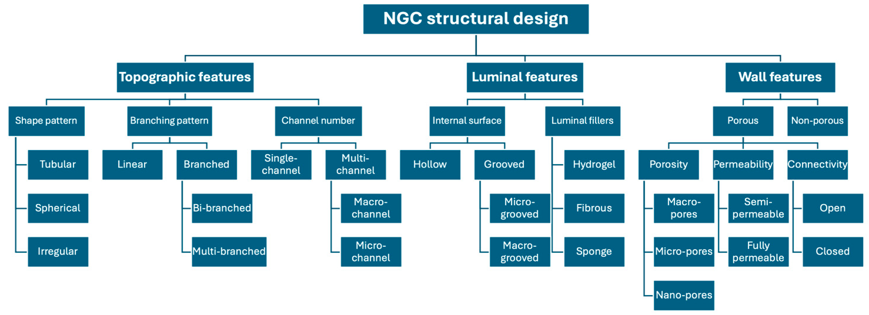
5. NGC Structural Features and Axonal Direction
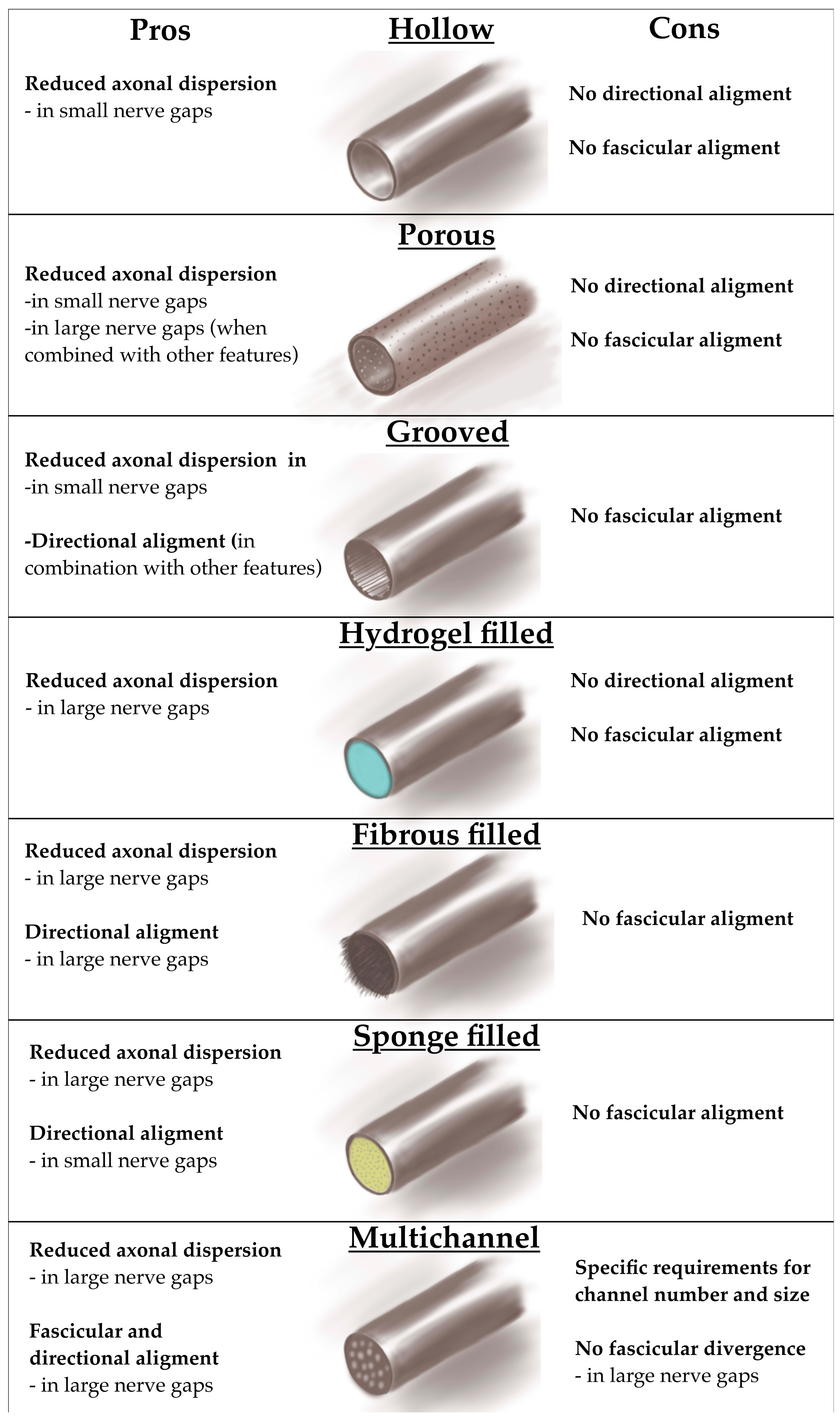
Various structural features may be included in NGC designing strategy (Figure 3) [8,17,18,19,20]. The ongoing research in the field usually involve simultaneous implementation of different strategies. There is no globally accepted consensus on classification of NGC design strategies. The simplified and most used NGC structure classification with their pros and cons is presented in the Figure 4 [13].
Scheme 2.
Structural features of NGC designing strategy.
5.1. Single Hollow Design
The single hollow design presents a fundamental NGC model featuring a solitary luminal cylindrical tube that serves to guide regenerative axons across the nerve gap. This structure prevents lateral axonal deviation and shields them from surrounding compression while concentrating growth factors and impeding the infiltration of scar tissue [71].
Some studies emphasized the potential superiority of the single hollow NGCs over end-to-end repair of gaps up to 5mm [72,73]. The so-called “tubular repair” by intentionally leaving a 2-5 mm gap between the nerve stumps is believed to provide space for natural axonal alignment and “preferential motor reinnervation”, contrary to the definite tubular misalignment provided by end-to-end nerve repair [73]. In a study by Liu et al. [73], the authors used retrograde tracing to evaluate the mismatched reinnervation following 3 and 6 mm rat sciatic nerve repair using AGNs and single hollow NGCs made of chitosan-poly(glycolide-co-lactide) (PGLA). The study results revealed a significantly reduced spreading of the peroneal motor axons into the tibial nerve in both 3 and 6 mm NGC repair groups compared to the ANG group. In a study by Tomita et al. [74], the authors used evoked motor potentials and sequential retrograde tracing for assessing mismatched innervation following 14 mm rat sciatic nerve repair using whole AGNs and AGNs combined with distally placed silicone single hollow NGC involving a 5 mm gap. They revealed a significantly higher innervation selectivity and functional recovery in cases of combined AGNs and NGCs.
The efficacy of a single hollow NGC decreases with increase of the nerve gap length, since it cannot provide continuous axonal direction towards the distal stump, resulting in their intraluminal dispersion and fasciculation [75,76,77]. In a study on 8 mm rat sciatic nerve gap repair [51], authors used stimulation with a suction electrode to reveal a similar level of mismatched reinnervation between ANGs and silicone single hollow NGCs. In a study on 10 mm rat sciatic nerve gap repair [36], authors used retrograde tracing to reveal a higher percentage of motoneurons projecting to both tibial and peroneal nerve branches in cases of PGLA single hollow NGCs compared to ANGs.
The single hollow NGCs can undergo structural modifications such as (1) incorporating porous walls, (2) integrating longitudinal luminal grooves, or (3) luminal fillers functionally enhancing with adhesive, bioactive, or neurotrophic molecules. These adaptations improve their efficacy by providing some axonal direction toward the distal stump and postponing synaptic mismatch. Thich is why they found their place in clinical practice for repairing defects under 3cm [78,79,80,81].
5.2. Porous Single Hollow Design
The porous single hollow NGC contains pores in the wall, which provides a vascular, nutritional, oxygen, and neurotrophic supply [81], as well as adhesive surface for migration of SCs and macrophages [82,83]. The size of the pores must be calculated to prevent fibroblast infiltration and induced scarring which may inhibit the axonal growth. The provided molecular supply through the porous wall enhance axonal direction towards the distal stump [82,84,85]. In a study on 8 mm rat sciatic nerve gap repair [51], the use of porous single hollow NGCs made of poly-L-lactate-e-caprolactone (PLC) led to reduced reinnervation mismatch, compared to the non-porous silicon NGCs and ANGs. Furthermore, NGCs with aligned pores are shown to enhance axonal guidance to their target destinations, while porous NGCs with thicker walls have shown superior nerve regeneration capacity by achieving an optimal balance between porosity and stiffness [86].
5.3. Grooved Single Hollow Design
The grooved single hollow NGC is integrated with longitudinal microgrooves that completely cover the luminal surface, providing enhanced glial migration and axonal direction [87]. In their study, Zhang et al. [88] investigated how micro-grooved surface influences axonal growth, focusing on the mechanisms that determine whether axons align along the grooves or cross over the ridges. They found that narrower and shallower grooves tend to promote axonal alignment along the grooves, while wider and deeper grooves increase the likelihood of axons crossing the ridges.
No studies have demonstrated the superiority of grooved NGCs over flat-surface NGCs. In both designs, axonal fasciculation occurs, likely because axons within grooves struggle to maintain directional growth when interacting with centrally positioned, dispersed axons. However, when grooved features are combined with porous or fiber-filled designs, they significantly enhance nerve regeneration [86].
5.4. Filled Single Hollow Design
Single hollow NGCs may contain intraluminal fillers, that resemble the endoneurial-like structure, or mimic the ECM composition and morphology, providing favorable conditions to enhance nerve regeneration. These fillers might comprise longitudinally aligned fibers [89,90], porous sponges, or hydrogels [91,92].
5.4.1. Hydrogel-Filled Single-Hollow Design
The single hollow NGC filled with hydrogel contains intraluminal properties that closely resemble native tissues. The hydrogel forms a 3D environment that more accurately replicates biological conditions and has ability to absorb and retain water, creating a hydrated structure similar to the extracellular matrix. The mechanical properties of the hydrogel, including its stiffness, porosity, architecture, and degradability, play a crucial role in influencing cellular behavior and viability. However, the hydrogel can not induce the straightforward guidance of the regenerative axons [93,94,95,96].
5.4.2. Fibrous-Filled Single-Hollow Design
The fibrous filled single hollow NGCs may involve filaments, microfibers, or nanofibers. Filaments are larger, thread-like structures with diameters in the micrometer to millimeter range. They provide strong mechanical support and guide axonal growth over larger gaps but have a lower surface-area-to-volume ratio, which limits cellular interaction. Microfibers are smaller, with diameters ranging from 1 to 100 μm, and offer improved flexibility and biomimicry, facilitating better alignment and interaction with SCs and the ECM. They are suitable for moderate nerve gaps but do not replicate the fine architecture of natural tissues as effectively as nanofibers. Nanofibers, with diameters below 1 micrometer, closely mimic the nanoscale architecture of the ECM. They have a high surface-area-to-volume ratio, promoting superior cellular adhesion and axonal growth. However, they may lack the mechanical strength needed for large-gap repairs and often require integration with other materials [50,68,97,98,99,100,101].
In a study by Daly et al. [42], NGCs filled with collagen fibers provided significantly higher targeted reinnervation following 1cm rat sciatic nerve repair, compared to the single-hollow NGCs and ANGs.
5.4.3. Nanosponge-Filled Single-Hollow Design
The nanofiber sponge filling provides a solid and very porous environment with a microarchitecture that resembles the neural tissue more than the fibrous-filling NGCs. Multiple diffusely spread channels provide easy migration of the Schwann cells, unable axonal dispersion, and guide them toward the distal stump. However, such diffuse channel distribution has no potential to cope with the challenges of intraneural topographic anatomy [102,103,104]
5.5. Multichannel Design
The multichannel NGCs are presented with multiple straightforward channels that can direct a group of axons, keeping them organized and separated from each other as they grow. This structure is designed to mimic the natural fascicular organization of nerves and provides a physical pathway to prevent misalignment and facilitate organized axonal growth [105,106,107,108,109]. In addition, multichannel NGCs can promote adhesion and proliferation of SCs, and promote the differentiation of stem cells.
In a study by Yao et al. [43] involving collagen multi-channel NGCs for 1cm rat sciatic nerve repair, simultaneous tracing showed a significantly lower percentage of motor neurons with double projections in cases of two- and four-channel, compared with single-channel repair. The similar results were achieved in a study by Ruiter et al. [36], involving 1cm rat sciatic nerve repair using poly(lactic-co-glycolic acid) (PLGA) NGCs with seven channels. Ina study by Park et al. [110], a 1 cm rat sciatic nerve repair using NGC with hundreds of microchannels showed higher gastrocnemius muscle density compared to the single channel NGC at 4 to 12 weeks.
6. Key Perspectives
All NGCs prevent initial axonal dispersion reinnervation mismatch in small nerve gaps. Multichannel and fiber/sponge-filled NGCs are recognized as the most promising designs for reducing axonal misdirection and supporting regeneration in longer nerve gaps. These designs offer improved axonal guidance over single-hollow conduits, though no single structure has been proven superior due to the variability in materials and fabrication techniques, which may independently impact outcomes.
Fiber-filled NGCs, especially those with porous structures, create an enriched microenvironment that guides axonal and cellular growth toward the distal stump. Sponge-filled NGCs provide a dispersed, porous scaffold that also offers directional cues but lacks the level of organized guidance that multichannel conduits or aligned nanofibers offer.
Multichannel NGCs mimic the natural fascicular organization of nerves, effectively directing grouped axonal growth and minimizing misalignment. However, their design may not offer as favorable a microenvironment for cellular growth as fiber- or sponge-filled conduits.
Despite their potential, multichannel and fiber/sponge-filled NGCs face biotechnological challenges in clinical adoption. Ensuring an appropriate degradation timeline while maintaining structural integrity poses a challenge, as the level of softness required for biodegradability can compromise the structural durability of the conduit.
No design strategy for NGCs currently ensures 100% recovery in peripheral nerve injuries. Designs like multichannel and fiber/sponge-filled provide straightforward axonal direction resulting in unpredictable degree of reinnervation mismatch, due to unpredictable intraneural branching topography.
These perspectives highlight the current advancements and limitations in NGC designs and underline the need for further research and development to address clinical challenges in peripheral nerve repair.
7. Future Perspectives
Personalized 3D printed conduits with tubular branching patterns that match the intraneural fascicular topography of a patient sound promising as a future advance in NGC designing strategy.
Johnson et al. [111] demonstrated the usage of 3D scanner for intraoperative visualization of rat proximal and distal nerve stump morphology, computational design modeling, and printing of tailored NGCs to match the stump fascicular distribution. Zhu et al. [112] developed rapid continuous 3D-printing platform for fabrication of NGCs with various architectures and adjusting structural parameters to match the mechanical properties of the injury site.
In one study, Yao et al. demonstrated the usage of micro-magnetic resonance imaging (micro-MRI) for scanning of fresh human nerve samples and their incorporation into computer software for 3D reconstruction [113]. In another study, Yao et al. used 3T MRI to scan the patient healthy nerve at the contralateral side and designed a tailored nerve graft according to information from previously formed data base, in accordance to specific injury situation [114].
8. Summary
In summary, while NGC designs have advanced significantly, current structures still face limitations in fully directing axonal growth and ensuring complete functional recovery. Multichannel and fiber/sponge-filled NGCs show promise for minimizing axonal misdirection, yet challenges remain in creating personalized, durable, and bio-compatible designs. Future approaches integrating 3D printing and machine learning for tailored patient-specific NGCs hold the potential to overcome these limitations, enhancing outcomes in peripheral nerve repair.
Author Contributions
For research articles with several authors, a short paragraph specifying their individual contributions must be provided. The following statements should be used “Conceptualization, A.M. and M.A.; methodology, A.M.; validation, A.S., M.L. and D.A.; formal analysis, L.V.; investigation, L.V.; writing—original draft preparation, A.M.; writing—review and editing, A.M.; visualization, J.G.; supervision, L.R.; project administration, J.P.; All authors have read and agreed to the published version of the manuscript.”.
Funding
This research received no external funding.
Conflicts of Interest
The authors declare no conflicts of interest.
Acknowledgments
The first author would like to express his sincere gratitude to Srđan Stevanović for his continuous support during the preparation of this paper.
References
- Rasulić, L.; Nikolić, L.; Lepić, M.; Savić, A.; Vitošević, F.; Novaković, N.; Radojević, S.; Mićić, A.; Lepić, S.; Mandić-Rajčević, S. Useful functional recovery and quality of life after surgical treatment of peroneal nerve injuries. Front. Surg. 2022, 9, 1005483. [Google Scholar] [CrossRef] [PubMed]
- Córdoba-Mosqueda, M.E.; Rasulić, L.; Savić, A.; Grujić, J.; Vitošević, F.; Lepić, M.; Mićić, A.; Radojević, S.; Mandić-Rajčević, S.; Jovanović, I.; et al. Quality of life and satisfaction in patients surgically treated for cubital tunnel syndrome. Neurol. Res. 2022, 45, 138–151. [Google Scholar] [CrossRef] [PubMed]
- Faroni, A.; Mobasseri, S.A.; Kingham, P.J.; Reid, A.J. Peripheral nerve regeneration: Experimental strategies and future perspectives. Adv. Drug Deliv. Rev. 2015, 82-83, 160–167. [Google Scholar] [CrossRef] [PubMed]
- Slutsky, D. A Practical Approach to Nerve Grafting in the Upper Extremity. Atlas Hand Clin. 2005, 10, 73–92. [Google Scholar] [CrossRef]
- DeLeonibus, A.; Rezaei, M.; Fahradyan, V.; Silver, J.; Rampazzo, A.; Gharb, B.B. A meta-analysis of functional outcomes in rat sciatic nerve injury models. Microsurgery 2021, 41, 286–295. [Google Scholar] [CrossRef] [PubMed]
- FF IJ, Nicolai JP, Meek MF. Sural nerve donor-site morbidity: thirty-four years of follow-up. Ann Plast Surg. 2006, 57, 391–395.
- Muheremu, A.; Ao, Q. Past, Present, and Future of Nerve Conduits in the Treatment of Peripheral Nerve Injury. BioMed Res. Int. 2015, 2015, 1–6. [Google Scholar] [CrossRef]
- Vijayavenkataraman, S. Nerve guide conduits for peripheral nerve injury repair: A review on design, materials and fabrication methods. Acta Biomaterialia 2020, 106, 54–69. [Google Scholar] [CrossRef]
- Supra, R.; Agrawal, D.K. Peripheral Nerve Regeneration: Opportunities and Challenges. J. Spine Res. Surg. 2023, 05, 10–18. [Google Scholar] [CrossRef] [PubMed]
- Grinsell, D.; Keating, C.P. Peripheral Nerve Reconstruction after Injury: A Review of Clinical and Experimental Therapies. BioMed Res. Int. 2014, 2014, 698256. [Google Scholar] [CrossRef]
- Scheib, J.; Höke, A. Advances in peripheral nerve regeneration. Nat. Rev. Neurol. 2013, 9, 668–676. [Google Scholar] [CrossRef] [PubMed]
- Cheng, H.; Bai, J.; Zhou, X.; Chen, N.; Jiang, Q.; Ren, Z.; Li, X.; Su, T.; Liang, L.; Jiang, W.; et al. Electrical stimulation with polypyrrole-coated polycaprolactone/silk fibroin scaffold promotes sacral nerve regeneration by modulating macrophage polarisation. Biomater Transl. 2024, 5, 157–174. [Google Scholar] [CrossRef] [PubMed]
- Amalakanti, S.; Mulpuri, R.P.; Avula, V.C.R. Recent advances in biomaterial design for nerve guidance conduits: a narrative review. Adv. Technol. Neurosci. 2024, 1, 32–42. [Google Scholar] [CrossRef]
- Tiwari, A.P.; Lokai, T.; Albin, B.; Yang, I.H. A Review on the Technological Advances and Future Perspectives of Axon Guidance and Regeneration in Peripheral Nerve Repair. Bioengineering 2022, 9, 562. [Google Scholar] [CrossRef] [PubMed]
- Zeng, Z.; Yang, Y.; Deng, J.; Rahman, M.S.U.; Sun, C.; Xu, S. Physical Stimulation Combined with Biomaterials Promotes Peripheral Nerve Injury Repair. Bioengineering 2022, 9, 292. [Google Scholar] [CrossRef]
- de Ruiter, G.C.W.; Spinner, R.J.; Verhaagen, J.; Malessy, M.J.A. Misdirection and guidance of regenerating axons after experimental nerve injury and repair. J. Neurosurg. 2014, 120, 493–501. [Google Scholar] [CrossRef] [PubMed]
- Yan, Y.; Yao, R.; Zhao, J.; Chen, K.; Duan, L.; Wang, T.; Zhang, S.; Guan, J.; Zheng, Z.; Wang, X.; et al. Implantable nerve guidance conduits: Material combinations, multi-functional strategies and advanced engineering innovations. Bioact. Mater. 2021, 11, 57–76. [Google Scholar] [CrossRef] [PubMed]
- Sarker, M.D.; Naghieh, S.; McInnes, A.D.; Schreyer, D.J.; Chen, X. Regeneration of peripheral nerves by nerve guidance conduits: Influence of design, biopolymers, cells, growth factors, and physical stimuli. Progress in Neurobiology 2018, 171, 125–150. [Google Scholar] [CrossRef]
- Sarker; Naghieh, S.; McInnes, A.D.; Schreyer, D.J.; Chen, X. Strategic Design and Fabrication of Nerve Guidance Conduits for Peripheral Nerve Regeneration. Biotechnol. J. 2018, 13, e1700635. [Google Scholar] [CrossRef] [PubMed]
- Yao, L.; de Ruiter, G.C.; Wang, H.; Knight, A.M.; Spinner, R.J.; Yaszemski, M.J.; Windebank, A.J.; Pandit, A. Controlling dispersion of axonal regeneration using a multichannel collagen nerve conduit. Biomaterials 2010, 31, 5789–5797. [Google Scholar] [CrossRef] [PubMed]
- John, A.; Rossettie, S.; Rafael, J.; Cox, C.; Ducic, I.; Mackay, B. Assessment of Motor Function in Peripheral Nerve Injury and Recovery. Orthop. Rev. 2022, 14, 37578. [Google Scholar] [CrossRef] [PubMed]
- Liu, X.; He, B.; Zhu, Z.; Zhu, Q.; Zhou, X.; Zheng, C.; Li, P.; Zhu, S.; Zhu, J.; B, H.; et al. Factors predicting sensory and motor recovery after the repair of upper limb peripheral nerve injuries. Neural Regen. Res. 2014, 9, 661–672. [Google Scholar] [CrossRef] [PubMed]
- Adidharma, W.; Khouri, A.N.; Lee, J.C.; Vanderboll, K.; Kung, T.A.; Cederna, P.S.; Kemp, S.W.P. Sensory nerve regeneration and reinnervation in muscle following peripheral nerve injury. Muscle Nerve 2022, 66, 384–396. [Google Scholar] [CrossRef] [PubMed]
- Liu, X.G.; Pang, R.P.; Zhou, L.J.; Wei, X.H.; Zang, Y. Neuropathic Pain: Sensory Nerve Injury or Motor Nerve Injury? Adv Exp Med Biol. 2016, 904, 59–75. [Google Scholar]
- Saragaglia, D.; Hassan Chamseddine, A. Injuries to Peripheral Nerves. Traumatology for the Emergency Doctor; Springer Nature: Cham, Switzerland, 2024; pp. 41–43. [Google Scholar]
- Khan, H.; Perera, N. Peripheral nerve injury: an update. Orthop. Trauma 2020, 34, 168–173. [Google Scholar] [CrossRef]
- Tian, R.; Zhou, Y.; Ren, Y.; Zhang, Y.; Tang, W. Wallerian degeneration: From mechanism to disease to imaging. Heliyon 2024, 11. [Google Scholar] [CrossRef]
- Huang, Y.; Wu, L.; Zhao, Y.; Guo, J.; Li, R.; Ma, S.; Ying, Z. Schwann cell promotes macrophage recruitment through IL-17B/IL-17RB pathway in injured peripheral nerves. Cell Rep. 2024, 43, 113753. [Google Scholar] [CrossRef] [PubMed]
- Contreras, E.; Bolívar, S.; Navarro, X.; Udina, E. New insights into peripheral nerve regeneration: The role of secretomes. Exp. Neurol. 2022, 354, 114069. [Google Scholar] [CrossRef] [PubMed]
- Benga, A.; Zor, F.; Korkmaz, A.; Marinescu, B.; Gorantla, V. The neurochemistry of peripheral nerve regeneration. Indian J. Plast. Surg. 2017, 50, 005–015. [Google Scholar] [CrossRef]
- Cattin, A.-L.; Lloyd, A.C. The multicellular complexity of peripheral nerve regeneration. Curr. Opin. Neurobiol. 2016, 39, 38–46. [Google Scholar] [CrossRef]
- Kong, L.; Gao, X.; Qian, Y.; Sun, W.; You, Z.; Fan, C. Biomechanical microenvironment in peripheral nerve regeneration: from pathophysiological understanding to tissue engineering development. Theranostics 2022, 12, 4993–5014. [Google Scholar] [CrossRef]
- Rotterman, T.M.; García, V.V.; Housley, S.N.; Nardelli, P.; Sierra, R.; Fix, C.E.; et al. Structural Preservation Does Not Ensure Function at Sensory Ia-Motoneuron Synapses following Peripheral Nerve Injury and Repair. J Neurosci. 2023, 43, 4390–4404. [Google Scholar] [CrossRef]
- Koerber, H.; Seymour, A.W.; Mendell, L.M. Mismatches between peripheral receptor type and central projections after peripheral nerve regeneration. Neurosci. Lett. 1989, 99, 67–72. [Google Scholar] [CrossRef] [PubMed]
- Dun, X.-P.; Parkinson, D.B. Visualizing Peripheral Nerve Regeneration by Whole Mount Staining. PLOS ONE 2015, 10, e0119168. [Google Scholar] [CrossRef]
- de Ruiter, G.C.; Spinner, R.J.; Malessy, M.J.; Moore, M.J.; Sorenson, E.J.; Currier, B.L.; et al. Accuracy of motor axon regeneration across autograft, single-lumen, and multichannel poly(lactic-co-glycolic acid) nerve tubes. Neurosurgery 2008, 63, 144–153, discussion 153–155. [Google Scholar] [CrossRef] [PubMed]
- Puigdellívol-Sánchez, A.; Prats-Galino, A.; Molander, C. Estimations of topographically correct regeneration to nerve branches and skin after peripheral nerve injury and repair. Brain Res. 2006, 1098, 49–60. [Google Scholar] [CrossRef]
- Wang, K.; Qin, B. [Research progress of peripheral nerve mismatch regeneration]. Zhongguo Xiu Fu Chong Jian Wai Ke Za Zhi 2021, 35, 387–391. [Google Scholar]
- Lee, J.I.; A Gurjar, A.; Talukder, M.A.H.; Rodenhouse, A.; Manto, K.; O’brien, M.; Karuman, Z.; Govindappa, P.K.; Elfar, J.C. Purposeful Misalignment of Severed Nerve Stumps in a Standardized Transection Model Reveals Persistent Functional Deficit With Aberrant Neurofilament Distribution. Mil. Med. 2021, 186, 696–703. [Google Scholar] [CrossRef]
- Gordon, T. Peripheral Nerve Regeneration and Muscle Reinnervation. Int. J. Mol. Sci. 2020, 21, 8652. [Google Scholar] [CrossRef] [PubMed]
- Gordon, T. Peripheral Nerve Regeneration and Muscle Reinnervation. Int. J. Mol. Sci. 2020, 21, 8652. [Google Scholar] [CrossRef]
- Daly, W.T.; Yao, L.; Abu-Rub, M.T.; O'Connell, C.; Zeugolis, D.I.; Windebank, A.J.; Pandit, A.S. The effect of intraluminal contact mediated guidance signals on axonal mismatch during peripheral nerve repair. Biomaterials 2012, 33, 6660–6671. [Google Scholar] [CrossRef] [PubMed]
- Yao, L.; de Ruiter, G.C.; Wang, H.; Knight, A.M.; Spinner, R.J.; Yaszemski, M.J.; Windebank, A.J.; Pandit, A. Controlling dispersion of axonal regeneration using a multichannel collagen nerve conduit. Biomaterials 2010, 31, 5789–5797. [Google Scholar] [CrossRef] [PubMed]
- Uschold, T.; Robinson, G.A.; Madison, R.D. Motor neuron regeneration accuracy: Balancing trophic influences between pathways and end-organs. Exp. Neurol. 2007, 205, 250–256. [Google Scholar] [CrossRef] [PubMed]
- Brushart, T.M.; Gerber, J.; Kessens, P.; Chen, Y.-G.; Royall, R.M. Contributions of Pathway and Neuron to Preferential Motor Reinnervation. J. Neurosci. 1998, 18, 8674–8681. [Google Scholar] [CrossRef]
- Abdullah, M.; O'Daly, A.; Vyas, A.; Rohde, C.; Brushart, T. Adult motor axons preferentially reinnervate predegenerated muscle nerve. Exp. Neurol. 2013, 249, 1–7. [Google Scholar] [CrossRef]
- Brushart, T.; Kebaish, F.; Wolinsky, R.; Skolasky, R.; Li, Z.; Barker, N. Sensory axons inhibit motor axon regeneration in vitro. Exp. Neurol. 2020, 323, 113073. [Google Scholar] [CrossRef] [PubMed]
- Maki, Y.; Yoshizu, T.; Tajima, T.; Narisawa, H. The Selectivity of Regenerating Motor and Sensory Axons. J. Reconstr. Microsurg. 1996, 12, 547–551. [Google Scholar] [CrossRef]
- Maki, Y.; Yoshizu, T.; Tsubokawa, N. Selective regeneration of motor and sensory axons in an experimental peripheral nerve model without endorgans. Scand. J. Plast. Reconstr. Surg. Hand Surg. 2005, 39, 257–260. [Google Scholar] [CrossRef] [PubMed]
- Lu, Q.; Zhang, F.; Cheng, W.; Gao, X.; Ding, Z.; Zhang, X.; Lu, Q.; Kaplan, D.L. Nerve Guidance Conduits with Hierarchical Anisotropic Architecture for Peripheral Nerve Regeneration. Adv. Heal. Mater. 2021, 10, e2100427. [Google Scholar] [CrossRef] [PubMed]
- Valero-Cabré, A.; Navarro, X. Functional Impact of Axonal Misdirection after Peripheral Nerve Injuries followed by Graft or Tube Repair. J. Neurotrauma 2002, 19, 1475–1485. [Google Scholar] [CrossRef]
- Lee, J.I.; A Gurjar, A.; Talukder, M.A.H.; Rodenhouse, A.; Manto, K.; O’brien, M.; Karuman, Z.; Govindappa, P.K.; Elfar, J.C. Purposeful Misalignment of Severed Nerve Stumps in a Standardized Transection Model Reveals Persistent Functional Deficit With Aberrant Neurofilament Distribution. Mil. Med. 2021, 186, 696–703. [Google Scholar] [CrossRef]
- Alant, J.D.d.V.; Senjaya, F.; Ivanovic, A.; Forden, J.; Shakhbazau, A.; Midha, R. The Impact of Motor Axon Misdirection and Attrition on Behavioral Deficit Following Experimental Nerve Injuries. PLOS ONE 2013, 8, e82546. [Google Scholar] [CrossRef] [PubMed]
- Brushart, T. Motor axons preferentially reinnervate motor pathways. J. Neurosci. 1993, 13, 2730–2738. [Google Scholar] [CrossRef] [PubMed]
- Madison, R.D.; Robinson, G.A.; Chadaram, S.R. The specificity of motor neurone regeneration (preferential reinnervation). Acta Physiol. 2007, 189, 201–206. [Google Scholar] [CrossRef]
- Hizay, A.; Ozsoy, U.; Demirel, B.M.; Ozsoy, O.; Angelova, S.K.; Ankerne, J.; Sarikcioglu, S.B.; Dunlop, S.A.; Angelov, D.N.; Sarikcioglu, L. Use of a Y-Tube Conduit After Facial Nerve Injury Reduces Collateral Axonal Branching at the Lesion Site But Neither Reduces Polyinnervation of Motor Endplates Nor Improves Functional Recovery. Neurosurgery 2012, 70, 1554–1556, discussion 1556. [Google Scholar] [CrossRef]
- Guntinas-Lichius, O.; Hundeshagen, G.; Paling, T.; Angelov, D.N. Impact of different types of facial nerve reconstruction on the recovery of motor function: an experimental study in adult rats. Neurosurgery 2007, 61, 1276–1283, discussion 1283–1285. [Google Scholar] [CrossRef]
- de Ruiter, G.C.; Malessy, M.J.; Alaid, A.O.; Spinner, R.J.; Engelstad, J.K.; Sorenson, E.; Kaufman, K.; Dyck, P.J.; Windebank, A.J. Misdirection of regenerating motor axons after nerve injury and repair in the rat sciatic nerve model. Exp. Neurol. 2008, 211, 339–350. [Google Scholar] [CrossRef]
- Witzel, C.; Rohde, C.; Brushart, T.M. Pathway sampling by regenerating peripheral axons. J. Comp. Neurol. 2005, 485, 183–190. [Google Scholar] [CrossRef]
- Cajal SRy. Cajal's Degeneration and Regeneration of the Nervous System. DeFelipe, J., Jones, E.G., May, R.M., Eds.; Oxford University Press; 1991 22 Mar 2012.
- Chen, B.; Chen, Q.; Parkinson, D.B.; Dun, X.-P. Analysis of Schwann Cell Migration and Axon Regeneration Following Nerve Injury in the Sciatic Nerve Bridge. Front. Mol. Neurosci. 2019, 12, 308. [Google Scholar] [CrossRef] [PubMed]
- Min, Q.; Parkinson, D.B.; Dun, X. Migrating Schwann cells direct axon regeneration within the peripheral nerve bridge. Glia 2020, 69, 235–254. [Google Scholar] [CrossRef] [PubMed]
- Fried, K.; Govrin-Lippmann, R.; Devor, M. Close apposition among neighbouring axonal endings in a neuroma. J. Neurocytol. 1993, 22, 663–681. [Google Scholar] [CrossRef]
- Tahir, M.; Lanzarin, L.D.; A J, B. Clinical Evidence of Retrograde Axonal Growth in Chronic Brachial Plexus Injury. Cureus 2024, 16, e62424. [Google Scholar] [CrossRef]
- Valero-Cabré, A.; Tsironis, K.; Skouras, E.; Navarro, X.; Neiss, W.F. Peripheral and Spinal Motor Reorganization after Nerve Injury and Repair. J. Neurotrauma 2004, 21, 95–108. [Google Scholar] [CrossRef] [PubMed]
- Li, C.; Rassekh, N.; O'Daly, A.; Kebaisch, F.; Wolinsky, R.; Vyas, A.; Skolasky, R.; Hoke, A.; Brushart, T. Preferential motor reinnervation is modulated by both repair site and distal nerve environments. Exp. Neurol. 2024, 385, 115066. [Google Scholar] [CrossRef] [PubMed]
- Konschake, M.; Burger, F.; Zwierzina, M. Peripheral Nerve Anatomy Revisited: Modern Requirements for Neuroimaging and Microsurgery. Anat. Rec. 2019, 302, 1325–1332. [Google Scholar] [CrossRef] [PubMed]
- Spivey, E.C.; Khaing, Z.Z.; Shear, J.B.; Schmidt, C.E. The fundamental role of subcellular topography in peripheral nerve repair therapies. Biomaterials 2012, 33, 4264–4276. [Google Scholar] [CrossRef] [PubMed]
- Paulin, E.; Bowen, E.C.; Dogar, S.; Mukit, M.; Lebhar, M.S.; Galarza, L.I.; Edwards, S.R.; Walker, M.E. A Comprehensive Review of Topography and Axon Counts in Upper-Extremity Peripheral Nerves: A Guide for Neurotization. J. Hand Surg. Glob. Online 2024, 6, 784–795. [Google Scholar] [CrossRef]
- Delgado-Martínez, I.; Badia, J.; Pascual-Font, A.; Rodríguez-Baeza, A.; Navarro, X. Fascicular Topography of the Human Median Nerve for Neuroprosthetic Surgery. Front. Neurosci. 2016, 10, 286. [Google Scholar] [CrossRef]
- Daly, W.; Yao, L.; Zeugolis, D.; Windebank, A.; Pandit, A. A biomaterials approach to peripheral nerve regeneration: bridging the peripheral nerve gap and enhancing functional recovery. J. R. Soc. Interface 2012, 9, 202–221. [Google Scholar] [CrossRef]
- Zhang, P.; Han, N.; Wang, T.; Xue, F.; Kou, Y.; Wang, Y.; Yin, X.; Lu, L.; Tian, G.; Gong, X.; et al. Biodegradable Conduit Small Gap Tubulization for Peripheral Nerve Mutilation: A Substitute for Traditional Epineurial Neurorrhaphy. Int. J. Med Sci. 2013, 10, 171–175. [Google Scholar] [CrossRef]
- Liu, D.; Mi, D.; Zhang, T.; Zhang, Y.; Yan, J.; Wang, Y.; Tan, X.; Yuan, Y.; Yang, Y.; Gu, X.; et al. Tubulation repair mitigates misdirection of regenerating motor axons across a sciatic nerve gap in rats. Sci. Rep. 2018, 8, 3443. [Google Scholar] [CrossRef]
- Tomita, K.; Kubo, T.; Matsuda, K.; Hattori, R.; Fujiwara, T.; Yano, K.; Hosokawa, K. Effect of conduit repair on aberrant motor axon growth within the nerve graft in rats. Microsurgery 2007, 27, 500–509. [Google Scholar] [CrossRef]
- Sarker; Naghieh, S.; McInnes, A.D.; Schreyer, D.J.; Chen, X. Strategic Design and Fabrication of Nerve Guidance Conduits for Peripheral Nerve Regeneration. Biotechnol. J. 2018, 13, e1700635. [Google Scholar] [CrossRef] [PubMed]
- Ribeiro-Resende, V.T.; Koenig, B.; Nichterwitz, S.; Oberhoffner, S.; Schlosshauer, B. Strategies for inducing the formation of bands of Büngner in peripheral nerve regeneration. Biomaterials 2009, 30, 5251–5259. [Google Scholar] [CrossRef] [PubMed]
- Taras, J.S.; Jacoby, S.M. Repair of Lacerated Peripheral Nerves With Nerve Conduits. Tech. Hand Up. Extremity Surg. 2008, 12, 100–106. [Google Scholar] [CrossRef] [PubMed]
- Johnson, E.O.; Soucacos, P.N. Nerve repair: Experimental and clinical evaluation of biodegradable artificial nerve guides. Injury 2008, 39, 30–36. [Google Scholar] [CrossRef] [PubMed]
- Chiono, V.; Tonda-Turo, C.; Ciardelli, G. Chapter 9: Artificial scaffolds for peripheral nerve reconstruction. Int Rev Neurobiol. 2009, 87, 173–198. [Google Scholar]
- Chiono, V.; Tonda-Turo, C. Trends in the design of nerve guidance channels in peripheral nerve tissue engineering. Prog. Neurobiol. 2015, 131, 87–104. [Google Scholar] [CrossRef]
- Gu, J.; Hu, W.; Deng, A.; Zhao, Q.; Lu, S.; Gu, X. Surgical repair of a 30 mm long human median nerve defect in the distal forearm by implantation of a chitosan-PGA nerve guidance conduit. J. Tissue Eng. Regen. Med. 2011, 6, 163–168. [Google Scholar] [CrossRef] [PubMed]
- Wan, T.; Wang, Y.-L.; Zhang, F.-S.; Zhang, X.-M.; Zhang, Y.-C.; Jiang, H.-R.; Zhang, M.; Zhang, P.-X. The Porous Structure of Peripheral Nerve Guidance Conduits: Features, Fabrication, and Implications for Peripheral Nerve Regeneration. Int. J. Mol. Sci. 2023, 24, 14132. [Google Scholar] [CrossRef]
- Tao, J.; Hu, Y.; Wang, S.; Zhang, J.; Liu, X.; Gou, Z.; Cheng, H.; Liu, Q.; Zhang, Q.; You, S.; et al. A 3D-engineered porous conduit for peripheral nerve repair. Sci. Rep. 2017, 7, 46038. [Google Scholar] [CrossRef]
- Russo, T.; Scialla, S.; D’albore, M.; Cruz-Maya, I.; De Santis, R.; Guarino, V. An Easy-to-Handle Route for Bicomponent Porous Tubes Fabrication as Nerve Guide Conduits. Polymers 2024, 16, 2893. [Google Scholar] [CrossRef]
- Johnson, L.D.V.; Aleemardani, M.; Atkins, S.; Boissonade, F.M.; Claeyssens, F. Emulsion templated composites: Porous nerve guidance conduits for peripheral nerve regeneration. Mater. Des. 2024, 239, 112779. [Google Scholar] [CrossRef]
- Mankavi, F.; Ibrahim, R.; Wang, H. Advances in Biomimetic Nerve Guidance Conduits for Peripheral Nerve Regeneration. Nanomaterials 2023, 13, 2528. [Google Scholar] [CrossRef] [PubMed]
- Carvalho, C.R.; Oliveira, J.M.; Reis, R.L. Modern Trends for Peripheral Nerve Repair and Regeneration: Beyond the Hollow Nerve Guidance Conduit. Front. Bioeng. Biotechnol. 2019, 7, 337. [Google Scholar] [CrossRef] [PubMed]
- Zhang, D.; Suo, H.; Qian, J.; Yin, J.; Fu, J.; Huang, Y. Physical understanding of axonal growth patterns on grooved substrates: groove ridge crossing versus longitudinal alignment. Bio-Design Manuf. 2020, 3, 348–360. [Google Scholar] [CrossRef]
- Matsumoto, K.; Ohnishi, K.; Kiyotani, T.; Sekine, T.; Ueda, H.; Nakamura, T.; Endo, K.; Shimizu, Y. Peripheral nerve regeneration across an 80-mm gap bridged by a polyglycolic acid (PGA)–collagen tube filled with laminin-coated collagen fibers: a histological and electrophysiological evaluation of regenerated nerves. Brain Res. 2000, 868, 315–328. [Google Scholar] [CrossRef] [PubMed]
- Wang, X.; Hu, W.; Cao, Y.; Yao, J.; Wu, J.; Gu, X. Dog sciatic nerve regeneration across a 30-mm defect bridged by a chitosan/PGA artificial nerve graft. Brain 2005, 128, 1897–1910. [Google Scholar] [CrossRef]
- Ceballos, D.; Navarro, X.; Dubey, N.; Wendelschafer-Crabb, G.; Kennedy, W.R.; Tranquillo, R.T. Magnetically aligned collagen gel filling a collagen nerve guide improves peripheral nerve regeneration. Exp Neurol. 1999, 158, 290–300. [Google Scholar] [CrossRef]
- Tonda-Turo, C.; Gentile, P.; Saracino, S.; Chiono, V.; Nandagiri, V.K.; Muzio, G.; Canuto, R.A.; Ciardelli, G. Comparative analysis of gelatin scaffolds crosslinked by genipin and silane coupling agent. Int. J. Biol. Macromol. 2011, 49, 700–706. [Google Scholar] [CrossRef] [PubMed]
- Novajra, G.; Tonda-Turo, C.; Vitale-Brovarone, C.; Ciardelli, G.; Geuna, S.; Raimondo, S. Novel systems for tailored neurotrophic factor release based on hydrogel and resorbable glass hollow fibers. Mater. Sci. Eng. C 2013, 36, 25–32. [Google Scholar] [CrossRef] [PubMed]
- Du, J.; Liu, J.; Yao, S.; Mao, H.; Peng, J.; Sun, X.; Cao, Z.; Yang, Y.; Xiao, B.; Wang, Y.; et al. Prompt peripheral nerve regeneration induced by a hierarchically aligned fibrin nanofiber hydrogel. Acta Biomater. 2017, 55, 296–309. [Google Scholar] [CrossRef]
- Rochkind, S.; Nevo, Z. Recovery of Peripheral Nerve with Massive Loss Defect by Tissue Engineered Guiding Regenerative Gel. BioMed Res. Int. 2014, 2014, 1–7. [Google Scholar] [CrossRef] [PubMed]
- Huang, Z.; Kankowski, S.; Ertekin, E.; Almog, M.; Nevo, Z.; Rochkind, S.; Haastert-Talini, K. Modified Hyaluronic Acid-Laminin-Hydrogel as Luminal Filler for Clinically Approved Hollow Nerve Guides in a Rat Critical Defect Size Model. Int. J. Mol. Sci. 2021, 22, 6554. [Google Scholar] [CrossRef] [PubMed]
- Mao, W.; Lee, E.; Cho, W.; Kang, B.-J.; Yoo, H.S. Cell-directed assembly of luminal nanofibril fillers in nerve conduits for peripheral nerve repair. Biomaterials 2023, 301, 122209. [Google Scholar] [CrossRef]
- Aigner, T.; Haynl, C.; Salehi, S.; O'Connor, A.; Scheibel, T. Nerve guidance conduit design based on self-rolling tubes. Mater. Today Bio 2020, 5, 100042. [Google Scholar] [CrossRef] [PubMed]
- Zhao, Q.; Lu, S.-B.; Quan, Q.; Meng, H.-Y.; Chang, B.; Liu, G.-B.; Cheng, X.-Q.; Tang, H.; Wang, Y.; Peng, J. Aligned fibers enhance nerve guide conduits when bridging peripheral nerve defects focused on early repair stage. Neural Regen. Res. 2019, 14, 903–912. [Google Scholar] [CrossRef] [PubMed]
- Farzamfar, S.; Salehi, M.; Tavangar, S.M.; Verdi, J.; Mansouri, K.; Ai, A.; Malekshahi, Z.V.; Ai, J. A novel polycaprolactone/carbon nanofiber composite as a conductive neural guidance channel: an in vitro and in vivo study. Prog. Biomater. 2019, 8, 239–248. [Google Scholar] [CrossRef] [PubMed]
- Hu, Y.; Zhang, H.; Wei, H.; Cheng, H.; Cai, J.; Chen, X.; Xia, L.; Wang, H.; Chai, R. Scaffolds with anisotropic structure for neural tissue engineering. Eng. Regen. 2022, 3, 154–162. [Google Scholar] [CrossRef]
- Sun, B.; Zhou, Z.; Wu, T.; Chen, W.; Li, D.; Zheng, H.; El-Hamshary, H.; Al-Deyab, S.S.; Mo, X.; Yu, Y. Development of Nanofiber Sponges-Containing Nerve Guidance Conduit for Peripheral Nerve Regeneration in Vivo. ACS Appl. Mater. Interfaces 2017, 9, 26684–26696. [Google Scholar] [CrossRef] [PubMed]
- Hou, Y.; Wang, X.; Wang, Y.; Chen, X.; Wei, B.; Zhang, J.; Zhu, L.; Kou, H.; Li, W.; Wang, H. Electrospun Nanofibrous Conduit Filled with a Collagen-Based Matrix (ColM) for Nerve Regeneration. Molecules 2023, 28, 7675. [Google Scholar] [CrossRef]
- Zhao, R.; Jiang, L.; Du, J.; Xu, B.; Li, A.; Wang, W.; Zhao, S.; Li, X. Fluffy sponge-reinforced electrospun conduits with biomimetic structures for peripheral nerve repair. Compos. Part B: Eng. 2021, 230, 109482. [Google Scholar] [CrossRef]
- Zheng, S.; Wei, H.; Cheng, H.; Qi, Y.; Gu, Y.; Ma, X.; Sun, J.; Ye, F.; Guo, F.; Cheng, C. Advances in nerve guidance conduits for peripheral nerve repair and regeneration. Am J Stem Cells 2023, 12, 112–123. [Google Scholar]
- Ye, W.; Li, H.; Yu, K.; Xie, C.; Wang, P.; Zheng, Y.; Zhang, P.; Xiu, J.; Yang, Y.; Zhang, F.; et al. 3D printing of gelatin methacrylate-based nerve guidance conduits with multiple channels. Mater. Des. 2020, 192, 108757. [Google Scholar] [CrossRef]
- Pawelec, K.; Koffler, J.; Shahriari, D.; Galvan, A.R.; Tuszynski, M.; Sakamoto, J. Microstructure and in vivo characterization of multi-channel nerve guidance scaffolds. Biomed. Mater. 2018, 13, 044104. [Google Scholar] [CrossRef] [PubMed]
- Jiang, Z.; Zhang, Y.; Wang, Y.; Wang, S.; Chang, J.; Liu, W.; Han, B. Multichannel nerve conduit based on chitosan derivates for peripheral nerve regeneration and Schwann cell survival. Carbohydr. Polym. 2022, 301, 120327. [Google Scholar] [CrossRef] [PubMed]
- Dinis, T.; Elia, R.; Vidal, G.; Dermigny, Q.; Denoeud, C.; Kaplan, D.; Egles, C.; Marin, F. 3D multi-channel bi-functionalized silk electrospun conduits for peripheral nerve regeneration. J. Mech. Behav. Biomed. Mater. 2015, 41, 43–55. [Google Scholar] [CrossRef] [PubMed]
- Park, D.; Kim, D.; Park, S.J.; Choi, J.H.; Seo, Y.; Kim, D.-H.; Lee, S.-H.; Hyun, J.K.; Yoo, J.; Jung, Y.; et al. Micropattern-based nerve guidance conduit with hundreds of microchannels and stem cell recruitment for nerve regeneration. npj Regen. Med. 2022, 7, 1–11. [Google Scholar] [CrossRef]
- Johnson, B.N.; Lancaster, K.Z.; Zhen, G.; He, J.; Gupta, M.K.; Kong, Y.L.; Engel, E.A.; Krick, K.D.; Ju, A.; Meng, F.; et al. 3D Printed Anatomical Nerve Regeneration Pathways. Adv. Funct. Mater. 2015, 25, 6205–6217. [Google Scholar] [CrossRef] [PubMed]
- Zhu, W.; Tringale, K.R.; Woller, S.A.; You, S.; Johnson, S.; Shen, H.; Schimelman, J.; Whitney, M.; Steinauer, J.; Xu, W.; et al. Rapid continuous 3D printing of customizable peripheral nerve guidance conduits. Mater. Today 2018, 21, 951–959. [Google Scholar] [CrossRef]
- Qi, J.; Zhu, Q.-T.; Yao, Z.; Yan, L.-W.; Wang, T.; Qiu, S.; Lin, T.; He, F.-L.; Yuan, R.-H.; Liu, X.-L. A rapid micro-magnetic resonance imaging scanning for three-dimensional reconstruction of peripheral nerve fascicles. Neural Regen. Res. 2018, 13, 1953–1960. [Google Scholar] [CrossRef]
- Yao, Z.; Yan, L.-W.; Qiu, S.; He, F.-L.; Gu, F.-B.; Liu, X.-L.; Qi, J.; Zhu, Q.-T. Customized Scaffold Design Based on Natural Peripheral Nerve Fascicle Characteristics for Biofabrication in Tissue Regeneration. BioMed Res. Int. 2019, 2019, 1–10. [Google Scholar] [CrossRef] [PubMed]
- Chang, P.S.; Lee, T.Y.; Kneiber, D.; Dy, C.J.; Ward, P.M.; Kazarian, G.; Apostolakos, J.; Brogan, D.M. Design and In Vivo Testing of an Anatomic 3D-Printed Peripheral Nerve Conduit in a Rat Sciatic Nerve Model. HSS J. 2024. [Google Scholar] [CrossRef]
- Stewart, C.E.; Kan, C.F.K.; Stewart, B.R.; Sanicola, H.W.; Jung, J.P.; Sulaiman, O.A.R.; Wang, D. Machine intelligence for nerve conduit design and production. J. Biol. Eng. 2020, 14, 25. [Google Scholar] [CrossRef] [PubMed]
- Venkata Krishna, D.; Ravi Sankar, M. Engineered approach coupled with machine learning in biofabrication of patient-specific nerve guide conduits - Review. Bioprinting 2023, 30, e00264. [Google Scholar] [CrossRef]
Scheme 1.
Factors promoting axonal misdirection.
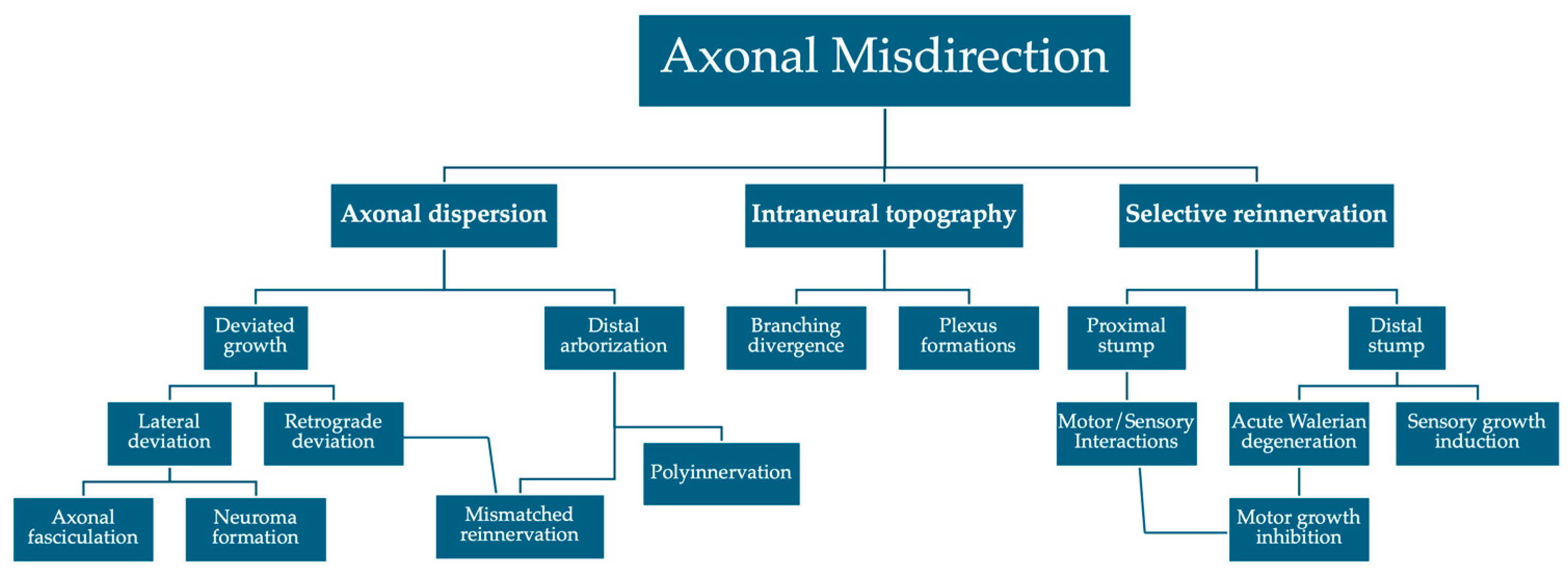
Figure 1.
Intraneural branching topography. (A) Divergent fascicular branching; (B) Intraneural plexus formations.
Figure 1.
Intraneural branching topography. (A) Divergent fascicular branching; (B) Intraneural plexus formations.
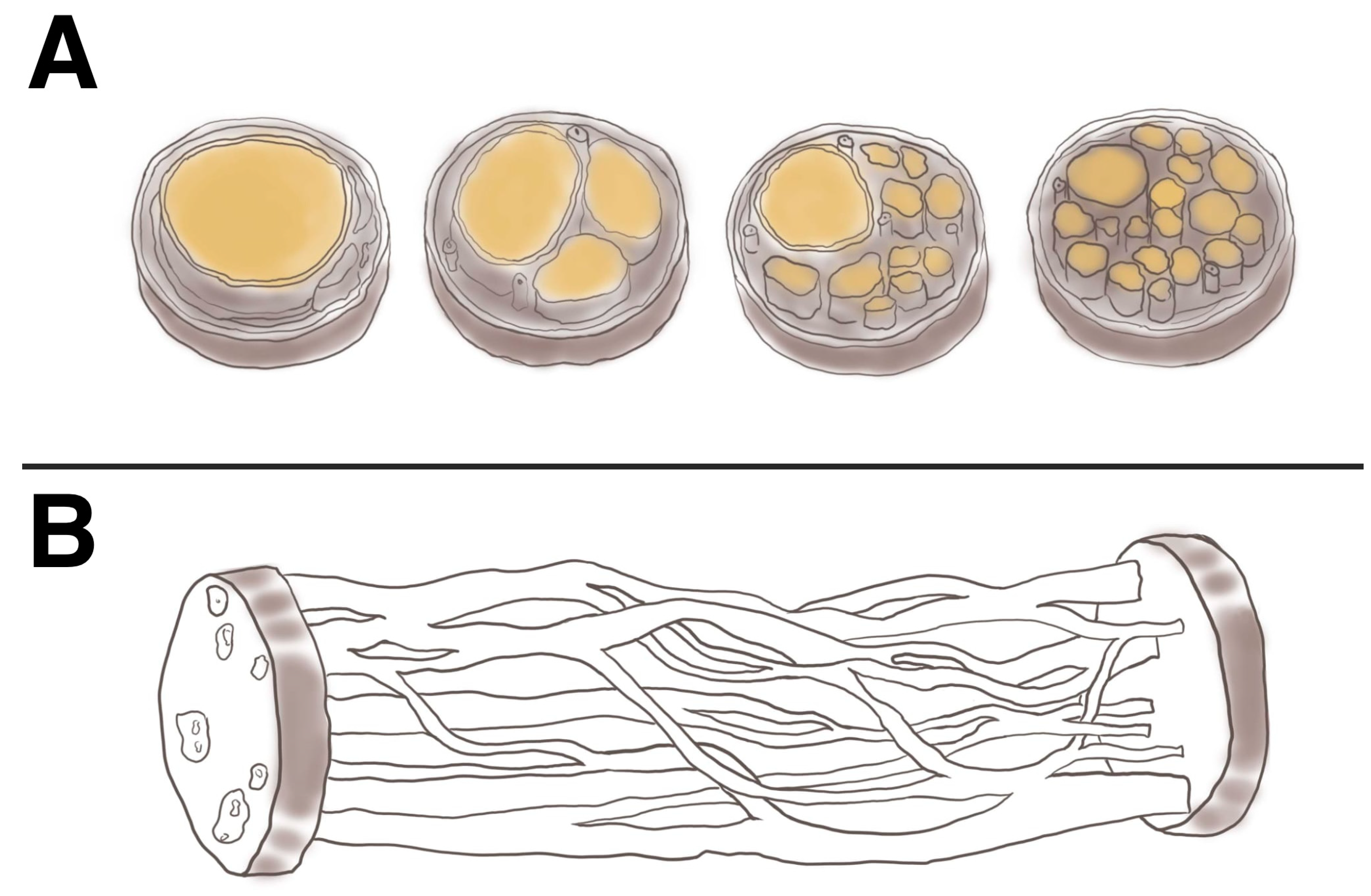
Figure 2.
Simplified classification of NGC structural designs with pros and cons in preventing axonal misdirection.
Figure 2.
Simplified classification of NGC structural designs with pros and cons in preventing axonal misdirection.
Disclaimer/Publisher’s Note: The statements, opinions and data contained in all publications are solely those of the individual author(s) and contributor(s) and not of MDPI and/or the editor(s). MDPI and/or the editor(s) disclaim responsibility for any injury to people or property resulting from any ideas, methods, instructions or products referred to in the content. |
© 2024 by the authors. Licensee MDPI, Basel, Switzerland. This article is an open access article distributed under the terms and conditions of the Creative Commons Attribution (CC BY) license (http://creativecommons.org/licenses/by/4.0/).
Copyright: This open access article is published under a Creative Commons CC BY 4.0 license, which permit the free download, distribution, and reuse, provided that the author and preprint are cited in any reuse.


