
Submitted:
29 November 2024
Posted:
02 December 2024
You are already at the latest version
Abstract
Biopolymer based nano drug delivery systems are emerging and promising strategy for addressing the challenges of cancer treatment and wound healing. These natural nanocarrier like alginate, chitosan, gelation and hyaluronic acid. These nano systems offers biocompatibility, biodegradibility and reduced toxicity of the therapeutics. The nanoparticles can be loaded with growth factors which accelerating tissue These provide targeted delivery sites and also modulate the microenvironment of the tissue by sustained drug release and reducing the damage to healthy tissues, thus promoting cancer treatment and wound tissue regeneration in synergistic manner. Drug delivery system (DDS) technology include varities of approaches such as liposomes, nanofibers, inorganic and lipid nanoparticles. The focus of this chapter to provide the recent developments in biopolymer-based strategies and their applications for the future prospects for clinical implementation.
Keywords:
Drug delivery system
; nanoparticles
; biopolymers
1. Introduction
Cancer disease are among the biggest and prevalent issues facing modern medicine with implications for society and the economy. Additionally, they are primary cause of death in the developed nations. The two most popular methods of treating malignant neoplasms are chemotherapy and radiotherapy. Their side effect are therefore a serious problem and significant obstacle for the contemporary oncology. Skin homeostatsis issues are among the most significant side effects of chemotherapy and radiation therapy [1]. Chronic wounds may develop as a result of surgery itself, adjuvant therapies like chemotherapy and radiotherapy. Both chemotherapy and radiotherapy are adjuvant treatment that have a number of systemic side effects such as skin disruptions. Oncological patients experience tissue loss and continuous search for an effective healing treatment. Biomaterials, growth factors, tissue enginering products dervived from in vitro cultured allogenic or autologous cells and trational dressings are some of the intringuing techniques [2]. These days, there is increased interest in the application nanomaterials in the pharmaceutical industry particularly in drug delivery systems(DDS) [3]. DDS based nanotechnology is a new multidisplinary program in the biomedical field that addresses issues like drug side effects , plasma inconsistency, therapeutic potency, poor intestinal absorption mechanism through degradation and bioavailability due to reduced solubility [4]. The aim of this chapter address the dual challenges of cancer treatment and wound healing by focusing on biopolymer based nano drug delivery systems which can be optimized to enhance the cancer treatment and wound tissue regeneration.
2. Biopolymer: Properties and Applications
Biopolymers are broad and incredibly adaptable class of chemicals that are either manufactured from biological sources or produced by organism. Biopolymers are made up of identical repeating units named monomers that are linked together [5]. Diverse natural and synthetic biomaterials, biodegradable and non-degradable are explored potentially as drug delivery for tissue engineering with medical applications. The key features of biopolymer are biocompatibility, biodegradability and antibacterial activity. There is lots of similarity is chemical structures and composition of the macromolecules of the natural extracellular environment [6,7,8].
2.1. Types of Biopolymer
2.1.1. Chitosan
One of well- known polysaccrides that is natural in origin and chitin byproduct is chitosan. copolymers Glucosamine and N-acetyle-glucosamine linked by β-1,4-glycosidic bonds [9]. The research that utilized solid lipid nanoparticles (SLN) loaded with all-trans retinoic acid (ATRA) and wrapped in chitosan film. Under the controlled conditions the medications are released by chitinosan film and the SLN-ATRA accelerated wound closure by minimizing scarring, collagen deposition boosted and lowered the leucocyte infiltration in the wound area. Chitosan-encased SLN-ATRA is a suitable option for the treatment of diabetic wounds and promoting wound tissue recovery [10]. Chitosan is known for the antibacterial and antibiofilm properties which was studied. A chitosan film release nitric oxide (NO) (CS/NO film) was formed. The result depicted that NO was released simulated wound fluid contentious for 72 hour. Furthermore, CS/NO represented more enhanced antibiofilm activity and substantially increased the antimicrobial action against MRSA and decreased bacterial viability. CS/NO film surged the elimination of biofilms, minimized wound size and encouraged collagen deposition and epithelialzation. Hence, it can be used in the future to curing infected wounds [11].
2.1.2. Alginate
It is also known as alginic acid, is an anionic polymer which is widely distributed in the cell walls of brown algae, particularly in Ascophyllum and Laminaria species. They are produced by copolymerizing d-mannuronic acid and I-guluronic acid. [9]. They are unbranched linear polysaccharides include distinct amounts of (1→4)-linked β-d mannuronic acid and α-I-guluronic acid residues. These are unbranched linear polysaccharides can be connected to other physiologically active molecules, having tractable porosity and are biodegradable [12]. Hydrogels dominating role in wound closure was demonstrated by the full healing of the PVA/alginate hydrogel wrapping the new tea polyphenol nanospheres (TPN) after 5 days of injury. The PI3K/AKT pathway is triggered by TPN@H which reduce inflammation and improves wound healing [13].
2.1.3. Hyaluronic Acid
Hyaluronic acid (HA) is a naturally occurring GAG that is highly hydrophilic, non-immunogenic, non-sulfated and anionic. It is found extensively distributed throughout the connective tissues, neural tissues, synovial fluids and epithelia. It is comprised of cockscomb, cartilage skin, vitreous humor. It comprises of 2-acetamide-2-deoxy- α-d-glucose and β-d-gluconic acid that are bounded by numerous (1,3) and (1,4) glycoside bonds [9]. HA is widely used in the biomedical field because of bacteriostatic effect [14]. It is also used in tissue engineering, ocular surgery, wound healing [15] and along with material for implant preparation in reconstructive plastic surgery [16].In order to conduct research on hyaluronic acid oligosaccharides ,Huang et al prepared an ointment that contained blend of hyaluronan fragments. In addition to developing tubes of endothleial cells in the midst of high glucose o-HA significantly boosted migration and proliferation. Applying O-HA ointment accelerates the wound healing by enhancing angiogenesis in the injured skin region. This suggest that using O-HA topically in clinical setting may be a useful strategy for treatment diabetes patient wound [17].
2.1.4. Gelation
Gelatin is primarily made from denatured protein collagen via hydrolysis process that produces significant peptides which initiate signal transduction and cellular adhesion pathways during wound healing. It is biocompatible, biodegradable, and non-immunogen [18]. Gelatin promotes the homeostasis stage, gelatin absorbs watery waste products from wound and residues in the tissue regeneration. Because of these features the creation of scaffolds for wound closure and regeneration of tissues. Furthermore, it is utilized in the development of absorbent-adhesive pads and surgical wound dressing [19,20].
3. Nano Drug Delivery Systems
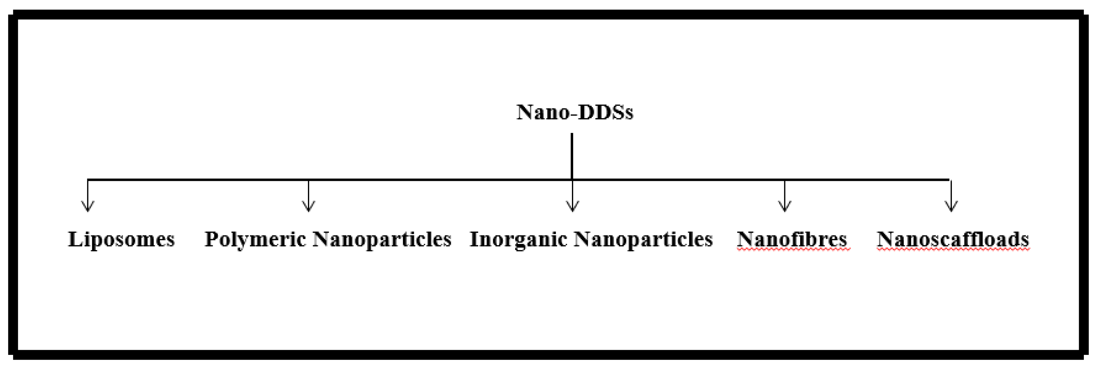
NDDS are drug delivery systems that can be made from a range of biomaterials and having particle diameter within the nanoscale. They having ability to improve drug stability, sustain release and controlled release [21]. The order to promote wound healing and skin regeneration, a variety of nano-DDSs containing therapeutic agents are agents are emerging at an unprecedented rate. These include liposomes, polymeric nanomaterials, inorganic nanoparticles, lipid nanoparticles, nanofibrous structures and nanohydrogel [22,23,24].
Figure 1.
It represent various Nano DDSs.
3.1. Liposomes
Liposomes are synthetic members primarily made up of amphiphilic molecules that assemble into a bilayer structure resembling skin cell member. The hollow portion of the lipid bilayer contains drugs, making liposomes sophisticated drug delivery nano carriers [25]. They are biocompatible with skin, biodegradable and nontoxic, they can hold hydrophilic medications (like growth factors) in the inner water cavity and hydrophobic agents in the bilayer [26,27]. Liposomes preserve the drugs release while protecting the encapsulating medication. Moreover after application, liposomes efficiently covers the wound and produce a moist environment on the surface that promotes the wound healing [28].
3.2. Inorganic Nanoparticles
Inorganic nanoparticles are defined as those that have been stripped from the inorganic sources like metallic nanoparticles, carbon-based ceramics, ceramics nanoparticles etc. [29]. Inorganic nanoparticles like silver nanoparticles which is frequently used as antimicrobial agents because of inherent properties as materials. They having strong antibacterial properties and comparable benefits for wound healing. Consequently, in order to achieve a syngisitic promoting effect of both materials and drugs, as it is more desirable in research to combine inorganic nanoparticles [30].
3.3. Polymeric Nanoparticles
Polymeric Nanoparticles are biocompatible colloidal system with precise formulation parameters [31]. It is possible to achieve reduced degradation rates and controlled release of drugs in the wound area by embedding or conjugating with biodegrable polymers. The field of nano-drug delivery systems is paying more attention to polymeric nanoparticles because of these advantages [32]. The core -shell structure of polymeric nanoparticles contains drugs whereas the hydrophilic polymeric outer surface offer stearic stability [33].
3.4. Nanofibers
Nanofibres are created from natural and synthetic continuous polymer which can be used in tissue engineering as 3D scaffloads or nanofibrous sheets [34]. Nanofibres typically having diameter of less than 100nm which is important class of nanomaterials [35]. Nanofibres include numerous exceptional characterstics like large surface area, variable porous rate, fantastic material selection flexibility and advanced fabrication technology [36]. These nanofibrous structures are intended to function as a substitute for artificial dermal analogs by simulating the extracellular matrix, promoting cell attachment and improving cell-drug interaction [37,38].
3.5. Nanohydrogels
Nanohydrogels is a three dimensional polymeric network that is assumed to be the best formulation for controlling wounds because of its porous three dimensional structure which allows to absorb aqueous fluids [39],preventing dehydration and fostering a beneficial moist environment for wound healing [40], additionally it is non-adhesive which can preserve the wound bed and allowing oxygen to penetrate which is essential for wound healing [41], the soft texture of nanohydrogel makes the treatment process comfortable [42].
3.6. Lipid Nanoparticles
Lipid nanoparticles are created by glycerophospholipids, cationic lipids, sterol lipids and PEGylated lipids coated with oligonucleotides [43]. Nanostructured lipid carriers (NLCs) and solid lipid nanoparticle (SLNs) are two types of LNPs which can improve the stability and solubility of medications that are encapsulated [44].

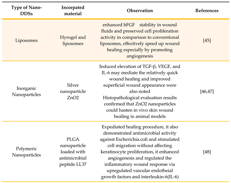
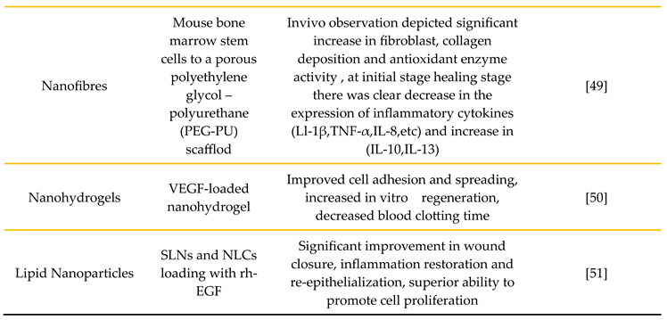
Table 1.
It depicts various advantages of Nano-DDSs.
4. Cancer Treatment and Wound Healing Mechanisms
It is demonstrated both malignant process and wound healing overlap certain characteristics [52,53,54]. Consequently, it makes appropriate to search and discuss regarding procedure. Wound healing is a complicated process with several stages. The main phases are tissue remodeling, proliferation and inflammation. Trama healing is an expression to describe the traits of this process. i)Following the trauma, platelet accumulation and blood vessel constriction occur to halt the bleeding. Then other cells linked to inflammation are drawn to the site: neutrophiles are drawn in the early stages, whereas monocytes and macrophages show up later. Inflammatory response may initiate by numerous cytokines, chemokines, DAMP, and PAMP. The hallmark of the inflammatory phase is hemostasis which seals the wound and stops further harm. Chemotaxis and increased vascular permeability are the features of phases that aid in cell movement to get rid of germs and cellular debris. ii) The proliferation phase begins when granulation tissue fills the wound defect. In order to help stabilize wounds, fibroblasts multiply and create new collagens and glycosaminoglycans. As a result, new blood vessels form and eventually an immature scar seals the edges of the wound. iii) The maturation phase begins after the damaged site is repaired, the site reaches its maximum strength and the scar formed. When the skin wound occurs, the edges of wound are drawn together and epithelization occurs [55,56].
5. Applications of Biopolymer Based Nanocarriers in Cancer Wound Healing
Nanotechnology is the new therapeutic approach that use nanoparticles to diagnose and cue cancer [57,58]. NPs are used in cancer treatment because of distinct size, which is typically between 1 and 1000 nanometer, but ideally between 5 and 200nm for drug delivery applications. NPs drug delivery system provide clear benefits for cancer treatment compared to free medication administration:- 1)enhance the therapeutic index of the pharmaceutical agents that are loaded as opposed to those that are administered via traditional dose forms. 2)enhance medication effectiveness by maintaining stable therapeutic drug levels over time. 3) decrease the drug toxicity via controlled drug release and increase the medication solubility along this stability to enhance pharmacokinetics [59]. Additionally, anticancer can be incorporated into nanoparticles to reduce chemoresistance to drug activity, which improves therapeutic selectivity for cancer cells and decreases drug toxicity against normal cells [60]. Furthermore, functionalizing the surface of the nanocarriers certain antibodies or Ab-fragments that identify specific epitopes of tumor-associated antigens (TAA) and tumor-specific antigens(TSA) improves their selectivity for cancer cells [61]. Since their lymphatic clearance at the tumor site is hampered nanocarriers are maintained in the tumor interstitium and gradually accumulation to the tumor tissues [62]. The release of drugs into tumroal interstitium can be regulated by modifying the nanoparticle structure such as the polymer utilized and thickness of the polymer wall covering [59].
Engineering techniques based on nanotechnology have accelerated the development of these biopolymers into a fresh group of wound care solutions [63]. Studies are conducted to investigate these compounds potential to aid the healing process [64]. Fucoidan promotes angiogenesis and wound healing by stimulating heparin binding cytokines such as FGF-1 and FGF-2 in the wound exudates [65]. Fucoidan has the ability to modify TGF-β1’s impact on wound healing [66]. The polysaccharides EP22, which was isolated from Pseudomonas stutzeri AS22, demonstrated effective wound healing in rats, as evidenced by neatly arranged dermal and epidermal layers [67]. The teams research has demonstrated that EPSs isolated from Nitratireductor sp. PRIM-31 and Rhizobium sp. PRIM-18 exhibit in vitro wound healing properties that are mediated by fibroblast migration and proliferation [68,69]. Likewise, it has been claimed that a several polysaccharides generated from fungi have ability to heal wounds. He et al. extracted polysaccharide from Lachnum sp. and validated its ability to heal tissue [70].
6. Challenges and Future Prospects
The challenges faced by these systems are stability and biocompatibility of the biomaterials as these can degrade under varying physiological conditions like pH and temp. This may trigger the inflammatory response and lead to difficulty in targeting specific tissue sites due to the poor penetration and specificity can limit the binding. Lastly, preparation of the nano drugs on large scale can be costly and may raise the sustainability issues as complexity of biopolymer synthesis which can be major hurdle in the future. The term clinical trials should be conducted to avoid toxicity and immunogenicity. The future prospects of the nanodrugs are the development of the multifunctional nanodrugs which accelerate the cancer wound healing, personalized targeted therapies but the high reproducibility and cost effective too.
7. Conclusions
Biopolymer based nano drug delivery systems hold immense potential for the cancer wound healing by offering synergistic solution to the conventional therapies. Additionally, its biocompatible, biodegradability to incorporate bioactive molecules which deliver therapeutic agents in a controlled and targeted manner. It is offering the targeted drug delivery, promoting tissue regeneration and reducing the side effects in wound healing. Apart from this drug delivery system is boon for therapeutics solutions as it is providing personalized medicine and regenerative therapies and significantly improving the clinical management for the cancer treatment and wound treatment.
References
- Deptuła, M.; Zieliński, J.; Wardowska, A.; Pikuła, M. Wound healing complications in oncological patients: perspectives for cellular therapy. [CrossRef]
- Pikuła, M.; Langa, P.; Kosikowska, P.; Trzonkowski, P. Stem cells and growth factors in wound healing. Postepy Hig Med Dosw 2015, 69, 874–885. [Google Scholar] [CrossRef] [PubMed]
- Jacob, J.; Haponiuk, J.T.; Thomas, S.; Gopi, S. Biopolymer based nanomaterials in drug delivery systems: A review. [CrossRef]
- Kukoyi, A.R. Economic impacts of natural polymers, in: O. Olatunji (Ed.),Natural Polymers: Industry Techniques and Applications; Springer: Switzerland, 2016. [Google Scholar]
- Wróblewska-Krepsztul, J.; et al. Biopolymers for Biomedical and Pharmaceutical Applications: Recent Advances and Overview of Alginate Electrospinning. Nanomaterials 2019, 9, 404. [Google Scholar] [CrossRef] [PubMed]
- Yu, L.; Dean, K.; Li, L. Polymer blends and composites from renewable resources. Progress in Polymer Science 2006, 31, 576–602. [Google Scholar] [CrossRef]
- Oh, J.K.; Lee, D.I.; Park, J.M. Biopolymer-based microgels/nanogels for drug delivery applications. Progress in Polymer Science 2009, 34, 1261–1282. [Google Scholar] [CrossRef]
- Uebersax, L.; Merkle, H.P.; Meinel, L. Biopolymer-based growth factor delivery for tissue repair: from natural concepts to engineered systems. Tissue Eng Part B Rev 2009, 15, 263–289. [Google Scholar] [CrossRef]
- Raina, N.; Rani, R.; Pahwa, R.; Gupta, M. Biopolymers and treatment strategies for wound healing: an insight view. International Journal of Polymeric Materials and Polymeric Biomaterials 2020, 71, 359–375. [Google Scholar] [CrossRef]
- Arantes, V. T.; Faraco, A. A.; Ferreira, F. B.; Oliveira, C. A.; Martins-Santos, E.; Cassini-Vieira, P.; Barcelos, L. S.; Ferreira, L. A.; Goulart, G. A. Retinoic Acid-Loaded Solid Lipid Nanoparticles Surrounded by Chitosan Film Support Diabetic Wound Healing in In Vivo Study. Coll. Surf. B. Biointerf. 2020, 188, 110749. [Google Scholar] [CrossRef]
- Choi, M.; Hasan, N.; Cao, J.; Lee, J.; Hlaing, S. P.; Yoo, J. W. Chitosan-Based Nitric Oxide-Releasing Dressing for Anti-Biofilm and In Vivo Healing Activities in MRSA Biofilm-Infected Wounds. Int. J. Biol. Macromol. 2020, 142, 680–692. [Google Scholar] [CrossRef]
- Thu, H. E.; Zulfakar, M. H.; Ng, S. F. Alginate Based Bilayer Hydrocolloid Films as Potential Slow-Release Modern Wound Dressing. Int. J. Pharm. 2012, 434, 375–383. [Google Scholar] [CrossRef]
- Chen, G.; He, L.; Zhang, P.; Zhang, J.; Mei, X.; Wang, D.; Zhang, Y.; Ren, X.; Chen, Z. Encapsulation of Green Tea Polyphenol Nanospheres in PVA/alginate hydrogel for promoting wound healing of diabetic rats by regulating PI3K/AKT pathway. Mater. Sci. Eng. C Mater. Biol. Appl. 2020, 110, 110686. [Google Scholar] [CrossRef]
- Pandey, R.P.; Vidic, J.; Mukherjee, R.; Chang, C.M. Experimental Methods for the Biological Evaluation of Nanoparticle-Based Drug Delivery Risks. Pharmaceutics. 2023, 15, 612. [Google Scholar] [CrossRef] [PubMed]
- Pandey, R.P.; Mukherjee, R.; Priyadarshini, A.; Gupta, A.; Vibhuti, A.; Leal, E.; Sengupta, U.; Katoch, V.M.; Sharma, P.; Moore, C.E.; Raj, V.S.; Lyu, X. Potential of nanoparticles encapsulated drugs for possible inhibition of the antimicrobial resistance development. Biomed Pharmacother. 2021, 141, 111943. [Google Scholar] [CrossRef] [PubMed]
- Park, S. N.; Lee, H. J.; Lee, K. H.; Suh, H. Biological Characterization of EDC-Crosslinked Collagen-Hyaluronic Acid Matrix in Dermal Tissue Restoration. Biomaterials 2003, 24, 1631–1641. [Google Scholar] [CrossRef] [PubMed]
- Neuman, M. G.; Nanau, R. M.; Oruña-Sanchez, L.; Coto, G. Hyaluronic Acid and Wound Healing. J. Pharm. Pharm. Sci. 2015, 18, 53–60. [Google Scholar] [CrossRef]
- Sparavigna, A.; Fino, P.; Tenconi, B.; Giordan, N.; Amorosi, V.; Scuderi, N. A New Dermal Filler Made of Cross-Linked and Auto-Cross-Linked Hyaluronic Acid in the Correction of Facial Aging Defects. J. Cosmet. Dermatol. 2014, 13, 307–314. [Google Scholar] [CrossRef]
- Wang, Y.; Han, G.; Guo, B.; Huang, J. Hyaluronan Oligosaccharides Promote Diabetic Wound Healing by Increasing Angiogenesis. Pharmacol. Rep. 2016, 68, 1126–1132. [Google Scholar] [CrossRef]
- Yoon, D. S.; Lee, Y.; Ryu, H. A.; Jang, Y.; Lee, K.-M.; Choi, Y.; Choi, W. J.; Lee, M.; Park, K. M.; Park, K. D.; Lee, J. W. Cell Recruiting Chemokine-Loaded Sprayable Gelatin Hydrogel Dressings for Diabetic Wound Healing. Acta Biomater. 2016, 38, 59–68. [Google Scholar] [CrossRef]
- Kawai, K.; Suzuki, S.; Tabata, Y.; Nishimura, Y. Accelerated Wound Healing through the Incorporation of Basic Fibroblast Growth Factor-Impregnated Gelatin Microspheres into Artificial Dermis Using a Pressure-Induced Decubitus Ulcer Model in Genetically Diabetic Mice. Br. J. Plast. Surg. 2005, 58, 1115–1123. [Google Scholar] [CrossRef]
- Iwakura, A.; Tabata, Y.; Tamura, N.; Doi, K.; Nishimura, K.; Nakamura, T.; Shimizu, Y.; Fujita, M.; Komeda, M. Gelatin Sheet Incorporating Basic Fibroblast Growth Factor Enhances Healing of Devascularized Sternum in Diabetic Rats. Circulation 2001, 104, I325–I329. [Google Scholar] [CrossRef]
- Qamar, Z.; Qizilbash, F.F.; Iqubal, M.K. , et al. Nano-based drug delivery system: recent strategies for the treatment of ocular disease and future perspective. Recent Pat Drug Deliv Formul. 2019, 13, 246–254. [Google Scholar] [CrossRef]
- Gainza, G.; Villullas, S.; Pedraz, J.L.; Hernandez, R.M.; Igartua, M. Advances in drug delivery systems (DDSs) to release growth factors for wound heal-ing and skin regeneration. Nanomedicine. 2015, 11, 1551–1573. [Google Scholar] [CrossRef] [PubMed]
- Navarro, M.; Planell, J.A. Nanotechnology in regenerative medicine. Drug Deliv Syst. 2011, 21, 623–626. [Google Scholar]
- Kumar, S.; Dhiman, R.; Prudencio, C.R.; da Costa, A.C.; Vibhuti, A.; Leal, E.; Chang, C.M.; Raj, V.S.; Pandey, R.P. Chitosan: Applications in Drug Delivery System. Mini Rev Med Chem. 2023, 23, 187–191. [Google Scholar] [PubMed]
- Korrapati, P.S.; Karthikeyan, K.; Satish, A.; Krishnaswamy, V.R.; Venugopal, J.R.; Ramakrishna, S. Recent advancements in nanotechnological strategies in selection, design and delivery of biomolecules for skin regeneration. Mater Sci Eng C Mater Biol Appl. 2016, 67, 747–765. [Google Scholar] [CrossRef]
- Ahmed, K.S.; Hussein, S.A.; Ali, A.H.; Korma, S.A.; Lipeng, Q.; Jinghua, C. Liposome: composition, characterisation, preparation, and recent innovation in clinical applications. J Drug Target. 2019, 27, 742–761. [Google Scholar] [CrossRef]
- Mitragotri, S.; Burke, P.A.; Langer, R. Overcoming the challenges in adminis-tering biopharmaceuticals: formulation and delivery strategies. Nat Rev Drug Discov. 2014, 13, 655–672. [Google Scholar] [CrossRef]
- Degim, Z.; Celebi, N.; Alemdaroglu, C.; Deveci, M.; Ozturk, S.; Ozogul, C. Evalu-ation of chitosan gel containing liposome-loaded epidermal growth factor on burn wound healing. Int Wound J. 2011, 8, 343–354. [Google Scholar] [CrossRef]
- Manca, M.L.; Matricardi, P.; Cencetti, C.; Peris, J.E.; Melis, V.; Carbone, C.; Escribano, E.; Zaru, M.; Fadda, A.M.; Manconi, M. Combination of argan oil and phospholipids for the development of an effective liposome-like formulation able to improve skin hydration and allantoin dermal delivery. Int J Pharm. 2016, 505, 204–211. [Google Scholar] [CrossRef]
- Mofazzal Jahromi, M.A.; Sahandi Zangabad, P.; Moosavi Basri, S.M.; Sahandi Zangabad, K.; Ghamarypour, A.; Aref, A.R.; Karimi, M.; Hamblin, M.R. Nanomedicine and advanced technologies for burns: preventing infection and facilitating wound healing. Adv Drug Deliv Rev. 2018, 123, 33–64. [Google Scholar] [CrossRef]
- Wang, W.; Lu, K.j.; Yu, C.; et al. Nano-drug delivery systems in wound treatment and skin regeneration. J Nanobiotechnol 2019, 17, 82. [Google Scholar] [CrossRef]
- Huang, S.; Fu, X. Naturally derived materials-based cell and drug delivery systems in skin regeneration. J Control Release. 2010, 142, 149–159. [Google Scholar] [CrossRef] [PubMed]
- Bonifacio, B.V.; Silva, P.B.; Ramos, M.A.; Negri, K.M.; Bauab, T.M.; Chorilli, M. Nanotechnology-based drug delivery systems and herbal medicines:a review. Int J Nanomedicine. 2014, 9, 1–15. [Google Scholar] [CrossRef] [PubMed]
- Discher, D.E.; Eisenberg, A. Polymer vesicles. Science. 2002, 297, 967–973. [Google Scholar] [CrossRef] [PubMed]
- Hromadka, M.; Collins, J.B.; Reed, C.; Han, L.; Kolappa, K.K.; Cairns, B.A.; Andrady, T.; van Aalst, J.A. Nanofber applications for burn care. J Burn Care Res. 2008, 29, 695. [Google Scholar] [CrossRef]
- Xue, J.; Xie, J.; Liu, W.; Xia, Y. Electrospun nanofibers: new concepts, materials, and applications. Acc Chem Res. 2017, 50, 1976–1987. [Google Scholar] [CrossRef]
- Rasouli, R.; Barhoum, A.; Bechelany, M.; Dufresne, A. Nanofibers for biomedical and healthcare applications. Macromol Biosci. 2019, 19, e1800256. [Google Scholar] [CrossRef]
- Reddy, V.J.; Radhakrishnan, S.; Ravichandran, R.; Mukherjee, S.; Balamurugan, R.; Sundarrajan, S.; Ramakrishna, S. Nanofbrous structured biome-metic strategies for skin tissue regeneration. Wound Repair Regen. 2013, 21, 1–16. [Google Scholar] [CrossRef]
- Tocco, I.; Zavan, B.; Bassetto, F.; Vindigni, V. Nanotechnology-based therapies for skin wound regeneration. J Nanomater. 2012, 2012, 4. [Google Scholar] [CrossRef]
- Bhattacharya, M.; Malinen, M.M.; Lauren, P.; Lou, Y.R.; Kuisma, S.W.; Kanninen, L.; Lille, M.; Corlu, A.; Guguen-Guillouzo, C.; Ikkala, O. Nanofbrillar cellulose hydrogel promotes three-dimensional liver cell culture. J Control Release. 2012, 164, 291–298. [Google Scholar] [CrossRef]
- Pachuau, L. Recent developments in novel drug delivery systems for wound healing. Expert Opin Drug Deliv. 2015, 12, 1895–1909. [Google Scholar] [CrossRef]
- Anumolu, S.S.; Menjoge, A.R.; Deshmukh, M.; Gerecke, D.; Stein, S.; Laskin, J.; Sinko, P.J. Doxycycline hydrogels with reversible disulfide crosslinks for dermal wound healing of mustard injuries. Biomaterials. 2011, 32, 1204–1217. [Google Scholar] [CrossRef] [PubMed]
- Hajimiri, M.; Shahverdi, S.; Esfandiari, M.A.; Larijani, B.; Atyabi, F.; Rajabiani, A.; Dehpour, A.R.; Amini, M.; Dinarvand, R. Preparation of hydrogel embedded polymer-growth factor conjugated nanoparticles as a diabetic wound dressing. Drug Dev Ind Pharm. 2015, 42, 1. [Google Scholar] [CrossRef]
- Witzigmann, D.; Kulkarni, J.A.; Leung, J.; Chen, S.; Cullis, P.R.; van der Meel, R. Lipid nanoparticle technology for therapeutic gene regulation in the liver. Adv Drug Deliv Rev. 2020, 159, 344–363. [Google Scholar] [CrossRef] [PubMed]
- de Souza, M.L.; Dos Santos, W.M.; de Sousa, A. , et al. Lipid nanoparticles as a skin wound healing drug delivery system: discoveries and advances. Curr Pharm Des. 2020, 26, 4536–4550. [Google Scholar] [CrossRef] [PubMed]
- Xu, H.L.; Chen, P.P.; ZhuGe, D.L.; Zhu, Q.Y.; Jin, B.H.; Shen, B.X.; Xiao, J.; Zhao, Y.Z. Liposomes with silk fbroin hydrogel core to stabilize bFGF and promote the wound healing of mice with deep second-degree scald. Adv Healthc Mater. 2017, 6, 1700344. [Google Scholar] [CrossRef]
- Tian, J.; Wong, K.K.; Ho, C.M.; Lok, C.N.; Yu, W.Y.; Che, C.M.; Chiu, J.F.; Tam, P.K. Topical delivery of silver nanoparticles promotes wound healing. Chem MedChem. 2007, 2, 129–136. [Google Scholar] [CrossRef]
- Ali, S.S.; Morsy, R.; El-Zawawy, N.A.; Fareed, M.F.; Bedaiwy, M.Y. Synthesized zinc peroxide nanoparticles (ZnO2-NPs): a novel antimicrobial, antielastase, anti-keratinase, and anti-inflammatory approach toward polymicrobial burn wounds. Int J Nanomed. 2017, 12, 6059–6073. [Google Scholar] [CrossRef]
- Chereddy, K.K.; Her, C.H.; Comune, M.; Moia, C.; Lopes, A.; Porporato, P.E.; Vanacker, J.; Lam, M.C.; Steinstraesser, L.; Sonveaux, P. PLGA nanoparticles loaded with host defense peptide LL37 promote wound healing. J Control Release. 2014, 194, 138–147. [Google Scholar] [CrossRef]
- Geesala, R.; Bar, N.; Dhoke, N.R.; Basak, P.; Das, A. Porous polymer scaffold for on-site delivery of stem cells–protects from oxidative stress and potentiates wound tissue repair. Biomaterials. 2016, 77, 1–13. [Google Scholar] [CrossRef]
- Lokhande, G.; Carrow, J.K.; Thakur, T.; Xavier, J.R.; Parani, M.; Bayless, K.J.; Gaharwar, A.K. Nanoengineered injectable hydrogels for wound healing application. Acta Biomater. 2018, 70, 35–47. [Google Scholar] [CrossRef]
- Dutt, Y.; Pandey, R.P.; Dutt, M.; Gupta, A.; Vibhuti, A.; Raj, V.S.; Chang, C.; Priyadarshini, A. Liposomes and phytosomes: Nanocarrier systems and their applications for the delivery of phytoconstituents. Coord. Chem. Rev. 2023, 491, 0010–8545. [Google Scholar] [CrossRef]
- Gunjan, Vidic, J.; Manzano, M.; Raj, V.S.; Pandey, R.P.; Chang, C.M. Comparative meta-analysis of antimicrobial resistance from different food sources along with one health approach in Italy and Thailand. One Health. 2022, 16, 100477.
- Ruby, M.; Gifford, C.C.; Pandey, R.; Raj, V.S.; Sabbisetti, V.S.; Ajay, A.K. Autophagy as a Therapeutic Target for Chronic Kidney Disease and the Roles of TGF-β1 in Autophagy and Kidney Fibrosis. Cells 2023, 12, 412. [Google Scholar] [CrossRef]
- Gainza, G.; Pastor, M.; Aguirre, J.J.; Villullas, S.; Pedraz, J.L.; Hernandez, R.M.; Igartua, M. A novel strategy for the treatment of chronic wounds based on the topical administration of rhEGF-loaded lipid nanoparticles: in vitro bioactivity and in vivo effectiveness in healing-impaired db/db mice. J Control Release. 2014, 185, 51–61. [Google Scholar] [CrossRef]
- Khatri, P.; Rani, A.; Hameed, S.; Chandra, S.; Chang, C.-M.; Pandey, R.P. Current Understanding of the Molecular Basis of Spices for the Development of Potential Antimicrobial Medicine. Antibiotics 2023, 12, 270. [Google Scholar] [CrossRef]
- Dvorak, H.F. Tumors: wounds that do not heal. Similarities between tumor stroma generation and wound healing. N Engl J Med. 1986, 315, 1650–1659. [Google Scholar] [CrossRef]
- Rybinski, B.; Franco-Barraza, J.; Cukierman, E. The wound healing, chronic fibrosis, and cancer progression triad. Physiol Genomics. 2014, 46, 223–244. [Google Scholar] [CrossRef]
- Thorsson, V.; Gibbs, D.L.; Brown, S.D.; Wolf, D.; Bortone, D.S.; Ou Yang, T.H. , et al. The Immune landscape of cancer. Immunity. 2018, 48, 812–830.e14. [Google Scholar] [CrossRef]
- Wallace, H.A.; Zito, P.M. Wound Healing Phases. StatPearls. Treasure Island (FL): StatPearls Publishing (2019).
- Ellis, S.; Lin, E.J.; Tartar, D. Immunology of Wound Healing. Curr Dermatol Rep. 2018, 7, 350–358. [Google Scholar] [CrossRef]
- Kumar, C.S. Nanomaterials for cancer therapy; Wiley-VCH: Weinheim, Germany, 2006. [Google Scholar]
- Ferrari, M. Cancer nanotechnology: opportunities and challenges. Nat Rev Cancer 2005, 5, 161–171. [Google Scholar] [CrossRef]
- Himanshu RPrudencio, C.; da Costa, A.C.; Leal, E.; Chang, C.M.; Pandey, R.P. Systematic Surveillance and Meta-Analysis of Antimicrobial Resistance and Food Sources from China and the USA. Antibiotics 2022, 11, 1471. [Google Scholar] [CrossRef]
- Nanocarriers for cancer-targeted drug delivery, Preeti Kumari, Balaram Ghosh, and Swati Biswas. [CrossRef]
- Serpe, L. Conventional chemotherapeutic drug nanoparticles for cancer treatment. Nanotechnol Life Sci 2006, 1–38. [Google Scholar] [CrossRef]
- Gullotti, E.; Yeo, Y. Extracellularly activated nanocarriers: a new paradigm of tumor targeted drug delivery. Mol Pharm 2009, 6, 1041–1051. [Google Scholar] [CrossRef] [PubMed]
- Ozawa, M.G.; Zurita, A.J.; Dias-Neto, E. , et al. Beyond receptor expression levels: the relevance of target accessibility in ligand directed pharma codelivery systems. Trends Cardiovasc Med 2008, 18, 126–133. [Google Scholar] [CrossRef] [PubMed]
- Hamdan, S.; Pastar, I.; Drakulich, S.; Dikici, E.; Tomic-Canic, M.; Deo, S.; Daunert, S. Nanotechnology-driven therapeutic interventions in wound healing: potential uses and applications. ACS Cent Sci 2017, 3, 163–175. [Google Scholar] [CrossRef] [PubMed]
- Sahana, T.G.; Rekha, P.D. Biopolymers: Applications in wound healing and skin tissue engineering Molecular Biology Reports. [CrossRef]
- Kim, B.S.; Park, J.Y.; Kang, H.J.; Kim, H.J.; Lee, J. Fucoidan/FGF-2 induces angiogenesis through JNK- and p38-mediated activation of AKT/MMP-2 signaling. Biochem Biophys Res Commun 2014, 450, 1333–1338. [Google Scholar] [CrossRef]
- O’Leary, R.; Rerek, M.; Wood, E.J. Fucoidan modulates the effect of transforming growth factor (TGF)-β1 on fibroblast proliferation and wound repopulation in in vitro models of dermal wound repair. Biol Pharm Bull 2004, 27, 266–270. [Google Scholar] [CrossRef]
- Maalej, H.; Moalla, D.; Boisset, C.; Bardaa, S.; Ayed, H.B.; Sahnoun, Z.; Rebai, T.; Nasri, M.; Hmidet, N. Rhelogical, dermal wound healing and in vitro antioxidant properties of exopolysaccharide hydrogel from Pseudomonas stutzeri AS22. Colloids Surf B 2014, 123, 814–824. [Google Scholar] [CrossRef]
- Priyanka, P.; Arun, A.B.; Ashwini, P.; Rekha, P.D. Functional and cell proliferative properties of an exopolysaccharide produced by Nitratireductor sp. PRIM-31. Int J Biol Macromol 2016, 85, 400–404. [Google Scholar]
- Pandey, R.P.; Nascimento, M.S.; Franco, C.H.; Bortoluci, K.; Silva, M.N.; Zingales, B.; Gibaldi, D.; Castaño Barrios, L.; Lannes-Vieira, J.; Cariste, L.M.; Vasconcelos, J.R.; Moraes, C.B.; Freitas-Junior, L.H.; Kalil, J.; Alcântara, L.; Cunha-Neto, E. Drug Repurposing in Chagas Disease: Chloroquine Potentiates Benznidazole Activity against Trypanosoma cruzi In Vitro and In Vivo. Antimicrob Agents Chemother. 2022, 66, e0028422. [Google Scholar] [CrossRef]
- Priyanka, P.; Arun, A.B.; Ashwini, P.; Rekha, P.D. Versatile properties of an exopolysaccharide R-PS18 produced by Rhizobium sp. PRIM-18. Carbohydr Polym 2015, 126, 215–221. [Google Scholar] [CrossRef] [PubMed]
- He, Y.; Ye, M.; Du, Z.; Wang, H.; Wu, Y.; Yang, L. Purification, characterization and promoting effect on wound healing of an exopolysaccharide from Lachnum YM405. Carbohydr Polym 2014, 105, 169–176. [Google Scholar] [CrossRef] [PubMed]
- Pandey, R.P.; Mukherjee, R.; Chang, C.M. Antimicrobial resistance surveillance system mapping in different countries. Drug Target Insights. 2022, 16, 36–48. [Google Scholar] [CrossRef] [PubMed]
- Pandey RP, Gunjan, Himanshu, Mukherjee R, Chang CM. Nanocarrier-mediated probiotic delivery: a systematic meta-analysis assessing the biological effects. Sci Rep. 2024, 14, 631. [Google Scholar] [CrossRef]
- Dutt, Y.; Pandey, R.P.; Dutt, M.; Gupta, A.; Vibhuti, A.; Raj, V.S.; Chang, C.-M.; Priyadarshini, A. Silver Nanoparticles Phytofabricated through Azadirachta indica: Anticancer, Apoptotic, and Wound-Healing Properties. Antibiotics 2023, 12, 121. [Google Scholar] [CrossRef]
- Villanova, F.; Milagres, F.A.P.; Brustulin, R.; Araújo, E.L.L.; Pandey, R.P.; Raj, V.S.; Deng, X.; Delwart, E.; Luchs, A.; Costa, A.C.D.; Leal, É. A New Circular Single-Stranded DNA Virus Related with Howler Monkey Associated Porprismacovirus 1 Detected in Children with Acute Gastroenteritis. Viruses 2022, 14, 1472. [Google Scholar] [CrossRef]
- Himanshu, Mukherjee R, Vidic J, Leal E, da Costa AC, Prudencio CR, Raj VS, Chang CM, Pandey RP. Nanobiotics and the One Health Approach: Boosting the Fight against Antimicrobial Resistance at the Nanoscale. Biomolecules 2023, 13, 1182. [Google Scholar] [CrossRef]
- Ramos, E.D.S.F.; Rosa, U.A.; Ribeiro, G.O.; Villanova, F.; Milagres, F.A.P.; Brustulin, R.; Morais, V.D.S.; Araújo, E.L.L.; Pandey, R.P.; Raj, V.S.; Sabino, E.C.; Deng, X.; Delwart, E.; Luchs, A.; Leal, É.; da Costa, A.C. Multiple clades of Husavirus in South America revealed by next generation sequencing. PLoS One 2021, 16, e0248486. [Google Scholar] [CrossRef]
Disclaimer/Publisher’s Note: The statements, opinions and data contained in all publications are solely those of the individual author(s) and contributor(s) and not of MDPI and/or the editor(s). MDPI and/or the editor(s) disclaim responsibility for any injury to people or property resulting from any ideas, methods, instructions or products referred to in the content. |
© 2024 by the authors. Licensee MDPI, Basel, Switzerland. This article is an open access article distributed under the terms and conditions of the Creative Commons Attribution (CC BY) license (http://creativecommons.org/licenses/by/4.0/).
Copyright: This open access article is published under a Creative Commons CC BY 4.0 license, which permit the free download, distribution, and reuse, provided that the author and preprint are cited in any reuse.


