
Submitted:
29 November 2024
Posted:
29 November 2024
You are already at the latest version
Abstract
Dielectric barrier discharge microplasma has various applications such as flow control, surface treatment, air treatment or biomedical applications. Microplasma was used for the inactivation of Escherichia coli, Staphylococcus aureus and Pseudomonas aeruginosa. Bacterial strains spread on Petri dishes containing Nutrient Agar were treated with microplasma and after incubation, inhibition zones were observed. By comparison, the experiments carried out with the already-grown bacteria on the Petri dish did not show any inhibitory response. Environmental air was used as discharge gas. The reactive oxygen and nitrogen species mainly carry out the inactivation process. A negative pulse voltage energized the microplasma electrodes. The microplasma treatment was the most potent against S. aureus, followed by E. coli, and P. aeruginosa, which was the least susceptible bacteria from the tested strains. An increase in the inhibitory efficiency was observed with the increase of discharge voltage from -1.5 kV to -1.7 kV. This research proved the efficiency of microplasma in biological decontamination and provides valuable insights of the inactivation of bacteria carried out with a technology that is suitable for easy integration and portability.
Keywords:
Dielectric barrier discharge
; microplasma
; Escherichia coli
; Staphylococcus aureus
; Pseudomonas aeruginosa
1. Introduction
Microplasma technology proved to be a more economical and ecological alternative to classic technologies for flow control [1,2], air treatment [3,4], surface treatment of polymers and glass [5,6,7], or biomedical applications [8,9]. Among biomedical applications, nonthermal plasma is of interest for the sterilization of bacterial contaminations since traditional methods could encounter limitations due to acquired resistance to antibiotics [10,11,12]. Moreover, nonthermal plasma is a more feasible alternative than other methods that use chemicals or heat. Biological decontamination can be achieved using atmospheric pressure nonthermal plasma with various types of plasma sources that can be classified considering their electrode geometry or the frequency of excitation [13]. The electrode geometry furthermore determines if the decontamination process is carried out for flat surfaces, complex geometries or is a localized process [14,15].
Previous studies showed how nonthermal plasma can be used for the bacterial decontamination i.e. Escherichia coli [16,17,18,19], Pseudomonas aeruginosa [20,21,22], and Staphylococcus aureus [23,24,25,26,27,28,29,30,31,32,33,34,35,36,37,38,39]. To inactivate these bacterial strains with nonthermal plasma, air was used as discharge gas [16,17,18,19,20,21,22,23,24,27,28,29,30,31,32,34,35,36,37,38,39], nitrogen [22,25], oxygen [38], mixtures of argon with oxygen [23,33,38], argon with air or helium with air [26]. Nonthermal plasma in dielectric barrier discharge (DBD) configuration was mostly used [16,17,18,19,21,22,23,24,25,26,27,29,31,32,33,34,35,36,37,39]. Other configurations were direct current (DC) microjet and corona discharge [20,30,38]. Although nonthermal plasma systems are already industrial-type produced [16,18], the discharge voltage is still high (more than 10 kV). Studies were also carried out by researchers that reduced the discharge voltage required for nonthermal plasma generation below 5 kV [19,25,31,34]. As mentioned also by Juozaitienė et. al. [17], the majority of nonthermal plasma devices used for plasma medicine applications, are based on high-voltage devices that are expensive and difficult to operate.
This research describes the use of microplasma as a type of atmospheric nonthermal plasma. It is a type of surface DBD that is generated facing two grid electrodes having a micrometer size dielectric material in between. Due to the micrometer size thickness of the dielectric material, a high intensity electric field generates microplasma at relatively low discharge voltages of about 1 kV. Conventionally nonthermal plasma is generated at tens of kilovolts. Because low discharge voltages are necessary to generate microplasma, less electrical insulation is required along with small-size power supplies and reactors. Also, microplasma is generated at atmospheric pressure, thus, costly vacuum enclosures are not necessary. The system of electrodes presented in this paper has the high voltage electrode covered on the back side with a dielectric layer, while the microplasma is generated at the surface of the grounded electrode, making the system safe to use. Moreover, the electrode can be scaled to larger or smaller surfaces while the voltage required voltage to ignite the discharge remains the same. The electrode is also flexible thus it can be bended and folded to fit various shapes. A portable decontamination system using microplasma could be fabricated due to the small size of electrode system and power supply thus making microplasma a more economical, safer and accessible technology for the sterilization of bacteria. A DBD plasma system similar to the one in this research was reported by Gershman et. al. although the discharge voltages are higher, over 2 kV.
The particle kinetics shows that microplasma produces effectively reactive species and charged particles at gas temperatures near the room temperature. It is a highly non-equilibrium characteristic that recommends microplasma for various applications [40]. This combined with the portability makes microplasma the ideal technology for biological decontamination. In this research we have investigated the microplasma treatment of Escherichia coli, Pseudomonas aeruginosa and Staphylococcus aureus.
2. Materials and Methods
2.1. Experimental Setup
In Figure 1(a) shows the schematic view of the experimental setup developed for the bacteria treatment using microplasma. The microplasma electrode is placed on a support that has electrical connections and also provides through a gas inlet the gas flow to the electrode as shown in Figure 1(b). During experiments the electrode was placed at a distance of 2 mm above the surface of the agar containing bacteria.
A pump (Mitsumi, R14-1604), was used to flow the air. It has a miniature size and it can be powered with voltages in range of 4.5-12 V thus making it suitable for battery use and furthermore for integration in a portable system. Room air with relative humidity RH at 39-41% was used and the air flow rate was set at 0.6 L/min. An ozone monitor (NORM ADKS-1 O3 Ozon) was placed at 2 mm under the microplasma electrode to measure the ozone concentration. The discharge voltage was increased from -1.2 kV to -1.7 kV with 1 kV incremental steps and ozone concentration was measured at each voltage value and at an air flow rate of 0.6 L/min.
A Marx Generator (laboratory manufactured) with MOSFET switches was developed to be used as the pulse power supply to energize the microplasma electrodes. The output of Marx Generator is a negative pulse with voltage peak values up to -3 kV. In our experiments the discharge voltages were set at -1.5 kV and -1.7 kV. The frequency was 500 Hz. A pulse wave was chosen to energize the electrodes since it has the advantage of producing less ozone than an AC excitation [23], thus is less harmful to humans. The energy efficiency is higher than the sinusoidal microplasma for pulsed microplasma because of a longer microplasma time off period that results in a lower power consumption [41]. The discharge voltage and corresponding discharge current were measured using a high voltage probe (Tektronix, Tek P5100), and a current probe (Tektronix, A622), respectively. The probes were connected to a digital oscilloscope (Tektronix, MSO 4104).
2.2. Microplasma Electrode
The microplasma electrode was facing the target to be sterilized.
Microplasma was generated at atmospheric pressure by a surface DBD type electrode using a configuration having a dielectric material of 25 µm thickness between a grid-shaped wire with 1.2 mm width on the high voltage side and 0.2 mm width on the ground side as shown in Figure 2 (a) and (b). A back dielectric layer of 50 µm thickness covers the high voltage electrode. The electrode has a diameter of 30 mm and the grid-shape is arranged with an interval of 3.5 mm.
The electrode has 31 perforated holes, with diameter Ø 2 mm, to allow the gas to flow and furthermore bring the active species generated by microplasma to a specific target. Microplasma discharge appeared near the grid of the grounded electrode as shown in Figure 2 (b) and (c).
Previously we have reported using a similar type of electrode, sterilization results obtained using a gas flow rate of 2 L/min [23]. Staphylococcus aureus was inactivated using pulse and AC voltages to energize microplasma electrodes. Air and Ar/O2 mixtures were used as discharge gases.
The output of the Marx generator energized the electrodes at a negative pulse voltage with pulse width of 16.9 µs and a pulse rising time of 548 ns as shown in Figure 3. The measured discharge current is the total current, Itot,
where Idisp is the displacement current, that occurs due to the charging of the capacitance formed by the microplasma electrodes, and Icond is the conduction current, that flows through the plasma.
The displacement current, Idisp,
where V is the applied voltage. The capacitance of the microplasma electrode C, was calculated considering the current at the discharge voltage of -400 V where the measured current is the displacement current Idisp, and there is no microplasma discharge [42,43]. Thus, Itot=Idisp, and for the microplasma electrode it was calculated C=37 pF. After knowing C, we have calculated:
The microplasma instantaneous power is obtained by multiplying the voltage V and conduction current Icond waveforms. The microplasma discharge power was calculated by integrating the power waveform in time and divided with the period corresponding to the frequency of 500 Hz. The calculated discharge power considering the conduction current Icond at the discharge voltages -1.5 kV and -1.7 kV was 2.52 W and 2.91 W respectively.
2.3. Ozone Generation
The concentrations of ozone generated by the microplasma increased with discharge voltage at a gas flow rate of 0.6 L/min, as shown in Figure 4. At the discharge voltage of -1.2 kV no ozone was measured. The values of ozone concentration at -1.5 kV and -1.7 kV were 12.8 ppm and 19.2 ppm respectively.
2.4. Microbiology Assay
Three bacterial strains were used to determine the inhibitory capacity of the microplasma: Escherichia coli (ATCC: 25922), Pseudomonas aeruginosa (ATCC: 27853), and Staphylococcus aureus (ATCC: 25923). The microbiological assays were conducted according to EUCAST protocols [44,45,46]. The 24 h samples were adjusted to 10-5 dilution and spread with a sterile swab on 10 cm Ø Petri dishes containing Nutrient Agar. The volume capacity of the swab is of 200 µL, and we estimated a volume of ~50 µL to be dispersed on the surface of the Petri dish. Two experimental designs were approached. The dishes were left to rest for 30 minutes, then the microplasma device was applied for 5 minutes at -1.5 and -1.7 kV. The plates were then incubated for 24 h at 35°C. Based on the best inhibitory efficiency, the next experimental design was employed. Here, using the same bacterial density, the strains were spread on the agar plates and left to grow for 24 h, after which the plasma was applied at -1.7 kV, for 5 minutes. The plates were incubated for another 24 h post-treatment and the results were quantified.
The approximate D values in Joule, for ~50 µL of bacterial suspension, were calculated as:
Initial concentration stands for initial concentration of bacteria.
2.5. Statistical Analyses
One Way ANOVA and Student’s t test were performed using Origin 8 (Origin Lab Corporation, USA) to determine the statistical significance between control groups and treated samples, at p levels below 0.05.
3. Results
Three bacterial strains were used to assess the antimicrobial efficacy of microplasma under two experimental designs (Figure 5).
The bacteria were inoculated on agar plates and the microplasma was applied at -1.5 kV and -1.7 kV for 5 minutes. At -1.5 kV only two out of the three bacterial strains were inhibited. The experiments were carried in duplicate, and only for S. aureus both replicates had a clear inhibition zone. At -1.7 kV the developmental of bacterial colonies was inhibited for all three strains, with clear inhibition zones that could be measured and compared (Figure 6). The microplasma had the best effect against S. aureus in this case as well, followed by E. coli, and P. aeruginosa, which was the least susceptible from the tested strains. Where possible, the colony forming units (CFU) were counted (Table 1). For both E. coli and S. aureus a decrease in CFU was observed, however, the results were not statistically significant compared to the untreated controls.
4. Discussions
The microplasma discharge in air generates Reactive Oxygen Species (ROS) such as O, O2(a1Δg), and O3, and Reactive Nitrogen Species (RNS) such as NO, NO2, and ONOO-, [23,47,48,49]. The discharge using room air benefits from the presence of water vapors thus H2O2, OH, HNO3, and HNO2 are also generated.
The mechanisms responsible for the generation of ROS and RNS by microplasma are initiated by the electron impact dissociation of O2, N2, and H2O (humidity in room air). Some ROS and RNS are generated at this stage and also the reactive products that furthermore are necessary to form other ROS and RNS [50]:
e + O2 → O + O + e
e + N2 → N + N + e
e + H2O → OH + H + e
e + H2O → H + H + O + e
e + H2O → H2 + O + e.
The electrons are losing energy mainly through inelastic collisions that are responsible for producing excited states of O2, N2, and H2O. These excited electronically, rotationally, or vibrationally states are O2(r), O2(v), O2∗, O2(1S), N2(r), N2(v), N2∗, or H2O(v). The formation of NO consists in the excited electronically states of N2 (N2∗) which are quenched dissociatively by O2 to produce O atoms, and furthermore participate in reactions with O. The three-body reaction of O2 and O produces Ozone (O3) [51]:
where M is a third collision partner: O, O2 or O3.
O + O2 + M → O3 + M
The direct three-body recombination is the mechanism for production of O2(a1Δg) [52]:
O + O + M → O2(a1Δg) + M.
The third body M can be in the room air primarily O2/N2 but also can be a third oxygen atom.
ROS, RON and also UV light affect the cell envelope and internal components of bacteria. Due to these active species the cellular envelope collapses and the intracellular components (DNA) is damaged [25,53]. T. Zhu et al. pointed out the differences between the inactivation mechanism of E. coli and S. aureus. For E. coli, a gram-negative bacteria, the main reason for the inactivation process occurs due to the action of ROS and RNS on the cell envelope thus the cell envelope of E. coli is affected and leads to cellular swelling, rupture and cytoplasmic leakage. Furthermore, a denaturation of biomacromolecules occurs because ROS and RNS enters excessively into the intracellular milieu. For S. aureus, a gram-positive bacteria, the deactivation mechanism consists of the accumulation in excess of ROS and RNS on internal structures. S. aureus has a larger cellular volume and also the peptidoglycan structure is thicker, thus, when the cell envelope is compromised there are minimal changes in particle size and consequently the likelihood of cytoplasmic leakage is reduced. Microplasma treatment causes damage to the intracellular macromolecules due to accumulation in excess of intracellular ROS that furthermore causes cellular death. For P. aeruginosa, a gram-negative bacteria, the inactivation mechanism is similar with E. Coli. According to Mosaka et. al. [20], a thinner peptidoglycan layer of P. aeruginosa compared with gram-positive bacteria and the presence of an outer membrane that contains proteins and lipopolysac-charide, which are sensitive to ROS and RNS in P. aeruginosa, will cause after microplasma treatment, cell breakage.
In various research papers is investigated the use of argon in mixture with air or oxygen as discharge gas for the plasma treatment of bacteria [23,33,38]. The discharge voltage is lowered when argon is used thus the power supply and the electrical insulation is reduced but the argon is expensive compared with air. Moreover, the use of room air is a more economical and practical solution since is freely available thus a portable microplasma device needs only a pump to generate a gas flow. The value of air flow rate was set considering our previous research [23]. In this research the value of the gas flow rate was set at 0.6 L/min due to the effect observed at a flow rate of 2 L/min [23]. The gas flowing to the holes, as shown in Figure 2 (b), has to have and optimum velocity, thus transporting the active species to the target but not flowing directly to the target through hole without taking the active species from the microplasma generated area near the grid of the electrode. Although in the gas velocity was higher compared with this research, on the agar the area directly under the electrode had spots of grown bacteria after the treatment. These spots were facing the electrode holes thus we concluded that the flow was too high and the gas was flowing directly to the agar without taking active species. Thus, after trials we reached the value of 0.6 L/min for the gas flow that allowed the gas to gather the active species generated by microplasma and transport them to the agar surface. Besides the gas flow from the pump, the air is directed to the target also by the vortexes induced by microplasma electrode due to the electrohydrodynamic (EHD) phenomenon [54].
At a gas flow of 0.6 L/min the calculated time for the gas to pass through the microplasma electrode’s holes and cross the distance of 2 mm to reach the agar that contains bacteria, was calculated to be at least 5 ms. The lifetime of highly reactive neutral species O, OH, and NO is about 0.1 ms and about 1 ms for N, O2(a1Δg), and HO2 in the afterglow region [55]. According to Sakiyama et. al. these reactive species could be delivered to a surface to be treated by having a gap of under 1 mm and an introduced convective flow of at least 10 m/s. Considering this we can conclude that the inactivation of bacteria was mostly due to the long-lived neutrals such as H2O2, N2O, NO2, HNO2, O3, NO3, HNO3 and N2O5, although the simulation results shown in [55], indicate that the interpretation of results can be affected due to a significant evolution in time of the reactive species density. To benefit also from the effect of the reactive neutral species with shorter lifetime the distance between microplasma and bacteria should be decreased, as Gershman et. al. reported results with bacteria inoculated glass coverslip in direct contact with plasma. This is possible with our microplasma electrode system since microplasma is generated on the grounded electrode side. These types of experiments will be carried out in our future research. The values of ozone concentration as shown in Figure 4 are 12.8 ppm and 19.2 ppm at -15 kV and -1.7 kV respectively. These values are lower than what we reported in with microplasma electrodes energized at AC voltage. Among long-lived neutrals ozone plays a major role in the sterilization process. The lower concentration obtained with pulse microplasma compared with AC driven microplasma recommend microplasma energized by pulse power as a safer alternative for the biological decontamination due to ozone hazard for human health at higher concentrations.
For the experimental design 1, the inhibition zone as shown in Figure 6, corresponding to the microplasma treatment at -1.7 kV, shows an area of bacterial inhibition larger than the microplasma electrode area of 3 cm diameter. Due to the gas flow the active species are spread thus for all bacterial strains an inhibition area with a diameter of at least 4.5 cm was measured. The colony forming units counts as shown in Table 1 represent all the bacteria grown on the Petri dish. Because the Petri dish diameter is larger than the microplasma electrode diameter, for a more correct assessment of the inhibition efficiency of microplasma, it should be considered the area below the electrode. In this case as shown in Figure 5, at -1.7 kV discharge voltage, the microplasma inhibition efficiency could be considered that was 100% for all bacterial strains.
The Experimental design 2 in which the bacteria was inoculated on agar plates and incubated for 24 h, treated with microplasma at -1.7 kV for 5 min, and after re-incubated for an additional 24 h, showed no visible effect on bacteria after microplasma treatment.
The bacteria was inhibited 100% on the surface of agar below the electrode, as shown in Figure 5. Thus, the CFU count was carried out for the bacteria that appeared mostly on the sides of the agar plates where the gas active species generated by microplasma did not reach. The discharge power of microplasma electrode is proportional with the surface area of the electrode. It can be considered that in the case of swabbing 1 mL of bacterial liquid the surface of the bacterial spread on agar will increase 20 times compared with 50 µL of bacterial liquid and consequently, to inactivate this area, an electrode 20 times larger will be necessary. Thus, discharge power will increase 20 times. According to this the D values calculated for 1 mL of bacterial liquid are the D values calculated for 50 µL multiplied with 20. The D values are shown in Table 2. For E. coli the initial concentration of 6.96×10³ CFU/mL was reduced to 5.58×10³ CFU/mL at -1.5 kV and 4.45×10³ CFU/mL at -1.7 kV after 5 minutes of treatment. The experiments were carried out using 50 µL of bacterial liquid, thus the D values were 157.5 kJ/mL at -1.5 kV and 89.9 kJ/mL at -1.7 kV. Considering only the inactivation of 50 µL of bacterial liquid the D values were 7.88 kJ and 4.49 kJ at -1.5. kV and -1.7 kV respectively.
The experiments carried out with S. aureus had the initial concentration of 4.1×10³ CFU/mL. After 5 minutes of treatment time the concentration was reduced to 0.71×10³ CFU/mL at -1.5 kV. This corresponds to a D value of 19.9 kJ/mL and a D value considering 50 µL of bacterial liquid of 0.99 kJ. The calculated D values in Joule based on D values reported by Zhu et. al. [16], after 50 seconds of treatment time were 18 kJ/mL and 40 kJ/mL for E. coli and S. aureus respectively, considering 1000 W input power and a 4 cm distance between plasma jet and bacterial sample. Liao et. al. reported a 4.95 log CFU/mL reduction of S. aureus after 45 seconds of treatment time at 60 W input power. This corresponds to a D value of 0.55 kJ/mL. In another study from Liao et. al. a 0.85 log CFU/mL reduction was reported for S. aureus and a 1.72 log CFU/mL reduction for E. Coli using a DBD system. The results were obtained at a power of 50 W after 30 seconds of treatment. These correspond to the calculated D values in Joule of 1.76 kJ/mL and 0.87 kJ/mL for S. aureus and E. coli respectively. For the values obtained with S. aureus at -1.7 kV as shown in Table 1 was difficult to calculate the D values since the average control values were higher than the treated values. This can be explained by the variations in the initial bacterial count due to the method of swabbing that we have used to spread bacteria on the Petri dish. Nonetheless the effect of inactivation is clear as shown in Figure 5 and Figure 6. Considering only a 45 mm diameter area below electrode, as shown in Figure 5, where inhibition was 100%, the D value for E. coli at -1.7 kV was 5.6 kJ/mL and 0.28 kJ for 50 µL. These values are comparable with the results reported in [16,24,37] although our results are obtained at relatively low discharge voltage of -1.7 kV compared with voltages of more than 10 kV. Gershman et. al. obtained a 5.8 log CFU/mL reduction of E. coli using a flexible DBD device similar with ours at a discharge voltage of 3 kV. The discharge power reported in was 0.5 W/cm2 and the electrode surface area was 2 cm2. The authors used bacteria inoculated and dried glass coverslips to measure the surviving bacterial load and 20 μL of the bacterial culture were placed onto each coverslip. Considering that for the inactivation of 1 mL of bacterial liquid the surface area of electrode will increase 50 times thus the calculated D value in Joule is 1.55 kJ/mL. In our experiments the bacteria to be inactivated was placed 2 mm from the microplasma electrode while the results from Gershman et. al. [19], were obtained with the bacteria inoculated glass coverslip directly in contact with the discharge. Microplasma reported in this paper was generated at only -1.7 kV with also a flexible electrode thus making it a safer and economical alternative than other types of plasma for biological decontamination.
The colony forming units of P. aeruginosa were difficult to count thus the calculations of D values were not carried out. Considering the higher initial concentration of CFU/mL and an inhibition of 100% on an area with 45 mm diameter, after the microplasma treatment of P. aeruginosa at -1.7 kV, the D values in Joule would be lower than what was calculated for E. coli and S. aureus.
5. Conclusions
The biological decontamination of Escherichia coli, Pseudomonas aeruginosa, and Staphylococcus aureus was carried out by microplasma energized at -1.5 kV and -1.7 kV negative pulse voltage. Inhibition zones with 100% inhibition efficiency with at least 45 mm diameter were measured on the agar plates for all bacterial strains after microplasma treatment at -1.7 kV discharge voltage. The inhibition efficiency increased with the increase of discharge voltage. The calculated D values considering the inhibition zones below the electrode were for E. coli 5.4 kJ/mL at -1.7 kV. No inhibition was observed after the microplasma treatment of already grown bacteria, indicating that the system is more suited as a prophylactic measure, rather than a complete decontamination of a surface. The process of bacteria inactivation is due to action of ROS and RON. The results obtained recommend microplasma as a safer and more economical alternative to classical sterilization technologies and also other plasma technologies due the low discharge voltage and reduced size that furthermore can translate in the portability of the microplasma system.
Author Contributions
CRediT. Marius Gabriel Blajan: Conceptualization, Data curation, Formal analysis, Investigation, Methodology, Software, Supervision, Validation, Visualization, Writing – original draft, review and editing. Alexandra Ciorita: Conceptualization, Data curation, Formal analysis, Investigation, Methodology, Software, Supervision, Validation, Visualization, Writing – original draft, review and editing. Emanoil Surducan: Conceptualization, Funding acquisition, Resources, Validation. Vasile Surducan: Data curation, Formal analysis, Investigation. Kazuo Shimizu: Conceptualization, Investigation, Validation.
Acknowledgments
Marius Gabriel Blajan, Alexandra Ciorita, Emanoil Surducan and Vasile Surducan acknowledge financial support from the MCID through the “Nucleu” Program within the National Plan for Research, Development and Innovation 2022-2027, project PN 23 24 02 01.
References
- Blajan, M., Mizuno, Y., Ito, A., Shimizu, K., 2016. Microplasma actuator for EHD induced flow. IEEE Transactions on Industry Applications 53, 2409–2415. [CrossRef]
- Shimizu, K., Mizuno, Y., Blajan, M., Yoneda, H., 2016. Characteristics of an atmospheric nonthermal microplasma actuator. IEEE Transactions on Industry Applications 53, 1452–1458. [CrossRef]
- Shimizu, K., Ishii, T., Blajan, M., 2010a. Emission spectroscopy of pulsed power microplasma for atmospheric pollution control. IEEE Transactions on Industry Applications 46, 1125–1131.
- Shimizu, K., Blajan, M., Kuwabara, T., 2011. Removal of indoor air contaminant by atmospheric microplasma. IEEE Transactions on Industry Applications 47, 2351–2358.
- Blajan, M., Umeda, A., Muramatsu, S., Shimizu, K., 2011. Emission spectroscopy of pulsed powered microplasma for surface treatment of PEN film. IEEE Transactions on Industry Applications 47, 1100–1108.
- Shimizu, K., Umeda, A., Muramatsu, S., Blajan, M., 2010b. Basic study on surface treatment of functional resin film by pulsed atmospheric microplasma. IEEJ Transactions on Fundamentals and Materials 130, 858–864.
- Shimizu, K., Umeda, A., Blajan, M., 2011. Surface treatment of polymer film by atmospheric pulsed microplasma: Study on gas humidity effect for improving the hydrophilic property. Japanese Journal of Applied Physics 50, 08KA03.
- Shimizu, K., Yamada, M., Kanamori, M., Blajan, M., 2010. Basic study of bacteria inactivation at low discharge voltage by using microplasmas. IEEE Transactions on Industry Applications 46, 641–649.
- Shimizu, K., Kristof, J., Blajan, M.G., 2019. Applications of dielectric barrier discharge microplasma. In: IntechOpen eBooks. [CrossRef]
- Li, H., Kang, Z., Jiang, E., Song, R., Zhang, Y., Qu, G., Wang, T., Jia, H., Zhu, L., 2021. Plasma induced efficient removal of antibiotic-resistant Escherichia coli and antibiotic resistance genes, and inhibition of gene transfer by conjugation. Journal of Hazardous Materials 419, 126465.
- Zhang, A., Jiang, X., Ding, Y., Jiang, N., Ping, Q., Wang, L., Liu, Y., 2023. Simultaneous removal of antibiotics and antibiotic resistance genes in wastewater by a novel nonthermal plasma/peracetic acid combination system: Synergistic performance and mechanism. Journal of Hazardous Materials 452, 131357.
- Li, H., Zhang, R., Zhang, J., Wang, Q., Wang, Y., Zhou, J., Wang, T., 2023. Conjugation transfer of plasma-induced sublethal antibiotic resistance genes under photoreactivation: Alleviation mechanism of intercellular contact. Journal of Hazardous Materials 455, 131620.
- Ehlbeck, J., Schnabel, U., Polak, M., Winter, J., Von Woedtke, T., Brandenburg, R., Von Dem Hagen, T., Weltmann, K., 2010. Low temperature atmospheric pressure plasma sources for microbial decontamination. Journal of Physics D Applied Physics 44, 013002.
- Weltmann, K.-D., Polak, M., Masur, K., Von Woedtke, T., Winter, J., Reuter, S., 2012. Plasma processes and plasma sources in medicine. Contributions to Plasma Physics 52, 644–654. [CrossRef]
- Weltmann, K.-D., Kindel, E., Von Woedtke, T., Hähnel, M., Stieber, M., Brandenburg, R., 2010. Atmospheric-pressure plasma sources: Prospective tools for plasma medicine. Pure and Applied Chemistry 82, 1223–1237. [CrossRef]
- Zhu, T., Fu, S., Xie, W., Li, F., Liu, Y., 2024. Comparison of inactivation characteristics of Escherichia coli and Staphylococcus aureus in water by rotary plasma jet sterilization. Environmental Technology & Innovation 36, 103746.
- Juozaitienė, V., Jonikė, V., Mardosaitė-Busaitienė, D., Griciuvienė, L., Kaminskienė, E., Radzijevskaja, J., Venskutonis, V., Riškevičius, V., Paulauskas, A., 2024. Application of cold plasma therapy for managing subclinical mastitis in cows induced by Streptococcus agalactiae, Streptococcus uberis and Escherichia coli. Veterinary and Animal Science 25, 100378.
- Wang, Q., Lavoine, N., Salvi, D., 2022. Cold atmospheric pressure plasma for the sanitation of conveyor belt materials: Decontamination efficacy against adherent bacteria and biofilms of Escherichia coli and effect on surface properties. Innovative Food Science & Emerging Technologies 84, 103260.
- Gershman, S., Harreguy, M.B., Yatom, S., Raitses, Y., Efthimion, P., Haspel, G., 2021. A low power flexible dielectric barrier discharge disinfects surfaces and improves the action of hydrogen peroxide. Scientific Reports 11. [CrossRef]
- Mosaka, T.B.M., Unuofin, J.O., Daramola, M.O., Tizaoui, C., Iwarere, S.A., 2024. Rapid susceptibility of Carbapenem resistant Pseudomonas aeruginosa and its resistance gene to non-thermal plasma treatment in a batch reactor. Journal of Water Process Engineering 65, 105915.
- Zhao, Y., Shao, L., Duan, M., Liu, Y., Sun, Y., Zou, B., Wang, H., Dai, R., Li, X., Jia, F., 2024. TMT-based quantitative proteomics and non-targeted metabolomic analyses reveal the inactivation mechanism of cold atmospheric plasma against Pseudomonas aeruginosa. Food Control 165, 110608.
- Zhao, Y., Shao, L., Jia, L., Zou, B., Dai, R., Li, X., Jia, F., 2022. Inactivation effects, kinetics and mechanisms of air- and nitrogen-based cold atmospheric plasma on Pseudomonas aeruginosa. Innovative Food Science & Emerging Technologies 79, 103051.
- Blajan, M.G., Yahaya, A.G., Kristof, J., Okuyama, T., Shimizu, K., 2022. Inactivation of Staphylococcus aureus by microplasma. IEEE Transactions on Industry Applications 59, 434–440. [CrossRef]
- Liao, X., Xiang, Q., Liu, D., Chen, S., Ye, X., Ding, T., 2017b. Lethal and Sublethal Effect of a Dielectric Barrier Discharge Atmospheric Cold Plasma on Staphylococcus aureus. Journal of Food Protection 80, 928–932. [CrossRef]
- Choi, M.-S., Jeon, E.B., Kim, J.Y., Choi, E.H., Lim, J.S., Choi, J., Park, S.Y., 2020. Impact of non-thermal dielectric barrier discharge plasma on Staphylococcus aureus and Bacillus cereus and quality of dried blackmouth angler (Lophiomus setigerus). Journal of Food Engineering 278, 109952. [CrossRef]
- Liu, X., Hong, F., Guo, Y., Zhang, J., Shi, J., 2013. Sterilization of Staphylococcus aureus by an atmospheric Non-Thermal plasma jet. Plasma Science and Technology 15, 439–442. [CrossRef]
- Cotter, J.J., Maguire, P., Soberon, F., Daniels, S., O’Gara, J.P., Casey, E., 2011. Disinfection of meticillin-resistant Staphylococcus aureus and Staphylococcus epidermidis biofilms using a remote non-thermal gas plasma. Journal of Hospital Infection 78, 204–207. [CrossRef]
- Tian, Y., Sun, P., Wu, H., Bai, N., Wang, R., Zhu, W., Zhang, J., Liu, F., 2010. Inactivation of Staphylococcus aureus and Enterococcus faecalis by a direct-current, cold atmospheric-pressure air plasma microjet. Journal of Biomedical Research 24, 264–269.
- Yoo, J.H., Baek, K.H., Heo, Y.S., Yong, H.I., Jo, C., 2020. Synergistic bactericidal effect of clove oil and encapsulated atmospheric pressure plasma against Escherichia coli O157:H7 and Staphylococcus aureus and its mechanism of action. Food Microbiology 93, 103611. [CrossRef]
- Korachi, M., Gurol, C., Aslan, N., 2010. Atmospheric plasma discharge sterilization effects on whole cell fatty acid profiles of Escherichia coli and Staphylococcus aureus. Journal of Electrostatics 68, 508–512. [CrossRef]
- Ji, S.H., Ki, S.H., Ahn, J.H., Shin, J.H., Hong, E.J., Kim, Y.J., Choi, E.H., 2018. Inactivation of Escherichia coli and Staphylococcus aureus on contaminated perilla leaves by Dielectric Barrier Discharge (DBD) plasma treatment. Archives of Biochemistry and Biophysics 643, 32–41. [CrossRef]
- Han, L., Patil, S., Boehm, D., Milosavljević, V., Cullen, P.J., Bourke, P., 2015. Mechanisms of Inactivation by High-Voltage Atmospheric Cold Plasma Differ for Escherichia coli and Staphylococcus aureus. Applied and Environmental Microbiology 82, 450–458. [CrossRef]
- Kondeti, V.S.S.K., Phan, C.Q., Wende, K., Jablonowski, H., Gangal, U., Granick, J.L., Hunter, R.C., Bruggeman, P.J., 2018. Long-lived and short-lived reactive species produced by a cold atmospheric pressure plasma jet for the inactivation of Pseudomonas aeruginosa and Staphylococcus aureus. Free Radical Biology and Medicine 124, 275–287. [CrossRef]
- Bayliss, D.L., Shama, G., Kong, M.G., 2013. Restoration of antibiotic sensitivity in meticillin-resistant Staphylococcus aureus following treatment with a non-thermal atmospheric gas plasma. International Journal of Antimicrobial Agents 41, 398–399. [CrossRef]
- Huang, M., Zhuang, H., Zhao, J., Wang, J., Yan, W., Zhang, J., 2019. Differences in cellular damage induced by dielectric barrier discharge plasma between Salmonella Typhimurium and Staphylococcus aureus. Bioelectrochemistry 132, 107445. [CrossRef]
- Joshi, S.G., Paff, M., Friedman, G., Fridman, G., Fridman, A., Brooks, A.D., 2010. Control of methicillin-resistant Staphylococcus aureus in planktonic form and biofilms: A biocidal efficacy study of nonthermal dielectric-barrier discharge plasma. American Journal of Infection Control 38, 293–301. [CrossRef]
- Liao, X., Li, J., Suo, Y., Ahn, J., Liu, D., Chen, S., Hu, Y., Ye, X., Ding, T., 2017a. Effect of preliminary stresses on the resistance of Escherichia coli and Staphylococcus aureus toward non-thermal plasma (NTP) challenge. Food Research International 105, 178–183. [CrossRef]
- Gök, V., Aktop, S., Özkan, M., Tomar, O., 2019. The effects of atmospheric cold plasma on inactivation of Listeria monocytogenes and Staphylococcus aureus and some quality characteristics of pastırma—A dry-cured beef product. Innovative Food Science & Emerging Technologies 56, 102188. [CrossRef]
- Burts, M.L., Alexeff, I., Meek, E.T., McCullers, J.A., 2009. Use of atmospheric non-thermal plasma as a disinfectant for objects contaminated with methicillin-resistant Staphylococcus aureus. American Journal of Infection Control 37, 729–733. [CrossRef]
- Iza, F., Kim, G.J., Lee, S.M., Lee, J.K., Walsh, J.L., Zhang, Y.T., Kong, M.G., 2008. Microplasmas: sources, particle kinetics, and biomedical applications. Plasma Processes and Polymers 5, 322–344. [CrossRef]
- Heeren, T., Ueno, T., Wang, N.D., Namihira, T., Katsuki, S., Akiyama, H., 2005. Novel dual Marx Generator for microplasma applications. IEEE Transactions on Plasma Science 33, 1205–1209.
- Rusterholtz, D.L., Lacoste, D.A., Stancu, G.D., Pai, D.Z., Laux, C.O., 2013. Ultrafast heating and oxygen dissociation in atmospheric pressure air by nanosecond repetitively pulsed discharges. Journal of Physics D Applied Physics 46, 464010.
- Zhang, L., Wang, K., Wu, K., Guo, Y., Liu, Z., Yang, D., Zhang, W., Luo, H., Fu, Y., 2024. Air disinfection by nanosecond pulsed DBD plasma. Journal of Hazardous Materials 472, 134487. [CrossRef]
- European Committee on Antimicrobial Susceptibility Testing. 2020. Available online: http://www.eucast.org.
- Leclercq, R., Canton, R., Brown, D.F.J., Giske, C.G., Heisig, P., MacGowan, A.P., Kahlmeter, G. EUCAST expert rules in antimicrobial susceptibility testing. Clin. Microbiol. Infect. 2013, 19, 141–160. [PubMed]
- Matuschek, E., Brown, D.F.J., Kahlmeter, G. Development of the EUCAST disk diffusion antimicrobial susceptibility testing method and its implementation in routine microbiology laboratories. Clin. Microbiol. Infect. 2014, 20, O255–O266. [CrossRef] [PubMed]
- Moldgy, A., Nayak, G., Aboubakr, H.A., Goyal, S.M., Bruggeman, P.J., 2020. Inactivation of virus and bacteria using cold atmospheric pressure air plasmas and the role of reactive nitrogen species. Journal of Physics D Applied Physics 53, 434004. [CrossRef]
- Morent, R., De, N., 2011. Inactivation of bacteria by Non-Thermal plasmas. In: InTech eBook, doi: 10.5772/1861. Available online: https://www.intechopen.com/chapters/17641.
- Graves, D.B., 2012. The emerging role of reactive oxygen and nitrogen species in redox biology and some implications for plasma applications to medicine and biology. Journal of Physics D Applied Physics 45, 263001.
- Polito, J., Quesada, M.J.H., Stapelmann, K., Kushner, M.J., 2023. Reaction mechanism for atmospheric pressure plasma treatment of cysteine in solution. Journal of Physics D Applied Physics 56, 395205.
- Eliasson, B., Hirth, M., Kogelschatz, U., 1987. Ozone synthesis from oxygen in dielectric barrier discharges. Journal of Physics D Applied Physics 20, 1421–1437.
- Pejaković, D.A., Copeland, R.A., Cosby, P.C., Slanger, T.G., 2007. Studies on the production of O2(a1Δg, υ = 0) and O2(b1Σg+, υ = 0) from collisional removal of O2(A3Σu+, υ′ = 6–10). Journal of Geophysical Research Atmospheres 112. [CrossRef]
- Fridman, A., 2008. Plasma Biology and Plasma Medicine. In: Cambridge University Press eBooks. pp. 848–914. [CrossRef]
- Blajan, M., Nonaka, D., Kristof, J., Shimizu, K., 2019. Study of Induced EHD flow by microplasma vortex Generator. IEEE Transactions on Plasma Science 47, 5345–5354. [CrossRef]
- Sakiyama, Y., Graves, D.B., Chang, H.-W., Shimizu, T., Morfill, G.E., 2012. Plasma chemistry model of surface microdischarge in humid air and dynamics of reactive neutral species. Journal of Physics D Applied Physics 45, 425201. [CrossRef]
Figure 1.
Experimental setup. (a) Schematic image of the experimental setup; (b) Image of the microplasma electrode support system.
Figure 1.
Experimental setup. (a) Schematic image of the experimental setup; (b) Image of the microplasma electrode support system.
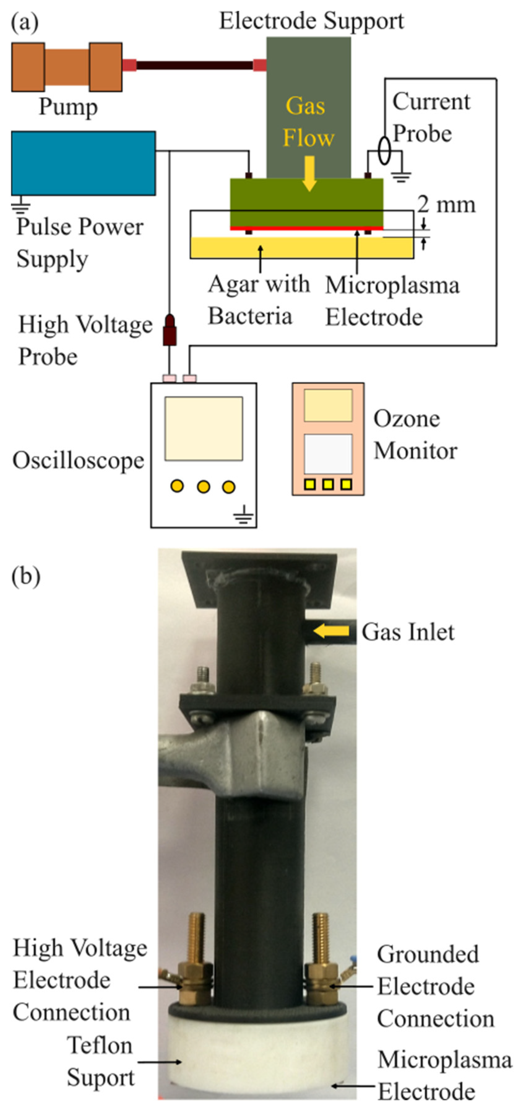
Figure 2.
Microplasma electrode system. (a) Image of the microplasma electrode system with grounded front side electrode and high voltage back side electrode; (b) Schematic image of the cross-section of the microplasma electrode system; (c) Image of the microplasma electrode during discharge.
Figure 2.
Microplasma electrode system. (a) Image of the microplasma electrode system with grounded front side electrode and high voltage back side electrode; (b) Schematic image of the cross-section of the microplasma electrode system; (c) Image of the microplasma electrode during discharge.
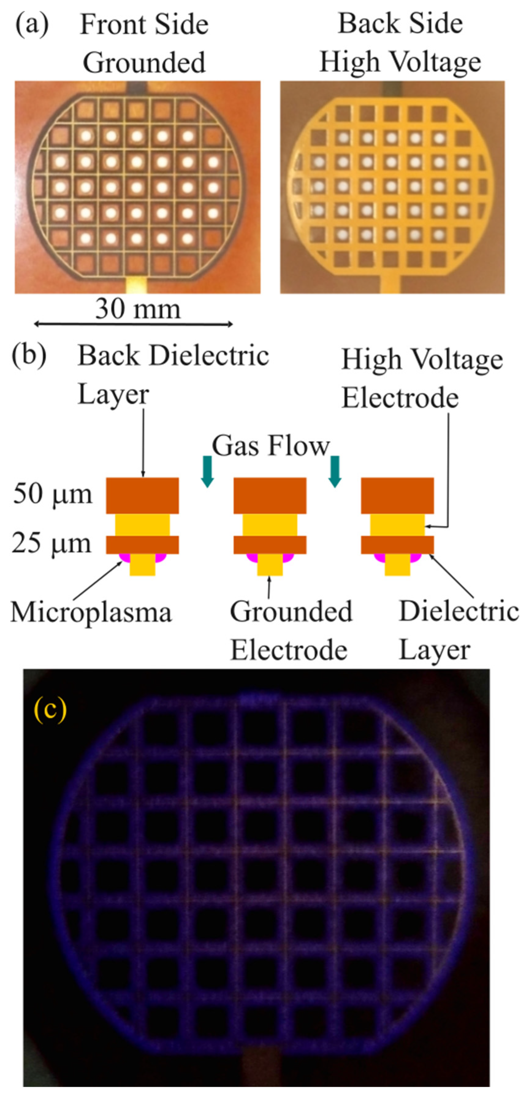
Figure 3.
Waveforms of the discharge voltage and corresponding discharge current.
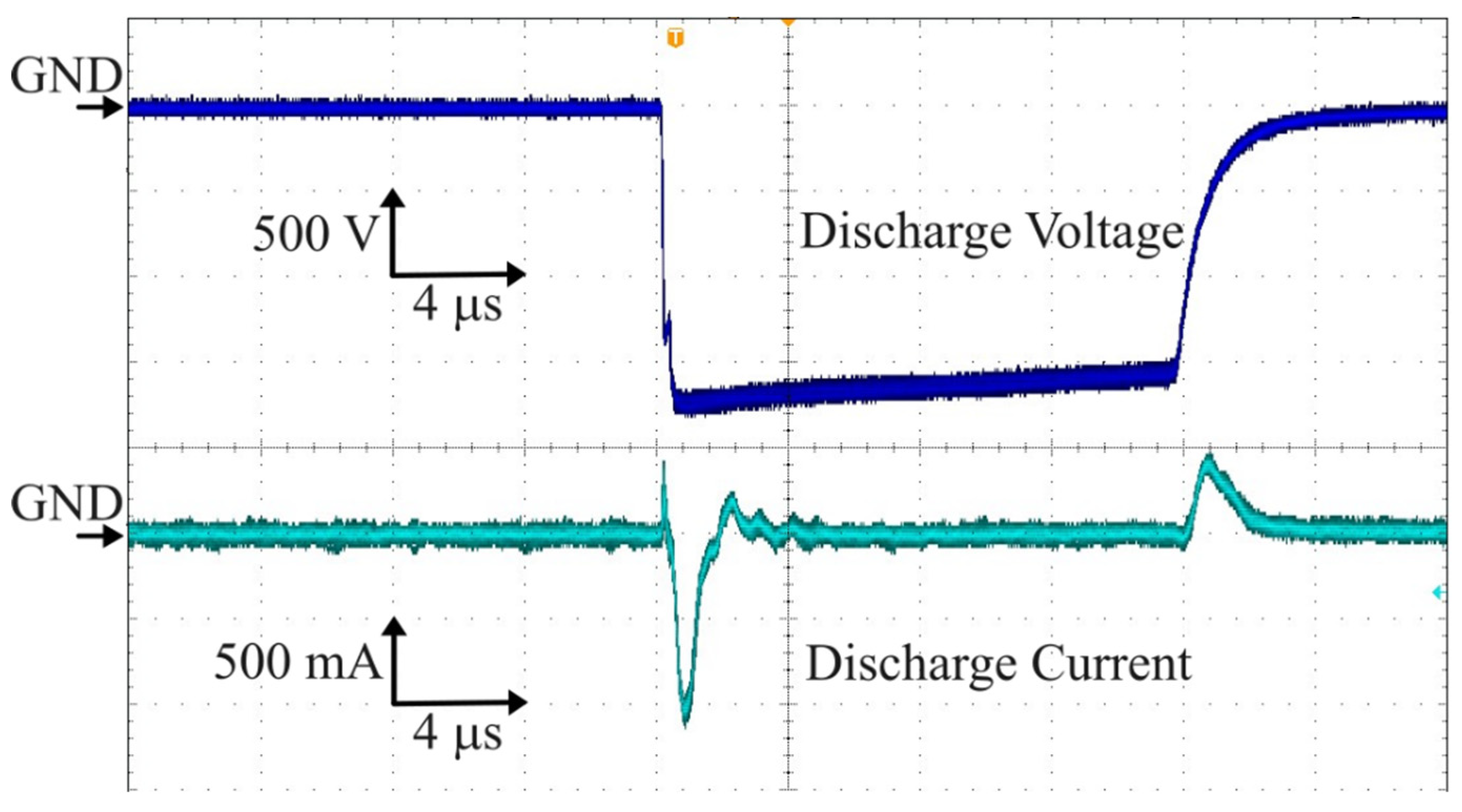
Figure 4.
Ozone concentration increases with discharge voltage.
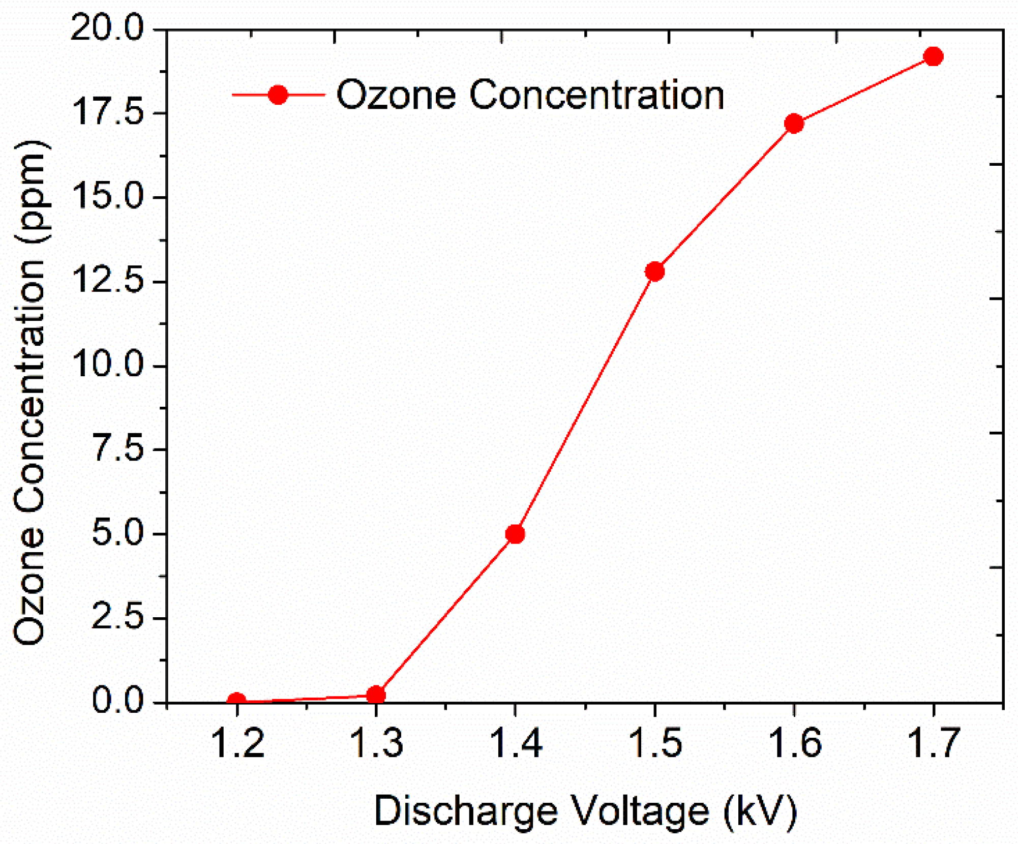
Figure 5.
Representative images of the bacterial strains treated with microplasma; Experimental design 1: bacteria inoculated on agar plates and incubated for 24 h after microplasma treatment; Experimental design 2: 24 h bacteria inoculated on agar plates, treated with microplasma at -1.7 kV for 5 min, and re-incubated for an additional 24 h.
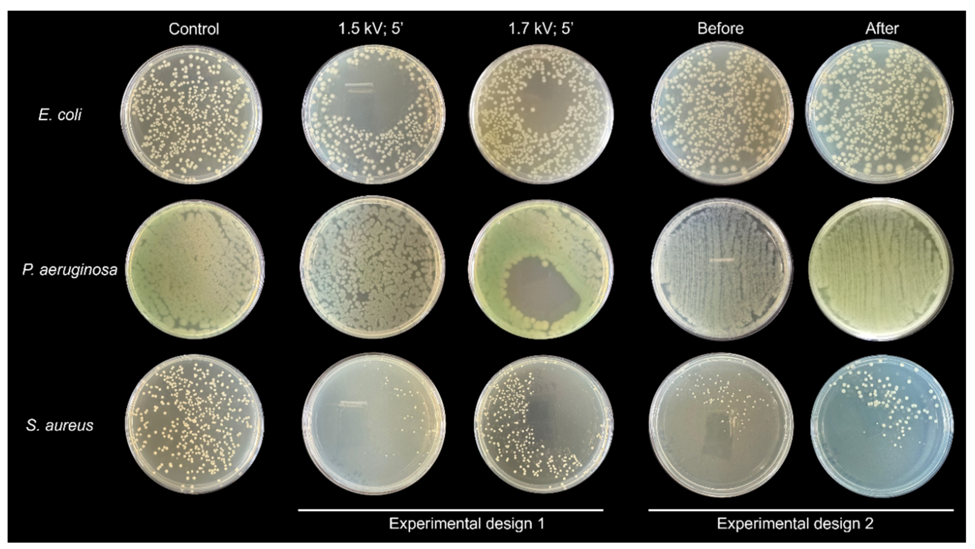
Figure 6.
Inhibition zone measurement for bacterial strains treated with microplasma at -1.7 kV.
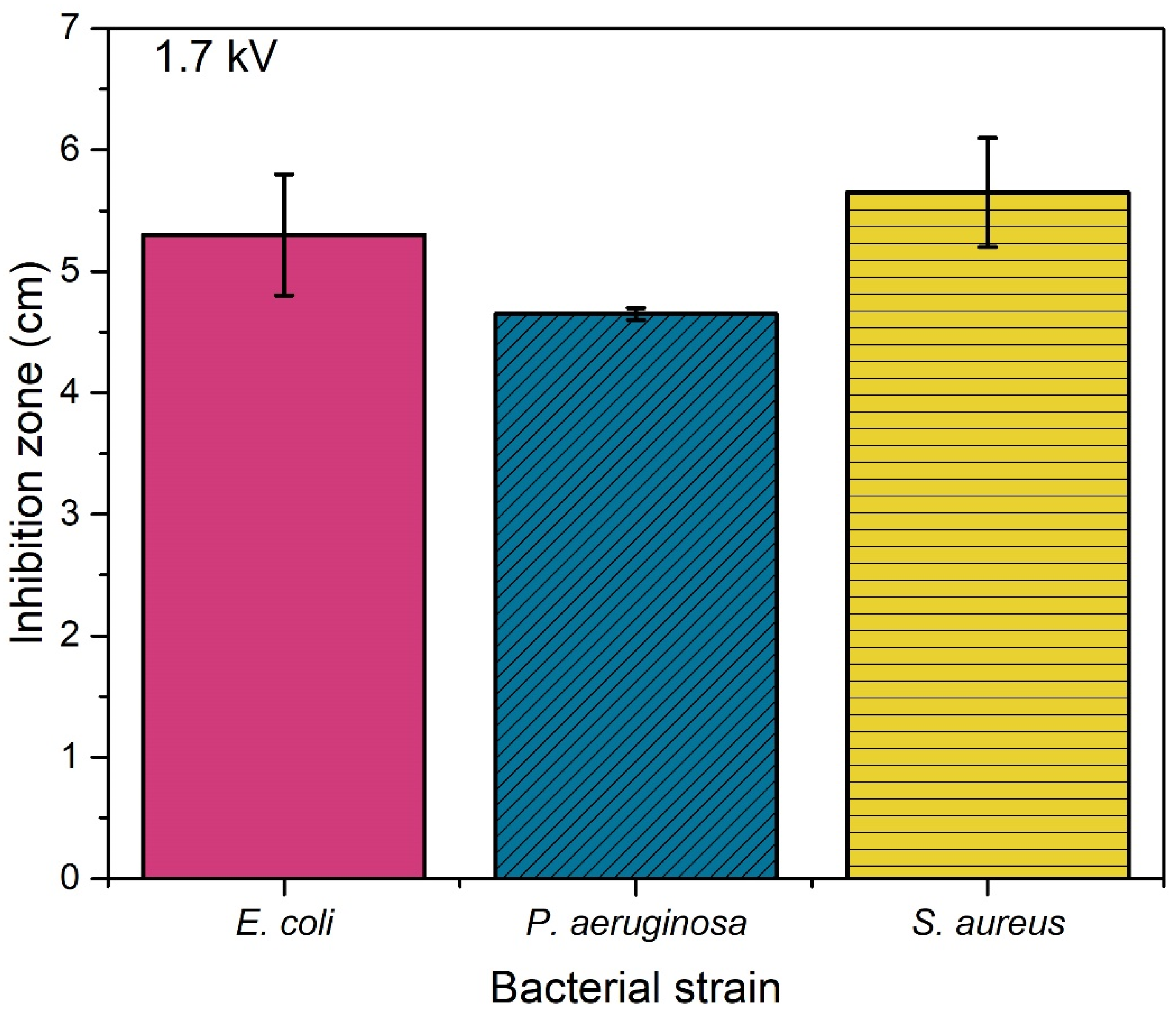

Table 1.
Colony forming units developed after 24 h after microplasma treatment at -1.5 and -1.7 kV.
| CFU | ||||
| -1.5 kV | -1.7 kV | |||
| Treated | Control | Treated | Control | |
| E. coli | 279±77.7 | 348±44.6 | 222.5 ±13.4 | 348±44.6 |
| P. aeruginosa | N.A. | N.A. | N.A. | N.A. |
| S. aureus | 35.5±6.3 | 205±103.9 | 287 ±69.3 | 205±103.9 |
mean ±s.d.; N.A. – not applicable.
Table 2.
D values in Joule after microplasma treatment at -1.5 and -1.7 kV.
| D values | ||||||
| -1.5 kV | -1.7 kV | -1.7 kV 45 mm diameter area |
||||
| Bacterial liquid quantity | 50 µL (kJ/50 µL) | 1 mL (kJ/mL) | 50 µL (kJ/50 µL) | 1 mL (kJ/mL) | 50 µL (kJ/50 µL) | 1 mL (kJ/mL) |
| E. coli | 7.88 | 157.5 | 4.49 | 89.9 | 0.28 | 5.6 |
| P. aeruginosa | N.A. | N.A. | N.A. | N.A. | N.A. | N.A. |
| S. aureus | 0.99 kJ | 19.9 | N.A. | N.A. | N.A. | N.A. |
N.A. – not applicable.
Disclaimer/Publisher’s Note: The statements, opinions and data contained in all publications are solely those of the individual author(s) and contributor(s) and not of MDPI and/or the editor(s). MDPI and/or the editor(s) disclaim responsibility for any injury to people or property resulting from any ideas, methods, instructions or products referred to in the content. |
© 2024 by the authors. Licensee MDPI, Basel, Switzerland. This article is an open access article distributed under the terms and conditions of the Creative Commons Attribution (CC BY) license (https://creativecommons.org/licenses/by/4.0/).
Copyright: This open access article is published under a Creative Commons CC BY 4.0 license, which permit the free download, distribution, and reuse, provided that the author and preprint are cited in any reuse.


