Preprint
Article

This version is not peer-reviewed.

The Role of Individual Residues in the N-Terminus of Arrestin-1 in Rhodopsin Binding

Submitted:

28 November 2024

Posted:

29 November 2024

You are already at the latest version

Abstract

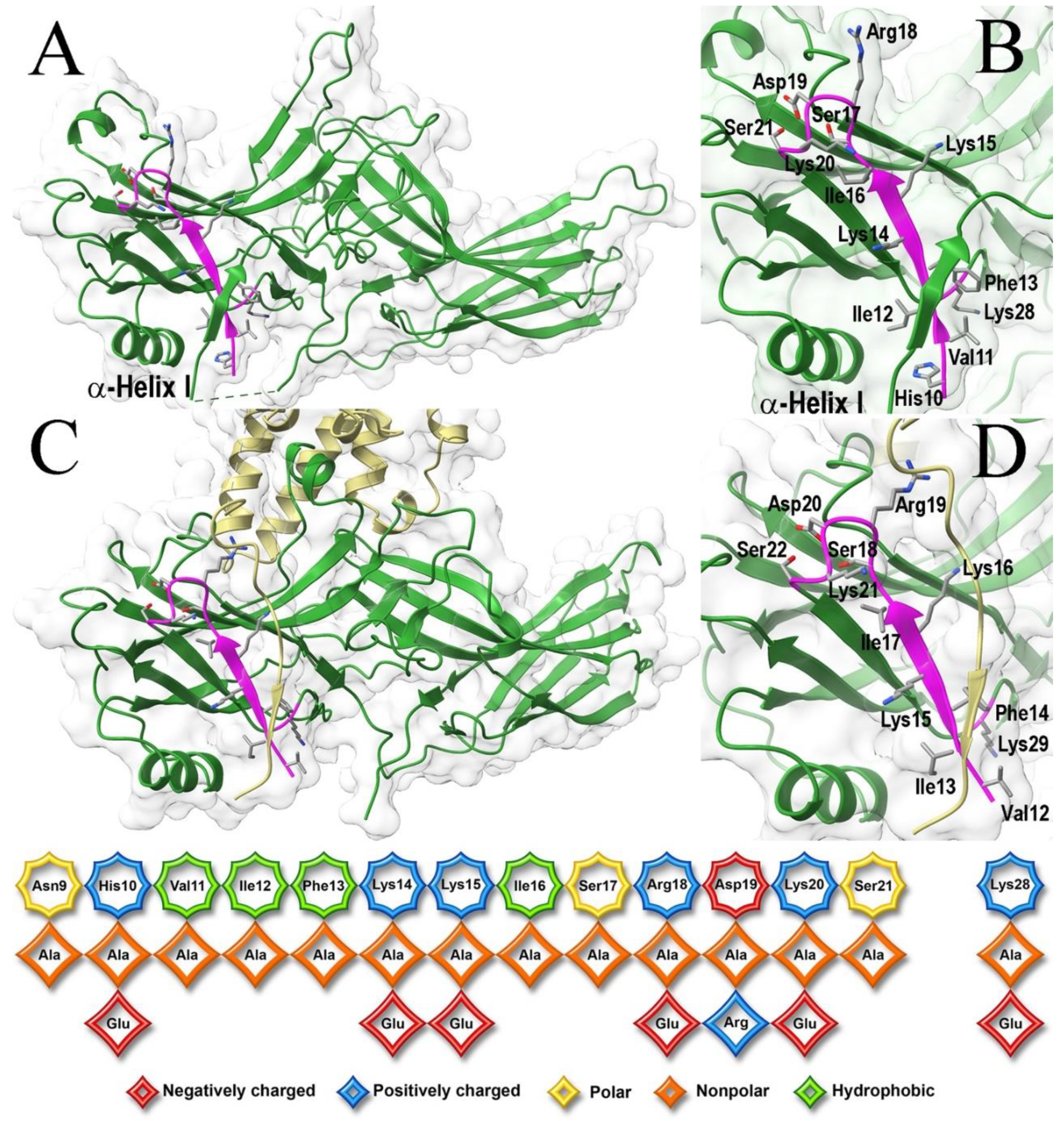
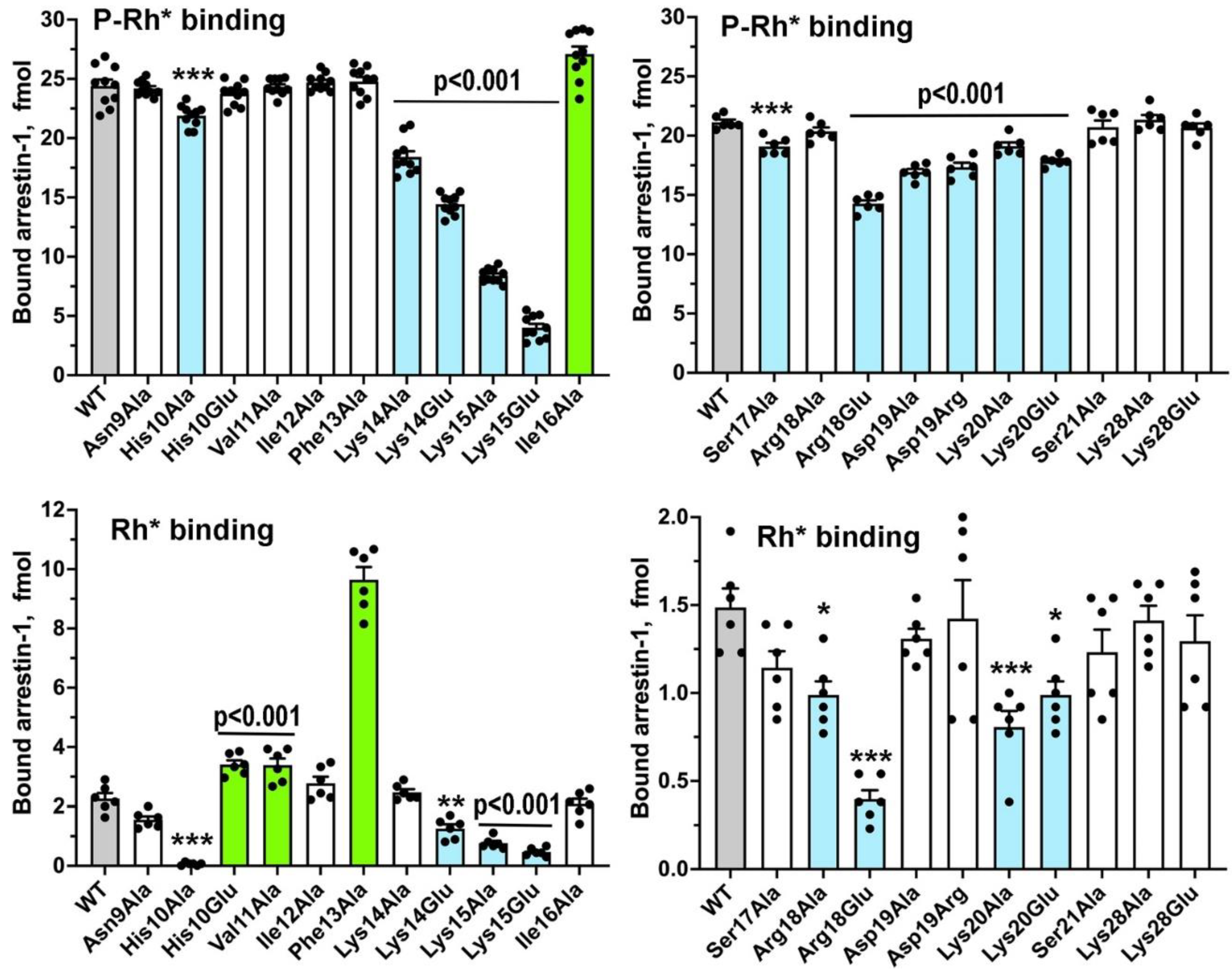
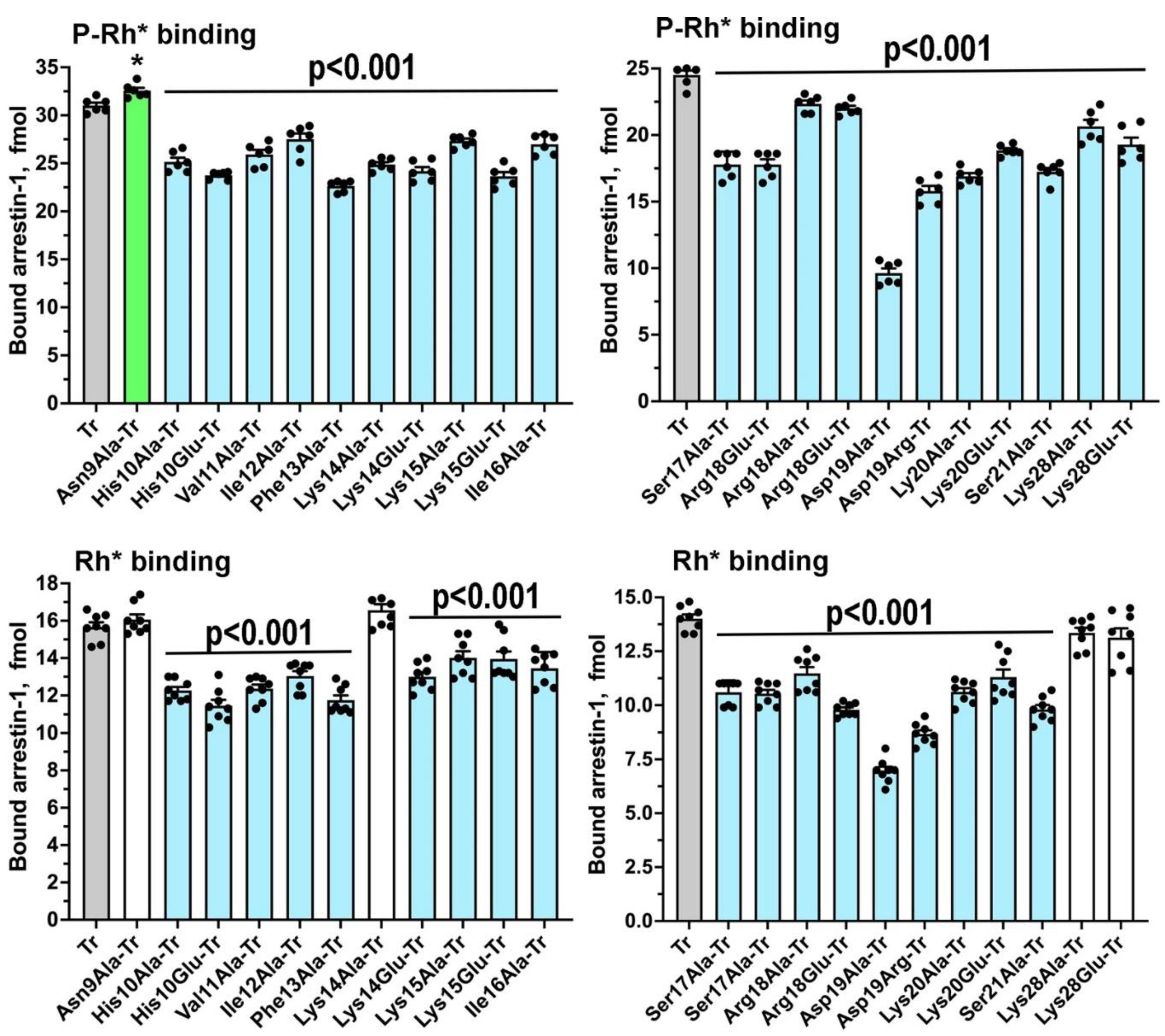
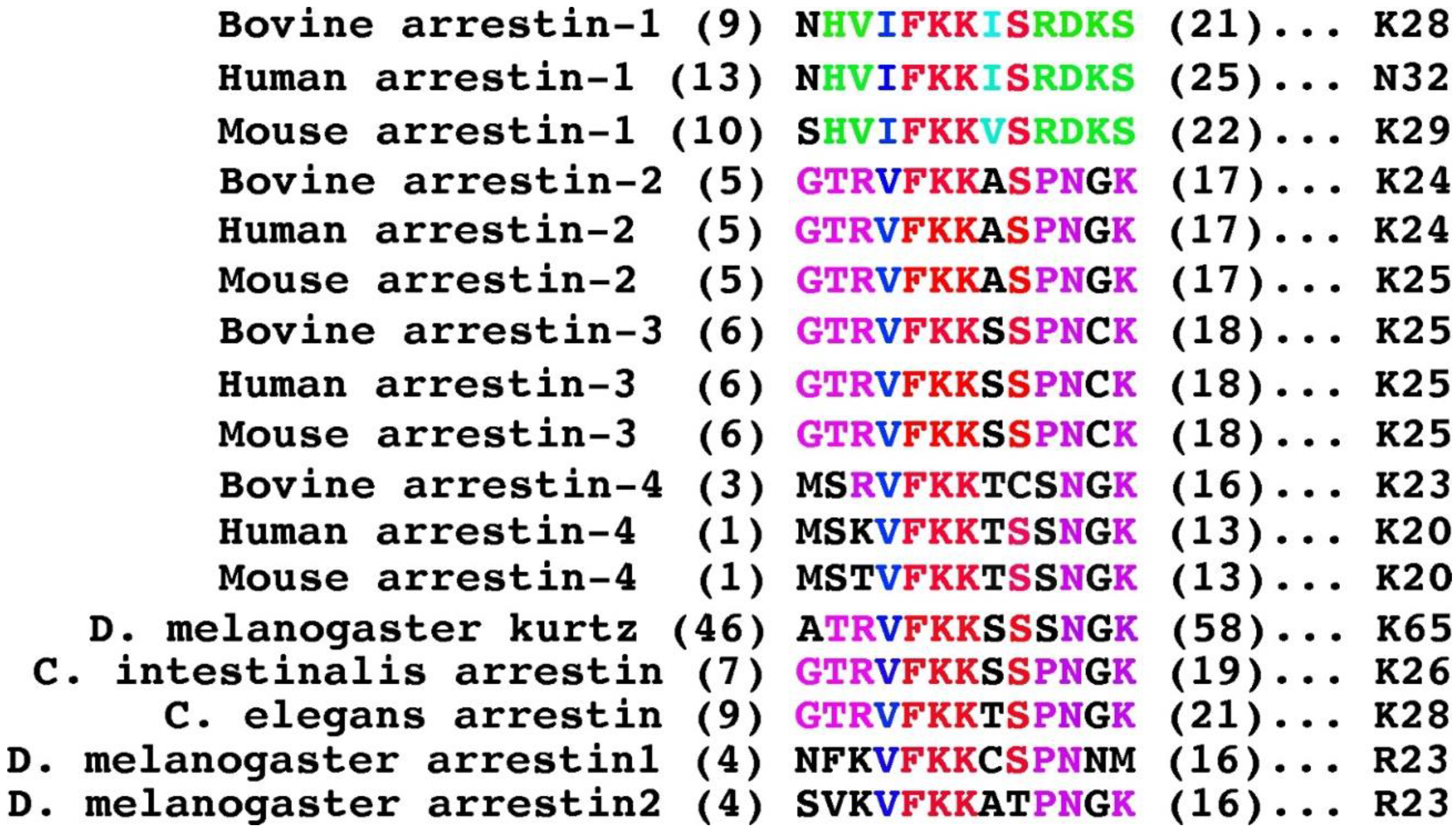
Sequences and three-dimensional structures of the four vertebrate arrestins are very similar, yet in sharp contrast to other subtypes arrestin-1 demonstrates exquisite selectivity for the active phosphorylated form of its cognate receptor, rhodopsin. The N-terminus participates in receptor binding and serves as the anchor of the C-terminus, the release of which facilitates arrestin transition into receptor-binding state. We tested the effects of substitutions of fourteen residues in the N-terminus of arrestin-1 on the binding to phosphorylated and unphosphorylated light-activated rhodopsin of wild type protein and its enhanced mutant with C-terminal deletion that demonstrates higher binding to both functional forms of rhodopsin. Profound effects of mutations identified lysine-15 as the main phosphate sensor and phenylalanine-13 as the key anchor of the C-terminus. These residues are conserved in all arrestin subtypes. Substitutions of five other residues reduced arrestin-1 selectivity, indicating that wild type residues participate in fine-tuning of arrestin- 1 binding to rhodopsin. Differential effects of numerous substitutions in wild type and an enhanced mutant arrestin-1 shows that these two proteins bind rhodopsin differently.

Keywords: 
;  ;  ;  

1. Introduction

Arrestin-1a was the first member of the family discovered [1] and cloned [2]. Arrestin-1 directly competes with the visual G protein transducin [3,4] ensuring rapid and reproducible shutoff of rhodopsin signaling with sub-second kinetics in vivo [5,6,7,8,9,10,11]. After its first non-visual homologue was cloned [12], and shown to play similar role in the signaling of β2-adrenergic receptor [13] the paradigm of two-step homologous desensitization of the family of G protein-coupled receptors (GPCRs) was established: active receptor is phosphorylated by a specific kinase, whereupon an arrestin protein binds to the active phosphoreceptor, shutting off its G protein-mediated signaling [14].
All arrestins bind active phosphorylated GPCRs with significantly higher affinity than unphosphorylated ones (reviewed in [15]). This makes biological sense: the time between the GPCR activation and phosphorylation provides a window of opportunity for G proteins, ensuring that each receptor activation event results in signaling. This is particularly important in rod photoreceptors that respond to single photons [16,17,18]. Arrestin-1 preferentially binds light-activated phosphorylated rhodopsin (P-Rh*) [19], demonstrating 10-20 times lower binding to the light-activated unphosphorylated form (Rh*) [20]. The ability of arrestin-1 to discriminate between the unphosphorylated and phosphorylated form of its cognate receptor is unmatched in the family [21]. Due to this unique feature, the “coincidence detector” model explaining how arrestins discriminate among functional forms of a receptor was first developed based on the studies of arrestin-1 [20]. The model posits that the transition into a high-affinity receptor-binding state is triggered by simultaneous engagement of two sensors, one recognizing receptor-attached phosphates and the other responding to active GPCR conformation [20]. This model appears to be valid for all arrestins [15]. The arrestin N-terminus plays a critical role in receptor binding: it contains a pair of lysines serving as the phosphate sensor [22,23] and the anchor of the C-terminus [24,25,26,27,28], the release of which accompanies arrestin binding to receptors [29,30,31,32,33,34,35,36,37]. Thus, a comprehensive analysis of this part of the arrestin molecule is necessary for two reasons: first, to identify specific structural features of arrestin-1 underlying its unique selectivity; second, to determine the functional role of residues conserved among arrestin subtypes that are likely involved in arrestin-receptor interaction and contribute to phosphorylation recognition. Here we tested the role of the arrestin-1 N-terminal residues in its binding to P-Rh*, with a particular interest in their role in its remarkable preference for this functional form of rhodopsin over Rh*, focusing on the part of the N-terminus conserved in evolution (positions 9-21 in bovine protein; Figure 1).

2. Results

Here we targeted the conserved part of the N-terminus, as well as Lys28 located near this element in the folded arrestin-1 in its basal and rhodopsin-bound conformation (Figure 1 B,D). In addition to conventional alanine scanning, we introduced charge reversals of seven residues: six positively charged Lys, Arg, and His were replaced by negatively charged Glu; negatively charged Asp was replaced by positively charged Arg. Thus, the total number of mutations was 21 (Figure 1, bottom panel). All mutants were tested on the background of WT bovine arrestin-1, as well as on the background of its C-terminally truncated form 1-378 (Tr) with enhanced binding to both P-Rh* and Rh* [20]. Functional importance of the targeted element is supported by the finding that the binding to P-Rh* and Rh* was significantly affected by 12 and 11 mutations, respectively, on WT background (Figure 2), as well as by all 21 and 17 mutations, respectively, on Tr background (Figure 3).
The N-terminal-most mutation, Asn9Ala, produced the smallest effect: a slight increase of the Tr binding to P-Rh* and nothing else (Figure 2 and Figure 3), consistent with the idea that the distal N-terminus, which faces away from bound receptors in all solved structures of the complexes [29,30,31,32,33,34,35,36,37,38], does not play a role in the arrestin-GPCR interaction. Functional roles of positively charged His10, Arg18, Lys20, and Lys28 were never tested before. His10Ala reduced the binding to P-Rh* and Rh* on both backgrounds, whereas the effects of placing a negatively charged glutamate in this position were less severe: while the binding on the Tr background was negatively affected, His10Glu did not appreciably change P-Rh* binding and increased Rh* binding of WT arrestin-1 (Figure 2 and Figure 3). This suggests that His-10 contributes to arrestin-1 selectivity for P-Rh* and that upon rhodopsin binding its side chain is likely involved in H-bonding, rather than in a charge-charge interaction. Arg18Glu mutation reduced the binding to P-Rh* and Rh* on both backgrounds, whereas Arg18Ala substitution was detrimental only for the binding of Tr (Figure 2 and Figure 3). More severe effect of charge reversal suggests that in WT arrestin-1 this residue likely interacts with a negatively charged partner in rhodopsin. Its effect on Rh* binding indicates that its partner is not a rhodopsin-attached phosphate, but a negatively charged side chain of one of rhodopsin residues (Glu or Asp). Lys20Ala and Lys20Glu mutations reduced the binding of WT and Tr arrestin-1 to both forms of rhodopsin (Figure 2 and Figure 3), also suggesting that the interaction partner of this lysine is not a phosphate on rhodopsin, which is present only in P-Rh*. Although homologous residue in mouse arrestin-1 was found in the vicinity of one of the phosphates in P-Rh* in the structure of the complex [30], our data indicate that this lysine is not an important phosphate-binding residue. Lys28Ala and Lys28Glu mutations reduced only the binding of Tr to P-Rh*, suggesting that this residue does not play a significant role in the interaction of WT arrestin-1 with rhodopsin. The lack of conservation of this lysine in human arrestin-1 (Figure 4) is consistent with this conclusion.
Severe negative effects of the substitutions of Lys15, which were much stronger in WT arrestin-1 than in its Tr form (Figure 2 and Figure 3) demonstrate that it is one of the key players in rhodopsin binding and arrestin-1 transition into high-affinity rhodopsin-binding state. Substitutions of Lys14 were also detrimental, although less so than substitutions of Lys15 (Figure 2 and Figure 3). These data support the idea that this pair of lysines serves as the phosphate sensor of arrestin-1 [23]. More important role of Lys 15 than that of Lys14 is consistent with the finding that in the complex of mouse arrestin-1 with rhodopsin homologous Lys16 interacts with two rhodopsin-attached phosphates, whereas preceding Lys 15 (homolog of bovine Lys14) does not, although in the structure it contacts negatively charged rhodopsin residue Glu341 [30].
Elimination of the bulky hydrophobic side chain in position 16 by Ile16Ala mutation slightly increased the binding of WT arrestin-1 to P-Rh*, and somewhat decreased the binding of Tr form to both P-Rh* and Rh* (Figure 2 and Figure 3). Relatively small effects suggest that homologs of Ile16 do not play an important role in arrestin binding to receptors, consistent with the lack of conservation of the chemical nature of the residue in this position in different arrestin subtypes (Figure 4). The next residue, Ser17 in bovine arrestin-1, is conserved in the family (Figure 4). Its replacement with alanine was detrimental for the binding of WT and Tr arrestin-1 to both functional forms of rhodopsin (Figure 2 and Figure 3). In contrast, alanine substitution of Ser21, which is not conserved (Figure 4), did not affect the binding of WT arrestin-1, although it somewhat reduced the binding of Tr (Figure 2 and Figure 3). Unexpectedly, elimination of the side chain of Asp19 and charge reversal at this position had negative effects on WT arrestin-1 binding to P-Rh*, but not Rh* (Figure 2), suggesting an important role of the WT aspartate in arrestin-1 selectivity for P-Rh*. Negative effects of both substitutions of Asp19 on Tr background were significantly stronger (Figure 3). The finding that Asp19Ala mutation in Tr was more detrimental than Asp19Arg (Figure 3) suggests that the side chain of this residue participates in hydrogen bonding, rather than in charge-charge interactions with rhodopsin.
While three consecutive hydrophobic residues (Val11-Ile12-Phe13 in bovine arrestin-1) were hypothesized to serve as an anchor of the arrestin C-terminus by crystal structures of all arrestin subtypes [24,25,27,28,52], as well as by the functional testing [23,53], the role of individual residues in this cluster was not investigated. Judging by strong positive effects of alanine substitutions in WT arrestin-1 on Rh* binding (Figure 2), Phe13 appears to be critical, Val11 is much less important, whereas Ile12 does not play a significant role. This conclusion is consistent with strict conservation of Phe in homologous positions in other arrestin subtypes (Figure 4). The presence of bulky hydrophobic residue in the preceding position in all arrestins (Figure 4) also indicates its importance, although the data (Figure 2) suggest that this residue does not play a role in receptor binding. Interestingly, alanine substitutions of all three residues somewhat reduced the binding of Tr to both forms of rhodopsin (Figure 3). Conceivably, these residues directly participate in rhodopsin binding of the mutant, but not of the WT protein.

3. Discussion

Members of the arrestin family of proteins, like GPCRs, are present in all animal species. Mammals express four arrestin subtypes [54]. Arrestin-1 binding to P-Rh* is necessary for quenching light-induced rhodopsin signaling with sub-second kinetics [5,7,55], which ensures exceptional time resolution of rod photoreceptors [56,57,58,59,60]. Arrestin-4 binds cone opsins, playing a similar role in the shutoff of light response in cone photoreceptors [61]. Although the expression of arrestin-1 in cones is ~50 times higher, arrestin-4 is responsible for about half of this function [61]. Arrestin-2 and -3 are key players in homologous desensitization of most non-visual GPCRs [14,62]. GPCRs are the largest family of signaling proteins in animals [63] responding to a variety of stimuli, from hormones and neurotransmitters to light, odorants, peptides, proteins, extracellular calcium, etc. [64]. Humans express ~800 different GPCR subtypes and ~30% of clinically used drugs target various GPCRs [65,66,67,68,69]. In rhodopsin, inherent flexibility of seven-helix GPCR core [70,71,72] is suppressed by covalent binding of 11-cis-retinal, an inverse agonist, which ensures extremely low noise [57]. Exceptional selectivity of arrestin-1 for P-Rh* [21,55] also contributes to proper function of rod photoreceptors. Thus, understanding the molecular mechanisms of arrestin-mediated regulation of GPCR signaling, as well as the structural basis of unique selectivity of the arrestin-1 branch of the family, is biologically important.
All arrestins are elongated two-domain molecules with the C-terminus coming back from the C-domain and forming a strong hydrophobic interaction with the N-domain, in which the N-terminus and α-helix I participate [24,25,26,27,28,52,73] (Figure 1A). This three-element interaction and the polar core between the two arrestin domains (both terms from [24]) are the two critical intramolecular interactions holding arrestins in their basal state. Both need to be destabilized for arrestin transition into the receptor-binding conformation [74]. The first few N-terminal residues are not resolved in the structures of arrestin-1 [24,52,73] and other subtypes [25,26,27,28], as well as in receptor-bound arrestin-1 [29,30], -2 [31,32,33,34,35,36,37,75], and -3 [75], suggesting that this part does not have a preferred conformation in the basal or receptor-bound state. The length of the N-terminus (note the difference in the position of starting residue in Figure 4) and the identity of the first residues in it are not conserved in evolution [54], suggesting that these features are unlikely to be important for the function shared by all arrestins, binding cognate GPCRs. The fact that in the arrestin-rhodopsin complex the distal N-terminus faces away from the receptor [29,30] is consistent with this notion. Although the role of the conserved part of the arrestin-1 N-terminus (Figure 1) in P-Rh* binding is not in doubt, the effects of few mutations were tested: triple mutation Val11Ala+Ile12Ala+Phe13Ala, two point mutations (Lys14Ala and Lys15Ala) (the numbering corresponds to the bovine protein) [22,53]. The effects of alanine substitutions in this element on P-Rh* [76] and P-opsin [77] were tested only using a relatively low sensitivity assay. The role of individual hydrophobic side chains of residues 11-13 in the anchoring of the C-terminus to the N-domain has not been elucidated. Lysines 14 and 15 were proposed to serve as the phosphate sensor [22,23], but relative roles of individual lysines in this pair was not unambiguously established.
Traditional belief that the only possible function of residues exposed on the arrestin-1 surface is participation in rhodopsin binding was challenged by recent studies [78,79] revealing other functions: some residues specifically suppress the binding to Rh*, thereby increasing arrestin-1 selectivity for P-Rh*, others participate in receptor binding of enhanced mutants but not of WT protein. Our experiments yielded several new insights into the role of individual N-terminal residues. Three appear to be largely irrelevant for rhodopsin binding of WT arrestin-1: Asn9, Ser21, and Lys28 (Figure 2). In contrast, His10, Ser17, Arg18, Asp19, and Lys20, that were not implicated in rhodopsin binding before, were found to participate in the interaction (Figure 2). Selective effect of Asp19 substitutions on the binding to P-Rh*, but not to Rh*, suggests that this aspartate likely interacts with its partner on rhodopsin only upon receptor phosphorylation. As negatively charged Asp cannot bind receptor-attached phosphates, this implies that the rhodopsin C-terminus (containing all phosphorylation sites) changes its conformation after the phosphates are attached, so that Asp19 partner inaccessible in Rh* becomes accessible upon rhodopsin phosphorylation. This putative partner is not necessarily located on the C-terminus, as the conformational change in it would affect accessibility of residues on other cytoplasmic parts of rhodopsin (i.e., three intracellular loops). In crystal structure of the arrestin-1 complex with rhodopsin, this aspartate is located far from resolved rhodopsin elements [30]. However, solved structure contained mutant human rhodopsin (two activating mutations E113Q and M257Y, plus N2C and N282C to form a stabilizing disulfide bond absent in the WT protein) and fused mouse arrestin-1-(10-392) with triple alanine substitution in the C-terminus (L374A, V375A, F376A). The effects of Asp19 mutations suggest that the position of WT arrestin-1 relative to bound WT rhodopsin is not the same as in the solved structure.
Previous studies suggested that the two lysines (Lys14 and Lys15 in bovine arrestin-1) strictly conserved in the family [54] (Figure 4), interact with rhodopsin-attached phosphates [22,23]. Our data identified Lys15 as the main phosphate sensor, whereas Lys14 is less important (Figure 2). The three hydrophobic residues preceding these lysines in the linear sequence (Val11, Ile12, Phe13) were probed as a group, but not individually [80]. Crystal structure of arrestin-1 in its basal conformation [24] as well as functional evidence [23] shows that these residues, along with the three leucines in the α-helix I (Figure 1A), anchor the arrestin-1 C-terminus in the basal state. Alanine substitution of individual residues showed that Phe13 is a critical anchor, possibly assisted by Val11, whereas Ile12 does not play this role (Figure 2). Phenylalanine in position homologous to Phe13 is strictly conserved in the arrestin family [54] (Figure 4). The preceding position is invariably occupied by a hydrophobic residue with a bulky side chain, but the first residue in this triplet is not conserved (Figure 4). Conceivably Ile12 and its homologs in other arrestins are important for the folding and/or other functions, while the homologs of Phe13 are sufficient to anchor the arrestin C-terminus. Juxtaposition of the key phosphate sensor Lys15 and key anchor of the C-terminus Phe13 (Figure 1) suggests the molecular mechanism whereby receptor-attached phosphate(s) trigger the release of the C-terminus (anchored to the N-domain in the basal state of all arrestins [24,25,27,28]), which is the key event triggering arrestin transition unto receptor binding conformation [74]. Apparently, even a small shift induced by the phosphate interaction with Lys15 would move Phe13 out of position favorable for holding the C-terminus in place. The data identified seven residues in the arrestin-1 N-terminus (His10, Val11, Phe 13, Lys 15, Ser17, Asp19, and Lys20) that enhance its selectivity for P-Rh*. Importantly, four of these (His10, Val11, Asp19, and Lys20) are specific for the mammalian arrestin-1 proteins (Figure 4). Thus, this study identified several key N-terminal residues (Figure 2) that, along with previously identified residues [23,78,79,81], ensure exquisite selectivity of arrestin-1 for P-Rh*
Significant differences of the effects of ten (out of 21 tested) point mutations (His10Glu, Val11Ala, Ile12Ala, Phe13Ala, Ile16Ala, Ser17Ala, Arg18Ala, Ser21Ala, Lys28Ala, and Lys28Glu) on rhodopsin binding of WT arrestin-1 and its Tr form (compare Figure 2 and Figure 3) support the idea that WT arrestins and enhanced mutants bind GPCRs differently [23,78,79,81]. Various mutants were used in virtually all structural work: arrestin-2 with activating polar core mutation Arg169Glu in structures with β1-adrenergic [35] and 5HT2B serotonin [32] receptors, arrestin-1-(10-392) with triple alanine substitution in the C-terminus (that detaches it from the N-domain) with rhodopsin [29,30], arrestin-2 with homologous triple alanine substitution with neurotensin NTS1 [37] receptor, arrestin-2 with various deletions in the C-terminus with M2 muscarinic [36], V2 vasopressin [31], neurotensin NTS1 [37], and β1-adrenergic [38] receptors, etc. Functional analysis of arrestin-1 interaction with rhodopsin suggests that solved structures of the arrestin-receptor complexes utilizing enhanced arrestins and mutant receptors do not necessarily reveal how WT arrestins bind cognate WT receptors. Distance measurements between rhodopsin and bound arrestin-1 [29,30], as well as in-cell study of arrestin-2 interactions with parathyroid hormone receptor PTH1R [82] show that the complex of the same arrestin with the same receptor has multiple shapes, only one of which is revealed by the structures. Direct binding assay reports the sum of all binding modes. Thus, detected differences between WT arrestin-1 and its Tr mutant (compare Figure 2 and Figure 3) do not mean that WT protein does not bind rhodopsin the way Tr does. The results show that in case of WT protein Tr-like binding mode is responsible for a smaller fraction of the population of complexes than can be detected by statistical analysis.
Functionally important residues are usually conserved in evolution [83]. In the mammalian arrestin-1 proteins eleven out of fourteen residues tested are identical, with one additional conservative substitution (Ile vs Val after the pair of lysines) (Figure 4). Only two residues are not conserved, the homologs of Asn9 and Lys28. Indeed, their substitutions did not affect rhodopsin binding of WT arrestin-1 (Figure 2). The sequences of the N-termini of human, bovine, and mouse arrestin-2 and -3 are significantly different from arrestin-1. The N-terminal regions of the two non-visual subtypes are virtually identical, with only a single difference: Gly in arrestin-2 corresponds to Cys in arrestin-3 (Figure 4). As Gly occupies this position in “ancient” arrestins (Figure 4), it is likely the original, whereas Cys is a specific acquisition of the arrestin-3 branch. The sequence of cone-specific arrestin-4 from the three mammalian species is closer to that of arrestin-2 and -3 than to arrestin-1. This is likely one of the reasons why arrestin-4 is less selective for active phosphorylated receptors, similar to non-visual arrestin-2 and -3 [27]. The first three residues, Gly-Thr-Arg, are conserved in non-visual and “ancient” arrestins (magenta in Figure 4), but strikingly different in visual subtypes: mammalian arrestin-1, -4 and arrestin1 and 2 from Drosophila (Figure 4). Functional role of this element in non-visual subtypes remains to be elucidated. Four residues are conserved in virtually all arrestin proteins (red in Figure 4). The only exceptions are bovine arrestin-4 where Ser is replaced with its geometrical (but not chemical) analog Cys and Drosophila arrestin2, where the same Ser is replaced by chemically similar Thr (Figure 4). In addition, one substitution is conservative: Ile in arrestin-1 corresponds to Val in all other arrestins (blue in Figure 4). Interestingly, Phe preceding the two lysines and the second residue after them, Ser, are conserved even in species far removed from vertebrates: round worm C. elegans, tunicate C. intestinalis, and fly Drosophila (in Figure 4). All four conserved residues play important roles in receptor binding: main anchor of the C-terminus (Phe13), main (Lys15) and auxiliary (Lys14) phosphate sensors, as well as Ser17 that was implicated in arrestin-rhodopsin interaction for the first time by this study. Conservation of the phosphate sensor and the anchor of the C-terminus is consistent with the idea that all arrestins use similar molecular mechanism of transitioning into receptor-binding conformation. Although arrestins are required for the shutoff of rhodopsin signaling in Drosophila photoreceptors [84], the binding of arrestin in flies is triggered by rhodopsin activation and does not require its phosphorylation [85,86,87]. The conservation of lysines that serve as phosphate sensors in vertebrate arrestins (Figure 4) suggests that negatively charged side chain(s) in fly rhodopsin become accessible upon its activation and “attract” arrestin the way the phosphates do in vertebrates. The structure of the complex of one of Drosophila visual arrestins with rhodopsin is necessary to test this hypothesis. The reason for the conservation of several other residues in arrestins from non-vertebrate species, the sequences of which are closer to mammalian non-visual subtypes than to arrestin-1 (Figure 4), remain to be elucidated. Our data suggest that Arg18, Asp19, and Lys20, which are conserved in mammalian arrestin-1 proteins, are necessary for the binding of rhodopsin: their substitutions significantly reduced P-Rh* binding (Figure 2 and Figure 3). In arrestin-2, -3, and -4 this sequence is replaced with Pro-Asn-Gly, Pro-Asn-Cys, and Ser-Asn-Gly, respectively. This element in C. intestinalis and C. elegans proteins is the same as in vertebrate arrestin-2, whereas in Drosophila non-visual arrestin kurtz and visual arrestin2 it is the same as in human and mouse arrestin-4; Drosophila arrestin1 has Pro-Asn-Asn sequence in this place (Figure 4). As these species are very far removed from mammals, this indicates that only limited variability of functionally important elements is allowed by evolution. As these three residues play a role in rhodopsin binding of arrestin-1 (Figure 2), it is likely that this element in other arrestins participates in their binding to cognate GPCRs. Its function, as well as the role of Gly vs Cys difference between arrestin-2 and -3, needs to be tested experimentally.
To summarize, we showed that four residues conserved in all arrestins are critical for receptor binding and that these plus four additional residues specific for mammalian arrestin-1 proteins enhance the selectivity of this subtype for P-Rh*.

4. Materials and Methods

Materials. Radiolabeled [γ-32P]ATP and [14C]leucine were from Perkin-Elmer (Waltham, MA). Restriction endonucleases, Vent DNA polymerase, and Quick T4 DNA ligase were from New England Biolabs (Ipswich, MA). Rabbit reticulocyte lysate was made in bulk by Ambion (Austin, TX). SP6 RNA polymerase was expressed in E. coli and purified, as described [88]. DNA purification kits for mini (3 ml of bacterial culture), midi (50 ml) and maxi (100 ml) preparations were from Zymo Research (Irvine, CA). All other reagents were from Sigma-Aldrich (St. Louis, MO).
Mutagenesis and plasmid construction. For in vitro mRNA synthesis bovine arrestin-1 was subcloned into modified pGEM2 vector (Promega; Madison, WI) with “idealized” 5’-UTR that does not require a cap for efficient translation [88] between Eco RI and Hind III sites, as described [79,81]. Mutations were introduced by PCR. Two restriction sites, Eco RI preceding “idealized” 5’-UTR and Bam HI in the coding sequence of bovine arrestin-1 [88] were used to subclone mutant fragments generated by PCR. Mutations were confirmed by dideoxy sequencing (GenHunter Corporation, Nashville, TN). Eco RI – Bam HI fragments (164 bp) containing mutations were excised from WT constructs and subcloned into pGEM2-based construct encoding arrestin-1-(1-378) (Tr) mutant with much higher binding to Rh* [79,81].
In vitro transcription, translation, calculation of specific activity of produced arrestin proteins, and preparation of different functional forms of rhodopsin were performed as described recently [79,81].
Direct binding assay was performed, as described [20]. Briefly, 1 nM arrestin-1 (50 fmol, specific activity 10.9 – 12.9 dpm/fmol) in the translation mix from which ribosomes were removed by high-speed centrifugation was incubated with 0.3 μg of indicated functional forms of rhodopsin (P-Rh* or Rh*) in 50 μl of 50 mM Tris-HCL, pH 7.4, 100 mM potassium acetate, 1 mM EDTA, 1 mM DTT for 5 min at 37oC under room light. After incubation the samples were cooled on ice for 1-2 min, then bound to rhodopsin-containing membranes arrestin-1 was separated from free arrestin-1 and unincorporated [14C]-leucine present in the translation mix at 4oC by gel-filtration on 2-ml Sepharose 2B-CL column. Radiolabeled arrestin-1 eluting with rhodopsin was quantified by liquid scintillation counting (Tri-Carb; PerkinElmer, Waltham, MA). Relatively small (<10% of the total) non-specific “binding” (likely reflecting arrestin-1 aggregation during the assay) was determined in samples without rhodopsin and subtracted.
Data Analysis and Statistics. Statistical analysis was performed by one-way ANOVA (analysis of variance) with post-hoc Dunnett’s comparison to WT or Tr, respectively, with correction for multiple comparisons using Prism 9 software (GraphPad, Boston, MA). P values < 0.05 were considered statistically significant and indicated directly when applied to several values or as follows: *p < 0.05; **, p<0.01; ***p < 0.001.
Footnote
aWe use systematic names of arrestin proteins, where the number after the dash indicates the order of cloning: arrestin-1 (historic names S-antigen, 48 kDa protein, visual or rod arrestin; SAG in HUGO database), arrestin-2 (β-arrestin or β-arrestin1; ARRB1 in HUGO database), arrestin-3 (β-arrestin2 or hTHY-ARRX; ARRB2 in HUGO database), and arrestin-4 (cone or X-arrestin; ARR3 in HUGO database).

Author Contributions

Conceptualization, S.A.V and V.V.G.; formal analysis, S.A.V. and E.V.G.; investigation, S.A.V. and T.P.; writing—original draft preparation, S.A.V. and V.V.G.; writing—review and editing, S.A.V., V.V.G., and E.V.G.; visualization, S.A.V. and V.V.G.; supervision, V.V.G.; funding acquisition, V.V.G. All authors have read and agreed to the published version of the manuscript.

Funding

This research was funded by NIH grant RO1 EY011500 and Cornelius Vanderbilt Chair, Vanderbilt University (V.V.G.). The APC was funded by Cornelius Vanderbilt Chair, Vanderbilt University.

Data Availability Statement

The data are presented in the manuscript. Raw binding data obtained in each experiment are available upon request.

Conflicts of Interest

The authors declare no conflict of interest.

References

  1. Kuhn, H. , Light-regulated binding of rhodopsin kinase and other proteins to cattle photoreceptor membranes. Biochemistry 1978, 17, 4389–4395. [Google Scholar] [CrossRef] [PubMed]
  2. Shinohara, T.; Dietzschold, B.; Craft, C.M.; Wistow, G.; Early, J.J.; Donoso, L.A.; Horwitz, J.; Tao, R. , Primary and secondary structure of bovine retinal S antigen (48 kDa protein). Proceedings of the National Academy of Sciences 1987, 84, 6975–6979. [Google Scholar] [CrossRef] [PubMed]
  3. Wilden, U. , Duration and amplitude of the light-induced cGMP hydrolysis in vertebrate photoreceptors are regulated by multiple phosphorylation of rhodopsin and by arrestin binding. Biochemistry 1995, 34, 1446–1454. [Google Scholar] [CrossRef] [PubMed]
  4. Krupnick, J.G.; Gurevich, V.V.; Benovic, J.L. , Mechanism of quenching of phototransduction. Binding competition between arrestin and transducin for phosphorhodopsin. J Biol Chem 1997, 272, 18125–18131. [Google Scholar] [CrossRef] [PubMed]
  5. Xu, J.; Dodd, R.L.; Makino, C.L.; Simon, M.I.; Baylor, D.A.; Chen, J. , Prolonged photoresponses in transgenic mouse rods lacking arrestin. Nature 1997, 389, 505–509. [Google Scholar] [CrossRef]
  6. Chen, C.K.; Burns, M.E.; Spencer, M.; Niemi, G.A.; Chen, J.; Hurley, J.B.; Baylor, D.A.; Simon, M.I. , Abnormal photoresponses and light-induced apoptosis in rods lacking rhodopsin kinase. Proc Nat Acad Sci USA 1999, 96, 3718–3722. [Google Scholar] [CrossRef]
  7. Mendez, A.; Burns, M.E.; Roca, A.; Lem, J.; Wu, L.W.; Simon, M.I.; Baylor, D.A.; Chen, J. , Rapid and reproducible deactivation of rhodopsin requires multiple phosphorylation sites. Neuron 2000, 28, 153–164. [Google Scholar] [CrossRef]
  8. Lamb, T.D.; Kraft, T.W. , Quantitative modeling of the molecular steps underlying shut-off of rhodopsin activity in rod phototransduction. Mol Vis 2016, 22, 674–696. [Google Scholar]
  9. Makino, C.L.; Wen, X.H.; Lem, J. , Piecing together the timetable for visual transduction with transgenic animals. Curr Opin Neurobiol 2003, 13, 404–412. [Google Scholar] [CrossRef]
  10. Field, G.D.; Rieke, F. , Mechanisms regulating variability of the single photon responses of mammalian rod photoreceptors. Neuron 2002, 35, 733–747. [Google Scholar] [CrossRef]
  11. Peinado Allina, G.; Fortenbach, C.; Naarendorp, F.; Gross, O.P.; Pugh, E.N. J.; Burns, M.E. , Bright flash response recovery of mammalian rods in vivo is rate limited by RGS9. J Gen Physiol 2017, 149, 443–454. [Google Scholar] [CrossRef] [PubMed]
  12. Lohse, M.J.; Benovic, J.L.; Codina, J.; Caron, M.G.; Lefkowitz, R.J. , beta-Arrestin: A protein that regulates beta-adrenergic receptor function. Science 1990, 248, 1547–1550. [Google Scholar] [CrossRef] [PubMed]
  13. Lohse, M.J.; Andexinger, S.; Pitcher, J.; Trukawinski, S.; Codina, J.; Faure, J.P.; Caron, M.G.; Lefkowitz, R.J. , Receptor-specific desensitization with purified proteins. Kinase dependence and receptor specificity of beta-arrestin and arrestin in the beta 2-adrenergic receptor and rhodopsin systems. J Biol Chem 1992, 267, 8558–8564. [Google Scholar] [CrossRef]
  14. Carman, C.V.; Benovic, J.L. , G-protein-coupled receptors: Turn-ons and turn-offs. Curr Opin Neurobiol 1998, 8, 335–344. [Google Scholar] [CrossRef] [PubMed]
  15. Gurevich, V.V.; Gurevich, E.V. , The molecular acrobatics of arrestin activation. Trends Pharmacol Sci 2004, 25, 105–111. [Google Scholar] [CrossRef]
  16. Baylor, D.A.; Lamb, T.D.; Yau, K.W. , Responses of retinal rods to single photons. J Physiol 1979, 288, 613–634. [Google Scholar] [CrossRef]
  17. Vuong, T.M.; Chabre, M.; Stryer, L. , Millisecond activation of transducin in the cyclic nucleotide cascade of vision. Nature 1984, 311, 659–661. [Google Scholar] [CrossRef]
  18. Ashmore, J.F.; Falk, G. , The single-photon signal in rod bipolar cells of the dogfish retina. J Physiol 1980, 300, 151–166. [Google Scholar] [CrossRef]
  19. Kuhn, H.; Hall, S.W.; Wilden, U. , Light-induced binding of 48-kDa protein to photoreceptor membranes is highly enhanced by phosphorylation of rhodopsin. FEBS Lett. 1984, 176, 473–478. [Google Scholar] [CrossRef]
  20. Gurevich, V.V.; Benovic, J.L. , Visual arrestin interaction with rhodopsin: Sequential multisite binding ensures strict selectivity towards light-activated phosphorylated rhodopsin. J. Biol. Chem. 1993, 268, 11628–11638. [Google Scholar] [CrossRef]
  21. Gurevich, V.V. , Arrestins: A Small Family of Multi-Functional Proteins. Int J Mol Sci 2024, 25, 6284. [Google Scholar] [CrossRef] [PubMed]
  22. Vishnivetskiy, S.A.; Schubert, C.; Climaco, G.C.; Gurevich, Y.V.; Velez, M.-G.; Gurevich, V.V. , An additional phosphate-binding element in arrestin molecule: Implications for the mechanism of arrestin activation. J. Biol. Chem. 2000, 275, 41049–41057. [Google Scholar] [CrossRef]
  23. Vishnivetskiy, S.A.; Zheng, C.; May, M.B.; Karnam, P.C.; Gurevich, E.V.; Gurevich, V.V. , Lysine in the lariat loop of arrestins does not serve as phosphate sensor. J Neurochem 2021, 156, 435–444. [Google Scholar] [CrossRef] [PubMed]
  24. Hirsch, J.A.; Schubert, C.; Gurevich, V.V.; Sigler, P.B. , The 2.8 A crystal structure of visual arrestin: A model for arrestin’s regulation. Cell 1999, 97, 257–269. [Google Scholar] [CrossRef]
  25. Han, M.; Gurevich, V.V.; Vishnivetskiy, S.A.; Sigler, P.B.; Schubert, C. , Crystal structure of beta-arrestin at 1.9 A: Possible mechanism of receptor binding and membrane translocation. Structure 2001, 9, 869–880. [Google Scholar] [CrossRef] [PubMed]
  26. Milano, S.K.; Pace, H.C.; Kim, Y.M.; Brenner, C.; Benovic, J.L. , Scaffolding functions of arrestin-2 revealed by crystal structure and mutagenesis. Biochemistry 2002, 41, 3321–3328. [Google Scholar] [CrossRef]
  27. Sutton, R.B.; Vishnivetskiy, S.A.; Robert, J.; Hanson, S.M.; Raman, D.; Knox, B.E.; Kono, M.; Navarro, J.; Gurevich, V.V. , Crystal Structure of Cone Arrestin at 2.3Å: Evolution of Receptor Specificity. J Mol Biol 2005, 354, 1069–1080. [Google Scholar] [CrossRef]
  28. Zhan, X.; Gimenez, L.E.; Gurevich, V.V.; Spiller, B.W. , Crystal structure of arrestin-3 reveals the basis of the difference in receptor binding between two non-visual arrestins. J Mol Biol 2011, 406, 467–478. [Google Scholar] [CrossRef]
  29. Kang, Y.; Zhou, X.E.; Gao, X.; He, Y.; Liu, W.; Ishchenko, A.; Barty, A.; White, T.A.; Yefanov, O.; Han, G.W.; et al. , Crystal structure of rhodopsin bound to arrestin determined by femtosecond X-ray laser. Nature 2015, 523, 561–567. [Google Scholar] [CrossRef]
  30. Zhou, X.E.; He, Y.; de Waal, P.W.; Gao, X.; Kang, Y.; Van Eps, N.; Yin, Y.; Pal, K.; Goswami, D.; White, T.A.; Barty, A.; Latorraca, N.R.; Chapman, H.N.; Hubbell, W.L.; Dror, R.O.; Stevens, R.C.; Cherezov, V.; Gurevich, V.V.; Griffin, P.R.; Ernst, O.P.; Melcher, K.; Xu, H.E. , Identification of Phosphorylation Codes for Arrestin Recruitment by G protein-Coupled Receptors. Cell 2017, 170, 457–469. [Google Scholar] [CrossRef]
  31. Bous, J.; Fouillen, A.; Orcel, H.; Trapani, S.; Cong, X.; Fontanel, S.; Saint-Paul, J.; Lai-Kee-Him, J.; Urbach, S.; Sibille, N.; Sounier, R.; Granier, S.; Mouillac, B.; Bron, P. , Structure of the vasopressin hormone-V2 receptor-β-arrestin1 ternary complex. Sci Adv 2022, 8, eabo7761. [Google Scholar] [CrossRef] [PubMed]
  32. Cao, C.; Barros-Álvarez, X.; Zhang, S.; Kim, K.; Dämgen, M.A.; Panova, O.; Suomivuori, C.M.; Fay, J.F.; Zhong, X.; Krumm, B.E.; Gumpper, R.H.; Seven, A.B.; Robertson, M.J.; Krogan, N.J.; Hüttenhain, R.; Nichols, D.E.; Dror, R.O.; Skiniotis, G.; Roth, B.L. , Signaling snapshots of a serotonin receptor activated by the prototypical psychedelic LSD. Neuron 2022, 110, 3154–3167. [Google Scholar] [CrossRef] [PubMed]
  33. Huang, W.; Masureel, M.; Qianhui, Q.; Janetzko, J.; Inoue, A.; Kato, H.E.; Robertson, M.J.; Nguyen, K.C.; Glenn, J.S.; Skiniotis, G.; Kobilka, B.K. , Structure of the neurotensin receptor 1 in complex with β-arrestin 1. Nature 2020, 579, 303–308. [Google Scholar] [CrossRef] [PubMed]
  34. Liao, Y.Y.; Zhang, H.; Shen, Q.; Cai, C.; Ding, Y.; Shen, D.D.; Guo, J.; Qin, J.; Dong, Y.; Zhang, Y.; Li, X.M. , Snapshot of the cannabinoid receptor 1-arrestin complex unravels the biased signaling mechanism. Cell 2023, 186, 5784–5797. [Google Scholar] [CrossRef]
  35. Lee, Y.; Warne, T.; Nehmé, R.; Pandey, S.; Dwivedi-Agnihotri, H.; Chaturvedi, M.; Edwards, P.C.; García-Nafría, J.; Leslie, A.G. W.; Shukla, A.K.; Tate, C.G. , Molecular basis of β-arrestin coupling to formoterol-bound β(1)-adrenoceptor. Nature 2020, 583, 862–866. [Google Scholar] [CrossRef]
  36. Staus, D.P.; Hu, H.; Robertson, M.J.; Kleinhenz, A.L. W.; Wingler, L.M.; Capel, W.D.; Latorraca, N.R.; Lefkowitz, R.J.; Skiniotis, G. , Structure of the M2 muscarinic receptor-β-arrestin complex in a lipid nanodisc. Nature 2020, 579, 297–302. [Google Scholar] [CrossRef]
  37. Yin, W.; Li, Z.; Jin, M.; Yin, Y.L.; de Waal, P.W.; Pal, K.; Yin, Y.; Gao, X.; He, Y.; Gao, J.; Wang, X.; Zhang, Y.; Zhou, H.; Melcher, K.; Jiang, Y.; Cong, Y.; Zhou, X.E.; Yu, X.; Xu, H.E. , A complex structure of arrestin-2 bound to a G protein-coupled receptor. Cell Res 2019, 29, 971–983. [Google Scholar] [CrossRef]
  38. Nguyen, A.H.; Thomsen, A.R. B.; Cahill, T.J., 3rd; Huang, R.; Huang, L.Y.; Marcink, T.; Clarke, O.B.; Heissel, S.; Masoudi, A.; Ben-Hail, D.; Samaan, F.; Dandey, V.P.; Tan, Y.Z.; Hong, C.; Mahoney, J.P.; Triest, S.; Little, J., 4th; Chen, X.; Sunahara, R.; Steyaert, J.; Molina, H.; Yu, Z.; des Georges, A.; Lefkowitz, R.J. , Structure of an endosomal signaling GPCR-G protein-β-arrestin megacomplex. Nat Struct Mol Biol 2019, 26, 1123–1131. [Google Scholar] [CrossRef]
  39. Smith, W.C. , A splice variant of arrestin from human retina. Exp Eye Res 1996, 62, 585–592. [Google Scholar] [CrossRef]
  40. Tsuda, M.; Syed, M.; Bugra, K.; Whelan, J.P.; McGinnis, J.F.; Shinohara, T. , Structural analysis of mouse S-antigen. Gene 1988, 73, 11–20. [Google Scholar] [CrossRef]
  41. Sterne-Marr, R.; Gurevich, V.V.; Goldsmith, P.; Bodine, R.C.; Sanders, C.; Donoso, L.A.; Benovic, J.L. , Polypeptide variants of beta-arrestin and arrestin3. J Biol Chem 1993, 268, 15640–15648. [Google Scholar] [CrossRef] [PubMed]
  42. Parruti, G.; Peracchia, F.; Sallese, M.; Ambrosini, G.; Masini, M.; Rotilio, D.; De Blasi, A. , Molecular analysis of human beta-arrestin-1: Cloning, tissue distribution, and regulation of expression. Identification of two isoforms generated by alternative splicing. J Biol Chem 1993, 268, 9753–9761. [Google Scholar] [CrossRef] [PubMed]
  43. Kingsmore, S.F.; Peppel, K.; Suh, D.; Caron, M.G.; Lefkowitz, R.J.; Seldin, M.F. , Genetic mapping of the beta-arrestin 1 and 2 genes on mouse chromosomes 7 and 11 respectively. Mamm Genome 1995, 6, 306–307. [Google Scholar] [CrossRef]
  44. Rapoport, B.; Kaufman, K.D.; Chazenbalk, G.D. , Cloning of a member of the arrestin family from a human thyroid cDNA library. Mol Cell Endocrinol 1992, 84, R39–43. [Google Scholar] [CrossRef]
  45. Maeda, T.; Ohguro, H.; Sohma, H.; Kuroki, Y.; Wada, H.; Okisaka, S.; Murakami, A. , Purification and characterization of bovine cone arrestin (cArr). FEBS Lett. 2000, 470, 336–340. [Google Scholar] [CrossRef]
  46. Murakami, A.; Yajima, T.; Sakuma, H.; McLaren, M.J.; Inana, G. , X-arrestin: A new retinal arrestin mapping to the X chromosome. FEBS Lett. 1993, 334, 203–209. [Google Scholar] [CrossRef]
  47. Hyde, D.R.; Mecklenburg, K.L.; Pollock, J.A.; Vihtelic, T.S.; Benzer, S. , Twenty Drosophila visual system cDNA clones: One is a homolog of human arrestin. Proc Natl Acad Sci U S A 1990, 87, 1008–1012. [Google Scholar] [CrossRef]
  48. Yamada, T.; Takeuchi, Y.; Komori, N.; Kobayashi, H.; Sakai, Y.; Hotta, Y.; Matsumoto, H. , A 49-kilodalton phosphoprotein in the Drosophila photoreceptor is an arrestin homolog. Science 1990, 246, 483–486. [Google Scholar] [CrossRef]
  49. Johnson, E.C.; Tift, F.W.; McCauley, A.; Liu, L.; Roman, G. , Functional characterization of kurtz, a Drosophila non-visual arrestin, reveals conservation of GPCR desensitization mechanisms. Insect Biochem Mol Biol 2008, 38, 1016–1022. [Google Scholar] [CrossRef]
  50. Nakagawa, M.; Orii, H.; Yoshida, N.; Jojima, E.; Horie, T.; Yoshida, R.; Haga, T.; Tsuda, M. , Ascidian arrestin (Ci-arr), the origin of the visual and nonvisual arrestins of vertebrate. Eur J Biochem 2002, 269, 5112–5118. [Google Scholar] [CrossRef]
  51. Palmitessa, A.; Hess, H.A.; Bany, I.A.; Kim, Y.M.; Koelle, M.R.; Benovic, J.L. , Caenorhabditus elegans arrestin regulates neural G protein signaling and olfactory adaptation and recovery. J Biol Chem 2005, 280, 24649–24662. [Google Scholar] [CrossRef] [PubMed]
  52. Sander, C.L.; Luu, J.; Kim, K.; Furkert, D.; Jang, K.; Reichenwallner, J.; Kang, M.; Lee, H.J.; Eger, B.T.; Choe, H.W.; Fiedler, D.; Ernst, O.P.; Kim, Y.J.; Palczewski, K.; Kiser, P.D. , Structural evidence for visual arrestin priming via complexation of phosphoinositols. Structure 2022, 30, 263–277. [Google Scholar] [CrossRef] [PubMed]
  53. Zheng, C.; Weinstein, L.D.; Nguen, K.K.; Grewal, A.; Gurevich, E.V.; Gurevich, V.V. , GPCR binding and JNK activation by arrestin-3 have different structural requirements. Cells 2023, 12, 1563. [Google Scholar] [CrossRef] [PubMed]
  54. Indrischek, H.; Prohaska, S.J.; Gurevich, V.V.; Gurevich, E.V.; Stadler, P.F. , Uncovering missing pieces: Duplication and deletion history of arrestins in deuterostomes. BMC Evol Biol 2017, 17, 163. [Google Scholar] [CrossRef]
  55. Gross, O.P.; Burns, M.E. , Control of rhodopsin’s active lifetime by arrestin-1 expression in mammalian rods. J Neurosci 2010, 30, 3450–3457. [Google Scholar] [CrossRef]
  56. Burns, M.E.; Baylor, D.A. , Activation, deactivation, and adaptation in vertebrate photoreceptor cells. Annu Rev Neurosci 2001, 24, 779–805. [Google Scholar] [CrossRef]
  57. Hofmann, K.P.; Lamb, T.D. , Rhodopsin, light-sensor of vision. Prog Retin Eye Res 2023, 93, 101116. [Google Scholar] [CrossRef]
  58. Field, G.D.; Uzzell, V.; Chichilnisky, E.J.; Rieke, F. , Temporal resolution of single-photon responses in primate rod photoreceptors and limits imposed by cellular noise. J Neurophysiol 2019, 121, 255–268. [Google Scholar] [CrossRef]
  59. Burns, M.E. , Deactivation mechanisms of rod phototransduction: The Cogan lecture. Invest Ophthalmol Vis Sci 2010, 51, 1282–1288. [Google Scholar] [CrossRef]
  60. Chichilnisky, E.J.; Rieke, F. , Detection sensitivity and temporal resolution of visual signals near absolute threshold in the salamander retina. J Neurosci 2005, 25, 318–330. [Google Scholar] [CrossRef]
  61. Nikonov, S.S.; Brown, B.M.; Davis, J.A.; Zuniga, F.I.; Bragin, A.; Pugh, E.N., Jr; Craft, C.M. , Mouse cones require an arrestin for normal inactivation of phototransduction. Neuron 2008, 59, 462–474. [Google Scholar] [CrossRef] [PubMed]
  62. Peterson, Y.K.; Luttrell, L.M. , The Diverse Roles of Arrestin Scaffolds in G Protein-Coupled Receptor Signaling. Pharmacol Rev 2017, 69, 256–297. [Google Scholar] [CrossRef] [PubMed]
  63. Fredriksson, R.; Lagerstrom, M.C.; Lundin, L.G.; Schioth, H.B. , The G-protein-coupled receptors in the human genome form five main families. Phylogenetic analysis, paralogon groups, and fingerprints. Mol Pharmacol 2003, 63, 1256–1272. [Google Scholar] [CrossRef] [PubMed]
  64. Bockaert, J.; Pin, J.P. , Molecular tinkering of G protein-coupled receptors: An evolutionary success. EMBO J 1999, 18, 1723–1729. [Google Scholar] [CrossRef]
  65. Hauser, A.S.; Attwood, M.M.; Rask-Andersen, M.; Schiöth, H.B.; Gloriam, D.E. , Trends in GPCR drug discovery: New agents, targets and indications. Nat Rev Drug Discov 2017, 16, 829–842. [Google Scholar] [CrossRef]
  66. Morales, P.; Scharf, M.M.; Bermudez, M.; Egyed, A.; Franco, R.; Hansen, O.K.; Jagerovic, N.; Jakubík, J.; Keserű, G.M.; Kiss, D.J.; Kozielewicz, P.; Larsen, O.; Majellaro, M.; Mallo-Abreu, A.; Navarro, G.; Prieto-Díaz, R.; Rosenkilde, M.M.; Sotelo, E.; Stark, H.; Werner, T.; Wingler, L.M. , Progress on the development of Class A GPCR-biased ligands. Br J Pharmacol 2024, in press. [Google Scholar] [CrossRef]
  67. Zhang, M.; Chen, T.; Lu, X.; Lan, X.; Chen, Z.; Lu, S. , G protein-coupled receptors (GPCRs): Advances in structures, mechanisms, and drug discovery. Signal Transduct Target Ther 2024, 9, 88. [Google Scholar] [CrossRef]
  68. Jones, R.D. , Information Transmission in G Protein-Coupled Receptors. Int J Mol Sci 2024, 25, 1621. [Google Scholar] [CrossRef]
  69. Majumdar, S.; Chiu, Y.T.; Pickett, J.E.; Roth, B.L. , Illuminating the understudied GPCR-ome. Drug Discov Today 2024, 29, 103848. [Google Scholar] [CrossRef]
  70. Manglik, A.; Kim, T.H.; Masureel, M.; Altenbach, C.; Yang, Z.; Hilger, D.; Lerch, M.T.; Kobilka, T.S.; Thian, F.S.; Hubbell, W.L.; Prosser, R.S.; Kobilka, B.K. , Structural Insights into the Dynamic Process of β2-Adrenergic Receptor Signaling. Cell 2015, 161, 1101–1111. [Google Scholar] [CrossRef]
  71. Xu, J.; Hu, Y.; Kaindl, J.; Risel, P.; Hübner, H.; Maeda, S.; Niu, X.; Li, H.; Gmeiner, P.; Jin, C.; Kobilka, B.K. , Conformational Complexity and Dynamics in a Muscarinic Receptor Revealed by NMR Spectroscopy. Mol Cell 2019, 75, 53–65.e7. [Google Scholar] [CrossRef] [PubMed]
  72. Van Eps, N.; Caro, L.N.; Morizumi, T.; Kusnetzow, A.K.; Szczepek, M.; Hofmann, K.P.; Bayburt, T.H.; Sligar, S.G.; Ernst, O.P.; Hubbell, W.L. , Conformational equilibria of light-activated rhodopsin in nanodiscs. Proc Natl Acad Sci U S A 2017, 114, E3268–E3275. [Google Scholar] [CrossRef] [PubMed]
  73. Granzin, J.; Stadler, A.; Cousin, A.; Schlesinger, R.; Batra-Safferling, R. , Structural evidence for the role of polar core residue Arg175 in arrestin activation. Sci Rep 2015, 5, 15808. [Google Scholar] [CrossRef] [PubMed]
  74. Sente, A.; Peer, R.; Srivastava, A.; Baidya, M.; Lesk, A.M.; Balaji, S.; Shukla, A.K.; Babu, M.M.; Flock, T. , Molecular mechanism of modulating arrestin conformation by GPCR phosphorylation. Nat Struct Mol Biol 2018, 25, 538–545. [Google Scholar] [CrossRef]
  75. Chen, Q.; Schafer, C.T.; Mukherjee, S.; Gustavsson, M.; Agrawal, P.; Yao, X.Q.; Kossiakoff, A.A.; Handel, T.M.; Tesmer, J.J. G. , ACKR3-arrestin2/3 complexes reveal molecular consequences of GRK-dependent barcoding. bioRxiv, 2023; bioRxiv:2023.07.18.549504. [Google Scholar]
  76. Ostermaier, M.K.; Peterhans, C.; Jaussi, R.; Deupi, X.; Standfuss, J. , Functional map of arrestin-1 at single amino acid resolution. Proc Natl Acad Sci U S A 2014, 111, 1825–1830. [Google Scholar] [CrossRef]
  77. Peterhans, C.; Lally, C.C.; Ostermaier, M.K.; Sommer, M.E.; Standfuss, J. , Functional map of arrestin binding to phosphorylated opsin, with and without agonist. Sci Rep 2016, 6, 28686. [Google Scholar] [CrossRef]
  78. Vishnivetskiy, S.A.; Weinstein, L.D.; Zheng, C.; Gurevich, E.V.; Gurevich, V.V. , Functional Role of Arrestin-1 Residues Interacting with Unphosphorylated Rhodopsin Elements. Int J Mol Sci 2023, 24, 8903. [Google Scholar] [CrossRef]
  79. Vishnivetskiy, S.A.; Huh, E.K.; Karnam, P.C.; Oviedo, S.; Gurevich, E.V.; Gurevich, V.V. , The Role of Arrestin-1 Middle Loop in Rhodopsin Binding. Int J Mol Sci 2022, 23, 13887. [Google Scholar] [CrossRef]
  80. Vishnivetskiy, S.A.; Francis, D.J.; Van Eps, N.; Kim, M.; Hanson, S.M.; Klug, C.S.; Hubbell, W.L.; Gurevich, V.V. , The role of arrestin alpha-helix I in receptor binding. J. Mol. Biol. 2010, 395, 42–54. [Google Scholar] [CrossRef]
  81. Vishnivetskiy, S.A.; Huh, E.K.; Gurevich, E.V.; Gurevich, V.V. , The finger loop as an activation sensor in arrestin. J Neurochem 2021, 157, 1138–1152. [Google Scholar] [CrossRef]
  82. Aydin, Y.; Böttke, T.; Lam, J.H.; Ernicke, S.; Fortmann, A.; Tretbar, M.; Zarzycka, B.; Gurevich, V.V.; Katritch, V.; Coin, I. , Structural details of a class B GPCR-arrestin complex revealed by genetically encoded crosslinkers in living cells. Nat Commun 2023, 14, 1151. [Google Scholar] [CrossRef] [PubMed]
  83. Kimura, M.; Ohta, T. , On some principles governing molecular evolution. Proc Natl Acad Sci U S A 1974, 71, 2848–2852. [Google Scholar] [CrossRef] [PubMed]
  84. Dolph, P.J.; Ranganathan, R.; Colley, N.J.; Hardy, R.W.; Socolich, M.; Zuker, C.S. , Arrestin function in inactivation of G protein-coupled receptor rhodopsin in vivo. Science 1993, 260, 1910–1916. [Google Scholar] [CrossRef] [PubMed]
  85. Alloway, P.G.; Dolph, P.J. , A role for the light-dependent phosphorylation of visual arrestin. Proc Natl Acad Sci U S A 1999, 96, 6072–6077. [Google Scholar] [CrossRef]
  86. Bentrop, J.; Paulsen, R. , Light-modulated ADP-ribosylation, protein phosphorylation and protein binding in isolated fly photoreceptor membranes. Eur J Biochem 1986, 161, 61–67. [Google Scholar] [CrossRef]
  87. Satoh, A.K.; Xia, H.; Yan, L.; Liu, C.H.; Hardie, R.C.; Ready, D.F. , Arrestin translocation is stoichiometric to rhodopsin isomerization and accelerated by phototransduction in Drosophila photoreceptors. Neuron 2010, 67, 997–1008. [Google Scholar] [CrossRef]
  88. Gurevich, V.V. , Use of bacteriophage RNA polymerase in RNA synthesis. Methods Enzymol 1996, 275, 382–397. [Google Scholar]
Figure 1. N-terminus of arrestin-1. A. Crystal structure of basal bovine arrestin-1 (PDB ID 1CF1 [24]). B. Enlarged area shows the residues targeted in this study. C. Crystal structure of mouse arrestin-1-3A bound to rhodopsin (PDB ID 5W0P [30]). D. Enlarged area of the complex shows the residues targeted in this study. In panels A, B, C, and D mutated part is shown in magenta, with side chains of residues shown as stick models. The rest of the protein shown in green. Surface of arrestin-1 is shown in pale gray. Rhodopsin in panels C and D is shown in yellow. Note the first Asn mutated here is not resolved in the structures and that the numbers in mouse arrestin-1 (labeled in panel D) are N+1 relative to the bovine protein (labeled in panel B). Bottom panel shows the native sequence of the N-terminus of bovine arrestin-1 and mutations introduced. All residues are color coded, as follows: small non-polar, orange; uncharged polar, yellow; positively charged, blue; negatively charged, red. Images in panels A, B, C, and D were created in DS ViewerPro 6.0 (Dassault Systèmes, San Diego, CA).
Preprints 141142 g001
Figure 2. The effect of N-terminal mutations on WT background on arrestin-1 binding to rhodopsin. The binding of indicated mutants of arrestin-1 to P-Rh* and Rh* was determined using radiolabeled arrestins, produced in cell-free translation, in the direct binding assay with purified phosphorylated or unphosphorylated light-activated bovine rhodopsin, as described in Methods. Small black circles represent individual measurements (n=6-8). The binding to P-Rh* and Rh* was analyzed separately in each of the two groups. Statistical significance of the differences between WT arrestin-1 and mutants was determined by one-way ANOVA followed by Dunnet post hoc comparison to WT with correction for multiple comparisons. Statistical significance (p value) is either shown (directly applicable to all bars under the line) or indicated, as follows: *p < 0.05; **, p<0.01; ***p < 0.001 to WT. Bars corresponding to mutants with increased or decreased binding are colored green and light blue, respectively; uncolored bars correspond to no significant difference from WT. Bars corresponding to WT are gray.
Preprints 141142 g002
Figure 3. The effect of N-terminal mutations on Tr background on arrestin-1 binding to rhodopsin. The binding of indicated mutants to P-Rh* and Rh* was determined using radiolabeled arrestins, produced in cell-free translation, in the direct binding assay with purified phosphorylated or unphosphorylated light-activated bovine rhodopsin, as described in Methods. Small black circles represent individual measurements (n=6). The binding to P-Rh* and Rh* was analyzed separately in each of the two groups. Statistical significance of the differences between the parental Tr and mutants was determined by ANOVA, followed by Dunnet post hoc test with correction for multiple comparisons. Statistical significance of the differences (p value) is either shown (a[[lies to all bars under the line) or indicated, as follows: *p < 0.05 to Tr. Bars corresponding to mutants with increased or decreased binding are colored green and light blue, respectively; uncolored bars correspond to no significant difference from Tr. Bars corresponding to Tr are gray.
Preprints 141142 g003
Figure 4. Conservation of the N-terminal sequence in arrestins. The numbers of the first and last residue in each arrestin are shown in parentheses before and after the sequence in single-letter code, respectively. Strictly conserved residues are shown in red, conservative substitutions in blue (light blue when the chemical nature of the residue is conserved only in arrestin-1 proteins), residues conserved only in arrestin-1 from different mammalian species are shown in green, residues conserved only in the other subtypes, including arrestins from round worm C. elegans, tunicate Ciona intestinalis, and fly Drosophila, are shown in magenta. Note that C. elegans and C. intestinalis have only one arrestin, whereas Drosophila has three: arrestin1 and 2 are expressed in photoreceptors, kurtz is the non-visual subtype. The sequences are from: arrestin-1 bovine [2], human [39], mouse [40]; arrestin-2 bovine [41], human [42], mouse [43]; arrestin-3 bovine [41], human [44], mouse [43]; arrestin-4 bovine [45], human [46], mouse (GenBank AF156979); Drosophila melanogaster arrestin1 [47], arrestin2 [48], kurtz [49]; Ciona intestinalis arrestin [50]; C. elegans arrestin [51].
Preprints 141142 g004
Disclaimer/Publisher’s Note: The statements, opinions and data contained in all publications are solely those of the individual author(s) and contributor(s) and not of MDPI and/or the editor(s). MDPI and/or the editor(s) disclaim responsibility for any injury to people or property resulting from any ideas, methods, instructions or products referred to in the content.
Copyright: This open access article is published under a Creative Commons CC BY 4.0 license, which permit the free download, distribution, and reuse, provided that the author and preprint are cited in any reuse.
Prerpints.org logo

Preprints.org is a free preprint server supported by MDPI in Basel, Switzerland.

Subscribe

Disclaimer

Terms of Use

Privacy Policy

Privacy Settings

© 2025 MDPI (Basel, Switzerland) unless otherwise stated