Preprint
Article

This version is not peer-reviewed.

The interaction of Taurine and HIIT on PI3K/AKT and Txnip in mice C57BL/6 fed HFD

Submitted:

25 November 2024

Posted:

26 November 2024

You are already at the latest version

Abstract

Background: Non-alcoholic fatty liver disease (NAFLD) has become one of the most prevalent diseases in recent decades, expanding alongside obesity in society. It typically begins with insulin resistance and progresses through the activation of inflammatory pathways.Purpose: The aim of this study was to explore the effects of taurine supplementation and high-intensity interval training (HIIT) on the TXNIP/NLRP3/CASPASE1 and PI3K/AKT/PTEN pathways in male miceC57BL/6 fed a high fat diet. Methods: Fifty-four male C57BL/6J mice were randomly divided into two groups: a control diet group (n=8) and a high-fat diet (HFD) group to induce NAFLD (n=48). After 12 weeks, HFD group further divided into six subgroups: HFD control (n=8), HFD+ HIIT (n=8), HFD+ 2.5%taurine supplementation (n=8), HFD+HIIT+ 2.5% taurine supplementation (n=8), HFD+ 5%taurine supplementation (n=8), and HFD+ HIIT+ 5%taurine supplementation (n=8). The exercise protocol consisted of 10 weeks of HIIT, five days per week, and taurine supplementation was administered in water solubility throughout the entire period.Results: There were significant differences between the control and HFD groups in terms of weight and liver enzymes levels. Additionally, a significant difference was observed in the HOMA2 index between HFD control and the other intervention groups. The HFD affected the PI3K/AKT/PTEN and TXNIP/NLRP3/CASPASE1 pathways. Taurine supplementation and HIIT had a positive impact on the phosphorylation of PI3K and AKT and expression of PTEN and also TXNIP and caspase1 reversing the effects of the HFD. results indicated that a lower dose of taurine had a stronger effect, particularly on the TXNIP pathway.Conclusion: In summary, combining HIIT with a lower dose of Taurine supplementation may be more effective than either intervention alone in mitigating the progression of NAFLD in male C57BL/6 mice.

Keywords: 
;  ;  ;  ;  

Introduction

Obesity and metabolic syndrome have significantly increased in recent years. One of the most prevalent and dangerous causes of this increase is non-alcoholic fatty liver disease (NAFLD) [2,3]. According to previous studies, NAFLD is the main cause of 90% of deaths in obese individuals [26]. NAFLD includes simple steatosis(SS) and non-alcoholic steato hepatitis (NASH) and ultimately progresses to cirrhosis and hepatic cell carcinoma (HCC) [25]. Insulin resistance(IR) triggers disease, and oxidative stress and inflammatory responses are primary factors in NAFLD progression [8,37]. Central obesity and high-fat diets(HFD) increase plasma fatty lipids, which serve as major triggers for metabolic diseases and non-alcoholic hepatitis. These conditions typically begin with insulin resistance and progress due to increased oxidative stress, inflammatory disorders, or dysfunction of the PI3K/AKT signaling pathway [16,24,26]. TXNIP, an essential regulator of lipid and glucose metabolism, directly inhibits thioredoxin, an antioxidant and anti-apoptotic molecule, potentially leading to increased ROS and mitochondrial dysfunction in IR [27]. TXNIP triggers inflammation in non-alcoholic hepatitis through insulin resistance and attaches to pyrin domain containing 3 (NLRP3) after dissociating from thioredoxin TRX through oxidative stress [7,8,30]. NLRP3 inflammation in the cytosolic pattern recognition receptor (PRR) family triggers caspase1 and the immune system [46]. NLRP3 is directly associated with risk signals that are thought to increase the risk of obesity in the immune system in preexisting diseases, inflammation, and insulin resistance [30,37]. Some studies have shown that NAFLD and NLRP3 activation lead to fibrosis and liver damage in mice [7,37]. This may have affected the PI3K/AKT pathway. Accordingly, one study hypothesized that selective AMPK pathway activity in muscle and fat tissues and PI3K/AKT pathway stimulation might be related to enhanced IR in rats with NLRP3 suppression [36]. TXNIP increases insulin resistance by activating the phosphatase protein PTEN, which inhibits the PI3K/AKT pathway. This pathway is crucial for insulin signaling, insulin resistance, immune system regulation, and managing NAFLD by modulating inflammatory cytokines [19,23]. Additionally, the PI3K/AKT pathway is essential for regulating glucose metabolism via the FOXO1 and GSK3 pathways, as well as lipid metabolism through the mTOR1 and SREBP enzyme pathways [7]. According to the American Association for the Study of Hepatic Disease (AASLD), physical activity reduces hepatic steatosis in patients with NAFLD, but its impact on exercise intensity and duration [32]. Exercise also reduces TXNIP expression by lowering blood sugar levels and oxidative stress [18]. A previous study indicated that high-intensity exercise is more beneficial than moderate-intensity exercise for reducing lipid profiles, reducing insulin resistance, and increasing liver enzyme activity [22].Taurine, an abundant amino acid, decreases obesity and diabetes levels, has physiological effects like anti-inflammatory, antioxidant, and immunosuppressive properties, and protects the liver from liver damage, especially liver steatosis, in animal models of NAFLD and NASH induced by HFD [19,27]. Taurine supplementation affects TXNIP gene expression, modulates its stimulatory action, and inhibits the TXNIP/NLRP3 pathway in liver tissue depending on the dosage, but its mechanism remains unclear [20,21]. Several studies have examined the effects of different doses of taurine on the PI3K/AKT/PTEN pathway. Murakami's study showed that HFD-induced non-alcoholic hepatitis can be reduced by two different doses of taurine (2% and 5%), resulting in decreased liver enzyme levels and a slower progression of the disease. Furthermore, as the dosage increases, the rate of liver enzyme production is further reduced [36]. The TXNIP/NLRP3 pathway is also thought to target HFD-induced NAFLD caused by HFD [8]. The PI3K/AKT pathway is also important in the treatment of NAFLD [11,33]. We examined the effects of two taurine dosages, 2.5% and 5%, combined with HIIT, on a HFD induced disease model in C57BL/6 mice. This study aimed to determine which dosage would exert the most significant effect on the TXNIP/NLRP3/CASPASE1 and PI3K/AKT/PTEN pathways, potentially offering insights into the suppression of NAFLD progression.

Methods

Animal Models

NAFLD was induced in 54 male C57BL/6J mice (6–8 weeks old) purchased from the Pasteur Institute of Iran using HFD. After a week of adaptation, the mice were randomly divided into two groups: control (n = 8) and HFD (n = 46). The control group (Con) was fed standard food containing 10% fat. HFD groups received HFD (60% fat) for 12 weeks, and 46 mice were further divided into six subgroups. HFD control group (H), HFD plus HIIT(HT), HFD plus taurine supplementation (2.5%) (HTL), HFD plus HIIT plus taurine supplementation (2.5%) (HTTL), HFD plus taurine supplementation (5%) (HTH), HFD plus HIIT+ Taurine supplementation (5%) (HTTH). Taurine supplementation was started simultaneously with high-intensity interval training and consumed as a solution in water. All animals were housed four per cage in an animal lab under standard conditions on a 12-hour light-dark cycle at 22–24°C and were provided free access to food and water. The mice were weighed weekly throughout the experimental period. Food consumption was measured every week. After the experimental period, all mice were kept sedentary for two days and fasted overnight before being sacrificed for the collection of blood and liver tissue.

Exercise Protocol

To familiarize animals with the exercise training, preceding the primary exercise intervention, the exercise groups (HT, HTTL, and HTTH) engaged in a 15-minute daily treadmill session at a rate of 5 m/min for a duration of 1 week prior to starting the HIIT. After treadmill familiarization, VO2max was determined through a precisely calibrated assessment, during which mice initiated running at a velocity of 8 m/min on a 5-degree incline, and the pace of the treadmill progressively rose by 1.8 meters per minute every 2 minute until the point of exhaustion. The aerobic capacity of the animals in terms of VO2max was determined based on the relationship between VO2max, speed, and treadmill slope. Exercise training was performed on a five-day weekly basis for 10 weeks. Each training session included 30 min of running, comprising a warm-up period lasting 6 min at an intensity level of 50%, a cool-down phase lasting 6 min at an intensity range of 50 to 60%, and the main training segment lasting 18 min. The main training exercise included three sets of running at a velocity of 15 m/min (85–90% of VO2max) with a 5 °slope for 4 minutes with 2-minute intervals of slow running (at an intensity of 50–60% as active rest intervals). After every two weeks of exercise training, VO2max was reassessed to gain new exercise intensity, with the exception of the final two-week period. Electrical shock was not used to reduce the effect of stress during the training sessions. To ensure that all exercise mice were running, if they stopped running, they were gently pushed forward with the trainers’ hands and encouraged to continue running.

Blood Analysis

Blood samples were obtained after the completion of all behavioral assessments. Animals that had fasted overnight were anesthetized through intraperitoneal administration of a ketamine-xylazine combination (ketamine, 37.5 mg/kg and xylazine, 12.5 mg/kg). Blood samples were collected using a syringe via cardiac puncture. Alanine aminotransferase (ALT) and aspartate aminotransferase (AST) levels in the serum were quantified using commercially available kits (Bio-Rex FARS). Insulin plasma levels were determined using an Ultra-Sensitive Mouse Insulin ELISA Kit, while liver glycogen was assessed using a Mouse Glycogen ELISA kit.

Histological Analysis of Liver Sections

Fresh liver specimens were fixed in 4% paraformaldehyde solution for 24 h, embedded in paraffin, and sliced into 4-µm sections for hematoxylin and eosin (H&E) staining

Western Blot Analysis

Proteins were extracted from mouse livers and primary mouse cells. For western blotting, tissues were lysed with RIPA buffer, and lysates were clarified by centrifugation at 14,000 rpm for 20 minutes at 4°C. Protein concentration was determined using the Bradford Protein Quantification Kit (DB0017, DNAbioTech, Iran), following the manufacturer’s instructions. Tissue lysates were mixed with an equal volume of 2X Laemmli sample buffer. Lysates (20 μg) were then subjected to SDS-PAGE after boiling for 5 minutes and subsequently transferred to a 0.2 μm Immobilon™ polyvinylidene difluoride (PVDF) membrane (Cat No: 162-0177; Bio-Rad Laboratories, CA, USA). The membranes were blocked with 5% bovine serum albumin (BSA; Cat No: A-7888; Sigma Aldrich, MO, USA) in 0.1% Tween 20 for 1 hour. The membranes were then incubated with anti-TXNIP (Cat No: ab188865, Abcam), anti-NLRP3 (Cat No: ab263899, Abcam), anti-Caspase-1 (Cat No: ab138483, Abcam), anti-PI3K (Cat No: ab302958, Abcam), anti-pPI3K (Cat No: ab182651, Abcam), anti-AKT2 (Cat No: ab131168, Abcam), anti-pAKT2 (Cat No: ab38513, Abcam), anti-PTEN (Cat No: ab267787, Abcam), and anti-β-actin loading control antibodies (Cat No: ab8227, Abcam) for 1 hour at room temperature. Subsequently, the membranes were washed three times with TBST and incubated with goat anti-rabbit IgG H&L (HRP) (Cat No: ab6721; Abcam) secondary antibody. The membranes were then treated with enhanced chemiluminescence (ECL) for 1–2 minutes. Protein expression was normalized to that of β-actin.

Assessment of Insulin Sensitivity in Mice Subjects

Insulin sensitivity was assessed by using the homeostasis model assessment-2(HOMA2) index using the online-based calculator on the Diabetes Trials Unit of the University of Oxford website (https://www.dtu.ox.ac.uk/homacalculator/) pro-viding values for insulin resistance (HOMA-IR)

Statistical Analysis

Statistical analyses were conducted using SPSS 26.0 (IBM Corporation, Armonk, NY, USA). Data normality was assessed with the Shapiro-Wilk test. For normally distributed data, a one-way analysis of variance (ANOVA) was performed. In cases of significant findings, a Tukey HSD post-hoc test was used to compare pairs of means. Statistical significance was set at p < 0.05.

Results

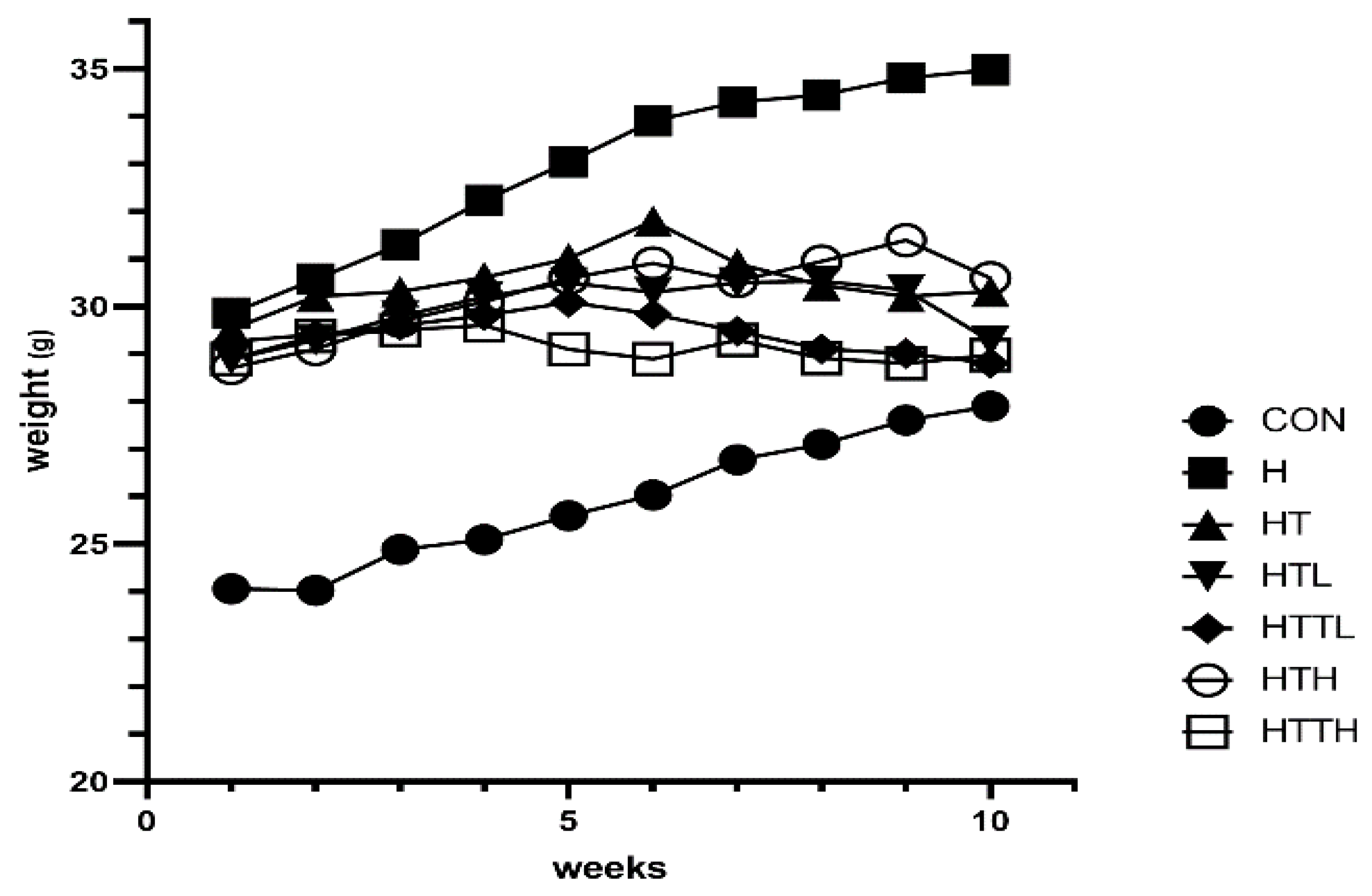
Body Weight and Blood Samples

Weight of the HFD group increased by 10–20% compared to that of the control group (Figure 1). Exercise and taurine (at two different doses) suppressed this increase in body weight. The data in table1 shows levels of liver enzymes (ALT and AST) increased in HFD groups, there was significant difference between CON and HFD groups. Notably, only ALT levels were significantly reduced in the HTTL group compared to the HFD group. However, neither exercise nor taurine supplementation produced statistically significant changes in these liver enzymes.

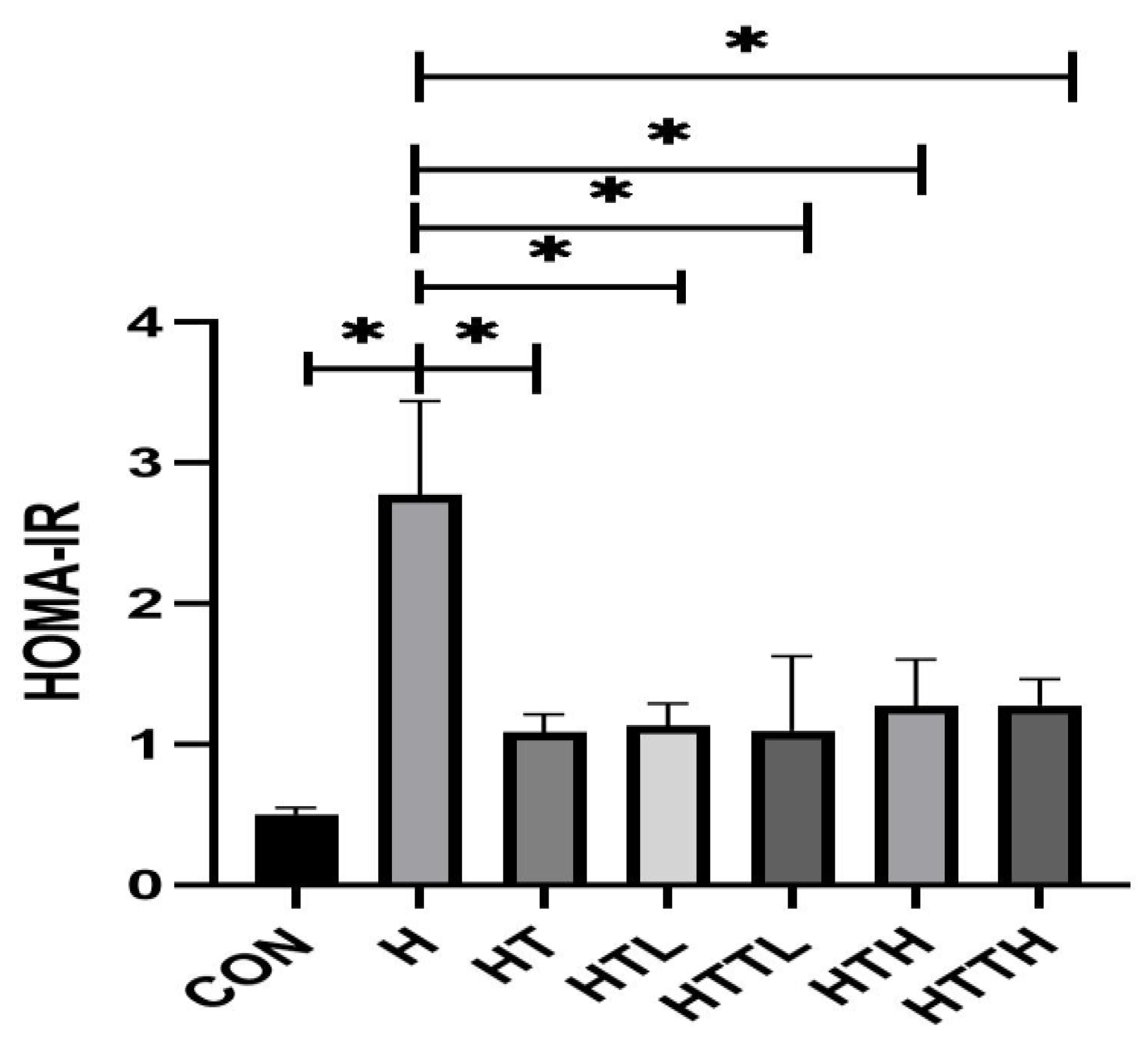
Effects of Different Doses of Taurine and HIIT on Fasting Blood Glucose, Insulin and Liver Glycogen and Insulin Sensitivity

HFD significantly reduced liver glycogen storage, and there was a significant difference between the CON and the other experimental groups. Furthermore, there were significant differences between the HTL and HTTL groups compared to others. These findings suggest that only a low dose of taurine, whether administered alone or in combination with exercise, exerts a positive effect on glycogen content. The HFD also increased insulin level. There was a significant difference between CON and other study groups. There was significant difference between HTTL and HTH. Additionally, the HFD elevated fasting blood glucose levels, with significant differences detected between the CON group and the H, HT, HTL, HTTL, and HTTH groups. A significant difference was also observed between the H group and the HT, HTL, HTTL, HTH, and HTTH groups (Table 1). both fasting blood glucose and plasma insulin levels were significantly elevated in the HFD group mice(Table 1). Based on these measurements, we calculated insulin resistance by homeostasis model assessment (HOMA) index. HFD- fed mice exhibited higher HOMA-insulin resistance (HOMA-IR) and there was a significant difference between HFD and other experimental groups(Figure 2).

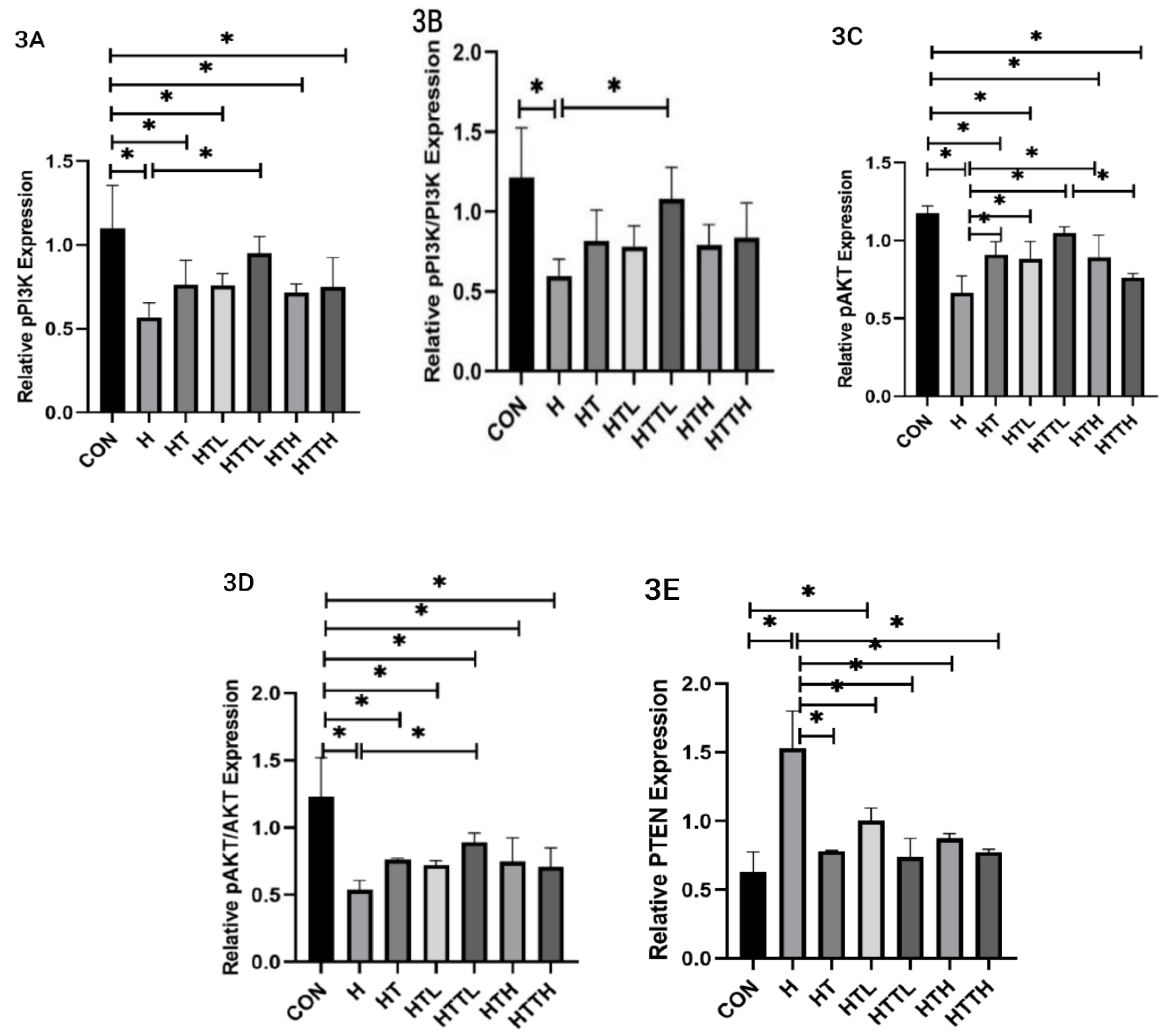
Effects of Different Doses of Taurine and HIIT on PI3K, AKT, and PTEN Expression in the Liver

There was no significant difference in PI3K protein synthesis between the HFD and CON groups. However, the phosphorylation of PI3K decreased, and there was a significant difference between the CON and other study groups. Exercise and taurine had no effect on pPI3K, except there was a significant difference between HTTL and H. There was no change in AKT expression; however, there were some changes in AKT phosphorylation. The pAKT levels decreased in the HFD groups, and there were significant differences between the CON and HFD groups. AKT phosphorylation was ameliorated by taurine and exercise, resulting in a significant difference between the HFD and HT, HTL, HTTL, and HTH groups. PTEN enzyme levels increased as a result of the HFD, with significant differences between the CON and HFD groups. A significant difference was observed between the CON and the HTL groups. Increased PTEN levels were reduced with the use of different doses of taurine and HIIT, there was significant differences between the H group and other groups (Figure 3A, B,C,D,E).

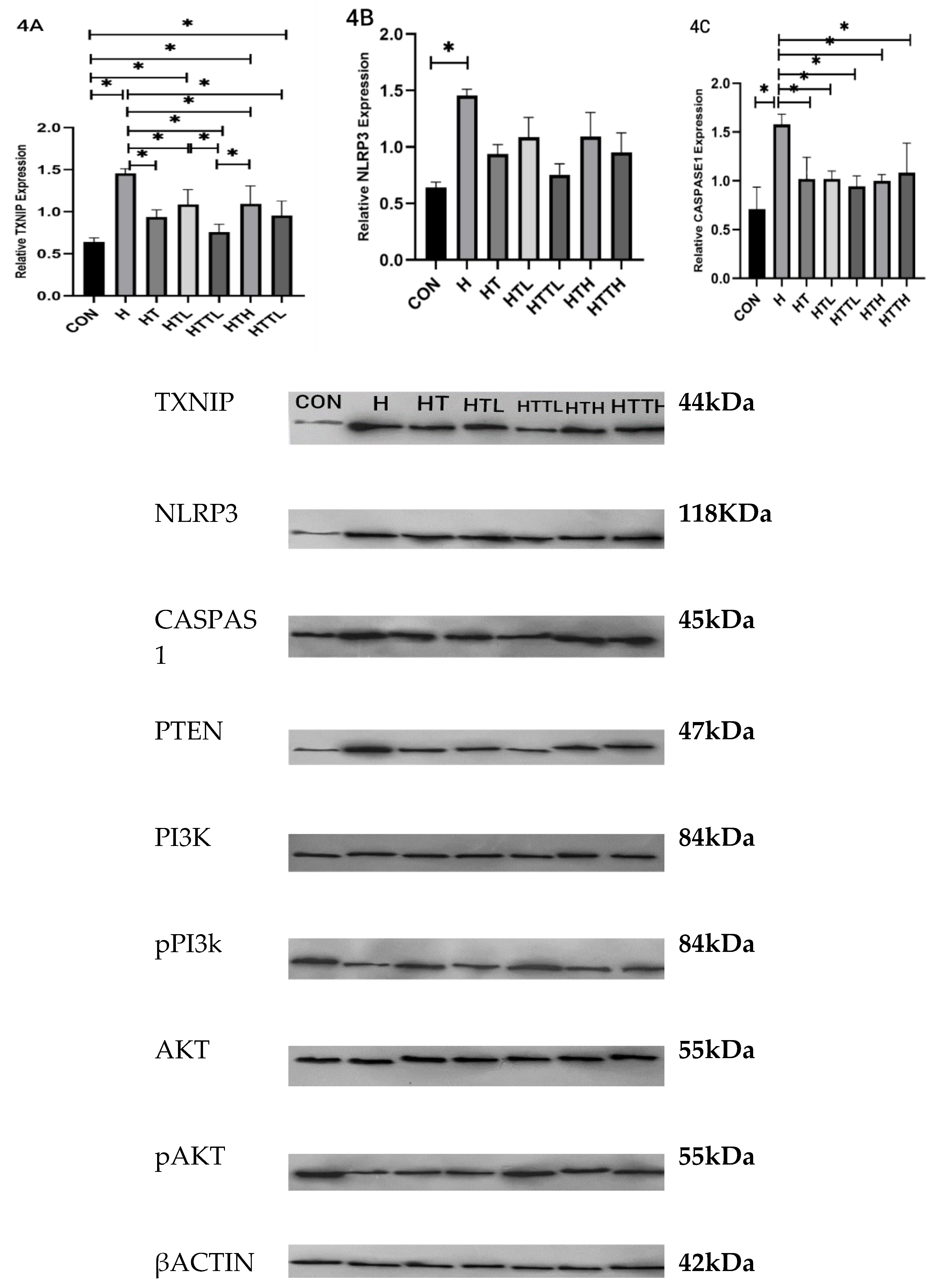
Effects of Different Doses of Taurine and HIIT on TXNIP, NLRP3, and Caspase-1 Expression in the Liver

The levels of TXNIP, NLRP3, and CASPASE1 proteins in the HFD groups were increased, and there were significant differences between the CON and HFD groups, which could result in an activated inflammatory pathway. Exercise suppressed the increase in TXNIP levels, with a significant difference between the H and HT groups. Additionally, there were significant differences between the H and HTL and HTTL and HTH groups. In terms of the quantity of protein, TXNIP combined with 2.5% taurine and HIIT exercise was at its lowest amount and had the best effect. None of the taurine interventions or exercise had an effect on NLRP3 protein levels and did not make a significant difference. There were significant differences between the H group and the other groups in the expression of caspase1. Thus, low and high dosages of HIIT and taurine were efficient (Figure 4A–C).

Discussions

This study found that both taurine and HIIT can reduce insulin resistance and liver inflammation in mice fed HFD. However, the effect of taurine was various with different doses.
Obesity has increased because of sedentary lifestyles and unhealthy eating habits, which contribute to the high prevalence of NAFLD. The biggest challenges in today's society is the diagnosis and treatment of NAFLD [23]. Similar to other investigations, this study showed that diet increased the AST and ALT levels. It also increases liver fat and inflammation [26,42], as shown in the H&E images. Studies have shown that 7% weight loss can help reduce inflammation and cellular issues. Therefore, exercise and diet can be used to manage obesity and improve NAFLD [16]. Exercise as a safe and inexpensive method plays a key role in regulating inflammation [9]. HIIT has been shown to potentially reduce fibrosis and liver inflammation due pathophysiological changes in patients with NAFLD [29]. In addition, previous studies have shown that taurine supplements effectively manage metabolic disorders, such as insulin resistance and obesity, potentially preventing NAFLD in the liver [12]. Furthermore, taurine due to its ability to increase glucose and lipid metabolism and reduce inflammation and fibrosis simultaneously, can decrease liver steatosis in wild mouse models of NAFLD [18,22].
TXNIP, a key regulator of cell redox, is crucial for developing acute liver diseases. So, it may treat NAFLD [4,8]. This study also showed that HFD may be due to the increase in ROS, which can increase TXNIP protein levels [46]. Our study found that two doses of taurine reduced the TXNIP protein levels. This was a significant change compared with that in the HFD group. This finding is similar to a previous study that showed that taurine reduces TXNIP activity [18]. In addition, taurine can inhibit TXNIP by affecting the calcium channels in diabetic patients [27]. The present study also found that HIIT alone affected TXNIP and led to significant differences. This study for the first time, examined the combined effect of two doses of taurine and exercise. We found Lower dosage with HIIT is the most efficient on TXNIP, but the higher dose was not efficient. It may be because increasing it can stimulate its stimulants and factors [31,36]. One study found that NLRP3 inflammation is crucial for obesity sensitivity. It regulates inflammation and boosts insulin signaling. Both depend on Caspase1 activity triggers [37]. Our study found that HFD intake increased NLRP3. Another study found that removing NLRP3 from mice can protect against obesity caused by nutrition, insulin resistance, and systemic inflammation [9]. Cheng et al. reported that NLRP3 inflammation decreases in the liver by taurine [15]. However, in the present study, there were no significant differences. We observed a significant decrease in CASPASE1 expression in liver tissue. Several studies have shown that exercise reduces TXNIP/NLRP3 expression in different tissues [13,17,40]. In this study, HIIT did not reduced activity of NLRP3 significantly. This may be due to mitochondrial dysfunction caused by high-intensity exercise, which might impact pre-inflammatory factors [45]. However, 12 weeks of exercise decreased NLRP3 levels in a healthy diet [43]. Another study found that moderate-intensity exercise had the best effect [17]. For example, Grey et al. found that moderate exercise decreased NLRP3 expression, and HIIT had no effect [14]. Moreover, in this study, because of HFD, it could not suppress the NLRP3.
Figure 5. HFD increased IR and inflammation in hepatocyte and taurine and HIIT improve them. Representative images (×20 objective) of liver sections stained with H&E.
Preprints 140773 g005
HIIT) and taurine, in both high and low doses, significantly reduced the activation of caspase-1. One of the key characteristics of taurine is its antioxidant property. This property is crucial because it can lead to the specific reduction of TXNIP and caspase 1 through various interventions. These interventions may include antioxidant treatments, metabolic regulation, or the use of inhibitors that target oxidative stress. Consequently, the decrease in these proteins can be attributed to the antioxidant effects of taurine.
TXNIP activates PTEN protein, which inhibits insulin messaging. TXNIP appears to prevent insulin sensitivity by increasing the insulin response to conditions like obesity that increase insulin resistance [35]. This study demonstrated that both HIIT and two different dosages of taurine resulted in a decrease in PTEN levels, likely due to the reduction of TXNIP. These findings are consistent with previous research indicating that HIIT and taurine can lower PTEN levels [5,17].Without TXNIP and PTEN, the PI3K/AKT pathway increases, thereby enhancing glucose transfer and metabolism. This study revealed both doses of taurine and exercises (individually and combined) decrease insulin resistance. Taurine converts PIP2 to PIP3 and triggers AKT activation, while the exact structure and function of this phenomenon are not yet fully understood. Two other studies have shown that taurine can activate the inhibited PI3K/AKT pathway [6,10]. Our study demonstrated that a 2.5% taurine intake alone results in a significant change in AKT protein expression compared to the HFD group, as found by researchers in the same study by Zhang et al. [44]. In a different study, the use of 5% taurine was found to enhance protein phosphorylation. [5]. In another study, it showed no effect at lower concentrations [6]. Similarly, another study demonstrated that consuming 5% taurine increased AKT levels in the liver, indicating an increase in its phosphorylation [5]. Previous studies indicate that exercise alone does not affect PI3K [28,34].we found the same result. However, Abdolahi et al. showed that aerobic exercise impacts PI3K in the livers of diabetic mice [1]. We found HIIT influenced AKT and led to a significant difference. This result is similar to those achieved in other studies that showed HIIT and low-intensity exercise affected AKT in fatty tissues and the liver, meanwhile, it found that high-intensity exercise was better at removing metabolic disorders [39]. Another study found that HIIT exercises increased PI3K/AKT phosphorylation [41] . Li et al. reported HIIT to be more effective than moderate-intensity exercise [17]. In contrast, other researchers indicated that exercising with high volume and intensity, which generates significant oxidative stress, only triggered the activation of IRS2 and did not affect the PI3K/AKT pathway [38]. However, this study demonstrated that high-intensity interval training (HIIT) does influence pAKT. The present study found that a combination of 2.5% taurine and HIIT led to an increase in AKT phosphorylation. This study demonstrated a significant improvement compared with the obese diet group. These results indicated that various doses of taurine reduced TXNIP levels and decreased CASPASE1 activity, simultaneously improving the inflammatory pathway. Additionally, there may be an enhancement in the redox conditions. This improvement could be attributed to the effect of TXNIP on PTEN, which reduces insulin resistance and increases PI3K/AKT activity. Interestingly, a lower dose of taurine appears to be more effective than a higher dose because it positively affects TXNIP activity and stimulates receptors. Furthermore, high-intensity interval training (HIIT) may help slow the progression of non-alcoholic fatty liver disease (NAFLD) by addressing its triggers.

Conclusions

In our study, we discovered that taurine, administered at doses of 2.5% and 5% and combined with HIIT, can beneficially influence insulin resistance, a key trigger of the disease, and inflammation, which worsens the condition. This effect is largely due to the combined effect of TXNIP. Furthermore, the findings suggest that a 2.5% dosage of taurine is more effective than a 5% dosage.

Abbreviation

Nonalcoholic Fatty Liver Disease (NAFLD), High-Fat Diet (HFD), Alanine Aminotransferase (ALT), Aspartate Aminotransferase (AST), Phosphatidylinositol 3-Kinases (PI3K), Protein Kinase B (AKT), Phosphatase and TENsin Homolog (PTEN), Thioredoxin Interacting Protein (TXNIP), NLR Family Pyrin Domain Containing 3 (NLRP3).

References

  1. Abdollahi M, Marandi SM, Ghaedi K. ; et al. Insulin-Related Liver Pathways and the Therapeutic Effects of Aerobic Training, Green Coffee, and Chlorogenic Acid Supplementation in Prediabetic Mice. Oxid Med Cell Longev. 2022, 2022, 5318245. [Google Scholar] [CrossRef]
  2. Berná, G.; Romero-Gomez, M. The role of nutrition in non-alcoholic fatty liver disease: Pathophysiology and management. Liver Int. 2020, 40, 102–108. [Google Scholar] [CrossRef] [PubMed]
  3. Brunt EM, Wong VW, Nobili V. ; et al. Nonalcoholic fatty liver disease. Nat Rev Dis Primers. 2015, 1, 15080. [Google Scholar] [CrossRef] [PubMed]
  4. Cao Z, Fang Y, Lu Y. ; et al. Melatonin alleviates cadmium-induced liver injury by inhibiting the TXNIP-NLRP3 inflammasome. J Pineal Res. 2017, 62. [Google Scholar] [CrossRef]
  5. Cappelli AP, Zoppi CC, Barbosa-Sampaio HC. ; et al. Taurine-induced insulin signalling improvement of obese malnourished mice is associated with redox balance and protein phosphatases activity modulation. Liver Int. 2014, 34, 771–783. [Google Scholar] [CrossRef]
  6. Chen, B.; Abaydula, Y.; Li, D.; Tan, H.; Ma, X. Taurine ameliorates oxidative stress by regulating PI3K/Akt/GLUT4 pathway in HepG2 cells and diabetic rats. Journal of Functional Foods. 2021, 85, 104629. [Google Scholar] [CrossRef]
  7. Dai B, Wu Q, Zeng C. ; et al. The effect of Liuwei Dihuang decoction on PI3K/Akt signaling pathway in liver of type 2 diabetes mellitus (T2DM) rats with insulin resistance. J Ethnopharmacol. 2016, 192, 382–389. [Google Scholar] [CrossRef]
  8. Ding C, Zhao Y, Shi X. ; et al. New insights into salvianolic acid A action: Regulation of the TXNIP/NLRP3 and TXNIP/ChREBP pathways ameliorates HFD-induced NAFLD in rats. Scientific Reports. 2016, 6, 28734. [Google Scholar] [CrossRef]
  9. Ding, Y.; Xu, X. Effects of regular exercise on inflammasome activation-related inflammatory cytokine levels in older adults: a systematic review and meta-analysis. J Sports Sci. 2021, 39, 2338–2352. [Google Scholar] [CrossRef]
  10. Farese, R.V.; Jr Zechner, R.; Newgard, C.B.; Walther, T.C. The problem of establishing relationships between hepatic steatosis and hepatic insulin resistance. Cell Metab. 2012, 15, 570–573. [Google Scholar] [CrossRef]
  11. Feng J, Qiu S, Zhou S. ; et al. mTOR: A Potential New Target in Nonalcoholic Fatty Liver Disease. International Journal of Molecular Sciences. 2022, 23, 9196. [Google Scholar] [CrossRef] [PubMed]
  12. Gentile CL, Nivala AM, Gonzales JC. ; et al. Experimental evidence for therapeutic potential of taurine in the treatment of nonalcoholic fatty liver disease. Am J Physiol Regul Integr Comp Physiol. 2011, 301, R1710–R1722. [Google Scholar] [CrossRef] [PubMed]
  13. Javaid, H.M.A.; Sahar, N.E.; ZhuGe, D.L.; Huh, J.Y. Exercise Inhibits NLRP3 Inflammasome Activation in Obese Mice via the Anti-Inflammatory Effect of Meteorin-like. Cells. 2021, 10, 3480. [Google Scholar] [CrossRef] [PubMed]
  14. Khakroo Abkenar, I.; Rahmani-Nia, F.; Lombardi, G. The Effects of Acute and Chronic Aerobic Activity on the Signaling Pathway of the Inflammasome NLRP3 Complex in Young Men. Medicina 2019, 55, 105. [Google Scholar] [CrossRef]
  15. Lee, C.C.; Chen, W.T.; Chen, S.Y.; Lee, T.M. Taurine Alleviates Sympathetic Innervation by Inhibiting NLRP3 Inflammasome in Postinfarcted Rats. J Cardiovasc Pharmacol. 2021, 77, 745–755. [Google Scholar] [CrossRef]
  16. Lewis, J.R.; Mohanty, S.R. Nonalcoholic fatty liver disease: a review and update. Dig Dis Sci. 2010, 55, 560–578. [Google Scholar] [CrossRef]
  17. Li Y, Xu P, Wang Y. ; et al. Different Intensity Exercise Preconditions Affect Cardiac Function of Exhausted Rats through Regulating TXNIP/TRX/NF-ĸB(p65)/NLRP3 Inflammatory Pathways. Evid Based Complement Alternat Med. 2020, 2020, 5809298. [Google Scholar] [CrossRef]
  18. Liu X, Zhang YR, Cai C.; et al. Taurine Alleviates Schistosoma-Induced Liver Injury by Inhibiting the TXNIP/NLRP3 Inflammasome Signal Pathway and Pyroptosis. Infect Immun. [CrossRef]
  19. Mohamed, I.N.; Hafez, S.S.; Fairaq, A.; Ergul, A.; Imig, J.D.; El-Remessy, A.B. Thioredoxin-interacting protein is required for endothelial NLRP3 inflammasome activation and cell death in a rat model of high-fat diet. Diabetologia. 2014, 57, 413–423. [Google Scholar] [CrossRef]
  20. Mohamed, I.N.; Sarhan, N.R.; Eladl, M.A.; El-Remessy, A.B.; El-Sherbiny, M. Deletion of Thioredoxin-interacting protein ameliorates high fat diet-induced non-alcoholic steatohepatitis through modulation of Toll-like receptor 2-NLRP3-inflammasome axis: Histological and immunohistochemical study. Acta Histochem. 2018, 120, 242–254. [Google Scholar] [CrossRef]
  21. Murakami, S. Role of taurine in the pathogenesis of obesity. Molecular Nutrition & Food Research. 2015, 59, 1353–1363. [Google Scholar] [CrossRef]
  22. Murakami, S.; Ono, A.; Kawasaki, A.; Takenaga, T.; Ito, T. Taurine attenuates the development of hepatic steatosis through the inhibition of oxidative stress in a model of nonalcoholic fatty liver disease in vivo and in vitro. Amino Acids. 2018, 50, 1279–1288. [Google Scholar] [CrossRef] [PubMed]
  23. Neuschwander-Tetri, B.A. Non-alcoholic fatty liver disease. BMC Med. 2017, 15, 45. [Google Scholar] [CrossRef] [PubMed]
  24. Peyrou M, Bourgoin L, Foti M. PTEN in Non-Alcoholic Fatty Liver Disease/Non-Alcoholic Steatohepatitis and Cancer. Digestive Diseases. 2010, 28, 236–246. [Google Scholar] [CrossRef] [PubMed]
  25. Polyzos, S.A.; Kountouras, J.; Mantzoros, C.S. Obesity and nonalcoholic fatty liver disease: From pathophysiology to therapeutics. Metabolism 2019, 92, 82–97. [Google Scholar] [CrossRef]
  26. Powell, E.E.; Wong, V.W.; Rinella, M. Non-alcoholic fatty liver disease. Lancet 2021, 397, 2212–2224. [Google Scholar] [CrossRef]
  27. Qayyum, N.; Haseeb, M.; Kim, M.S.; Choi, S. Role of Thioredoxin-Interacting Protein in Diseases and Its Therapeutic Outlook. Int J Mol Sci. 2021, 22, 2754. [Google Scholar] [CrossRef]
  28. Razavi, S.; Nazarali, P.; Alizadeh, R.; rezaeinezhad, N. Effects of Aerobic Interval Training with Citrus aurantium Consumption on Gene Expression of AMPK and PI3K in Liver Tissues of Elderly Rats. Research. Iranian Journal of Nutrition Sciences and Food Technology 2022, 17, 1–11. [Google Scholar]
  29. Ross, L.M.; Porter, R.R.; Durstine, J.L. High-intensity interval training (HIIT) for patients with chronic diseases. J Sport Health Sci. 2016, 5, 139–144. [Google Scholar] [CrossRef]
  30. Roy, A.; Sil, P.C. Taurine protects murine hepatocytes against oxidative stress-induced apoptosis by tert-butyl hydroperoxide via PI3K/Akt and mitochondrial-dependent pathways. Food Chemistry. 2012, 131, 1086–1096. [Google Scholar] [CrossRef]
  31. Satsu H, Gondo Y, Shimanaka H. ; et al. Signaling Pathway of Taurine-Induced Upregulation of TXNIP. Metabolites. 2022, 12, 636. [Google Scholar] [CrossRef]
  32. Schaffer, S.; Kim, H.W. Effects and Mechanisms of Taurine as a Therapeutic Agent. Biomol Ther. 2018, 26, 225–241. [Google Scholar] [CrossRef] [PubMed]
  33. Shamsan E, Almezgagi M, Gamah M. ; et al. The role of PI3k/AKT signaling pathway in attenuating liver fibrosis: a comprehensive review. Review. Frontiers in Medicine. 2024, 11, 1389329. [Google Scholar] [CrossRef] [PubMed]
  34. Shykholeslami, Z.; Abdi, A.; Hosseini, S.A.; Barari, A. Effect of Continuous Aerobic Training with Citrus Aurantium L. on Mitogen-Activated Protein Kinase and Phosphatidylinositol 3-Kinases Gene Expression in the Liver Tissue of the Elderly Rats. Research. Journal title. 2021, 29, 81–89. [Google Scholar] [CrossRef]
  35. Surai, P.F.; Earle-Payne, K.; Kidd, M.T. Taurine as a Natural Antioxidant: From Direct Antioxidant Effects to Protective Action in Various Toxicological Models. Antioxidants 2021, 10, 1876. [Google Scholar] [CrossRef] [PubMed]
  36. Tu, Y.; Zhang, P.; Pang, X. Thioredoxin-interacting Protein as a Common Regulation Target for Multiple Drugs in Clinical Therapy/Application. Cancer Translational Medicine. 2015, 1. [Google Scholar] [CrossRef]
  37. Vandanmagsar B, Youm YH, Ravussin A. ; et al. The NLRP3 inflammasome instigates obesity-induced inflammation and insulin resistance. Nat Med. 2011, 17, 179–188. [Google Scholar] [CrossRef]
  38. Wang, H.; Wang, J.; Zhu, Y.; Yan, H.; Lu, Y. Effects of Different Intensity Exercise on Glucose Metabolism and Hepatic IRS/PI3K/AKT Pathway in SD Rats Exposed with TCDD. Int J Environ Res Public Health. 2021, 18, 13141. [Google Scholar] [CrossRef]
  39. Wang, N.; Liu, Y.; Ma, Y.; Wen, D. High-intensity interval versus moderate-intensity continuous training: Superior metabolic benefits in diet-induced obesity mice. Life Sci. 2017, 191, 122–131. [Google Scholar] [CrossRef]
  40. Wang Q, Hu J, Liu Y. ; et al. Aerobic Exercise Improves Synaptic-Related Proteins of Diabetic Rats by Inhibiting FOXO1/NF-κB/NLRP3 Inflammatory Signaling Pathway and Ameliorating PI3K/Akt Insulin Signaling Pathway. J Mol Neurosci. 2019, 69, 28–38. [Google Scholar] [CrossRef]
  41. Wang Y, Guo Y, Xu Y. ; et al. HIIT Ameliorates Inflammation and Lipid Metabolism by Regulating Macrophage Polarization and Mitochondrial Dynamics in the Liver of Type 2 Diabetes Mellitus Mice. Metabolites 2022, 13, 14. [Google Scholar] [CrossRef]
  42. Wong VW, Wong GL, Tsang SW. ; et al. Metabolic and histological features of non-alcoholic fatty liver disease patients with different serum alanine aminotransferase levels. Aliment Pharmacol Ther. 2009, 29, 387–396. [Google Scholar] [CrossRef] [PubMed]
  43. Yang W, Liu L, Wei Y. ; et al. Exercise suppresses NLRP3 inflammasome activation in mice with diet-induced NASH: a plausible role of adropin. Lab Invest. 2021, 101, 369–380. [Google Scholar] [CrossRef] [PubMed]
  44. Zhang M, Shi X, Luo M. ; et al. Taurine ameliorates axonal damage in sciatic nerve of diabetic rats and high glucose exposed DRG neuron by PI3K/Akt/mTOR-dependent pathway. Amino Acids. 2021, 53, 395–406. [Google Scholar] [CrossRef] [PubMed]
  45. Zhang, T.; Ding, S.; Wang, R. Research Progress of Mitochondrial Mechanism in NLRP3 Inflammasome Activation and Exercise Regulation of NLRP3 Inflammasome. Int J Mol Sci. 2021, 22, 10866. [Google Scholar] [CrossRef]
  46. Zheng T, Yang X, Li W. ; et al. Salidroside Attenuates High-Fat Diet-Induced Nonalcoholic Fatty Liver Disease via AMPK-Dependent TXNIP/NLRP3 Pathway. Oxid Med Cell Longev. 2018, 2018, 8597897. [Google Scholar] [CrossRef]
Figure 1. weight of subjects in different groups. Control (CON), High Fat Diet (H), High Fat Diet + HIIT (HT), High Fat Diet and Taurine 2/5% (HTL), High Fat Diet + HIIT + Taurine 2/5% (HTTL), High Fat Diet + Taurine 5% (HTH), High.
Preprints 140773 g001
Figure 2. HOMA2 index values: insulin resistance (HOMA-IR).
Preprints 140773 g002
Figure 3. HIIT and taurine in two different doses decreased IR. 3A) PI3K phosphorylation, 3B) pPI3k/PI3K, 3C) pAKT expression, 3D) pAKT/AKT, 3E) PTEN expression. Significant alterations (p < 0.05, marked by asterisks are observed, especially with combined treatments.
Preprints 140773 g003
Figure 4. HIIT and taurine in two different doses decreased inflammation phosphorylation, 4A) TXNIP expression, 4B) NLRP3 expression, 4C) Caspase-1 expression. Significant alterations (p < 0.05, marked by asterisks are observed, especially with combined treatments. Data are mean ± SEM for 6-8 mice per. D) western blot analyses of TXNIP, NLRP3, CASPASE1, PTEN, PI3K, pPI3k, AKT and pAKT in hepatocyte.
Preprints 140773 g004
Table 1. ALT, AST, Blood Glucose, Fasting Insulin, and Liver Glycogen for C57BL/6 Mice, Control (CON), HFD (H), HFD + HIIT (HT), HFD and Taurine 2.5% (HTL), HFD + HIIT + Taurine 2.5% (HTTL), High Fat Diet + Taurine 5%(HTH), HFD+HIIT+taurine5%(HTTH).
Group ALT (U/L) AST (U/L) Blood Glucose (mg/dl) Fasting Insulin (µlu/ml) Liver Glycogen (mg/g)
CON 62.33±5.78 159.66±40.70 125±19.20 3.55±0.39 26.94±4.43
H 209.66±85.54 326.16±57.56 382.25±22.21 6.51±.37 11.83±3.47
HT 134.50±30.07 253.66±90.77 233.50±39.07 6.43±0.22 15.02±1.03
HTL 146.83±37.60 278.66±91.78 237.50±45.80 6.55±0.42 19.91±2.20
HTTL 119.16±37.99 217.50±84.09 255.5±56.03 5.65±1.84 21.72±1.92
HTH 157.16±34.12 290.66±89.66 194.25±56.93 7.96±1.13 14.11±1.35
HTTH 153.16±33.03 284.00±83.63 281.50±40.35 6.38±0.15 15.72±1.58
Disclaimer/Publisher’s Note: The statements, opinions and data contained in all publications are solely those of the individual author(s) and contributor(s) and not of MDPI and/or the editor(s). MDPI and/or the editor(s) disclaim responsibility for any injury to people or property resulting from any ideas, methods, instructions or products referred to in the content.
Copyright: This open access article is published under a Creative Commons CC BY 4.0 license, which permit the free download, distribution, and reuse, provided that the author and preprint are cited in any reuse.
Prerpints.org logo

Preprints.org is a free preprint server supported by MDPI in Basel, Switzerland.

Subscribe

Disclaimer

Terms of Use

Privacy Policy

Privacy Settings

© 2025 MDPI (Basel, Switzerland) unless otherwise stated