
Submitted:
25 November 2024
Posted:
26 November 2024
You are already at the latest version
Abstract
Nanomaterial properties such as size, structure, and composition can be controlled by manipulating radiation, such as gamma rays, X-rays, and electron beams. This control allows scientists to create materials with desired properties that can be used in a wide range of applications, from electronics to medicine. This use of radiation for nanotechnology is revolutionizing the way we design and manufacture materials. Additionally, radiation-induced nanomaterials are more cost-effective and energy-efficient. This technology is also having a positive impact on the environment, as materials are being produced with fewer emissions, less energy, and less waste. This cutting-edge technology is opening up new possibilities and has become an attractive option for many industries, from medical advancements to energy storage. It is also helping to make the world a better place by reducing our carbon footprint and preserving natural resources. This review aims to meticulously point out the synthesis approach and highlights significant progress in generating radiation-induced nanomaterials with tunable and complex morphologies. This comprehensive review article is essential for researchers to design innovative materials for advancements in health care, electronics, energy storage, and environmental remediation.
Keywords:
Nano material
; electron beam irradiation
; gamma radiation
; X-ray beam
; Application of nanomaterials
; metallic nanoparticle
1. Introduction
Nanomaterials have emerged as a pivotal class of materials with transformative potential across multiple industries due to their unique properties at the nanoscale. These materials, characterized by dimensions typically between 1 and 100 nanometers [1,2], exhibit distinctive mechanical, optical, electrical, and chemical properties that are not present in their bulk counterparts. Nonmaterial are increasingly important in today's world due to their unique and versatile properties that can be harnessed across a wide range of applications, driving innovation and efficiency in various fields. The increased surface area to volume ratio at this scale enhances their reactivity, making them ideal for applications in catalysis, electronics, and medicine [3]. Their high surface area to volume ratio enhances chemical reactivity, making them valuable in catalysis and environmental applications such as pollution control and water purification. In medicine, nanomaterials are revolutionizing drug delivery systems, allowing for targeted therapies that minimize side effects and improve efficacy, as well as enhancing diagnostic imaging techniques [4]. In the realm of electronics, nanomaterials enable the miniaturization of components, leading to the development of faster, smaller, and more energy-efficient devices. Additionally, they contribute to advancements in energy storage and conversion, such as in the development of high-capacity batteries and efficient solar cells. The ability to tailor the properties of nanomaterials for specific needs also offers significant potential in the development of strong yet lightweight materials for use in transportation and construction. Overall, nanomaterials are a driving force in advancing technology and improving the quality of life, addressing global challenges, and contributing to sustainable development [5].
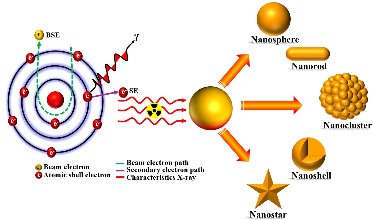
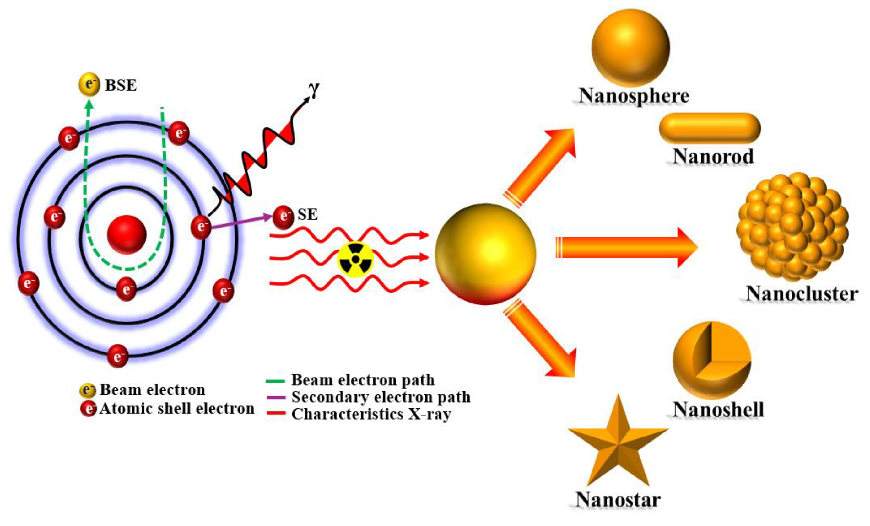
In other events, radioactivity, nuclear reactors, Hiroshima and Nagasaki, and Chernobyl are common images that people think about radiation. However, radiation also exists in many different forms, including visible light, infrared light, solar radiation, UV light that produces a suntan, and radio and television signals. The word radiation refers to the way that energy is released from a nucleus in the form of waves or particles. This energy can travel through space, and when it interacts with matter, it can cause changes in the matter. These changes can be in the form of heat, light, or other forms of energy. Radiation is a vital component of life on Earth because it surrounds every living organism. It is an important part of photosynthesis, the process by which plants use light to convert carbon dioxide and water into glucose and oxygen. As both plants and animals use radiation to send and receive signals, it is also crucial for interspecies communication. Radiation is also an important part of the human body, as it is used in medical imaging and cancer treatments. Radiation is used predominately to detect nuclear waste and measure long-distance objects. It is important for the weather forecasting sector, as it is used to detect changes in air pressure and temperature, volcanic activity and earthquakes. The nanomaterials in driving technological advancements stem from their versatile properties and superior performance compared to their bulk counterparts. Traditionally, nanoparticles have been synthesized by reducing metal ions into neutral particles using harmful reducing agents. However, recent efforts have focused on developing green radiation technologies that leverage radiation sources instead of hazardous chemicals for nanoparticle production. This shift aims to enhance the safety and environmental sustainability of synthesis methods while maintaining the effectiveness of the resulting nanoparticles [6]. The synthesis of nanomaterials using radiation technology is an innovative and impactful approach that leverages various forms of radiation, including gamma rays, electron beams, and ultraviolet (UV) light [7,8,9,10,11,12], to produce nanoparticles with well-defined characteristics. This method offers exceptional precision in controlling reaction conditions, allowing nanoparticles that exhibit uniform size and shape, essential factors for their performance in specific applications. The synthesis process typically begins with the preparation of a precursor solution containing metal salts or organic molecules, which are then subjected to irradiation. This irradiation effectively generates free radicals, instigating chemical reactions that lead to the formation of nanomaterials [13,14]. One of the most significant advantages of using radiation technology for synthesis is the high purity of the resulting nanoparticles, as the technique often eliminates the need for additional chemical reagents, thereby reducing the risk of contamination. Moreover, radiation methods are more environmentally friendly, generating less chemical waste and minimizing the reliance on toxic substances compared to traditional synthesis methods, which aligns with sustainable practices in materials science [15,16,17]. The energy efficiency of radiation-based processes further enhances their appeal, as they generally require less energy expenditure than conventional thermal methods. The resulting nanomaterials find valuable applications across diverse fields, including biomedicine, where they are used for targeted drug delivery and advanced imaging techniques, as well as in electronics, enabling the miniaturization of components for faster, more efficient devices. Additionally, these nanoparticles play a vital role in catalysis, enhancing reaction rates and selectivity in chemical processes. Despite the numerous advantages, challenges such as the scalability of the process for industrial applications and the need for consistent reproducibility must be addressed. Continued research and innovation in radiation synthesis techniques hold the promise of expanding the capabilities and applications of nanomaterials [1,18,19], making them an important area of focus in the advancement of nanotechnology and its integration into various industries. Gamma rays, ultraviolet (UV) light, and electron beam irradiation play crucial roles in the synthesis of nanomaterials [10,11], each providing unique mechanisms and advantages that enhance the formation and properties of nanoparticles. Gamma irradiation, known for its high energy and penetrating power, generates free radicals in precursor solutions, initiating chemical reactions that reduce metal ions into high-purity nanoparticles, essential for applications in catalysis, electronics, and biomedicine. Meanwhile, UV irradiation excites atoms or molecules in precursor solutions, promoting environmentally friendly reactions without the use of toxic chemicals, effective for synthesizing metal nanoparticles and semiconductor nanostructures with controlled morphology and composition. Electron beam irradiation directs high-energy electrons onto substrates or solutions, allowing precise control over synthesis parameters and facilitating various reactions, which results in the generation of consistent nanoparticles [20]. Together, these modern radiation techniques offer significant advantages over conventional methods, leading to enhanced functionalities and diverse applications of nanomaterials in multiple industries.
The synthesis of nanomaterials, while offering significant advantages, presents several challenges associated with both top-down and bottom-up approaches [21,22,23]. In top-down techniques like mechanical milling and lithography, a key challenge is achieving precise control over the size, shape, and distribution of nanoparticles (as shown in Scheme 1). Variability in these parameters can critically affect the performance of the materials in specific applications. Moreover, these methods often require complex, expensive equipment and controlled conditions, which can escalate production costs and limit their feasibility for large-scale manufacturing [24]. On the other hand, bottom-up approaches, such as chemical vapor deposition and sol-gel processes, face challenges related to the purity and stability of the synthesized nanomaterials. Contaminants or agglomeration during the synthesis can significantly alter the desired properties and effectiveness of the nanoparticles. Additionally, these methods may involve toxic reagents or byproducts, raising environmental and safety concerns that must be addressed. Both approaches also struggle with reproducibility. Minor variations in synthesis conditions can lead to inconsistent results, complicating the development process and hindering the ability to create standardized materials. Furthermore, there is a continuous need to develop more energy-efficient and sustainable synthesis methods that minimize environmental impact while still yielding high-quality nanomaterials. Addressing these challenges requires ongoing research and innovation to refine existing techniques and explore new synthesis pathways that enhance the capabilities and applications of nanomaterials [8,9,10,11].
In this review, we will focus on the synthesis of nanomaterials using modern radiation technologies, such as gamma rays, and electron beams, offering a comprehensive exploration of these innovative methods. We will highlight the advantages of utilizing radiation technology over traditional top-down and bottom-up approaches, emphasizing the unique benefits this contemporary technique presents.
2. Synthesis of Nanomaterials by Radiation Method and Application
2.1. Electron Beam Synthesis
Electron beam irradiation is an emerging technique in the synthesis and modification of nanomaterials, providing unparalleled precision and control in nanotechnology applications [25,26,27,28]. This method harnesses high-energy electrons to interact with materials, facilitating reactions such as excitation, ionization, and radiolysis, which are essential for producing and tailoring nanostructures [29]. One of the primary advantages of e-beam irradiation is its capability to induce nucleation and growth of nanoparticles without the need for chemical reagents, leading to environmentally friendly synthesis processes that align with green chemistry principles [30]. In metallic nanoparticle production, e-beam irradiation can precisely control the size and distribution of particles like gold and silver, optimizing their functionality for applications in catalysis and electronics [31,32,33,34,35,36]. In semiconductor fabrication, the technique allows for the creation of quantum dots and nanowires, critical for next-generation electronic devices. Additionally, e-beam irradiation enhances the properties of polymer nanocomposites through cross-linking, making them more suitable for demanding applications in the automotive and aerospace industries.
The irradiation dose and its rate play crucial roles in making the size and shape of nanostructures. The metallic ions reduction to zero-valent atoms depends on the overall reducing classes generated by ionizing radiation, determined by the energy dose deposited in the medium. These reduction reactions occur alongside association and coalescence processes, affecting both free ions and agglomerated ions. The growth and size of nanomaterials are influenced by the rate of reducing species production, which is tied to the energy deposition rate, or dose rate, as depicted in Scheme 2 and Scheme 3. At high dose rates numerous reducing species form quickly, leading to rapid ion-reduction and the simultaneous production of numerous free atoms. Each atom acts as a nucleation center, initiating nanoparticle growth, resulting in many small aggregates with a narrow size distribution.
Conversely, at low dose rates fewer reduced species form, causing a slower reduction rate compared to association processes. Initially, few free atoms are produced, and reducing species react more with existing oligomers. With fewer nucleation centers, there are fewer aggregates and particles than at high dose rates, producing larger nanoparticles from the same initial metallic ion concentration.
2.1.1. Electron-Matter Interactions
Recent progress in the understanding of electron-matter interactions, particularly using electron beams in transmission electron microscopy (TEM) and field emission-scanning electron microscopy (FE-SEM), has established these methods as pivotal tools for the remote manipulation of nanomaterials' functional properties. This technological advancement is characterized by high spatial and temporal resolution, as well as digital control, making them attractive to researchers in the field. The investigation of electron beam interactions with matter is a fundamental aspect of modern science, revealing intricate quantum phenomena associated with electron dynamics. In solid materials, electrons encounter two essential forces: Coulombic repulsion between themselves and attraction toward the atomic nuclei. The interactions between electrons (and/or photons) and atoms at the quantum level yield significant insights into material properties. In TEM, a high-energy electron beam transmits through a sample, forming images that capitalize on the wave-like nature of electrons, similar to techniques used in conventional light microscopy. This capability is critical for analyzing microstructures, characterizing nanoscale materials, and obtaining chemical insights at the atomic scale. However, the high energy associated with the electron beam can induce radiation damage, which may lead to structural alterations and defects that negatively affect the physical and chemical properties of the materials, increasing the risk of failure in practical applications [37,38].The electron beam generated in TEM and FE-SEM provides a versatile platform for imaging metal nanoparticles, enabling spectroscopic analyses that confirm the presence of specific elements on a localized level. Additionally, e-beam irradiation in TEM not only aids in the fabrication of nanomaterials but also facilitates the investigation of their morphology, structural characteristics, and chemical transformations. These capabilities are essential for developing innovative nanostructures that conventional chemical and physical methods may not be able to produce. Thus, TEM is recognized as a crucial instrument in the field of nanoscale materials engineering. Jiang [39] has compiled a variety of beam damage phenomena associated with oxides in scanning TEM (STEM) and highlighted the shortcomings of currently accepted mechanisms. Similarly, El Mel and Bittencourt [40] have reviewed the processes of solid-to-hollow conversion induced by e-beam irradiation in TEM, citing important examples from the literature. Gonzalez-Martinez et al. [41] conducted a comprehensive review focused on available work related to electron beam-induced synthesis techniques with in-situ capabilities, emphasizing the e-beam as a key agent driving the synthesis of nanostructures within the TEM. Demonstrations in recent years have shown that the electron beam in TEM serves as a powerful tool for the fabrication and manipulation of nanostructures, allowing precise control at the nanoscale and even at the individual nanoparticle level Furthermore, S. Jesse et al. [42] recently reviewed advancements in using focused electron beams to create freestanding nanoscale 3D structures, explore radiolysis and the fabrication potential of liquid precursors, achieve epitaxial crystallization of amorphous oxides with atomic layer precision, and visualize and control the motion of individual dopants within a 3D crystal lattice.
2.1.2. Transition Metal Nano Materials
The synthesis of transition metal nanoparticles through electron beam irradiation has gained traction in nanomaterials research due to its rapid processing capabilities and precise control over particle size and morphology. This method facilitates the direct reduction of transition metal cations to metallic nanoparticles without requiring additional reducing agents. In solution-phase synthesis, dilute metal salt solutions are irradiated, leading to the formation of uniform nanoparticles that are particularly suitable for applications in catalysis, electronics, and sensing. Transition metals such as platinum, palladium, and nickel have been effectively synthesized using this approach. Conversely, solid-phase synthesis involves irradiating transition metal precursors in thin films or nanocomposites, resulting in well-dispersed nanoparticles and tailored interactions with the substrate. A major advantage of this technique is the ability to control synthesis parameters-such as electron dose and exposure time-allowing for manipulation of nanoparticle growth and properties. The elimination of chemical residues and the potential for one-step synthesis also enhance its scalability for industrial applications. While the advantages are significant, challenges exist in optimizing the process for practical implementation. Continued research is essential to refine synthesis protocols and explore new transition metal systems. Electron beam irradiation is a promising method for the synthesis of transition metal nanoparticles, offering opportunities for developing advanced nanomaterials for various applications in catalysis, energy, and electronics. Future studies should focus on optimizing these techniques to maximize their industrial applicability. In the following sections, we will briefly describe a few of the most important transition metal and metal compound nanoparticles synthesized by electron beam irradiation.
2.1.3. Gold Nanoparticles
A novel and straightforward strategy has been developed for synthesizing composites of gold nanoparticles (AuNPs) incorporated within polyvinyl alcohol (PVA) films using electron beam irradiation. The PVA/Au nanocomposites exhibited a prominent surface plasmon resonance peak at approximately 540 nm, indicative of the presence of Au nanoparticles. With increasing irradiation doses, the formation of AuNPs was observed, ranging in size from 10 to 65 nm and displaying diverse morphologies. Notably, the optical energy band gap significantly decreased from 5.8 eV for pure PVA to 3.5 eV for samples subjected to a 300 kGy irradiation dose (as shown in Figure 1). Additionally, the incorporation of Au nanoparticles into the PVA matrix, paired with elevated electron beam irradiation, led to a reduction in the thermal stability of the PVA/Au nanocomposite films. AC conductivity measurements at room temperature showed stable behavior, followed by a sharp increase in conductivity as a function of the applied field frequency across various irradiation doses (as shown in Figure 1) [43].
The interest in polymer/metal nanoparticle composites stems from their electrical properties, which closely resemble those of metals, while their processing techniques and mechanical attributes are similar to those of plastics. Consequently, the ability to manipulate the physical and electrical characteristics of these composites is essential for determining their potential applications [44,45]. Additionally, there is an increasing focus on the synthesis of gold nanoparticles (AuNPs) because of their exceptional properties across various research fields. Gold nanoparticles are recognized as the most stable and compatible metal nanoparticles, making them highly suitable for the development of advanced smart devices [46,47]. Kim et al. [48] obtained uniform Au nanoparticles by subjecting gold(I)-alkane thiolate complexes to electron beam irradiation.
On the other hand, poly metal nanoparticles have also been synthesized by highly efficient electron beam irradiation process and have a good application in sensor, antimicrobial agents, optical data storage, photo catalysis and photovoltaics [49,50,51,52]. Y. Ohkubo et.al. [53] investigated an efficient method for synthesizing Au-Pd bimetallic nanoparticles with random alloy structures on carbon supports using high-energy electron beam irradiation. This straightforward process involves the reduction of metal precursor ions through radicals formed by radiolysis in an aqueous solution, eliminating the need for surfactants or organic solvents. The researchers discovered that the addition of citric acid and sodium hydroxide, along with careful pH control before irradiation, significantly influences the structural characteristics of the nanoparticles. This was confirmed by advanced analytical techniques such as X-ray diffraction (XRD) and extended X-ray absorption fine structure (EXAFS). By employing metal ion complexing agents and adjusting the solution's pH, the study highlights a refined approach to achieving tailored bimetallic nanoparticles with desired random alloy structures, showcasing its potential in advancing nanomaterial synthesis. The electron beam radiation condition of the bimetallic nanomaterial synthesis procedure is also reported in other literature [54,55].
2.1.4. Platinum Nanoparticles
Platinum nanoparticles supported on various oxide substrates, particularly TiO2 (Pt/TiO2), have garnered significant interest due to their notable chemical properties and effectiveness as catalysts for CO oxidation [56,57]. Satoru Kageyama and colleagues investigated Pt/TiO2 composite nanoparticles synthesized via electron beam irradiation [58]. In their study, the synthetic solution was subjected to electron beam irradiation at room temperature for 6.7 seconds at a dose of 20 kGy and an energy of 4.8 MeV (at the Japan Electron Beam Irradiation Service) to enhance preferential CO oxidation. This innovative method allows for the creation of stabilizer-free Pt/TiO2 composite nanoparticles, with the microstructures analyzed using transmission electron microscopy. The resulting Pt nanoparticles, with sizes ranging from 2 to 4 nm, were successfully deposited on TiO2 without the addition of stabilizers. The size and morphology of the Pt nanoparticles were significantly influenced by the concentrations of Pt ions and 2-propanol. The catalytic performance for preferential CO oxidation was evaluated across a temperature range of 60 to 140 °C. Remarkably, the Pt/TiO2 catalyst featuring spherical Pt nanoparticles achieved a CO conversion rate of 67% with 100% selectivity at a low temperature of 60 °C following electron beam irradiation.
2.1.5. Silver Nanoparticles
Electron beam (EB) irradiation is an effective approach for producing stable silver nanoparticles (AgNPs) while minimizing the interference from inherent impurities associated with chemical reactions. This prototype experiment utilized linear electron beam accelerators at two different EB absorbed dose rates: 2 kGy/min and 7-8 kGy/s, along with various absorbed dose levels. It established optimal conditions for generating AgNPs through radiolysis alone or through a combination of radiolysis and chemical reduction. A high dose rate is essential for achieving a good yield of AgNPs via radiolysis (as shown in Figure 2), enabling a rapid production process. In contrast, at lower absorbed dose rates, the inclusion of a stabilization agent is recommended. By adjusting the experimental parameters, someone can modulate the balance between chemical and radiolytic reduction processes, allowing for the synthesis of nanoparticles with specific characteristics tailored to various applications [59].
In another investigation, a method combining gamma, electron beam, and synchrotron X-ray irradiation was employed for synthesizing silver nanoparticles in poly (vinyl pyrrolidone) (PVP) [60]. A one-pot synthesis approach was developed to prepare silver nanoparticles in an aqueous PVP solution using synchrotron X-ray radiation. The reduction of metal ions, leading to homogeneous nucleation and nanoparticle formation, is facilitated by hydrated electrons (eaqˉ) and hydrogen atom radicals (H·), which are generated from the radiolysis of water by synchrotron X-rays. A comparative analysis assessed the effectiveness of this synthesis method against the gamma and electron beam irradiation techniques. The gamma radiation method produced nanoparticles with an average size of 8 nm, while synchrotron X-ray irradiation yielded particles in the range of 10-15 nm. Notably, gamma radiolysis and electron beam irradiation resulted in smaller-sized particles with a narrower size distribution compared to those synthesized via X-ray radiolysis. The study also examined the influence of various experimental parameters, such as the concentration of Ag+ ions and PVP, on nanoparticle formation.
I. Ion conducted a study on the synthesis of silver nanoparticles embedded in micro-hydrogel particles using electron beam irradiation [61]. The research focuses on creating a hybrid micro-hydrogel that exhibits antibacterial properties. This micro-hydrogel matrix is composed of biocompatible poly(vinyl alcohol) (PVA) that is functionalized with antibacterial agents, specifically graphene oxide and silver nanoparticles. The polymer composites were synthesized through electron beam irradiation at absorbed doses of 10, 25, and 50 kGy to facilitate the formation of silver nanoparticles. The PVA hydrogel itself was produced by irradiating a 20 mL solution of 10% (wt./vol.) PVA with a molecular weight of 400,000 g/mol, in conjunction with graphene oxide and silver salt at a concentration of 1 mg/mL, and additionally functionalized with pyridine and iron salt.
F. Yang et al. [62] have developed a method that employs a displacement reaction combined with spontaneous electrolysis to efficiently synthesize switching riddle nanowires of silver tetracyanoquinodimethane (AgTCNQ) over large areas. The authors observed that under electron beam irradiation, silver nanodots and nanoclusters begin to precipitate. The pattern of separation between the Ag nanoclusters and the AgTCNQ matrix resembles that seen in various well-established solid electrolytes, such as Ag2S [63], RbAg4I5 [64], which are commonly used in electrochemical metallization memories or atomic switches [65,66]. In a related study demonstrated the application of electron beam irradiation for the fabrication of nanoparticles and nanorods on silver surfaces, achieving exceptional nanometric precision. Their work showcased both precise control over the dimensions of the nanorods and high placement accuracy.
The electron beam radiation synthesis of silver nanoparticles was also reported in other literature [67,68,69,70], which described the electron beam irradiation technique as effective for enabling rapid reactions while eliminating unwanted chemical residues. According to their studies, this method is particularly advantageous because it allows for the controlled growth of silver nanocrystals from dilute solutions of silver nitrate using scanning transmission electron microscopy (STEM) irradiation. The ability to finely tune the initiation and growth processes is crucial for advancing practical applications of this technique, especially in the development of designer nanostructures intended for nano-electronic and nanophotonic applications. Subsequently, achieving precise control over these parameters not only enhances the quality and characteristics of the nanostructures but also accommodates the specific requirements needed for integration into advanced technological platforms. Several research groups have recently made a significant advancement in the production of silver (Ag) nanoparticles and nanowires from various silver-based materials through irradiation with a commonly used electron beam generated by field emission scanning electron microscopy (FE-SEM) or transmission electron microscopy (TEM). This technique involves materials such as α-Ag2WO4 [71], β-Ag2MoO4 [72,73], β-AgVO3 [74], Ag3PO4 [75], and Ag2CrO4 [76]. For α-Ag2WO4 case, in situ FE-SEM imaging reveals key insights into the growth of silver (Ag) on α-Ag2WO4 crystals following 30 kV electron exposure without heating. The initial image captures the crystal surface prior to exposure, establishing a baseline. After 6 minutes, modeled surface defects emerge, acting as nucleation sites for Ag nanoparticles, indicating that energetic electrons disrupt the crystal lattice. By 10 minutes, these nanoparticles evolve into Ag nanorods, reflecting the influence of electron interactions on metal morphology (as shown in Figure 3). These findings enhance the understanding of electron-driven growth mechanisms in semiconductor materials, highlighting the interplay between electron exposure and material response, which is crucial for applications in nanotechnology and materials engineering.
Overall, the interaction of these semiconductors with the e-beam facilitates the reduction of Ag cations, leading to the formation and growth of metallic Ag on their surfaces, which results in Ag nanoparticles associated with semiconductors (as shown in Figure 4). Several recent publications have discussed the mechanisms underlying the formation of these new structures [77,78]. For instance, Lin et al. [79] investigated the electronic reconstruction of α-Ag2WO4 nanorods and their potential applications in visible-light photocatalysis. Meanwhile, studies by Xu et al. [80] and Thomas et al. [81] focused on evaluating the photocatalytic activity of α-Ag2WO4 particles and nanoparticles, respectively. Additionally, Sreedevi et al. [82] analyzed the impact of 8 MeV e-beam irradiation on the structural and optical properties of α-Ag2WO4 nanoparticles, emphasizing the concurrent growth of Ag nanoparticles surfaces.
Silver nanoparticles are extensively utilized as optical labels due to their sensitivity to surface-enhanced Raman scattering effects [83,84,85]. J. Gong conducted an in-situ synthesis of Ag nanoparticles using electron beam irradiation within the transmission electron microscope (TEM) chamber at room temperature, thoroughly exploring the growth mechanism [86]. The size of the Ag nanoparticles was precisely controlled by adjusting the electron beam current density. Two distinct growth stages were identified: the initial stage was dominated by the discharging effect, while the subsequent stage was influenced by the heating effect. This nanoparticle synthesis technique may be applicable to the fabrication of other metallic nanoparticles as well. In the synthesis of Ag nanoparticles, the precursor initially appeared fluffy with an electron beam current density (EBCD) of 5 × 10³ A/m² (as shown in Figure 5a). Increasing the EBCD to 1.5 × 10⁴ A/m² caused significant shrinking of the precursor (as shown in Figure 5b), but no further alterations occurred at this level over an extended duration, similar to the behavior recorded during nanoparticle growth. As the EBCD increased to 6 × 10 A/m², the precursor transformed into a compact sphere (as shown in Figure 5c). A proposed growth mechanism, illustrated in Figures 5 (d-f), implies that when the electron beam irradiates the precursor, electron-nucleus interactions ionize some atoms, resulting in a positively charged electrostatic field. Notably, this synthesis process does not require surfactants, making it a streamlined approach for producing Ag nanoparticles.
2.1.6. Other Metal Nano Materials
Apart from nanoparticles, electron beams in TEM have been adeptly utilized to generate and control a variety of nanostructures. For instance, nanoscale gallium (Ga) particles were produced by triggering a charging explosion via in-situ electron beam irradiation on silica-coated Ga microspheres [87]. Similarly, the conversion of indium sulfide (In2S3) nanosheets into indium oxide (In2O3) nanocrystals was achieved through electron beam exposure, inducing chemical bond alterations within In2S3 [88]. Moreover, sodium nanostructures were noted because of irradiating NaCl powders [89]. Wang et al. [90] reported the synthesis of single-crystalline Cu nanorods via electron beam irradiation, outlining a mechanism that manipulates the electron exposure on Cu powders situated on carbon films. M.-Y. Yen et al. [91] investigated how Cu nanoparticles evolved from CuCl through in situ TEM irradiation. Copper-containing materials, such as zeolites, can also produce nanorods with high aspect ratios [92]. Furthermore, Padhi et al. [93] demonstrated that electron beam irradiation can transform Cu2(OH)3NO3 nanoflakes into nanocrystalline CuO. Various growth processes driven by electron beams highlight their potential for technological applications, including those involving cobalt and indium [94,95]. Del Angel et al. [96] introduced the in-situ creation of nickel nanoparticles by targeting NiO/ZrO2-CeO2 and NiO with an electron beam, while Huang et al. [97] examined the impacts of irradiation on Ti3AlC2 samples, noting that aluminum was primarily sputtered without inducing amorphization. J.-H. Ahn et al. [98] have developed a novel synthesis method for TiO2 nanoparticles using electron beam (E-beam) irradiation, specifically aimed at enhancing their application as anode materials in lithium-ion batteries. This method successfully produces small TiO2 nanoparticles with a narrow particle size distribution (as shown in Figure 6). The study investigates the effects of E-beam irradiation on both the synthesis of these nanoparticles and their electrochemical performance as alternative anode materials for Li-ion batteries. The findings reveal that TiO2 nanoparticles generated via E-beam irradiation exhibit superior cycling performance and rate capability compared to those synthesized through conventional hydrolysis methods. This enhanced electrochemical performance is primarily attributed to the smaller particle size and narrow size distribution, which together increase the surface area, providing a greater number of reaction sites and reducing the diffusion length for Li+ ions within the TiO2 nanoparticles. This research highlights the potential of E-beam irradiation as an effective approach for producing high-performance TiO2 anode materials in lithium-ion batteries. Gonzalez-Martinez et al. [99] recently proposed a catalyst-free method for the room-temperature growth of crystalline aluminum borate nanowires using e-beam irradiation in a transmission electron microscope (TEM). An increasing number of studies have observed the formation of lead (Pb) nanoparticles through e-beam irradiation on various lead halide perovskites, including CH3NH3PbX3 [100,101], (C4H9NH3)2PbBr4 [102], and CsPbX3 [103,104].
In a notable study, Kim et al. [105] synthesized Bi nanoparticles by irradiating BiCl3 films using TEM under extreme conditions at a high voltage of 400 kV. Conversely, Sepulveda-Guzman et al. [106] demonstrated the formation of Bi nanoparticles from NaBiO3 precursors in a TEM without prior treatment and under milder conditions. They hypothesized that the radiolysis process decomposes NaBiO3, yielding Bi5+ ions and neutral Bi0 that cluster into small crystalline seeds, which then grow into larger nanoparticles through Ostwald ripening. Recently, Chang et al. [107] reported in situ observation of Bi nanoparticles growing on BiOCl photocatalysts using real-time TEM. Their findings highlighted how e-beam irradiation accelerates nanoparticle growth while examining the associated crystallinity and elemental changes occurring during the synthesis.
A multi-metal CrMnFeCoNi nanoparticles synthesis was reported by Y. Liu et al. [108]. They showed the effective laser solid-phase synthesis of CrMnFeCoNi nanoparticles by irradiating mixed metal precursors on a laser-induced graphene (LIG) support featuring a three-dimensional (3D) porous structure. The resulting CrMnFeCoNi nanoparticles are encapsulated within multiple layers of graphene, forming graphene shell-encapsulated high-entropy alloy (HEA) nanoparticles. This study investigates the mechanisms involved in the laser solid-phase synthesis of HEA nanoparticles on LIG supports through a combination of theoretical simulation and experimental observations. Key processes considered include the adsorption of mixed metal precursors, thermal decomposition, reduction via electrons from laser-induced thermionic emission, and the splitting of liquid beads. Remarkably, the production rate achieved with the current laser setup is as high as 30 g/h. The laser-synthesized graphene shell-encapsulated CrMnFeCoNi nanoparticles (as schematically depicted in Figure 7), when loaded on LIG-coated carbon paper, are utilized as binder-free integrated 3D electrodes. These electrodes demonstrate excellent electrocatalytic activity for the oxygen evolution reaction, achieving an overpotential of 293 mV at a current density of 10 mA/cm², along with exceptional stability over 428 hours in alkaline media. This performance surpasses that of commercial RuO2 catalysts and other relevant catalysts synthesized using alternative methods. Additionally, this work highlights the versatility of the laser synthesis technique, successfully producing CrMnFeCoNi oxide, sulfide, and phosphide nanoparticles.
2.1.7. Carbon-Based Nanocomposite
Boostani and colleagues examined the use of powder metallurgy to create SiC nanoparticles and graphene nanosheets, which were utilized as thermally active materials in aerospace applications. These metal matrix nanocomposites showed exceptional mechanical qualities, such as enhanced tensile elongation, particularly in aluminum matrices reinforced with ceramic nanoparticles, and outperforming conventional strengthening methods. The use of nano ZrO2 particles also helped decrease deterioration caused by atomic oxygen (AO) in metal nanocomposites [109,110,111]. Uddin and others focused on developing flame-retardant nanocomposite coatings for aircraft applications [112]. They synthesized graphene oxide (GO) using the Hummers method and applied it through a layup technique followed by vacuum bagging. Burn tests indicated that the burn length was shorter when nanocomposites were included. Although these approaches target the aerospace industry, the application of fused deposition modeling (FDM) is limited by its decreased material strength. Carbon-based materials such as carbon nanotubes and graphene also benefit from this technique, as e-beam irradiation can modify their properties to improve electrical and thermal conductivities. However, challenges such as equipment costs and scalability need addressing for broader adoption. Despite these hurdles, the precision and versatility offered by e-beam irradiation make it a valuable tool in advancing nanomaterial research and technology, underpinning its potential to revolutionize the field of nanotechnology. As research progresses, further refinement of this technique is expected to enhance its application scope, driving innovation across diverse scientific and industrial domains.
2.2. Gama Radiation Induced Synthesis
Gamma radiation stands as a potent and innovative methodology in the synthesis of nanomaterials, significantly contributing to advancements in the field of nanotechnology. This technique leverages the unique properties of gamma rays particularly their high energy, deep penetration, and uniform energy distribution to induce chemical reactions that lead to the formation of nanoparticles [113]. One of the primary benefits of using gamma radiation is its ability to initiate these reactions without the necessity of adding chemical catalysts or reducing agents. This absence of additional chemicals not only reduces the risk of contamination but also aligns with the principles of green chemistry, making the process more environmentally friendly compared to conventional synthesis methods [114]. The experimental setup of gamma radiation synthesis is shown in Scheme 4.
Gamma ray irradiation has been shown to exhibit a bonding effect that plays a significant role in the synthesis and properties of various materials. This effect refers to the alterations in chemical bonds and interactions that occur when materials are exposed to gamma radiation. During gamma ray exposure, the energy from the radiation can cause ionization and excitation of atoms and molecules, leading to the breaking and forming of chemical bonds. This process can facilitate the reduction of metal ions, enhance cross-linking in polymers, and promote the formation of nanoparticles or composites with improved structural integrity. In the context of nanomaterials synthesis, gamma ray irradiation can lead to the controlled formation of size and shape, as well as the distribution of nanoparticles, by influencing the bonding interactions between the nanoparticles and the supporting matrix or substrate. Additionally, the resulting materials often exhibit enhanced physical and chemical properties, such as increased stability, conductivity, and reactivity, making them suitable for applications in fields like catalysis, energy storage, and environmental remediation.
In the synthesis of metallic nanoparticles, gamma radiation is particularly effective. For example, the radiolytic reduction of metal ions such as silver (Ag+) and gold (Au3+) can result in the formation of finely dispersed metal nanoparticles, which are integral in various industrial and biomedical applications [115]. Silver nanoparticles produced in this manner are renowned for their antimicrobial properties and are used extensively in medical devices, textiles, and coatings to inhibit microbial growth. Similarly, gold nanoparticles synthesized through gamma radiation hold significant potential in fields ranging from drug delivery to cancer treatment, as well as in the development of biosensors due to their excellent conductivity and biocompatibility. Beyond metals, gamma radiation plays a crucial role in the modification and enhancement of polymer materials. By inducing cross-linking in polymers, it can result in the formation of polymer nanocomposites, which exhibit superior mechanical strength, thermal stability, and chemical resistance [116]. These enhanced properties expand the application range of polymers, making them suitable for use in areas such as aerospace, automotive, and electronics. The ability to control the extent of cross-linking with precision further allows for the tailoring of material properties to meet specific requirements. Moreover, gamma radiation can modify existing nanostructures, altering surface properties or creating defects strategically, thus making them more suitable for targeted applications [117]. For instance, introducing defects can enhance the catalytic activity of certain nanomaterials, broadening their use in chemical reactions or environmental remediation. This versatility and adaptability of the gamma radiation technique underscores its importance in the nanotechnology landscape, where precise control over material properties is essential. Gamma radiation-induced synthesis represents a versatile and sustainable approach in the development of nanomaterials, fostering advancements across multiple disciplines. The method's capacity to produce a wide range of nanoparticles with controlled properties makes it a critical tool in the continuous evolution of nanotechnology, promoting innovations that address both current and future scientific and industrial challenges [118]. As the demand for efficient and environmentally conscious synthesis methods grows, the role of gamma radiation in nanomaterial synthesis is likely to expand further, entrenching its value in the pursuit of technological breakthroughs.
2.2.1. Metal Nano Particles and Nanocomposites
Nanostructures have gained significant attention in the biology research area for their antimicrobial applications due to their unique properties and enhanced reactivity compared to bulk materials. Various forms of nanostructures, such as metallic nanoparticles (e.g., silver, gold, copper), carbon nanotubes, and nanocomposites, exhibit potent antimicrobial effects against a wide range of microorganisms. These nanostructures operate through several mechanisms, including the generation of reactive oxygen species (ROS), disruption of microbial cell membranes, inhibition of biofilm formation, and the release of toxic metal ions. Their ability to efficiently target and eliminate pathogens makes them suitable for applications in medical devices, coatings, wound dressings, and filtration systems, ultimately contributing to improved infection control and environmental sanitation [119,120,121].
Gamma irradiation techniques have enabled the successful fabrication of various nanometals and their corresponding polymer composites. Notably, colloidal silver and gold nanoparticles (NPs) have been synthesized from metal salts in aqueous solutions using a 60Co gamma-ray source. When assessing the efficacy of this method compared to traditional chemical reduction, silver NPs produced via gamma irradiation demonstrated significantly higher concentrations and a narrower size distribution. For gold NPs, however, both methods yielded comparable results. Specifically, the gamma irradiation of a 1.0 × 10⁻³ M AgNO3 solution resulted in silver colloids that were nearly 100 times more concentrated than those achieved through citrate reduction. Furthermore, increasing the AgNO3 concentration to 2.0 × 10⁻² M further enhanced silver colloid concentrations via the radiation method [122]. Alternative precursors, including silver sulfate and silver chloride, have also been employed for synthesis. Additionally, a distinctive nanostructural hybrid particle powder composed of Ag-TiO2 was created through a combination of gamma irradiation and hydrothermal treatment. This innovative approach enhanced visible luminescence, which is attributed to the inter-band transitions within TiO2 nanoparticles, facilitated by energy transfer from the silver nanoparticles [123]. The synthesis of graphene oxide/gold nanoparticle (GO/Au NP) composites via gamma-ray irradiation has shown promising results for nanomaterial production. This method is an efficient alternative to conventional gold nanoparticle synthesis (as shown in Figure 8A), being simple, rapid, and cost-effective. The low doses of gamma irradiation (1–20 kGy) were employed to anchor gold nanoparticles onto graphene oxide sheets by D.P. Kepi´c et. al. [124]. GO was selected because of its large surface area and excellent dispersibility due to oxygen-containing functional groups. Gamma irradiation effectively reduced chloroauric acid, resulting in uniformly distributed spherical Au NPs on the GO surface while simultaneously reducing GO and partially restoring its structure. The Au NPs synthesized at 1 kGy exhibited sizes up to 20 nm, whereas higher doses (5 and 10 kGy) led to larger particles, and the highest dose of 20 kGy resulted in a broad size range from several nanometers to 120 nm (as shown in Figure 8B). Ni²⁺ ions in an aqueous solution attempt to synthesize freestanding nickel nanoparticles by gamma radiation. During the process, a black precipitate formed, which we identified as metallic nickel particles due to their observable attraction to a strong magnet. This magnetic response suggested the presence of nickel, as the particles took on unique shapes within the solution. Further investigation using transmission electron microscopy (TEM) revealed that the size of the resulting particles was approximately 2 nm [125]. Recent research explored the synthesis of freestanding nickel nanoparticles using gamma irradiation from a MDS Nordion 1000 Elite Cs-137 source at a dose rate of 0.12 Gy s⁻¹ [126]. By reducing nickel chloride (NiCl₂) in an aqueous solution, metallic nickel nanoparticles were produced. These nanoparticles exhibited ferromagnetic-like behavior, with magnetization approximately 30% lower than that of bulk nickel. Additionally, they showed a high electrochemically active surface area (ECSA) of 39.2 m² g⁻¹ and good electrochemical reversibility, indicating their potential for energy storage applications. Overall, synthesized nickel nanoparticles are promising candidates for energy storage devices, thanks to their unique magnetic properties and effective electrochemical characteristics, warranting further investigation into their practical applications.
Recent studies have investigated the gamma irradiation-assisted synthesis of silver nanoparticle-embedded graphene oxide-TiO₂ nanotube (GAT) nanocomposites for the photodegradation of organic dyes. The research demonstrated that gamma irradiation significantly enhances the composite synthesis process. The resulting GAT nanocomposites exhibited impressive efficiency in degrading Rhodamine B (RhB) after just 60 minutes of exposure to natural sunlight, as confirmed by UV-Vis absorption spectroscopy. These findings underscore the potential of gamma irradiation as a clean and controllable method for fabricating valuable nanocomposite materials aimed at wastewater purification and other environmental applications. The study also explored the influence of the irradiation process, examining both one-step and two-step methodologies at varying doses (5, 10, 15, 20, and 25 kGy) on the physicochemical properties of the GAT nanocomposites. Overall, the results highlight the promise of this approach in developing effective solutions for environmental remediation [127]. Figures 9a and 9b depict the GAT nanocomposite employed for the photodegradation of rhodamine B and the generation of metal atoms resulting from water radiolysis induced by gamma irradiation.
In the study conducted by M. Bekhit et al., a decahedron-like silver nanostructure (D-AgNs) was successfully synthesized in an aqueous solution using gamma-radiolysis, with polyvinylpyrrolidone (PVP) serving as a capping agent, eliminating the need for a separate reducing agent. The UV-Vis absorption spectra displayed prominent surface plasmon resonance (SPR) bands between 350 and 600 nm, confirming the successful formation of colloidal D-AgNs. Furthermore, the data indicated that these synthesized D-AgNs exhibited remarkable antimicrobial and antibiofilm properties, making them suitable for applications across different fields. Notably, they show promises for disinfecting wastewater contaminated with harmful pathogenic microbes, as well as for biomedical applications, particularly in combating bacteria responsible for urinary tract infections (UTIs) and unicellular fungi (as shown in Figure 10) [128].
Mohamed Bakr Mohamed and M.H. Abdel-Kader [129] reported that the crystallite size of ZnS increased from 4 nm to 10 nm as the annealing temperature rose from 300 to 500 °C. For the PVA/ZnS nanocomposite, the addition of ZnS nanoparticles improved the extinction coefficient at 300 °C, but this enhancement diminished with further increases in annealing temperature. According to S. Sugumaran et al. [130] study, the optical transmittance of PVA/Al2O3 thin films was approximately 80% for the as-grown films, which improved with higher annealing temperatures. The band gap energy decreased from 3.74 to 3.78 eV with increasing temperature, and the dielectric constant values ranged between 8 and 16, exceeding those of pure PVA. The dielectric loss values were determined to be between 0.1 and 0.6.
2.2.2. Metal Perovskites Nanocomposites
Numerous prior studies have emphasized the characteristics and potential applications of various perovskites. Materials such as SrZrO3, SrRuO3, CaGeO3, PbTiO3, SrTiO3, BaTiO3, GdFeO3, and CaTiO3 have garnered attention, especially calcium titanate (CaTiO3), due to its exceptional optoelectronic, ferroelectric, and photocatalytic properties [131]. CaTiO3 is categorized as an n-type semiconductor and possesses a perovskite structure. A nanocomposite film was developed from polyvinyl alcohol (PVA), silver nanoparticles, and calcium titanate (CaTiO3) using gamma radiation-induced reduction methods. The study explored how the film's structural, optical, direct current (DC) electrical conductivity, and dielectric properties varied with temperature (as shown in Figure 11). It was observed that as the temperature increased, the average crystallite sizes of CaTiO3 and silver nanoparticles decreased from 19.8 to 9.7 nm and from 25 to 14.8 nm, respectively, while the optical band gap rose from 5.75 to 5.84 eV at 373 K [132]. Another study showed the application of synthesized novel PVA/Ag/CaTiO3. Gamma radiation is utilized for synthesizing nanomaterials in optoelectronics, allowing for precise control over material properties and uniform nanostructure formation while reducing hazardous chemicals. This method enhances the purity of quantum dots for displays, thin films for LEDs, and up conversion materials, offering significant potential to improve the performance of optoelectronic devices [133,134].
2.2.3. Carbon-Based Nanomaterials
Graphene-based nanocomposites combine graphene with other materials to enhance properties such as mechanical strength, electrical conductivity, and thermal performance. These composites leverage graphene's high surface area and unique characteristics, making them suitable for applications in electronics, energy storage, automotive, and biomedical fields. Manufacturing methods include solution mixing and in-situ polymerization, but challenges remain in achieving uniform dispersion and scalability. The ongoing research aims to maximize the benefits of these advanced materials for various practical uses [135,136,137]. A recent study investigates the shielding capabilities of a lightweight graphene-based composite with a density of approximately 1 g/cm³. The findings indicate that the linear attenuation coefficient is dependent on radiation energy, corroborating the predictions made by the XCOM model. Additionally, the mass attenuation coefficient for selected radiation energies is comparable to that of established shielding materials, exceeding 0.2 cm²/g at higher energy levels. This research supports the effectiveness of the XCOM model in predicting gamma and X-ray radiation attenuation for novel materials, positioning the graphene-based composite as a viable candidate for radiation shielding applications [138].
3. Conclusions and Future Direction
In this comprehensive review, we have explored the profound impact of harnessing radiation-specifically electron beam and gamma ray irradiation in the field of nanotechnology. In contrast to conventional methods of nanomaterial synthesis, such as chemical vapor deposition, sol-gel processes, and hydrothermal synthesis, radiation techniques offer notable advantages. Traditional methods often require high temperatures, extensive processing times, or potentially hazardous chemicals, which can limit their application and scalability. Radiation-induced synthesis provides a cleaner, more precise, and environmentally friendly alternative. These techniques allow for the modification and fabrication of nanomaterials under ambient conditions, minimizing the need for high temperatures and reducing energy consumption. The ability of radiation methods to manipulate nanostructures at the atomic level offer enhanced control over material properties, leading to superior structural and functional attributes. This precision opens opportunities for designing materials tailored to specific applications, from electronics and biomedicine to advanced textiles and catalysts. The techniques discussed offer significant advantages in the precise, controlled synthesis of nanomaterials, enhancing their structural and functional attributes. These methodologies have demonstrated remarkable potential across a diverse array of applications, including the development of high-efficiency photovoltaic cells that improve solar energy capture, targeted drug delivery systems that increase treatment efficacy while minimizing side effects, and novel sensors for environmental monitoring that provide rapid and sensitive detection of pollutants. Further specific applications include the creation of supercapacitors with enhanced energy storage capabilities, water purification systems utilizing nanomaterials with superior adsorption properties, and antibacterial coatings that leverage the properties of silver nanoparticles for improved healthcare surfaces.
The review underscores that radiation-induced synthesis is not only a powerful tool for material design but also a versatile approach that can be tailored to meet specific industrial and research needs. The ability to manipulate nanostructures at the atomic level opens new possibilities for innovation and development, setting the stage for breakthroughs in various technological sectors. Looking forward, future research should focus on addressing challenges such as scalability, cost-effectiveness, and safety to make these techniques more commercially viable. Further exploration into the synergistic effects of combining radiation methods with other nanofabrication techniques could unveil new pathways for material innovation. Additionally, investigating the environmental impact and long-term stability of radiation-synthesized nanomaterials will be crucial for sustainable implementation. Through continued research and innovation, radiation-induced techniques will undoubtedly play a pivotal role in shaping the future landscape of nanotechnology.
Author Contributions
Conceptualization, M.I. and M.S.A.; validation, S.Y. and H. Y. K; data curation, S.Y. and H.Y.K.; funding acquisition, K.-W.N.; resources, K.-W.N.; supervision, M.I. and K.-W.N.; writing—original draft, M.I. and M.S.A.; Writing—review and editing, M.I., M.S.A., and K.-W.N. All authors have read and agreed to the published version of the manuscript.
Funding
This work was supported by the Nano & Material Technology Development Program funded through the National Research Foundation of Korea (NRF) (Grant No. RS-2024-00446825) and MSIT (Grant No. 2022R1A2C2009459).
Data Availability Statement
No new data were created or analyzed in this study.
Conflicts of Interest
The authors declare no conflict of interest.
References
- Mekuye, B.; Abera, B. Nanomaterials: An overview of synthesis, classification, characterization, and applications. Nano Sel. 2023, 4, 486–501. [Google Scholar] [CrossRef]
- Zahra, Z.; Habib, Z.; Chung, S.; Badshah, M. A. Exposure route of TiO2 NPs from industrial applications to wastewater treatment and their impacts on the agro-environment. Nanomaterials 2020, 10, 1469. [Google Scholar] [CrossRef] [PubMed]
- Saba, H. A Review on nanoparticles: their synthesis and types. Res. J. Recent Sci. 2014, 4, 9–11. [Google Scholar]
- Mhlanga, N.; Mphuthi, N.; Van der Walt, H.; Nyembe, S.; Mokhena, T.; Sikhwivhilu, L. Nanostructures and nanoparticles as medical diagnostic imaging contrast agents: A review. Mater Today Chem. 2024, 40, 102233. [Google Scholar] [CrossRef]
- Pokrajac, L.; Abbas, A.; Chrzanowski, W.; et al. Nanotechnology for a Sustainable Future: Addressing Global Challenges with the International Network4Sustainable Nanotechnology. ACS Nano. 2021, 15, 18608–18623. [Google Scholar] [CrossRef]
- De Coelho Escobar, C.; Dos Santos, J.H.Z. Effect of the sol-gel route on the textural characteristics of silica imprinted with Rhodamine B. J. Sep. Sci. 2014, 37, 868–875. [Google Scholar] [CrossRef]
- Shang, Y.; Min, C.; Hu, J.; Wang, T.; Liu, H.; Hu, Y. Synthesis of gold nanoparticles by reduction of HAuCl4 under UV irradiation. Solid State Sci. 2013, 15, 17–23. [Google Scholar] [CrossRef]
- Susi, T.; Meyer, J.C.; Kotakoski, J. Quantifying transmission electron microscopy irradiation effects using two-dimensional materials. Nat. Rev. Phys. 2019, 1, 397–405. [Google Scholar] [CrossRef]
- Abyaneh, M.K.; Paramanik, D.; Varma, S.; Gosavi, S.W.; Kulkarni, S.K. Formation of gold nanoparticles in polymethylmethacrylate by UV irradiation. J. Phys. Appl. Phys. 2007, 40, 3771–3779. [Google Scholar] [CrossRef]
- Zhang, Y.; Ma, H.-L.; Cao, K.; Wang, L.; Zeng, X.; Zhang, X.; He, L.; Liu, P.; Wang, Z.; Zhai, M. Gamma irradiation-induced preparation of graphene–Ni nanocomposites with efficient electromagnetic wave absorption. Materials 2018, 11, 2145. [Google Scholar] [CrossRef]
- Kepi´c, D.P.; Kleut, D.N.; Markovi´c, Z.M.; Bajuk-Bogdanovi´c, D.V.; Pavlovi´c, V.B.; Krmpot, A.J.; Leki´c, M.M.; Jovanovi´c, D.J.; Todorovi´c-Markovi´c, B.M. One-step preparation of gold nanoparticles - exfoliated graphene composite by gamma irradiation at low doses for photothermal therapy applications. Mater. Char. 2021, 173, 110944. [Google Scholar] [CrossRef]
- Yang, D.; Velamakanni, A.; Bozoklu, G.; Park, S.; Stoller, M.; Piner, R.D.; Stankovich, S.; Jung, I.; Field, D.A.; Ventrice, C.A.; Ruoff, R.S. Chemical analysis of graphene oxide films after heat and chemical treatments by X-ray photoelectron and micro-Raman spectroscopy. Carbon 2009, 47, 145–152. [Google Scholar] [CrossRef]
- Liu, W.; Shen, R.; Liu, S.; et al. Free radical evolution and decay of PAN nanofibers formed by irradiation and thermal stabilization. Polym. Degrad. Stab. 2021, 188, 109570. [Google Scholar] [CrossRef]
- Liu, Y.; Zhang, S.; Pei, X.; et al. Free radical scavenging behavior of multidimensional nanomaterials in γ-irradiated epoxy resin and mechanical and thermal performance of γ-irradiated composites. Compos Part C: Open Access 2021, 4, 100095. [Google Scholar] [CrossRef]
- Čubová, K.; Čuba, V. Synthesis of inorganic nanoparticles by ionizing radiation – a review. Radiat. Phys. Chem. 2020, 169, 108774. [Google Scholar] [CrossRef]
- Guo, K.; Baidak, A.; Yu, Z. Recent advances in green synthesis and modification of inorganic nanomaterials by ionizing and non-ionizing radiation. J. Mater. Chem. A. 2020, 8, 23029–23058. [Google Scholar] [CrossRef]
- Ahmed, M.S.; Islam, M.; Hasan, M.K.; Nam, K.-W. A Comprehensive Review of Radiation-Induced Hydrogels: Synthesis, Properties, and Multidimensional Applications. Gels 2024, 10, 381. [Google Scholar] [CrossRef]
- El-Kady, M.M.; Ansari, I.; Arora, C.; et al. Nanomaterials: A comprehensive review of applications, toxicity, impact, and fate to environment. J. Mol. Liq. 2023, 370, 121046. [Google Scholar] [CrossRef]
- Ahmed, M.S.; Islam, M.; Raut, B.; Yun, S.; Kim, H.Y.; Nam, K.-W. A Comprehensive Review of Functional Gel Polymer Electrolytes and Applications in Lithium-Ion Battery. Gels 2024, 10, 563. [Google Scholar] [CrossRef]
- Gonzalez-Martinez, I.G.; Bachmatiuk, A.; Bezugly, V.; et al. Electron-beam induced synthesis of nanostructures: A review. Nanoscale 2016, 8, 11340–11362. [Google Scholar] [CrossRef]
- Abid, N.; Khan, A.M.; Shujait, S.; et al. Synthesis of nanomaterials using various top-down and bottom-up approaches, influencing factors, advantages, and disadvantages: A review. Adv. Colloid. Interface Sci. 2022, 300, 102597. [Google Scholar] [CrossRef] [PubMed]
- De Oliveira, P.F.M.; Torresi, R.M.; Emmerling, F.; Camargo, P.H.C. Challenges and opportunities in the bottom-up mechanochemical synthesis of noble metal nanoparticles. J. Mater. Chem. A. 2020, 8, 16114–16141. [Google Scholar] [CrossRef]
- Biswas, A.; Bayer, I.S.; Biris, A.S.; Wang, T.; Dervishi, E.; Faupel, F. Advances in top-down and bottom-up surface nanofabrication: Techniques, applications & future prospects. Adv. Colloid. Interface Sci. 2012, 170, 2–27. [Google Scholar] [PubMed]
- Jamkhande, P.G.; Ghule, N.W.; Bamer, A.H.; Kalaskar, M.G. Metal nanoparticles synthesis: an overview on methods of preparation, advantages and disadvantages, and applications. J. Drug Deliv. Sci Technol. 2019, 53, 101174. [Google Scholar] [CrossRef]
- Van Vu, K.L.; Tran, N.T.T.; Nguyen, D.N.; Nguyen, L.T.T.; Phan, T.D. Application of electron beam irradiation for selenium nanoparticles production using gum Arabic as stabilizer. Radiat. Phys. Chem. 2023, 211, 111061. [Google Scholar]
- Afify, T.A.; Ghazy, O.A.; Saleh, H.H.; Ali, Z.I. Efficient in situ synthetic routes of polyaniline/poly(vinyl alcohol)/TiO2 nanocomposites using gamma irradiation. J. Mol. Struct. 2018, 1153, 128–134. [Google Scholar] [CrossRef]
- Tanaka, S.I. Control and modification of nanostructured materials by electron beam irradiation. Quantum Beam Sci. 2021, 5, 23. [Google Scholar] [CrossRef]
- IAEA, 2010. Use of Mathematical Modelling in Electron Beam Processing: A Guidebook. IAEA, Austria.
- Grand, J.; Ferreira, S.R.; De Waele, V.; Mintova, S.; Nenoff, T. M. Nanoparticle Alloy Formation by Radiolysis. J. Phys. Chem. C. 2018, 122, 12573–12588. [Google Scholar] [CrossRef]
- Dhahri, S.; Shall, H.; Thabet Mliki, N. Towards sustainable Nanomaterials: Exploring green synthesis methods and their impact on electrical properties. Inorg. Chem. Commun. 2024, 168, 112872. [Google Scholar] [CrossRef]
- Ghobashy, M.M.; Sharshir, A.I.; Zaghlool, R.A.; Mohamed, F. Investigating the impact of electron beam irradiation on electrical, magnetic, and optical properties of XLPE/Co3O4 nanocomposites. Sci Rep. 2024, 14, 4829. [Google Scholar] [CrossRef]
- Harish, V.; Ansari, M.M.; Tewari, D.; et al. Cutting-edge advances in tailoring size, shape, and functionality of nanoparticles and nanostructures: A review. J. Taiwan Inst. Chem. Eng. 2023, 149, 105010. [Google Scholar] [CrossRef]
- Chen, X.; Cheng, L.; Li, H.; Barhoum, A.; Zhang, Y.; He, X.; et al. Magnetic nanofibers: unique properties, fabrication techniques, and emerging applications. ChemistrySelect 2018, 3, 9127–9143. [Google Scholar] [CrossRef]
- Rastogi, A.; Singh, P.; Haraz, F.A.; Barhoum, A. Chapter 19 - Biological synthesis of nanoparticles: an environmentally benign approach. In Barhoum A, Hamdy Makhlouf ASBT-F of N, editors. Micro Nano Technol; Elsevier, 2018; pp. 571–604. [Google Scholar] [CrossRef]
- Salem, S.S.; Hammad, E.N.; Mohamed, A.A.; El-Dougdoug, W. A comprehensive review of nanomaterials: types, synthesis, characterization, and applications. Biointerface Res. Appl. Chem. 2023, 13, 41. [Google Scholar]
- Barhoum, A.; Rehan, M.; Rahier, H.; Bechelany, M.; Van Assche, G. Seed-mediated hot- injection synthesis of tiny Ag nanocrystals on nanoscale solid supports and reaction mechanism. ACS Appl. Mater. Interfaces 2016, 8, 10551–10561. [Google Scholar] [CrossRef] [PubMed]
- Egerton, R.F.; Li, P.; Malac, M. Radiation damage in the TEM and SEM. Micron 2004, 35, 399–409. [Google Scholar] [CrossRef]
- Hu, Y.H.; Ruckenstein, E. Nano-structured Li2O from LiOH by electronirradiation. Chem. Phys. Lett. 2006, 430, 80–83. [Google Scholar] [CrossRef]
- Jiang, N. Electron beam damage in oxides: A review. Rep. Prog. Phys. 2016, 79, 016501. [Google Scholar] [CrossRef]
- El Mel, A.A.; Bittencourt, C. In situ conversion of nanostructures from solid to hollow in transmission electron microscopes using electron beam. Nanoscale 2016, 8, 10876–10884. [Google Scholar] [CrossRef]
- Gonzalez-Martinez, I.G.; Bachmatiuk, A.; Bezugly, V.; Kunstmann, J.; Gemming, T.; et al. Electron-beam induced synthesis of nanostructures: A review. Nanoscale 2016, 8, 11340–11362. [Google Scholar] [CrossRef]
- Jesse, S.; Borisevich, A.Y.; Fowlkes, J.D.; Lupini, A.R.; Rack, P.D.; et al. Directing Matter: Towards Atomic Scale 3D Nanofabrication. ACS Nano 2016, 1, 5600–5618. [Google Scholar] [CrossRef]
- Ghazy, O.A.; Saleh, H.H.; Shehata, M.M.; Hosni, H.M.; Ali, Z.I. Electron beam radiation induced solid-state synthesis of gold nanoparticles in polyvinyl alcohol films and their physico-chemical properties. Radiat. Phys. Chem. 2022, 191, 109848. [Google Scholar] [CrossRef]
- Ibrahim, H.M.; Reda, M.M.; Klingner, A. Preparation and characterization of green carboxymethylchitosan (CMCS) - Polyvinyl alcohol (PVA) electrospun nanofibers containing gold nanoparticles (AuNPs) and its potential use as biomaterials. Int. J. Biol. Macromol. 2020, 151, 821–829. [Google Scholar] [CrossRef]
- Mamunya, Ye.P.; Davydenko, V.V.; Pissis, P.; Lebedev, E.V. Electrical and thermal conductivity of polymers filled with metal powders. Eur. Polym. J. 2002, 38, 1887–1897. [Google Scholar] [CrossRef]
- Ghobashy, M. M.; Alkhursani Sh., A.; Alqahtani, H. A.; El-damhougy, T. K.; Madani, M. Gold nanoparticles in microelectronics advancements and biomedical applications. Mater. Sci. Engineering: B 2024, 301, 117191. [Google Scholar] [CrossRef]
- Xiao, T.; Huang, J.; Wang, D.; Meng, T.; Yang, X. Au and Au-Based nanomaterials: Synthesis and recent progress in electrochemical sensor applications. Talanta 2020, 206, 120210. [Google Scholar] [CrossRef] [PubMed]
- Kim, J.U.; Cha, S.H.; Shin, K.; Jho, J.Y.; Lee, J.C. Synthesis of Gold Nanoparticles from Gold(I)−Alkanethiolate Complexes with Supramolecular Structures through Electron Beam Irradiation in TEM. J. Am. Chem. Soc. 2005, 127, 9962–9963. [Google Scholar] [CrossRef]
- Lee, S.Y.; Jeon, H.C.; Yang, S.M. Unconventional methods for fabricating nanostructures toward high-fidelity sensors. J. Mater Chem. 2012, 22, 5900–5913. [Google Scholar] [CrossRef]
- Atwater, H.A.; Polman, A. Plasmonics for improved photovoltaic devices. Nat. Mater. 2010, 9, 205–213. [Google Scholar] [CrossRef]
- Ngoc Duy, N.; Kim Lan, N.T.; Van Phu, D.; Thuan, N. C.; Quoc, L. A.; Chung, C. V.; Thang, P.P. Study on the preparation of bimetallic silver-copper nanoparticles by electron beam irradiation. J. Nucl. Sci. Technol. 2024, 14, 34–39. [Google Scholar] [CrossRef]
- Zaleska-Medynska, A.; Marchelek, M.; Diak, M.; Grabowska, E. Noble metal-based bimetallic nanoparticles: the effect of the structure on the optical, catalytic and photocatalytic properties. Adv. Colloid Interface Sci. 2016, 229, 80–107. [Google Scholar] [CrossRef]
- Ohkubo, Y.; Shibata, M.; Kageyama, S.; Seino, S.; Nakagawa, T.; Kugai, J.; Nitani, H.; Yamamoto, T. A. Carbon-supported AuPd bimetallic nanoparticles synthesized by high-energy electron beam irradiation for direct formic acid fuel cell. J Mater. Sci. 2013, 48, 2142–2150. [Google Scholar] [CrossRef]
- Yamamoto, T.A.; Nakagawa, T.; Seino, S.; Nitani, H. Bimetallic nanoparticles of PtM (M = Au, Cu, Ni) supported on iron oxide: Radiolytic synthesis and CO oxidation catalysis. Appl. Catal. A Gen. 2010, 387, 195–202. [Google Scholar] [CrossRef]
- Belloni, J. Nucleation, growth and properties of nanoclusters studied by radiation chemistry: Application to catalysis. Catal. Today. 2006, 113, 141–156. [Google Scholar] [CrossRef]
- Sivakumar, M.; Towata, A.; Yasui, K.; et al. Fabrication of nanosized Pt on rutile TiO2 using a standing wave sonochemical reactor (SWSR) - observation of an enhanced catalytic oxidation of CO. Ultrason. Sonochem. 2010, 17, 213–218. [Google Scholar] [CrossRef] [PubMed]
- Shiraishi, Y.; Tsukamoto, D.; Sugano, Y.; Shiro, A.; Ichikawa, S.; Tanaka, S.; Hirai, T. Platinum Nanoparticles Supported on Anatase Titanium Dioxide as Highly Active Catalysts for Aerobic Oxidation under Visible Light Irradiation. ACS Catal. 2012, 2, 1984–1992. [Google Scholar] [CrossRef]
- Kageyama, S.; Sugano, Y.; Hamaguchi, Y.; et al. Pt/TiO2 composite nanoparticles synthesized by electron beam irradiation for preferential CO oxidation. Mater. Res. Bull. 2013, 48, 1347–1351. [Google Scholar] [CrossRef]
- Călinescu, I.; Martin, D.; Ighigeanu, D.; et al. Nanoparticles synthesis by electron beam radiolysis. Cent. Eur. J. Chem. 2014, 12, 774–781. [Google Scholar] [CrossRef]
- Misra, N.; Biswal, J.; Dhamgaye, V.P.; Lodha, G.S.; Sabharwal, S. A comparative study of gamma, electron beam, and synchrotron X-ray irradiation method for synthesis of silver nanoparticles in PVP. Adv. Mater. Lett. 2013, 4, 458–463. [Google Scholar] [CrossRef]
- Ion, I.; Stancu, E.; Mitu, C.M.; Marinescu, V.; Lungulescu, E.M.; Nicula, N.O. Synthesis of Silver Nanoparticles Embedded in Micro-Hydrogel Particles by Electron Beam Irradiation. Chem. Proc. 2022, 7, 22. [Google Scholar] [CrossRef]
- Yang, F.; Zhao, Q.; Xu, C.; Zou, Y.; Dong, H.; et al. Unveiling the Switching Riddle of Silver Tetracyanoquinodimethane Towards Novel Planar Single Crystalline Electrochemical Metallization Memories. Adv. Mater. 2016, 28, 7094–7100. [Google Scholar] [CrossRef]
- Sone, H.; Tamura, T.; Miyazaki, K.; Hosaka, S. Nano-dots formation on silver sulphide surface using electron beam irradiation. Microelectron Eng. 2006, 83, 1487–1490. [Google Scholar] [CrossRef]
- Lee, M.; O’Hayre, R.; Prinz, F.B.; Gür, T.M. Electrochemical nanopatterning of Ag on solid-state ionic conductor RbAg4I5 using atomic force microscopy. Appl. Phys. Lett. 2004, 85, 3552–3554. [Google Scholar] [CrossRef]
- Ohno, T.; Hasegawa, T.; Tsuruoka, T.; Terabe, K.; Gimzewski, J.K.; et al. (2011) Short-term plasticity and long-term potentiation mimicked in single inorganic synapses. Nat. Mater. 2011, 10, 591–595. [Google Scholar] [CrossRef] [PubMed]
- Liang, X.F.; Chen, Y.; Chen, L.; Yin, J.; Liu, Z.G. Electric switching and memory devices made from RbAg4I5 films. Appl. Phys. Lett. 2007, 90, 22508. [Google Scholar] [CrossRef]
- Bogle, K.A.; Dhole, S.D.; Bhoraskar, V.N. Silver nanoparticles: synthesis and size control by electron irradiation. Nanotechnol. 2006, 17, 3204. [Google Scholar] [CrossRef]
- Belloni, J. Nucleation, growth and properties of nanoclusters studied by radiation chemistry: Application to catalysis. Catal. Today 2006, 113, 141–156. [Google Scholar] [CrossRef]
- Belloni, J.; Mostafavi, M.; Remita, H.; Marignier, J.L.; Marie, O.D. Radiationinduced synthesis of mono- and multi-metallic clusters and nanocolloids. New J. Chem. 1998, 22, 1239–1255. [Google Scholar] [CrossRef]
- Henglein, A. Physicochemical properties of small metal particles in solution: "microelectrode" reactions, chemisorption, composite metal particles, and the atom-to-metal transition. J. Phys. Chem. 1993, 97, 5457–5471. [Google Scholar] [CrossRef]
- Longo, E.; Cavalcante, L.S.; Volanti, D.P.; Gouveia, A.F.; Longo, V.M.; et al. Direct in situ observation of the electron-driven synthesis of Ag filaments on α-Ag2WO4 crystals. Sci. Rep. 2013, 3, 1676. [Google Scholar] [CrossRef]
- Andrés, J.; Ferrer, M.M.; Gracia, L.; Beltran, A.; Longo, V.M.; et al. A combined experimental and theoretical study on the formation of Ag filaments on β-Ag2MoO4 induced by electron irradiation. Part. Part. Syst. Charact. 2015, 32, 646–651. [Google Scholar] [CrossRef]
- Fabbro, M.T.; Saliby, C.; Rios, L.R.; La Porta, F.A.; Gracia, L.; et al. Identifying and rationalizing the morphological, structural, and optical properties of beta Ag2MoO4 microcrystals, and the formation process of Ag nanoparticles on their surfaces: combining experimental data and first-principles calculations. Sci. Technol. Adv. Mater. 2015, 16, 065002. [Google Scholar] [CrossRef] [PubMed]
- de Oliveira, R.C.; Assis, M.; Teixeira, M.M.; da Silva, M.D.P.; Li, M.S.; et al. An Experimental and Computational Study of β-AgVO3: Optical Properties and Formation of Ag Nanoparticles. J. Phys. Chem. C 2016, 120, 12254–12264. [Google Scholar] [CrossRef]
- Botelho, G.; Sczancoski, J.C.; Andres, J.; Gracia, L.; Longo, E. Experimental and Theoretical Study on the Structure, Optical Properties, and Growth of Metallic Silver Nanostructures in Ag3PO4. J. Phys. Chem. C 2015, 119, 6293–6306. [Google Scholar] [CrossRef]
- Fabbro, M.T.; Gracia, L.; Silva, G.S.; Santos, L.P.S.; Andrés, J.; et al. Understanding the formation and growth of Ag nanoparticles on silver chromate induced by electron irradiation in electron microscope: A combined experimental and theoretical study. J Solid State Chem. 2016, 239, 220–227. [Google Scholar] [CrossRef]
- Longo, E.; Volanti, D.P.; Longo, V.M.; Gracia, L.; Nogueira, I.C.; et al. Toward an Understanding of the Growth of Ag Filaments on α-Ag2WO4 and Their Photoluminescent Properties: A Combined Experimental and Theoretical Study. J. Phys. Chem. C 2014, 118, 1229–1239. [Google Scholar] [CrossRef]
- Andrés, J.; Gracia, L.; Gonzalez-Navarrete, P.; Longo, V.M.; Avansi, W.; et al. Structural and electronic analysis of the atomic scale nucleation of Ag on α-Ag2WO4 induced by electron irradiation. Sci. Rep. 2014, 4, 5391. [Google Scholar] [CrossRef]
- Lin, Z.; Li, J.; Zheng, Z.; Yan, J.; Liu, P.; et al. Electronic Reconstruction of α-Ag2WO4 Nanorods for Visible-Light Photocatalysis. ACS Nano 2015, 9, 7256–7265. [Google Scholar] [CrossRef]
- Xu, D.; Cheng, B.; Zhang, J.; Wang, W.; Yu, J.; et al. Photocatalytic activity of Ag2MO4 (M = Cr, Mo, W) photocatalysts. J. Mater Chem. A 2015, 3, 20153–20166. [Google Scholar] [CrossRef]
- Thomas, A.; Janáky, C.; Samu, G.F.; Huda, M.N.; Sarker, P.; et al. Time- and Energy-Efficient Solution Combustion Synthesis of Binary Metal Tungstate Nanoparticles with Enhanced Photocatalytic Activity. Chem. Sus. Chem. 2015, 8, 1652–1663. [Google Scholar] [CrossRef]
- Sreedevi, A.; Priyanka, K.P.; Babitha, K.K.; Ganesh, S.; Vargheses, T. Influence of electron beam irradiation on structural and optical properties of α-Ag2WO4 nanoparticles. Micron 2016, 88, 1–6. [Google Scholar]
- Dukes, K.D.; Christensen, K. A.; Chumanov, G. Core-shell silver nanoparticles for optical labeling of cells. Anal. Biochem. 2014, 458, 43–48. [Google Scholar] [CrossRef] [PubMed]
- Beck, F.; Loessl, M.; Baeumner, A.J. Signaling strategies of silver nanoparticles in optical and electrochemical biosensors: considering their potential for the point-of-care. Mikrochim. Acta. 2023, 190, 91. [Google Scholar] [CrossRef] [PubMed]
- Tsao, C.-W.; Zheng, Y.-S.; Sun, Y.-S.; Cheng, Y.-C. Surface-enhanced Raman scattering (SERS) spectroscopy on localized silver nanoparticle-decorated porous silicon substrate. Analyst 2021, 146, 7645–7652. [Google Scholar] [CrossRef] [PubMed]
- Gong, J.; Liu, H.; Jiang, Y.; et al. In-situ synthesis of Ag nanoparticles by electron beam irradiation. Mater Charact. 2015, 110, 1–4. [Google Scholar] [CrossRef]
- Chen, Y.; Huang, Y.; Liu, N.; Su, J.; Li, L.; Gao, Y. Fabrication of nanoscale Ga balls via a Coulomb explosion of microscale silica-covered Ga balls by TEM electron-beam irradiation. Sci. Rep. 2015, 5, 11313. [Google Scholar] [CrossRef]
- Yang, J.; Li, C.; Quan, Z.; et al. One-step aqueous solvothermal synthesis of In2O3 nanocrystal. Cryst Growth Des. 2008, 8, 695–699. [Google Scholar] [CrossRef]
- Neng, W.; Sun, L.T.; Hu, X. H.; et al. Charge supported growth and super plasticity of sodium nanostructures. Cryst. Growth Des. 2012, 12, 3899–3905. [Google Scholar] [CrossRef]
- Wang, P.I.; Zhao, Y.P.; Wang, G.C.; Lu, T.M. Novel growth mechanism of single crystalline Cu nanorods by electron beam irradiation. Nanotechnol. 2004, 15, 218. [Google Scholar] [CrossRef]
- Yen, M.Y.; Chiu, C.W.; Chen, F.R.; Kai, J.J.; Lee, C.Y.; et al. Convergent Electron Beam Induced Growth of Copper Nanostructures: Evidence of the Importance of a Soft Template. Langmuir 2004, 20, 279–281. [Google Scholar] [CrossRef]
- Anderson, P.A.; Edmondson, M.J.; Edwards, P.P.; Gameson, I.; Meadows, P.J.; et al. Production of Ultrafine Single-Crystal Copper Wires through Electron Beam Irradiation of Cu-containing Zeolite X. J. Inorg. General Che. 2005, 631, 443–447. [Google Scholar] [CrossRef]
- Padhi, S.K.; Gottapu, S.N.; Krishna, M.G. Electron-beam irradiation induced transformation of Cu2(OH)3NO3 nanoflakes into nanocrystalline CuO. Nanoscale 2016, 8, 11194–11201. [Google Scholar] [CrossRef] [PubMed]
- Gnanavel, T.; Möbus, G. In-situ cobalt nanocrystal synthesis by intense electron beams in TEM. J. Phys.: Conf. Ser. 2012, 371, 012047. [Google Scholar]
- Assis, M.; Macedo, N. G.; Machado, T. R.; Ferrer, M. M.; Gouveia, A. F.; et al. Laser/Electron Irradiation on Indium Phosphide (InP) Semiconductor: Promising Pathways to In Situ Formation of Indium Nanoparticles. Part. Part. Syst. Charact. 2018, 35, 1800237. [Google Scholar] [CrossRef]
- del Angel, P.; Rodriguez-Hernandez, J.H.; Garcia-Borquez, A.; de la Fuente, J.A.M. Nucleation and growth of Ni0 nanoparticles and thin films by TEM electron irradiation. Catal. Today 2013, 212, 194–200. [Google Scholar] [CrossRef]
- Huang, X.; Feng, Y.; Dou, Y.; Tang, H.; Ding, D.; et al. Effect of electron irradiation on Ti3AlC2. Scr. Mater. 2016, 113, 114–117. [Google Scholar] [CrossRef]
- Ahn, J.H.; Eom, J.Y.; Kim, J.H.; Kim, H.W.; Lee, B.C.; Kim, S.S. Synthesis of TiO2 nanoparticles induced by electron beam irradiation and their electrochemical performance as anode materials for Li-ion batteries. J. Electrochem. Sci. Technol. 2015, 6, 75–80. [Google Scholar] [CrossRef]
- Gonzalez, M.I.G.; Gemming, T.; Mendes, R.; Bachmatiuk, A.; Bezugly, V.; et al. In-situ Quasi-Instantaneous e-beam Driven Catalyst-Free Formation of Crystalline Aluminum Borate Nanowires. Sci. Rep. 2016, 6, 22524. [Google Scholar] [CrossRef]
- Sichert, J.A.; Tong, Y.; Mutz, N.; Vollmer, M.; Fischer, S.; et al. Quantum Size Effect in Organometal Halide Perovskite Nanoplatelets. Nano Lett. 2015, 15, 6521–6527. [Google Scholar] [CrossRef]
- Zhu, F.; Men, L.; Guo, Y.; Zhu, Q.; Bhattacharjee, U.; et al. Shape Evolution and Single Particle Luminescence of Organometal Halide Perovskite Nanocrystals. ACS Nano 2015, 9, 2948–2959. [Google Scholar] [CrossRef]
- Dou, L.; Wong, A.B.; Yu, Y.; Lai, M.; Kornienko, N.; et al. Atomically thin two dimensional organic-inorganic hybrid perovskites. Sci. 2015, 349, 1518–1521. [Google Scholar] [CrossRef]
- Imran, M.; Di Stasio, F.; Dang, Z.; Canale, C.; Khan, A.H.; et al. Colloidal Synthesis of Strongly Fluorescent CsPbBr3 Nanowires with Width Tunable down to the Quantum Confinement Regime. Chem. Mater. 2016, 28, 6450–6454. [Google Scholar] [CrossRef] [PubMed]
- Shamsi, J.; Dang, Z.; Bianchini, P.; Canale, C.; Di Stasio, F.; et al. Colloidal Synthesis of Quantum Confined Single Crystal CsPbBr3 Nanosheets with Lateral Size Control up to the Micrometer Range. J Am. Chem. Soc. 2016, 138, 7240–7243. [Google Scholar] [CrossRef] [PubMed]
- Kim, S.H.; Choi, Y.S.; Kang, K.; Yang, S.I. Controlled growth of bismuth nanoparticles by electron beam irradiation in TEM. J. Alloys Compd. 2007, 427, 330–332. [Google Scholar] [CrossRef]
- Sepulveda, G.S.; Elizondo, V.N.; Ferrer, D.; Torres, C.A.; Gao, X.; et al. In situ formation of bismuth nanoparticles through electron-beam irradiation in a transmission electron microscope. Nanotechnol. 2007, 18, 335–604. [Google Scholar]
- Chang, X.; Wang, S.; Qi, Q.; Gondal, M.A.; Rashid, S.G.; et al. Insights into the growth of bismuth nanoparticles on 2D structured BiOCl photocatalysts: an in-situ TEM investigation. Dalton Trans. 2015, 44, 15888–15896. [Google Scholar] [CrossRef]
- Liu, Z. Laser solid-phase synthesis of graphene shell-encapsulated high-entropy alloy nanoparticles. Light Sci. Appl. 2024, 13, 270. [Google Scholar] [CrossRef]
- Boostani, A.F.; Yazdani, S.; Mousavian, R.T.; Tahamtan, S.; Khosroshahi, R.A.; Wei, D.; et al. Strengthening mechanisms of graphene sheets in aluminium matrix nanocomposites. Mater. Des. 2015, 88, 983–989. [Google Scholar] [CrossRef]
- Lv, M.; Wang, Q.; Wang, T.; Liang, Y. Effects of atomic oxygen exposure on the tribological performance of ZrO2 reinforced polyimide nanocomposites for low earth orbit space applications. Compos. Part B Eng. 2015, 77, 215–222. [Google Scholar] [CrossRef]
- Bhat, A.; Budholiya, S.; Raj, S.A.; et al. Review on nanocomposites based on aerospace applications. Nanotechnol. Rev. 2021, 10, 237–253. [Google Scholar] [CrossRef]
- He, W.; Song, P.; Yu, B.; Fang, Z.; Wang, H. Flame retardant polymeric nanocomposites through the combination of nanomaterials and conventional flame retardants. Prog. Mater. Sci. 2020, 114, 100687. [Google Scholar] [CrossRef]
- Bekhit, M.; Fathy, E.S.; Sharaf, A. Effect of gamma irradiation on properties of the synthesized PANI-Cu nanoparticles assimilated into PS polymer for electromagnetic interference shielding application. Sci. Rep. 2024, 14, 16403. [Google Scholar] [CrossRef] [PubMed]
- Bansal, N.; Arora, S. Exploring the impact of gamma rays and electron beam irradiation on physico-mechanical properties of polymers & polymer composites: A comprehensive review. Nucl. Instruments Methods Phys. Res. Sect. B Beam Interact with Mater. Atoms, 2024, 549, 165297. [Google Scholar]
- Naikwadi, A.T.; Sharma, B.K.; Bhatt, K.D.; Mahanwar, P.A. Gamma Radiation Processed Polymeric Materials for High Performance Applications: A Review. Front Chem. 2022, 10, 837111. [Google Scholar] [CrossRef] [PubMed]
- Mousa, E.; Taha, E.O.; Lotfy, S.; Anwar, A. Influence of Gamma irradiation on shape memory polymer nanocomposite for satellite deployment mechanism. Sci. Rep. 2024, 14, 23917. [Google Scholar] [CrossRef]
- Ladjouzi, S.; Guerbous, L. Gamma–radiation synthesis and characterization of nanocomposite hydrogels based on poly(vinyl-alcohol)/zirconium NPs/ europium, Radiat. Phys. Chem. 2024, 215, 111349. [Google Scholar]
- Ghoreishian, S.M.; Kang, S.M.; Seeta Rama Raju, G.; et al. γ-Radiolysis as a highly efficient green approach to the synthesis of metal nanoclusters: A review of mechanisms and applications. Chem. Eng. J. 2019, 360, 1390–1406. [Google Scholar] [CrossRef]
- Alhindawy, I.G.; Sayyed, M.I.; Almuqrin, A.H.; Mahmoud, K.A. Optimizing gamma radiation shielding with cobalt-titania hybrid nanomaterials. Sci. Rep. 2023, 13, 8936. [Google Scholar] [CrossRef]
- Ihsani, R.N.; Gareso, P.L.; Tahir, D. An overview of gamma radiation shielding: Enhancements through polymer-lead (Pb) composite materials. Radiat. Phys. Chem. 2024, 218, 111619. [Google Scholar] [CrossRef]
- Goel, M.; Mackeyev, Y.; Krishnan, S. Radiolabeled nanomaterial for cancer diagnostics and therapeutics: principles and concepts. Cancer Nanotechnol. 2023, 14, 1–36. [Google Scholar] [CrossRef]
- Taihua Li, H.; Gyu, P.; Seong-Ho, C. γ -Irradiation-induced preparation of Ag and Au nanoparticles and their characterizations. Mater. Chem. Phys. 2007, 105, 325–330. [Google Scholar]
- Liu, Y.; Fu, J.; Tan, Y.; et al. Method for preparing silver nanopowder in aqueous solution by 60Co-γ -ray radiation reduction. Patent CN 101,612,670, 2009. p. 6.
- Kepić, D.P.; Stefanović, A.M.; Budimir, M.D.; et al. Gamma rays induced synthesis of graphene oxide/gold nanoparticle composites: structural and photothermal study. Radiat. Phys. Chem. 2023, 202, 110545. [Google Scholar] [CrossRef]
- Soroka, I.L.; Tarakina, N.V.; Hermansson, A.; Bigum, L.; Widerberg, R.; Andersson, M.S.; Mathieu, R.; Paulraj, A.R.; Kiros, Y. Radiation-induced synthesis of nanoscale Co- and Ni-based electro-catalysts on carbon for the oxygen reduction reaction. Dalton Trans., 2017, 46, 9995–10002. [Google Scholar] [CrossRef] [PubMed]
- Yang, Y.; Johansson, M.; Wiorek, A.; et al. Gamma-radiation induced synthesis of freestanding nickel nanoparticles. Dalt. Trans. 2021, 50, 376–383. [Google Scholar] [CrossRef]
- Vu, D.K.N.; Nguyen, D.K.V. Gamma Irradiation-Assisted Synthesis of Silver Nanoparticle-Embedded Graphene Oxide-TiO2 Nanotube Nanocomposite for Organic Dye Photodegradation. J. Nanomater. 2021, 2021, 679637. [Google Scholar] [CrossRef]
- Bekhit, M.; El-Sayyad, G.S.; Sokary, R. Gamma Radiation-Induced Synthesis and Characterization of Decahedron-Like Silver Nanostructure and Their Antimicrobial Application. J Inorg. Organomet. Polym. Mater. 2023, 33, 2906–2923. [Google Scholar] [CrossRef]
- Mohamed, M. B.; Abdel-Kader, M.H. Effect of annealed ZnS nanoparticles on the structural and optical properties of PVA polymer nanocomposite. Mater. Chem. Phys. 2020, 241, 122285. [Google Scholar] [CrossRef]
- Sugumaran, S.; Bellan, C.S.; Nadimuthu, M. Characterization of composite PVA–Al2O3 thin films prepared by dip coating method. Iran. Polym. J. 2015, 24, 63–74. [Google Scholar] [CrossRef]
- Yan, Y.; Yang, H.; Yi, Z.; Li, R.; Wang, X.; et al. Enhanced photocatalytic performance and mechanism of Au@CaTiO3 composites with Au nanoparticles assembled on CaTiO3 nanocuboids. Micromachines 2019, 10, 254. [Google Scholar] [CrossRef]
- Abdel Maksoud, M.I.A.; Abdelhaleem, S.; Tawfik, E.K.; Awed, A.S. Gamma radiation-induced synthesis of novel PVA/Ag/CaTiO3 nanocomposite film for flexible optoelectronics. Sci. Rep. 2023, 13, 12385. [Google Scholar] [CrossRef]
- Meera, K.; Ramesan, M.T. Performance of boehmite nanoparticles reinforced carboxymethyl chitosan/polyvinyl alcohol blend nanocomposites tailored through green synthesis. J. Polym. Environ. 2023, 31, 447–460. [Google Scholar] [CrossRef]
- Ramesan, M.T.; Anjitha, T.; Parvathi, K.; et al. Nano zinc ferrite fller incorporated polyindole/poly(vinyl alcohol) blend: Preparation, characterization, and investigation of electrical properties. Adv. Polym. Technol. 2018, 37, 3639–3649. [Google Scholar] [CrossRef]
- Berger, M. J.; et al. XCOM: Photon cross section database (Version 1.5). National Institute of Standards and Technology, Gaithersburg, MD http://physics.nist.gov/xcom.
- Hubbell, J.H. Review of photon interaction cross section data in the medical and biological context. Phys. Med. Biol. 1999, 44, R1–R22. [Google Scholar] [CrossRef] [PubMed]
- Hubbell, J.H. Review and history of photon cross section calculations. Phys. Med. Biol. 2006, 51, R245–R262. [Google Scholar] [CrossRef] [PubMed]
- Filak-Mędoń, K.; Fornalski, K.W.; Bonczyk, M.; et al. Graphene-based nanocomposites as gamma- and X-ray radiation shield. Sci Rep. 2024, 14, 18998. [Google Scholar] [CrossRef]
Scheme 1.
The scheme illustrates top-down and bottom-up approaches for the synthesis of nanostructures.
Scheme 1.
The scheme illustrates top-down and bottom-up approaches for the synthesis of nanostructures.
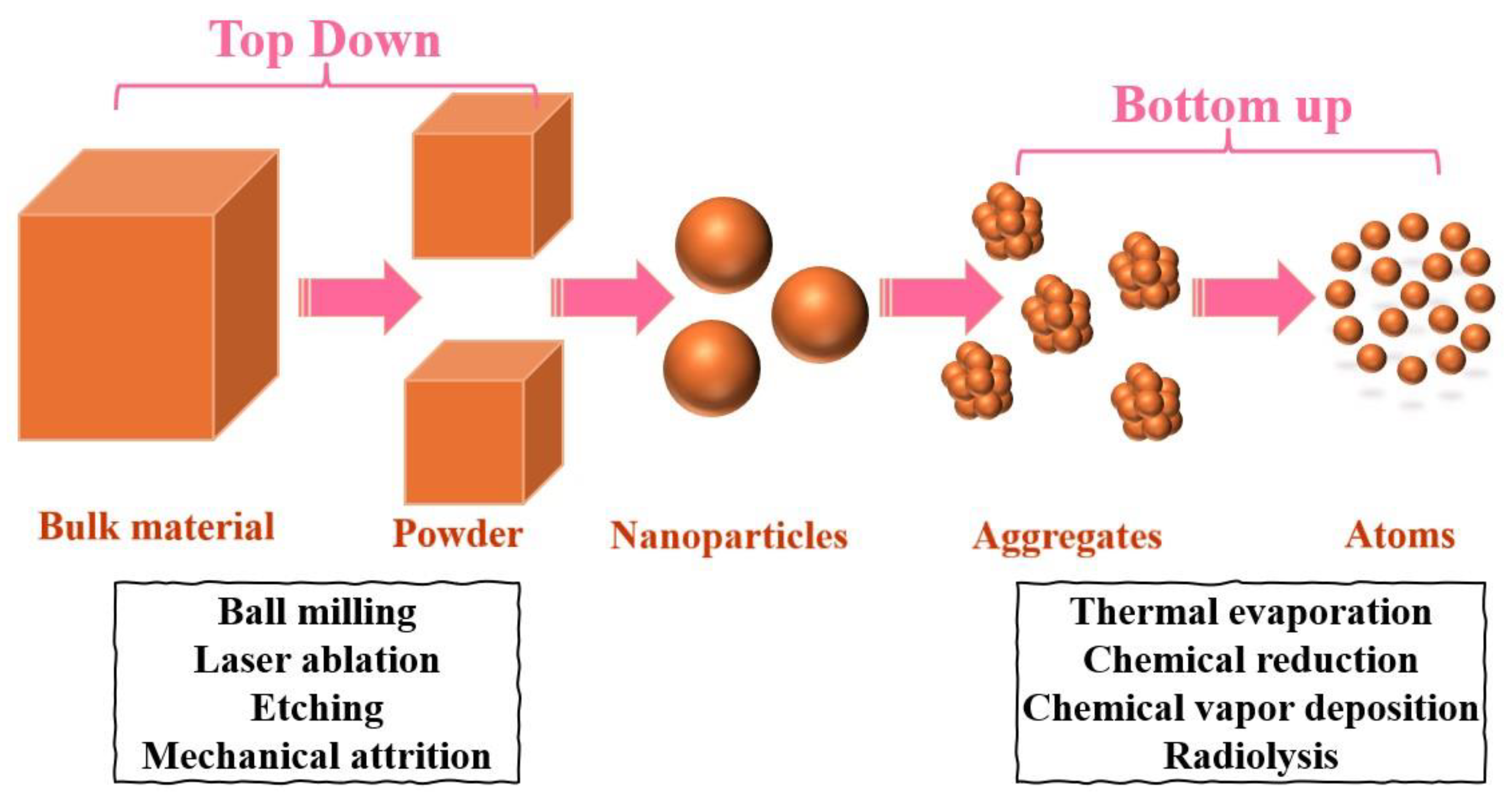
Scheme 2.
Schematic illustration of nanomaterial synthesis by high irradiation dose.
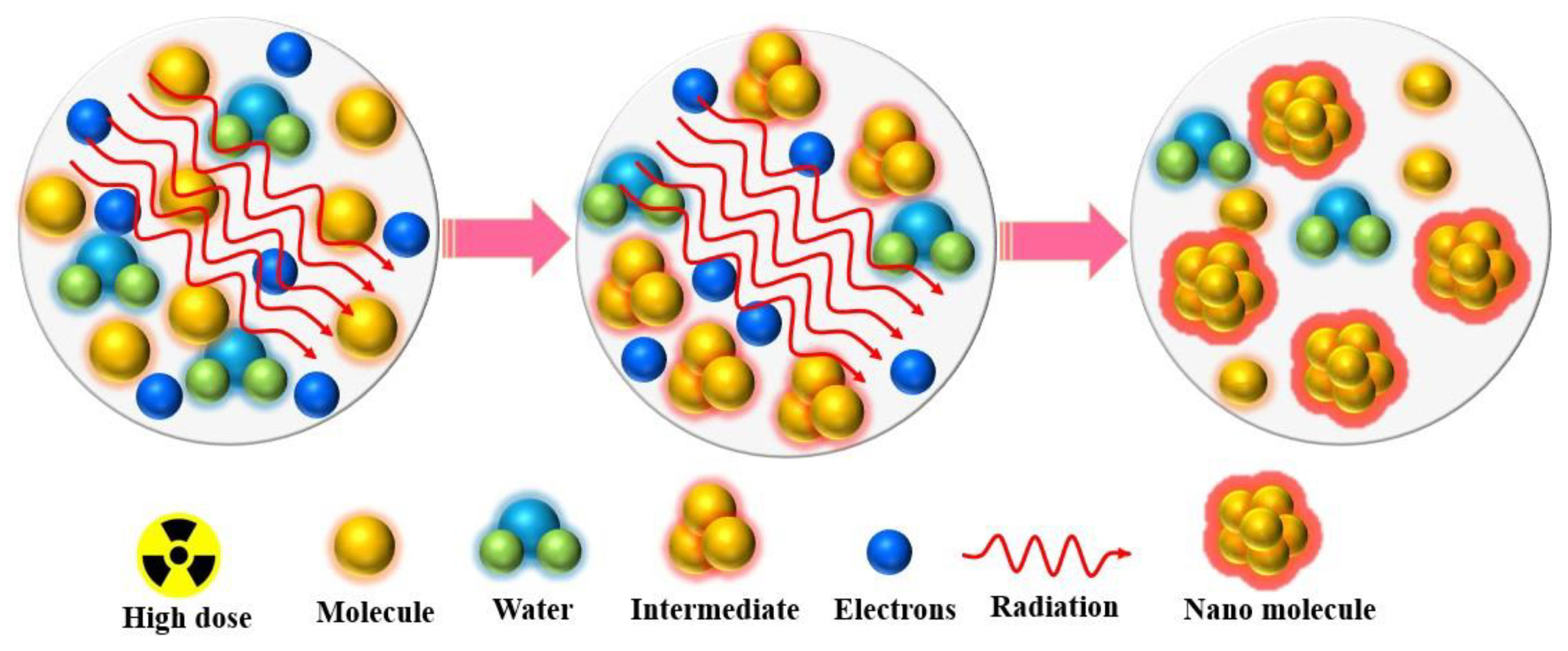
Scheme 3.
Schematic illustration of nanomaterial synthesis by low irradiation dose.
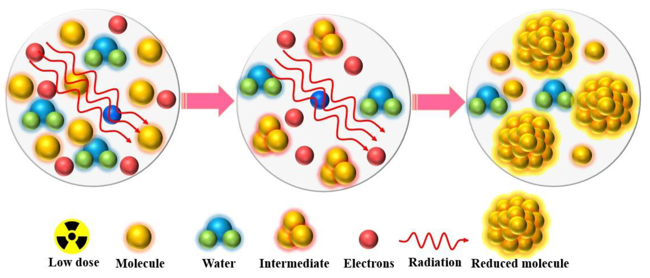
Figure 1.
(a) Conductivity of specimens, both non-irradiated and irradiated, at ambient temperature. (b) Optical band gap and energy need for activation to the electron beam exposure for PVA/Au nanocomposite, before and after irradiation. (c) Schematic of the preparation of PVA/Au NPs film, reproduced with permission from ref. [43], Elsevier.
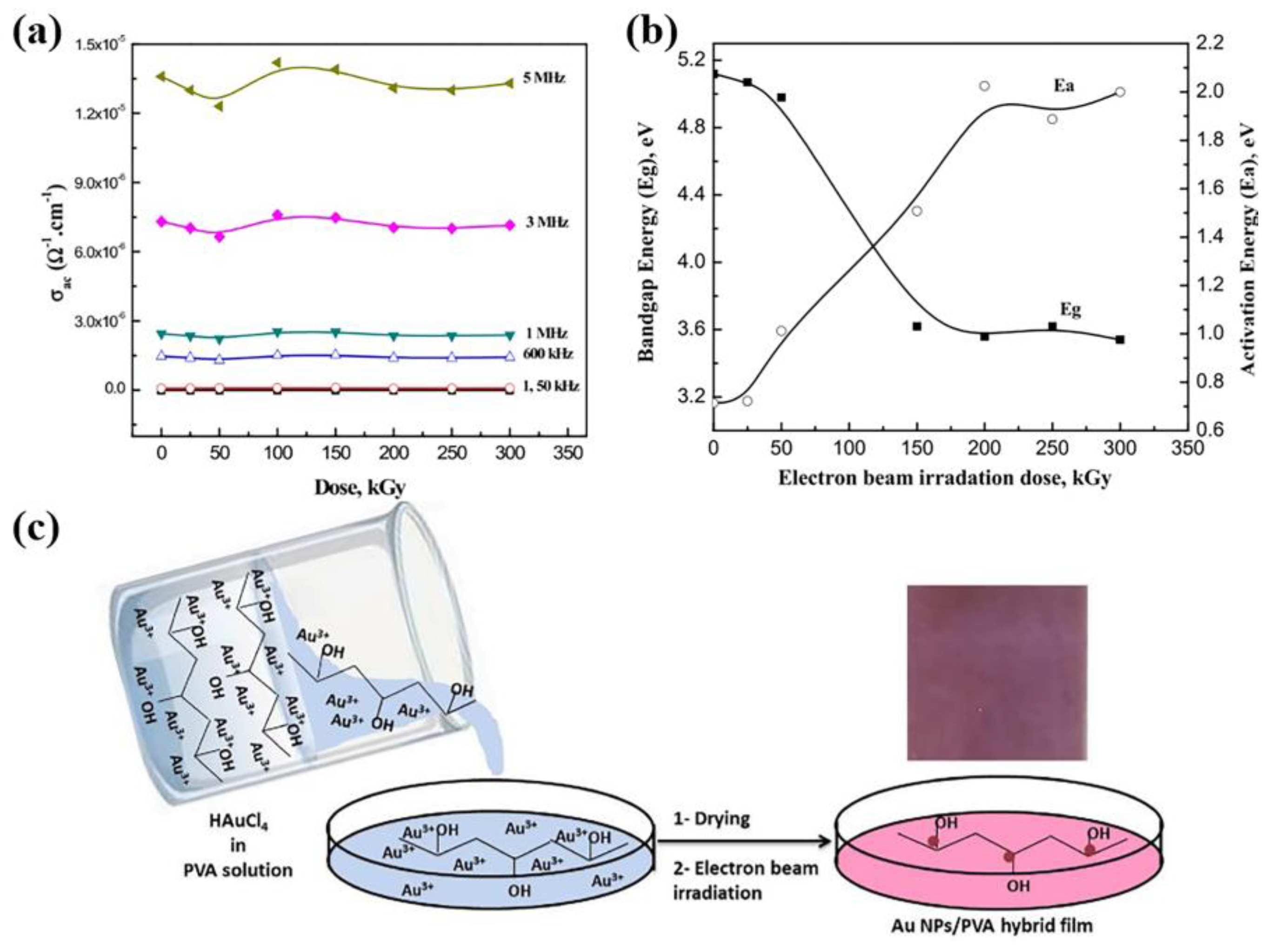
Figure 2.
Electron beam (EB) irradiation experimental setup for AgNPs preparation. There are several components, including: 1 - Exit head for electron accelerators (6 MeV, 2 kGy/min or 5 MeV, 5-10 kGy/s); 2 - EB collection monitor in ring shape (for calculating dose rate and accumulating dose); 3 - EB field; 4 - Al foil; 5 - Reaction vessel; 6 - Irradiated samples (thickness is adjusted for density and EB energy of the samples); 7 - Magnetic stirrer; 8 - Pump used to measure precursor solution; 9 - Argon bubbling system , reproduced from ref. [59], Published by De Gruyter Open Access.
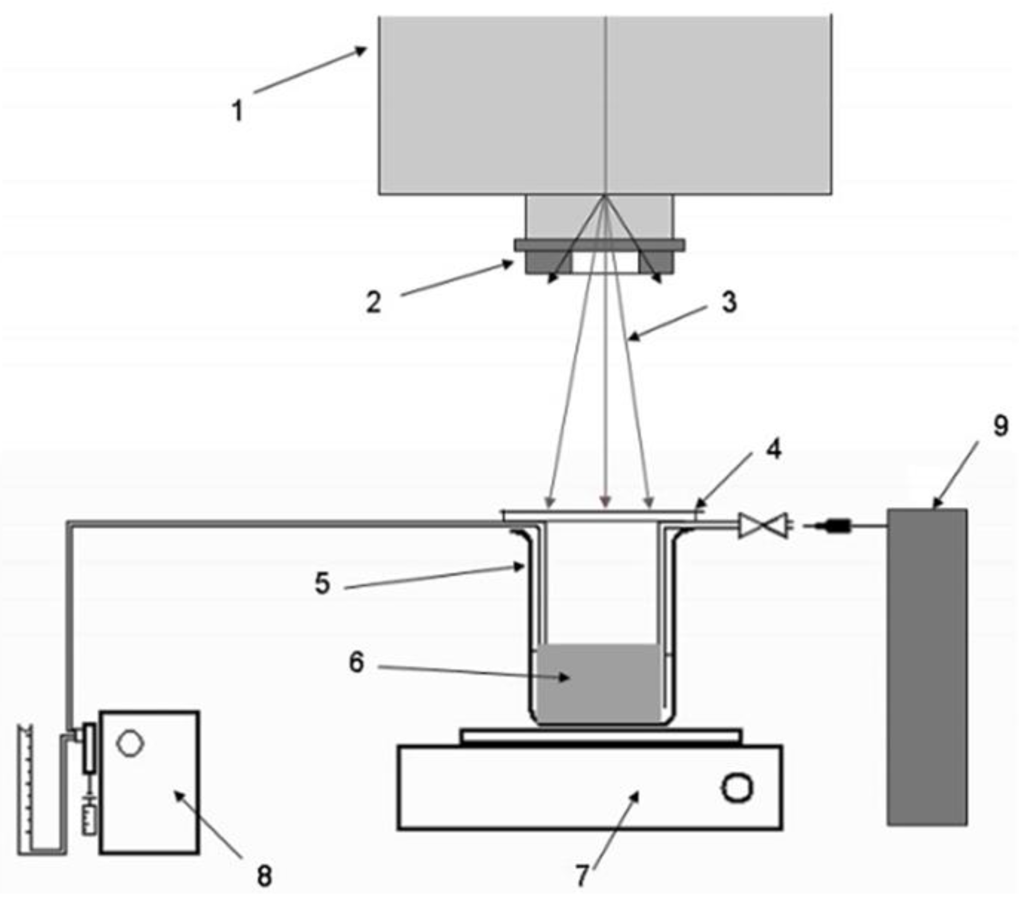
Figure 3.
A series of in situ FE-SEM images depicts Ag growth on α-Ag2WO4 crystals after 30 kV electron exposure without heating: (a) The initial image shows the crystal surface before exposure, (b) after 6 minutes, modeled defects indicate the formation of Ag nanoparticles. (c) After 10 minutes, the images reveal the development of Ag nanorods in the same region, reproduced from ref. [71], Nature publication.
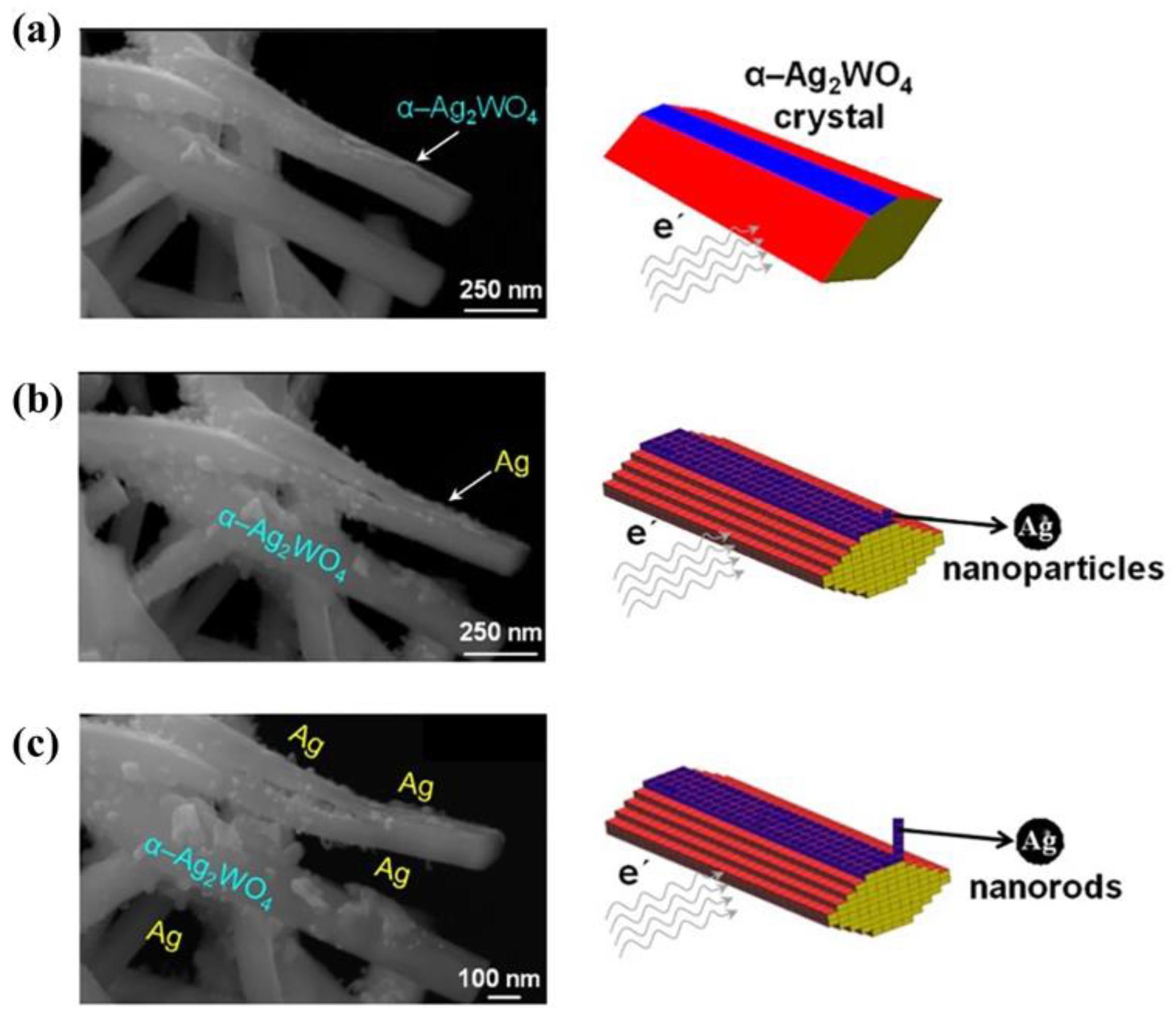
Figure 4.
FE-SEM images of β-Ag2MoO4 microcrystals (left) and silver nanoparticle formation on β-Ag2MoO4 facets at 5, 10, 15, and 20 kV (right); with area distribution histograms for silver particles across different voltages after 5 minutes, reproduced from ref. [73], Taylor & Francis.
Figure 4.
FE-SEM images of β-Ag2MoO4 microcrystals (left) and silver nanoparticle formation on β-Ag2MoO4 facets at 5, 10, 15, and 20 kV (right); with area distribution histograms for silver particles across different voltages after 5 minutes, reproduced from ref. [73], Taylor & Francis.
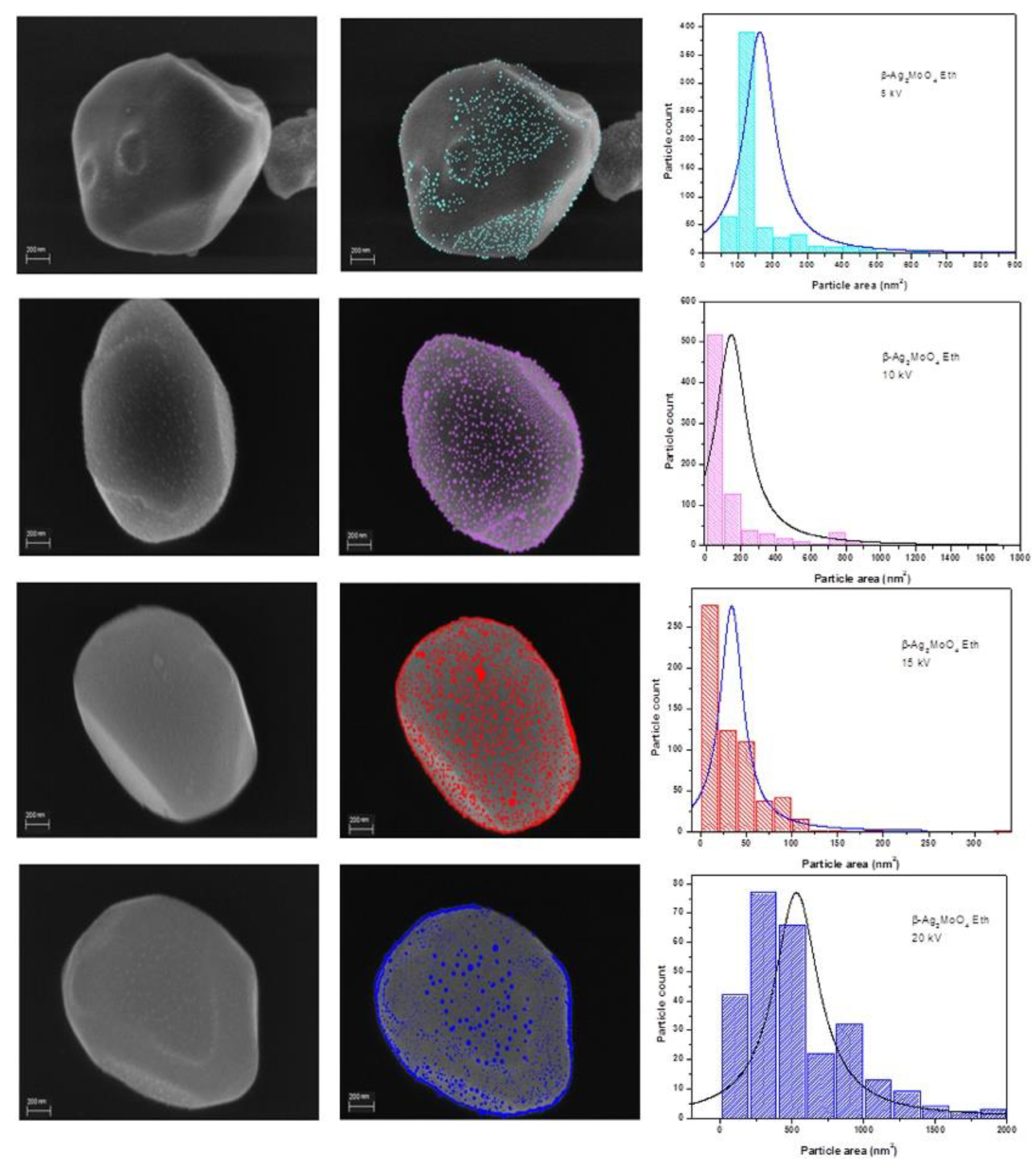
Figure 5.
(a–c) TEM images showing different stages of a precursor under different EBCD. (d–f) Schematic diagrams showing Ag atoms escaping from the precursor, reproduced with permission from ref. [86], Elsevier.
Figure 5.
(a–c) TEM images showing different stages of a precursor under different EBCD. (d–f) Schematic diagrams showing Ag atoms escaping from the precursor, reproduced with permission from ref. [86], Elsevier.
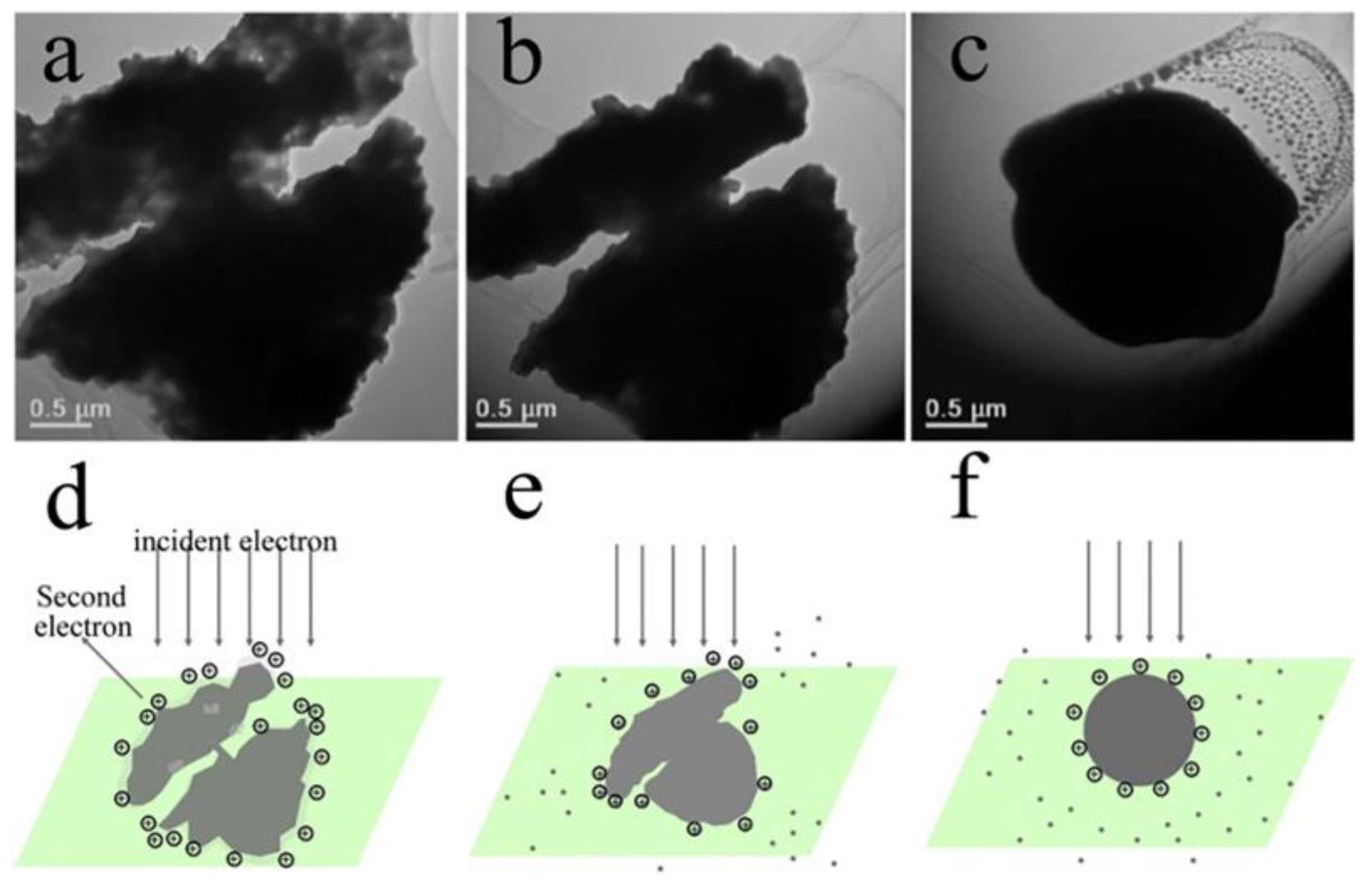
Figure 6.
SEM images of (a-b) the hydrolysis synthesized TiO2 nanoparticles and (c-d) the E-beam irradiated TiO2 nanoparticles, reproduced from ref. [98], The Korean Electrochemical Society.
Figure 6.
SEM images of (a-b) the hydrolysis synthesized TiO2 nanoparticles and (c-d) the E-beam irradiated TiO2 nanoparticles, reproduced from ref. [98], The Korean Electrochemical Society.
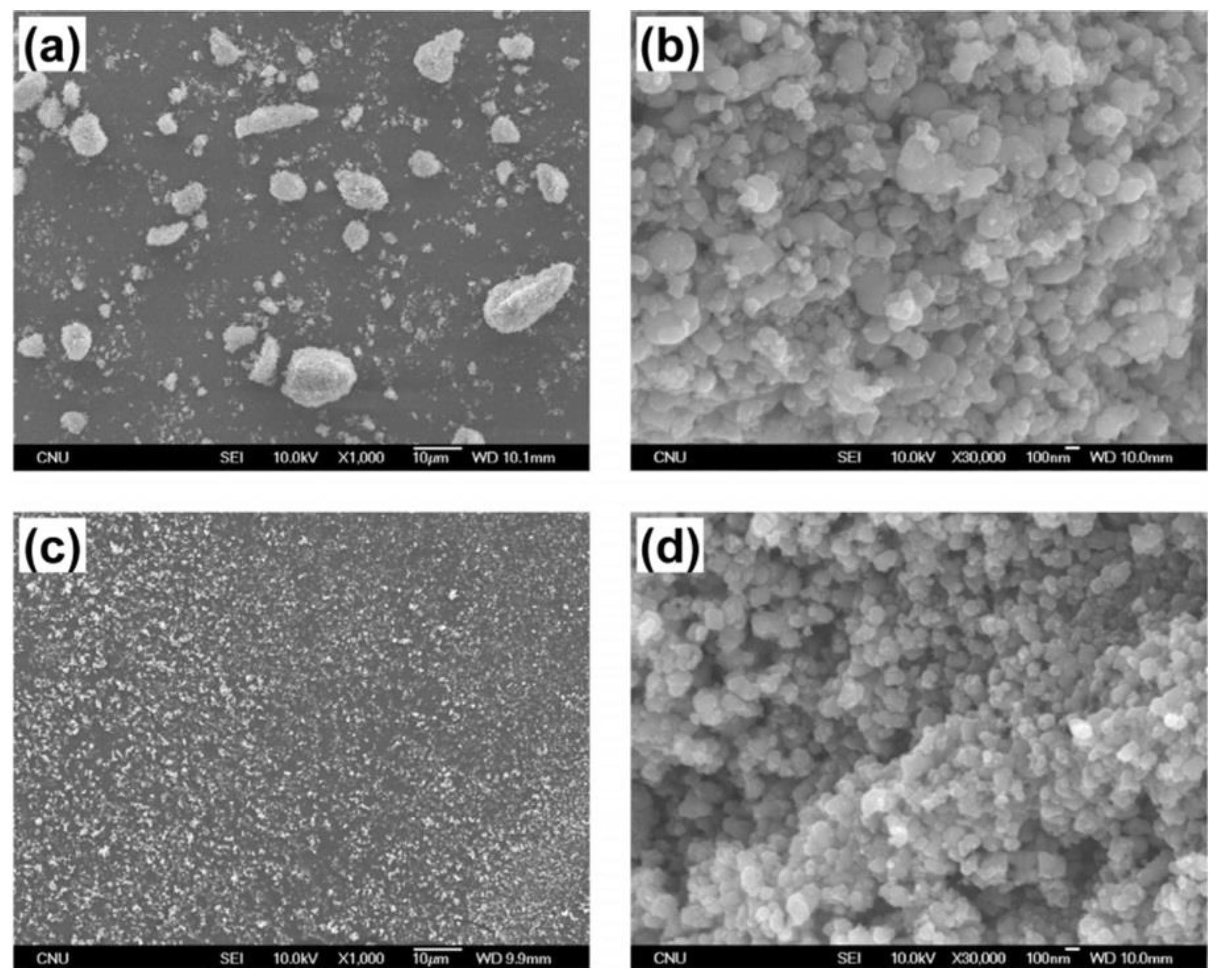
Figure 7.
Schematic diagram of laser synthesis for CrMnFeCoNi high-entropy alloy (HEA) nanoparticles and iron-doped HEA/lignin composite (IE-HEA/LIG), reproduced from ref. [108], Nature Publication.
Figure 7.
Schematic diagram of laser synthesis for CrMnFeCoNi high-entropy alloy (HEA) nanoparticles and iron-doped HEA/lignin composite (IE-HEA/LIG), reproduced from ref. [108], Nature Publication.
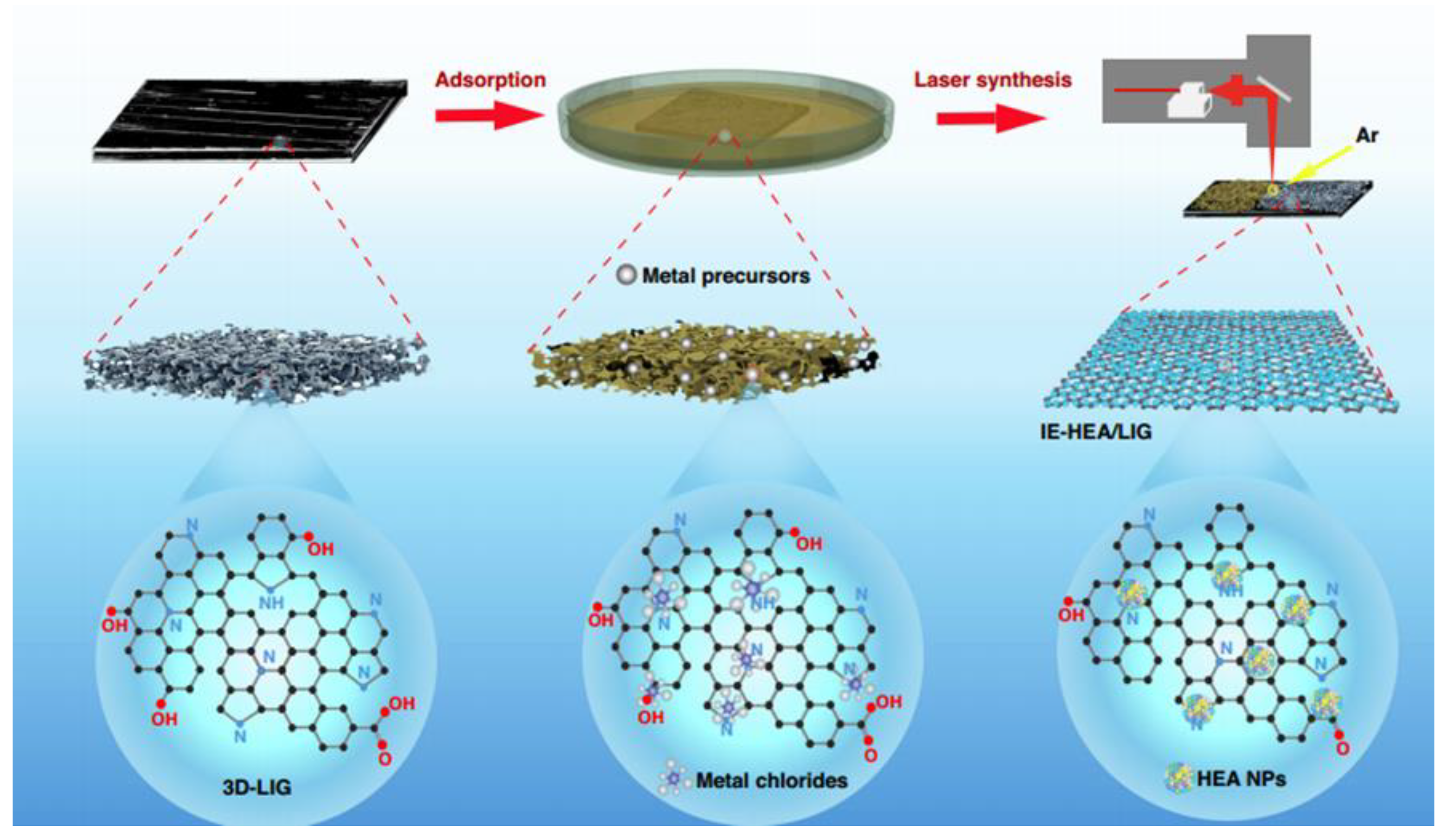
Scheme 4.
Experimental setup of gamma radiation synthesis.
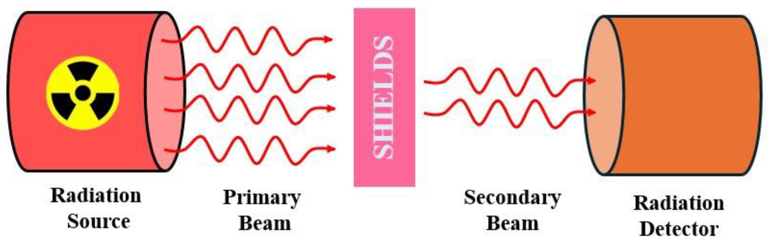
Figure 8.
(A) Experimental setup for the determination of photothermal properties of GO/Au NP composites. (B) TEM images of GO/Au NP composites prepared at a) 1 kGy, b) 5 kGy, c) 10 kGy, and d) 20 kGy irradiation dose, reproduced with permission from ref. [124], Elsevier.
Figure 8.
(A) Experimental setup for the determination of photothermal properties of GO/Au NP composites. (B) TEM images of GO/Au NP composites prepared at a) 1 kGy, b) 5 kGy, c) 10 kGy, and d) 20 kGy irradiation dose, reproduced with permission from ref. [124], Elsevier.
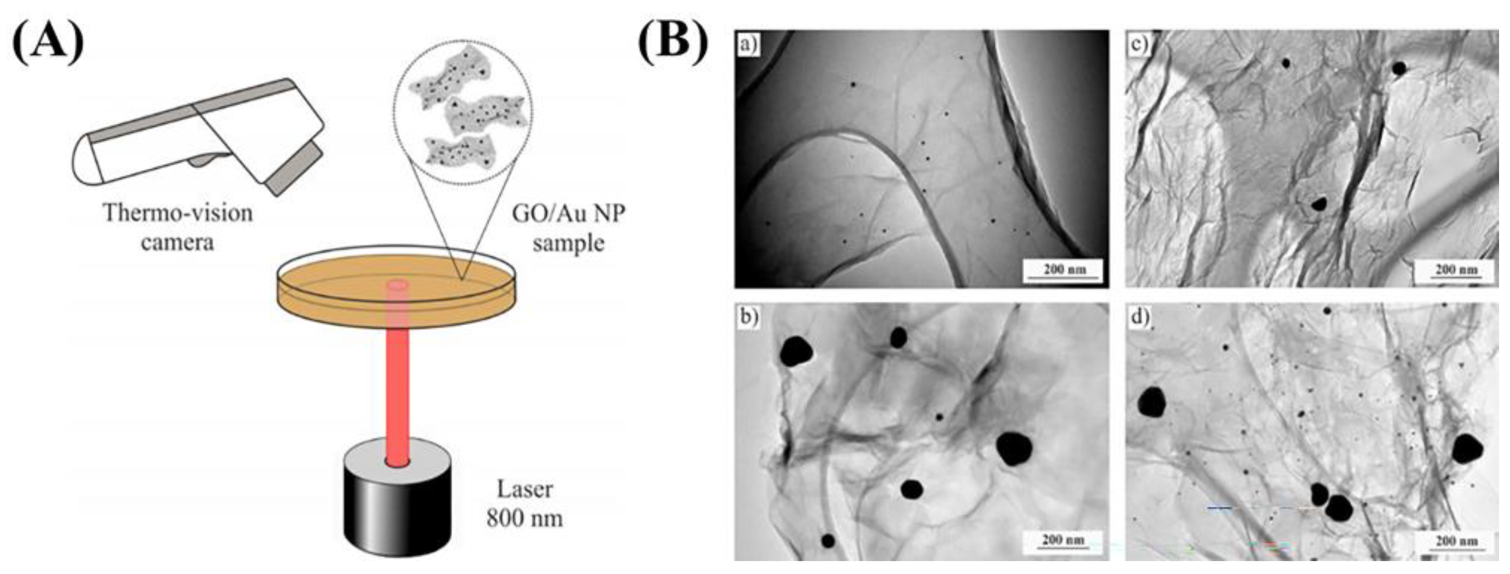
Figure 9.
(a) Concept developing the GAT nanocomposite to facilitate the photodecomposition of rhodamine B. (b) Gamma irradiation-induced water radiolysis and the subsequent formation of metal atoms, reproduced from ref. [127], Wiley & Sons.
Figure 9.
(a) Concept developing the GAT nanocomposite to facilitate the photodecomposition of rhodamine B. (b) Gamma irradiation-induced water radiolysis and the subsequent formation of metal atoms, reproduced from ref. [127], Wiley & Sons.
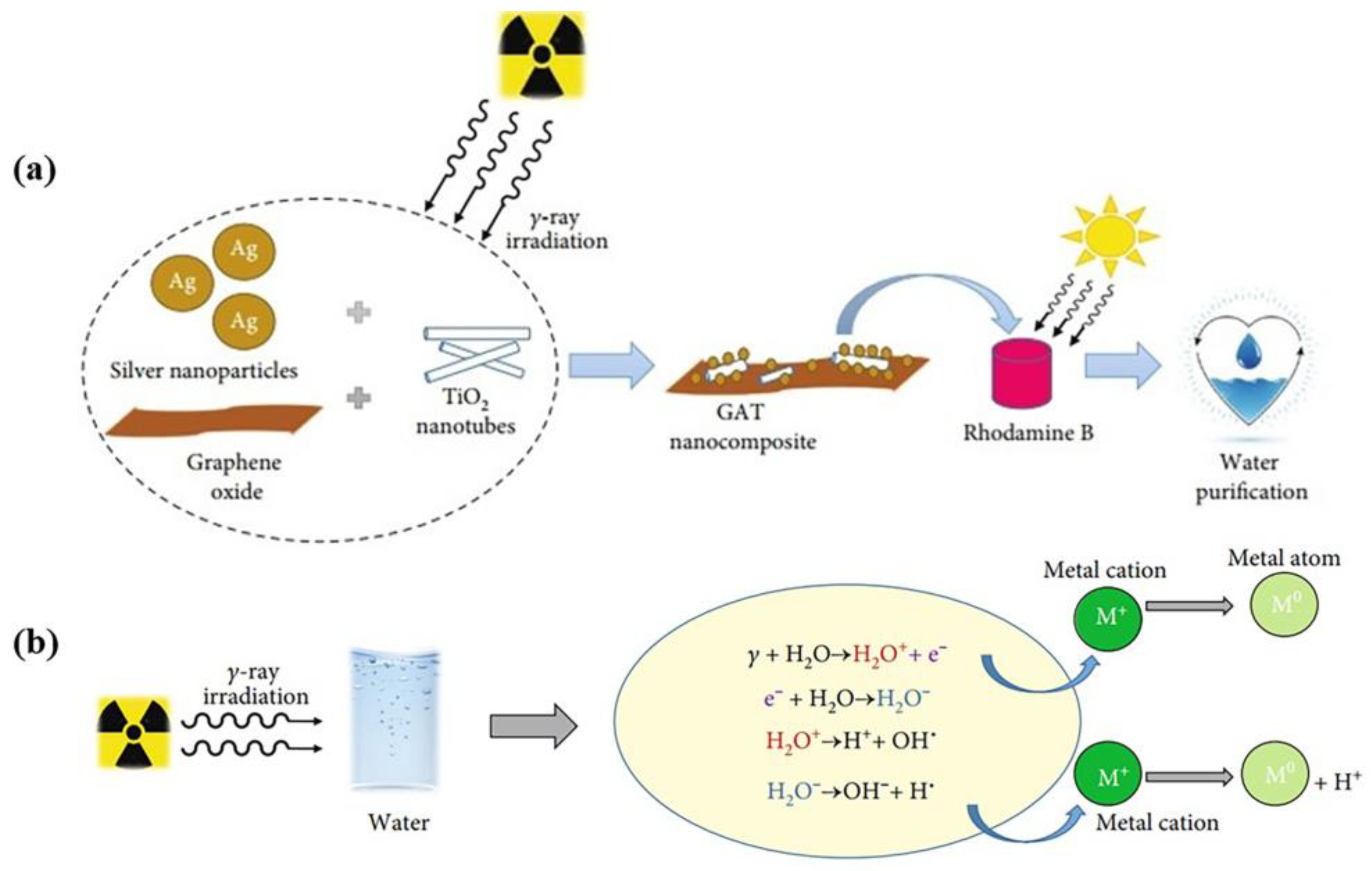
Figure 10.
Schematic overview of the antibacterial action of PVP@D-AgNs: 1. They adhere to bacterial surfaces, disrupting membranes. 2. They generate reactive oxygen species (ROS), weakening the cell wall. 3. They block ion movement. 4. They enter cells, impair organelles, and cause cell lysis., reproduced from ref. [128], Spinger.
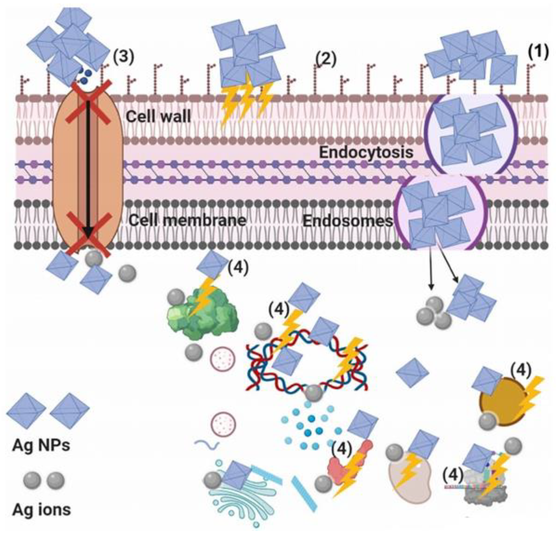
Figure 11.
Schematic representation of the PVA/Ag/CaTiO3 nanocomposite film synthesis, reproduced from ref. [132], Nature publication.
Figure 11.
Schematic representation of the PVA/Ag/CaTiO3 nanocomposite film synthesis, reproduced from ref. [132], Nature publication.
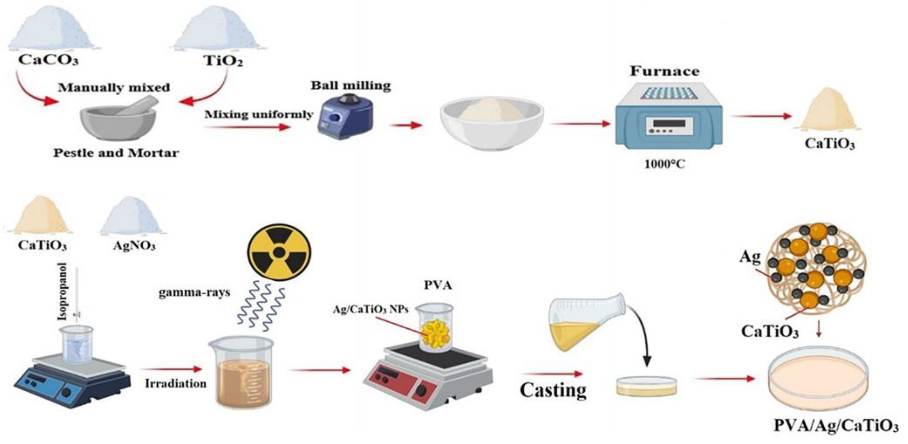
Disclaimer/Publisher’s Note: The statements, opinions and data contained in all publications are solely those of the individual author(s) and contributor(s) and not of MDPI and/or the editor(s). MDPI and/or the editor(s) disclaim responsibility for any injury to people or property resulting from any ideas, methods, instructions or products referred to in the content. |
© 2024 by the authors. Licensee MDPI, Basel, Switzerland. This article is an open access article distributed under the terms and conditions of the Creative Commons Attribution (CC BY) license (http://creativecommons.org/licenses/by/4.0/).
Copyright: This open access article is published under a Creative Commons CC BY 4.0 license, which permit the free download, distribution, and reuse, provided that the author and preprint are cited in any reuse.


