Preprint
Article

This version is not peer-reviewed.

Effect of Selenium, Copper and Manganese Nanocomposites in Arabinogalactan Matrix on Potato Colonization by Phytopathogens Clavibacter sepedonicus and Pectobacterium carotovorum

A peer-reviewed article of this preprint also exists.

Submitted:

20 November 2024

Posted:

21 November 2024

You are already at the latest version

Abstract

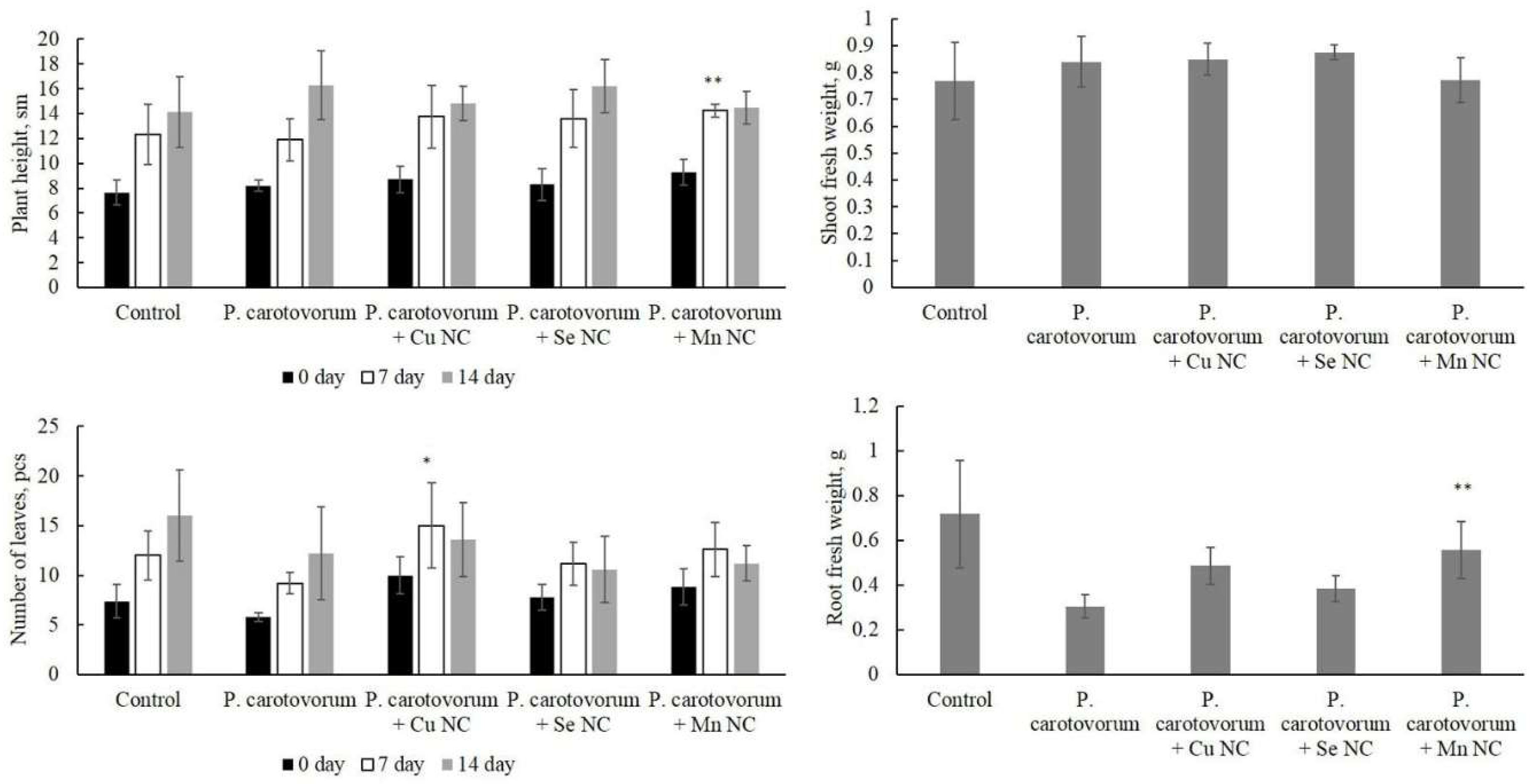
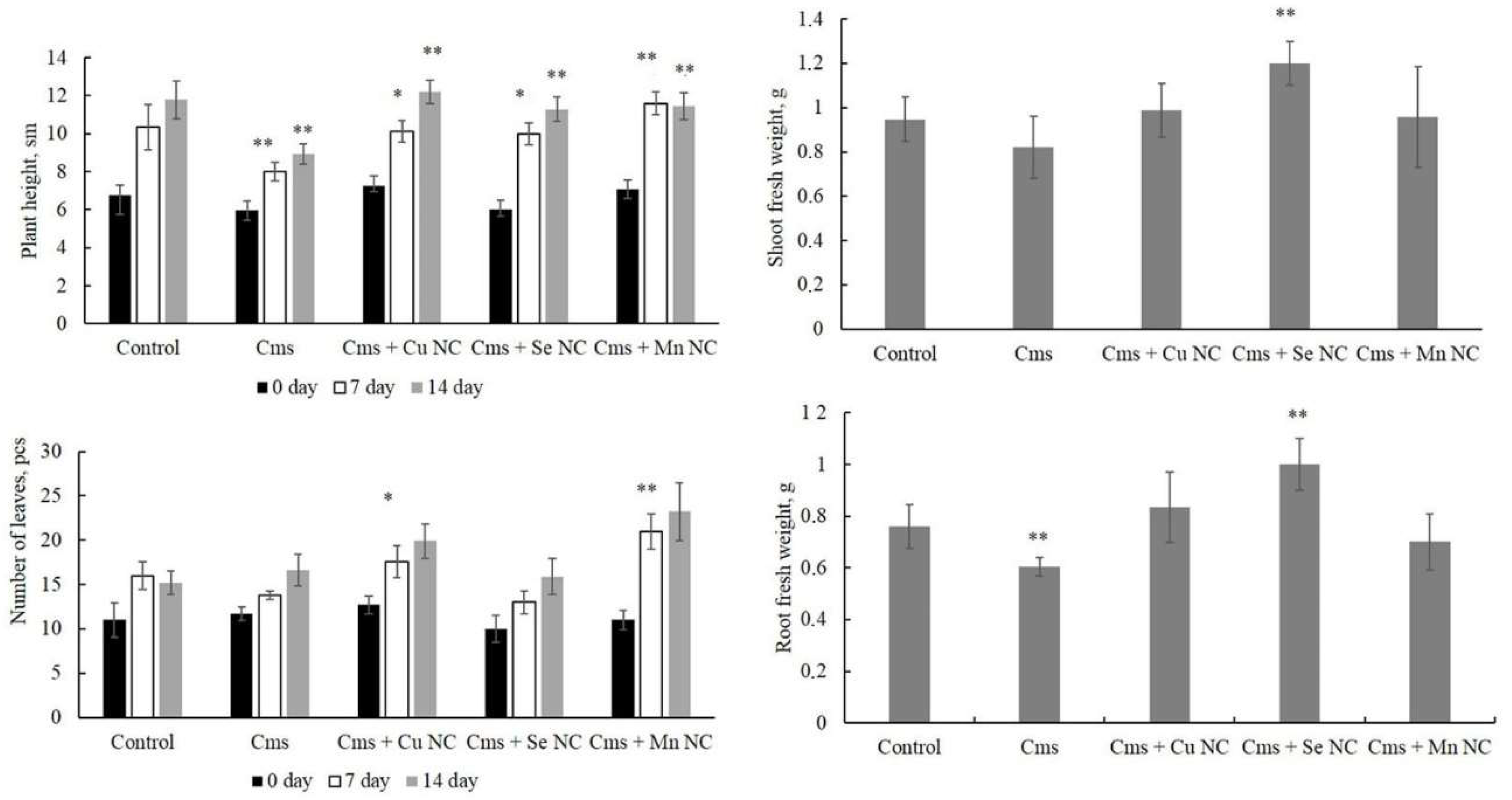
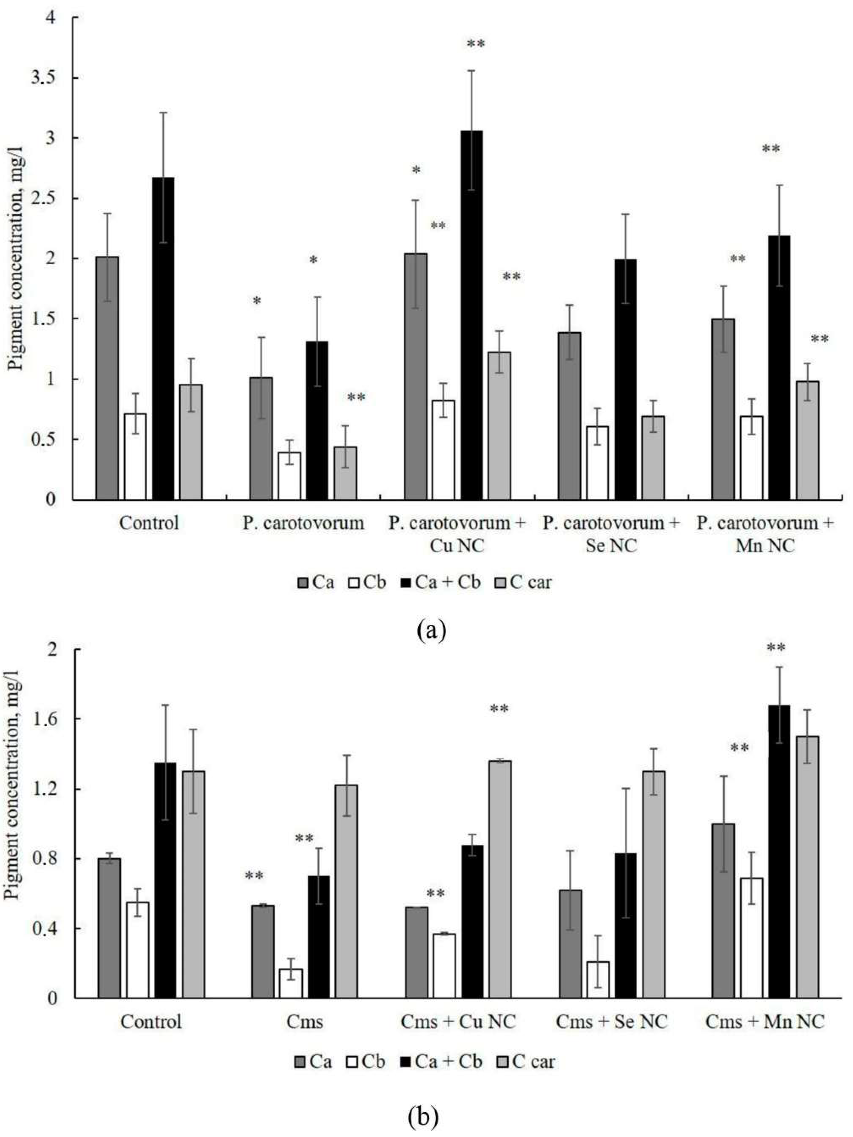
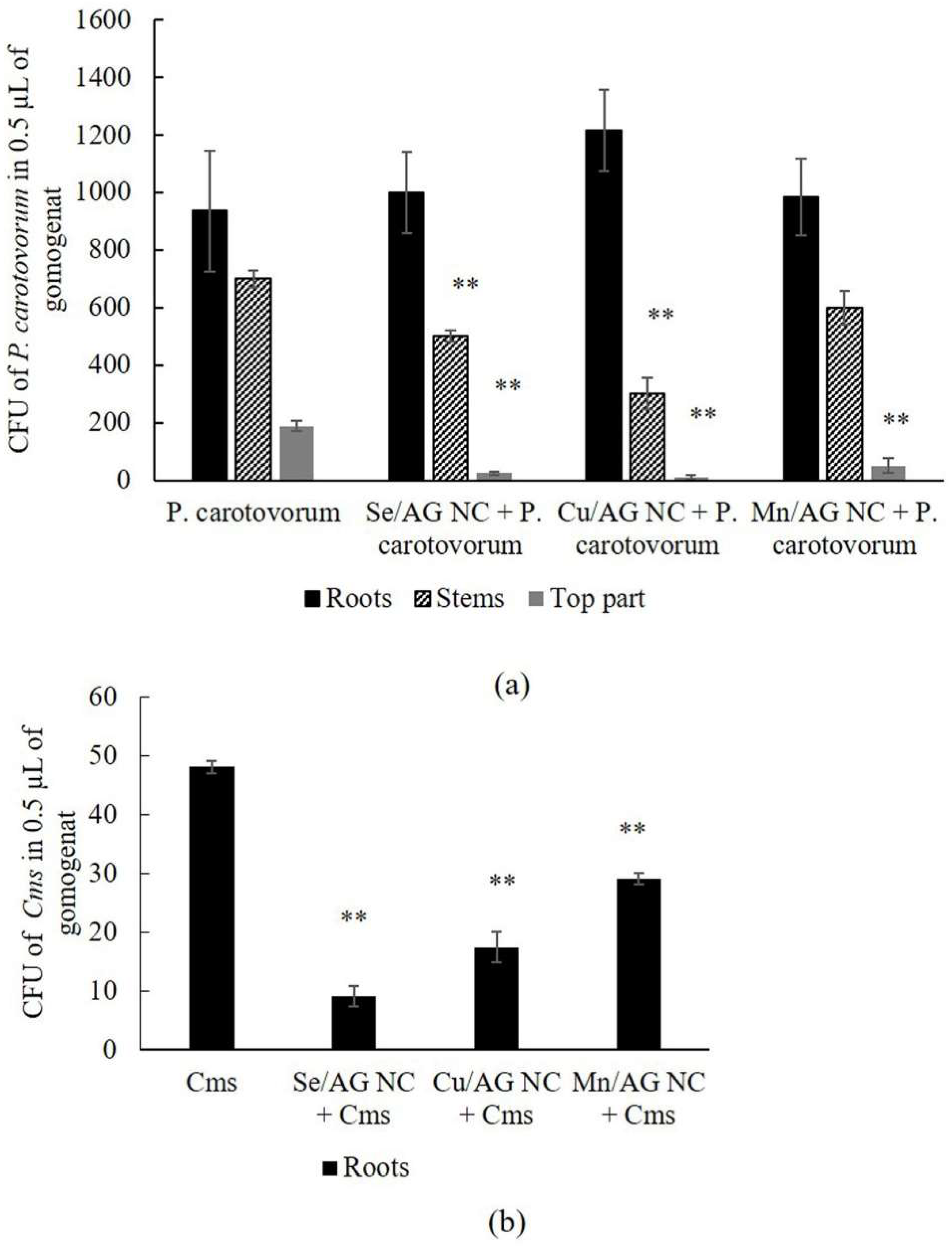
The effect of chemically synthesized nanocomposites (NCs) of selenium (Se/AG NC), copper oxide (Cu/AG NC) and manganese hydroxide (Mn/AG NC) based on the natural polymer arabinogalactan (AG) on the processes of growth, development and colonization of potato plants in vitro was studied upon infection with the causative agent of potato blackleg – the Gram-negative bacterium Pectobacterium carotovorum and the causative agent of ring rot – the Gram-positive bacterium Clavibacter sepedonicus (Cms). It was shown that infection of potatoes with P. carotovorum reduced root formation of plants and the concentration of pigments in leaf tissues. Treatment of plants with Cu/AG NC before infection with P. carotovorum stimulated leaf formation and increased the concentration of pigments in them. A similar effect was observed when potatoes were exposed to Mn/AG NC, and an increase in growth and root formation was also observed. Infection of plants with Cms inhibited plant growth. Treatment with each of the NCs mitigated this negative effect of the phytopathogen. At the same time, Se/AG and Mn/AG NCs promoted leaf formation. Se/AG NC increased the biomass of Cms-infected plants. Treatment of plants with NCs before infection showed a decrease in the intensity of colonization of plants by bacteria. The Se/AG NC had the maximum effect, which is probably due to its high antioxidant capacity. Thus, the NCs are able to mitigate the negative effect of bacterial phytopathogens on vegetation and the intensity of colonization by these bacteria during infection of cultivated plants.

Keywords: 
;  ;  ;  ;  ;  ;  ;  ;  

1. Introduction

Modern climate change promotes expansion of many phytopathogens to the new northern and eastern areas [1,2]. These microorganisms include the causative agent of potato black leg - the Gram-negative bacterium Pectobacterium carotovorum, which remains highly virulent at elevated temperatures, can quickly spread through the vascular system of the plant and remain latent when seeds are stored at low temperatures [3]. During the growing season, the symptoms of black leg are similar to the symptoms of potato ring rot (wilt, yellowing of leaves), caused by the Gram-positive bacterium Clavibacter sepedonicus (Cms). Both P. carotovorum and Cms are quarantine species in many countries around the world [4,5]. Currently used pesticides are aimed primarily at combating fungal infections of potatoes and are ineffective against bacterial phytopathogens. In this regard, the development of new plant protection agents is highly needed. In order to meet this task, the possibility of using nanosubstances is being considered [6,7,8]. The use of nanocompounds for the development of agents that increase plant resistance to diseases seems promising.
Previously, we studied a number of nanocomposites (NCs) of selenium (Se) and silver (Ag) in native matrices as agents for regulating the abundance of Cms. The studied NCs are Se or Ag nanoparticles (NPs) stabilized by natural polymers such as arabinogalactan (AG) isolated from Siberian larch (Larix sibirica L.) wood, potato starch, carrageenan and humic acids. Se/AG NC with a Se content of 6.4% was characterized by high antibacterial activity against Cms [9,10]. In addition, this NC did not have a negative effect on potato plants when visually observed over time [11,12]. However, the detailed influence of Se/AG NC on the interaction of potato with the bacterium Cms has not yet been elucidated. Therefore, the aim of this work is to study the influence of Se, Cu and manganese (Mn) NC based on AG on the colonization of potato plants in vitro by bacterial pathogens P. carotovorum and Cms.

2. Results

The effect of phytopathogens alone and in combination with NCs on potato growth and development, as well as the biomass of roots and aboveground parts of plants, were assessed in detail in vitro (Figure 1 and Figure 2). Plants without any treatment (pure control), as well as the effect of phytopathogens without the addition of NCs, were used as controls. In these experiments, the effect of NCs on plants alone was not studied, since such experiments were carried out previously, and the results are described in detail in [13,14,15].
The results showed that infection of plants with the phytopathogen P. carotovorum did not have a pronounced negative effect, except for the weight of flesh root compared to the pure control (Figure 1). Treatment of the infected potato with NCs also did not lead to any significant results, except for apparent stimulation of potato growth (height) under the influence of Mn/AG NC, number of leaves under the influence of Cu/AG NC on the 7th day of the experiment, and weight of flesh root under the influence of Mn/AG NC when compared with infected samples without NCs treatment.
The results showed that infection of plants with the phytopathogen Cms resulted in a decrease in the intensity of plant growth on days 7-14 of the experiment. At the same time, treatment of plants with NC mitigated this negative effect of Cms infection, all NCs stimulated plant growth on days 7 and 14 of the experiment compared to the treatment with infection only. A reliable increase in the number of leaves on day 7 of the experiment was found in plants treated with Cu/AG and Mn/AG NCs. Se/AG NC significantly increased the biomass of fresh shoots and the biomass of roots of potato plants infected with Cms (Figure 2).
To assess the physiological state of the plants at the end of the experiment, the concentration of pigments in the leaf tissues was determined (Figure 3). It was found that infection with P. carotovorum causes a reliable decrease in the concentration of pigments in the tissues of potato leaves. Cu/AG NC significantly increased the concentration of all the studied pigments in the tissues of infected potatoes; their values reached the levels of pigment concentration in uninfected plants. No significant stimulation of an increase in the concentration of pigments was found when treating plants with NC Se/AG compared to infection. Mn-containing NC increased the content of chlorophyll b and carotenoids (Figure 3a).
When infecting plants with Cms, a decrease in the concentration of chlorophylls was found. Cu-containing NC increased the concentration of chlorophyll b in the tissues of plant leaves during infection. Se-containing NC did not have a pronounced effect on the studied trait. Mn-containing NC increased the concentration of chlorophylls in potato tissues during infection (Figure 3b).
The results showed that infection of plants with Cms and P. carotovorum led to a negative effect on the morphometric parameters of plants and causing a decrease in the concentration of pigments in leaf tissues. NCs mitigated the negative effect of infection, bringing the studied parameters to the level of the control without infection. However, it is important to understand to what extent the plant was seeded with microorganisms and how NCs affect this process. To address this issue we studied the effect of treating plants with NCs before infection on the intensity of potato colonization by P. carotovorum and Cms bacteria.
Potato plants were treated with NCs and incubated for 1 hour, since during this time the level of ROS in potato tissues increases [10,14]. The plants were then infected with Cms and after two days of coincubation and complete colonization with the pathogen, microbiological inoculations of the homogenate obtained from potato tissues were carried out according to [16]. It was found that bacteria were inoculated from all zones of plants both in the control without NCs treatment and with NCs treatment (Figure 4).
A high number of CFUs of P. carotovorum was observed in all infected plant parts without NC treatments (Figure 4a). All NCs reduced bacterial CFUs in the apical (top) part. Cu/AG and Se/AG NCs reduced CFUs in the stem part of plants. The most pronounced effect was noted when treating plants with Se/AG and Cu/AG NCs (Figure 4a). The number of CFUs decreased when treating plants infected by Cms with any NC compared to plants untreated with NCs. The maximum effect was observed in the Se/AG NC +Cms treatment (Figure 4b).

3. Discussion

In this study, we analyzed the effect of Cu/AG, Se/AG and Mn/AG NCs embedded in an AG matrix on potatoes infected in vitro with the necrotrophic Gram-negative phytopathogenic bacterium P. carotovorum and the biotrophic Gram-positive phytopathogenic bacterium Cms. The effect of NCs on vegetation, morphometric parameters, pigment concentration and the intensity of plant colonization by the pathogen were analyzed. The use of these particular phytopathogens in our experiments was motivated by the fact that these bacteria are extremely virulent and resistant to exposure factors [17], and are quarantine objects in different countries [4,5]. Both bacteria are characterized by the ability to form biofilms, which is a key factor in the virulence of P. carotovorum [18,19] and Cms, leading to the formation of biofilms in the vascular pathways of plants and clogging them. P. carotovorum has a large set of pectolytic enzymes and toxins [20]. This bacterium damages plant tissues due to enzymes that destroy the plant cell wall (pectinases, cellulases and proteinases released through type II secretion systems). The pathogenicity factors of the necrotrophic pathogen include a complex of enzymes, including pectate lyases, exopolysaccharides and lipopolysaccharides. Plants are able to demonstrate resistance due to the binding of toxins and their removal, as well as the presence of phenols that inactivate exoenzymes [21]. Cms also has exopolysaccharides and is able to secrete cellulase, protease, endoglucanase, xylanase, glycosyl hydrolase and serine proteases [5,19]. The defense response of potatoes to Cms invasion is largely associated with the activation of antioxidant enzymes during infection, but data on this response are still insufficient.
We have shown earlier the fungicidal effect of Se NC against the phytopathogenic fungus Phytophthora cactorum [22] and the antibacterial effect of Se NC on Cms [13]. It was found that Se NC is able to reduce the growth of the bacterial culture of Cms, inhibit biofilm formation and increase the number of dead cells in the bacterial suspension [13]. In our experiments in vitro, it was revealed that Se / AG and Mn / AG NCs have bacteriostatic, antibiofilm, bactericidal effects on Cms [13,14]. The antibacterial effect of NCs on Cms may be due to the suppression of bacterial respiration, as evidenced by a decrease in cell dehydrogenase activity and a change in the fatty acid composition of bacterial cell walls under the influence of NCs, as well as an effect on the transmembrane potential of cells [23]. There are also data on the suppression of the viability of P. carotovorum by nanocompounds [24]. The antibacterial activity of biosynthesized chitosan NP against P. carotovorum has been demonstrated in [25]. CuS NPs inhibited the motility of P. carotovorum [26]. Ag NP exhibited high antibacterial activity against P. carotovorum; it completely inhibited the growth of bacteria, led to the destruction of the bacterial cell membrane and inhibition of biofilm formation [27].
The effect of nanocompounds on phytopathosystems is usually presented in the published data by studying the expression of genes and the activity of plant stress enzymes [28,29,30,31,32]. There are practically no studies on the intensity of plant colonization by the pathogen. Therefore, in our study, we studied how NCs affect the colonization of plants by the pathogen during their artificial infection and pre-treatment with NC. The results we obtained are summarized in Table 1.
Cu and Mn belong to the group of microelements essential for living organisms. These elements play an important role in biochemical processes. Thus, in potatoes, microelements activate enzymes, participate in the synthesis of vitamins, and promote the adsorption of moisture, which in turn has a positive effect on plant growth and increases their resistance to stress. Cu is a component of enzymes involved in carbohydrate and protein metabolism, and is involved in photosynthesis and respiration [33]. Cu is also involved in many vital physiological functions of plants, acting as a catalyst in oxidation-reduction reactions in mitochondria, chloroplasts, and cell cytoplasm [34] or as an electron carrier in the process of plant respiration [35].
Our experiments showed that Cu/AG NC had a positive effect on plant growth when infected with Cms and on their development when infected with P. carotovorum. The stimulating effect of Cu/AG NC is also confirmed by published data [36,37,38,39,40,41]. For instance, it was shown that Cu/AG NC stimulated an increase in germination, shoot and root length during the cultivation of seeds of the Zea mays maize hybrid variety Hema [36]. Spraying the leaves of Dracocephalum moldavica L. with CuO NP increased the shoot biomass by 23% compared to the control [37]. The use of Cu/AG NC had a positive effect on the morphological and physiological parameters of sweet basil Ocimum basilicum L. [38]. A positive effect of CuO NC on the growth and development of various woody crops was also noted [39,40,41].
The stimulating effect of Cu/AG NC can be explained by an increase in the concentration of photosynthetic pigments in the tissues of plant leaves. Cu/AG NC are capable of influencing the intensity of photosynthesis and the functioning of the antioxidant system of plants. A number of studies confirm this [37,42]. For example, spraying the leaves of D. moldavica L. with CuO NC increased the content of chlorophyll a by 77% and increased the content of chlorophyll b by 123% compared to the control [37]. It was shown that spraying the leaves of avocado plants with CuO NC stimulated the intensity of photosynthesis [42].
In our experiments, Se/AG NC did not have a pronounced effect on the growth and development of plants infected with P. carotovorum. However, when infecting potatoes with Cms, this NC stimulated all morphometric parameters: it increased the intensity of potato growth and leaf formation, root and fresh shoot biomass. Differences in the stimulating effect of this NC may be associated with its different effects on the studied pathogens. We have shown that Se/AG NC has a complex effect on Cms - it suppresses biofilm formation, respiration, and has bactericidal and bacteriostatic effects [10,13]. Similar effects of Se/AG NC were also shown for P. carotovorum, but their expression was less pronounced, which is apparently due to the difference in the structure of the bacterial cell wall. There are published data on the positive effect of Se NP on the physiological parameters of plants [43,44,45]. For example, exogenous spraying with nanoselenium enhances the growth of tobacco Nicotiana tabacum L. [43] and peanut Arachis hypogaea L. [44] plants. Melissa officinalis plants were treated with different concentrations of nanoselenium (10 and 50 mg/l) [45]. When treating plants with Se NP at a concentration of 10 mg/l, a sharp increase in biomass, activation of lateral buds and stimulation of lateral root development were observed [45].
In the experiments presented in this paper, the high biological activity was observed for Mn/AG NC. This activity of Mn/AG NC may be associated with the small size of Mn NPs. It is known that the absorption of Mn NPs by plants depends on their size. Typically, the pore size of plant cell walls is in the range of 3-8 nm, so large Mn NPs will not penetrate cell walls [46], and Mn NPs in the NCs we studied were in the specified size range of 3-6 nm [14]. Mn/AG NC stimulated plant growth and development during infection in both experimental variants, and also stimulated root formation during infection of plants with P. carotovorum. The stimulating effect of Mn is described in published data [47,48,49,50]. For example, it was shown that MnFe2O4 NPs were captured from the roots and migrate to the leaves, which helps to improve the growth parameters of barley [47]. FeOₓ NP, MnOₓ NP and bimetallic MnOₓ/FeOₓ NP had positive effects on the growth of Zea mays plants, particularly on seed germination rate, root growth and biomass growth of maize seedlings [48]. MnO2 NPs increased plant length, root length, leaf and flower number in bean (Phaseolus vulgaris) [49]. The application of ZnO-MnO NPs at a concentration of 270 ppm increased root size, dry biomass, chlorophyll content and leaf area in cabbage (Brássica oleracea) plants [50].
The stimulating effect of NC Mn/AG may be associated with an increase in the concentration of pigments in potato leaf tissues under its influence. Mn is known to affect the photosynthetic activity of plants [49,51,52]. This effect is associated with increased chloroplast stability, enhanced chlorophyll and carotenoid biosynthesis, increased ROS inactivation, activation of H+-ATPase gene expression, activation of ribulose-1,5-bisphosphate carboxylase/oxygenase (Rubisco), improved absorption of mineral elements, effects on the electron transport chain and oxidative phosphorylation, effects on ATP and NADPH synthesis. It is believed that the mechanism of activation of photosynthesis under the influence of NP metal oxides is associated, due to their small size, with their ability to penetrate plant chloroplasts, reach the reaction center of photosystem II, enhance electron transfer and light absorption in plant chloroplasts [53].
In the present study, a decrease in the intensity of plant root colonization by P. carotovorum and Cms was revealed. At the same time, NC did not completely block the penetration of bacteria into plant roots. This is probably due to the high infectious load on plants under the conditions of the model experiment and the fact that roots are the first plant organ to come into contact with pathogens. In addition, NC treatment occurred before plant infection. As a result, NPs from the NC composition could already move with the xylem flow to the aboveground plant organs, where the effect of some NCs has already been noted. It has been shown that roots absorb NPs through the main root, pores in the root cell wall, and damaged areas [54]. The intensity of colonization of the stem zone of plants decreased under the influence of Cu/AG NC when plants were infected with P. carotovorum and under the influence of Se/AG NC when potatoes were infected with Cms. A decrease in the contamination of the apical zone of potatoes was observed under the influence of all NCs. The decrease in the intensity of plant colonization by P. carotovorum in the apical zone of plants may be due to the fact that pre-treatment of plants with NC led to the absorption of NC by the plant and its distribution throughout the plant, which could lead to an increase in the activity of antioxidant enzymes and an effect on the expression of protective genes. It is known that Se NPs have high antioxidant activity [55]. Thus, it is known that Se NPs affect the activity of antioxidant enzymes in various plant organs - nitrate reductase in leaves and peroxidase (PER) in roots [30]. It has been shown that metal NPs can affect the expression levels of genes encoding vacuolar proton exchanger synthesis, superoxide dismutase (SOD), cytochrome P450-dependent oxidase and PER [56], microRNA genes [57] and genes encoding aquaporins in seeds [58]. Takehara et al. [59] demonstrated that MgO NPs induce strong immunity against Fusarium wilt in tomato, which is also associated with differential expression of several genes. These data provided new insights into the possible genetic mechanisms explaining this immunity. It is known that gene expression in plant cells can also be altered by Cu NP exposure. Thus, it was shown on the downy birch Betula pubescens that infection of plants with the pathogen Alternaria alternata significantly increased the level of transcripts of the MYB46 transcription factor, protective proteins LEA8, phenylalanine-ammonia lyase PAL, pathogen-dependent proteins PR-1 and PR-10 in birch microclones. When NP was added to the cultivation medium and simultaneously exposed to the phytopathogen, the expression of the MYB46, PR-1 and PR-10 genes decreased by 5.4 times. The obtained effect is associated with a decrease in the pathogenic load caused by exposure to NP and simultaneous stimulation of clones in vitro [41]. In the tissues of Glycine max soybean seedlings treated with CuO NP, a decrease in the expression activity of a number of genes involved in the cell division process was found [60]. It has been shown that the use of chitosan-polyvinyl alcohol hydrogel (Cs-PVA) in combination with Cu NPs promotes an increase in the expression of genes encoding the synthesis of jasmonic acid and SOD in tomato plant tissues under salt stress, mitigating its consequences [61]. A decrease in the expression of the pathogen-associated protein 1 (PR1) and polyphenol oxidase precursor (PoP) genes was observed in the tissues of Capsicum annuum pepper and tomato when treating plants infected with the pathogen Xanthomonas euvesicatoria with a nanocomposite consisting of Cu and Ag NPs of silver deposited on reduced graphene oxide [62].
For example, it was shown that foliar application of Cu NP improved the quality of Solanum lycopersicum tomato fruits and increased the synthesis of biologically active compounds, as well as the antioxidant effects of catalase (CAT) and SOD [63]. It was shown that spraying maize plants with Cu NP in combination with aspartic acid under field experiment conditions leads to a significant decrease in ROS in the treated plants due to an increase in the activity of antioxidant enzymes (PER, SOD, ascorbate PER, etc.) under stress caused by lead toxicity [64]. It was shown that treatment of Phaséolus vulgáris bean plants with Cu NPs reduced ROS generation by increasing the activity of antioxidant enzymes (CAT, ascorbate PER, etc.), as well as inhibiting the activity of ROS-producing enzymes, such as glutathione PER and NADPH oxidase. In addition, under the influence of Cu NPs, the amount of malondialdehyde in bean tissues decreased by 50% [65]. Some NPs, such as Mn nanoforms, exhibit antioxidant activity against ROS [66,67]. The general mechanism of the antioxidant effect of Mn NP is associated with the induction of photosynthesis processes, stimulation of SOD activity, and an increase in the level of proline and phenolic compounds [68]. The antioxidant effect of Mn NP is explained by the high content of Mn ions in the NP, each of which is capable of capturing free radicals and neutralizing them. Therefore, Mn NPs, unlike other antioxidants, such as ascorbic acid, have a prolonged effect. MnO2 NPs affect the activity of antioxidant enzymes: CAT, SOD and glutathione PER. They inactivate OH and inhibits apoptosis processes induced by a high content of ROS in animal cell tissues [69]. Due to such antioxidant properties, Mn NPs have been actively studied for medicinal purposes, in particular for their anticancer activity [70]. Mn3O4, MnO, and MnO2 have enzyme-like activity, which allows them to inactivate ROS [71,72,73]. Stimulation of SOD by 28.16%, ascorbate PER by 52.38%, and CAT by 28.57% was observed in rice seeds nanoprimed by FeS and MnS NPs in comparison with control (seeds primed only by water) [74]. Mn3O4 NPs have enzymatic activity similar to SOD and CAT, and were also capable of scavenging hydroxyl radicals [73].
Thus, the published results and the data obtained in this paper confirm that treatment with Mn, Cu and Se NPs increases plant resistance to pathogen infection. The maximum effect in limiting bacterial penetration was found for Se/AG NC, which can be explained by the high antioxidant capacity of Se/AG NC compared to metal-containing NC.

4. Materials and Methods

4.1. Synthesis of Nanocomposites (NCs)

The synthesis of NCs was carried out according to previously described methods: Se/AG NC in [13], Mn/AG NC in [13,14] and Cu/AG NC in [15], respectively.

4.2. Potato Plants

Potato plants of the Lukyanovsky variety, susceptible to infections, were used in the experiments in vitro [75]. Microclonal propagation of plants was carried out in test tubes using cuttings on Murashige-Skoog agar nutrient medium (Sigma, USA). The plants were cultivated under controlled conditions for 20 days at 26 °C and illumination of 5–6 klx.

4.3. Bacteria

The strains of the potato ring rot pathogen Cms Ac-1405 and phytopathogenic Gram-negative bacterium Pectobacterium carotovorum subsp. carotovorum strain VKM B-1274 were obtained from the All-Russian Collection of Microorganisms (G.K. Skryabin Institute of Biochemistry and Physiology of Microorganisms, Russian Academy of Sciences, Pushchino, Moscow Region, Russia). Cms bacteria were grown on a medium with glucose, peptone and yeast extract (GPY) [76]. P. carotovorum bacteria were cultured on meat-peptone (MP) agar media; for experiments, they were grown on MP broth. The antibiofilm effect of NCs was determined using the plate method [77].

4.4. Nanocomposite Treatment

In a series of experiments on the effect of phytopathogen infection and NC treatment on the potato growth rate, potato plants at the age of 4 tillering nodes were transferred to a liquid Murashige-Skoog nutrient medium in test tubes with the addition of an aqueous solution of Se/AG, Mn/AG and Cu/AG NCs separately, one plant per test tube (a total of 5 test tubes per variant). After 1 hour of incubation of plants with NC, 1 ml of a bacterial suspension of the phytopathogen (titer 4×10⁹ colony forming units (CFUs) per ml) per 10 ml of medium was added to the Murashige-Skoog nutrient medium in the test tubes. In the present experiments, variants with only NC treatment of plants without infection were not included due to the fact that such studies were conducted in previous works and the absence of a negative effect of NC on potato plants was shown [13,14,15]. Plants for measuring biometric parameters and for the experiment on the effect of NC on the intensity of colonization by phytopathogens were prepared simultaneously (see Table 2).
In a series of experiments on the effect of infection and NC on the biometric characteristics of potato plants, the plant growth rate and leaf count were recorded daily for 7 days. At the end of the experiment, the mass of fresh shoots and roots, and the pigment content in leaf tissues were measured in the experiment with the phytopathogen and in the control without it. The pigment content was determined spectrometrically using 80% acetone [78].
The intensity of colonization of potato plants by bacteria was determined using microbiological cultures. After incubation for two days together with a bacterial suspension of the phytopathogen under controlled conditions, the plants were sterilized for 10 min in a solution of 10% sodium hypochlorite with the addition of two drops of Tween-80 detergent (Sigma, USA) and washed three times with sterile water. The roots, stems, and apical parts of plants were then separately ground in a sterile porcelain mortar and pestle. The resulting homogenate was diluted multiple times, and plated on YPGA medium by grinding it into a dish with a Drygalski spatula. The dishes were incubated at 26 °C in the dark for 7 days, and then CFUs were determined.
The obtained data were statistically analyzed using the Microsoft Excel software package and the SigmaPlot v.12.5 program (SYSTAT Software, Chicago, IL, USA). The Shapiro–Wilk test was used to check the measurements for normality. The data obtained after treatment were statistically compared with controls using either the nonparametric Kruskal–Wallis U-test for traits that did not follow normal distribution or Student's test for traits that followed normal distribution.

5. Conclusions

It was shown in this study that infection of potatoes with phytopathogenic bacteria leads to a decrease in the morphometric parameters of potatoes in vitro, a decrease in the concentration of pigments in leaf tissues, and an intensive colonization of all plant zones by pathogens P. carotovorum and the root zone by the pathogen Cms. Treatment of plants with NC mitigated these negative effects of phytopathogens. Treatment of plants with Cu/AG NC before infection with P. carotovorum stimulated leaf formation and increased the concentration of pigments in them. A similar effect was observed when potatoes were exposed to Mn/AG NC, and an increase in growth and root formation was also observed. Both Se/AG and Mn/AG NCs promoted leaf formation. Se/AG NC also increased the biomass of Cms-infected plants. Treatment of plants with each NC before infection showed a decrease in the intensity of colonization of the apical zone of plants by bacteria. Se/AG NC had the maximum effect, which is probably due to its high antioxidant capacity. The presented data show the potential of the studied NC for use as a phytoprotector for valuable agricultural plants.

Author Contributions

Conceptualization, A.I.P.; methodology, A.I.P. and K.V.K.; software, O.V.Z.; validation, A.I.P. and K.V.K.; formal analysis, A.I.P. and K.V.K.; investigation, A.I.P.; resources, A.I.P.; data curation O.V.Z. and A.I.P.; writing—original draft preparation, A.I.P. and K.V.K.; writing—review and editing, A.I.P. and K.V.K.; visualization, A.I.P. and K.V.K.; supervision, K.V.K.; project administration, A.I.P.; funding acquisition, A.I.P. and O.V.Z. All authors have read and agreed to the published version of the manuscript.

Funding

This research was funded by the federal program No. 0277-2021-0004 (121031300011-7) of the Ministry of Science and Higher Education of the Russian Federation within the framework of the basic project “Study of molecular mechanisms of physiological processes and allelopathy in plant-microbial relationships”. This research was partially supported by state assignment of the Ministry of Science and Higher Education of the Russian Federation (theme 1023080200005-3-1.6.19).

Conflicts of Interest

The authors declare no conflicts of interest.

References

  1. Ayliffe, M.; Sørensen, C.K. Plant nonhost resistance: paradigms and new environments. Curr. Opin. Plant Biol. 2019, 50, 104–113. [Google Scholar] [CrossRef] [PubMed]
  2. Newbery, F.; Qi, A.; Fitt, B.D.L. Modelling impacts of climate change on arable crop diseases: progress, challenges and applications. Curr. Opin. Plant Biol. 2016, 32, 101–109. [Google Scholar] [CrossRef] [PubMed]
  3. Toth, I.K.; van der Wolf, J.M.; Saddler, G.; Lojkowska, E.; Hélias, V.; Pirhonen, M.; Tsror, L.; Elphinstone, J.G. Dickeya species: an emerging problem for potato production in Europe. Plant Pathol. 2011, 60, 385–399. [Google Scholar] [CrossRef]
  4. van der Wolf, J.M.; Acuña, I.; De Boer, S.H.; Brurberg, M.B.; Cahill, G.; Charkowski, A.O.; Coutinho, T.; Davey, T.; Dees, M.W.; Degefu, Y.; Dupuis, B.; Elphinstone, J.G.; Fan, J.; Fazelisanagri, E.; Fleming, T.; Gerayeli, N.; Gorshkov, V.; Helias, V.; le Hingrat, Y.; Johnson, S.B.; Keiser, A.; Kellenberger, I.; Li, X.; Lojkowska, E.; Martin, R.; Perminow, J.I.; Petrova, O.; Motyka-Pomagruk, A.; Rossmann, S.; Schaerer, S.; Sledz, W.; Toth, I.K.; Tsror, L.; van der Waals, J.E.; de Werra, P.; Yedidia, I. Diseases Caused by Pectobacterium and Dickeya Species Around the World. In Plant Diseases Caused by Dickeya and Pectobacterium Species, Van Gijsegem, F., van der Wolf, J.M., Toth, I.K., Eds.; Springer International Publishing: Cham, 2021; pp. 215–261. [Google Scholar]
  5. Osdaghi, E.; van der Wolf, J.M.; Abachi, H.; Li, X.; De Boer, S.H.; Ishimaru, C.A. Bacterial ring rot of potato caused by Clavibacter sepedonicus: A successful example of defeating the enemy under international regulations. Mol. Plant Pathol. 2022, 23, 911–932. [Google Scholar] [CrossRef]
  6. Mittal, D.; Kaur, G.; Singh, P.; Yadav, K.; Ali, S.A. Nanoparticle-Based Sustainable Agriculture and Food Science: Recent Advances and Future Outlook. Front. nanotechnol. 2020, 2. [Google Scholar] [CrossRef]
  7. Scott-Fordsmand, J.J.; Fraceto, L.F.; Amorim, M.J.B. Nano-pesticides: the lunch-box principle-deadly goodies (semio-chemical functionalised nanoparticles that deliver pesticide only to target species). J. Nanobiotechnology 2022, 20, 13. [Google Scholar] [CrossRef]
  8. Gupta, A.; Rayeen, F.; Mishra, R.; Tripathi, M.; Pathak, N. Nanotechnology applications in sustainable agriculture: An emerging eco-friendly approach. Plant Nano Biology 2023, 4, 100033. [Google Scholar] [CrossRef]
  9. Perfileva, A.I.; Nozhkina, O.A.; Graskova, I.A.; Sidorov, A.V.; Lesnichaya, M.V.; Aleksandrova, G.P.; Dolmaa, G.; Klimenkov, I.V.; Sukhov, B.G. Synthesis of selenium and silver nanobiocomposites and their influence on phytopathogenic bacterium Clavibacter michiganensis subsp. sepedonicus. Russ. Chem. Bull. 2018, 67, 157–163. [Google Scholar] [CrossRef]
  10. Perfileva, A.I.; Moty’leva, S.M.; Klimenkov, I.V.; Arsent’ev, K.Y.; Graskova, I.A.; Sukhov, B.G.; Trofimov, B.A. Development of antimicrobial nano-selenium biocomposite for protecting potatoes from bacterial phytopathogens. Nanotechnologies in Russia 2017, 12, 553–558. [Google Scholar] [CrossRef]
  11. Graskova, I.A.; Perfileva, A.I.; Nozhkina, O.A.; Dyakova, A.V.; Nurminsky, V.N.; Klimenkov, I.V.; Sudakov, N.P.; Borodina, T.M.; Aleksandrova, G.P.; Lesnichaya, M.V.; Sukhov, B.G.; Trofimov, B.A. The effect of nanoscale selenium on the causative agent of ring rot and potato in vitro. Khimiya Rastitel'nogo Syr'ya 2019, 3, 345–354. [Google Scholar] [CrossRef]
  12. Perfileva, A.I.; Nozhkina, O.A.; Graskova, I.A.; Dyakova, A.V.; Pavlova, A.G.; Aleksandrova, G.P.; Klimenkov, I.V.; Sukhov, B.G.; Trofimov, B.A. Selenium nanocomposites having polysaccharid matrices stimulate growth of potato plants in vitro infected with ring rot pathogen. Dokl. Biol. Sci. 2019, 489, 184–188. [Google Scholar] [CrossRef] [PubMed]
  13. Perfileva, A.I.; Nozhkina, O.A.; Ganenko, T.V.; Graskova, I.A.; Sukhov, B.G.; Artem’ev, A.V.; Trofimov, B.A.; Krutovsky, K.V. Selenium nanocomposites in natural matrices as potato recovery agent. Int. J. Mol. Sci. 2021, 22, 4576. [Google Scholar] [CrossRef] [PubMed]
  14. Khutsishvili, S.S.; Perfileva, A.I.; Nozhkina, O.A.; Ganenko, T.V.; Krutovsky, K.V. Novel nanobiocomposites based on natural polysaccharides as universal trophic low-dose micronutrients. Int. J. Mol. Sci. 2021, 22, 12006. [Google Scholar] [CrossRef] [PubMed]
  15. Khutsishvili, S.S.; Perfileva, A.I.; Kon’kova, T.V.; Lobanova, N.A.; Sadykov, E.K.; Sukhov, B.G. Copper-containing bionanocomposites based on natural raw arabinogalactan as effective vegetation stimulators and agents against phytopathogens. Polymers 2024, 16, 716. [Google Scholar] [CrossRef]
  16. Perfileva, A.I. The effect of iodoacetic acid sodium salt (pesticides-inhibitor of mitochondrions) on potatoes plants by the example of model system in vitro. Izvestiya Vuzov. Prikladnaya Khimiya i Biotekhnologiya (Proceedings of Universities. Applied Chemistry and Biotechnology) 2013, 1, 91–94. [Google Scholar]
  17. Stevens, L.H.; Tom, J.Y.; van der Zouwen, P.S.; Mendes, O.; Poleij, L.M.; van der Wolf, J.M. Effect of temperature treatments on the viability of Clavibacter sepedonicus in infected potato tissue. Potato Res. 2021, 64, 535–552. [Google Scholar] [CrossRef]
  18. Pu, H.; Xu, Y.; Lin, L.; Sun, D.-W. Biofilm formation of Pectobacterium carotovorum subsp. carotovorum on polypropylene surface during multiple cycles of vacuum cooling. IJFST 2021, 56, 3495–3506. [Google Scholar] [CrossRef]
  19. Gryń, G.; Franke, K.; Nowakowski, M.M.; Nowakowski, M. Latent infection by Clavibacter sepedonicus and correlation with ring rot symptoms development in potato cultivars. Potato Res. 2021, 64, 459–468. [Google Scholar] [CrossRef]
  20. Wang, H.; Yang, Z.; Du, S.; Ma, L.; Liao, Y.; Wang, Y.; Toth, I.; Fan, J. Characterization of Pectobacterium carotovorum proteins differentially expressed during infection of Zantedeschia elliotiana in vivo and in vitro which are essential for virulence. Mol. Plant Pathol. 2018, 19, 35–48. [Google Scholar] [CrossRef]
  21. Davidsson, P.R.; Kariola, T.; Niemi, O.; Palva, T. Pathogenicity of and plant immunity to soft rot Pectobacteria. Frontiers in Plant Sci. 2013, 4. [Google Scholar] [CrossRef]
  22. Perfileva, A.I.; Tsivileva, O.M.; Nozhkina, O.A.; Karepova, M.S.; Graskova, I.A.; Ganenko, T.V.; Sukhov, B.G.; Krutovsky, K.V. Effect of natural polysaccharide matrix-based selenium nanocomposites on Phytophthora cactorum and Rhizospheric microorganisms. Nanomaterials 2021, 11, 2274. [Google Scholar] [CrossRef] [PubMed]
  23. Lesnichaya, M.V.; Malysheva, S.F.; Belogorlova, N.A.; Graskova, I.A.; Gazizova, A.V.; Perfilyeva, A.I.; Nozhkina, O.A.; Sukhov, B.G. Synthesis and antimicrobial activity of arabinogalactan-stabilized selenium nanoparticles from sodium bis(2-phenylethyl)diselenophosphinate. Russ. Chem. Bull. 2019, 68, 2245–2251. [Google Scholar] [CrossRef]
  24. Perfileva, A.I.; Kharasova, A.R.; Nozhkina, O.A.; Sidorov, A.V.; Graskova, I.A.; Krutovsky, K.V. Effect of nanopriming with selenium nanocomposites on potato productivity in a field experiment, soybean germination and viability of Pectobacterium carotovorum. Horticulturae 2023, 9, 458. [Google Scholar] [CrossRef]
  25. El-Naggar, N.E.-A.; Bashir, S.I.; Rabei, N.H.; Saber, W.I.A. Innovative biosynthesis, artificial intelligence-based optimization, and characterization of chitosan nanoparticles by Streptomyces microflavus and their inhibitory potential against Pectobacterium carotovorum. Sci. Rep. 2022, 12, 21851. [Google Scholar] [CrossRef]
  26. Yan, W.; Fu, X.; Gao, Y.; Shi, L.; Liu, Q.; Yang, W.; Feng, J. Synthesis, antibacterial evaluation, and safety assessment of CuS NPs against Pectobacterium carotovorum subsp. carotovorum. Pest. Manag. Sci. 2022, 78, 733–742. [Google Scholar] [CrossRef]
  27. Usman, O.; Mohsin Baig, M.M.; Ikram, M.; Iqbal, T.; Islam, S.; Syed, W.; Al-Rawi, M.B.A.; Naseem, M. Green synthesis of metal nanoparticles and study their anti-pathogenic properties against pathogens effect on plants and animals. Sci. Rep. 2024, 14, 11354. [Google Scholar] [CrossRef]
  28. Danish, M.; Altaf, M.; Robab, M.I.; Shahid, M.; Manoharadas, S.; Hussain, S.A.; Shaikh, H. Green synthesized silver nanoparticles mitigate biotic stress induced by meloidogyne incognita in Trachyspermum ammi (L.) by improving growth, biochemical, and antioxidant enzyme activities. ACS Omega 2021, 6, 11389–11403. [Google Scholar] [CrossRef]
  29. Wang, L.; Ning, C.; Pan, T.; Cai, K. Role of Silica Nanoparticles in abiotic and biotic stress tolerance in plants: A Review. Int. J. Mol. Sci. 2022, 23. [Google Scholar] [CrossRef]
  30. Khan, Z.; Thounaojam, T.C.; Chowdhury, D.; Upadhyaya, H. The role of selenium and nano selenium on physiological responses in plant: a review. Plant Growth Regul. 2023, 100, 409–433. [Google Scholar] [CrossRef]
  31. Maqsood, M.F.; Shahbaz, M.; Khalid, F.; Rasheed, Y.; Asif, K.; Naz, N.; Zulfiqar, U.; Zulfiqar, F.; Moosa, A.; Alamer, K.H.; Attia, H. Biogenic nanoparticles application in agriculture for ROS mitigation and abiotic stress tolerance: A review. Plant Stress 2023, 10, 100281. [Google Scholar] [CrossRef]
  32. Tortella, G.; Rubilar, O.; Pieretti, J.C.; Fincheira, P.; de Melo Santana, B.; Fernández-Baldo, M.A.; Benavides-Mendoza, A.; Seabra, A.B. Nanoparticles as a promising strategy to mitigate biotic stress in agriculture. Antibiotics 2023, 12, 338. [Google Scholar] [CrossRef] [PubMed]
  33. Garcia, L.; Welchen, E.; Gonzalez, D.H. Mitochondria and copper homeostasis in plants. Mitochondrion 2014, 19, 269–274. [Google Scholar] [CrossRef] [PubMed]
  34. Fargašová, A. Toxicity comparison of some possible toxic metals (Cd, Cu, Pb, Se, Zn) on young seedlingsof Sinapis alba L. PSE. 2004, 50, 33–38. [Google Scholar] [CrossRef]
  35. Yruela, I. Copper in plants: acquisition, transport and interactions. Functional plant biology : FPB 2009, 36, 409–430. [Google Scholar] [CrossRef]
  36. Maithreyee, M.N.; Gowda, R. Influence of nanoparticles in enhancing seed quality of aged seed. MJAS 2015, 49, 310–313. [Google Scholar]
  37. Nekoukhou, M.; Fallah, S.; Pokhrel, L.R.; Abbasi-Surki, A.; Rostamnejadi, A. Foliar enrichment of copper oxide nanoparticles promotes biomass, photosynthetic pigments, and commercially valuable secondary metabolites and essential oils in dragonhead (Dracocephalum moldavica L.) under semi-arid conditions. Sci. Total Environ. 2023, 863, 160920. [Google Scholar] [CrossRef]
  38. Abbasifar, A.; Shahrabadi, F.; ValizadehKaji, B. Effects of green synthesized zinc and copper nano-fertilizers on the morphological and biochemical attributes of basil plant. J. Plant Nutr. 2020, 43, 1104–1118. [Google Scholar] [CrossRef]
  39. Grodetskaia, T.A.; Fedorova, O.A.; Evlakov, P.M.; Baranov, O.Y.; Zakharova, O.V.; Gusev, A.A. Effect of copper oxide and silver nanoparticles on the development of tolerance to alternaria alternata in poplar in vitro clones. Nanobiotechnology Reports 2021, 16, 231–238. [Google Scholar] [CrossRef]
  40. Fedorova, O.; Grodetskaya, T.; Evtushenko, N.; Evlakov, P.; Gusev, A.; Zakharova, O. The impact of copper oxide and silver nanoparticles on woody plants obtained by in vitro method. IOP Conference Series: Earth and Environmental Science 2021, 875, 012048. [Google Scholar] [CrossRef]
  41. Grodetskaya, T.A.; Evlakov, P.M.; Fedorova, O.A.; Mikhin, V.I.; Zakharova, O.V.; Kolesnikov, E.A.; Evtushenko, N.A.; Gusev, A.A. Influence of copper oxide nanoparticles on gene expression of birch clones in vitro under stress caused by phytopathogens. Nanomaterials 2022, 12. [Google Scholar] [CrossRef]
  42. López-Luna, J.; Nopal-Hormiga, Y.; López-Sánchez, L.; Mtz-Enriquez, A.I.; Pariona, N. Effect of methods application of copper nanoparticles in the growth of avocado plants. Sci. Total Environ. 2023, 880, 163341. [Google Scholar] [CrossRef] [PubMed]
  43. Jiang, C.; Zu, C.; Shen, J.; Shao, F.; Li, T. Effects of selenium on the growth and photosynthetic characteristics of flue-cured tobacco (Nicotiana tabacum L.). Acta Soc. Bot. Pol. 2015, 84, 71–77. [Google Scholar] [CrossRef]
  44. Hussein, H.-A.A.; Darwesh, O.M.; Mekki, B.B.; El-Hallouty, S.M. Evaluation of cytotoxicity, biochemical profile and yield components of groundnut plants treated with nano-selenium. Biotechnol. Rep. 2019, 24, e00377. [Google Scholar] [CrossRef] [PubMed]
  45. Feng, T.; Chen, S.S.; Gao, D.Q.; Liu, G.Q.; Bai, H.X.; Li, A.; Peng, L.X.; Ren, Z.Y. Selenium improves photosynthesis and protects photosystem II in pear (Pyrus bretschneideri), grape (Vitis vinifera), and peach (Prunus persica). Photosynthetica 2015, 53, 609–612. [Google Scholar] [CrossRef]
  46. Ma, X.; Geiser-Lee, J.; Deng, Y.; Kolmakov, A. Interactions between engineered nanoparticles (ENPs) and plants: Phytotoxicity, uptake and accumulation. Sci. Total Environ. 2010, 408, 3053–3061. [Google Scholar] [CrossRef]
  47. Tombuloglu, H.; Tombuloglu, G.; Slimani, Y.; Ercan, I.; Sozeri, H.; Baykal, A. Impact of manganese ferrite (MnFe2O4) nanoparticles on growth and magnetic character of barley (Hordeum vulgare L.). Environ. Pollut. 2018, 243, 872–881. [Google Scholar] [CrossRef]
  48. de França Bettencourt, G.M.; Degenhardt, J.; Zevallos Torres, L.A.; de Andrade Tanobe, V.O.; Soccol, C.R. Green biosynthesis of single and bimetallic nanoparticles of iron and manganese using bacterial auxin complex to act as plant bio-fertilizer. Biocatal. Agric. Biotechnol. 2020, 30, 101822. [Google Scholar] [CrossRef]
  49. Salama, D.M.; Abd El-Aziz, M.E.; Osman, S.A.; Abd Elwahed, M.S.A.; Shaaban, E.A. Foliar spraying of MnO2-NPs and its effect on vegetative growth, production, genomic stability, and chemical quality of the common dry bean. Arab j. basic appl. sci. 2022, 29, 26–39. [Google Scholar] [CrossRef]
  50. Murgueitio-Herrera, E.; Falconí, C.E.; Cumbal, L.; Gómez, J.; Yanchatipán, K.; Tapia, A.; Martínez, K.; Sinde-Gonzalez, I.; Toulkeridis, T. Synthesis of iron, zinc, and manganese nanofertilizers, using andean blueberry extract, and their effect in the growth of cabbage and lupin plants. Nanomaterials 2022, 12, 1921. [Google Scholar] [CrossRef]
  51. Landa, P. Positive effects of metallic nanoparticles on plants: Overview of involved mechanisms. PPB. 2021, 161, 12–24. [Google Scholar] [CrossRef]
  52. Kasote, D.M.; Lee, J.H.J.; Jayaprakasha, G.K. Manganese oxide nanoparticles as safer seed priming agent to improve chlorophyll and antioxidant profiles in watermelon seedlings. Nanomaterials 2021, 11. [Google Scholar] [CrossRef] [PubMed]
  53. Ogunyemi, S.O.; Abdallah, Y.; Ibrahim, E.; Zhang, Y.; Bi, J.a.; Wang, F.; Ahmed, T.; Alkhalifah, D.H.M.; Hozzein, W.N.; Yan, C.; et al. Bacteriophage-mediated biosynthesis of MnO2NPs and MgONPs and their role in the protection of plants from bacterial pathogens. Front. microbiol. 2023, 14. [Google Scholar] [CrossRef] [PubMed]
  54. Wang, X.; Xie, H.; Wang, P.; Yin, H. Nanoparticles in plants: uptake, transport and physiological activity in leaf and root. Materials 2023, 16, 3097. [Google Scholar] [CrossRef] [PubMed]
  55. Sentkowska, A.; Pyrzyńska, K. Antioxidant properties of selenium nanoparticles synthesized using tea and herb water extracts. Appl. Sci. 2023, 13, 1071. [Google Scholar] [CrossRef]
  56. Abideen, Z.; Hanif, M.; Munir, N.; Nielsen, B.L. Impact of Nanomaterials on the regulation of gene expression and metabolomics of plants under salt stress. Plants 2022, 11, 691. [Google Scholar] [CrossRef]
  57. Owusu Adjei, M.; Zhou, X.; Mao, M.; Xue, Y.; Liu, J.; Hu, H.; Luo, J.; Zhang, H.; Yang, W.; Feng, L.; Ma, J. Magnesium oxide nanoparticle effect on the growth, development, and microRNAs expression of Ananas comosus var. bracteatus. J. Plant Interact. 2021, 16, 247–257. [Google Scholar] [CrossRef]
  58. Nile, S.H.; Thiruvengadam, M.; Wang, Y.; Samynathan, R.; Shariati, M.A.; Rebezov, M.; Nile, A.; Sun, M.; Venkidasamy, B.; Xiao, J.; et al. Nano-priming as emerging seed priming technology for sustainable agriculture—recent developments and future perspectives. J. Nanobiotechnology 2022, 20, 254. [Google Scholar] [CrossRef]
  59. Takehara, Y.; Fijikawa, I.; Watanabe, A.; Yonemura, A.; Kosaka, T.; Sakane, K.; Imada, K.; Sasaki, K.; Kajihara, H.; Sakai, S.; Mizukami, Y.; Haider, M.S.; Jogaiah, S.; Ito, S. Molecular analysis of MgO nanoparticle-induced immunity against fusarium wilt in tomato. Int. J. Mol. Sci. 2023, 24, 2941. [Google Scholar] [CrossRef]
  60. Liu, C.; Yu, Y.; Liu, H.; Xin, H. Effect of different copper oxide particles on cell division and related genes of soybean roots. PPB 2021, 163, 205–214. [Google Scholar] [CrossRef]
  61. Hernández-Hernández, H.; Juárez-Maldonado, A.; Benavides-Mendoza, A.; Ortega-Ortiz, H.; Cadenas-Pliego, G.; Sánchez-Aspeytia, D.; González-Morales, S. Chitosan-PVA and copper nanoparticles improve growth and overexpress the sod and ja genes in tomato plants under salt stress. Agronomy 2018, 8, 175. [Google Scholar] [CrossRef]
  62. Bytešníková, Z.; Pečenka, J.; Tekielska, D.; Kiss, T.; Švec, P.; Ridošková, A.; Bezdička, P.; Pekárková, J.; Eichmeier, A.; Pokluda, R.; Vojtěch, A.; Richtera, L. Reduced graphene oxide-based nanometal-composite containing copper and silver nanoparticles protect tomato and pepper against Xanthomonas euvesicatoria infection. Chem. biol. technol. agric. 2022, 9, 84. [Google Scholar] [CrossRef]
  63. López-Vargas, E.R.; Ortega-Ortíz, H.; Cadenas-Pliego, G.; De Alba Romenus, K.; Cabrera de la Fuente, M.; Benavides-Mendoza, A.; Juárez-Maldonado, A. Foliar application of copper nanoparticles increases the fruit quality and the content of bioactive compounds in tomatoes. Appl. Sci. 2018, 8, 1020. [Google Scholar] [CrossRef]
  64. Ullah, R.; Ullah, Z.; Iqbal, J.; Chalgham, W.; Ahmad, A. Aspartic acid-based nano-copper induces resilience in Zea mays to applied lead stress via conserving photosynthetic pigments and triggering the antioxidant biosystem. Sustainability 2023, 15, 12186. [Google Scholar] [CrossRef]
  65. Hidouri, S.; Karmous, I. Clue of zinc oxide and copper oxide nanoparticles in the remediation of cadmium toxicity in Phaseolus vulgaris L. via the modulation of antioxidant and redox systems. Environ. Sci. Pollut. Res. Int. 2022, 29, 85271–85285. [Google Scholar] [CrossRef] [PubMed]
  66. Chaudhary, P.; Nadda, A.; Chaudhary, A.; Khati, P. Advances in nanotechnology for smart agriculture: Techniques and applications; 2023. [CrossRef]
  67. Haque, S.; Tripathy, S.; Patra, C.R. Manganese-based advanced nanoparticles for biomedical applications: future opportunity and challenges. Nanoscale 2021, 13, 16405–16426. [Google Scholar] [CrossRef]
  68. Salehi, H.; Cheheregani Rad, A.; Raza, A.; Djalovic, I.; Prasad, P.V.V. The comparative effects of manganese nanoparticles and their counterparts (bulk and ionic) in Artemisia annua plants via seed priming and foliar application. Front. Plant Sci. 2023, 13. [Google Scholar] [CrossRef]
  69. Zhang, Y.; Chen, L.; Sun, R.; Lv, R.; Du, T.; Li, Y.; Zhang, X.; Sheng, R.; Qi, Y. Multienzymatic antioxidant activity of manganese-based nanoparticles for protection against oxidative cell damage. ACS Biomaterials Science & Engineering 2022, 8, 638–648. [Google Scholar] [CrossRef]
  70. Cai, X.; Zhu, Q.; Zeng, Y. Manganese oxide nanoparticles as mri contrast agents in tumor multimodal imaging and therapy. Int. J. Nanomedicine 2019, 14, 8321–8344. [Google Scholar] [CrossRef]
  71. Ragg, R.; Schilmann, A.M.; Korschelt, K.; Wieseotte, C.; Kluenker, M.; Viel, M.; Völker, L.; Preiß, S.; Herzberger, J.; Frey, H.; Heinze, K.; Blümler, P.; Tahir, M.N.; Nataliof, F.; Tremel, W. Intrinsic superoxide dismutase activity of MnO nanoparticles enhances the magnetic resonance imaging contrast. J. Mater. Chem. B 2016, 4, 7423–7428. [Google Scholar] [CrossRef]
  72. Li, W.; Liu, Z.; Liu, C.; Guan, Y.; Ren, J.; Qu, X. Manganese dioxide nanozymes as responsive cytoprotective shells for individual living cell encapsulation. Angew Chem. Int. Ed. Engl. 2017, 56, 13661–13665. [Google Scholar] [CrossRef]
  73. Yao, J.; Cheng, Y.; Zhou, M.; Zhao, S.; Lin, S.; Wang, X.; Wu, J.; Li, S.; Wei, H. ROS scavenging Mn3O4 nanozymes for in vivo anti-inflammation. Chem. Sci. 2018, 9, 2927–2933. [Google Scholar] [CrossRef] [PubMed]
  74. Khepar, V.; Ahuja, R.; Sidhu, A.; Samota, M.K. Nano-sulfides of Fe and Mn efficiently augmented the growth, antioxidant defense system, and metal assimilation in rice seedlings. ACS Omega 2023, 8, 30231–30238. [Google Scholar] [CrossRef] [PubMed]
  75. Romanenko, A.S.; Riffel, A.A.; Graskova, I.A.; Rachenko, M.A. The role of extracellular ph-homeostasis in potato resistance to ring rot pathogen. J. Phytopathol. 1999, 147, 679–686. [Google Scholar] [CrossRef]
  76. Roozen, N.J.M.; Van Vuurde, J.W.L. Development of a semi-selective medium and an immunofluorescence colony-staining procedure for the detection of Clavibacter michiganensis subsp. sepedonicus in cattle manure slurry. Neth. J. Plant Pathol. 1991, 97, 321–334. [Google Scholar] [CrossRef]
  77. Shaginian, I.A.; Danilina, G.A.; Chernukha, M.; Alekseeva, G.V.; Batov, A.B. Biofilm formation by strains of Burkholderia cepacia complex in dependence of their phenotypic and genotypic characteristics. Zhurnal mikrobiologii, epidemiologii i immunobiologii 2007, 3–9. [Google Scholar]
  78. Chazaux, M.; Schiphorst, C.; Lazzari, G.; Caffarri, S. Precise estimation of chlorophyll a, b and carotenoid content by deconvolution of the absorption spectrum and new simultaneous equations for Chl determination. TPJ 2022, 109, 1630–1648. [Google Scholar] [CrossRef]
Figure 1. Effect of treatment with Cu/AG, Se/AG and Mn/AG NCs on potato growth (height), number of leaves, weight of flesh root and shoots infected by P. carotovorum. ** P < 0.05 and * P < 0.01 – significance levels when compared with infected samples without NCs treatment.
Preprints 140293 g001
Figure 2. Effect of Cms infestation and Cu/AG, Se/AG and Mn/AG NCs treatment on potato plant growth, leaf number, root weight and fresh shoot weight. **P < 0.05 and *P < 0.01 are significance levels when compared with infested samples without the NCs treatment.
Preprints 140293 g002
Figure 3. Effect of P. carotovorum (a) and Cms (b) infections and Cu/AG, Se/AG and Mn/AG NCs treatment on the concentration of chlorophyll a (Ca) and b (Cb) and carotenoids (C car) in potato plant leaf tissues. **P < 0.05 and *P < 0.01 are significance levels when compared with infected samples without NCs treatment, and the P. carotovorum and Cms infections alone were compared with the pure control.
Preprints 140293 g003
Figure 4. Effect of infection with P. carotovorum (a) and Cms (b) in Petri dishes for top, stems and roots of the potato plants and treatment with Se/AG, Cu/AG and Mn/AG NCs on the intensity of colonization of potato plants by these phytopathogens, respectively, measured as a number of colony-forming units (CFUs) of P. carotovorum and Cms in 0.5 ml homogenate. *P < 0.01 – significance level when compared with infected samples without NCs treatment. Please, notice that unlike (a) no data were presented separately for top parts, stems and roots in (b) because unlike P. carotovorum Cms were found only in roots, but not in top parts, leaves and stems.
Preprints 140293 g004
Table 1. Summary data on the effect of infection of potato plants with phytopathogens P. carotovorum and Cms and treatment with Cu/AG, Se/AG and Mn/AG NC on morphometric parameters, pigment concentration and bacterial contamination intensity.
Treatment P. carotovorum Cms
Infection • reduction of root biomass • decrease in plant growth rate
• reduction of pigment concentration • decrease in pigment concentration
NC Cu/AG + Infection • stimulation of leaf formation during infection • stimulation of plant growth intensity
• increase in pigment concentration • increase in chlorophyll b concentration
• reduction in CFUs of the phytopathogen in the apical and stem zone of plants • reduction of CFU of phytopathogen in the root zone of plants
NC Se/AG + Infection • reduction of CFUs of phytopathogen in the apical and stem zone of plants • reduction of pathogen phytotoxicity
• stimulation of plant growth intensity and leaf formation
• increase in biomass of fresh shoots and roots
• reduction of CFU of phytopathogen in the root zone of plants
NC Mn/AG + Infection • reduction of pathogen phytotoxicity • stimulation of plant growth rate and leaf formation
• stimulation of plant growth and leaf formation during infection • increase in chlorophyll concentration
• increase in root formation
• increase in the concentration of chlorophyll b and carotenoids • reduction of CFU of phytopathogen in the root zone of plants
• reduction in CFUs of the phytopathogen in the apical zone of plants
Table 2. Plant material used in the treatment variants and control.
Treatment variant Number of plants used for measuring biometric parameters taken daily for 7 days Number of plants in the experiment to determine the intensity of colonization by the pathogen seeded two days after infection with the phytopathogen
Control (without infection and NC) 5 3
P. carotovorum 5 3
P. carotovorum + Se/АG NC 5 3
P. carotovorum + Mn/АG NC 5 3
P. carotovorum + Cu/АG NC 5 3
Cms 5 3
Cms + Se/АG NC 5 3
Cms + Mn/АG NC 5 3
Cms + Cu/АG NC 5 3
Disclaimer/Publisher’s Note: The statements, opinions and data contained in all publications are solely those of the individual author(s) and contributor(s) and not of MDPI and/or the editor(s). MDPI and/or the editor(s) disclaim responsibility for any injury to people or property resulting from any ideas, methods, instructions or products referred to in the content.
Copyright: This open access article is published under a Creative Commons CC BY 4.0 license, which permit the free download, distribution, and reuse, provided that the author and preprint are cited in any reuse.
Prerpints.org logo

Preprints.org is a free preprint server supported by MDPI in Basel, Switzerland.

Subscribe

Disclaimer

Terms of Use

Privacy Policy

Privacy Settings

© 2025 MDPI (Basel, Switzerland) unless otherwise stated