Preprint
Article

This version is not peer-reviewed.

Transcriptomic Analysis of HPV-Positive Oesophageal Tissue Reveals Upregulation of Genes Linked to Cell Cycle and DNA Replication

A peer-reviewed article of this preprint also exists.

Submitted:

17 November 2024

Posted:

19 November 2024

You are already at the latest version

Abstract

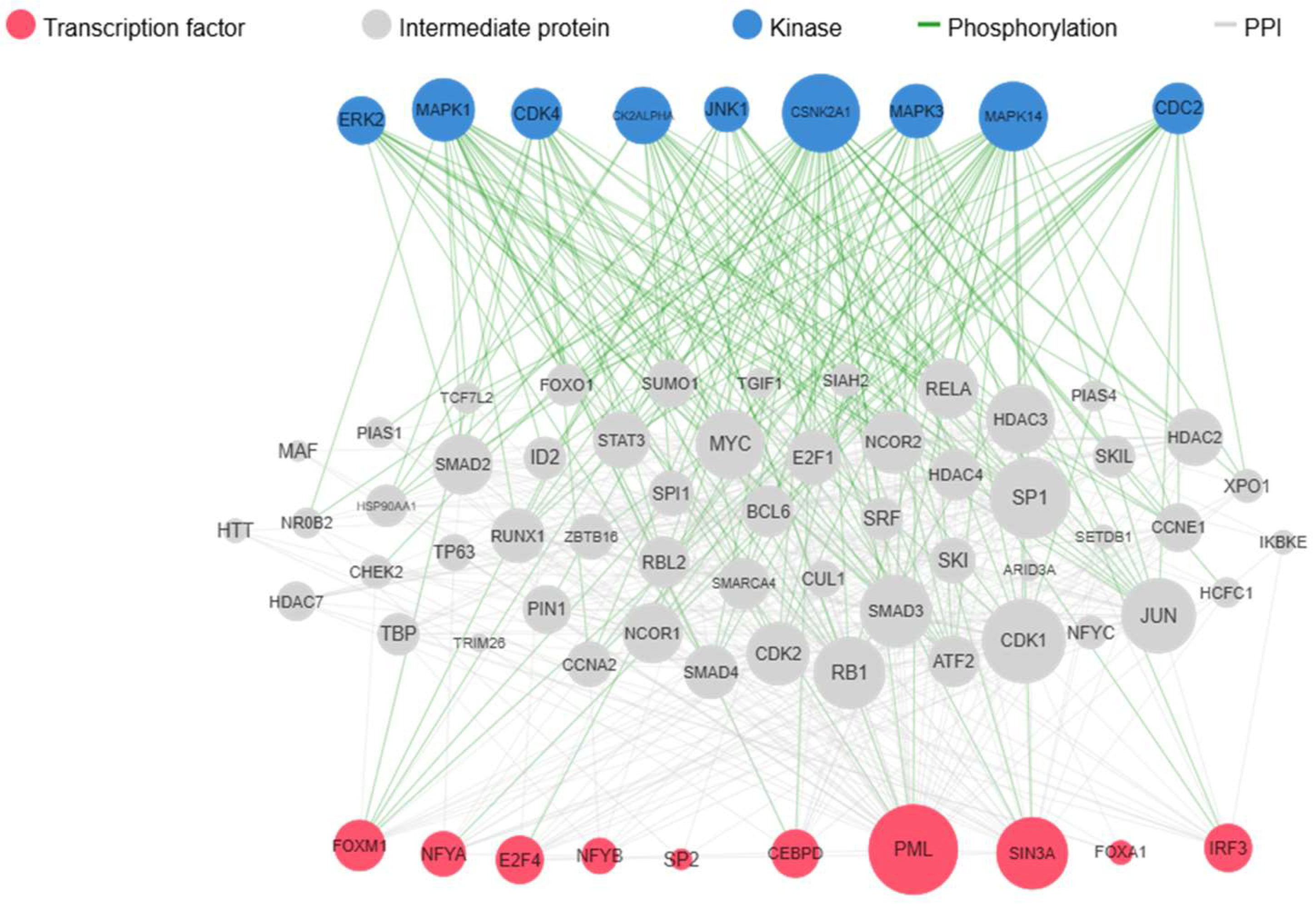
Human papillomavirus (HPV) is a prevalent sexually transmitted infection, implicated in various cancers, yet its influence in non-cancerous oesophageal tissue remains unclear. This study aims to investigate the gene expression changes associated with high-risk HPV (HR-HPV) in non-cancerous oesophageal tissue to elucidate potential early oncogenic mechanisms. Using RNA sequencing, we compared transcriptomic profiles of HPV-positive and HPV-negative non-cancerous oesophageal tissues. Differential gene expression analysis revealed significant upregulation of cell cycle and DNA replication pathways in HPV-positive samples, specifically involving key genes such as CCNA2, DSN1, and MCM10, which are known to regulate cellular proliferation and genomic stability. Additionally, kinase and transcription factor enrichment analyses highlighted HR-HPV-associated regulatory molecules, including E2F4 and CSNK2A1, suggesting HPV's role in modulating host cell cycle control. These findings support the hypothesis that HPV infection may initiate cellular alterations in oesophageal tissue, potentially predisposing it to malignancy. This study contributes to understanding HPV's impact in non-cancerous tissues and identifies possible biomarkers for early HPV-related cellular changes, offering insights into HPV-driven cancer development beyond traditionally associated sites.

Keywords: 
;  ;  ;  ;  ;  ;  ;  

1. Introduction

Human Papillomaviruses (HPVs) are small, circular, double-stranded DNA viruses, approximately 55 nm in diameter, with a non-enveloped icosahedral capsid structure[1]. As the most prevalent sexually transmitted infections (STIs) worldwide, HPVs infect a significant portion of the global population, resulting in a wide range of health conditions [2]. Over 200 HPV types have been identified, categorised based on their oncogenic potential into two main groups: high-risk HPVs (HR-HPVs) and low-risk HPVs (LR-HPVs). While LR-HPVs are primarily associated with benign conditions such as cutaneous and genital warts, HR-HPVs are strongly linked to several cancers, including those of the cervix, anus, oropharynx, vulva, penis, and skin, as well as precursor lesions associated with these cancers[2,3,4].
HR-HPVs encode three principal oncoprotein, E5, E6, and E7, which play essential roles in cellular transformation and cancer progression. The E5 protein, for instance, interacts with major histocompatibility complex (MHC) class I molecules, retaining them within the Golgi apparatus and thereby preventing their transport to the cell surface. This action helps the virus evade immune surveillance, supporting persistent infection[5,6]. Additionally, the E6 protein promotes cellular immortalisation by binding to the tumour suppressor protein p53, leading to its ubiquitination and degradation, which interferes with the normal apoptotic response. Similarly, the E7 protein fosters uncontrolled cell proliferation by inactivating the RB protein, thus releasing E2F transcription factors that drive DNA synthesis and cell division [7].
Although the presence and role of HR-HPVs in various cancers are well established, their presence in non-cancerous tissue is often overlooked. Investigating HPV infection in tissues where malignancy is not yet developed could provide vital insights into early viral activity and its potential role in cancer initiation. Understanding HPV interaction with host cells and identifying altered gene expression or protein profile in non-cancerous tissue may reveal mechanisms that contribute to cellular transformation and identify biomarkers associated with early HPV infection, which could aid in developing diagnostic tools for early detection.
The presence of HPV in oesophagus and its potential implications are crucial, though not well understood. While extensive research has established a clear association between HPV and various cancers, the role of HPV infection in the oesophagus is not conclusive in terms of cancer and is especially understudied in non-cancerous tissue. In this study, we addressed this gap by evaluating the differential gene expression in HPV-positive oesophageal samples and examining how these changes impact regulatory molecules. Our findings revealed an upregulated expression of key genes involved in cell proliferation, which may drive infected tissue toward pathological changes.

2. Results

2.1. Pathological Characteristics and Detection of HR-HPV

The histopathological evaluation of the samples was carried out by the pathology department of Kingston Hospital and all six samples were reported to be non-cancerous inflamed tissue. Tissue specimens from the fresh non-cancerous tissues were examined for the presence of DNA of 14 HR-HPV types using type-specific real time PCR.
Data obtained revealed that HR-HPV DNA was present in three non-cancerous oesophageal tissue samples tested. All the positive samples had HR-HPV 56 infection. One of the three HPV positive samples had an additional infection of HR-HPV 18 infection showing co-infection with HR-HPV 56. The remaining three samples were HR-HPV infection free. The amplification of β-globin gene was positive in all extracted DNA, indicating an adequate quality of DNA. Positive and negative controls confirmed that there was no evidence of contamination, indicating a successful PCR amplification.

2.2. Transcriptomic Profiling of Non-Cancerous Oesophageal Tissue with and Without HR-HPV Infection

To investigate the impact of HPV on the gene expression profile of oesophageal tissue, we performed RNA sequencing (RNA-Seq) to analyse changes in gene expression across three HPV-positive and three HPV-negative oesophageal samples. The subsequent analysis of differential gene expression revealed that 18 genes were consistently and significantly upregulated in HPV-Positive samples compared to HPV-negative non-cancerous oesophageal human tissue samples (Figure 1).

2.3. Analysis of Differentially Expressed Genes of Non-Cancerous Oesophageal Tissue with and Without HR-HPV Infection Using Pathway Enrichment Analysis

Differentially upregulated genes in HPV positive oesophageal tissue were analysed using pathway enrichment analysis. The results showed that the genes significantly upregulated following HPV infection are involved in cell proliferation including pathways of “Cell Cycle” and “DNA replication” (Table 1). Notably, some genes are overlapping between the pathways, indicating their central role in cell proliferation.

2.4. Investigation of Differentially Expressed Genes of Non-Cancerous Oesophageal Tissue with and Without HR-HPV Infection via Kinase Enrichment Analysis

To identify molecular networks modulated by HR-HPV infection, we used Kinase Enrichment Analysis (KEA) to examine key kinases and transcription factors that regulate the differentially expressed genes expression between HPV-positive and HPV-negative groups. The results highlighted the prominence of transcription factors including E2F4, FOXM1, CEBPD, IRF3, NFYA along with kinases CSNK2A1, CDK4, CDK2ALPHA, MAPK14 and CDK1 (Table 2A,B). These molecules play an important role in altering the expression of observed genes indicating a complex interaction between HPV and cellular regulatory network. The upstream pathway analysis of upregulated transcription factors and kinases indicated that HPV infection influences key regulatory protein involved in cell cycle including CDK1, RB1, CDK2, and SP1 (Figure 2). The observation of these transcription factors and kinases as the key regulators of upregulated genes in HPV positive non-cancerous samples points to the pathways that HPV might be influencing to alter cellular functions, even in non-cancerous states.

3. Discussion

Human Papillomavirus (HPV), the most prevalent sexually transmitted infection globally, affects millions and contributes to a range of diseases from benign warts to cancers across multiple sites[2,3,4,8]. High-risk HPV (HR-HPV) types are implicated in around 750,000 cancer cases worldwide each year[9]. While HPV’s role in cancers of the cervix and head and neck is well documented, its potential effects on the oesophageal tissue remain poorly understood, particularly in non-cancerous tissue. HPV infection in the oesophagus was first proposed in the early 1980s [10], but despite numerous studies, its connection to oesophageal cancer remains inconclusive, complicated by variations in geography, HPV type, and detection methods [11].
Given the complexity of cancer biology, investigating HPV’s effects on the host genome in non-cancerous tissues offers unique insights, as cancer-associated genomic changes can arise independently of HPV infection. Our study explores whether the presence and activity of HPV in non-cancerous oesophageal tissues may alter the host genome, potentially initiating a progression toward oesophageal cancer driven solely by HPV expression. Using RNA-Seq analysis, we compared the transcriptome of HPV-positive non-cancerous oesophageal tissues to HPV-negative controls, identifying significant alterations in gene expression linked to the cell cycle (p-value: 7.49 x 10⁻⁸) and DNA replication pathways (p-value: 8.69 x 10⁻⁵). The upregulation of cell cycle regulation pathways in HPV-positive, non-cancerous oesophageal tissues may cause abnormal cell proliferation, a hallmark of carcinogenesis [12].
Key cell cycle regulatory genes, including CCNA2, NEDD1, and DSN1, were significantly overexpressed in these samples. For instance, CCNA2 is pivotal in cell cycle control, with dysregulation linked to tumour development [13]. Likewise, NEDD1 and DSN1, crucial for microtubule organisation and chromosome segregation during cell division, are linked to genomic instability and abnormal cell division when overexpressed [14,15,16]. The dysregulation of cell cycle pathways in our findings aligns with established mechanisms in HPV-related cancers, such as cervical and head and neck cancers, where HPV oncoproteins E6 and E7 disrupt cellular controls, inducing genomic instability and cell cycle dysregulation [17,18].
Additionally, our results indicate a significant upregulation in DNA replication pathways, with key genes such as CASC5 and MCM10 overexpressed in HPV-positive samples. MCM10, part of the mini-chromosome maintenance complex, is essential for DNA replication and genomic stability, and its aberrant expression is implicated in various cancers, including cervical cancer [19,20]. Similarly, CASC5, essential for chromosome segregation during cell division, is associated with cancer when dysregulated [21,22]. This upregulation suggests that HPV infection may influence DNA replication, potentially increasing genomic instability in non-cancerous tissue both of which are considered key contributors to the hall marks of cancers [23].
The upregulation of genes in cell cycle and DNA replication pathways correlates with the documented mechanisms of HPV E6 and E7 oncoproteins. E7 facilitates unchecked cell proliferation by inhibiting the retinoblastoma protein (pRB), allowing the premature entry of cells into the S-phase [24]. Additionally, E6 promotes the degradation of the p53 tumour suppressor, which usually pauses cell division in response to DNA damage, preventing apoptosis. The degradation of p53 permits cells to accumulate genetic mutations, contributing to genomic instability and malignancy risk [25]. These established mechanisms in cervical, head, and neck cancers suggest that similar pathways may be disrupted by HPV in non-cancerous oesophageal tissue, contributing to cell cycle and DNA replication disturbances.
Longitudinal studies have shown that persistent HR-HPV infection in normal tissues of the cervix and head and neck can lead to cancer over time [26,27,28]. HR-HPV is central to the transformation of normal cervical tissue to carcinoma, primarily through cell cycle dysregulation and genomic instability. Similarly, studies on HPV infection in normal head and neck tissues indicate that while most infections are transient, persistent infection can lead to oral and oropharyngeal cancers [29,30]. Our findings align with this pattern, suggesting that HPV infection in non-cancerous oesophageal tissue may similarly initiate cellular changes that could drive cancer development over time.
To investigate further molecular network modulated by HPV, transcription factor and kinase enrichment analysis was performed revealing the involvement of transcription factors such as E2F4, FOXM1, and Kinases CSNK2A1 and CDK4. These transcription factors are integral to cycle regulation, differentiation, and DNA synthesis. Elevated expressions of these transcription factors are associated with tumorigenesis in variety of cancers [31,32,33,34,35,36,37,38]. CSNK2A1 a serine/threonine kinase is a catalytic subunit of Casein Kinase II (CK2), involved in various cellular processes including cell cycle regulation, apoptosis, signalling pathways, growth and metabolism [39]. Overexpression of CSNK2A1 is reported to play important role in various cancers including gastric cancer and breast cancer[40,41]. CDK4, a serine/threonine kinase, is associated to cell cycle regulation, especially the transition from G1 to S phase. CDK4 pairs with D-type cyclins and drive cell cycle progression, thereby promoting proliferation [42]. Overexpression of CDK4 has been observed in variety of cancers including breast cancers, glioblastomas multiforme, gliomas, and meningiomas [43]. Our pathway analysis reveals the impact of HR-HPV infection on essential proteins involved in cell cycle and DNA replication pathways including SP1, E2F1, CDK1, and CDK2 [44,45]. The identification of these kinases and transcription factors point to complex regulatory network that may be disrupted by HPV infection, further supporting the virus’s role in creating pro-proliferative environment conducive to oncogenic transformation.
In conclusion, our study suggests that HR-HPV infection in non-cancerous oesophageal tissue leads to cell cycle dysregulation and may encourage precancerous changes through genomic instability. The upregulation of key genes and regulators, such as FOXM1, CSNK2A1, and CDK4, highlights potential biomarkers for early detection of HPV related cellular changes. These findings emphasise the significance of HPV’s impact on oesophagus, suggesting that even in non-cancerous tissue, HR-HPV may exert oncogenic pressure and predispose cells to malignancy. To our knowledge, this is the first study to compare gene expression in HPV-negative and HPV-positive non-cancerous oesophageal tissue, providing insights into the molecular alterations induced by early HPV infection.

4. Materials and Methods

4.1. Oesophageal Tissue Specimen Collection

The study was conducted with the approval of Health Research Authority (HRA) and Health and Care Research Wales (HCRW) under project ID 250010. All procedures strictly adhered to approved guidelines and regulations. Informed consent was obtained from the patients participating in the study. The study protocol included the protection of patient identifiable information from the researcher team.
Patients referred to Kingston Hospital, London by general practitioners (GPs), because of gastrointestinal complaints, were consented and oesophageal biopsies (3-6 per patient) were collected by endoscopy team in the endoscopy unit of Kingston Hospital. The diagnosis of all oesophageal tissue specimen was formally reported by Kingston Hospital pathology department as part of standard clinical diagnosis of patients.
Following excision, the integrity of biopsy tissue was immediately preserved by submerging them in ALLPROTECT reagent (QIAGEN, Hilden, Germany). This step aimed to stabilise DNA, RNA, and protein while maintaining aseptic handling to prevent contamination of samples. The specimens were transferred to Kingston University in accordance with World Health Organisation transport guidelines for infectious material [46].

4.2. Genomic Material Extraction and Purification

To avoid the cross-contamination between specimens, separate disposable items such as gloves, petri dishes, tubes, and surgical blades, were used for the handling of each specimen. Cellular DNA, RNA, and protein were extracted from the oesophageal specimens using column-based extraction kit. The samples were fragmented and homogenised in lysis buffer using tissue lyser (QIAGEN, Hilden, Germany) and further processed with QIAshredder spin column (QIAGEN, Hilden, Germany).
The extraction of genomic material and protein was conducted using GenElute RNA/DNA/Protein Plus Purification Kit (Sigma-Aldrich, USA) according to manufactures protocol. Purification of these biological molecules in this kit is based on spin column chromatography allowing the sequential isolation of genomic DNA, total RNA, and protein from a single sample. The concentration and purity of eluted genomic material was assessed using spectrophotometer BIODROP DUO+ (Biochrom, UK).

4.3. Detection and Genotyping of HPV DNA

The methodology employed for the detection and identification of HPVs involved the utilisation of real time- polymerisation chain reaction (PCR). The AmpliSens HPV HCR genotype-Titre-FRT PCR kit (Ecoli s.r.o., Bratislava, Slovak Republic) was used according to manufacturer’s direction for the detection and genotyping of 14 high risk HPV types including type 16, 18, 31, 33, 35, 39, 45, 51, 52, 56, 58, 59, 66 and 68 in 52 oesophageal tissue specimens, extracted at Kingston University laboratories, UK. The presence of HPV was detected using Stratagene Mx3005p (Agilent, USA). Each PCR run contained the quality control measures. This included positive control, negative control, and internal control (β-globin) primers. These measures were integral to mitigating the risk of false positive or false negative results. To avoid cross-contamination, the DNA extraction and PCR amplification were carried out in separate laboratories following strict aseptic protocol.

4.4. RNA Sequencing

The RNA sequencing was performed on six non-cancerous oesophageal tissue sample, divided in two groups: a HPV positive group and a HPV negative group, with three samples in each. All 6 samples had RIN value greater than 7.5. The library preparation, sequencing and bioinformatics analysis was performed by CeGaT GmbH, Tubingen, Germany. SMART-Seq standard kit (Takara, Kusatsu, Japan) was utilised for library preparation and multiplexed libraries were sequenced using the Illumina NovaSeq 6000 platform at 100 bp paired-end reads. The sequencing depth for each sample was >20 million reads. All samples passed quality control based on the manufacturer’s standards.

4.5. Bioinformatics Analysis

Diverse bioinformatics tools were utilised for the analysis of sequence reads. Sequence reads were de-multiplexed with Illumina bcl2fastq (vs. 2.20) and adaptors were trimmed using Skewer (vs. 0.2.2) [47]. STAR (vs. 2.7.3) was utilised for the alignment of trimmed raw reads to hg19-cegat [48]. Pseudo-autosomal regions (PAR) on Y-Chromosome were masked as reads from these regions are also mapped to their corresponding location on X-Chromosome. Normalised counts were calculated using DESeq2 (version 1.24.0) in R (version 3.6.1) [49]. For the functional enrichment analysis, the RNA-Seq data obtained from HPV-positive and HPV-negative oesophageal tissue were used. Gene Set Enrichment Analysis (GSEA) was performed using the GSEA software (version number: 4.3.0) [50,51]. The RNA-Seq data sets were pre-processed and normalized, and the resulting gene expression profiles were analysed against a comprehensive collection of gene sets derived from public databases, such as MSigDB [51]. The GSEA algorithm computed an enrichment score for each gene set, indicating the extent to which the gene set was overrepresented among the differentially expressed genes.
EnrichR, an online platform, was used for comprehensive gene set enrichment analysis[52,53,54]. The pre-processed RNA-Seq data sets were uploaded to EnrichR and analysed following the provided instructions. EnrichR identifies pathways associated with differentially expressed genes. The analysis yielded enriched pathways results along with corresponding statistical significance. Outputs from GSEA and EnrichR were utilised to understand the biological processes and pathways impacted with the infection of HR-HPVs in Human oesophageal tissue. The kinase enrichment analysis was confirmed using Expression2 Kinase (X2K) software (vs 0.0.4) [55,56].

Author Contributions

Conceptualization, G.H.A.; Methodology, M.O.S, G.H.A. M.OC., and U.B.; Validation, G.H.A. and M.O.S.; Formal Analysis, G.H.A., M.O.S., U.B. and M.O.C; Data Curation, M.O.S., G.H.A., M.O.C and U.B.; Manuscript Writing and Editing, M.O.S. and G.H.A.; Sample Collection, M.O.S., and Y.P.; Supervision, G.H.A.; Project Administration, G.H.A.; Funding Acquisition, G.H.A. All authors have read and agreed to the published version of the manuscript.

Funding

This research was funded by the Laurie Todd Foundation, UK, and Kingston Hospital Charity, UK.

Institutional Review Board Statement

The study was conducted according to the guidelines of the Declaration of Helsinki and approved by the Health Research Authority (HRA) and Health and Care Research Whales (HCRW) under project ID 250010, September 2019.

Informed Consent Statement

Informed consent was obtained from all subjects involved in the Written informed consent has been obtained from the patients to publish this paper.

Data Availability Statement

Data is available upon request from the authors.

Acknowledgments

The author gratefully acknowledges the support provided by the Laurie Todd Foundation, UK, and the Kingston Hospital Charity, UK, whose funding made this research possible. Special appreciation is extended to the patients whose tissue donations were instrumental to this study, as well as to the dedicated surgical team at Kingston Hospital (Kingston upon Thames, London, UK) for their invaluable assistance. Furthermore, the authors would like to acknowledge Kingston University London for the provision of state-of-the-art laboratory facilities, which significantly contributed to the success of this investigation.

Conflicts of Interest

The authors declare no conflicts of interest. The funders had no role in the design of the study; in the collection, analyses, or interpretation of data; in the writing of the manuscript; or in the decision to publish the results.

References

  1. Graham, S. V. The Human Papillomavirus Replication Cycle, and Its Links to Cancer Progression: A Comprehensive Review. Clin Sci 2017, 131. [Google Scholar] [CrossRef] [PubMed]
  2. Kombe Kombe, A.J.; Li, B.; Zahid, A.; Mengist, H.M.; Bounda, G.-A.; Zhou, Y.; Jin, T. Epidemiology and Burden of Human Papillomavirus and Related Diseases, Molecular Pathogenesis, and Vaccine Evaluation. Front Public Health 2021, 8. [Google Scholar] [CrossRef] [PubMed]
  3. de Sanjosé, S.; Brotons, M.; Pavón, M.A. The Natural History of Human Papillomavirus Infection. Best Pract Res Clin Obstet Gynaecol 2018, 47, 2–13. [Google Scholar] [CrossRef] [PubMed]
  4. Tramontano, L.; Sciorio, R.; Bellaminutti, S.; Esteves, S.C.; Petignat, P. Exploring the Potential Impact of Human Papillomavirus on Infertility and Assisted Reproductive Technology Outcomes. Reprod Biol 2023, 23, 100753. [Google Scholar] [CrossRef] [PubMed]
  5. Ashrafi, G.H.; Haghshenas, M.R.; Marchetti, B.; O’Brien, P.M.; Campo, M.S. E5 Protein of Human Papillomavirus Type 16 Selectively Downregulates Surface HLA Class I. Int J Cancer 2005, 113, 276–283. [Google Scholar] [CrossRef] [PubMed]
  6. Ashrafi, G.H.; Salman, N.A. Pathogenesis of Human Papillomavirus – Immunological Responses to HPV Infection. In Human Papillomavirus - Research in a Global Perspective; InTech, 2016.
  7. Bhattacharjee, R.; Das, S.S.; Biswal, S.S.; Nath, A.; Das, D.; Basu, A.; Malik, S.; Kumar, L.; Kar, S.; Singh, S.K.; et al. Mechanistic Role of HPV-Associated Early Proteins in Cervical Cancer: Molecular Pathways and Targeted Therapeutic Strategies. Crit Rev Oncol Hematol 2022, 174, 103675. [Google Scholar] [CrossRef]
  8. Jensen, J.E.; Becker, G.L.; Jackson, J.B.; Rysavy, M.B. Human Papillomavirus and Associated Cancers: A Review. Viruses 2024, 16, 680. [Google Scholar] [CrossRef]
  9. Pešut, E.; Đukić, A.; Lulić, L.; Skelin, J.; Šimić, I.; Milutin Gašperov, N.; Tomaić, V.; Sabol, I.; Grce, M. Human Papillomaviruses-Associated Cancers: An Update of Current Knowledge. Viruses 2021, 13, 2234. [Google Scholar] [CrossRef]
  10. Syrjänen, K.; Pyrhönen, S.; Aukee, S.; Koskela, E. Squamous Cell Papilloma of the Esophagus: A Tumour Probably Caused by Human Papilloma Virus (HPV). Diagn Histopathol 1982, 5, 291–296. [Google Scholar]
  11. Abbas, G.; Krasna, M. Overview of Esophageal Cancer. Ann Cardiothorac Surg 2017, 6, 131–136. [Google Scholar] [CrossRef] [PubMed]
  12. Hanahan, D.; Weinberg, R.A. The Hallmarks of Cancer. Cell 2000, 100, 57–70. [Google Scholar] [CrossRef] [PubMed]
  13. Lu, Y.; Su, F.; Yang, H.; Xiao, Y.; Zhang, X.; Su, H.; Zhang, T.; Bai, Y.; Ling, X. E2F1 Transcriptionally Regulates CCNA2 Expression to Promote Triple Negative Breast Cancer Tumorigenicity. Cancer Biomarkers 2022, 33, 57–70. [Google Scholar] [CrossRef] [PubMed]
  14. Manning, J.; Kumar, S. NEDD1: Function in Microtubule Nucleation, Spindle Assembly and Beyond. Int J Biochem Cell Biol 2007, 39, 7–11. [Google Scholar] [CrossRef] [PubMed]
  15. Peng, Q.; Wen, T.; Liu, D.; Wang, S.; Jiang, X.; Zhao, S.; Huang, G. DSN1 Is a Prognostic Biomarker and Correlated with Clinical Characterize in Breast Cancer. Int Immunopharmacol 2021, 101, 107605. [Google Scholar] [CrossRef] [PubMed]
  16. Chuang, T.-P.; Wang, J.-Y.; Jao, S.-W.; Wu, C.-C.; Chen, J.-H.; Hsiao, K.-H.; Lin, C.-Y.; Chen, S.-H.; Su, S.-Y.; Chen, Y.-J.; et al. Over-Expression of AURKA, SKA3 and DSN1 Contributes to Colorectal Adenoma to Carcinoma Progression. Oncotarget 2016, 7, 45803–45818. [Google Scholar] [CrossRef]
  17. Duensing, S.; Münger, K. Human Papillomaviruses and Centrosome Duplication Errors: Modeling the Origins of Genomic Instability. Oncogene 2002, 21, 6241–6248. [Google Scholar] [CrossRef]
  18. Scarth, J.A.; Patterson, M.R.; Morgan, E.L.; Macdonald, A. The Human Papillomavirus Oncoproteins: A Review of the Host Pathways Targeted on the Road to Transformation. Journal of General Virology 2021, 102. [Google Scholar] [CrossRef]
  19. Yeeles, J.T.P.; Deegan, T.D.; Janska, A.; Early, A.; Diffley, J.F.X. Regulated Eukaryotic DNA Replication Origin Firing with Purified Proteins. Nature 2015, 519, 431–435. [Google Scholar] [CrossRef]
  20. Cheng, J.; Lu, X.; Wang, J.; Zhang, H.; Duan, P.; Li, C. Interactome Analysis of Gene Expression Profiles of Cervical Cancer Reveals Dysregulated Mitotic Gene Clusters. Am J Transl Res 2017, 9, 3048–3059. [Google Scholar]
  21. Ghongane, P.; Kapanidou, M.; Asghar, A.; Elowe, S.; Bolanos-Garcia, V.M. The Dynamic Protein Knl1 – a Kinetochore Rendezvous. J Cell Sci 2014. [CrossRef]
  22. Cui, Y.; Zhang, C.; Ma, S.; Guo, W.; Cao, W.; Guan, F. <scp>CASC5</Scp> Is a Potential Tumour Driving Gene in Lung Adenocarcinoma. Cell Biochem Funct 2020, 38, 733–742. [Google Scholar] [CrossRef] [PubMed]
  23. Hanahan, D.; Weinberg, R.A. Hallmarks of Cancer: The Next Generation. Cell 2011, 144, 646–674. [Google Scholar] [CrossRef] [PubMed]
  24. BOYER, S.N.; WAZER, D.E.; BAND, V. E7 Protein of Human Papilloma Virus-16 Induces Degradation of Retinoblastoma Protein through the Ubiquitin-Proteasome Pathway. Cancer Res 1996, 56, 4620–4624. [Google Scholar] [PubMed]
  25. Bieging, K.T.; Mello, S.S.; Attardi, L.D. Unravelling Mechanisms of P53-Mediated Tumour Suppression. Nat Rev Cancer 2014, 14, 359–370. [Google Scholar] [CrossRef] [PubMed]
  26. Cox, J.T.; Schiffman, M.; Solomon, D. Prospective Follow-up Suggests Similar Risk of Subsequent Cervical Intraepithelial Neoplasia Grade 2 or 3 among Women with Cervical Intraepithelial Neoplasia Grade 1 or Negative Colposcopy and Directed Biopsy. Am J Obstet Gynecol 2003, 188, 1406–1412. [Google Scholar] [CrossRef]
  27. Kitchener, H.C.; Almonte, M.; Wheeler, P.; Desai, M.; Gilham, C.; Bailey, A.; Sargent, A.; Peto, J. HPV Testing in Routine Cervical Screening: Cross Sectional Data from the ARTISTIC Trial. Br J Cancer 2006, 95, 56–61. [Google Scholar] [CrossRef]
  28. Derchain, S.F.; Sarian, L.O.; Naud, P.; Roteli-Martins, C.; Longatto-Filho, A.; Tatti, S.; Branca, M.; Eržen, M.; Serpa-Hammes, L.; Matos, J.; et al. Safety of Screening with Human Papillomavirus Testing for Cervical Cancer at Three-Year Intervals in a High-Risk Population: Experience from the LAMS Study. J Med Screen 2008, 15, 97–104. [Google Scholar] [CrossRef]
  29. Gillison, M.L.; Broutian, T.; Pickard, R.K.L.; Tong, Z.; Xiao, W.; Kahle, L.; Graubard, B.I.; Chaturvedi, A.K. Prevalence of Oral HPV Infection in the United States, 2009-2010. JAMA 2012, 307, 693. [Google Scholar] [CrossRef]
  30. Giuliano, A.R.; Viscidi, R.; Torres, B.N.; Ingles, D.J.; Sudenga, S.L.; Villa, L.L.; Luiza Baggio, M.; Abrahamsen, M.; Quiterio, M.; Salmeron, J.; et al. Seroconversion Following Anal and Genital HPV Infection in Men: The HIM Study. Papillomavirus Research 2015, 1, 109–115. [Google Scholar] [CrossRef]
  31. Waghray, A.; Schober, M.; Feroze, F.; Yao, F.; Virgin, J.; Chen, Y.Q. Identification of Differentially Expressed Genes by Serial Analysis of Gene Expression in Human Prostate Cancer. Cancer Res 2001, 61, 4283–4286. [Google Scholar]
  32. Rakha, E.A.; Pinder, S.E.; Paish, E.C.; Robertson, J.F.; Ellis, I.O. Expression of E2F-4 in Invasive Breast Carcinomas Is Associated with Poor Prognosis. J Pathol 2004, 203, 754–761. [Google Scholar] [CrossRef] [PubMed]
  33. Rakha, E.A.; Armour, J.A.L.; Pinder, S.E.; Paish, C.E.; Ellis, I.O. High-resolution Analysis of 16q22.1 in Breast Carcinoma Using DNA Amplifiable Probes (Multiplex Amplifiable Probe Hybridization Technique) and Immunohistochemistry. Int J Cancer 2005, 114, 720–729. [Google Scholar] [CrossRef] [PubMed]
  34. Hsu, J.; Sage, J. Novel Functions for the Transcription Factor E2F4 in Development and Disease. Cell Cycle 2016, 15, 3183–3190. [Google Scholar] [CrossRef] [PubMed]
  35. Zheng, Q.; Fu, Q.; Xu, J.; Gu, X.; Zhou, H.; Zhi, C. Transcription Factor E2F4 Is an Indicator of Poor Prognosis and Is Related to Immune Infiltration in Hepatocellular Carcinoma. J Cancer 2021, 12, 1792–1803. [Google Scholar] [CrossRef] [PubMed]
  36. Qi, L.; Ren, Z.; Li, W. E2F4 Transcription Factor Is a Prognostic Biomarker Related to Immune Infiltration of Head and Neck Squamous Cell Carcinoma. Sci Rep 2022, 12, 12132. [Google Scholar] [CrossRef] [PubMed]
  37. Liao, G.-B.; Li, X.-Z.; Zeng, S.; Liu, C.; Yang, S.-M.; Yang, L.; Hu, C.-J.; Bai, J.-Y. Regulation of the Master Regulator FOXM1 in Cancer. Cell Communication and Signaling 2018, 16, 57. [Google Scholar] [CrossRef]
  38. Rachmadi, L.; Billianti Susanto, Y.D.; Sitinjak, D.; Manatar, A.F.; Saraswati, M.; Adham, M. HPV Infection Is Associated with FoxM1 Overexpression in Dysplastic Changes of Sinonasal Inverted Papilloma. Asian Pac J Cancer Prev 2022, 23, 4293–4298. [Google Scholar] [CrossRef]
  39. Strum, S.W.; Gyenis, L.; Litchfield, D.W. CSNK2 in Cancer: Pathophysiology and Translational Applications. Br J Cancer 2022, 126, 994–1003. [Google Scholar] [CrossRef]
  40. Bae, J.S.; Park, S.-H.; Jamiyandorj, U.; Kim, K.M.; Noh, S.J.; Kim, J.R.; Park, H.J.; Kwon, K.S.; Jung, S.H.; Park, H.S.; et al. CK2α/CSNK2A1 Phosphorylates SIRT6 and Is Involved in the Progression of Breast Carcinoma and Predicts Shorter Survival of Diagnosed Patients. Am J Pathol 2016, 186, 3297–3315. [Google Scholar] [CrossRef] [PubMed]
  41. Jiang, C.; Ma, Z.; Zhang, G.; Yang, X.; Du, Q.; Wang, W. <p>CSNK2A1 Promotes Gastric Cancer Invasion Through the PI3K-Akt-MTOR Signaling Pathway</P>. Cancer Manag Res 2019, Volume 11, 10135–10143. [Google Scholar] [CrossRef]
  42. Baker, S.J.; Poulikakos, P.I.; Irie, H.Y.; Parekh, S.; Reddy, E.P. CDK4: A Master Regulator of the Cell Cycle and Its Role in Cancer. Genes Cancer 2022, 13, 21–45. [Google Scholar] [CrossRef] [PubMed]
  43. Deshpande, A.; Sicinski, P.; Hinds, P.W. Cyclins and Cdks in Development and Cancer: A Perspective. Oncogene 2005, 24, 2909–2915. [Google Scholar] [CrossRef] [PubMed]
  44. Jungert, K.; Buck, A.; Wichert, G. von; Adler, G.; König, A.; Buchholz, M.; Gress, T.M.; Ellenrieder, V. Sp1 Is Required for Transforming Growth Factor-β–Induced Mesenchymal Transition and Migration in Pancreatic Cancer Cells. Cancer Res 2007, 67, 1563–1570. [Google Scholar] [CrossRef] [PubMed]
  45. Engeland, K. Cell Cycle Regulation: P53-P21-RB Signaling. Cell Death Differ 2022, 29, 946–960. [Google Scholar] [CrossRef] [PubMed]
  46. World Health Organization (WHO). Guidance on Regulations for the Transport of Infectious Substances, 2023–2024; 2024. [Google Scholar]
  47. Jiang, H.; Lei, R.; Ding, S.-W.; Zhu, S. Skewer: A Fast and Accurate Adapter Trimmer for next-Generation Sequencing Paired-End Reads. BMC Bioinformatics 2014, 15, 182. [Google Scholar] [CrossRef]
  48. Dobin, A.; Davis, C.A.; Schlesinger, F.; Drenkow, J.; Zaleski, C.; Jha, S.; Batut, P.; Chaisson, M.; Gingeras, T.R. STAR: Ultrafast Universal RNA-Seq Aligner. Bioinformatics 2013, 29, 15–21. [Google Scholar] [CrossRef]
  49. Love, M.I.; Huber, W.; Anders, S. Moderated Estimation of Fold Change and Dispersion for RNA-Seq Data with DESeq2. Genome Biol 2014, 15, 550. [Google Scholar] [CrossRef]
  50. Subramanian, A.; Tamayo, P.; Mootha, V.K.; Mukherjee, S.; Ebert, B.L.; Gillette, M.A.; Paulovich, A.; Pomeroy, S.L.; Golub, T.R.; Lander, E.S.; et al. Gene Set Enrichment Analysis: A Knowledge-Based Approach for Interpreting Genome-Wide Expression Profiles. Proceedings of the National Academy of Sciences 2005, 102, 15545–15550. [Google Scholar] [CrossRef]
  51. Liberzon, A.; Birger, C.; Thorvaldsdóttir, H.; Ghandi, M.; Mesirov, J.P.; Tamayo, P. The Molecular Signatures Database Hallmark Gene Set Collection. Cell Syst 2015, 1, 417–425. [Google Scholar] [CrossRef]
  52. Xie, Z.; Bailey, A.; Kuleshov, M. V.; Clarke, D.J.B.; Evangelista, J.E.; Jenkins, S.L.; Lachmann, A.; Wojciechowicz, M.L.; Kropiwnicki, E.; Jagodnik, K.M.; et al. Gene Set Knowledge Discovery with Enrichr. Curr Protoc 2021, 1. [Google Scholar] [CrossRef]
  53. Chen, E.Y.; Tan, C.M.; Kou, Y.; Duan, Q.; Wang, Z.; Meirelles, G.V.; Clark, N.R.; Ma’ayan, A. Enrichr: Interactive and Collaborative HTML5 Gene List Enrichment Analysis Tool. BMC Bioinformatics 2013, 14, 128. [Google Scholar] [CrossRef] [PubMed]
  54. Kuleshov, M. V.; Jones, M.R.; Rouillard, A.D.; Fernandez, N.F.; Duan, Q.; Wang, Z.; Koplev, S.; Jenkins, S.L.; Jagodnik, K.M.; Lachmann, A.; et al. Enrichr: A Comprehensive Gene Set Enrichment Analysis Web Server 2016 Update. Nucleic Acids Res 2016, 44, W90–W97. [Google Scholar] [CrossRef] [PubMed]
  55. Chen, E.Y.; Xu, H.; Gordonov, S.; Lim, M.P.; Perkins, M.H.; Ma’ayan, A. Expression2Kinases: MRNA Profiling Linked to Multiple Upstream Regulatory Layers. Bioinformatics 2012, 28, 105–111. [Google Scholar] [CrossRef] [PubMed]
  56. Clarke, D.J.B.; Kuleshov, M. V; Schilder, B.M.; Torre, D.; Duffy, M.E.; Keenan, A.B.; Lachmann, A.; Feldmann, A.S.; Gundersen, G.W.; Silverstein, M.C.; et al. EXpression2Kinases (X2K) Web: Linking Expression Signatures to Upstream Cell Signaling Networks. Nucleic Acids Res 2018, 46, W171–W179. [Google Scholar] [CrossRef] [PubMed]
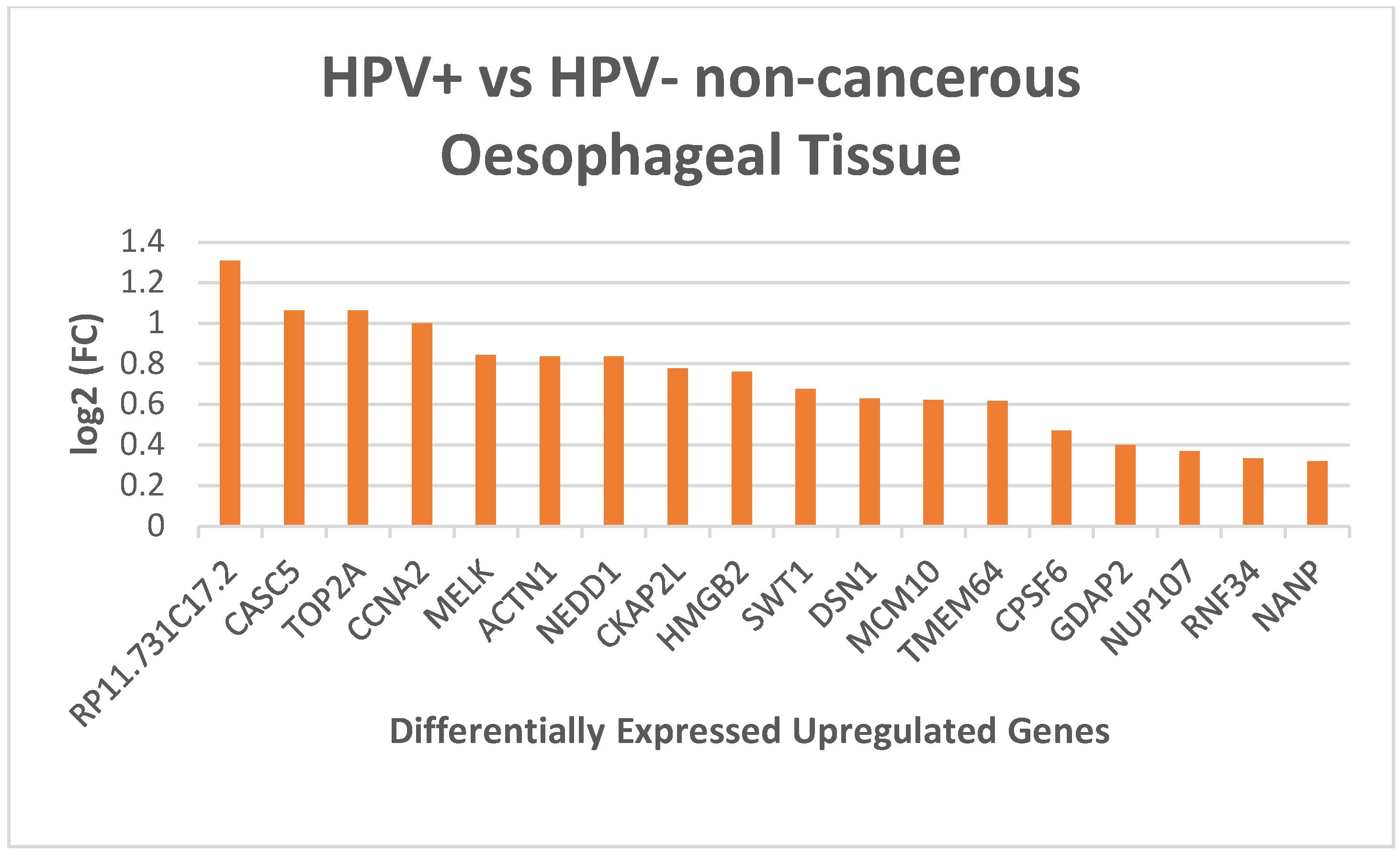
Figure 1. Differentially expressed genes between HPV positive and HPV negative non-cancerous human oesophageal tissue samples.
Preprints 139908 g001
Figure 2. Complete upstream pathways connecting enriched transcription factors to kinases through known protein-protein interaction. Red nodes highlight top transcription factors predicted to regulate our upregulated genes. Grey nodes depict proteins that interact with these transcription factors. Blue nodes represent kinases predicted to phosphorylate within the network. Green lines represent phosphorylation interactions between kinases and their targets, while grey lines indicate protein-protein physical interactions.
Preprints 139908 g002
Table 1. Pathway enrichment analysis of upregulated genes in HPV positive oesophageal tissue. The table includes the description of pathways, Number of overlapping genes from the differentially overexpressed genes, the p-value, and the overlapping genes.
Description Number of overlap genes p-Value Overlap Genes
Cell Cycle 7 7.49 x 10⁻⁸ TOP2A; CCNA2; NEDD1; DSN1; NUP107; CASC5; MCM10
DNA Replication 5 8.69 x 10⁻⁵ CCNA2; DSN1; NUP107; CASC5; MCM10
Table 2. Pathway enrichment analysis of upregulated genes in HPV positive oesophageal tissue. The table includes the description of pathways, Number of overlapping genes from the differentially overexpressed genes, the p-value, and the overlapping genes.
A
Transcription Factors p-Value
E2F4 1.6 x 10⁻⁸
FOXM1 1.5 x 10⁻⁴
CEBPD 9.1 x 10 -4
B
Regulatory Kinases p-Value
CSNK2A1 2.2 x 10⁻¹⁵
CDK4 7.0 x 10⁻¹⁵
MAPK14 1.05 x 10⁻¹²
Disclaimer/Publisher’s Note: The statements, opinions and data contained in all publications are solely those of the individual author(s) and contributor(s) and not of MDPI and/or the editor(s). MDPI and/or the editor(s) disclaim responsibility for any injury to people or property resulting from any ideas, methods, instructions or products referred to in the content.
Copyright: This open access article is published under a Creative Commons CC BY 4.0 license, which permit the free download, distribution, and reuse, provided that the author and preprint are cited in any reuse.
Prerpints.org logo

Preprints.org is a free preprint server supported by MDPI in Basel, Switzerland.

Subscribe

Disclaimer

Terms of Use

Privacy Policy

Privacy Settings

© 2025 MDPI (Basel, Switzerland) unless otherwise stated