Preprint
Review

This version is not peer-reviewed.

Hypothalamic Mechanisms Contributing to Cancer Cachexia

Submitted:

13 November 2024

Posted:

15 November 2024

Read the latest preprint version here

Abstract

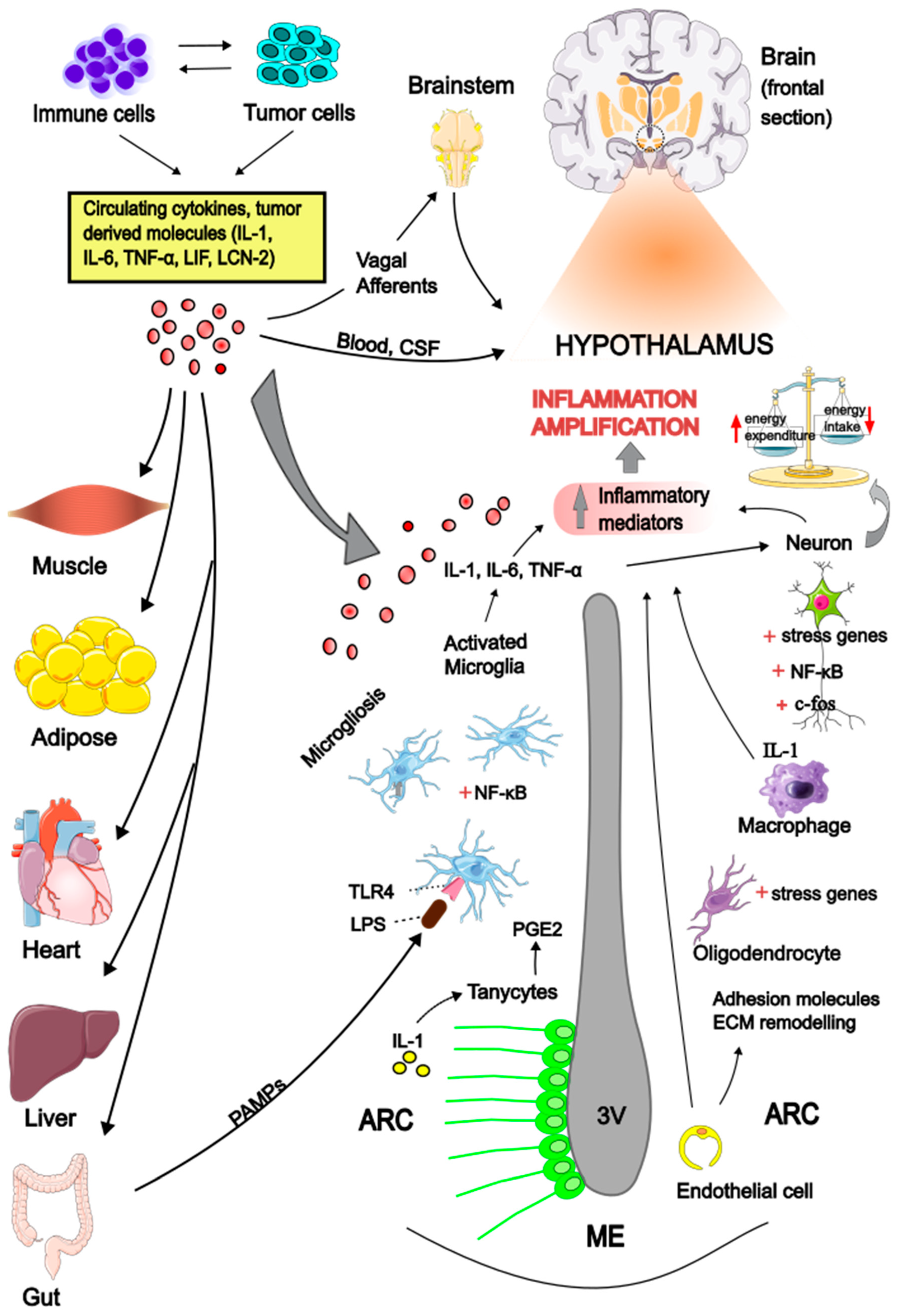
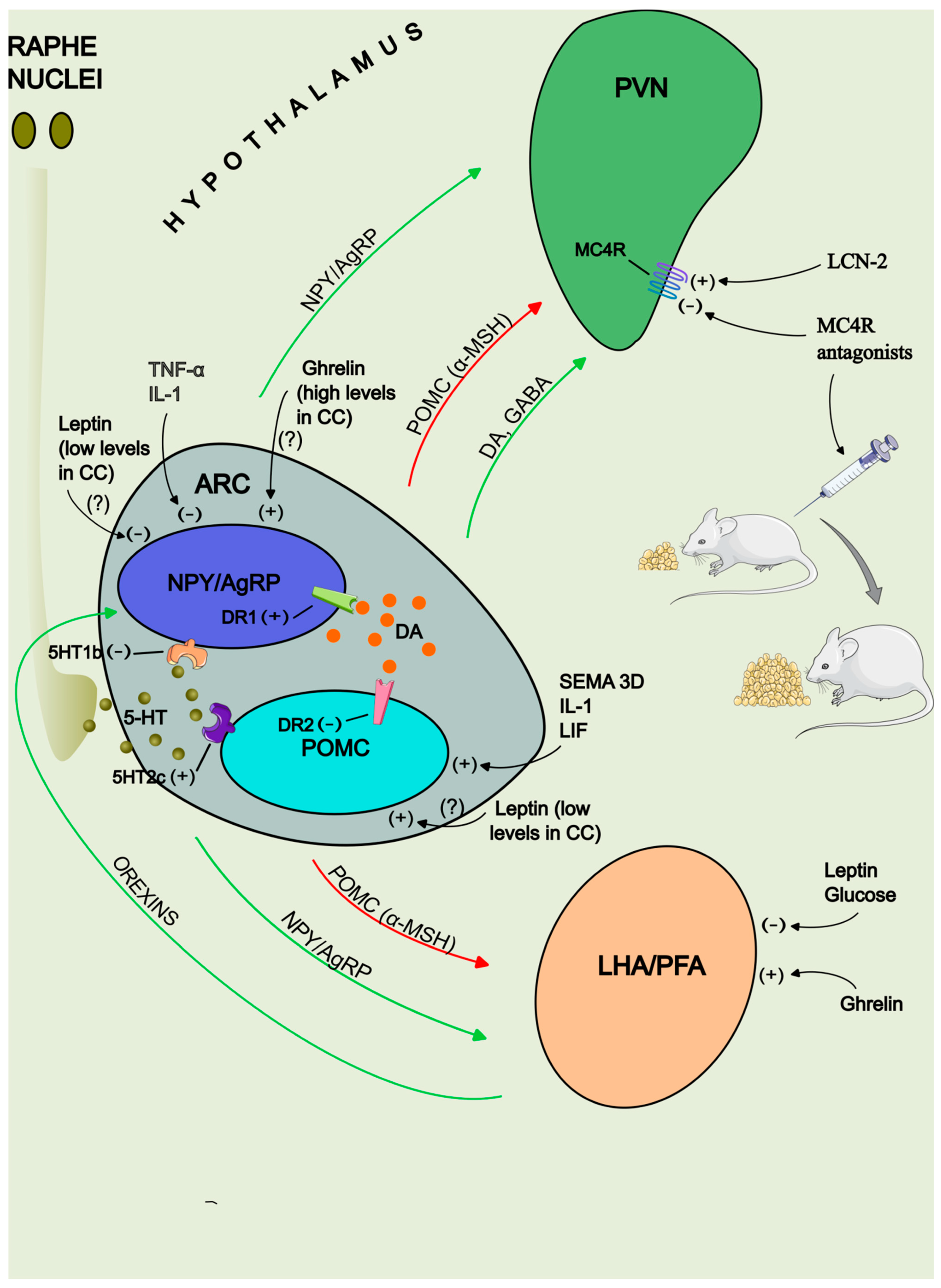
Cachexia is a complex multiorgan syndrome associated with various chronic diseases, characterized by anorexia and increased tissue wasting in the context of chronic inflammation. A specific form of this syndrome, known as cancer cachexia (CC), occurs alongside different types of tumors. The pathogenesis of CC is multifactorial, with inflammatory mediators and hormones released by either the tumor or the host identified as key drivers of the peripheral catabolic process through several direct mechanisms. Accumulating evidence indicates that the central nervous system (CNS) is also recognized as an integral component in the pathogenesis of CC. Hypothalamus has emerged as a critical brain area that senses and amplifies peripheral stimuli, generating an inappropriate neuronal signaling, leading to deregulation of feeding behavior and impaired control of energy homeostasis. Circulating cytokines may act in concert with hormones and neurotransmitters and perturbate the hypothalamic melanocortin system, shifting its activity towards the anorexigenic pathway and increase energy expenditure. The purpose of this review is to provide insights on the potential mechanisms mediating the hypothalamic inflammation in the context of anorexia and cachexia associated with cancer.

Keywords: 
;  ;  ;  ;  ;  

1. Methods

We performed an extensive search of the PubMed database, retrieving manuscripts published up to October 2024. In the search strategy the keywords cancer cachexia, anorexia, hypothalamus, cytokines, neurotransmitters and neuroendocrine were used with derivations and different combinations. We also assessed the references of the retrieved articles to identify any further relevant publication.

2. Introduction

Cachexia is a serious clinical condition associated with several illnesses including chronic kidney disease [1], cardiac failure [2], chronic obstructive pulmonary disease [3], acquired immunodeficiency syndrome (AIDS) [4] and cancer [5]. Cancer cachexia (CC) occurs in almost 80% of patients with advanced cancer and it is directly correlated with impaired quality of life and short survival [6]. The principal cachexia features include loss of appetite, inflammation, insulin resistance and skeletal muscle wasting, with or without fat mass loss [7]. The pathogenesis of this syndrome is complex and is gradually being unraveled. Inflammatory mediators and tumor derived factors have been proposed as direct triggers of the catabolic process in skeletal muscle and adipose tissue. Even though muscle and fat loss remain the principal features of CC, there is growing evidence that organs such as liver, heart and gut are also affected by cachexia factors.
Increasing body of evidence suggests that the brain mediates several metabolic and behavioral aspects associated with CC by sensing and amplifying peripheral inflammatory signals and altering the pattern of neurotransmitters and neuropeptides involved in feeding. The hypothalamus is an important structure for appetite regulation and body energy expenditure and may play a crucial role in CC development. The mediobasal hypothalamus (MBH), which contains the ventromedial arcuate nucleus (ARC) and the media eminence (ME) complex, is a key region that lacks the blood brain barrier (BBB) and functions as a ‘brain window’ for sensing circulating inflammatory molecules and hormones [8]. In the ARC resides the melanocortin system (MCS), a critical neural system comprised of neurons with opposite effect on the control of appetite and the homeostatic feedback regulation of energy balance [9]. Upon peripheral stimuli, the MCS may respond inappropriately and disrupt the balance between orexigenic (appetite-stimulating) and anorexigenic (appetite-suppressing) pathways, leading to anorexia and hypercatabolic state [10]. In addition, it is known that hypothalamus is an important site for autonomic and endocrine homeostasis. Heightened sympathetic tone and aberrant neuroendocrine function have been observed in experimental cachexia models and are recognized as critical determinants of cancer associated tissue loss and increased energy wasting [11,12,13,14,15]. The paraventricular nucleus (PVN), specifically the parvocellular division, serves a crucial role in this process since it is able to integrate neuronal signals from hypothalamic nuclei and brainstem centers involved in appetite regulation (i.e. nucleus tractus solitarii-NTS and dorsal motor nucleus of the vagus-DMNV) and either generate a coordinated sympathetic output through projections to preganglionic neurons in the spinal cord [16] or secrete hypophysiotropic factors into the neurohemal zone of the median eminence modulating the neuroendocrine response [17].
The purpose of this review is to give information about cachexia mediators and neural pathways involved in central nervous system (CNS) dysfunction in the setting of cancer. The interplay between inflammatory molecules and neuronal groups will be discussed and how neuropeptides and neurohormones are affected to alter energy homeostasis and food intake. Although the process is complicated and involves multiple brain centers, this study will focus on the contribution of the hypothalamic mechanisms in driving this pathologic condition. Sheding light on the underlying pathophysiological pathways could help us identify novel molecular targets and expand the therapeutic options for cancer-related anorexia.

3. Cytokines Signaling in the Brain

Elevated circulating levels of pro-inflammatory cytokines have been reported in cancer patients suffering from cachexia and associated with poor treatment outcomes [18,19,20,21,22,23]. These molecules have been shown to act on peripheral tissues stimulating catabolism and centrally affecting the mechanisms that regulate energy homeostasis [Figure 1]. Cytokines directly access the brain through the ME, where the BBB is incomplete, and reach the ARC. In addition, upon binding to their relative receptors on the BBB endothelial and glial cells can stimulate the synthesis of other chemical mediators such as prostaglandin-E2 (PGE2) and nitric oxide (NO) [24]. Tanycytes are specialized glial cells lined at the floor of the 3rd ventricle in the MBH with processes that extend into the ventromedial hypothalamus (VMH) and the ARC [25]. Tanycytes have a prominent role in sensing blood-borne signals and generating an appropriate hypothalamic response. A recent study showed that IL-1 induced the transcription of the enzyme cyclooxygenase-2 (COX-2), involved in the PGE2 biosynthesis, by activating the nuclear factor-κB (NF-κB) pro-inflammatory signaling pathway in the tanycytes of the MBH and suppressed feeding in mice [26]. Finally, cytokines may communicate peripheral signals to brainstem centers via the vagal sensory terminals and ultimately to the hypothalamus [Figure 1] [27].
Cytokines interact in the CNS through a complex network in order to induce anorexia in chronic pathologic states. Experiments in rats revealed that the combined intracerebroventricular (i.c.v.) administration of cytokines, at doses that yield pathophysiological concentrations in the cerebrospinal fluid (CSF), may elicit greater anorexia than the treatment with individual cytokine alone, indicating a synergistic or additive activity [28].
Several anorexigenic cytokines have been implicated in the pathophysiology of CC and will be discussed separately [29,30]. High cytokine levels have been detected in the hypothalami of tumor-bearing animals and associated with worsening of cancer associated anorexia [31,32,33,34,35,36,37,38]. Pharmacological activation of neuronal adenosine 5'-monophosphate-activated protein kinase (AMPK), a key component molecule involved in the regulation of energy homeostasis, was accompanied by the reduction of the hypothalamic levels of these cytokines, reversed anorexia and prolonged life span in animals [32].
1. IL-1
Interleukin-1 (IL-1) is the prototype of the family of cytokines and in the brain plays a pivotal role in the organization of the hypothalamic cytokine network in response to systemic inflammatory challenges [39]. Under acute inflammatory stimuli and in chronic inflammatory illnesses such as cancer, the hypothalamic activity of IL-1β increases and correlates with the suppression of appetite [31,34,35,36,37,38,39,40]. A series of experiments in anorectic cancer animal models reported increased concentration of IL-1 in the CSF which correlated inversely with food intake [41]. IL-1 interferes with the activity of the MCS as reported by studies using central administration of IL-1β and animal models of CC [42,43]. Selective treatment with intra-VMH injection of molecules that antagonize the IL-1 activity reduced the severity of cancer associated anorexia [44].
2. TNF-α
Another cytokine implicated in the induction of cachexia in acute and chronic diseases is tumor-necrosis-factor-α (TNF-α) [45]. Rodents that received TNF-α both centrally and peripherally exhibited decreased nutritional intake and body mass [46,47]. Its anorectic properties may be attributed to its suppressive action on chemosensitive neurons in the lateral hypothalamic area (LHA) and orexigenic neurons in the ARC [48,49]. Moreover, increased hypothalamic TNF-α signaling has been shown to induce thermogenesis in the brown adipose tissue (BAT) mediated by an increase in β-adrenergic tone and rise of total body oxygen consumption, typical features of chronic cachectic states [50]. In experimental cancer models the use of recombinant human soluble TNF-α receptor or anti-TNF-α monoclonal antibodies improved food intake, body weight and increased survival [47,50]. However, in clinical trials blockade of the TNF activity alone was not sufficient to ameliorate anorexia and cachexia parameters, underlying the presence of a complex network of humoral mediators that collectively promote wasting [51,52].
3. IL-6 Family
The IL-6 family of cytokines is a group of cytokines that use the glycoprotein 130 (gp130) as the common signaling transducing component of the functional receptor complexes [53]. Among all the members of the IL-6 family, IL-6 and leukemia inhibitory factor (LIF) are considered to play a crucial role in the pathogenesis of anorexia and wasting syndrome in cancer. All these molecules signal through activation of the janus kinase/signal transducer and activator of transcription (JAK/STAT) pathway, which is the same pathway induced by leptin [53].
IL-6 is produced by both immune and tumor cells [29]. Plasma levels of IL-6 were increased significantly in mice with tumors and in oncologic patients and have been correlated with poor outcome [54,55,56,57,58]. Anti IL-6 antibodies successfully reversed the cachectic phenotype in C26 tumor bearing mice and body weight loss in cancer patients with high IL-6 serum levels [59,60]. However, IL-6 is not considered a universal driver of CC and mice bearing IL-6 knock out C26 tumors were still able to develop the cachectic phenotype [61]. In addition to the well documented peripheral catabolic effects, several publications pointed to a role of IL-6 in the CNS. IL-6 plays a prominent role in the anorexia of infection and increased brain production has been associated with the development of LPS induced sickness behavior [62]. Central delivery or overexpression of IL-6 in the CNS, dampened feeding and increased energy expenditure and tissue wasting [63,64,65]. It is likely that IL-6 exerts its modulatory effects on metabolism by acting at the hypothalamus and affecting the expression of neuropeptides implicated in the control of energy balance [63,64,66]. Increased IL-6 signaling in the VMH induced fatty acid oxidation in the skeletal muscle through stimulation of the α-adrenergic pathway [67].
LIF is a pleiotropic cytokine with paradoxically opposite effects on different cell types [29]. LIF is produced by multiple cancer cell lines and fosters tumor development, metastasis and peripheral tissue wasting [54,68,69,70,71,72,73]. LIF can reach the brain parenchyma from the blood by crossing the BBB [74] where interacts at the hypothalamic level with the appetite regulating neuropeptides and interferes also with the leptin secretion promoting hypophagia [68,73,75,76]. LIF can induce a sustained inflammatory reaction and a long-term anorectic response, in contrast with IL-1 and TNF-α, whose anorectic effect is attenuated after continuous administration [45]. In animals implanted with cancer cells LIF expression correlated with early onset and greater severity of anorexia-cachexia [76]. It has been demonstrated that LIF associated cachexia occurs independently of IL-6 because rLIF administration in wild type (WT) mice lacking IL-6- provoked anorexia and weight loss [54].

4. Macrophage inhibitory cytokine-1/Growth differentiation factor-15 (MIC-1, GDF-15)

MIC-1/GDF-15 is a stress response cytokine, member of the tumor-growth-factor beta (TGF-β) superfamily, whose expression is induced in chronic inflammatory processes and malignancies [77]. High MIC-1/GDF-15 circulating levels are linked to anorexia, weight loss and poor life span in cancer patients [22]. Mice bearing tumors overexpressing MIC-1/GDF-15 presented weight loss secondary to reduced nutritional intake and these effects were reversed upon use of antibody to MIC-1/GDF-15 [30]. Systemic administration of MIC-1/GDF-15 induced c-fos immunoreactivity in the ARC and PVN, implying that neurons in these appetite controlling centers act as potential downstream effectors of the MIC-1/GDF-15 action [30]. However, MIC-1/GDF-15 driven anorexia appears related to the activation of upstream feeding centers, since the MIC-1/GDF-15 specific receptors, named glial-derived neurotrophic factor (GDNF) receptor alpha-like (GFRAL), have been found to be restricted in the area postrema (AP) and NTS in the brainstem [78,79,80]. Further analysis extends beyond the scope of this review.

4.1. Hypothalamic Inflammation

Previous experiments demonstrated that peripheral administration of immune challenges and cytokines increased c-fos expression (marker of neural activity) in critical brain areas involved in feeding and metabolism, like the MBH and PVN [81,82,83]. In these centers high density of cytokine receptors is observed, implying that cyt[78–80okines target these hypothalamic centers to mediate anorexia and metabolic changes [84,85,86]. Cytokines can act in the brain through paracrine interactions inducing further their local production, independently of their concentration in plasma, thus maintaining and amplifying locally the inflammatory signaling [87]. In the hypothalamus an important source of cytokines seems to be the activated microglia [88]. Infectious agents have been shown to stimulate cytokine production by microglia, which in turn affect the appetite regulating neuronal networks via NF-κB signaling [35,89,90,91]. In tumor bearing animals CD11b-positive hypothalamic microglia showed high expression of m-RNA levels of IL-1β, IL-6 and TNF-α in the early stages of the disease and before the appearance of cachexia symptoms [15]. Neurons whose cell bodies are mainly located in the periventricular hypothalamic area are also able to synthesize and release cytokines [92,93,94]. Inflamed endothelial cells further contribute to the amplification of the inflammatory state by increasing the expression of genes involved in cell adhesion, cytokine signaling and extracellular matrix (ECM) remodelling, leading to altered permeability of the BBB [95]. Finally, oligodendrocytes and macrophages are also recruited and participate in the sustainment of the inflammatory response [95]. The pathophysiology of hypothalamic inflammation is illustrated in Figure 1.

4.2. Inflammatory Signals Across the Gut-Brain Axis

A mechanism linking gut microbiota with their metabolites and exacerbation of the hypothalamic inflammatory status is currently being investigated. Damage of the intestinal mucosa is well documented in CC animals and patients and may lead to transposition of bacterial components like lipopolysaccharide (LPS) to the systemic circulation [Figure 1] [58,96,97]. In animals with advanced cancer and severe appetite and weight loss, high LPS plasma levels correlated with deterioration of hypothalamic inflammation as evidenced by changes in the expression of immune checkpoint receptors in hypothalamic microglia [15]. In anorectic experimental cancer systems increased circulating LPS levels correlated directly with hypothalamic inflammation and degree of cachexia [98]. In vitro analysis showed that addition of LPS in cultured hypothalamic cells induced the release of IL-6 and this response was significantly amplified upon addition of tumor secretomes, indicating that LPS and tumor derived factors may act synergistically augmenting the inflammatory hypothalamic response [98]. It is well known that LPS induces neuroinflammation by interacting with the toll-like receptor 4 (TLR4), abundantly expressed in microglial cells, leading to phosphorylation and nuclear translocation of the NF-κB p65 subunit and the transcription of genes encoding cytokines [99,100,101]. Myeloid differentiation primary response gene 88 protein (MyD88) and TIR (Toll/interleukin-1 receptor) -domain-containing adaptor-inducing beta interferon (TRIF) are two key adaptor proteins for the TLR4-mediated downstream signaling [100,102]. Blockade of MyD88 signaling mitigated the hypothalamic inflammatory gene expression and significantly attenuated anorexia in pancreatic cancer models [36]. Likewise, TRIF deficient mice inoculated with tumor cells displayed down-regulated levels of cytokines and chemokines in the hypothalamus and improved anorexia and weight loss [35].

4.3. The Role of Hypothalamic Microglia

Several studies uncovered the contribution of glial cells in the control of energy metabolism in association with brain inflammation. Hypothalamic microglia were assigned a key role in the pathogenesis of acute sickness behavior and obesity [35,91,103,104]. In these conditions, pharmacological depletion of microglia has been reported to produce beneficial effects by attenuating hypothalamic inflammation and neuronal stress [91,103,104]. Few studies investigated the role of microglia in the development of CC. In a rodent model with implantation of the AH-130 ascites hepatoma cancer cell line, accumulation and activation of microglia were detected in both PVN and ARC where specifically increased sympathetic tone and induced the anorexigenic pathway activity [105]. Treatment with the microglia inhibitor minocycline suppressed microglia and neuronal activation and alleviated the clinical cachectic features [105]. In a murine model of pancreatic ductal adenocarcinoma (PDAC) microgliosis within the MBH was observed at an early stage of disease progression [106]. However, in this case pharmacologic microglia removal worsened cachexia indicators (anorexia, fatigue and muscle catabolism), suggesting that microglia may have a protective role in the PDAC-induced cachexia [106]. Indeed, in vitro analysis showed that microglia responded to tumor derived factors by increasing the production of arginase-1, an enzyme with potent anti-inflammatory neuroprotective properties, and attenuating the production of the pro-inflammatory cytokines IL-1 and TNF-α, both of which are implicated in neurotoxicity [106,107,108]. Microglia are highly heterogeneous cells and display differential phenotypic and functional features when activated depending on differences in the regional microenvironment [109]. Recent data reported the presence of two transcriptionally and functionally distinct subpopulations of activated microglia (increased expression of the marker ionized calcium-binding adaptor molecule 1 i.e. IBA1) in the MBH of pancreatic cancer models with different gene expression signatures and cachexia-related responses [95]. The IBA1+/CD45+ subset which expresses genes linked to a pro-inflammatory and neurotoxic profile and the IBA1+/CD45- subset which presents a transcriptional profile associated with neuronal growth, tissue remodeling and cell signaling [95]. These observations indicate that different clusters of disease related microglia coexist and at least one cluster appears to have a protective role in pancreatic cancer.

4.4. The Melanocortin System

The ARC in rodents (infundibular nucleus in humans) is located in the MBH and its role is critical for the maintenance of energy homeostasis. Within the ARC resides the MCS composed of two distinct subpopulations of neurons with opposite effects on feeding and energy balance [9]. One cluster includes the pro-opiomelanocortin (POMC) and cocaine and amphetamine regulated transcript (CART) expressing neurons. POMC is a pro-hormone that is processed to biologically active peptides including the alpha-melanocyte-stimulating hormone (α-MSH), which act as an agonist at its specific subtypes melanocortin 4 receptors (MC4R) at the PVN to suppress food intake and increase energy expenditure [9]. Adjacent to POMC/CART neurons a second cluster of neurons express the orexigenic peptides Agouti-related peptide (AgRP) and neuropeptide Y (NPY) [9]. AgRP is the inverse agonist for MC3R and MC4R and antagonizes the effects of α-MSH. Neuropeptide Y (NPY) is a potent stimulator of food intake through its combined action on both the receptor subtypes Y1 and Y5 [110]. Under positive energy balance the expression of AgRP and NPY in the ARC is decreased while that of POMC and CART is enhanced. Conversely, in conditions of negative energy balance, the activity of the anorexigenic POMC/CART neurons is suppressed and the activity of NPY/AgRP neurons is promoted, resulting in reduction of the basal metabolism and increase in food consumption [10]. In the context of cancer wasting syndrome, the hypothalamus does not respond appropriately to neuroendocrine signals and the equilibrium between the neuropeptidergic circuits is dysregulated (see Figure 2 for the potential interplay between inflammatory mediators, hormones and neuropeptides in the disruption[111–114 of the balance between orexigenic and anorexigenic circuits). Upregulated MC4R signaling was observed in the hypothalami of rodents with advanced cancer, anorexia and significant tissue loss, while central infusion of AgRP or other MC3/4-R antagonists ameliorated hypothalamic inflammation and cachexia [111,112,113,114]. This observation led to the hypothesis that interruption of the MC4R pathway may prove beneficial for the treatment of CC. Novel selective MC4R inhibitors with high bioavailability and BBB penetration capacity have been developed and when administered peripherally have been shown to protect against tumor-induced cachexia in animals [115,116]. Their efficacy and safety profile are currently being tested in phase 1 clinical trials in healthy volunteers [117].
While the impact of MC4R on body weight and feeding behaviour is clear, the role of MC3R is not well defined. The obesity phenotype of MC3R deficient mice is different than that of MC4R deficient mice. In contrast to MC4R deficient mice which develop early and severe obesity with remarkable hyperphagia, MC3R deficient mice display mild late onset obesity without hyperphagia [118,119]. It has been reported that MC3R is expressed on AgRP neurons and exerts an inhibitory effect on post-synaptic POMC and PVN neurons expressing the MC4R through γ-aminobutyric acid (GABA) release [120]. In physiologic states, blockade of MC3R potentiated the anorectic effect in response to administration of MC4R agonists and enhanced the sensitivity to multiple anorexigenic compounds (Glucagone-like peptid-1 i.e. GLP-1, cholecystokinin, leptin) [121]. In LPS and cancer induced cachexia in mice lacking the MC3R, decreased food consumption and enhanced tissue wasting were observed [122].
Cytokines may directly interact and influence the activity of the MCS [Figure 2]. For example, receptors for IL-1 and LIF have been detected on POMC neurons [42,75]. In response to IL-1 administration POMC neurons increased their firing rate and the release of α-MSH from hypothalamic explants [42]. Centrally LIF-treated mice exhibited decreased food consumption associated with increased POMC activity and a-MSH secretion [75]. Conversely, IL-1 and TNF-α down-regulated the activity of AgRP-producing neurons and inhibited the release of AgRP [49]. MIC-1/GDF-15 has been shown to rise and drop arcuate POMC and NPY m-RNA levels respectively, and presumably indirectly through projections from upstream hindbrain nuclei [30,78]. These changes were associated with reduced food intake and weight loss.
Semaphorins of class 3 (SEMA3) are signaling secreted molecules with a regulatory role in tumor microenvironment and cancer cell behavior [123]. Semaphorin 3D (SEMA3D) has been shown to interact with its receptors neurophilin and plexin located on POMC neurons to drive the development of the hypothalamic circuit and energy balance [Figure 2] [124]. In a recent paper increased SEMA-3D expression was reported in patients and animals with CC which was positively related to increased hypothalamic POMC levels [125]. Mice with deficiency of SEMA3D showed decreased POMC neural activity and improvement of cachexia parameters [125].
The levels of hypothalamic POMC m-RNA have been found to be low, high or even unchanged among different cancer models or vary in the same model as the disease progresses [31,32,38,56,76,95,126,127,128,129,130]. It has been postulated that acute or chronic inflammatory stimuli may exert opposing signals on POMC expression. In conditions of acute inflammation POMC expression is induced, whereas in chronic inflammation POMC expression is suppressed. Administration of LPS (a typical model that resembles acute inflammation) in animals, induced an extensive cytokine response that promoted POMC expression, leading to an anorexic state and increased energy expenditure [131]. In the contest of malignancy, early (acute) or advanced (chronic) stage of the disease is associated with high or reduced POMC levels, respectively. Cernackova et al found increased POMC transcript levels at day 21 in fibrosarcoma bearing Wistar rats, before cachexia was detected, and decreased POMC levels at day 28 when cachectic manifestations were remarkable [38]. Likewise, Suzuki et al noted a pronounced decrease of POMC expression in hypothalamus of gastric cancer bearing rats only in advanced-stage cachexia with severe anorexia [129]. A previous report revealed that the p65 subunit of the transcription factor NF-κB binds to the POMC promoter and activate its transcription in acute inflammatory conditions. However, in chronic inflammatory states due to increased methylation at and near the p65 binding site within the POMC promoter, the p65 subunit is unable to interact with the promoter, resulting in failure of the NF-κB-induced POMC transcription. At the same time p65 interacts with the phosphorylated signal transducer and activator of transcription 3 (STAT3) preventing its binding to the POMC promoter, further repressing its transcriptional activation [132].
Lipocalin-2 (LCN2) was recently identified as an endogenous osteoblast-derived appetite-suppressing factor with central action [133]. Mosialou et al showed that in normal mice when LCN2 was injected intraperitoneally was able to cross the BBB and inhibit food intake through a MC4R signaling dependent manner in neurons located in the PVN and VMH [Figure 2] [133]. Elevated hypothalamic and circulating levels of LCN2 have been detected in murine cancer models and correlated to reduced food consumption [37,95,114]. Conversely, tumor bearing mice with genetic deletion of LCN2 showed improvement of energy intake and tissue wasting [95,114]. It has been reported that tumor cells stimulate the neutrophils of the bone marrow compartment to produce and secrete LCN2 and that this circulating LCN2 is able to penetrate the BBB and negatively regulate feeding behaviour [114]. Huisman et al reported that in a pancreatic cancer murine model LCN2 was a strong stimulator of hypothalamic inflammation and affected gene expression in POMC neurons, but without affecting POMC expression itself [95]. Since anorexia in this model occurred independently of POMC expression and could be reversed with melanocortin antagonists, the authors concluded that LCN2 is likely to bind to and activate the MC4R, thus by-passing the physiologic melanocortin signaling pathway [114,115]. Further studies are warranted in order to determine the potential interaction between LCN2 and MC4R in pathologic conditions.

5. Neurotransmitters Implicated in Energy Balance

In this section the effect of the neurotransmitters NPY, 5-hydroxytriptamine (5-HT), dopamine (DA), orexins and nesfatin-1 in the regulation of energy balance with emphasis in CC will be discussed.

5.1. NPY

Although very high NPY transcript levels have been reported in the ARC, the main hypothalamic site of NPY peptide release is the PVN, highlighting the importance of the NPYergic ARC-PVN pathway in the control of food intake [134]. NPY is stimulated in conditions of negative energy balance such as fasting, exercise and suppressed after refeeding [134].
In CC the NPYergic system seems to be dysfunctional [135]. Central administration of NPY does not elicit feeding response in tumor-bearing rodents compared to non tumor bearing counterparts [136]. The levels of NPY m-RNA are either unaltered or increased in anorexic animals but without a compensatory increase in food intake [31,38,56,76,95,118,127,135,137,138,139]. Various hormonal and metabolic signals have been reported to affect the synthesis and secretion of NPY. Leptin and lactate decrease NPY m-RMA levels through AMPK inhibition, whereas ghrelin induces NPY gene expression [140,141,142] through AMPK activation. Similarly, increased glucocorticoids (GC) levels, as reported in CC, increase NPY transcripts in the ARC through AMPK signaling [143]. However, in contrast to elevated NPY transcript levels in hypothalamus, reduced protein levels are detected in the PVN, suggesting an impaired NPY transport and release at this specific region [144,145]. The increased serotonergic tone may contribute to this by interfering with NPY signaling at the post transcriptional level and repress its secretion [139]. Leptin has also been shown to suppress NPY secretion in hypothalamic cell lines [140].

5.2. 5-hydroxytryptamine

The neurotransmitter 5-hydroxytryptamine (5-HT) or serotonin has long been considered as a potent anorectic agent with central action. There is evidence that the serotonergic pathway originates from the raphe nucleus in the brainstem and lesions of these nuclei induce hyperphagia [146,147]. 5-HT neurons in the raphe nuclei project to the ARC where they regulate the neural activity of the MCS [146]. 5-HT leads to direct activation of the POMC neurons via interaction with 5-HT2c, and inhibition of the NPY/AgRP neurons via interaction with 5-HT1b subtype receptors [Figure 2] [148,149]. In experimental cancer systems elevated expression levels of 5-HT and HT1b have been observed in hypothalamic nuclei and correlated inversely with food consumption [150,151,152,153]. Tumor removal normalized these alterations and restored appetite [151,152]. Intrahypothalamic delivery of the non-selective 5-HT antagonist mianserin improved anorexia significantly in cachectic animals with cancer [154]. Cytokines may interfere with the 5-HT turnover or regulate the plasma pool of tryptophan, the precursor molecule of 5-HT [134]. IL-1 when injected peripherically reduced food intake and increased plasma tryptophan, suggesting an increase in brain 5-HT synthesis [155]. High plasma and hypothalamic levels of tryptophan and the metabolite 5-hydroxy indoleacetic acid (5-HIAA), marker of 5-HT release, have been reported in both tumor-bearing rats and anorectic cancer patients [156,157]. Administration of branched-chain amino acids BCAA, which compete with tryptophan for entry across the BBB, reduced tryptophan concentration in the brain, normalized brain serotonin activity and attenuated anorexia in cancer patients [158].
An interaction between NO and 5-HT metabolism in the regulation of food intake has also been postulated. More specifically, brain NO has been shown to inactivate irreversibly the enzyme tryptophan hydroxylase, thus, down-regulating the biosynthesis of 5-HT and stimulating appetite [159]. In conditions of food deprivation in rats the activity of the NO synthase (NOS) enzyme in the diencephalon is increased and is accompanied by the fall of 5-HT levels [160]. Delivery of molecules that impair NO synthesis was paralleled with an increase of 5-HT levels and significant reduction in food intake [160]. An endogenous inhibitor of NO synthesis has been described in patients with anorexia associated with chronic renal failure [161]. In tumor models the pattern of NOS expression among different hypothalamic and extrahypothalamic nuclei involved in appetite regulation suggested that NO did not appear as a main mediator behind feeding depression induced by the tumor but rather as a compensatory mechanism to increase energy intake in conditions of negative energy balance [55]. The precise interaction mechanism between NO and 5-HT in the control of food intake in CC remains to be elucidated.

5.3. Dopamine

DA is a critical brain messenger implicated in the regulation of appetite. Mice with dopamine deficiency exhibited hypophagia and death [162]. It appears that DA levels in the LHA are directly correlated to food consumption and rise proportionally to the quantity of meal consumed [163]. In food deprived animals the delivery of high fat diet induced a strong increase of DA in the hypothalamic ARC [164]. Low hypothalamic DA levels are generally reported in animals with CC [145,151]. Tumor resection returned DA levels to normal and restored appetite [151].
DA receptors (DRs) are divided in two groups, depending on whether they can activate or inhibit the enzyme adenylate cyclase; the stimulatory DA1-like receptor, comprising the DR1 and DR5 and the inhibitory DA2-like receptor group (comprising the DR2-DR4) families [165]. High hypothalamic expression of DR2 mRNA was reported in sarcoma bearing rats in the VMH and LHA which correlated with anorexia through a decrease in meal number [166]. Injection of the DR2 antagonist sulpiride in LHA or VMH promoted feeding in both tumor animals and free feeding counterparts [166].
An interesting relation between DA signaling in the ARC and homeostatic feeding response has recently been reported. More specifically, a cluster of NPY/AgRP neurons which express the stimulatory DR1 are predominantly activated upon DA stimulation, whereas a large proportion of POMC neurons expressing the inhibitory DR2 are inhibited, thereby promoting food intake and increasing energy expenditure [Figure 2] [164,167]. Rodents lacking DR1 expression in NPY/AgRP cells exhibited attenuated foraging [164]. Moreover, a group of dopaminergic neurons has been identified in the ARC which is different from the NPY/AgRP and POMC neurons and can be stimulated by fasting and ghrelin [168]. These neurons promote feeding by exerting an inhibitory effect on POMC neurons as well as on neurons in the PVN, through axonal release of DA and GABA [168]. The engagement of this dopamine-dependent control of energy homeostasis in the development of tumor induced cachexia remains to be explored.

5.4. Orexins

Orexins (A and B) are hypothalamic neuropeptides derived from the proteolytic cleavage of a 130 amino acid precursor peptide, termed prepro-orexin (PPO) [169]. Orexin-secreting neurons are placed mainly in the LHA/perifornical area (PFA) and are activated in response to NPY positive projections originating from ARC neurons [170]. They in turn, project and release their product to other hypothalamic centers, including PVN, ARC and NTS. Focusing on the CNS, orexins regulate multiple functions like sleep-wake cycle, autonomic function, feeding behavior and energy homeostasis [169]. Orexin neurons in the LHA sense and respond to peripheral circulating metabolic signals such as glucose, leptin and ghrelin [Figure 2] [169]. Central delivery of orexin-A in rats enhanced food intake, although continuous infusion did not produce substantial changes on feeding and body weight [171,172]. Prepro-orexin m-RNA levels are induced during periods of negative energy balance, such as periods of fasting [171,173]. There is evidence that LPS-induced hypothalamic inflammation disrupts the orexigenic signaling in the LHA and leads to reduced physical activity and hypophagia in animals [174]. In a similar manner, tumor-induced inflammation in anorectic rats was paralleled by a reduction in the number of orexin mRNA-expressing neurons, predominantly in the PFA [174]. The anorectic chemotherapeutic agent cisplatin has been associated with increased hypothalamic levels of proinflammatory cytokines and reduction of prepro-orexin m-RNA [104,175]. Direct delivery of orexin-A to the ARC enhanced significantly the feeding response and the use of neuropeptide Y1 receptor antagonist abolished this effect [175]. These findings indicate that orexin signaling in the ARC stimulates the feeding response via an NPY-dependent pathway.
It has been demonstrated that orexin neurons in the LHA are able to sense circulating glucose levels and increase orexin m-RNA levels in response to hypoglycemia promoting food intake [173]. Central administration of orexin increased the levels of insulin and prevented insulin resistance in peripheral tissues via the autonomic nervous system [176]. Since insulin resistance is frequently encountered in chronic cachectic diseases and associated with poor prognosis, restoration of normal insulin sensitivity by orexin may be a promising supportive therapeutic approach for CC [29].

5.5. Nesfatin-1

Nesfatin-1 is an 82-amino acid polypeptide that is cleaved from the precursor protein nucleobindin-2 (NUCB2) [177]. Nesfatin-1/NUCB2 was first detected in rat brains, in the PVN, ARC and NTS and latter in various peripheral tissues [177,178]. Rats with deficiency of Nesfatin-1/NUCB2 in the PVN increased feeding, fat accumulation and body weight [179]. Overexpression of Nesfatin-1/NUCB2 has been correlated with cancer invasion and metastatic potential [180,181]. Elevated Nesfatin-1/NUCB2 m-RNA levels in the PVN have been reported in tumor-bearing mice with anorexia implying a role for this molecule in the etiology of the syndrome [182]. Several mechanisms involved in the Nesfatin-1/NUCB2-induced anorexia have been proposed. Central administration of SHU9119, a melanocortin-3/4 receptor antagonist, abolished the anorexia induced by Nesfatin-1/NUCB2, suggesting a possible interaction with the MCS [183]. Increase of the oxytocinergic signaling from PVN neurons to POMC neurons in the NTS, interaction with the corticotropin releasing factor (CRF) receptor system and down-regulation of the NPY-expressing neurons in the ARC have also been reported [184,185,186]. Finally, Nesfatin-1/NUCB2 has been shown to evoke a sympatho-excitatory response through extracellular signal regulated kinase (ERK)-dependent signaling in PVN-CRF neurons and enhance fat lipolysis and fatty-acid oxidation in skeletal muscle in pathologic contexts other than CC[187,188]

6. Peripheral Hormone Signals: Leptin and Ghrelin

Leptin is a protein hormone derived from the adipocytes involved in satiety promotion and increase in energy expenditure [189]. Leptin signals in the cell by binding to its transmembrane receptors (Lep-R) and activating the JAK/STAT pathway [190]. Leptin interacts with both NPY/AgRP and POMC neurons in the ARC [Figure 2] producing opposing effects, however, a direct action on a population of neurons expressing the MC4R in the PVN has also been observed [191,192]. Low leptin plasma levels have been reported among patients and animals with CC [128,193,194,195], but without a concomitant compensatory stimulation of appetite or weight gain [18,128,193,194,195]. There is evidence that cytokines that share the same post receptor pathway with leptin, are able to upregulate the anorexigenic and down-regulate the orexigenic pathways by mimicking leptin action. In particular, MIC-1/GDF-15 and members of the IL-6 family induce the phosphorylation of STAT3, similar to leptin and have been demonstrated to trigger anorexia-cachexia in cancer animal models, providing an explanation for the persistence of anorexia and body weight loss, in spite of decreased leptin concentration [30,54,196]. Treatment with molecules that inhibit JAK activity suppressed the anorexia associated with cancer in animals, inhibited lipolysis and improved survival and all these effects were accompanied by the reduction of STAT3 phosphorylation in hypothalamic and adipose tissues [54].
Ghrelin, is an endogenous stomach derived ligand with high affinity for the growth hormone (GH) secretagogue receptor (GHS-R) [197]. Ghrelin promotes weight gain by increasing appetite and decreasing energy expenditure [198]. Independently of its action in regulating the activity of the pituitary GH releasing neurons, ghrelin directly activates the orexigenic NPY/AgRP neurons which express the GHS-R [199,200]. Ghrelin is also able to inhibit POMC neurons indirectly through an increase of the inhibitory GABAergic tone originating from the NPY/AgRP neurons [201,202]. Additionally, ghrelin is able to act peripherically by stimulating the GHS-Rs located at the vagal afferent terminals which in turn transmit the signal to the hypothalamus [203]. Ghrelin serum levels are frequently found increased in cachectic patients with cancer and in anorectic tumor carrying animals. However due to an altered ghrelin signaling (called ghrelin resistance) appetite remains suppressed [Fig 2] [111,204,205,206,207,208]. It has been postulated that anorexigenic proinflammatory cytokines can interfere with the appetite stimulatory effect induced by ghrelin [208]. Modulation of the orexigenic ghrelin signaling in anorectic tumor bearing rats due to hypothalamic interaction of 5-HT and CRF via the 5-HT2c has also been reported [13]. In addition, defect at the receptor level has been hypothesized and reduced expression of the GHS-R has been detected in anorectic animals treated with cisplatin [209]. In this study, oral administration of 5-HT2cR antagonist, enhanced ghrelin sensitivity through up-regulation of the hypothalamic levels of GHS-R and attenuated the cisplatin-induced anorexia [209]. However, in tumor-bearing animals the levels of hypothalamic GHS-R were found to be either similar to control animals or elevated and normalized after exogenous ghrelin administration at high doses [207,208]. The possibility of an impaired signaling downstream of the ghrelin-GHS-R system or a defective ghrelin transport across the BBB perhaps should be considered in the future. Administration of exogenous ghrelin in patients and animals with cancer increased appetite and food intake, suggesting that presumably further increase of plasma ghrelin levels may overcome the endogenous encountered resistance to the peptide, resembling that way the delivery of exogenous insulin for the treatment of insulin-resistant state in type II diabetes [205,207,210,211,212]. Ghrelin or ghrelin mimetics in animals with CC, along with the positive impact on orexigenic peptides exert also an anti-inflammatory hypothalamic action as reported by the decrease of IL-1 receptor transcripts levels [213]. Synthetic agonists of GHSR have been developed and are currently under clinical investigation for the treatment of anorexia associated with cancer. Anamorelin is such a molecule with a longer half-life than ghrelin and has been approved for the treatment of CC in Japan. Although it appears well tolerated and increase lean body mass and appetite, its efficacy in patients with very poor clinical conditions is questioned [214].

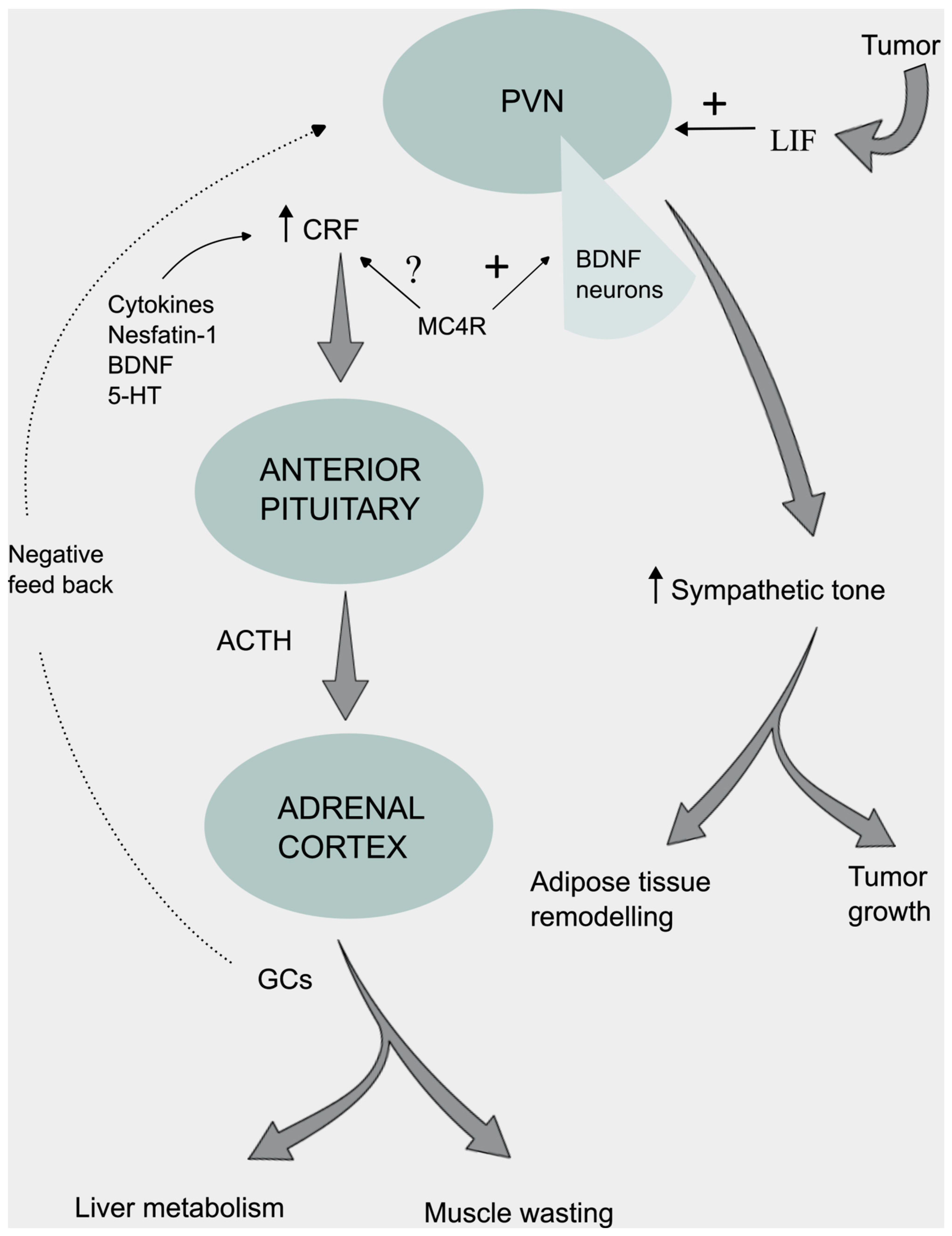
Neuroendocrine and Autonomic Regulation

The neuroendocrine system is regulated by hypothalamic neurons that send projections to the ME and secrete neurohormones into the portal hypophyseal system to excite cells in the anterior pituitary gland (adenohypophysis). Among these trophic factors, CRF is released by neurons located within the parvocellular zone of the PVN and stimulates the secretion of adrenocorticotropic hormone (ACTH) from the pituitary. ACTH induces the adrenal cortex to produce GC (cortisol in humans and corticosterone in rodents) which in turn exerts a negative feed-back control suppressing the hypothalamic-pituitary-adrenal axis (HPA) [215].
It is well accepted that GCs play a crucial role in muscle wasting by regulating specific intracellular signaling pathways which stimulate protein catabolism and inhibit protein synthesis [216]. Rise in endogenous GC levels have been reported in both cancer patients and tumor bearing animals and correlated with the severity of cachexia [14,15,38,217,218,219,220,221,222,223]. In adenomatous polyposis coli (APC) cachectic mice a rise in corticosterone levels in serum and in skeletal muscle was associated with the induction of the transcription of GC-responsive genes that promote muscle protein degradation (called atrogenes) and down-regulate phosphatidylinositol 3-kinase (PI3K)/AKT/mammalian target of the rapamycin (mTOR) anabolic pathway [14].
Although GCs act as downstream effectors of the HPA axis the upstream mechanism by which HPA is activated in cancer cachexia is unknown. CRF possess a strong hypophagic action and has assumed particular relevance in the setting of cancer [13,14,34,144,224]. In mice with terminal stage pancreatic cancer, severe muscle loss was associated with elevated hypothalamic CRF gene expression [34]. I.c.v. administration of a CRF antagonist in rats inoculated with AH-130 ascites hepatoma cells increased feeding, suggesting that the CRF system is activated in malignancy [13].
Cytokines such as IL-1, IL-6, TNF-α and GDF-15 have been shown to directly stimulate the expression of the CRF gene in hypothalamic 4B cells [66,225,226]. Central administration of serum anti-CRF abolished the IL-1-induced anorexia in Wistar rats and depletion of endogenous IL-6 was associated with reduced production of CRF [66,227]. The brain derived neurotrophic factor (BDNF) is a molecule with a central role in energy control also able to stimulate the anorexigenic CRF system. Single or continuous i.c.v. delivery of BDNF upregulated the CRF m-RNA levels predominantly in the PVN and local infusion of the a-helical CRF receptor antagonist counteracted the appetite suppressive effect evoked by BDNF [228].
Ultrastructural analysis revealed neuronal intercommunications between 5-HT axons and CRF-containing neurons in the PVN, indicating that a possible interaction between the central serotonergic system and CRF synthesis may exist [229]. In physiologic murine models, 5-HT agonists enhanced c-fos immunoreactivity in CRF-containing neurons in the parvocellular zone and increased CRF m-RNA expression [230]. The administration of 5-HT2cR antagonists decreased hypothalamic CRF level and ameliorated anorexia and body weight loss in tumor-bearing rats [13].
CRF positive neurons in the PVN also express the MC4R [231]. Several studies investigated the potential involvement of these receptors in the CRF induced anorexia with disaccording results [232,233]. Selective blockage of the MC4-R in rats did not affect the CRF-induced inhibition of food intake suggesting that the anorectic effect of CRF is independent of signaling pathways involving the MC4R [232]. On the contrary, the appetite suppressive effect following delivery of a synthetic α-MSH was abolished when a CRF receptor antagonist was used [233]. Experiments in CRF knock-out mice revealed that CRF was involved in the early phase of the α-MSH induced anorexia but not in the late phase, implying that other endogenous ligands of the CRF family might be responsible for the late anorexic effect [233]. A schematic representation of the molecules involved in the activation of the HPA axis in CC is presented in Figure 3.
The parvocellular PVN provides an important sympathoexcitatory output through connections with brainstem centers and pre-ganglionic neurons in the spinal cord as well as with local intrahypothalamic neurocircuits. Activation of the PVN-sympathetic adipose circuit has been reported in chronic restraint stress [234]. Increased sympathetic drive is documented in humans and experimental models of tumor cachexia and it is associated with white adipose tissue browning, resulting in lipid mobilization and augmented non-shivering thermogenesis [Figure 3] [11,235].
According to some lines of evidence PVN is an important site of action of BDNF and BDNF signaling plays a key role in regulating the sympathetic activity and energy metabolism. Moreover, BDNF has been shown to act as a downstream effector molecule of the melanocortin signaling pathway [236]. In anorectic tumor bearing rats at a late stage of cachexia a significant increase in the hypothalamic expression of MC4R was observed along with a remarkable increase in BDNF expression, underscoring the role of BDNF as an integral component of the melanocortin neurocircuity [129]. In the physiological context, BDNF signaling in the PVN increased the basal metabolism rate in part by stimulating the uncoupling protein-1 (UCP-1) expression (a protein involved in heat production) in BAT [237]. A recent work showed that leptin signaling in the arcuate AgRP and POMC cells regulates sympathetic innervation of adipose tissue through a downstream BDNF-expressing population of neurons in the PVN [238]. BDNF neurons placed in the medial and posterior part of PVN project their axons to the sympathetic preganglionic neurons in the intermedio lateral column of the spinal cord where BDNF acts via its corresponding tropomyosine receptor tyrosine kinase B (TrkB), thereby stimulating BAT thermogenesis [238,239]. In addition, the activity of the BDNF positive neurons in PVN is under the control of the DA signaling pathway originating from upstream brain nuclei. These neurons express DR1 and DR2 receptors and receive projections from dopaminergic neurons in the Substantia Nigra (upstream brain central nuclei) [240]. Reduced activity of the D2 signaling pathway disinhibited BDNF-neurons and increased BDNF release, which in turn affected BAT thermogenesis [240]. LIF receptors are also expressed in neurons in the PVN [68]. In several mouse allograft cancer models Xu et al showed that LIF was able to trigger neuronal activation in several brain regions including PVN [Figure 3] and promote tumor progression through a mechanism involving sympathetic activation and peripheral immune cellular response [68]. Sympathetic ablation led to inhibition of tumor growth without affecting significantly the LIF-mediated neuronal activation of the PVN, suggesting that cancer affects upstream brain centers to modulate the sympathoexcitatory outpout [68]. However, besides the clear antitumor effect, the cachectic parameters or the effects on adipose tissue were not directly evaluated and additional work investigating the correlation between LIF-induced sympathetic response and adipose tissue dysfunction is required.

7. Conclusions

CC is a multifactorial chronic condition related to abnormalities of the central physiological mechanisms that regulate food intake and energy balance. The hypothalamus has been recognized as an essential center that receives peripheral blood-borne signals and messages from other important brain structures which it transduces in perturbated neuronal signaling, abnormal neuroendocrine response and metabolic dysregulation. Hypothalamic inflammation has been shown to promote anorexia by triggering changes in orexigenic and anorexigenic neural circuits and induce energy deficit. Understanding the complex interrelationship between cytokines, tumor-derived factors and brain neurotransmitters and neuropeptides could not only shed light on the underlying pathophysiology of the disorder but also prove indispensable in unveiling the potential therapeutic interventions for the wasting syndrome associated with cancer. Disrupting neural signaling pathways and interfering pharmacologically with cytokine action might be therapeutically exploited in order to ameliorate the cachexia symptoms.

Author Contributions

Figures were drawn by Dimitrios Stagikas. Konstantinos I. Tsamis revised the manuscript. All authors contributed equally to the creation of the manuscript and approved the final version of the paper.

Funding

This study was not supported by any sponsor or funder.

Data Availability Statement

No datasets were generated or analysed during the current study.

Acknowledgments

Figures were created using the free graphic editor Inkscape. Specific icons and images were provided by the open-source databases smart.servier.com and reactome.com.

Declarations

Ethics approval and consent to participate: Not applicable for this manuscript.
Consent for publication The authors declare their consent for publica tion of the review article in Acta Physiologica.

Conflicts of Interest

There is not competing interest to declare.

References

  1. L. Koppe, D. Fouque, K. Kalantar-Zadeh, Kidney cachexia or protein-energy wasting in chronic kidney disease: facts and numbers. J. Cachexia Sarcopenia Muscle 10, 479-484 (2019).
  2. S.D. Anker, A.J. Coats, Cachexia in heart failure is bad for you. Eur. Heart J. 19, 191e3 (1998).
  3. M.L.N. McDonald, E.F.M. Wouters, E. Rutten, R. Casaburi, S.I. Rennard, M. Bamman, B. Celli, A. Agusti, R. Tal-Singer, C.P. Hersh, M. Dransfield, E.K. Silverman, It’s more than low BMI: prevalence of cachexia and associated mortality in COPD. Respir. Res. 20, 100 (2019).
  4. K. Mulligan, V.W. Tai, M. Schambelan, Cross-sectional and longitudinal evaluation of body composition in men with HIV infection. J. Acquir. Immune Defic. Syndr. Hum. Retrovirol. 15, 43e8 (1997).
  5. M.J. Tisdale, Biology of cachexia. J. Natl. Cancer Inst. 89, 1763-1773 (1997).
  6. M.J. Tisdale, Cachexia in cancer patients. Nat. Rev. Cancer 2, 862-871 (2002).
  7. W.J. Evans, J.E. Morley, J. Argiles, C. Bales, V. Baracos, D. Guttridge, A. Jatoi, K. Kalantar-Zadeh, H. Lochs, G. Mantovani, D. Marks, W.E. Mitch, M. Muscaritoli, A. Najand, P. Ponikowski, F. Rossi Fanelli, M. Schambelan, A. Schols, M. Schuster, D. Thomas, R. Wolfe, S.D. Anker, Cachexia: a new definition. Clin. Nutr. 27, 793–799 (2008).
  8. R.D. Cone, M.A. Cowley, A.A. Butler, W. Fan, D.L. Marks, M.J. Low, The arcuate nucleus as a conduit for diverse signals relevant to energy homeostasis. Int. J. Obesity 25, S63–S67 (2001).
  9. R.D. Cone, Anatomy and regulation of the central melanocortin system, Nat. Neurosci. 8, 571–578 (2005).
  10. A. Laviano, A. Inui, D.L. Marks, M.M. Meguid, C. Pichard, F. Rossi Fanelli, M. Seelander, Neural control of the anorexia-cachexia syndrome. Am. J. Physiol. Endocrinol. Metab. 295, E1000-1008 (2008).
  11. M. Petruzzelli, M. Schweiger, R. Schreiber, R. Campos-Olivas, M. Tsoli, J. Allen, M. Swarbrick, S. Rose-John, M. Rincon, G. Robertson, R. Zechner, E.F. Wagner, A switch from white to brown fat increases energy expenditure in cancer-associated cachexia. Cell Metab. 20, 433–447 (2014).
  12. R. Suriben, M. Chen, J. Higbee, J. Oeffinger, R. Ventura, B. Li, K. Mondal, Z. Gao, D. Ayupova, P. Taskar, D. Li, S.R. Starck, H.H. Chen, M. McEntee, S.D. Katewa, V. Phung, M. Wang, A. Kekatpure, D. Lakshminarasimhan, A. White, A. Olland, R. Haldankar, M.J. Solloway, J.Y. Hsu, Y. Wang, J. Tang, D.A. Lindhout, B.B. Allan, Antibody-mediated inhibition of GDF15–GFRAL activity reverses cancer cachexia in mice. Nat. Med. 26, 1264-1270 (2020).
  13. N. Fujitsuka, A. Asakawa, Y. Uezono, K. Minami, T. Yamaguchi, A. Niijima, T. Yada, Y. Maejima, U. Sedbazar, T. Sakai, T. Hattori, Y. Kase, A. Inui, Potentiation of ghrelin signaling attenuates cancer anorexia-cachexia and prolongs survival. Transl. Psychiatry 1, e23 (2011).
  14. A. Martin, J. Castells, V. Allibert, A. Emerit, C. Zolotoff, V. Cardot-Ruffino, Y.S. Gallot, B. Vernus, V. Chauvet, L. Bartholin, L. Schaeffer, A.C. Durieux, C. Hourdé, F.B. Favier, L. Mazelin, D. Freyssenet, Hypothalamic-pituitary-adrenal axis activation and glucocorticoid-responsive gene expression in skeletal muscle and liver of Apc mice. J. Cachexia Sarcopenia Muscle 13, 1686-1703 (2022).
  15. Y. Suda, K. Nakamura, F. Matsuyama, Y. Hamada, H. Makabe, M. Narita, Y. Nagumo, T. Mori, N. Kuzumaki, M. Narita, Peripheral-central network analysis of cancer cachexia status accompanied by the polarization of hypothalamic microglia with low expression of inhibitory immune checkpoint receptors. Mol Brain 17, 20. (2024).
  16. N. Nunn, M. Womack, C. Dart, R. Barrett-Jolley, Function and pharmacology of spinally-projecting sympathetic pre-autonomic neurones in the paraventricular nucleus of the hypothalamus. Curr. Neuropharmacol. 9, 262-277. (2011).
  17. A.G. Watts, S.E. Kanoski, G. Sanchez-Watts, W. Langhans, The physiological control of eating: signals, neurons, and networks. Physiol. Rev. 102, 689-813 (2022).
  18. G. Mantovani, A. Macciò, L. Mura, E. Massa, M.C. Mudu, C. Mulas, M.R. Lusso, C. Madeddu, A.J. Dessì, Serum levels of leptin and proinflammatory cytokines in patients with advanced –stage cancer at different sites, Mol. Med. 78, 554-561 (2000).
  19. J. Pfitzenmaier, R. Vessella, C.S. Higano, J.L. Noteboom, D. Wallace Jr, E. Corey, Elevation of cytokine levels in cachectic patients with prostate carcinoma. Cancer 97, 1211-1216 (2003).
  20. M. Krzystek-Korpacka, M. Matusiewicz, D. Diakowska, K. Grabowski, K. Blachut, I. Kustrzeba-Wojcicka, T. Banas, Impact of weight loss on circulating IL-1, IL-6, IL-8, TNF-alpha, VEGF-A, VEGF-C and midkine in gastroesophageal cancer patients. Clin. Biochem. 40, 1353-1360 (2007).
  21. C. Scheede-Bergdahl, H.L. Watt, B. Trutschnigg, R.D. Kilgour, A. Haggarty, E. Lucar, A. Vigano, Is IL-6 the best pro-inflammatory biomarker of clinical outcomes of cancer cachexia? Clin. Nutr. 31, 85-88 (2012).
  22. L. Lerner, T.G. Hayes, N. Tao, B. Krieger, B. Feng, Z. Wu, R. Nicoletti, M.I. Chiu, J. Gyuris, J.M. Garcia, Plasma growth differentiation factor 15 is associated with weight loss and mortality in cancer patients. J. Cachexia Sarcopenia Muscle 6, 317-324 (2015).
  23. Y.C. Hou, C.J. Wang, Y.J. Chao, H.Y. Chen, H.C. Wang, H.L. Tung, J.T. Lin, Y.S. Shan, Elevated Serum Interleukin-8 Level Correlates with Cancer-Related Cachexia and Sarcopenia: An Indicator for Pancreatic Cancer Outcomes. J. Clin. Med. 7, 502 (2018).
  24. W.A. Banks, J.L. Lynch, O. Tulin O. Price, in The Neuroimmunological Basis of Behavior and Mental Disorder, ed. by A. Siegel, S.S. Zalcman (Springer New York, 2009), p.3.
  25. V. Prevot, B. Dehouck, A. Sharif, P. Ciofi, P. Giacobini, J. Clasadonte, The versatile tanycyte: a hypothalamic integrator of reproduction and energy metabolism. Endocrine Reviews 39, 333–368 (2018).
  26. M. Böttcher, H. Müller-Fielitz, S.M. Sundaram, S. Gallet, V. Neve, K. Shionoya, A. Zager, N. Quan, X. Liu, R. Schmidt-Ullrich, R. Haenold, J. Wenzel, A. Blomqvist, D. Engblom, V. Prevot, M. Schwaninger, NF-κB signaling in tanycytes mediates inflammation-induced anorexia. Mol. Metab. 39, 101022 (2020).
  27. G.R. Johnston, N.R. Webster, Cytokines and the immunomodulatory function of the vagus nerve. Br. J. Anaesth. 102, 453-462 (2009).
  28. C.R. Plata-Salaman, Central nervous system mechanisms contributing to the cachexia–anorexia syndrome. Nutrition 16, 1009-1012 (2000).
  29. A.J. Grossberg, J.M. Scarlett, D.L. Marks, Hypothalamic mechanisms in cachexia Physiol. Behav. 100, 478-489 (2010).
  30. H. Johnen, S. Lin, T. Kuffner, D.A. Brown, V.W. Tsai, A.R. Bauskin, L. Wu, G. Pankhurst, L. Jiang, S. Junankar, M. Hunter, W.D. Fairlie, N.J. Lee, R.F. Enriquez, P.A. Baldock, E. Corey, F.S. Apple, M.M. Murakami, E.J. Lin, C. Wang, M.J. During, A. Sainsbury, H. Herzog, S.N. Breit, Tumor-induced anorexia and weight loss are mediated by the TGF-beta superfamily cytokine MIC-1. Nat. Med. 13, 1333-1340 (2007).
  31. C.R. Plata-Salamán, S.E. Ilyin, D. Gayle, Brain cytokine mRNAs in anorectic rats bearing prostate adenocarcinoma tumor cells. Am. J. Physiol. 275, R566-573 (1998).
  32. E.R. Ropelle, J.R. Pauli, K.G. Zecchin, M. Ueno, C.T. de Souza, J. Morari, M.C. Faria, L.A. Velloso, M.J. Saad, J.B. Carvalheira, A central role for neuronal adenosine 5'-monophosphate-activated protein kinase in cancer-induced anorexia. Endocrinology 148, 5220-5229 (2007).
  33. F.S. Lira, A.S. Yamashita, J.C. Rosa, F.L. Tavares, E. Caperuto, L.C. Carnevali Jr, G.D. Pimentel, R.V. Santos, M.L. Batista Jr, A. Laviano, F. Rossi-Fanelli, M. Seelaende, Hypothalamic inflammation is reversed by endurance training in anorectic-cachectic rats. Nutr. Metab. (Lond) 8, 60 (2011).
  34. K.A. Michaelis, X. Zhu, K.G. Burfeind, S.M. Krasnow, P.R. Levasseur, T.K. Morgan, D.L. Marks, Establishment and characterization of a novel murine model of pancreatic cancer cachexia. J. Cachexia Sarcopenia Muscle 8, 824–838. (2017).
  35. K.G. Burfeind, X. Zhu, P.R. Levasseur, K.A. Michaelis, M.A. Norgard, D.L. Marks, TRIF is a key inflammatory mediator of acute sickness behavior and cancer cachexia. Brain Behav. Immun. 73, 364–374 (2018).
  36. X. Zhu, K.G. Burfeind, K.A. Michaelis, T.P. Braun, B. Olson, K.R. Pelz, T.K. Morgan, D.L. Marks, MyD88 signalling is critical in the development of pancreatic cancer cachexia. J. Cachexia Sarcopenia Muscle 10, 378–390 (2019).
  37. B. Olson, M.A. Norgard, P.R. Levasseur, X. Zhu, D.L. Marks, Physiologic and molecular characterization of a novel murine model of metastatic head and neck cancer cachexia. J. Cachexia Sarcopenia Muscle 12, 1312-1332 (2021).
  38. A. Cernackova, A. Tillinger, J. Bizik, B. Mravec, L. Horvathova, Dynamics of cachexia-associated inflammatory changes in the brain accompanying intra-abdominal fibrosarcoma growth in Wistar rats. J. Neuroimmunol. 15, 376 (2023).
  39. S. Layé, G. Gheusi, S. Cremona, C. Combe, K. Kelley, R. Dantzer, P. Parnet, Endogenous brain IL-1 mediates LPS-induced anorexia and hypothalamic cytokine expression. Am. J. Physiol. Regul. Integr. Comp. Physiol. 279, R93-98 (2000).
  40. R.M. Bluthé, S. Layé, B. Michaud, C. Combe, R. Dantzer, P. Parnet, Role of interleukin-1beta and tumour necrosis factor-alpha in lipopolysaccharide-induced sickness behaviour: a study with interleukin-1 type I receptor-deficient mice. Eur. J. Neurosci. 12, 4447-4456 (2000).
  41. E.I. Opara, A. Laviano, M.M. Meguid, Correlation between food intake and cerebrospinal fluid interleukin 1 alpha in anorectic tumor-bearing rats. Nutrition 11, 678-679 (1995).
  42. J.M. Scarlett, E.E. Jobst, P.J. Enriori, D.D. Bowe, A.K. Batra, W.F. Grant, M.A. Cowley, D.L. Marks, Regulation of central melanocortin signaling by interleukin-1 beta. Endocrinology 148, 4217-4225 (2007).
  43. J.M. Scarlett, X. Zhu, P.J. Enriori, D.D. Bowe, A.K. Batra, P.R. Levasseur, W.F. Grant, M.M. Meguid, M.A. Cowley, D.L. Marks, Regulation of agouti-related protein messenger ribonucleic acid transcription and peptide secretion by acute and chronic inflammation. Endocrinology 149, 4837-4845 (2008).
  44. A. Laviano, T. Renvyle, M.M. Meguid, Z.J. Yang, C. Cangiano, F. Rossi Fanelli, Relationship between interleukin-1 and cancer anorexia. Nutrition 11, 680-683 (1995).
  45. K.G. Burfeind, K.A. Michaelis, D.L. Marks, The central role of hypothalamic inflammation in the acute illness response and cachexia. Semin. Cell Dev. Biol. 54, 42-52 (2016).
  46. A.P. Arruda, M. Milanski, T. Romanatto, C. Solon, A. Coope, L.C. Alberici, W.T. Festuccia, S.M. Hirabara, E. Ropelle, R. Curi, J.B. Carvalheira, A.E. Vercesi, L.A. Velloso, Hypothalamic actions of tumor necrosis factor alpha provide the thermogenic core for the wastage syndrome in cachexia. Endocrinology 151, 683–694 (2010).
  47. G.F. Torelli, M.M. Meguid, L.L. Moldawer, C.K. Edwards 3rd, H.J. Kim, J.L. Carter, A. Laviano, F. Rossi Fanelli, Use of recombinant human soluble TNF receptor in anorectic tumor-bearing rats. Am. J. Physiol. 277, R850-855 (1999).
  48. C.R. Plata-Salamán, Y. Oomura, Y. Kai, Tumor necrosis factor and interleukin-1 beta: suppression of food intake by direct action in the central nervous system. Brain Res. 448, 106-114 (1988).
  49. F.M. Chaves, N.S. Mansano, R. Frazão, J. Donato Jr, Tumor Necrosis Factor α and Interleukin-1β Acutely Inhibit AgRP Neurons in the Arcuate Nucleus of the Hypothalamus. Int. J. Mol. Sci. 21, 8928 (2020).
  50. A.P. Arruda, M. Milanski, T. Romanatto, C. Solon, A. Coope, L.C. Alberici, W.T. Festuccia, S.M. Hirabara, E. Ropelle, R. Curi, J.B. Carvalheira, A.E. Vercesi, L.A. Velloso, Hypothalamic actions of tumor necrosis factor alpha provide the thermogenic core for the wastage syndrome in cachexia. Endocrinology 151, 683-694 (2010).
  51. A. Jatoi, S.R. Dakhil, P.L. Nguyen, J.A. Sloan, J.W. Kugler, K.M. Rowland Jr, G.S. Soori, D.B. Wender, T.R. Fitch, P.J. Novotny, C.L. Loprinzi, A placebo-controlled double blind trial of etanercept for the cancer anorexia/weight loss syndrome: results from N00C1 from the North Central Cancer Treatment Group Cancer. 110, 1396-1403 (2007).
  52. A. Jatoi, H.L. Ritter, A. Dueck, P.L. Nguyen, D.A. Nikcevich, R.F. Luyun, B.I. Mattar, C.L. Loprinzi, A placebo-controlled, double-blind trial of infliximab for cancer-associated weight loss in elderly and/or poor performance non-small cell lung cancer patients (N01C9). Lung Cancer 68, 234-239 (2010).
  53. P.C. Heinrich, I. Behrmann, G. Müller-Newen, F. Schaper, L. Graeve, Interleukin-6-type cytokine signalling through the gp130/Jak/STAT pathway. Biochem. J. 334, 297-314 (1998).
  54. G. Arora, A. Gupta, T. Guo, A. Gandhi, A. Laine, D. Williams, C. Ahn, P. Iyengar, R. Infante, JAK Inhibitors Suppress Cancer Cachexia-Associated Anorexia and Adipose Wasting in Mice. JCSM Rapid Commun. 3, 115-128 (2020).
  55. W. Wang, E. Svanberg, D. Delbro, K. Lundholm, NOS isoenzyme content in brain nuclei as related to food intake in experimental cancer cachexia. Brain Res. Mol. Brain Res. 134, 205-214 (2005).
  56. B. Yu, Y. Zhao, S. Teng, Y. Ni, S. Xu, X. Wu, J. Zhang, X. Xu, Y. Fang, J. Shi, B. Zhang, Atractylodin alleviates cancer anorexia-cachexia syndrome by regulating NPY through hypothalamic Sirt1/AMPK axis-induced autophagy. Biochem. Biophys. Res. Commun. 625, 154-160 (2022).
  57. Z. Qiu, D. Wo, X. Zhong, J. Chen, E. Ma, J. He, J. Peng, W. Zhu, D.N. Ren, Babao Dan Alleviates Cancer Cachexia in Mice Via Inhibiting IL-6/STAT3 Signaling Pathway. Integr. Cancer Ther. 22, 15347354231168369 (2023).
  58. R.G.F. Costa, P.L. Caro, E.M. de Matos-Neto, J.D.C.C. Lima, K. Radloff, M.J. Alves, R.G. Camargo, A.F.M. Pessoa, E. Simoes, P. Gama, D.C. Cara, A.S.F. da Silva, W.O Pereira, L.F. Maximiano, P.S.M. de Alcântara, J.P. Otoch, G. Trinchieri, A. Laviano, M. Muscaritoli, M. Seelaender, Cancer cachexia induces morphological and inflammatory changes in the intestinal mucosa. J. Cachexia Sarcopenia Muscle 10, 1116-1127 (2019).
  59. J. Fujita, T. Tsujinaka, M. Yano, C. Ebisui, H. Saito, A. Katsume, K. Akamatsu, Y. Ohsugi, H. Shiozaki, M. Monden, Anti-interleukin-6 receptor antibody prevents muscle atrophy in colon-26 adenocarcinoma-bearing mice with modulation of lysosomal and ATP-ubiquitin-dependent proteolytic pathways. Int. J. Cancer 68, 637-643 (1996).
  60. Y. Du, X.Y. Liu, R.L. Pan, X.T. Zhang, X.Y. Si, M. Chen, M. Wang, L. Zhang, Tocilizumab for advanced non-small cell lung cancer with concomitant inflammatory cachexia: A single-centre study. J. Clin. Oncol. 42, 2647 (2024).
  61. Y.Y. Kwon, S. Hui, IL-6 promotes tumor growth through immune evasion but is dispensable for cachexia. EMBO Rep. 25, 2592-2609 (2024).
  62. M.D. Burton, N.L. Sparkman, R.W. Johnson, Inhibition of interleukin-6 trans-signaling in the brain facilitates recovery from lipopolysaccharide-induced sickness behavior. J. Neuroinflammation 8, 54 (2011).
  63. K. Timper, J.L. Denson, S.M. Steculorum, C. Heilinger, L. Engström-Ruud, C.M. Wunderlich, S. Rose-John, F.T. Wunderlich, J.C. Brüning, IL-6 Improves Energy and Glucose Homeostasis in Obesity via Enhanced Central IL-6 trans-Signaling. Cell Rep. 19, 267-280 (2017).
  64. V.C. Bobbo, D.F. Engel, C.P. Jara, N.F. Mendes, R. Haddad-Tovolli, T.P. Prado, D. Sidarta-Oliveira, J. Morar, L.A. Velloso, E.P. Araujo, Interleukin-6 actions in the hypothalamus protects against obesity and is involved in the regulation of neurogenesis. J. Neuroinflammation 18, 192 (2021).
  65. K. Wallenius, V. Wallenius, D. Sunter, S.L. Dickson, J.Q. Jansson, Intracerebroventricular interleukin-6 treatment decreases body fat in rats. Biochem. Biophys. Res. Commun. 293, 560-565. (2022).
  66. A. Benrick, E. Schéle, S.B. Pinnock, I. Wernstedt-Asterholm, S.L. Dickson, L. Karlsson-Lindahl, J.O. Jansson, Interleukin-6 gene knockout influences energy balance regulating peptides in the hypothalamic paraventricular and supraoptic nuclei. J. Neuroendocrinol. 21 620-628 (2009).
  67. C.K. Katashima, T. de Oliveira Micheletti, R.R. Braga, R.S. Gaspar, L.J.E. Goeminne, A. Moura-Assis, B.M. Crisol, R.S. Brícola, V.R.R. Silva, C. de Oliveira Ramos, A.L. da Rocha, M.R. Tavares, F.M. Simabuco, V.A. Matheus, L. Buscaratti, H. Marques-Souza, P. Pazos, D. Gonzalez-Touceda, S. Tovar, M. Del Carmen García, J.C.R. Neto, R. Curi, S.M. Hirabara, P.C. Brum, P.O. Prada, L.P. de Moura, J.R. Pauli, A.S.R. da Silva, D.E. Cintra, L.A. Velloso, E.R. Ropelle, Evidence for a neuromuscular circuit involving hypothalamic interleukin-6 in the control of skeletal muscle metabolism. Sci. Adv. 8, eabm7355 (2022).
  68. Q. Xu, Y. Cao, F. Kong, J. Liu, X. Chen, Y. Zhao, C.H. Lai, X. Zhou, H. Hu, W. Fu, J. Chen, J. Yang, Multiple cancer cell types release LIF and Gal3 to hijack neural signals. Cell Res. 34, 345-354 (2024).
  69. C. Di Giorgio, S. Marchianò, E. Marino, M. Biagioli, R. Roselli, M. Bordoni, R. Bellini, G. Urbani, A. Zampella, E. Distrutti, A. Donini, L. Graziosi, S. Fiorucci, Next-Generation Sequencing Analysis of Gastric Cancer Identifies the Leukemia Inhibitory Factor Receptor as a Driving Factor in Gastric Cancer Progression and as a Predictor of Poor Prognosis. Front. Oncol. 12, 939969 (2022).
  70. F. Zhang, Y. Yan, X. Cao, C. Guo, K. Wang, S. Lv, TGF-β-driven LIF expression influences neutrophil extracellular traps (NETs) and contributes to peritoneal metastasis in gastric cancer. Cell Death Dis. 15, 218 (2024).
  71. S.C. Kandarian, R.L. Nosacka, A.E. Delitto, A.R. Judge, S.M. Judge, J.D. Ganey, J.D. Moreira, R.W. Jackman, Tumour-derived leukaemia inhibitory factor is a major driver of cancer cachexia and morbidity in C26 tumour-bearing mice. J. Cachexia Sarcopenia Muscle 9, 1109-1120 (2018).
  72. X. Yang, J. Wang, C.Y. Chang, F. Zhou, J. Liu, H. Xu, M. Ibrahim, M. Gomez, G.L. Guo, H. Liu, W.X. Zong, F.E. Wondisford, X. Su, E. White, Z. Feng, W. Hu, Leukemia inhibitory factor suppresses hepatic de novo lipogenesis and induces cachexia in mice. Nat. Commun. 15, 627 (2024).
  73. G.K. Arora, A. Gupta, S. Narayanan, T. Guo, P. Iyengar, R.E. Infante, Cachexia-associated adipose loss induced by tumor-secreted leukemia inhibitory factor is counterbalanced by decreased leptin. JCI Insight 3, e121221 (2018).
  74. W. Pan, A.J. Kastin, J.M. Brennan, Saturable entry of leukemia inhibitory factor from blood to the central nervous system. J. Neuroimmunol. 106, 172-180 (2000).
  75. A.J. Grossberg, J.M. Scarlett, X. Zhu, D.D. Bowe, A.K. Batra, T.P. Braun, D.L. Marks, Arcuate nucleus proopiomelanocortin neurons mediate the acute anorectic actions of leukemia inhibitory factor via gp130. Endocrinology 151, 606-616 (2010).
  76. K. Terawaki, Y. Sawada, Y. Kashiwase, H. Hashimoto, M. Yoshimura, M. Suzuki, K. Miyano, Y. Sudo, S. Shiraishi, Y. Higami, K. Yanagihara, Y. Kase, Y. Ueta, Y. Uezono, New cancer cachexia rat model generated by implantation of a peritoneal dissemination-derived human stomach cancer cell line. Am. J. Physiol. Endocrinol. Metab. 306, E373-387 (2014).
  77. T. Ling, J. Zhang, F. Ding, L. Ma, Role of growth differentiation factor 15 in cancer cachexia (Review). Oncol. Lett. 26, 462 (2023).
  78. V.W. Tsai, R. Manandhar, S.B. Jørgensen, K.K. Lee-Ng, H.P. Zhang, C.P. Marquis, L. Jiang, Y. Husaini, S. Lin, A. Sainsbury, P.E. Sawchenko, D.A. Brown, S.N. Breit, The anorectic actions of the TGFβ cytokine MIC-1/GDF15 require an intact brainstem area postrema and nucleus of the solitary tract. PLoS One 9, e100370 (2014).
  79. A.A. Worth, R. Shoop, K. Tye, C.H. Feetham, G. D'Agostino, G.T. Dodd, F. Reimann, F.M. Gribble, E.C. Beebe, J.D. Dunbar, J.T. Alexander-Chacko, D.K. Sindelar, T. Coskun, P.J. Emmerson, S.M. Luckman, The cytokine GDF15 signals through a population of brainstem cholecystokinin neurons to mediate anorectic signaling. Elife 9, e55164 (2020).
  80. J.Y. Hsu, S. Crawley, M. Chen, D.A. Ayupova, D.A. Lindhout, J. Higbee, A. Kutach, W. Joo, Z. Gao, D. Fu, C. To, K. Mondal, B. Li, A. Kekatpure, M. Wang, T. Laird, G. Horner, J. Chan, M. McEntee, M. Lopez, D. Lakshminarasimhan, A. White, S.P. Wang, J. Yao, J. Yie, H. Matern, M. Solloway, R. Haldankar, T. Parsons, J. Tang, W.D. Shen, Y. Alice Chen, H. Tian, B.B. Allan, Non-homeostatic body weight regulation through a brainstem-restricted receptor for GDF15. Nature 550, 255–259 (2017).
  81. J.K. Elmquist, T.E. Scammell, C.D. Jacobsen, C.B. Saper, Distribution of Fos-like immunoreactivity in the rat brain following intravenous lipopolysaccharide administration. J. Comp. Neurol. 371, 85–103 (1996).
  82. S. Tolchard, A.S. Hare, D.J. Nutt, G. Clarke, TNF alpha mimics the endocrine but not the thermoregulatory responses of bacterial lipopolysaccharide (LPS): correlation with 83. FOS-expression in the brain. Neuropharmacology 35, 243–248 (1996).
  83. M. Herkenham, H.Y. Lee, R.A. Baker, Temporal and spatial patterns of c-fos mRNA induced by intravenous interleukin-1: a cascade of non-neuronal cellular activation at the blood-brain barrier. J. Comp. Neurol. 400, 175–196 (1998).
  84. A. Ericsson, C. Liu, R.P. Hart, P.E. Sawchenko, Type 1 interleukin-1 receptor in the rat brain: distribution, regulation, and relationship to sites of IL-1-induced cellular activation. J. Comp. Neurol. 361, 681–698 (1995).
  85. M. Utsuyama, K. Hirokawa, Differential expression of various cytokine receptors in the brain after stimulation with LPS in young and old mice. Exp. Gerontol. 37, 411–420 (2002).
  86. H. Yamakuni, M. Minami, M. Satoh, Localization of mRNA for leukemia inhibitory factor receptor in the adult rat brain. J. Neuroimmunol. 70, 45–53 (1996).
  87. S.K. Patra, S. Arora, Integrative role of neuropeptides and cytokines in cancer anorexia-cachexia syndrome. Clin. Chim. Acta 413, 1025-1034 (2012).
  88. S.J. Hopkins, N.J. Rothwell, Cytokines and the nervous system I: expression and recognition. Trends Neurosci. 18, 83-88 (1995).
  89. W.L. Reis, C.X. Yi, Y. Gao, M.H. Tschöp, J.E. Stern, Brain innate immunity regulates hypothalamic arcuate neuronal activity and feeding behavior. Endocrinology 156, 1303–1315 (2015).
  90. P.G. Jang, C. Namkoong, G.M. Kang, M.W. Hur, S.W. Kim, G.H. Kim, Y. Kang, M.J. Jeon, E.H. Kim, M.S. Lee, M. Karin, J.H. Baik, J.Y. Park, K.U. Lee, Y.B Kim, M.S. Kim, NF-κB activation in hypothalamic pro-opiomelanocortin neurons is essential in illness- and leptin-induced anorexia. J. Biol. Chem. 285, 9706–9715 (2010).
  91. M. Valdearcos, J.D. Douglass, M.M. Robblee, M.D. Dorfman, D.R. Stifler, M.L. Bennett, I. Gerritse, R. Fasnacht, B.A. Barres, J.P. Thaler, S.K. Koliwad, Microglial inflammatory signaling orchestrates the hypothalamic immune response to dietary excess and mediates obesity susceptibility. Cell Metab. 26, 185-197 (2017).
  92. C.D. Breder, C.A. Dinarello, C.B. Saper, Interleukin- 1 immunoreactive innervation of the human hypothalamus. Science 240, 321-324 (1988).
  93. L. Acarin, B. Gonzalez, B. Castellano, Neuronal, astroglial and microglial cytokine expression after an excitotoxic lesion in the immature rat brain. Eur. J. Neurosci. 12, 3505–3520 (2000).
  94. J.T. Dwarkasing, R.F. Witkamp, M.V. Boekschoten, M.C. Ter Laak, M.S. Heins, K. van Norren, Increased hypothalamic serotonin turnover in inflammation-induced anorexia. BMC Neurosci. 17, 26 (2016).
  95. C. Huisman, M.A. Norgard, P.R. Levasseur, S.M. Krasnow, M.G.P. van der Wijst, B. Olson, D.L. Marks, Critical changes in hypothalamic gene networks in response to pancreatic cancer as found by single-cell RNA sequencing. Mol. Metab. 58, 101441 (2022).
  96. L.B. Bindels, A.M. Neyrinck, A. Loumaye, E. Catry, H. Walgrave, C. Cherbuy, S. Leclercq, M. Van Hul, H. Plovier, B. Pachikian, L.G. Bermúdez-Humarán, P. Langella, P.D. Cani, J.P. Thissen, N.M. Delzenne, Increased gut permeability in cancer cachexia: mechanisms and clinical relevance. Oncotarget 9, 18224-18238 (2018).
  97. H. Liu, Y. Cheng, Y. Qu, G. Wu, Unraveling the gut microbiota and short-chain fatty acids characteristics and associations in a cancer cachexia mouse model. Microb. Pathog. 183, 106332 (2023).
  98. X. Li, T. Holtrop, F.A.C. Jansen, B. Olson, P. Levasseur, X. Zhu, M. Poland, W. Schalwijk, R.F. Witkamp, D.L. Marks, K. van Norren, Lipopolysaccharide-induced hypothalamic inflammation in cancer cachexia-anorexia is amplified by tumour-derived prostaglandin E2. J. Cachexia Sarcopenia Muscle 13, 3014-3027 (2022).
  99. P.A. Carpentier, D.S. Duncan, S.D. Miller, Glial toll-like receptor signaling in central nervous system infection and autoimmunity. Brain Behav. Immun. 22, 140–147 (2008).
  100. T. Kawai, S. Akira, The role of pattern-recognition receptors in innate immunity: update on Toll-like receptors. Nat. Immunol. 11, 373–384 (2010).
  101. T. Kawai, S. Akira, Signaling to NF-kappaB by Toll-like receptors. Trends Mol. Med. 13, 460–469 (2007).
  102. M. Yamamoto, S. Sato, H. Hemmi, K. Hoshino, T. Kaisho, H. Sanjo, O. Takeuchi, M. Sugiyama, M. Okabe, K. Takeda, S. Akira, Role of adaptor TRIF in the MyD88-independent toll-like receptor signaling pathway. Science 301, 640–643 (2003).
  103. S. Jin, J.G. Kim, J.W. Park, M. Koch, T.L. Horvath, B.J. Lee, Hypothalamic TLR2 triggers sickness behavior via a microglia-neuronal axis. Sci. Rep. 6, 29424 (2016).
  104. Y.T. Kim, B.S. Park, H.R. Yang, S. Yi, I.S. Nam-Goong, J.G. Kim, Exploring the potential hypothalamic role in mediating cisplatin-induced negative energy balance. Chem. Biol. Interact. 385, 110733 (2023).
  105. S. Kashihara, K. Shinohara, S. Ikeda, H. Tsutsui, Microglia contribute to cancer cachexia through affecting PVN neurons and POMC neurons. FASEB J. 34, 1-1 Experimental Biology 2020 Meeting Abstracts.
  106. K.G. Burfeind, X. Zhu, M.A. Norgard, P.R. Levasseur, C. Huisman, K.A. Michaelis, B. Olson, D.L. Marks, Microglia in the hypothalamus respond to tumor-derived factors and are protective against cachexia during pancreatic cancer. Glia 68, 1479-1494 (2020).
  107. C.I. Chang, J.C. Liao, L. Kuo, Arginase modulates nitric oxide production in activated macrophages. Am. J. Physiol. 274, H342–H348 (1998).
  108. L. Ye, Y. Huang, L. Zhao, Y. Li, L. Sun, Y. Zhou, G. Qian, J.C. Zheng, IL-1β and TNF-α induce neurotoxicity through glutamate production: a potential role for neuronal glutaminase. J. Neurochem. 125, 897-908 (2013).
  109. M. Olah, K. Biber, J. Vinet, H.W.G.M. Boddeke, Microglia phenotype diversity. CNS & Neurological Disorders. Drug Targets 10, 108–118 (2011).
  110. A.D. Nguyen, N.F. Mitchell, S. Lin, L. Macia, E. Yulyaningsih, P.A. Baldock, R.F. Enriquez, L. Zhang, Y.C. Shi, S. Zolotukhin, H. Herzog, A. Sainsbury, Y1 and Y5 Receptors Are Both Required for the Regulation of Food Intake and Energy Homeostasis in Mice. PLoS One 7, e40191 (2012).
  111. B.E. Wisse, R.S. Frayo, M.W. Schwartz, D.E. Cummings, Reversal of cancer anorexia by blockade of central melanocortin receptors in rats. Endocrinology 142, 3292-301 (2001).
  112. D.L. Marks, N. Ling, R.D. Cone, Role of the central melanocortin system in cachexia. Cancer Res. 61, 1432-1438 (2001).
  113. M.A. Joppa, K.R. Gogas, A.C. Foster, S. Markison, Central infusion of the melanocortin receptor antagonist agouti-related peptide (AgRP(83-132)) prevents cachexia-related symptoms induced by radiation and colon-26 tumors in mice. Peptides 28, 636-642 (2007).
  114. B. Olson, X. Zhu, M.A. Norgard, P.R. Levasseur, J.T. Butler, A. Buenafe, K.G. Burfeind, K.A. Michaelis, K.R. Pelz, H. Mendez, J. Edwards, S.M. Krasnow, A.J. Grossberg, D.L. Marks, Lipocalin 2 mediates appetite suppression during pancreatic cancer cachexia. Nat. Commun. 12, 2057 (2021).
  115. X. Zhu, M.F. Callahan, K.A. Gruber, M. Szumowski, D.L. Marks, Melanocortin-4 receptor antagonist TCMCB07 ameliorates cancer- and chronic kidney disease-associated cachexia. J. Clin. Invest. 130, 4921-4934 (2020).
  116. S.M. Axiak-Bechtel, S.B. Leach, J.R. Newton-Northup, R.J. Milner, S.A. Fox-Alvarez, L.I. Fagman, K.A. Young, D.J. Tate, Z.M. Wright, J.D. Chretin, J.W. Allen, S.K. Yoshimoto, K.A. Selting, B.K. Flesner, C.R. White, T. Mills, M. Aherne, P.J. Bergman, L. Qi, K.A. Gruber, M.F. Callahan, Safety of TCMCB07, a melanocortin-4 antagonist peptide, in dogs with naturally occurring cachexia. J. Vet. Intern. Med. 37, 2344-2355 (2023).
  117. L.A.K. Qi, X. Zhu, K. Gruber, E.J. Roeland, R. Potterfield, D. Marks, Preliminary data from the phase I study of TCMCB07, a study to assess the safety, tolerability and pharmacokinetics of the melanocortin antagonist TCMCB07 in healthy subjects. J. Clin. Oncol. 41, e15195 (2023).
  118. D. Huszar, C.A. Lynch, V. Fairchild-Huntress, J.H. Dunmore, Q. Fang, L.R. Berkemeier, W. Gu, R.A. Kesterson, B.A. Boston, R.D. Cone, F.J. Smith, L.A. Campfield, P. Burn, F. Lee, Targeted disruption of the melanocortin-4 receptor results in obesity in mice. Cell 88, 131–141 (1997).
  119. A.A. Butler, R A. Kesteson, K. Khong, M. Jane Cullen, M.A. Pelleymounter, J. Dekoning, M. Baetscher, R.D. Cone, A unique metabolic syndrome causes obesity in the melanocortin-3 receptor-deficient mouse. Endocrinology 141, 3518–3521 (2000).
  120. M. Ghamari-Langroudi, I. Cakir, R.N. Lippert, P. Sweeney, M.J. Litt, K.L.J. Ellacott, R.D. Cone, Regulation of energy rheostasis by the melanocortin-3 receptor. Sci. Adv. 4, eaat0866 (2018).
  121. Dahir, Y. Gui, Y. Wu, P.R. Sweeney, A.A. Rouault, S.Y. Williams, L.E. Gimenez, T.K. Sawyer, S.T. Joy, A.K. Mapp, R.D. Cone, Subthreshold activation of the melanocortin system causes generalized sensitization to anorectic agents in mice. J. Clin. Invest. 134, e178250 (2024).
  122. D.L. Marks, A.A. Butler, R. Turner, G. Brookhart, R.D. Cone, Differential Role of Melanocortin Receptor Subtypes in Cachexia. Endocrinology 144, 1513-1523 (2003).
  123. C. Bica, A. Tirpe, A. Nutu, C. Ciocan, S. Chira, E.S. Gurzau, C. Braicu, I. Berindan-Neagoe, Emerging roles and mechanisms of semaphorins activity in cancer.Life Sci. 318, 121499 (2023).
  124. A.A. van der Klaauw, S. Croizier, E. Mendes de Oliveira, L.K.J. Stadler, S. Park, Y. Kong, M.C. Banton, P. Tandon, A.E. Hendricks, J.M. Keogh, S.E. Riley, S. Papadia, E. Henning, R. Bounds, E.G. Bochukova, V. Mistry, S. O'Rahilly, R. Simerly, J.E.N. Minchin, I. Barroso, E.Y. Jones, S.G. Bouret, I.S. Farooqi, Human Semaphorin 3 Variants Link Melanocortin Circuit Development and Energy Balance. Cell 176, 729–742 (2019).
  125. Y. Zhang, Q. Xi, M. Zhong, Y. Jiang, Q. Zhuang, Z. Ding, S. Tan, J. Wang, H. Liu, Z. Zhang, B. Zhou, G. Wu, Tumor-derived semaphorin 3D promoting cancer cachexia via regulating hypothalamic pro-opiomelanocortin neurons. FASEB J. 37, e22980 (2023).
  126. B.E. Wisse, M.W. Schwartz, D.E. Cummings, Melanocortin Signaling and Anorexia in Chronic Disease States. Ann. N. Y. Acad. Sci. 994, 275-281 (2003).
  127. J.T. Dwarkasing, M. van Dijk, F.J. Dijk, M.V. Boekschoten, J. Faber, J.M. Argilès, A. Laviano, M. Müller, R.F. Witkamp, K. van Norren, Hypothalamic food intake regulation in a cancer-cachectic mouse model. J. Cachexia Sarcopenia Muscle 5, 159-169 (2014).
  128. H. Suzuki, H. Hashimoto, M. Kawasaki, M. Watanabe, H. Otsubo, T. Ishikura, H. Fujihara, H. Ohnishi, E. Onuma, H. Yamada-Okabe, Y. Takuwa, E. Ogata, T. Nakamura, Y. Ueta, Similar changes of hypothalamic feeding-regulating peptides mRNAs and plasma leptin levels in PTHrP-, LIF-secreting tumors-induced cachectic rats and adjuvant arthritic rats. Int. J. Cancer 128, 2215-2223 (2011).
  129. M. Suzuki, M. Narita, M. Ashikawa, S. Furuta, M. Matoba, H. Sasaki, K. Yanagihara, K. Terawaki, T. Suzuki, Y. Uezono, Changes in the melanocortin receptors in the hypothalamus of a rat model of cancer cachexia. Synapse 66, 747-751 (2012).
  130. W.T. Chance, S. Sheriff, R. Dayal, A. Balasubramaniam, Refractory hypothalamic alpha-mSH satiety and AGRP feeding systems in rats bearing MCA sarcomas. Peptides 24, 1909-1919 (2003).
  131. V. Sergeyev, C. Broberger, T. Hökfel, Effect of LPS administration on the expression of POMC, NPY, galanin, CART and MCH mRNAs in the rat hypothalamus. Brain Res. Mol. Brain Res. 90, 93-100 (2001).
  132. X. Shi, X. Wang, Q. Li, M. Su, E. Chew, E.T. Wong, Z. Lacza, G.K. Radda, V. Tergaonkar, W. Han, Nuclear factor κB (NF-κB) suppresses food intake and energy expenditure in mice by directly activating the Pomc promoter. Diabetologia 56, 925-936 (2013).
  133. I. Mosialou, S. Shikhel, J.M. Liu, A. Maurizi, N. Luo, Z. He, Y. Huang, H. Zong, R.A. Friedman, J. Barasch, P. Lanzano, L. Deng, R.L. Leibel, M. Rubin, T. Nickolas, W. Chung, L.M. Zeltser, K.W. Williams, J.E. Pessin, S. Kousteni, Corrigendum: MC4R-dependent suppression of appetite by bone-derived lipocalin 2. Nature 546, 440 (2017).
  134. J.T. Dwarkasing, D.L. Marks, R.F. Witkamp, K. van Norren, Hypothalamic inflammation and food intake regulation during chronic illness. Peptides 77, 60-66 (2016).
  135. N. Nara-ashizawa, T. Tsukada, K. Maruyama, Y. Akiyama, N. Kajimura, K. Yamaguchi, Response of hypothalamic NPY mRNAs to a negative energy balance is less sensitive in cachectic mice bearing human tumor cells. Nutr. Cancer 41, 111-118 (2001).
  136. W.T. Chance, A. Balasubramaniam, H. Thompson, B. Mohapatra, J. Ramo, J.E. Fischer, Assessment of feeding response of tumor-bearing rats to hypothalamic injection and infusion of neuropeptide Y. Peptides 17, 797-801 (1996).
  137. N. Nara-ashizawa, T. Tsukada, K. Maruyama, Y. Akiyama, N. Kajimura, K. Nagasaki, T. Iwanaga, K. Yamaguchi, Hypothalamic appetite-regulating neuropeptide mRNA levels in cachectic nude mice bearing human tumor cells. 50, 1213–1219 (2001).
  138. W.T. Chance, C. Xiao, R. Dayal, S. Sheriff, Alteration of NPY and Y1 receptor in dorsomedial and ventromedial areas of hypothalamus in anorectic tumor-bearing rats. Peptides 28, 295–301 (2007).
  139. J.T. Dwarkasing, M.V. Boekschoten, J.M. Argilès, M. van Dijk, S. Busquets, F. Penna, M. Toledo, A. Laviano, R.F. Witkamp, K. van Norren, Differences in food intake of tumour-bearing cachectic mice are associated with hypothalamic serotonin signalling. J. Cachexia Sarcopenia Muscle 6, 84-94 (2015).
  140. S.S. Dhillon, S.A. McFadden, J.A. Chalmers, M.L. Centeno, G.L. Kim, D.D. Belsham, Cellular Leptin Resistance Impairs the Leptin-Mediated Suppression of Neuropeptide Y Secretion in Hypothalamic Neurons. Endocrinology 152, 4138-4147 (2011).
  141. S.H. Cha, M.D. Lane, Central lactate metabolism suppresses food intake via the hypothalamic AMP kinase/malonyl-CoA signaling pathway. Biochem. Biophys Res Commun. 386, 212-216 (2009).
  142. S.H. Lockie, R. Stark, M. Mequinion, S. Ch'ng, D. Kong, D.C. Spanswick, A.J. Lawrence, Z.B. Andrews, Glucose Availability Predicts the Feeding Response to Ghrelin in Male Mice, an Effect Dependent on AMPK in AgRP Neurons Endocrinology 159, 3605-3614 (2018).
  143. H. Shimizu, H. Arima, M. Watanabe, M. Goto, R. Banno, I. Sato, N. Ozaki, H. Nagasaki, Y. Oiso, Glucocorticoids increase neuropeptide Y and agouti-related peptide gene expression via adenosine monophosphate-activated protein kinase signaling in the arcuate nucleus of rats. Endocrinology 149, 4544-4553 (2008).
  144. H.D. McCarthy, P.E. McKibbin, A.V. Perkins, E.A. Linton, G. Williams, Alterations in hypothalamic NPY and CRF in anorexic tumor-bearing rats. Am. J. Physiol. 264, E638–E643 (1993).
  145. M.M. Meguid, E.J. Ramos, A. Laviano, M. Varma, T. Sato, C. Chen, Y. Qi, U.N. Das, Tumor anorexia: effects on neuropeptide Y and monoamines in paraventricular nucleus. Peptides 25, 261–266 (2004).
  146. J.P. Voigt, H. Fink, Serotonin controlling feeding and satiety. Behav. Brain Res. 277, 14-31 (2015).
  147. M.A. Geyer, A. Puerto, D.B. Menkes, D.S. Segal, A.J. Mandell, Behavioral studies following lesions of the mesolimbic and mesostriatal serotonergic pathways. Brain Res. 106, 257–269 (1976).
  148. L.K. Heisler, E.E. Jobst, G.M. Sutton, L. Zhou, E. Borok, Z. Thornton-Jones, H.Y. Liu, J.M. Zigman, N. Balthasar, T. Kishi, C.E. Lee, C.J. Aschkenasi, C.Y. Zhang, J. Yu, O. Boss, K.G. Mountjoy, P.G. Clifton, B.B. Lowell, J.M. Friedman, T. Horvath, A.A. Butler, J.K. Elmquist, M.A. Cowley, Serotonin reciprocally regulates melanocortin neurons to modulate food intake. Neuron 51, 239–249 (2006).
  149. Y. Xu, J.E. Jones, D. Kohno, K.W. Williams, C.E. Lee, M.J. Choi, J.G. Anderson, L.K. Heisler, J.M. Zigman, B.B. Lowell, J.K. Elmquist, HT2CRs Expressed by Pro-Opiomelanocortin Neurons Regulate Energy Homeostasis. Neuron 60, 582-589 (2008).
  150. V. Bláha, Z.J. Yang, M.M. Meguid, J.K. Chai, A. Oler, Z. Zadák, Ventromedial nucleus of hypothalamus is related to the development of cancer-induced anorexia: in vivo microdialysis study. Acta Medica (Hradec Kralove) 41, 3-11 (1998).
  151. E.J. Ramos, S. Suzuki, M.M. Meguid, A. Laviano, T. Sato, C. Chen, U. Das, Changes in hypothalamic neuropeptide Y and monoaminergic system in tumor-bearing rats: pre- and post-tumor resection and at death. Surgery 136, 270-276 (2004).
  152. I.G. Makarenko, M.M. Meguid, L. Gatto, C. Chen, E.J. Ramos, C.G. Goncalves, M.V. Ugrumov, Normalization of hypothalamic serotonin (5-HT 1B) receptor and NPY in cancer anorexia after tumor resection: an immunocytochemical study. Neurosci. Lett. 383, 322-327 (2005).
  153. I.G. Makarenko, M.M. Meguid, L. Gatto, C.G. Goncalves, E.J. Ramos, C. Chen, M.V. Ugrumov, Hypothalamic 5-HT1B-receptor changes in anorectic tumor bearing rats. Neurosci Lett. 376, 71-75 (2005).
  154. A. Laviano, J.R. Gleason, M.M. Meguid, Z.J. Yang, C. Cangiano, F. Rossi Fanelli, Effects of intra-VMN mianserin and IL-1ra on meal number in anorectic tumor-bearing rats. J. Investig. Med. 48, 40-48 (2000).
  155. M. Sato, A. Laviano, M.M. Meguid, C. Chen, F. Rossi-Fanelli, K. Hatakeyama, Involvement of plasma leptin, insulin and free tryptophan in cytokine-induced anorexia. Clin. Nutr. 22, 139–146 (2003).
  156. W.T. Chance, M.F. von Meyenfeldt, J.E. Fischer, Changes in brain amines associated with cancer anorexia. Neurosci. Biobehav. Rev. 7, 471-479 (1983).
  157. C. Cangiano, A. Cascino, F. Ceci, A. Laviano, M. Mulieri, M. Muscaritoli, F. Rossi-Fanelli, Plasma and CSF tryptophan in cancer anorexia. J. Neural Transm. Gen. Sect. 81, 225-233 (1990).
  158. C. Cangiano, A. Laviano, M.M. Meguid, M. Mulieri, L. Conversano, I. Preziosa, F. Rossi-Fanelli, Effects of administration of oral branched-chain amino acids on anorexia and caloric intake in cancer patients. J. Natl. Cancer Inst. 88, 550-552 (1996).
  159. D.M. Kuhn, R. Arthur Jr, Molecular Mechanism of the Inactivation of Tryptophan Hydroxylase by Nitric Oxide: Attack on Critical Sulfhydryls that Spare the Enzyme Iron Center. J. Neurosci. 17, 7245-7251 (1997).
  160. F. Squadrito, G. Calapai, D. Altavilla, D. Cucinotta, B. Zingarelli, G.M. Campo, V. Arcoraci, L. Sautebin, G. Mazzaglia, A.P. Caputi, Food deprivation increases brain nitric oxide synthase and depresses brain serotonin levels in rats, Neuropharmacology 33, 83 – 86 (1994).
  161. A. Aguilera, J.A. Sanchez-Tomero, R. Selgas, NOS isoenzyme content in brain nuclei as related to food intake in experimental cancer cachexia. Brain activation in uremic anorexia. J. Ren Nutr. 17, 57-61 (2007).
  162. M.S. Szczypka, M.A. Rainey, D.S. Kim, W.A. Alaynick, B.T. Marck, A.M. Matsumoto, R.D. Palmiter, Feeding behavior in dopamine-deficient mice. Proc. Natl. Acad. Sci. U S A 96, 12138-12143 (1999).
  163. M.M. Meguid, Z.J. Yang, M. Koseki, Eating induced rise in LHA-dopamine correlates with meal size in normal and bulbectomized rats. Brain Res. Bull. 36, 487-490 (1995).
  164. Q. Zhang, Q. Tang, N.M. Purohit, J.B. Davenport, C. Brennan, R.K. Patel, E. Godschall, L.S. Zwiefel, A. Spano, J.N. Campbell, A.D. Güler, Food-induced dopamine signaling in AgRP neurons promotes feeding. Cell Rep. 41, 111718 (2022).
  165. M. Perez de la Mora, C. Hernandez-Mondragon, M. Crespo-Ramirez, J. Rejon-Orantes, D.O. Borroto-Escuela, K. Fuxe, Conventional and Novel Pharmacological Approaches to Treat Dopamine-Related Disorders: Focus on Parkinson’s Disease and Schizophrenia. Neuroscience 439, 301-318 (2020).
  166. T. Sato, M.M. Meguid, S.O. Fetissov, C. Chen, L. Zhang, Hypothalamic dopaminergic receptor expressions in anorexia of tumor-bearing rats. Am. J. Physiol. Regul. Integr. Comp. Physiol. 281, R1907-1916 (2001).
  167. I. Gaziano, S. Corneliussen, N. Biglari, R. Neuhaus, L. Shen, T. Sotelo-Hitschfeld, P. Klemm, L. Steuernagel, A.J. De Solis, W. Chen, F.T. Wunderlich, P. Kloppenburg, J.C. Brüning, Dopamine-inhibited POMCDrd2+ neurons in the ARC acutely regulate feeding and body temperature JCI Insight. 7, e162753 (2022).
  168. X. Zhang, A.N. van den Pol, Hypothalamic arcuate nucleus tyrosine hydroxylase neurons play orexigenic role in energy homeostasis. Nat. Neurosci. 19, 1341-1347 (2016).
  169. J. Gatfield, C. Brisbare-Roch, F. Jenck, C. Boss, Orexin receptor antagonists: a new concept in CNS disorders? Chem. Med.Chem. 5, 1197-1214 (2010).
  170. C.F. Elias, C.B. Saper, E. Maratos-Flier, N.A. Tritos, C. Lee, J. Kelly, J.B. Tatro, G.E. Hoffman, M.M. Ollmann, G.S. Barsh, T. Sakurai, M. Yanagisawa, J.K. Elmquist, Chemically defined projections linking the mediobasal hypothalamus and the lateral hypothalamic area. J. Comp. Neurol. 402, 442–459 (1998).
  171. T. Sakurai, A. Amemiya, M. Ishii, I. Matsuzaki, R.M. Chemelli, H. Tanaka, S.C. Williams, J.A. Richardson, G.P. Kozlowski, S. Wilson, J.R. Arch, R.E. Buckingham, A.C. Haynes, S.A. Carr, R.S. Annan, D.E. McNulty, W.S. Liu, J.A. Terrett, N.A. Elshourbagy, D.J. Bergsma, M. Yanagisawa, Orexins and orexin receptors: a family of hypothalamic neuropeptides and G protein-coupled receptors that regulate feeding behavior. Cell 92, 573-585 (1998).
  172. A. Yamanaka, T. Sakurai, T. Katsumoto, M. Yanagisawa, K. Goto, Chronic intracerebroventricular administration of orexin-A to rats increases food intake in daytime, but has no effect on body weight. Brain Res. 849, 248-252 (1999).
  173. X.J. Cai, P.S. Widdowson, J. Harrold, S. Wilson, R.E. Buckingham, J.R. Arch, M. Tadayyon, J.C. Clapham, J. Wilding, G. Williams, Hypothalamic orexin expression: modulation by blood glucose and feeding. Diabetes. 48, 2132-2137 (1999).
  174. A.J. Grossberg, X. Zhu, G.M. Leinninger, P.R. Levasseur, T.P. Braun, M.G. Myers Jr, D.L. Marks, Inflammation-induced lethargy is mediated by suppression of orexin neuron activity. J. Neurosci. 31, 11376–11386 (2011).
  175. F. Guo, L. Xu, S. Gao, X. Sun, N. Zhang, Y. Gong, Effect of orexin-A in the arcuate nucleus on cisplatin-induced gastric side effects in rats. Neurosci. Res. 143, 53-60 (2019).
  176. H. Tsuneki, E. Tokai, Y. Nakamura, K. Takahashi, M. Fujita, T. Asaoka, K. Kon, Y. Anzawa, T. Wada, I. Takasaki, K. Kimura, H. Inoue, M. Yanagisawa, T. Sakurai, T. Sasaoka, Hypothalamic orexin prevents hepatic insulin resistance via daily bidirectional regulation of autonomic nervous system in mice. Diabetes. 64, 459-470 (2015).
  177. 177. S. Oh-I, H. Shimizu, T. Satoh, S. Okada, S. Adachi S, K. Inoue, H. Eguchi, M. Yamamoto, T. Imaki, K. Hashimoto, T. Tsuchiya, T. Monden, K. Horiguchi, M. Yamada, M. Mori, Identification of nesfatin-1 as a satiety molecule in the hypothalamus. Nature 443, 709-712 (2006).
  178. S.K. Rupp, A. Stengel, Interactions between nesfatin-1 and the autonomic nervous system-an overview. Peptides 149, 170719 (2022).
  179. D. Stephan, N. Taege, R. Dore, J. Folberth, O. Jöhren, M. Schwaninger, H. Lehnert, C. Schulz, Knockdown of Endogenous Nucb2/Nesfatin-1 in the PVN Leads to Obese-Like Phenotype and Abolishes the Metformin- and Stress-Induced Thermogenic Response in Rats. Horm. Metab. Res. 54, 768-779 (2022).
  180. L. Ren, D. Bao, L. Wang, Q. Xu, Y. Xu, Z. Shi, Nucleobindin-2/nesfatin-1 enhances the cell proliferation, migration, invasion and epithelial-mesenchymal transition in gastric carcinoma. J. Cell. Mol. Med. 26, 4986-4994 (2022).
  181. S. Ning, C. Liu, K. Wang, Y. Cai, Z. Ning, M. Li, L. Zeng, NUCB2/Nesfatin-1 drives breast cancer metastasis through the up-regulation of cholesterol synthesis via the mTORC1 pathway. J. Transl. Med. 21, 362 (2023).
  182. J.R. Burgos, B.M. Iresjö, U. Smedh, MCG101-induced cancer anorexia-cachexia features altered expression of hypothalamic Nucb2 and Cartpt and increased plasma levels of cocaine- and amphetamine-regulated transcript peptides. Oncol. Rep. 35, 2425-2430 (2016).
  183. G.L.C. Yosten, W.K. Samson, Nesfatin-1 exerts cardiovascular effects in brain: possible interaction with the central melanocortin system. Am. J. Physiol. Regul. Integr. Comp. Physiol. 297, R330–R336 (2009).
  184. Y. Maejima, U. Sedbazar, S. Suyama, D. Kohno, E. Takano, N. Yoshida, M. Koike, Y. Uchiyama, K. Fujiwara, T. Yashiro, T.L. Horvath, M.O. Dietrich, S. Tanaka, K. Dezaki, S. Oh -I, K. Hashimoto, H. Shimizu, M. Nakata, M. Mori, T. Yada, Nesfatin-1-regulated oxytocinergic signaling in the paraventricular nucleus causes anorexia through a leptin-independent melanocortin pathway. Cell Metab 10, 355–365 (2009).
  185. A. Stengel, M. Goebel, L. Wang, J. Rivier, P. Kobelt, H. Moennikes, N.W.G. Lambrecht, Y. Tache, Central nesfatin-1 reduces dark-phase food intake and gastric emptying: differential role of corticotropin-releasing-factor 2 receptor. Endocrinology 150, 4911–4919 (2009).
  186. C.J. Price, W.K. Samson, A.V. Ferguson, Nesfatin-1 inhibits NPY neurons in the arcuate nucleus. Brain Res. 1230, 99-106 (2008).
  187. M. Tanida, H. Gotoh, N. Yamamoto, M. Wang, Y. Kuda, Y. Kurata, M. Mori, T. Shibamoto, Hypothalamic Nesfatin-1 Stimulates Sympathetic Nerve Activity via Hypothalamic ERK Signaling Diabetes 64, 3725-3736 (2015).
  188. Y. Liu, X. Chen, Y. Qu, L. Song, Q. Lin, M. Li, K. Su, Y. Li, J. Dong, Central nesfatin-1 activates lipid mobilization in adipose tissue and fatty acid oxidation in muscle via the sympathetic nervous system. Biofactors 46, 454-464 (2020).
  189. J.M. Friedman, Modern science versus the stigma of obesity. Nature Medicine 10, 563–569 (2004).
  190. H. Baumann, K.K. Morella, D.W. White, M. Dembski, P.S. Bailon, H. Kim, C.F. Lai, L.A. Tartaglia, The full-length leptin receptor has signaling capabilities of interleukin 6-type cytokine receptors. Proc. Natl. Acad. Sci. U S A 93, 8374-8378 (1996).
  191. M.A. Cowley, J.L. Smart, M. Rubinstein, M.G Cerdán, S. Diano, T.L. Horvath, R.D. Cone, M.J. Low, Leptin activates anorexigenic POMC neurons through a neural network in the arcuate nucleus. Nature 411, 480-484 (2001).
  192. M. Ghamari-Langroudi, D. Srisai, R.D. Cone, Multinodal regulation of the arcuate/paraventricular nucleus circuit by leptin. Proc. Natl. Acad. Sci. U S A 108, 355-360 (2011).
  193. J.P. Simons, A.M. Schols, L.A. Campfield, E.F. Wouters, W.H. Saris, Plasma concentration of total leptin and human lung-cancer-associated cachexia. Clin Sci (Lond) 93, 273-277 (1997).
  194. J. Smiechowska, A. Utech, G. Taffet, T. Hayes, M. Marcelli, J.M. Garcia, Adipokines in patients with cancer anorexia and cachexia. J. Investig Med. 58, 554–559 (2010).
  195. C. Bing, S. Taylor, M.J. Tisdale, G. Williams, Cachexia in MAC16 adenocarcinoma: suppression of hunger despite normal regulation of leptin, insulin and hypothalamic neuropeptide Y. J. Neurochem. 79, 1004-1112 (2001).
  196. P.D. Lambert, K.D. Anderson, M.W. Sleeman, V. Wong, J. Tan, A. Hijarunguru, T.L. Corcoran, J.D. Murray, K.E. Thabet, G.D. Yancopoulos, S.J. Wiegand, Ciliary neurotrophic factor activates leptin-like pathways and reduces body fat, without cachexia or rebound weight gain, even in leptin-resistant obesity. Proc. Natl. Acad. Sci. U S A 98, 4652-4657 (2001).
  197. M. Kojima, H. Hosoda, Y. Date, M. Nakazato, H. Matsuo, K. Kangawa, Ghrelin is a growth-hormone-releasing acylated peptide from stomach. Nature 402, 656-660 (1999).
  198. M. Nakazato, N. Murakami, Y. Date, M. Kojima, H. Matsuo, K. Kangawa, S. Matsukura, A role for ghrelin in the central regulation of feeding. Nature 409, 194-198 (2001).
  199. M.G. Willesen, P. Kristensen, J. Romer, Co-localization of growth hormone secretagogue receptor and NPY mRNA in the arcuate nucleus of the rat. Neuroendocrinology 70, 306-316 (1999).
  200. J. Kamegai, H. Tamura, T. Shimizu T, S. Ishii, H. Sugihara I. Wakabayashi, Chronic central infusion of ghrelin increases hypothalamic neuropeptide Y and Agouti-related protein mRNA levels and body weight in rats. Diabetes 50, 2438-2443 (2001).
  201. M.A. Cowley, R.G. Smith, S. Diano S. M. Tschöp, N. Pronchuk, K.L. Grove, C.J. Strasburger, M. Bidlingmaier, M. Esterman, M.L. Heiman, L.M. Garcia-Segura, E.A. Nillni, P. Mendez, M.J. Low, P. Sotonyi, J.M. Friedman, H. Liu, S. Pinto, W.F. Colmers, R.D. Cone, T.L. Horvath, The distribution and mechanism of action of ghrelin in the CNS demonstrates a novel hypothalamic circuit regulating energy homeostasis. Neuron 37, 649–661 (2003).
  202. S.R. Chen, H. Chen, J.J. Zhou, G. Pradhan, Y. Sun, H.L. Pan, D.P. Li, Ghrelin receptors mediate ghrelin-induced excitation of agouti-related protein/neuropeptide Y but not pro-opiomelanocortin neurons. J. Neurochem. 142, 512-520 (2017).
  203. Y. Date, N. Murakami, K. Toshinai, S. Matsukura, A. Niijima, H. Matsuo, K. Kangawa, M. Nakazato, The role of the gastric afferent vagal nerve in ghrelin-induced feeding and growth hormone secretion in rats. Gastroenterology 123, 1120-1128 (2002).
  204. Y. Shimizu, N. Nagaya, T. Isobe, M. Imazu, H. Okumura, H. Hosoda, M. Kojima, K. Kangawa, N. Kohno, Increased plasma ghrelin level in lung cancer cachexia. Clin. Cancer Res. 9, 774-778 (2003).
  205. J.M. Garcia, M. Garcia-Touza, R.A. Hijazi, G. Taffet, D. Epner, D. Mann, R.G. Smith, G.R. Cunningham, M. Marcelli, Active ghrelin levels and active to total ghrelin ratio in cancer-induced cachexia. J. Clin. Endocrinol. Metab. 90, 2920–2926 (2005).
  206. D. Murata, K. Azuma, N. Matsuo, K. Murotani, G. Matama, A. Kawahara, T. Sasada, T. Tokito, T. Hoshino, Survival and biomarkers for cachexia in non-small cell lung cancer receiving immune checkpoint inhibitors. Cancer Med. 12, 19471-19479 (2023).
  207. W. Wang, M. Andersson, B.M. Iresjo, C. Lonnroth, K. Lundholm, Effects of ghrelin on anorexia in tumor-bearing mice with eicosanoid-related cachexia. Int. J. Oncol. 28, 1393–1400 (2006).
  208. K. Terawaki, Y. Kashiwase, Y. Sawada, H. Hashimoto, M. Yoshimura, K. Ohbuchi, Y. Sudo, M. Suzuki, K. Miyano, S. Shiraishi, Y. Higami, K. Yanagihara, T. Hattori, Y Kase, Y. Ueta, Y Uezono, Development of ghrelin resistance in a cancer cachexia rat model using human gastric cancer-derived 85As2 cells and the palliative effects of the Kampo medicine rikkunshito on the model. PLoS One 12, e0173113 (2017).
  209. K. Yakabi, S. Kurosawa, M. Tamai, M. Yuzurihara, M. Nahata, S. Ohno, S. Ro, S. Kato, T. Aoyama, T. Sakurada, H. Takabayashi, T. Hattori, Rikkunshito and 5-HT2C receptor antagonist improve cisplatin-induced anorexia via hypothalamic ghrelin interaction. Regul Pept. 161, 97–105 (2010).
  210. N.M. Neary, C.J. Small, A.M. Wren, J.L. Lee, M.R. Druce, C. Palmieri C, G.S. Frost, M.A. Ghatei, R.C. Coombes, S.R. Bloom, Ghrelin increases energy intake in cancer patients with impaired appetite: acute, randomized, placebo-controlled trial. J Clin. Endocrinol. Metab. 89, 2832–2836 (2004).
  211. T. Hanada, K. Toshinai, N. Kajimura, N. Nara-Ashizawa, T. Tsukada, Y. Hayashi, K. Osuye, K. Kangawa, S. Matsukura, M. Nakazato, Anti-cachectic effect of ghrelin in nude mice bearing human melanoma cells. Biochem. Biophys. Res. Commun. 301, 275-279 (2003).
  212. H. Tsubouchi, S. Yanagi, A. Miura, N. Matsumoto, K. Kangawa, M. Nakazato, Ghrelin relieves cancer cachexia associated with the development of lung adenocarcinoma in mice. Eur. J. Pharmacol. 743, 1-10 (2014).
  213. M.D. DeBoer, X.X. Zhu, P. Levasseur, M.M. Meguid, S. Suzuki, A. Inui, J.E. Taylor, H.A. Halem, J.Z. Dong, R. Datta, M.D. Culler, D.L. Marks, Ghrelin treatment causes increased food intake and retention of lean body mass in a rat model of cancer cachexia. Endocrinology 148, 3004-3012 (2007).
  214. Y. Ishioka, H. Tanaka, T. Makiguchi, S. Fujishima, Y. Nunomura, H. Sakamoto, T. Shiratori, K. Taima, S. Tasaka, Predictors of efficacy of anamorelin in patients with non-small cell lung cancer and cachexia: A retrospective study. Oncol. Lett. 27, 22 (2023).
  215. L.J. Rohrbasser, H. Alsaffar, J. Blair, in Principles of Endocrinology and Hormone Action, ed. by A. Belfiore, D. LeRoith (Springer, Cham, Switzerland, 2018) P.287.
  216. A.L. Goldberg, Protein turnover in skeletal muscle. II. Effects of denervation and cortisone on protein catabolism in skeletal muscle. J. Biol. Chem. 244, 3223–3229 (1969).
  217. M.P. Cala, M.T. Agullo-Ortuno, E. Prieto-Garcia, C. Gonzalez-Riano, L. Parrilla-Rubio, C. Barbas, C.V. Díaz-García, A. García, C. Pernaut, J. Adeva, M.C. Riesco, F.J. Rupérez, J.A. Lopez-Martin, Multiplatform plasma fingerprinting in cancer cachexia: a pilot observational and translational study. J. Cachexia Sarcopenia Muscle 9, 348-357 (2018).
  218. S. Lundström, C.J. Fürst, Symptoms in advanced cancer: Relationship to endogenous cortisol levels. Palliat. Med. 17, 503-508 (2003).
  219. M.L. Knapp, S. al-Sheibani, P.G. Riches, I.W. Hanham, R.H. Phillips, Hormonal factors associated with weight loss in patients with advanced breast cancer. Ann. Clin. Biochem. 28, 480-486 (1991).
  220. D.E. Rivadeneira, H.A. Naama, M.D. McCarter, J. Fujita, D. Evoy, P. Mackrell J.M. Daly, Glucocorticoid blockade does not abrogate tumor-induced cachexia. Nutr. Cancer 35, 202-206 (1999).
  221. S.T. Russell, M.J. Tisdale, The role of glucocorticoids in the induction of zinc-alpha2 glycoprotein expression in adipose tissue in cancer cachexia. Br. J. Cancer 92, 876-881 (2005).
  222. Y. Tanaka, H. Eda, T. Tanaka, T. Udagawa, T. Ishikawa, I. Horii, H. Ishitsuka, T. Kataoka, T. Taguchi, Experimental cancer cachexia induced by transplantable colon 26 adenocarcinoma in mice. Cancer Res. 50, 2290-2295 (1990).
  223. P. Costelli, N. Carbo, L. Tessitore, G.J. Bagby, F.J. Lopez-Soriano, J.M. Argiles, F.M. Baccino, Tumor necrosis factor-alpha mediates changes in tissue protein turnover in a rat cancer cachexia model. J. Clin. Invest. 92, 2783–2789 (1993).
  224. N.J. Rothwell, Central effects of CRF on metabolism and energy balance. Neurosci. Biobehav. Rev. 14, 263–271 (1990).
  225. K. Kageyama, S. Kagaya, S. Takayasu, K. Hanada, Y. Iwasaki, T. Suda, Cytokines induce NF-κB, Nurr1 and corticotropin-releasing factor gene transcription in hypothalamic 4B cells. Neuroimmunomodulation 17, 305-313 (2010).
  226. M. Tasso, K. Kageyama, Y. Iwasaki, Y. Watanuki, K. Niioka, S. Takayasu, M. Daimon, Growth differentiation factor-15 stimulates the synthesis of corticotropin-releasing factor in hypothalamic 4B cells. Peptides 170, 171112 (2023).
  227. A. Uehara, C. Sekiya, Y. Takasugi, M. Namiki, A. Arimura, Anorexia induced by interleukin 1: involvement of corticotropin-releasing factor. Am. J. Physiol. 257, R613-617 (1989).
  228. K. Gotoh, T. Masaki, S. Chiba, H. Ando, K. Fujiwara, T. Shimasaki, K. Mitsutomi, I. Katsuragi, T. Kakuma, T. Sakata, H. Yoshimatsu, Brain-derived neurotrophic factor, corticotropin-releasing factor, and hypothalamic neuronal histamine interact to regulate feeding behavior. J. Neurochem. 125, 588-598 (2013).
  229. Z. Liposits, C. Phelix, W.K. Paull, Synaptic interaction of serotonergic axons and corticotropin releasing factor (CRF) synthesizing neurons in the hypothalamic paraventricular nucleus of the rat. A light and electron microscopic immunocytochemical study. Histochemistry 86, 541-549 (1987).
  230. L.D. Van de Kar, A. Javed, Y. Zhang, F. Serres, D.K. Raap, T.S. Gray, 5-HT2A receptors stimulate ACTH, corticosterone, oxytocin, renin, and prolactin release and activate hypothalamic CRF and oxytocin-expressing cells. J. Neurosci. 21, 3572-3579 (2001).
  231. X.Y. Lu, G.S. Barsh, H. Akil, S.J. Watson, Interaction between α-melanocyte-stimulating hormone and corticotropin-releasing hormone in the regulation of feeding and hypothalamo-pituitary-adrenal responses. J. Neurosci. 23, 7863–7872 (2003).
  232. A.V. Vergoni, A. Bertolini, J.E. Wikberg, H.B. Schioth Corticotropin-releasing factor (CRF) induced anorexia is not influenced by a melanocortin 4 receptor blockage. Peptides 20, 509–513 (1999).
  233. S. Kawashima, S. Sakihara, K. Kageyama, T. Nigawara, T. Suda, Corti cotropin-releasing factor (CRF) is involved in the acute anorexic effect of α-melanocyte-stimulating hormone: A study using CRF-deficient mice. Peptides 29, 2169-2174 (2008).
  234. J. Wang, L. Sun, J. You, H. Peng, H. Yan, J. Wang, F. Sun, M. Cui, S. Wang, Z. Zhang, X. Fan, D. Liu, C. Liu, C. Qiu, C. Chen, Z. Xu, J. Chen, W. Li, B. Liu, Role and mechanism of PVN-sympathetic-adipose circuit in depression and insulin resistance induced by chronic stress. EMBO Rep. 24, e57176 (2023).
  235. M. Erdem, D. Möckel, S. Jumpertz, C. John, A. Fragoulis, I. Rudolph, J. Wulfmeier, J. Springer, H. Horn, M. Koch, G. Lurje, T. Lammers, S. Olde Damink, G. van der Kroft, F. Gremse, T. Cramer, Macrophages protect against loss of adipose tissue during cancer cachexia J. Cachexia Sarcopenia Muscle 10, 1128-1142 (2019).
  236. B. Xu, E.H. Goulding, K. Zang, D. Cepoi, R.D. Cone, K.R. Jones, L.H. Tecott, L.F. Reichardt, Brain-derived neurotrophic factor regulates energy balance downstream of melanocortin-4 receptor. Nat. Neurosci. 6, 736-742 (2003).
  237. C. Wang, E. Bomberg, C.J. Billington, A.S. Levine, C.M. Kotz, Brain-derived neurotrophic factor (BDNF) in the hypothalamic ventromedial nucleus increases energy expenditure. Brain Res. 1336, 66-77 (2010).
  238. P. Wang, K.H. Loh, M. Wu, D.A. Morgan, M. Schneeberger, X. Yu, J. Chi, C. Kosse, D. Kim, K. Rahmouni, P. Cohen, J. Friedman, A leptin-BDNF pathway regulating sympathetic innervation of adipose tissue. Nature 583, 839-844 (2020).
  239. J.J. An, G.Y. Liao, C.E. Kinney, N. Sahibzada, B. Xu, Discrete BDNF neurons in the paraventricular hypothalamus control feeding and energy expenditure. Cell Metab. 22, 175–188 (2015).
  240. Y. Zhang, L. Zhou, H. Lian, Y. Zhang, S. Tong, Z. Wang, Dopamine receptor 2 downregulation and brain-derived neurotrophic factor upregulation in the paraventricular nucleus are correlated with brown adipose tissue thermogenesis in rats with bilateral substantia nigra lesions. J. Chem. Neuroanat. 117, 102016 (2021).
Figure 1. Hypothalamic inflammation in the pathophysiology of cancer cachexia (CC). Inflammatory mediators and tumor derived factors create a systemic inflammatory state which impacts on peripheral organs i.e. muscle, adipose, liver, heart and gut. The disruption of the intestinal barrier leads to the release of pathogen-associated molecular patterns (PAMPs) such as lipopolysaccharide (LPS) in the circulation. Both cytokines and LPS act through the blood brain barrier (BBB) at the hypothalamus where they induce microglial activation and increase the production of pro-inflammatory molecules via the nuclear factor-κB (NF-κB) pathway. Tanycytes participate in this process by secreting inflammatory and anorexigenic molecules such as prostaglandin-E2 (PGE2) and inflamed endothelial cells display altered expression of transmembrane proteins and cell adhesion molecules leading to alteration of the BBB permeability, which further contributes to the exacerbation of the pro-inflammatory environment. Chemokines and adhesion molecules recruit macrophages in the hypothalamus which secrete secondary pro-inflammatory mediators. Oligodendrocytes are also activated and upregulate the expression of genes involved in cell metabolism and adhesion. Finally, peripheral inflammatory signals are also delivered through the sensory vagal afferent fibers to the brainstem centers which are interconnected with the hypothalamus. Collectively, the proper function of the hypothalamic neuronal circuits is affected, translating in altered feeding behavior and energy expenditure. 3V third ventricle; ARC arcuate nucleus; c-fos; CSF cerebrospinal fluid; ECM extracellular matrix; IL-1 interleukin-1; IL-6 interleukin-6; LCN2 lipocalin-2; LIF leukemia inhibitory factor; ME media eminence; NF-κB nuclear factor-κB; TLR4 toll-like receptor 4; TNF-α tumor necrosis factor-α.
Preprints 139557 g001
Figure 2. Theoretical model of neurofunctional mechanisms in CC. Molecules such as IL-1, semaphorin 3D (SEMA 3D) and LIF stimulate the anorexigenic POMC neurons. Conversely, IL-1 and TNF-α inhibit the orexigenic NPY/AgRP neurons, ultimately leading to suppresion of appetite. Leptin and ghrelin also target neurons in the ARC. Low leptin and high ghrelin levels, are frequently encountered in CC in order to counterbalance the energy deficit. However, due to an altered hypothalamic responsiveness, “resistance” to both hormones is observed. The physiological effects of the neurotransmitters dopamine (DA) and γ-aminobutyric acid (GABA) are also illustrated. DA promotes feeding by exciting NPY/AgRP neurons expressing the stimulatory dopamine receptor 1 (DR1) and suppressing POMC neurons expressing the inhibitory DR2. Dopaminergic and GABAergic projections from the ARC to PVN also promote feeding. Low hypothalamic DA levels in CC have been documented, however, the impact of these dopaminergic neurocircuits in the development of CC is not known. Moreover, NPY/AgRP and POMC axons from the ARC project and regulate the activity of MC4R neurons in the PVN and orexin producing neurons in the lateral hypothalamic area/perifornical area (LHA/PFA). The tumor derived molecule lipocalin-2 (LCN-2) has been shown to suppress appetite through binding to its receptor MC4R in neurons in the PVN. On the contrary, molecules with MC4R antagonistic properties have been proven to be safe and effective in reversing CC symptoms in animals. Orexin neurons in the LHA/PFA respond to peripheral metabolic stimuli (leptin, ghrelin, glucose) and modulate feeding response by affecting the NPY neurons in the ARC. Suppression of the orexigenic signaling in the PFA has been reported in tumor induced inflammation models. Finally, serotonergic neuronal fibers originating from the nuclei del raphe activate POMC and inhibit NPY/AgRP neurons in the ARC via interaction with the 5-hydroxytryptamine (5-HT) stimulatory (5-HT2c) and inhibitory (5-HT1b) receptor subtypes, respectively. Enhanced 5-HT signaling is also well documented in CC. Green arrows: increase feeding, red arrows: decrease feeding, (+): stimulatory, (-): inhibitory.
Preprints 139557 g002
Figure 3. Activation of the HPA axis and sympathetic response in CC. Cytokines, nesfatin-1, brain derived neurotrophic factor (BDNF) and 5-HT induce the expression of the hypophagic corticotropin releasing factor (CRF) in the PVN. It is not clear the interaction between the CRF system and the melanocortin signaling pathway. CRF is transported to the anterior pituitary where stimulates the secretion of adrenocorticotropic hormone (ACTH). ACTH triggers the adrenal glands to produce glucocorticoids (GCs) which in turn affect the transcription of genes involved in muscle catabolism and hepatic metabolism. PVN plays also a crucial role in the regulation of the hypothalamic-sympathetic-adipose circuit. Increased sympathetic outflow and fat remodelling have been reported in CC. A cluster of pre-autonomic BDNF neurons in the PVN send projections to pre-ganglionic sympathetic neurons in the spinal cord in order to induce brown adipose tissue (BAT) thermogenesis. BDNF has been shown to act as a downstream mediator of the melanocortin signaling pathway and increased hypothalamic BDNF levels have been reported in anorectic tumor bearing rats. LIF originated from tumor cells acts on PVN neurons and increase the sympathetic tone which in turn promotes tumor growth.
Preprints 139557 g003
Disclaimer/Publisher’s Note: The statements, opinions and data contained in all publications are solely those of the individual author(s) and contributor(s) and not of MDPI and/or the editor(s). MDPI and/or the editor(s) disclaim responsibility for any injury to people or property resulting from any ideas, methods, instructions or products referred to in the content.
Copyright: This open access article is published under a Creative Commons CC BY 4.0 license, which permit the free download, distribution, and reuse, provided that the author and preprint are cited in any reuse.
Prerpints.org logo

Preprints.org is a free preprint server supported by MDPI in Basel, Switzerland.

Subscribe

Disclaimer

Terms of Use

Privacy Policy

Privacy Settings

© 2025 MDPI (Basel, Switzerland) unless otherwise stated