
Submitted:
11 November 2024
Posted:
12 November 2024
You are already at the latest version
Abstract
Cold atmospheric plasma (CAP) has gained attention as a non-invasive therapeutic option in oncology due to its selective cytotoxicity against cancer cells. CAP produces a complex mixture of reactive oxygen and nitro-gen species (RONS), which induce oxidative stress, leading to various forms of cell death, including apopto-sis, necrosis, autophagy, and ferroptosis. These mechanisms allow CAP to target cancer cells effectively while sparing healthy tissue, making it a versatile tool in cancer treatment. This review explores the molecular pathways modulated by CAP, including PI3K/AKT, MAPK/ERK, and p53, which are crucial in the regulation of cell survival and proliferation. Additionally, in vivo, in vitro and clinical studies supporting the efficacy of CAP are collected, providing additional evidence on its potential in oncological therapy.
Keywords:
cancer
; cold plasma
; reactve oxygen and ntrogen speces
; death cell
; autophagy
; ferroptosis
; oxidative stress
; apoptosis
1. Introduction
Cancer treatment is continuously evolving, and researchers are seeking more effective, targeted therapies that minimize damage to healthy tissue and overcome resistance to traditional treatments [1,2]. Despite advances in chemotherapy, radiotherapy, and targeted molecular therapies, many cancers remain resistant, and the side effects of these treatments can be debilitating for patients [3]. The emergence of alternative therapies, such as immunotherapy and photodynamic therapy, along with cold atmospheric plasma therapy, offers new hope to address these limitations. [4,5,6].
CAP, a partially ionized gas generated at room temperature, has garnered significant interest in oncology due to its ability to selectively attack cancer cells while preserving normal tissue. This selectivity is primarily due to the production of reactive oxygen and nitrogen species (ROS and RNS), which induce oxidative stress and damage cellular components, initiating various forms of cell death in malignant cells. CAP consists of a mixture of reactive species, charged particles, and electromagnetic fields, creating a microenvironment capable of interacting with biological tissues in complex ways [7,8,9].
This review aims to provide a comprehensive overview of the current state of re-search on CAP as a cancer treatment, focusing on the molecular mechanisms of action and key signaling pathways involved. It is crucial to understand how oxidative stress and signaling pathways regulate survival, proliferation, and cell death in the context of oncological treatment. In addition, in vitro, in vivo, and clinical studies are analyzed to assess CAP’s efficacy and safety across different stages of research.
2. Fundamentals of CAP
2.1. Definition and Properties of CAP
CAP is a partially ionized gas that operates at room temperature. It consists of a mixture of reactive species, including ions, electrons, neutral atoms and molecules, combined with electromagnetic fields. When energy is applied to a gas, it induces ionization, leading to the formation of charged particles, excited molecules, and free radicals. Unlike thermal plasmas found in the sun or lightning, CAP functions at near-ambient temperatures, making it particularly suitable for biological applications, as it minimizes thermal damage to tissues and can safely interact with living cells [10,11]. The properties of the CAP are described below.
- Temperature Disparity:
Cold atmospheric plasma (CAP) exhibits a significant temperature difference among its components. While electron temperatures can exceed 10,000 K, the heavy particles (ions and neutral atoms) remain relatively close to room temperature, approximately 25 °C to 100 °C. This state is known as non-local thermodynamic equilibrium, where distinct temperatures exist among different species within the plasma. This disparity enables selective and controlled interactions with biological targets, minimizing the risk of thermal damage to surrounding tissues [12].
- Ionization and Reactive Species:
The application of energy to a neutral gas in CAP results in ionization, producing a variety of reactive species, including reactive oxygen species (ROS) and reactive nitrogen species (RNS). Together, these are commonly referred to as reactive oxygen and nitrogen species (RONS), which play a crucial role in various applications, such as sterilization, cancer treatment, and material surface modification. Further discussion on the role and mechanisms of RONS in these applications will be provided in subsequent sections.
- Low Power Requirement:
Unlike thermal plasmas, which require high power inputs (up to 50 MW), CAP (a type of non-thermal plasma) can be generated with significantly lower power levels, typically in the range of a few watts to kilowatts. This substantial difference in power requirements makes CAP a more cost-effective and practical option for a wide range of applications, as it eliminates the need for complex, high-powered equipment [16].
- Operation Under Ambient Conditions
CAP can be generated at room temperature and atmospheric pressure, providing practical advantages for real-world applications. This ease of generation is particularly beneficial for use in fields such as medicine, food safety, and environmental science, where the ability to operate in ambient conditions enhances its utility [12].
- Surface Interaction and Modification
CAP can modify the properties of surfaces, including altering surface chemistry and improving adhesion. This is especially advantageous in biomedical applications, such as the preparation of implants and tissue engineering, where enhanced surface compatibility can significantly improve outcomes [12].
2.2. CAP Generation and Technology
CAP is generated from several technological methods, each with specific characteristics and applications. Figure 1 shows an overview of the most prominent methods. CAP based experiments typically involve the use of plasma jets, dielectric barrier discharge systems, or plasma-activated solutions to deliver reactive species to cancer cells or tissues [13]. The methodologies applied in cold atmospheric plasma research vary between preclinical and clinical studies.
Other promising technologies in the field of CAP include plasma-activated water (PAW) and plasma-activated saline solutions (PASS), both of which are gaining attention for their diverse applications. In both PAW and PASS technologies, plasma is brought into direct contact with the liquid solution, facilitating the transfer of reactive species from the plasma to the liquid, which imparts bioactive properties to the solution [16,19].
A flow of ionized gas generates cold plasma, which is then brought into contact with a liquid solution, such as water. This interaction transfers various RONS into the liquid, endowing it with bioactive properties. This plasma-activated water (PAW) has potential applications in areas such as disinfection, cancer treatment, and other biomedical fields that will be discussed in detail later Figure 2.
Also, PASS are generated by treating salt solutions with plasma, which produces a variety of reactive chlorine/oxygen-chlorine species (RCS).
In recent years, PAW has also been confirmed to possess outstanding biological activity in biomedical and agricultural sectors [20]. Typically employed as an antimicrobial or disinfectant solution. It is particularly beneficial for heat-sensitive samples, as it excludes heat, electric fields, and UV rays associated with direct plasma treatment. Furthermore, the reactivity and antimicrobial properties of PAW can remain stable over time, contingent upon the storage conditions [21,22].
PASS have several advantages. First, they offer great application versatility, such as cells, tissues and biomaterials, without the need for direct contact with plasma. Furthermore, these solutions are easy to store and transport, which facilitates their implementation in clinical settings. Finally, they are generally considered biocompatible, which reduces the risk of adverse side effects, contributing to their safety in biomedical applications [23,24].
2.3. CAP Applications
3. Mechanisms of Action of Cold Plasma in Cancer Cells
3.1. Generation of RONS
In CAP, the generation of RONS is crucial for various processes, including applications in medicine. ROS, such as hydroxyl radical (.OH), hydrogen peroxide (H₂O₂) and superoxide (O₂⁻), are products of chemical reactions initiated by the ionization of oxygen and water molecules in the plasma [32] .
On the other hand, RNS, which include molecules such as nitric oxide (NO) and peroxynitrite (ONOO⁻), are also generated in plasma. RNS are produced from interactions between gaseous components in plasma, especially when nitrogen reacts with oxygen. Like ROS, RNS are highly reactive and have a significant role in cell signaling and induction of oxidative stress [33]. Table 1 describes the main RONS.
Figure 4 shows a schematic of the effects of CAP exposure on cells, highlighting the cascade of events triggered by the generation of RONS, which in turn causes a state of oxidative stress in the cells [35]. This oxidative stress results in three main pathways of cellular damage:
- Lipid Peroxidation: The generation of RONS triggers the peroxidation of cell membrane lipids, especially polyunsaturated fatty acids. This process increases the porosity of the membrane and compromises its structural stability, facilitating a cycle of continuous oxidative damage by allowing an exacerbated entry of more RONS into the cell. This sustained damage to the membrane not only destabilizes its function but can also lead to cell death [35].
- Protein Oxidation: RONS attack intracellular proteins, altering their structure and functionality, which leads to their inactivation. This protein damage causes stress in the endoplasmic reticulum (ER), which attempts to manage misfolded or damaged proteins through the misfolded protein response (UPR). If the damage is irreparable, ER stress affects cellular homeostasis and can activate signaling pathways that promote apoptosis, thus contributing to programmed cell death.
- DNA damage: RONS cause genotoxic damage to DNA, causing mutations through direct modifications in nitrogenous bases, such as the formation of 8-oxoguanine. This damage activates DNA repair mechanisms in an attempt to maintain genetic integrity, but if the damage is extensive, these mechanisms may be insufficient. The accumulation of mutations and redox imbalance can drive the cell towards apoptosis or, in the case of normal cells, potentially contribute to carcinogenesis.
The mechanisms of RONS generation by CAP induce oxidative stress that damages lipids, proteins and DNA, which is relevant for its potential application in oncology
3.2. Selective Induction of Oxidative Stress in Cancer Cells
CAP exposure differentially affects normal and cancer cells, which can be exploited for a selective therapeutic approach in cancer treatment [36].
Cancer cells, due to their higher metabolic rate and proliferation, tend to have higher basal levels of RONS than normal cells. This difference in ROS levels is a crucial factor underlying the selectivity of CAP toward cancer cells. Figure 5 shows a comparison of RONS susceptibility between cancer cells and normal cells when exposed to CAP.
Cancer cells generate more ROS under basal conditions, and CAP exposure increases ROS levels beyond a critical threshold, causing DNA damage, cell cycle arrest, and apoptosis. In contrast, normal cells, with lower basal ROS levels, can better handle CAP-induced oxidative stress, which reduces their rate of cell death [37,38,39]. Also, normal cell membranes contain higher levels of cholesterol, which act as a barrier and reduce permeability to RONS and sensitivity to CAP. In cancer cells, membrane cholesterol levels are lower, which increases their permeability and sensitivity to CAP, allowing greater entry of RONS and promoting more significant damage. In addition, cancer cells have a higher expression of aquaporins, which facilitates the absorption of H₂O₂ generated by CAP and amplifies intracellular oxidative stress [40,41,42,43].
Catalase expression: Catalase is an enzyme that breaks down H₂O₂ and protects cells from oxidative damage. Normal cells typically have normal or high expression of catalase, which allows them to neutralize H₂O₂ and better resist oxidative stress. In contrast, cancer cells typically have reduced expression of catalase, making them more vulnerable to H₂O₂-induced damage generated by CAP [44,45,46,47].
3.3. Modulation of Apoptotic Pathways
Several studies have highlighted the differential activation of these pathways in response to cold atmospheric CAP treatment in cancerous versus normal cells. Table 2 summarizes the varying effects of CAP on key signaling pathways involved in cell survival and apoptosis in both normal and cancer cells. The activation of these pathways is critical in shaping cellular responses to the oxidative stress induced by CAP [48].
4. Dual Applications of CAP: From Tissue Regeneration to Apoptosis Induction in Cancer Cells
The application of CAP produces RONS, which have profound effects on cellular behavior, depending on exposure time and dosage. Prolonged exposure to CAP generates significant redox imbalances that can hinder cell proliferation or lead to cell destruction, an effect beneficial in preventing tumor regrowth [53].
Conversely, shorter exposure times of CAP can stimulate cell proliferation, enhance motility and migration, and activate inflammatory signaling pathways. These effects are particularly advantageous in healthy skin and immune cells, playing a crucial role in wound healing and tissue regeneration [54].
RONS have been shown to effectively regulate various biological processes, including antibacterial actions, apoptosis induction in cancer cells, and promotion of wound healing [55]. The ability of CAP to elicit a spectrum of biological effects is influenced by the dosage and exposure duration, which allows for its versatile application across multiple medical fields, from dermatology to oncology.
In the context of wound healing and cancer treatment, CAP exerts differential effects on cells, a phenomenon explained by the principle of "hormesis." Low and controlled doses of CAP can enhance cell regeneration and facilitate tissue repair in healthy tissues. In contrast, in malignant tissues, CAP can induce oxidative stress and activate mechanisms that lead to cancer cell apoptosis [56].
The therapeutic efficacy of CAP primarily stems from the generation of RONS, including free radicals, neutral molecules, and electromagnetic radiation such as UV light. These reactive species inflict direct damage on critical cellular components, including lipids, proteins, and DNA, thereby promoting the destruction of tumor cells. In wound healing applications, the ROS and RONS produced help modulate the cellular environment, enhancing the healing response and reducing microbial load [57,58,59].
The generation and concentration of these reactive species during CAP treatment depend on several factors: equipment configuration, gas type, power settings, exposure mode, and the distance between the plasma discharge and the target tissue. The electron energy distribution function (EEDF) is another crucial parameter, directly influencing plasma chemistry and, consequently, the type and quantity of ROS and RNS generated [34,60].
These differentiated effects of CAP are pivotal for its clinical applications, enabling the customization of dose and exposure time according to the therapeutic goals. This maximization of benefits in healthy tissues, alongside selective targeting for the destruction of malignant cells, underscores the significance of precise dose control in optimizing treatment efficacy. Figure 6 illustrates these effects and clinical applications, highlighting the importance of the hormesis principle in the therapeutic landscape of CAP.
5. Preclinical Evidence of CAP in Cancer Treatment
5.1. In Vitro Studies on the Anti-Cancer Effects of CAP
Numerous in vitro and in vivo studies have explored CAP's ability to selectively induce cell death (apoptosis for example) in cancer cells while sparing healthy tissues, aiming to establish its efficacy and safety profile for clinical applications. Table 3 presents a compilation of in vitro studies examining the effect of CAP on different types of cancer, revealing its ability to induce selective cytotoxicity and enhance the effectiveness of conventional treatments.
As for the results presented in Table 3, the therapeutic potential of CAP is highlighted. This not only induces direct DNA damage through the generation of RONS, but also activates signaling pathways such as p53 and PI3K/Akt, promoting apoptosis and modulating the tumor microenvironment. Furthermore, the versatility of CAP to induce four types of cell death: apoptosis, autophagy, pyroptosis and ferroptosis, positions it as a promising strategy in the treatment of cancer Figure 7.
5.2. In Vivo Studies on the Anti-Cancer Effects of CAP
To validate these findings, it is essential to move towards in vivo studies that evaluate the efficacy and safety of CAP in animal models. The following Table 4 presents a compilation of in vivo studies investigating the effects of CAP on various animal models of cancer.
In vivo studies have shown promising results in animal models. However, for CAP to become a viable therapeutic option in clinical practice, it is essential to conduct clinical trials that evaluate its efficacy and safety in human patients.
The following Table 5 presents a compilation of clinical studies that investigate the use of CAP in the treatment of different types of cancer.
Clinical studies described herein suggest the potential of CAP as an alternative in cancer treatment, highlighting its ability to induce selective death in tumor cells and preserve healthy tissue. Devices such as the INPen and CHCP have shown clinical benefits, including tumor size reduction, pain relief, and control of residual cells in surgical margins, suggesting that CAP could be an excellent complementary tool in oncological treatment protocols. Furthermore, its application in precancerous lesions, such as in the case of the treatment of CIN, opens the possibility of using CAP in a preventive and therapeutic context to preserve tissue and reduce the invasive impact of other procedures.
However, further research is required in larger-scale and long-term studies to fully understand the molecular mechanisms of CAP and establish its effectiveness and safety in combined treatment protocols.
6. Future Perspectives of CAP Use in Oncology
Device and Dosing Optimization: Further studies are needed to define optimal CAP dosing and application parameters in different cancer types. This includes adjusting the intensity, duration, and frequency of treatment, as well as tailoring devices to maximize safety and efficacy in specific tissues.
Research into Combination Therapies: Combining CAP with conventional treatments such as chemotherapy, radiotherapy, and immunotherapy promises to improve cancer cell response and reduce drug resistance. Future research could focus on how to effectively integrate CAP into current protocols, enhancing clinical benefits and reducing side effects.
Long-Term Studies and Advanced Clinical Trials: Although preliminary studies are encouraging, advanced phase clinical trials evaluating the long-term safety and effectiveness of CAP in cancer patients are needed. These studies will help establish guidelines for its use in clinical practice.
Understanding Molecular Mechanisms: Delving deeper into the mechanisms by which CAP induces different types of cell death (such as apoptosis, autophagy, pyroptosis, and ferroptosis) will allow us to identify new therapeutic targets and personalize the use of CAP according to tumor and patient characteristics.
Preventive Application and in Early Cancer: Since CAP has been shown to be effective in precancerous lesions such as cervical CIN, the possibility of using CAP in preventive contexts and in early stages of cancer opens. This could contribute to avoiding tumor progression and reducing the risk of cancer in high-risk patients.
7. Conclusions
CAP especially in combination with conventional therapies, could establish a new paradigm in cancer treatment. Its ability to selectively induce oxidative stress in cancer cells and key modular signaling pathways make it a versatile and effective anti-cancer tool. In vitro, in vivo, and clinical studies support these findings; However, it is essential to continue with deeper investigations. Conducting rigorous clinical trials is essential to better understand its effects and optimize its application alongside conventional therapies.
Author Contributions
“Conceptualization, D.D.M. and A.V.M.; Visualization, J.R.I. and J.N.S.C; Formal analysis, D.D.M, A.V.M.; writing—original draft preparation, D.D.M and J.N.S.C.; writing—review and editing, J.N.S.C and J.R.I; supervision, J.N.S.C and J.R.I. All authors have read and agreed to the published version of the manuscript.”.
Data Availability Statement
Not applicable.
Conflicts of Interest
The authors declare no conflicts of interest.
References
- Zhou Z, Li M. Targeted therapies for cancer. BMC Med. 2022;20:90. [CrossRef]
- Santhosh S, Kumar P, Ramprasad V, Chaudhuri A. Evolution of targeted therapies in cancer: opportunities and challenges in the clinic. Future Oncol. 2015;11(2):279-293. [CrossRef]
- Ward RA, Fawell S, Floc’h N, Flemington V, McKerrecher D, Smith PD. Challenges and Opportunities in Cancer Drug Resistance. Chem Rev. 2021;121:3297-3351. [CrossRef]
- Murillo D, Huergo C, Gallego B, Rodríguez R, Tornín J. Exploring the Use of Cold Atmospheric Plasma to Overcome Drug Resistance in Cancer. Biomedicines. 2023;11:208. [CrossRef]
- Dubuc A, Monsarrat P, Virard F, et al. Use of Cold-Atmospheric Plasma in Oncology: A Concise Systematic Review. Ther Adv Med Oncol. 2018;10:1758835918786475. [CrossRef]
- Liu B, Zhou H, Tan L, et al. Exploring treatment options in cancer: tumor treatment strategies. Signal Transduct Target Ther. 2024;9:175. [CrossRef]
- Metelmann HR, Seebauer C, Miller V, et al. Clinical experience with cold plasma in the treatment of locally advanced head and neck cancer. Clin Plasma Med. 2018;9:6-13. [CrossRef]
- Liu Z, Xu D, Liu D, et al. Production of simplex RNS and ROS by nanosecond pulse N2/O2 plasma jets with homogeneous shielding gas for inducing myeloma cell apoptosis. J Phys D Appl Phys. 2017;50(19):195204. [CrossRef]
- Gusti-Ngurah-Putu EP, Huang L, Hsu YC. Effective combined photodynamic therapy with lipid platinum chloride nanoparticles therapies of oral squamous carcinoma tumor inhibition. J Clin Med. 2019;8(12):2112. [CrossRef]
- Chaudhary K, Imam AM, Rizvi SZH, Ali J. Kinetic Theory. Rijeka, Croatia: InTech; 2018:107-127. [CrossRef]
- von Woedtke T, Laroussi M, Gherardi M. Foundations of plasmas for medical applications. Plasma Sources Sci Technol. 2022;31:39. [CrossRef]
- Tabares FL, Junkar I. Cold Plasma Systems and their Application in Surface Treatments for Medicine. Molecules. 2021;26(7):1903. [CrossRef]
- Braný D, Dvorská D, Halašová E, Škovierová H. Cold Atmospheric Plasma: A Powerful Tool for Modern Medicine. Int J Mol Sci. 2020;21(8):2932. [CrossRef]
- Von Woedtke T, Schmidt A, Bekeschus S, Wende K, Weltmann KD. Plasma Medicine: A Field of Applied Redox Biology. In Vivo. 2019;33:1011-1026. [CrossRef]
- Takamatsu T, Uehara K, Sasaki Y, et al. Investigation of Reactive Species Using Various Gas Plasmas. RSC Adv. 2014;4:39901-39905. [CrossRef]
- Kazemi A, Nicol MJ, Bilén SG, Kirimanjeswara GS, Knecht SD. Cold Atmospheric Plasma Medicine: Applications, Challenges, and Opportunities for Predictive Control. Plasma. 2024;7:233-257. [CrossRef]
- Chen Z, Chen G, Obenchain R, et al. Cold atmospheric plasma delivery for biomedical applications. Materials Today. 2022;54:153-188. [CrossRef]
- Puligundla P, Mok C. Microwave- and radio-frequency-powered cold plasma applications for food safety and preservation. Bermudez-Aguirre D, editor. Advances in Cold Plasma Applications for Food Safety and Preservation. Academic Press; 2020:309-329. [CrossRef]
- Wang Z, Chen Z, Liu Z, et al. The bactericidal effects of plasma-activated saline prepared by the combination of surface discharge plasma and plasma jet. J Phys D Appl Phys. 2021;54(38):385202. [CrossRef]
- Sajib SA, Billah M, Mahmud S, et al. Plasma activated water: The next generation eco-friendly stimulant for enhancing plant seed germination, vigor and increased enzyme activity, a study on black gram (Vigna mungo L.). Plasma Chem Plasma Process. 2020;40(1):119-143. [CrossRef]
- Shen J, Tian Y, Li Y, et al. Bactericidal effects against S. aureus and physicochemical properties of plasma activated water stored at different temperatures. Sci Rep. 2016;6:28505. [CrossRef]
- Zhao YM, Patange A, Sun DW, Tiwari B. Plasma-activated water: Physicochemical properties, microbial inactivation mechanisms, factors influencing antimicrobial effectiveness, and applications in the food industry. Compr Rev Food Sci Food Saf. 2020;19(6):3951-3979. [CrossRef]
- Khlyustova A, Labay C, Machala Z, Ginebra MP, Canal C. Important parameters in plasma jets for the production of RONS in liquids for plasma medicine: A brief review. Front Chem Sci Eng. 2019;13(2):238–252. [CrossRef]
- Zhao Y, Chen R, Tian E, et al. Plasma-activated water treatment of fresh beef: Bacterial inactivation and effects on quality attributes. IEEE Trans Radiat Plasma Med Sci.2020; 254:201–207. [CrossRef]
- Laroussi M. Cold Plasma in Medicine and Healthcare: The New Frontier in Low Temperature Plasma Applications. Front Phys. 2020;8. [CrossRef]
- Heinlin J, et al. Plasma Applications in Medicine with a Special Focus on Dermatology. J Eur Acad Dermatol Venereol.2011;25(1):1-11. [CrossRef]
- Dijksteel GS, Ulrich MMW, Vlig M, Sobota A, Middelkoop E, Boekema BKHL. Safety and bactericidal efficacy of cold atmospheric plasma generated by a flexible surface Dielectric Barrier Discharge device against Pseudomonas aeruginosa in vitro and in vivo. Ann Clin Microbiol Antimicrob. 2020;19(1):37. [CrossRef]
- Sainz-García E, Alba-Elías F. Advances in the Application of Cold Plasma Technology in Foods. Foods. 2023;12(7):1388. [CrossRef]
- Scholtz V, et al. Nonthermal Plasma - A Tool for Decontamination and Disinfection. Biotechnol Adv. 2015;33(6):1108-1119. [CrossRef]
- Domonkos M, Tichá P, Trejbal J, Demo P. Applications of Cold Atmospheric Pressure Plasma Technology in Medicine, Agriculture and Food Industry. Appl Sci. 2021;11(11):4809. [CrossRef]
- Gururani, P., Bhatnagar, P., Bisht, B., et al. 2021. Cold plasma technology: advanced and sustainable approach for wastewater treatment. Environmental Science and Pollution Research, 28, 65062–65082. [CrossRef]
- Zhou R, Zhou R, Wang P, Xian Y, Mai-Prochnow A, Lu X, Cullen PJ, Ostrikov K, Bazaka K. Plasma-activated water: Generation, origin of reactive species and biological applications. J Phys D Appl Phys. 2020;53:303001. [CrossRef]
- Khlyustova A, Labay C, Machala Z, Ginebra M-P, Canal C. Important parameters in plasma jets for the production of RONS in liquids for plasma medicine: A brief review. Front Chem Sci Eng. 2019;13:238-252. [CrossRef]
- Abdo AI, Kopecki Z. Comparing redox and intracellular signalling responses to cold plasma in wound healing and cancer. Curr Issues Mol Biol. 2024;46(5):4885-4923. [CrossRef]
- Su LJ, Zhang JH, Gomez H, et al. Reactive Oxygen Species-Induced Lipid Peroxidation in Apoptosis, Autophagy, and Ferroptosis. Oxid Med Cell Longev. 2019;2019:5080843. [CrossRef]
- Dharini M, Jaspin S, Mahendran R. Cold plasma reactive species: Generation, properties, and interaction with food biomolecules. Food Chem. 2023;405(Part A):134746. [CrossRef]
- Maheux S, Frache G, Thomann JS, Clément F, Penny C, Belmonte T, Duday D. Small unilamellar liposomes as a membrane model for cell inactivation by cold atmospheric plasma treatment. J Phys D Appl Phys. 2016;49:344001. [CrossRef]
- Sasaki S, Honda R, Hokari Y, Takashima K, Kanzaki M, Kaneko T. Characterization of plasma-induced cell membrane permeabilization: Focus on OH radical distribution. J Phys D Appl Phys. 2016;49:334002. [CrossRef]
- Van der Paal J, Neyts EC, Verlackt CCW, Bogaerts A. Effect of lipid peroxidation on membrane permeability of cancer and normal cells subjected to oxidative stress. Chem Sci. 2016;7:489-498. [CrossRef]
- Yusupov M, Yan D, Cordeiro RM, Bogaerts A. Atomic scale simulation of H2O2 permeation through aquaporin: Toward the understanding of plasma cancer treatment. J. Phys. D: Appl. Phys. 2018;51:125401. [CrossRef]
- Bienert GP, Chaumont F. Aquaporin-facilitated transmembrane diffusion of hydrogen peroxide. Biochim Biophys Acta. 2014;1840:1596-1604. [CrossRef]
- Reis A, Spickett CM. Chemistry of phospholipid oxidation. Biochim Biophys Acta. 2012;1818:2374–2387. [CrossRef]
- Van der Paal J, Verheyen C, Neyts EC, Bogaerts A. Hampering effect of cholesterol on the permeation of reactive oxygen species through phospholipid bilayer: Possible explanation for plasma cancer selectivity. Sci Rep. 2017;7:39526. [CrossRef]
- Oberley LW, Buettner GR. Role of superoxide dismutase in cancer: A review. Cancer Res. 1979;39:1141–1149.
- Zhu SJ, Wang KJ, Gan SW, Xu J, Xu SY, Sun SQ. Expression of aquaporin 8 in human astrocytomas: Correlation with pathologic grade. Biochem Biophys Res Commun. 2013;440(1):168-172. [CrossRef]
- Glorieux C, Dejeans N, Sid B, Beck R, Calderon PB, Verrax J. Catalase overexpression in mammary cancer cells leads to a less aggressive phenotype and an altered response to chemotherapy. Biochem Pharmacol. 2011;82(10):1384-1390.. [CrossRef]
- Semmler ML, Bekeschus S, Schäfer M, Bernhardt T, Fischer T, Witzke K, Seebauer C, Rebl H, Grambow E, Vollmar B, et al. Molecular mechanisms of the efficacy of cold atmospheric pressure plasma (CAP) in cancer treatment. Cancers. 2020;12:269. [CrossRef]
- Lignitto L, LeBoeuf SE, Homer H, Jiang S, Askenazi M, Karakousi TR, Pass HI, Bhutkar AJ, Tsirigos A, Ueberheide B, et al. Nrf2 activation promotes lung cancer metastasis by inhibiting the degradation of Bach1. Cell. 2019;178:316–329.e318. [CrossRef]
- Li MH, Cha YN, Surh YJ. Peroxynitrite induces HO-1 expression via PI3K/Akt-dependent activation of NF-E2-related factor 2 in PC12 cells. Free Radic Biol Med. 2006;41:1079–1091. [CrossRef]
- Li W, Yu KN, Bao L, Shen J, Cheng C, Han W. Non-thermal plasma inhibits human cervical cancer HeLa cells invasiveness by suppressing the MAPK pathway and decreasing matrix metalloproteinase-9 expression. Sci Rep. 2016;6:19720. [CrossRef]
- Shi L, Yu L, Zou F, Hu H, Liu K, Lin Z. Gene expression profiling and functional analysis reveals that p53 pathway-related gene expression is highly activated in cancer cells treated by cold atmospheric plasma-activated medium. PeerJ. 2017; 25:5:e3751. [CrossRef]
- Cheng X, Sherman J, Murphy W, Ratovitski E, Canady J, Keidar M. The effect of tuning cold plasma composition on glioblastoma cell viability. PLoS ONE. 2014;9(5):e98652. [CrossRef]
- Chen W, Jiang T, Wang H, Tao S, Lau A, Fang D, Zhang DD. Does Nrf2 contribute to p53-mediated control of cell survival and death? Antioxid Redox Signal. 2012;17:1670–1675. [CrossRef]
- Dubey SK, Parab S, Alexander A, Agrawal M, Achalla VPK, Pal UN, Pandey MM, Kesharwani P. Cold atmospheric plasma therapy in wound healing. Process Biochem. 2022;112:112–123. [CrossRef]
- Liu Z, Xu D, Liu D, Cui Q, Cai H, Li Q, Chen H, Kong MG. Production of simplex RNS and ROS by nanosecond pulse N2/O2 plasma jets with homogeneous shielding gas for inducing myeloma cell apoptosis. J Phys D Appl Phys. 2017;50:195204. [CrossRef]
- Privat-Maldonado A, Schmidt A, Lin A, Weltmann KD, Wende K, Bogaerts A, Bekeschus S. ROS from physical plasmas: Redox chemistry for biomedical therapy. Oxid Med Cell Longev. 2019;2019:9062098. [CrossRef]
- KogaIto CY et al. Cold atmospheric plasma as a therapeutic tool in medicine and dentistry. Plasma Chem Plasma Process. 2023;44:1393-1429. [CrossRef]
- Li J et al. A novel method for estimating the dosage of cold atmospheric plasmas in plasma medical applications. Appl Sci. 2021;11(23):11135. [CrossRef]
- Ermakov AM, Ermakova ON, Afanasyeva VA, Popov AL. Dose-Dependent Effects of Cold Atmospheric Argon Plasma on the Mesenchymal Stem and Osteosarcoma Cells In Vitro. Int J Mol Sci. 2021;22(13):6797. Published 2021 Jun 24. [CrossRef]
- Koritzer J, Boxhammer V, Schafer A, Shimizu T, Klampfl TG, Li YF, Welz C, Schwenk-Zieger S, Morfill GE, Zimmermann JL, Schlegel J. Restoration of sensitivity in chemo-resistant glioma cells by cold atmospheric plasma. PLoS ONE. 2013;8(5):e64498. [CrossRef]
- Tanaka H, Mizuno M, Ishikawa K, Nakamura K, Kajiyama H, Kano H, Kikkawa F, Hori M. Plasma-activated medium selectively kills glioblastoma brain tumor cells by down-regulating a survival signaling molecule AKT kinase. Plasma Med. 2011;1(265-277). [CrossRef]
- Conway GE, He Z, Hutanu AL, et al. Cold atmospheric plasma induces accumulation of lysosomes and caspase-independent cell death in U373MG glioblastoma multiforme cells. Sci Rep. 2019;9(1):12891. [CrossRef]
- Wang Y, Mang X, Li X, Cai Z, Tan F. Cold atmospheric plasma induces apoptosis in human colon and lung cancer cells through modulating mitochondrial pathway. Front Cell Dev Biol. 2022;10:915785. [CrossRef]
- Muttiah B, Mohd Nasir N, Mariappan V, Vadivelu J, Vellasamy KM, Yap SL. Targeting colon cancer and normal cells with cold plasma-activated water: Exploring cytotoxic effects and cellular responses. Phys Plasmas. 2024;31(8):083516. [CrossRef]
- Ruwan Kumara MH, Piao MJ, Kang KA, et al. Non-thermal gas plasma-induced endoplasmic reticulum stress mediates apoptosis in human colon cancer cells. Oncol Rep. 2016;36(4):2268-2274. [CrossRef]
- Aggelopoulos CA, Christodoulou AM, Tachliabouri M, et al. Cold atmospheric plasma attenuates breast cancer cell growth through regulation of cell microenvironment effectors. Front Oncol. 2022;11:826865. [CrossRef]
- Adil BH, Al-Shammari AM, Murbat HH. Breast cancer treatment using cold atmospheric plasma generated by the FE-DBD scheme. Clin Plasma Med. 2020;19-20:100103. [CrossRef]
- Lee S, Lee H, Jeong D, et al. Cold atmospheric plasma restores tamoxifen sensitivity in resistant MCF-7 breast cancer cells. Free Radic Biol Med. 2017;110:280-290. [CrossRef]
- Wang P, Zhou R, Zhou R, et al. Epidermal growth factor potentiates EGFR(Y992/1173)-mediated therapeutic response of triple negative breast cancer cells to cold atmospheric plasma-activated medium. Redox Biol. 2024;69:102976. [CrossRef]
- Misra VC, Pai BG, Tiwari N, et al. Excitation frequency effect on breast cancer cell death by atmospheric pressure cold plasma. Plasma Chem Plasma Process. 2023;43:467-490. [CrossRef]
- Wang Y, Mang X, Li X, Cai Z, Tan F. Cold atmospheric plasma induces apoptosis in human colon and lung cancer cells through modulating mitochondrial pathway. Front Cell Dev Biol. 2022;10:915785;. [CrossRef]
- Zhang C, Liu H, Li X, et al. Cold atmospheric plasma enhances SLC7A11-mediated ferroptosis in non-small cell lung cancer by regulating PCAF mediated HOXB9 acetylation. Redox Biol. 2024;75:103299. [CrossRef]
- Yang X, Chen G, Yu KN, et al. Cold atmospheric plasma induces GSDME-dependent pyroptotic signaling pathway via ROS generation in tumor cells. Cell Death Dis. 2020;11:295. [CrossRef]
- Verloy R, Privat-Maldonado A, Smits E, Bogaerts A. Cold atmospheric plasma treatment for pancreatic cancer—the importance of pancreatic stellate cells. Cancers (Basel). 2020;12(10):2782. [CrossRef]
- Liedtke KR, Bekeschus S, Kaeding A, et al. Non-thermal plasma-treated solution demonstrates antitumor activity against pancreatic cancer cells in vitro and in vivo. Sci Rep. 2017;7:8319. [CrossRef]
- Zimmermann T, Staebler S, Taudte RV, et al. Cold Atmospheric Plasma Triggers Apoptosis via the Unfolded Protein Response in Melanoma Cells. Cancers (Basel). 2023;15(4):1064. [CrossRef]
- Soni V, Adhikari M, Simonyan H, et al. In Vitro and In Vivo Enhancement of Temozolomide Effect in Human Glioblastoma by Non-Invasive Application of Cold Atmospheric Plasma. Cancers (Basel). 2021;13(17):4485. [CrossRef]
- Lin AG, Xiang B, Merlino DJ, et al. Non-thermal plasma induces immunogenic cell death in vivo in murine CT26 colorectal tumors. Oncoimmunology. 2018;7(9):e1484978. [CrossRef]
- Jung J-M, Yoon H-K, Kim S-Y, et al. Anticancer Effect of Cold Atmospheric Plasma in Syngeneic Mouse Models of Melanoma and Colon Cancer. Molecules. 2023;28(10):4171. [CrossRef]
- Guo B, Pomicter AD, Li F, et al. Trident cold atmospheric plasma blocks three cancer survival pathways to overcome therapy resistance. Proc Natl Acad Sci U S A. 2021;118(51):e2107220118. [CrossRef]
- Qi M, Zhao X, Fan R, et al. Cold Atmospheric Plasma Suppressed MM In Vivo Engraftment by Increasing ROS and Inhibiting the Notch Signaling Pathway. Molecules. 2022;27(18):5832. [CrossRef]
- Sato Y, Yamada S, Takeda S, et al. Effect of Plasma-Activated Lactated Ringer's Solution on Pancreatic Cancer Cells In Vitro and In Vivo. Ann Surg Oncol. 2018;25(1):299-307. [CrossRef]
- Vaquero J, Judée F, Vallette M, et al. Cold-Atmospheric Plasma Induces Tumor Cell Death in Preclinical In Vivo and In Vitro Models of Human Cholangiocarcinoma. Cancers (Basel). 2020;12(5):1280. [CrossRef]
- Kang SU, Cho JH, Chang JW, et al. Nonthermal plasma induces head and neck cancer cell death: The potential involvement of mitogen-activated protein kinase-dependent mitochondrial reactive oxygen species. Cell Death Dis. 2014;5(2):e1056. [CrossRef]
- Metelmann HR, Nedrelow DS, Seebauer C, et al. Head and neck cancer treatment and physical plasma. Clin Plasma Med. 2015;3:17-23. [CrossRef]
- Schuster M, Seebauer C, Rutkowski R, et al. Visible tumor surface response to physical plasma and apoptotic cell kill in head and neck cancer. J Cranio-Maxillofac Surg. 2016;44:1445-1452. [CrossRef]
- Canady J, Murthy SRK, Zhuang T, et al. The First Cold Atmospheric Plasma Phase I Clinical Trial for the Treatment of Advanced Solid Tumors: A Novel Treatment Arm for Cancer. Cancers. 2023;15(14):3688. [CrossRef]
- Metelmann HR, Seebauer C, Miller V, et al. Clinical experience with cold plasma in the treatment of locally advanced head and neck cancer. Clin Plasma Med. 2018;9:6-13. [CrossRef]
- Marzi J, Stope MB, Henes M, et al. Noninvasive Physical Plasma as Innovative and Tissue-Preserving Therapy for Women Positive for Cervical Intraepithelial Neoplasia. Cancers. 2022;14:1933. [CrossRef]
- Li W, Yu KN, Ma J, et al. Non-thermal plasma induces mitochondria-mediated apoptotic signaling pathway via ROS generation in HeLa cells. Arch Biochem Biophys. 2017;633:68-77. [CrossRef]
- Liu F, Zhou Y, Song W, et al. Cold Atmospheric Plasma Inhibits the Proliferation of CAL-62 Cells through the ROS-Mediated PI3K/Akt/mTOR Signaling Pathway. Sci Technol Nucl Install. 2022;3884695. [CrossRef]
- MA J.; Yu K.N.; Zhang H.; Nie L.; Cheng C.; Cui S.; Yang M.; Chen G.; Han W. Non-thermal plasma induces apoptosis accompanied by protective autophagy via activating JNK/Sestrin2 pathway.J Phys D Appl Phys.2020;53:465201. [CrossRef]
- Niu B., Liao K., Zhou Y., Wen T., Quan G., Pan X., et al. Application of glutathione depletion in cancer therapy: enhanced ROS-based therapy, ferroptosis, and chemotherapy. Biomaterials.2021;277:121110. [CrossRef]
Figure 1.
CAP generation methods.
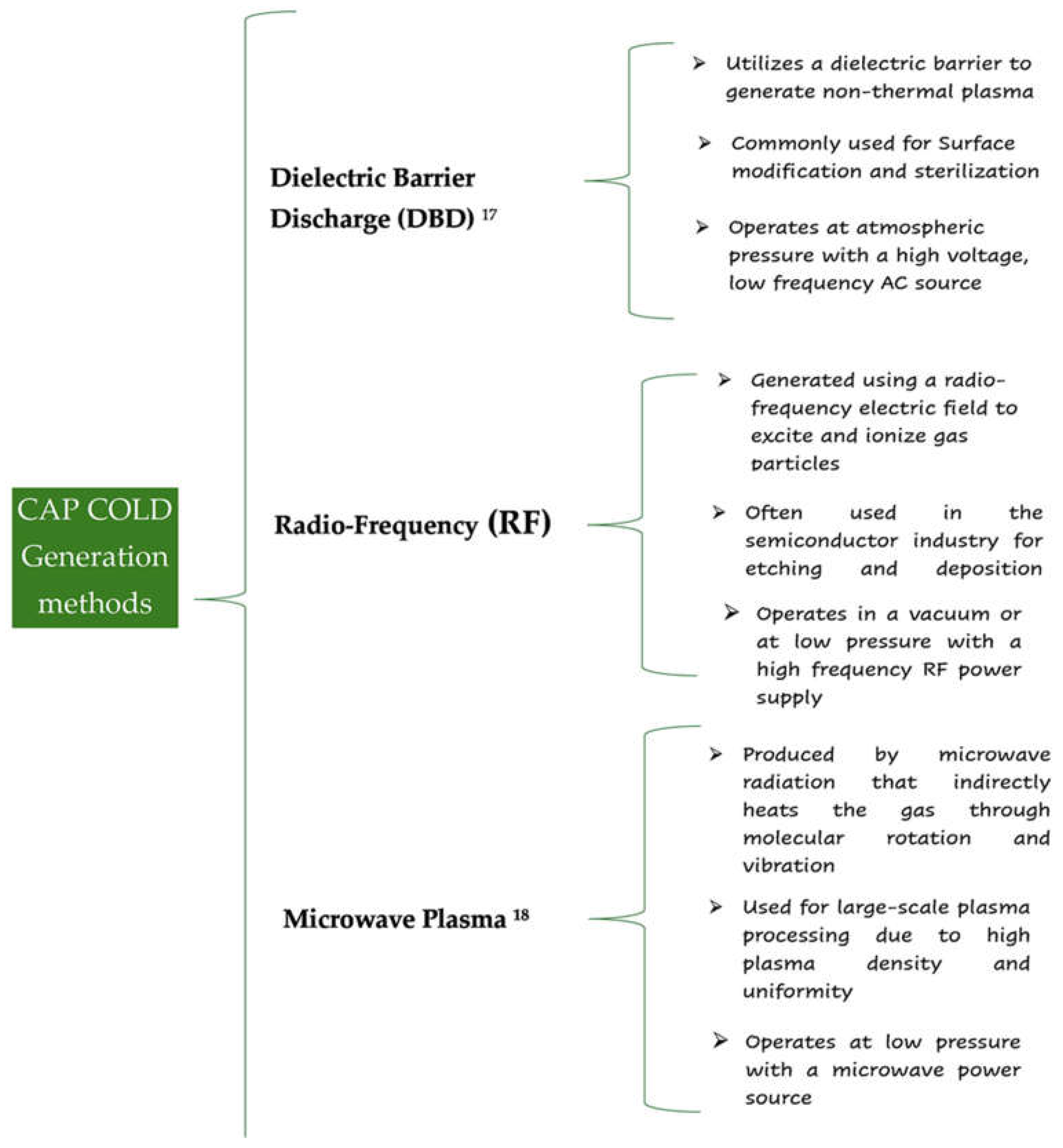
Figure 2.
Representation of the generation of PAW.
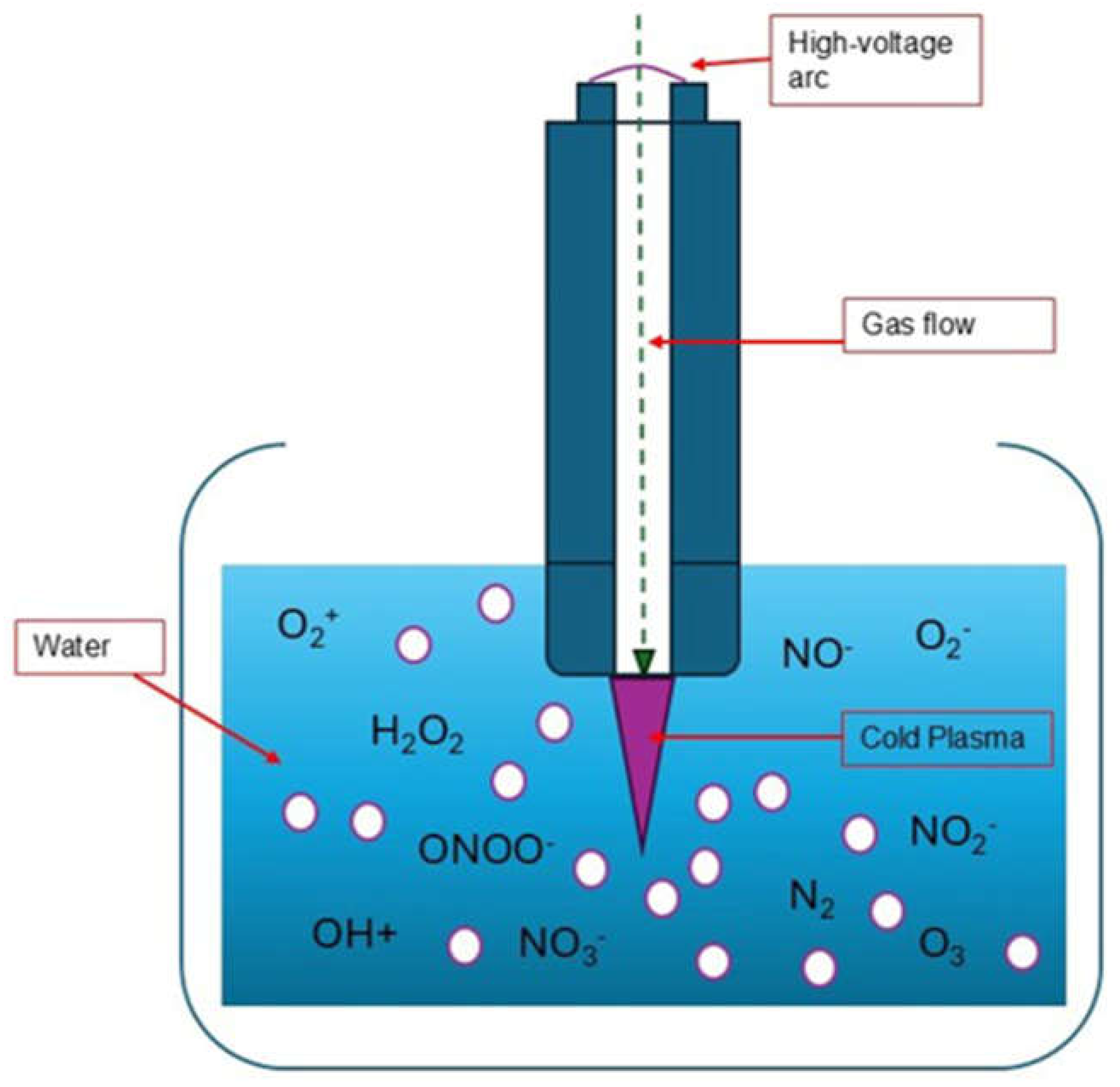
Figure 3.
Schematic representation of CAP Applications.
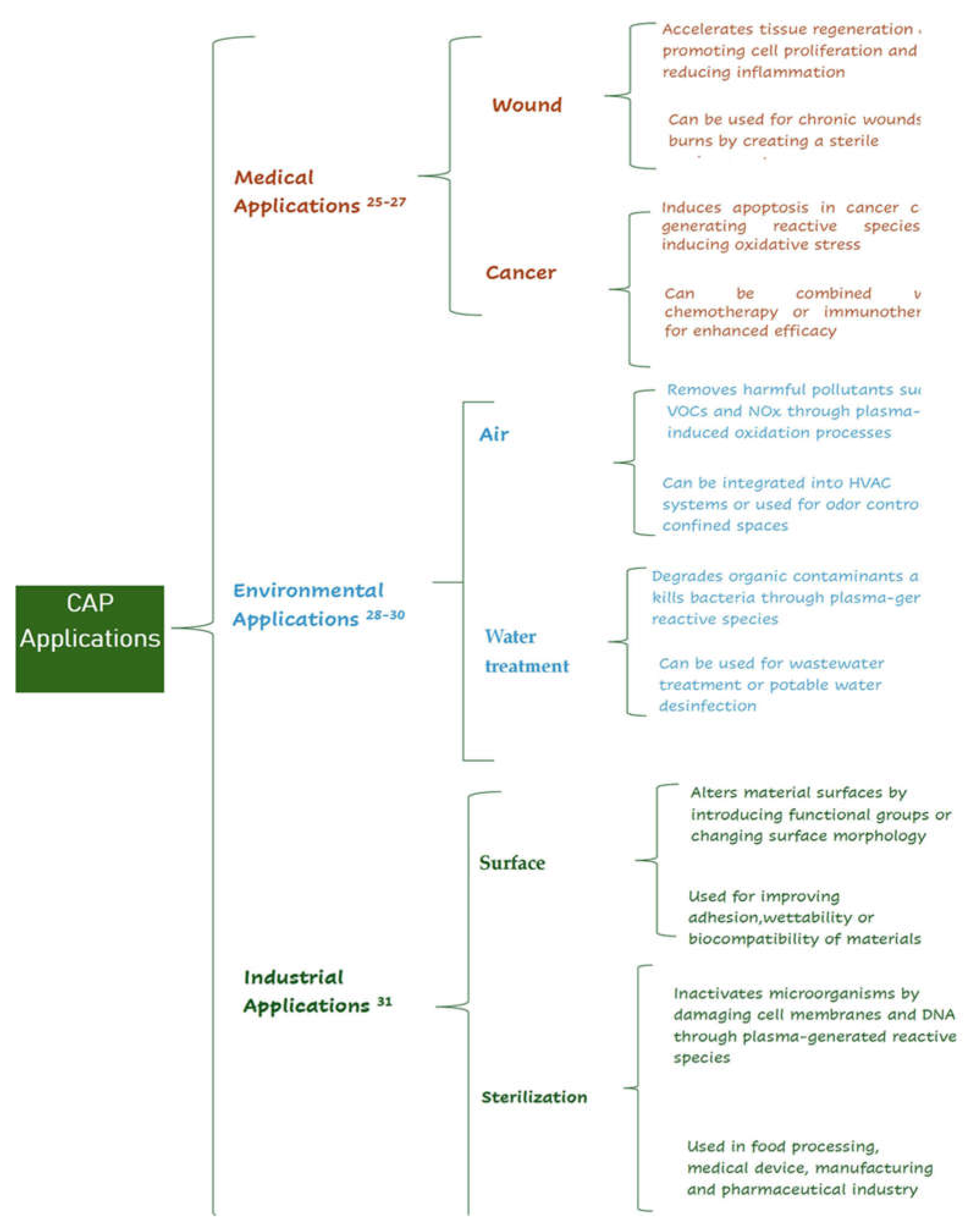
Figure 4.
Representative scheme of the effects on cells due to exposure to CAP on cells.
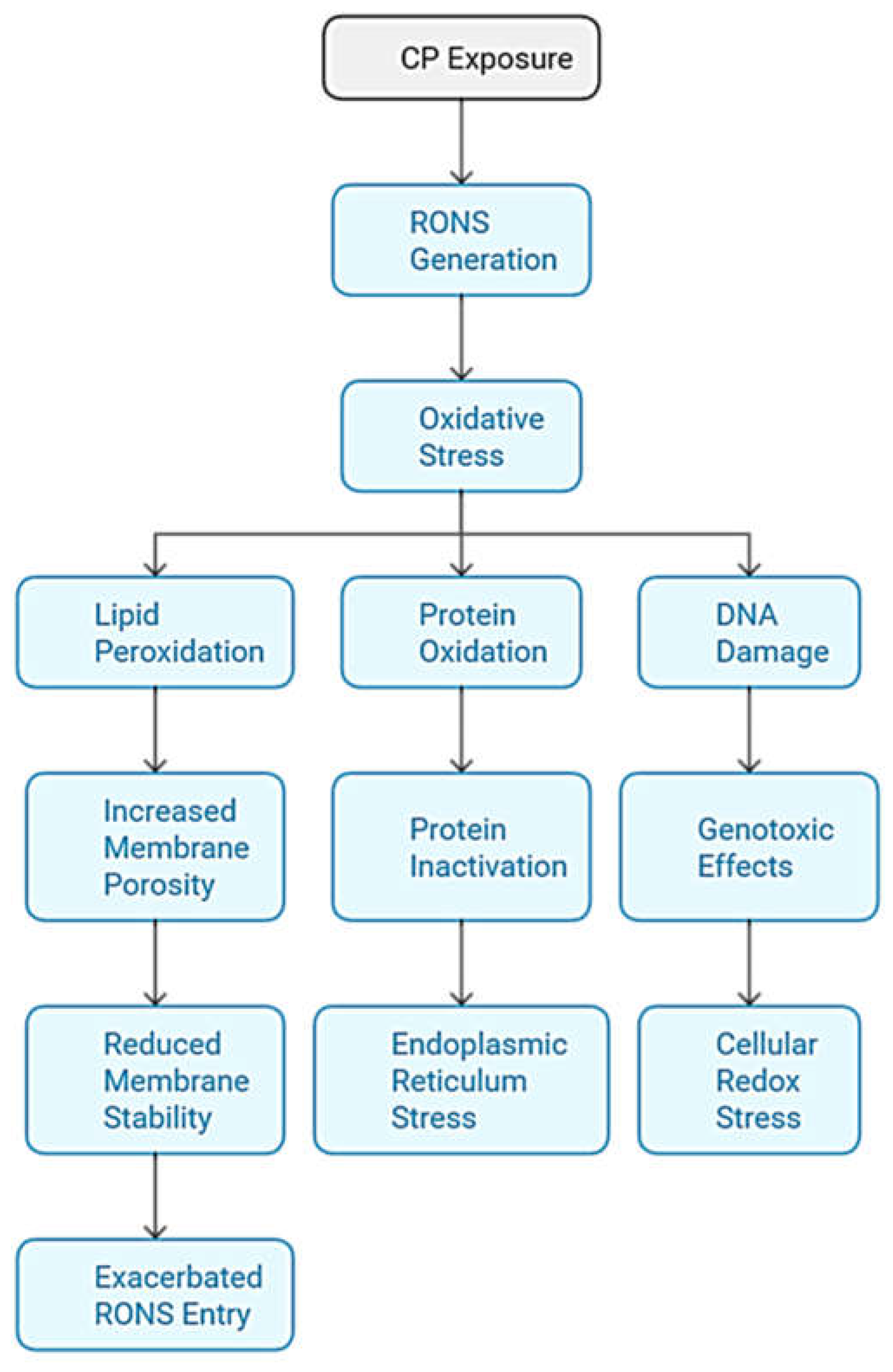
Figure 5.
Differences in the effects of CAP applications on cancer cells and normal cells.
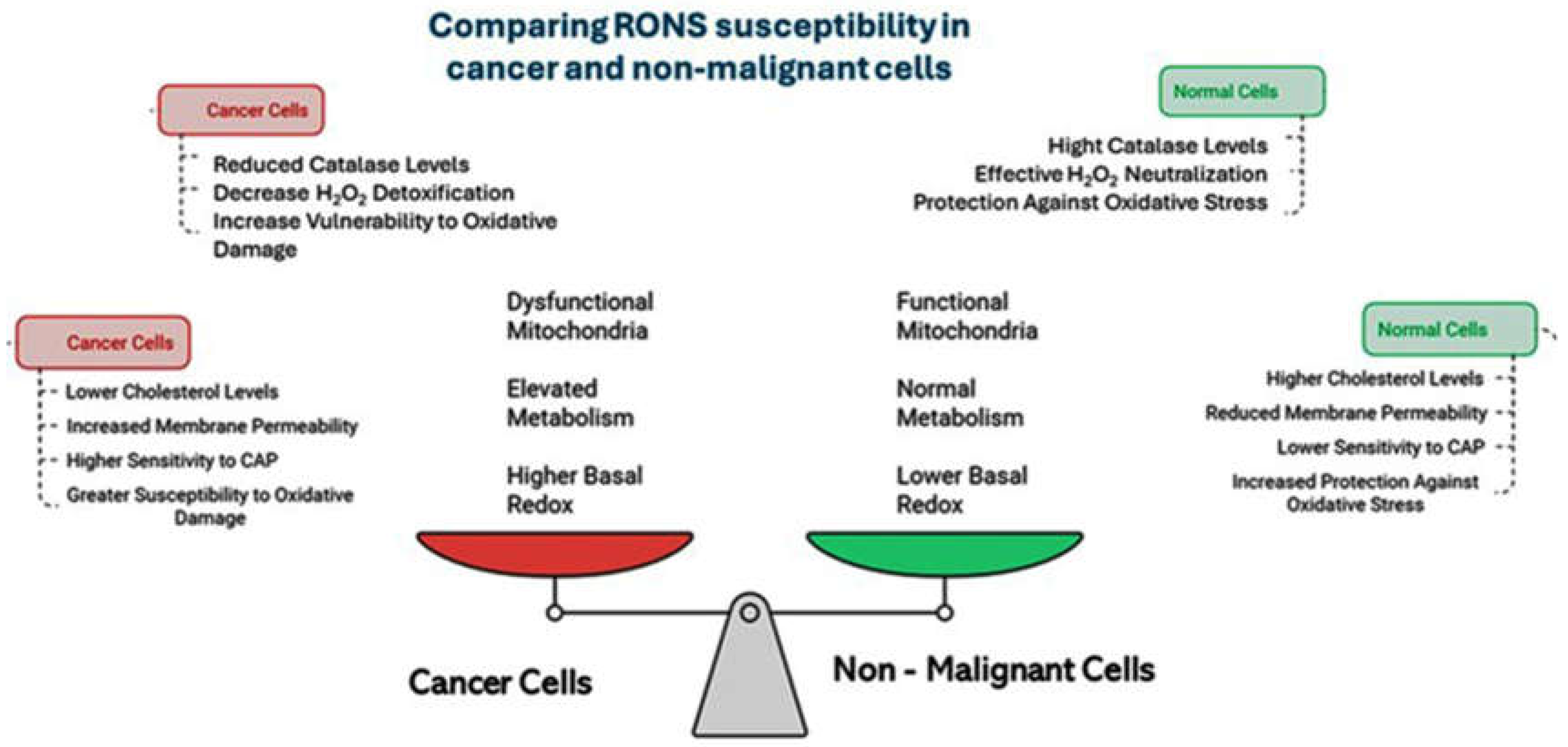
Figure 6.
Differentiated effects of CAP depending on the dose: Low doses healing; High doses of apoptoss in cancer cells. Artificial intelligence generated image from OpenAI." ChatGPT, OpenAI, 2024.
Figure 6.
Differentiated effects of CAP depending on the dose: Low doses healing; High doses of apoptoss in cancer cells. Artificial intelligence generated image from OpenAI." ChatGPT, OpenAI, 2024.
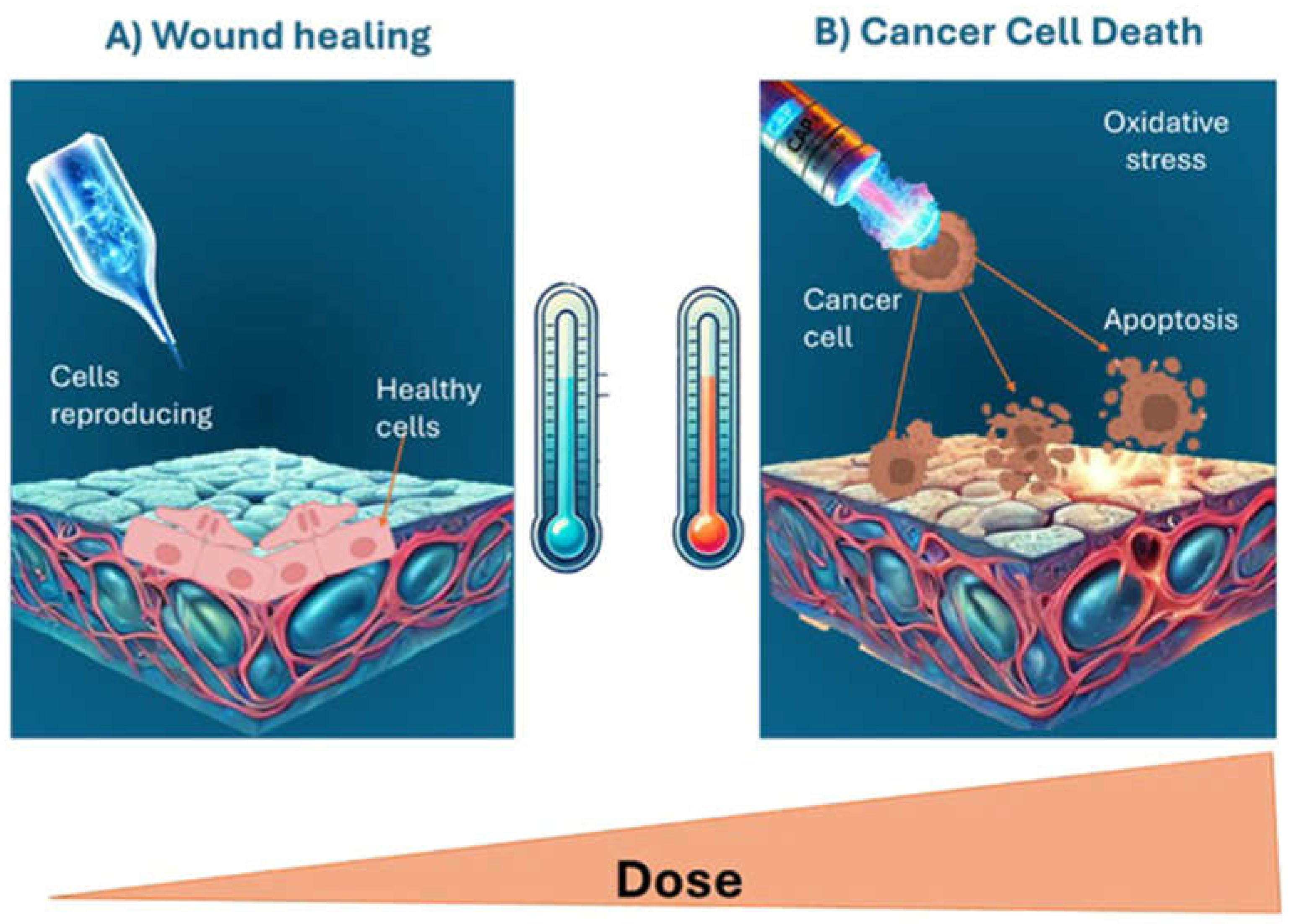
Figure 7.
Representation of the types of cell death induced by CAP and the associated signaling pathways, highlighting the role of ROS and other mechanisms in different types of cancer.
Figure 7.
Representation of the types of cell death induced by CAP and the associated signaling pathways, highlighting the role of ROS and other mechanisms in different types of cancer.
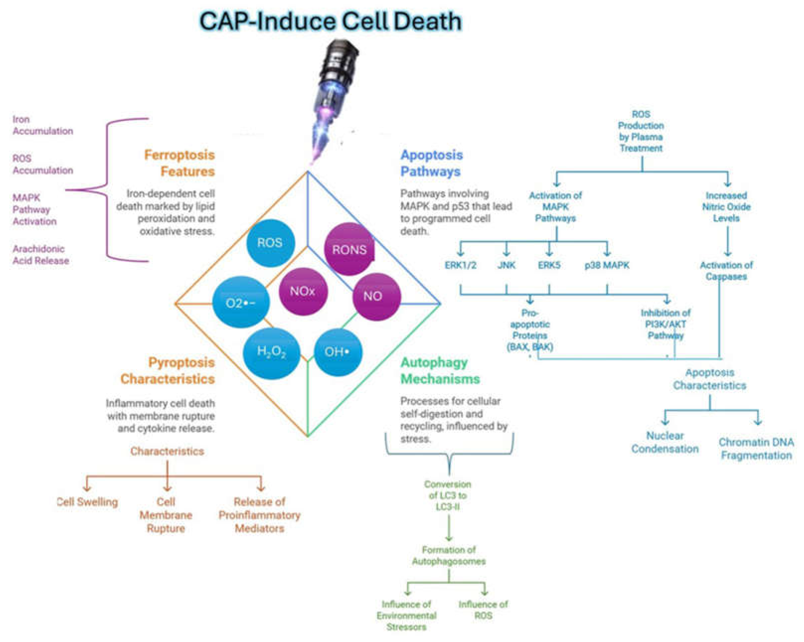

Table 1.
Classification of Reactive Species and generation mechanism.
| Type of Reactive Species | Name | Generation Pathway |
|---|---|---|
| ROS [33,34]. | Atomic Oxygen (O) | Generated by collisions between oxygen molecules and electrons in the plasma. |
| Hydroxyl Radicals (OH•) | Originates from the dissociation of water molecules, enhanced by UV radiation. | |
| Superoxide O2•− | Produced by collisions between electrons and oxygen molecules, and reactions between OH and ozone. | |
| Hydrogen Peroxide (H2O2) | Results from the combination of OH radicals in the plasma. | |
| Ozone (O3) | Formed from collisions between atomic oxygen (O) and oxygen molecules (O2) in the plasma. | |
| RNS [33,34]. | Nitrogen Oxides (NOx) | Includes nitrite (NO2− ) and nitrate (NO3−), generated by reactions between dissociated nitrogen and oxygen. |
| Nitric Oxide (NO) | Formed by dissolution from the gas phase or through secondary reactions in the plasma. |
Table 2.
Effects of CAP on key signaling pathways involved in normal and cancer cells.
| Pathway | Normal Cells | Cancer Cells | Reference |
|---|---|---|---|
| Nrf2 | - Activation of the Nrf2 pathway, a master regulator of the antioxidant response. - Promotes cell survival and enhances the ability to manage oxidative stress. |
- Suppression of the Nrf2 pathway, compromising antioxidant capacity. - Increases sensitivity to oxidative stress, predisposing to DNA damage and apoptosis. |
[49]. |
| PI3K/Akt | - Transient activation of the PI3K/Akt pathway, favoring cell survival and proliferation. - CAP does not significantly impact normal cell survival signaling. |
- Inhibition of the PI3K/Akt pathway by cold plasma reduces AKT phosphorylation, promoting apoptosis and decreasing proliferation. - Synergistic effects with chemotherapy enhance drug-induced apoptosis. |
[50]. |
| MAPK | - Less pronounced or transient activation of MAPK pathways, minimizing detrimental effects. | - Activation of stress-related kinases (e.g., JNK and p38) while inhibiting ERK1/2, leading to increased apoptosis and reduced proliferation. - CAP-induced activation of JNK promotes apoptotic cell death in cancer cells. |
[51]. |
| p53 Activation | - Typically unaltered in normal cells, maintaining regulatory functions of cell cycle and apoptosis. | - CAP treatment can restore p53 function, increasing expression and activation of apoptotic pathways. - Enhanced p53 activation leads to DNA damage response, promoting cell death. |
[52]. |
Table 3.
Compilation of in vitro studies examining the effect of CAP on mechanism ROS and others in different types of cancer.
Table 3.
Compilation of in vitro studies examining the effect of CAP on mechanism ROS and others in different types of cancer.
| Studie Type | Cancer Type | Study Description | Mechanism (ROS, Apoptosis, Others) | Specific Signaling Pathway | Reference |
|---|---|---|---|---|---|
| In Vitro | Glioblastoma | CAP increased the cytotoxicity of temozolomide in glioblastoma cells, suggesting chemosensitization. | ROS, Apoptosis, Direct DNA damage | p53, PI3K/Akt | [61,62,63] |
| Colon cancer | Induction of cell death by oxidative stress via CAP; potential use as an adjuvant therapy. | ROS, Apoptosis, Stress on the endoplasmic reticulum |
Caspasa-9, caspasa-3, PARP y Bax/ Bcl-2 | [63,64] |
|
| Breast cancer | Antiproliferative and apoptosis-inducing effect; potential for chemotherapy sensitization. Sensitization by epidermal growth factor (EGF) enhances the response of triple-negative breast cancer (TNBC) cells to CAP cold |
Apoptosis, Signaling pathway alteration This activation increases the production of reactive ROS and apoptotic signaling, |
Increased Bax/Bcl-2 ratio and cleavage of PARP-1. EGFR(Y992/1173) |
[67,68,69] [70] |
|
| Lung cancer | Reduction of viable cells and anti-metastatic activity observed. Inhibition proliferation, reduced migration Cell death in tumor cells PC9 expressing high levels of Gasdermin E (GSDME) in a dose-dependent manner |
ROS, Apoptosis, Microenvironment modulation ROS, Ferroptosis, ROS, Pyroptosis |
p38 MAPK, PI3/Akt Downregulation of the HOXB9/SLC7A11K JNK/cytochrome c/caspase-9/caspase-3 |
[71] [72] [73] |
|
| Pancreatic cancer | Reduction of metabolic activity and cell migration; favorable modulation of inflammatory profile. | ROS, Inflammatory regulation | NF-κB, IL-6 | [74,75] |
|
| Melanoma | CAP combined with nanoparticles enhanced selective toxicity towards cancer cells without damaging normal cells. | ROS, Microenvironment modulation | UPR signalling, Notch, Wnt/β-catenin | [76] |
|
Table 4.
Compilation of in vivo studies examining the effect of CAP on different models of cancer.
| Study Type | Cancer Type | Description | Mechanism of Action and Signaling Pathways | Reference |
|---|---|---|---|---|
| In vivo studies | Glioblastoma | CAP increased ROS production, sensitizing tumor cells to chemotherapy with temozolomide. | ROS, Apoptosis, p53, PI3K/Akt pathways; Significant reduction in tumor growth. | [77] |
| Colon cancer | CAP promoted danger signal release and stimulated adaptive immune response in mouse models. | ROS, Immune activation; Specific T cell response against GUCY2C. | [78,79] |
|
| Myeloid leukemia | CAP blocked three key cancer survival pathways: redox deregulation, glycolysis, and AKT/mTOR/HIF-1α signaling. | ROS, Apoptosis, AKT/mTOR, HIF-1α pathways; Reduced tumor growth and improved survival. | [80] |
|
| Multiple myeloma | CAP inhibited tumor implantation in mice, significantly prolonging survival time. | ROS, Apoptosis, Notch pathway inhibition; Reduced tumor cell proliferation. | [81] |
|
| Pancreatic cancer | A plasma-activated lactated Ringer's solution was developed to evaluate its antitumor effects. | ROS, Cytotoxic effects derived from activated lactic acid; Tumor volume reduction. | [82] |
|
| Cholangiocarcinoma | CAP induced DNA damage and apoptosis in subcutaneous xenografts of cancer cells. | ROS, DNA damage, Apoptosis; Activation of CHK1, p53, and 8-oxoguanine accumulation. | [83] |
|
| Head and neck cancer | CAP induced apoptosis and reduced cell viability in head and neck cancer models. | ROS, Apoptosis; Mitochondrial membrane potential modification and MAPK pathway activation. | [84] |
Table 5.
Compilation of in vivo studies examining the effect of CAP on different models of cancer.
| CAP APPLICATION DEVICE | Study Description | Result | Reference |
|---|---|---|---|
| kINPen | The study demonstrated that CAP treatment delivered using the kINPen MED device is safe, well tolerated, and effective in reducing tumor size in patients with head and neck cancer. CAP induced selective tumor cell death through oxidative stress without damaging surrounding healthy tissues. | Tumor size reduction in head and neck cancer. | [85] |
| Plasma jet, kINPen(®) MED (neoplas tools GmbH, Greifswald, Germany). | This study concluded that the use of a cold plasma device, specifically a dielectric barrier discharge (DBD) system, in patients with head and neck cancer showed visible responses on the tumor surface and significant apoptotic cell death. The treatment was well tolerated, with a favorable safety profile and no significant adverse effects. | Induction of apoptotic death in head and neck cancer. | [86] |
| Canady Helios Cold Plasma (CHCP) | The CHCP device was investigated in the first phase I clinical study, primarily to demonstrate safety. Preliminary findings were encouraging, showing that CHCP can control residual disease and improve patient survival. Ex vivo experiments on patient tissue samples confirmed CHCP-induced cancer cell death without harming normal cells, indicating its potential to control residual cancer cells at surgical margins. | Control of residual tumor cells in surgical margins in combination with surgery. | [87] |
| kINPen | This study concluded that CAP use in advanced head and neck cancer patients is safe and may induce positive clinical responses, such as pain reduction and improved quality of life. Two patients achieved partial remission, suggesting CAP's potential as an effective therapeutic option; however, further research is needed to fully understand its long-term mechanisms and efficacy. | Improving quality of life and reducing pain in patients with advanced head and neck cancer. | [88] |
| VIO3/APC3 (Erbe Elektromedizin) | This study concluded that noninvasive physical plasma (NIPP) is a safe and effective method for treating cervical intraepithelial neoplasia (CIN) grades 1 and 2. Using the cold plasma device, VIO3/APC3, with precise application control, the treatment preserved tissue while inducing lesion regression, making it a promising alternative to current excisional and ablative treatments. | Conservative treatment of CIN in women. | [89] |
Disclaimer/Publisher’s Note: The statements, opinions and data contained in all publications are solely those of the individual author(s) and contributor(s) and not of MDPI and/or the editor(s). MDPI and/or the editor(s) disclaim responsibility for any injury to people or property resulting from any ideas, methods, instructions or products referred to in the content. |
© 2024 by the authors. Licensee MDPI, Basel, Switzerland. This article is an open access article distributed under the terms and conditions of the Creative Commons Attribution (CC BY) license (http://creativecommons.org/licenses/by/4.0/).
Copyright: This open access article is published under a Creative Commons CC BY 4.0 license, which permit the free download, distribution, and reuse, provided that the author and preprint are cited in any reuse.


