Submitted:
04 November 2024
Posted:
06 November 2024
You are already at the latest version
Abstract
This review provides a comprehensive summary of the latest international research on detection methods for glucosinolates in cruciferous plants. The article examines various analytical tech-niques, including high-performance liquid chromatography (HPLC), liquid chromatog-raphy-mass spectrometry (LC-MS), and enzyme-linked immunosorbent assay (ELISA), while highlighting their respective advantages and limitations. Additionally, the review delves into recent advancements in sample preparation, extraction, and quantification methods, offering valuable insights into the accurate and efficient determination of glucosinolate content across diverse plant materials. Furthermore, it underscores the critical importance of standardization and validation of these methodologies to ensure reliable glucosinolate analysis in both scientific research and industrial applications.
Keywords:
glucosinolate
; cruciferous crops
; HPLC
; LC-MS/MS
; determination
1. Introduction
Glucosinolates represent a category of organic compounds that are present in cruciferous vegetables, including broccoli, cabbage, kale, and Brussels sprouts [1,2,3]. They are of significant interest due to their potential health benefits and their role as bioactive compounds in plant defense mechanisms. The detection and quantification of glucosinolates are crucial for both nutritional research and the advancement of plant breeding initiatives [4,5,6,7,8]. Glucosinolates are extensively distributed throughout various plant parts, including roots, stems, leaves, and seeds. This widespread presence underscores the importance of their analysis. The significance of glucosinolates is not only attributed to their direct consumption by humans and animals but also to the potential health benefits they may confer [9,10].
Although some glucosinolates and their degradation products have been identified as having anti-nutritional effects on certain animals, such as cattle, which can lead to negative consequences, other glucosinolates demonstrate specific anti-cancer activities [11,12,13,14]. Research suggests that these compounds may enhance the activity of phase II detoxification enzymes, which are linked to cancer prevention [13,15,16,17]. Furthermore, research has underscored the essential role of glucosinolates in sulfur metabolism, indicating that their biological functions merit further investigation [16].
The quantification of glucosinolates can be performed using two principal methodologies: one entails the indirect measurement of enzyme degradation products, while the other emphasizes the direct quantification of intact glucosinolates [18]. In indirect analysis, it is generally necessary to measure the products released during enzymatic or chemical reactions, such as isothiocyanates, oxazolidinethiones, thiocyanate ions, or glucose. The structures of these degradation products can reflect the characteristics of various parent glucosinolates. However, due to the lack of specificity in indirect methods for individual glucosinolates, these approaches are typically employed to quantify the total glucosinolate content in natural products [19,20,21].
Currently, a range of analytical techniques has been utilized to quantify glucosinolates in various plant tissues, including leaves, roots, buds, and seeds. The most frequently employed methods encompass HPLC, Gas Chromatography (GC), and Capillary Electrophoresis (CE) [22,23,24]. As previously noted, the content of glucosinolates can be analyzed either by directly measuring intact glucosinolates or by indirectly assessing their degradation products. However, certain methods, such as GC, may not be suitable for direct analysis due to the non-volatile or thermally unstable nature of some glucosinolates. Consequently, it is often necessary to convert these analytes into volatile derivatives to facilitate effective detection.
HPLC has been extensively utilized for the analysis of glucosinolates; however, the sample preparation process often necessitates desulfation. This procedure can result in incomplete desulfation, self-dimerization, and self-degradation, potentially compromising the reliability of the final data. Furthermore, the absence of standards for certain desulfated compounds poses a significant challenge to this methodology [25].
Mass Spectrometry (MS), owing to its high sensitivity, has been employed for the identification and quantification of glucosinolate compounds. A method has also been developed that integrates liquid chromatography with MS without necessitating desulfation. However, the accuracy and precision of this method are constrained by insufficient mass resolution. For instance, the mass-to-charge ratios (m/z) of glycosides and glycoproteins (m/z 422.0255 and m/z 422.0585, respectively) are remarkably similar. Although they share the same nominal mass of 422, their isotopic masses differ by only 0.033 Da, making it challenging to distinguish them through mass analysis alone. While these compounds can be separated chromatographically, the application of LC-MS remains limited if certain glucosinolate peaks significantly overlap or if there is substantial background interference in the monitoring channel [12,26].
Similar challenges arise when utilizing High-Resolution Mass Spectrometry (HRMS) to detect isomeric glucosinolates. This limitation can be mitigated by employing LC-MS/MS, which introduces an additional dimension of mass spectrometric detection, allowing for the simultaneous scanning of precursor and fragment ions, thereby enhancing selectivity and sensitivity. Furthermore, compared to LC-MS, LC-MS/MS typically requires shorter analysis times, facilitating the confirmation of known glucosinolates and the identification of novel compounds based on their fragmentation patterns [27,28].
Recently, a novel computerized system, GLS Finder, has been introduced, utilizing data generated from high-resolution HPLC-MS/MS to analyze glucosinolates in 49 common cruciferous vegetables. However, some reviewers have noted that the reported results may be ambiguous, as the number of chromatographic peaks for glucosinolates or their isomers exceeds the known quantity of glucosinolates present in each extract. This phenomenon may stem from the initial methods' inability to comprehensively identify all existing glucosinolates. Furthermore, comparing results with reported compositions serves as an effective validation method [29].
The accurate and effective detection of glucosinolates in plant materials is critically important for both scientific research and industrial applications. This review aims to summarize the latest advancements in detection methods for glucosinolates in cruciferous plants, providing valuable insights for future research.
2. Analytical Techniques for the Detection of Glucosinolates
Various analytical techniques have been employed for the detection of glucosinolates, including HPLC, LC-MS, GC, ultraviolet-visible spectrophotometry (UV-Vis), near-infrared spectroscopy (NIR), ELISA, and CE. Each technique presents distinct advantages and limitations regarding sensitivity, specificity, sample throughput, and cost. The selection of an appropriate method is contingent upon the specific requirements of the analysis, such as the number of glucosinolate species to be detected, the desired level of quantification, and available resources (Figure 1).
2.1. HPLC
HPLC is one of the most widely employed techniques for the analysis of glucosinolates, characterized by its high sensitivity, selectivity, and reproducibility. However, HPLC does have certain limitations, including extensive sample preparation requirements, costly equipment, and the necessity for skilled operators. The principle underlying this technique involves the separation and detection of thioglucoside components in vegetables, with quantification based on their retention time and peak area.
Initially, sample treatment is performed by chopping fresh cruciferous vegetables and extracting thioglucosides using suitable solvents (e.g., methanol or water). Following extraction, centrifugation and filtration are conducted to remove suspended matter; specifically, the extract is centrifuged to obtain a supernatant that is subsequently filtered. After obtaining the extract, chromatographic conditions are established by selecting appropriate columns and mobile phases while setting optimal flow rates and temperatures. Detection of thioglucosides can be achieved using either ultraviolet (UV) detectors or MS, allowing for accurate quantification of separated components through peak data recording [30,31].
2.2. LC-MS
LC-MS is a powerful technique that integrates the separation capabilities of liquid chromatography with mass spectrometric detection. It exhibits high sensitivity and specificity, enabling the simultaneous identification and quantification of multiple analytes. However, LC-MS is more complex and costly than HPLC, which limits its widespread application. The sample processing procedure closely resembles that of HPLC, involving the extraction of thioglucosides using appropriate solvents followed by separation via liquid chromatography upon completion. The separated components are then introduced into the mass spectrometer, which analyzes ions based on their mass-to-charge ratio (m/z) to provide molecular mass and structural information. Key advantages include enhanced sensitivity and selectivity, suitability for complex samples, and the capacity to yield insights into molecular structure [32,33,34,35].
2.3. ELISA
ELISA is a rapid, cost-effective, and straightforward method for the detection of glucosinolates. This technique relies on the specific binding of an antibody to a glucosinolate molecule. ELISA is well-suited for high-throughput analysis; however, it may be affected by cross-reactivity and limited specificity. The assay employs specific antibodies that bind to target molecules, generating a measurable signal through an enzymatic reaction, making it suitable for the quantitative analysis of particular thioglucosides [36,37]. Specific antibodies for the target thioglucoside are initially prepared, followed by sample processing once the antibodies are ready. The extracted sample is diluted and added to the ELISA plate, allowing the antigen to bind to the antibody. Subsequently, a substrate is introduced, and the enzyme reacts with this substrate to produce a measurable color change; absorbance is then quantified using a spectrophotometer. While this method offers advantages such as suitability for large-scale sample screening along with high sensitivity and specificity, it also presents limitations including the necessity of preparing specific antibodies in advance, a more complex procedure, and restricted capability for analyzing unknown compounds.
3. Sample Preparation, Extraction, and Quantification
3.1. Sample Preparation
The preparation of samples for glucosinolate analysis is a critical step that significantly impacts the integrity and quantification of these compounds. Various methods and conditions have been investigated to optimize glucosinolate extraction, each carrying distinct implications for the final results. A primary concern in glucosinolate sample preparation is the degradation of these compounds during processing. For instance, González-Hidalgo et al. emphasize that the time elapsed between harvesting and processing can lead to cellular damage, facilitating contact between glucosinolates and myrosinase, which results in rapid hydrolysis and a subsequent reduction in glucosinolate concentrations compared to fresh samples [38]. This degradation highlights the necessity of minimizing the interval between harvest and processing to preserve glucosinolate levels.
Furthermore, the selection of extraction solvent and method is crucial for optimizing glucosinolate recovery efficiency. Cold methanol extraction has demonstrated effectiveness, as it not only preserves glucosinolate content but also mitigates complications associated with elevated temperatures that may lead to degradation [39]. Doheny-Adams et al. demonstrate that cold methanol extraction surpasses other methods, particularly in preserving glucosinolate concentrations, thereby establishing it as a preferred technique for sample preparation [39]. Furthermore, the application of lyophilization (freeze-drying) may inadvertently diminish glucosinolate levels, as noted by Major et al., who observed that various drying methods did not significantly affect glucosinolate content in certain instances, indicating that careful consideration of drying techniques is crucial [40].
The extraction process can also be affected by the physical state of the plant material. For instance, mechanical processing of vegetables, such as shredding, may initially reduce glucosinolate levels but could subsequently result in an accumulation of these compounds over time due to the release of myrosinase [41]. This phenomenon suggests that while immediate processing may result in losses, subsequent reactions can potentially enhance glucosinolate levels, underscoring the necessity for a balanced approach in sample preparation.
Furthermore, the analytical methods utilized for glucosinolate quantification must be both robust and reliable. Techniques such as HPLC are frequently employed; however, the extraction and purification steps can be labor-intensive and time-consuming [42]. Grosser and Dam propose a streamlined HPLC method that simplifies the extraction process while ensuring accuracy [43]. This is critical as the complexity of glucosinolate profiles necessitates precise analytical techniques to ensure accurate quantification.
In conclusion, the preparation of samples for glucosinolate analysis necessitates meticulous attention to detail, encompassing the timing of processing, selection of extraction solvents, and employed methodologies. The interplay among these factors can significantly influence the final glucosinolate content and its bioactive potential. Therefore, adopting optimized extraction protocols and analytical methods is imperative for accurate glucosinolate profiling.
3.2. Extraction
Several extraction methods have been utilized to isolate glucosinolates from plant materials, including solvent extraction, ultrasound-assisted extraction (UAE), and microwave-assisted extraction (MAE). The selection of the optimal extraction method is critical for achieving high recovery rates and purity of glucosinolates. The effective extraction of glucosinolates—bioactive compounds present in cruciferous vegetables—can be accomplished through various techniques, each possessing unique advantages and challenges that are essential for optimizing extraction efficiency.
Solvent extraction remains a conventional method for isolating glucosinolates, where the choice of solvent significantly affects the yield and purity of the extracted compounds. For instance, studies have demonstrated that an 80% methanol solution can effectively inactivate myrosinase, an enzyme responsible for degrading glucosinolates, thereby preserving their concentrations during extraction [39,44]. Furthermore, the application of hydroalcoholic mixtures has been shown to enhance glucosinolate extraction yields, with varying efficiencies observed depending on solvent composition [45]. However, traditional solvent extraction often necessitates substantial time and solvent volumes, which may result in increased operational costs and environmental concerns [46].
In contrast, UAE has emerged as a more efficient alternative. This method employs ultrasonic waves to generate cavitation bubbles that disrupt plant cell walls and facilitate the release of glucosinolates into the solvent [47]. In contrast, UAE has emerged as a more efficient alternative. This method employs ultrasonic waves to generate cavitation bubbles that disrupt plant cell walls and facilitate the release of glucosinolates into the solvent [48,49]. For instance, studies have demonstrated that UAE achieves higher extraction efficiencies for glucosinolates from Camelina sativa by optimizing parameters such as solvent type and extraction duration [49]. Additionally, the application of UAE has been shown to improve the extraction of phenolic compounds, which are often co-extracted with glucosinolates, thereby further increasing the nutritional value of the extracts [50].
MAE is another innovative technique that has garnered attention for its capacity to enhance extraction efficiency. MAE utilizes microwave energy to heat both the solvent and sample, resulting in rapid extraction of glucosinolates due to the elevated temperature and pressure within the plant matrix [51,52]. Studies have demonstrated that MAE can yield results comparable to or superior than those obtained through UAE and traditional solvent extraction methods, particularly regarding extraction speed and solvent efficiency [46,51]. The integration of MAE with optimization techniques such as response surface methodology has been shown to maximize glucosinolate yields while minimizing solvent consumption [52].
In summary, while traditional solvent extraction methods are effective for glucosinolate extraction, UAE and MAE present significant advantages in terms of efficiency, yield, and environmental impact. The selection of the appropriate extraction method should be informed by the specific requirements of the target glucosinolates and the desired purity of the final extract.
3.3. Quantification
The quantification of glucosinolates can be accomplished by constructing calibration curves with standard compounds or by comparing the peak areas of samples against those of internal standards. The choice of a quantification method is dependent on the analytical technique utilized and the availability of suitable standards.
The quantification of glucosinolates, a class of sulfur-containing compounds predominantly present in the Brassicaceae family, is essential for elucidating their biological activities and nutritional significance. Various methodologies have been developed to accurately assess glucosinolate content in plant tissues, each possessing distinct advantages and limitations.
HPLC is one of the most widely employed techniques for quantifying glucosinolates. This method facilitates the separation and identification of individual glucosinolates based on their distinct chemical properties (Table 1). For instance, research has demonstrated that HPLC can effectively differentiate among various glucosinolates present in different cruciferous crops and Brassica species, thereby providing comprehensive profiles of their content [53,54,55]. The quantification process generally entails the extraction of glucosinolates from plant tissues, followed by hydrolysis to liberate glucose, which is subsequently measured spectrophotometrically [56,57]. This methodology has been validated in numerous studies, affirming its reliability for both total and individual glucosinolate measurements (Table 2) [52,58,59,60].

Table 1.
The individual compounds of glucosinolates found in cruciferous plants.
| Family | Species | Tissues and organs | GSLs compounds |
|---|---|---|---|
| Brassicaceae | Brassica napus | Seed, leaves, stems, roots | 6-15 |
| Brassica juncea | 7-17 | ||
| Capsella bursa - pastoris (L.) Medic. | 3-7 | ||
| Brassica oleracea L. var. botrytis L. | 4-9 | ||
| Brassica rapa | 2-11 | ||
| Brassica carinata A Braun | Seed, leaves, stems | 3-8 | |
| Camelina sativa | 3-12 | ||
| Camelina rumelica subsp. rumelica | |||
| Camelina macrocarpa | |||
| Brassica oleracea L. var capitata | Seeds, leaves | 3-12 | |
| Brassica oleracea L. convar capitata var alba | Florets, seedlings | 3-14 | |
| Brassica oleracea L. var italica | Seed, leaves, stems, roots, seedlings | 7-16 | |
| Raphannus sativus L. | Roots, seeds | 6-14 | |
| Arabidopsis thaliana | Leaf, florets, flowers, seedlings | 3-23 |
Concurrently, spectrophotometric methods have been developed for the rapid and cost-effective quantification of glucosinolates. For instance, a straightforward spectrophotometric approach was proposed to estimate total glucosinolates in mustard de-oiled cake, illustrating the potential for less resource-intensive techniques [66]. Furthermore, near-infrared spectroscopy (NIRS) has emerged as a promising alternative for non-destructive analysis of glucosinolates in intact seeds and plant tissues [67,68]. This method capitalizes on the unique spectral signatures of glucosinolates, enabling swift assessments without extensive sample preparation.
The quantification of glucosinolates is influenced by a variety of factors, including plant species, developmental stage, and environmental conditions. For instance, glucosinolate content can vary significantly among different cultivars of Brassica and in response to abiotic stresses such as salinity and temperature [52,69,70,71]. Moreover, genetic factors play a pivotal role in shaping the glucosinolate profiles of various plant varieties, which can be leveraged for breeding programs aimed at enhancing nutritional quality [72,73,74]. Additionally, the biological activity of glucosinolates is intricately linked to their hydrolysis products—such as isothiocyanates—which are recognized for their health-promoting properties [52,75,76]. Therefore, understanding quantification methods not only facilitates the assessment of the nutritional value of Brassica vegetables but also provides insights into their potential health benefits."
The quantification of glucosinolates is a complex process that can be accomplished using various analytical techniques, primarily HPLC and spectrophotometry. The choice of method may depend on specific research goals, available resources, and the nature of the samples being analyzed. Further research into the factors affecting glucosinolate content will enhance our understanding of these important phytochemicals.
4. Standardization and Validation of Glucosinolate Detection Methods
4.1. Standardization
Standardizing glucosinolate detection methods is crucial to ensure accurate and reliable results. This involves establishing standardized protocols for sample preparation, extraction, and quantification, as well as using certified reference materials and participating in inter-laboratory comparison studies. The standardization of these methods is essential for ensuring consistency and reliability in the analysis of bioactive compounds found in cruciferous vegetables, which offer significant health benefits. Various methodologies have been developed and validated, each with its own advantages and limitations.
HPLC is a widely utilized technique for glucosinolate analysis. Vastenhout et al. have demonstrated a method for evaluating the kinetics of glucosinolate hydrolysis using HPLC, confirming the linearity of absorbance with concentration through UV-Vis spectroscopy, which is essential for accurate quantification [77]. Similarly, Gallaher et al. developed and validated a spectrophotometric method for quantifying total glucosinolates in cruciferous vegetables, highlighting the reliability and speed of their approach, which has broad applicability [78]. The use of HPLC in combination with MS has also been emphasized by Frank et al., who employed LC-TOF-MS to screen mustard seeds for glucosinolates, demonstrating the method's effectiveness in identification and quantification of these compounds [79].
Additionally, Nuclear Magnetic Resonance (NMR) spectroscopy has been suggested as a potential approach for quantifying glucosinolates. Yuan et al. observed that although NMR is not commonly utilized for this purpose, it offers advantages such as eliminating the need for calibration standards, which can streamline the analysis process [53]. This is particularly pertinent when considering the complexity of plant matrices, where conventional methods may encounter difficulties due to interference from other compounds.
The extraction protocols employed prior to analysis are equally critical and can significantly influence the results. Neal et al. described a method for extracting glucosinolates from Arabidopsis thaliana leaves, where the identities of the peaks were confirmed using standards, thereby ensuring the reliability of the findings [80]. Furthermore, Major et al. discussed the impact of myrosinase activity during extraction, emphasizing that enzymatic activity can lead to a reduction in total glucosinolate concentration, thus necessitating meticulous control over extraction conditions [40].
Recent advancements have also concentrated on optimizing extraction methods to enhance glucosinolate yield. For instance, Meza et al. developed a UPLC-DAD method that refined sample preparation procedures to achieve high specificity and accuracy in glucosinolate quantification [81]. Furthermore, ultrasound-assisted extraction has been investigated as a more environmentally friendly alternative, demonstrating potential in improving extraction efficiency while minimizing solvent usage [82].
Thus, the standardization of glucosinolate detection methods necessitates a combination of reliable analytical techniques, optimized extraction protocols, and careful consideration of enzymatic activity. The integration of HPLC with MS, along with emerging methodologies such as NMR and ultrasound-assisted extraction, constitutes a comprehensive approach to glucosinolate analysis that can enhance the accuracy and reproducibility of results across various studies.
4.2. Validation
The validation of glucosinolate detection methods entails the assessment of parameters such as linearity, precision, accuracy, limit of detection (LOD), and limit of quantification (LOQ). This process ensures that the method is appropriate for its intended purpose and yields reliable results. The validation of these methods is essential to confirm their reliability and suitability for specific applications. Key parameters for validation include linearity, precision, accuracy, LOD, and LOQ; each parameter plays a crucial role in verifying that the analytical methods employed can consistently produce valid results.
Linearity refers to the method's capacity to yield results that are directly proportional to the analyte concentration within a specified range. For instance, studies have shown that HPLC methods can achieve exceptional linearity for glucosinolate quantification, with correlation coefficients (R2) frequently exceeding 0.99, thereby indicating a robust linear relationship between concentration and response [43,83]. This is crucial for ensuring that the method can accurately quantify glucosinolates across a spectrum of concentrations typically encountered in plant samples.
Precision, which evaluates the reproducibility of the method, is another critical parameter for validation. It is typically assessed through repeatability and intermediate precision studies. For instance, the precision of HPLC methods for glucosinolate analysis has been reported with relative standard deviations (RSDs) generally below 5%, indicating high reproducibility in measurements [39,43]. Similarly, methodologies such as UPLC-DAD have demonstrated consistent results across various laboratories, further reinforcing the reliability of these techniques [52,81].
Accuracy, which quantifies the proximity of measured values to true values, is a fundamental aspect of method validation. Various studies have utilized recovery experiments to evaluate accuracy, wherein known quantities of glucosinolates are added to samples and the recovery rates are subsequently calculated. Reports indicate that recovery rates for glucosinolates typically range from 90% to 110%, thereby confirming the reliability of the methods employed [39,83]. Such accuracy is essential for ensuring that reported glucosinolate levels accurately reflect true concentrations in the samples.
LOD and LOQ are critical for assessing the sensitivity of analytical methods. LOD denotes the lowest concentration of the analyte that can be reliably detected, while LOQ represents the lowest concentration that can be quantified with acceptable precision and accuracy. Studies have demonstrated that HPLC methods can achieve LODs in the low micromolar range for various glucosinolates, rendering them suitable for detecting trace levels of these compounds in complex matrices [43,83]. This sensitivity is particularly vital in food safety and nutritional studies, where low glucosinolate levels may carry significant biological implications. The validation of glucosinolate detection methods through an assessment of linearity, precision, accuracy, LOD, and LOQ is essential to ensure result reliability. A rigorous evaluation of these parameters confirms that the employed methods are appropriate for their intended purposes, thereby bolstering research and applications across fields such as nutrition, food science, and plant biology.
5. Future Prospects
5.1. Development of New Analytical Techniques
The development of novel analytical techniques, such as ion mobility spectrometry (IMS) and NMR, has the potential to significantly enhance the sensitivity, selectivity, and throughput of glucosinolate analysis. Glucosinolates, sulfur-containing compounds found in cruciferous vegetables, are of considerable interest due to their health benefits and roles in plant defense mechanisms. Traditional methods for glucosinolate analysis—primarily HPLC—while effective, may be limited in terms of sensitivity and specificity. The integration of advanced analytical techniques like IMS and NMR can effectively address these limitations.
NMR spectroscopy is particularly valuable due to its non-destructive and highly informative characteristics. Recent advancements in NMR technology, including the development of hyperpolarization techniques, have significantly enhanced the sensitivity of NMR measurements. For instance, hyperpolarized water has been employed to improve the detection of various compounds, including glucosinolates, by increasing the signal-to-noise ratio in NMR experiments [84,85]. Furthermore, the application of dynamic nuclear polarization (DNP) has demonstrated promise in further enhancing NMR sensitivity, facilitating the detection of low-abundance metabolites—crucial for analyzing glucosinolates within complex biological matrices [86].
Furthermore, the incorporation of magnetic nanoparticles in conjunction with NMR has emerged as a potent strategy for enhancing detection capabilities. Magnetic nanoparticles can alter the local magnetic environment, thereby influencing the relaxation rates of adjacent nuclei and improving the overall sensitivity of NMR measurements [87,88]. This approach is particularly advantageous for glucosinolate analysis, as it facilitates the detection of these compounds at lower concentrations and within more complex samples.
IMS is another promising technique that can complement NMR in the analysis of glucosinolates. IMS facilitates rapid separation and identification of ions based on their mobility in a gas phase, thereby providing high-throughput analytical capabilities. The integration of IMS with MS further enhances the specificity of glucosinolate detection by enabling the identification of specific molecular ions associated with glucosinolates and their degradation products [89,90]. This dual approach significantly improves the analytical workflow, allowing researchers to obtain comprehensive profiles of glucosinolates across various samples.
The integration of advanced analytical techniques, such as NMR and IMS, offers a significant opportunity to enhance the analysis of glucosinolates. These methods not only improve sensitivity and selectivity but also facilitate high-throughput analysis, rendering them invaluable tools in the investigation of these important phytochemicals.
5.2. Miniaturization and Automation
The adoption of advanced analytical techniques, including NMR and IMS, provides a considerable opportunity to enhance the analysis of glucosinolates. These methodologies not only improve sensitivity and selectivity but also facilitate high-throughput analysis, thereby establishing them as essential tools in the investigation of these important phytochemicals.
HPLC and Ultra-Performance Liquid Chromatography (UPLC) represent the cutting edge of glucosinolate analysis. Notably, UPLC is recognized for its superior separation efficiency, reduced solvent consumption, and shorter run times compared to conventional HPLC methods. For example, Meza et al. demonstrated that UPLC provides a more environmentally sustainable quantification of glucosinolates in Camelina seeds, thereby minimizing ecological impact while enhancing throughput [81]. Similarly, Shi et al. underscored the efficacy of HPLC coupled with diode array detection for quantifying glucosinolates, highlighting its significance in high-throughput applications [91]. These methodologies enable the simultaneous detection of multiple glucosinolates, thus streamlining the analytical process.
Furthermore, the integration of MS with chromatographic techniques has significantly enhanced glucosinolate detection. Wu et al. employed LC-MS/MS in multiple reaction monitoring mode to analyze glucosinolate profiles in red cabbage, demonstrating the method's sensitivity and its capacity to rapidly provide detailed compositional data [35]. This combination improves detection limits and facilitates the identification of various glucosinolates within complex matrices, which is crucial for comprehensive profiling in high-throughput applications.
Automation plays a pivotal role in these advancements, facilitating the rapid processing of samples with minimal human intervention. Techniques such as automated sample preparation and robotic liquid handling systems can significantly reduce analysis time and variability in results. However, the reference by Li et al. does not specifically address glucosinolate analysis; rather, it focuses on genetically modified organisms [92]. Consequently, it is inappropriate to support claims regarding automation in glucosinolate detection based on this source. Furthermore, the proposed application of biosensors and microfluidic devices for real-time monitoring and analysis of glucosinolates lacks direct corroboration from the cited references [93,94].
In conclusion, the miniaturization and automation of glucosinolate detection methods are revolutionizing analytical practices by facilitating high-throughput analysis, minimizing resource consumption, and expediting analysis times. The integration of advanced chromatographic techniques with MS, alongside automation, is paving the way for more efficient and effective glucosinolate profiling.
5.3. Application of Machine Learning(ML) and Artificial Intelligence
ML and artificial intelligence algorithms can be employed to optimize detection methods, predict glucosinolate content, and classify plant materials based on their glucosinolate profiles. The application of ML and artificial intelligence (AI) in refining detection techniques, forecasting glucosinolate levels, and categorizing plant materials according to their glucosinolate profiles represents an emerging area of research that holds significant promise for agricultural and food sciences. Glucosinolates, a class of sulfur-containing compounds predominantly found in Brassica species, are recognized for their health benefits, including cancer prevention and antioxidant properties [52,85,95]. The integration of ML and AI has the potential to enhance the efficiency and accuracy of glucosinolate analysis, which is critical for both breeding programs and food quality assessment.
One of the primary applications of ML in this context is the optimization of detection methods. Traditional techniques for glucosinolate quantification, such as HPLC, while effective, can be time-consuming and necessitate extensive sample preparation [81]. Recent advancements in ML algorithms have demonstrated their ability to analyze complex datasets generated from chromatographic techniques, enabling rapid identification and quantification of glucosinolates [96]. For instance, the GLS-Finder platform employs UPLC coupled with HRMS to conduct qualitative and semi-quantitative analyses of glucosinolates, significantly reducing analysis time [96]. Furthermore, ML can be trained to predict glucosinolate profiles based on various growth conditions and environmental factors, thereby facilitating more targeted breeding strategies [85,97].
In addition to optimizing detection methods, ML and artificial intelligence (AI) can be utilized to predict glucosinolate content in various plant materials. Factors such as soil composition, climatic conditions, and plant genetics significantly influence glucosinolate levels [97]. Statistical modeling approaches, including regression analysis and neural networks, have been effectively employed to forecast glucosinolate concentrations in crops such as Chinese cabbage and kale based on these variables [85,97]. For instance, studies have demonstrated that environmental factors like temperature and humidity are correlated with glucosinolate accumulation, facilitating predictive models that assist in selecting optimal growing conditions for desired glucosinolate profiles [89,97].
Furthermore, artificial intelligence (AI) can facilitate the classification of plant materials based on their glucosinolate content. By employing supervised learning techniques, researchers can categorize different cultivars of Brassica species according to their glucosinolate profiles, which is essential for both breeding and consumer preferences [95]. ML algorithms are capable of analyzing spectral data from methods such as hyperspectral imaging to classify plant materials rapidly and non-destructively, providing a valuable tool for quality control in agricultural practices [98]. This classification capability can also extend to identifying plant varieties with enhanced health-promoting properties, thereby guiding breeding programs aimed at improving nutritional quality [95]. Thus, the integration of ML and AI into glucosinolate research offers significant advancements in detection methods, predictive modeling, and the classification of plant materials. These technologies not only enhance the efficiency of glucosinolate analysis but also contribute to the development of crops with optimized health benefits, aligning with the increasing demand for functional foods in the market.
Author Contributions
Conceptualization, Z.L.; Writing-original draft preparation, X.L., D.W.; Writing—review and editing, F.H., J.S., S.L.,Y.L., Y.H., F.G.,M.Z; Supervision, Z.L.; Project administration, Z.L. All authors have read and agreed to the published version of the manuscript.
Funding
This work was supported by the National Nature Science Foundation (32172580), the China Agriculture Research System (CARS-23-A05), the Agricultural Science and Technology Innovation Program (ASTIP).
Data Availability Statement
The data presented in this study are available on request from the corresponding author.
Acknowledgments
Figure 1 was created with BioRender.com, and thanks for the support of Yafei Seed Co., Ltd.
Conflicts of Interest
The authors declare no conflicts of interest.
References
- Kamal, R.M.; Abdull Razis, A.F.; Mohd Sukri, N.S.; Perimal, E.K.; Ahmad, H.; Patrick, R.; Djedaini-Pilard, F.; Mazzon, E.; Rigaud, S. Beneficial health effects of glucosinolates-derived isothiocyanates on cardiovascular and neurodegenerative diseases. Molecules 2022, 27, 624. [Google Scholar] [CrossRef] [PubMed]
- Nguyen, V.T.; Stewart, J.; Lopez, M.; Ioannou, I.; Allais, F. Glucosinolates: Natural occurrence, biosynthesis, accessibility, isolation, structures, and biological activities. Molecules 2020, 25, 4537. [Google Scholar] [CrossRef] [PubMed]
- Sheu, M.-J.; Yeh, M.-C.; Tsai, M.-C.; Wang, C.-C.; Chang, Y.-L.; Wang, C.-J.; Huang, H.-P. Glucosinolates Extracts from Brassica juncea Ameliorate HFD-Induced Non-Alcoholic Steatohepatitis. Nutrients 2023, 15, 3497. [Google Scholar] [CrossRef]
- Kapusta-Duch, J.; Kopec, A.; Piatkowska, E.; Borczak, B.; Leszczynska, T. The beneficial effects of Brassica vegetables on human health. Roczniki Państwowego Zakładu Higieny 2012, 63. [Google Scholar]
- McNaughton, S.; Marks, G. Development of a food composition database for the estimation of dietary intakes of glucosinolates, the biologically active constituents of cruciferous vegetables. British Journal of Nutrition 2003, 90, 687–697. [Google Scholar] [CrossRef]
- Šamec, D.; Urlić, B.; Salopek-Sondi, B. Kale (Brassica oleracea var. acephala) as a superfood: Review of the scientific evidence behind the statement. Critical reviews in food science and nutrition 2019, 59, 2411–2422. [Google Scholar] [CrossRef]
- Vanduchova, A.; Anzenbacher, P.; Anzenbacherova, E. Isothiocyanate from broccoli, sulforaphane, and its properties. Journal of medicinal food 2019, 22, 121–126. [Google Scholar] [CrossRef]
- Zeng, W.; Yang, J.; He, Y.; Zhu, Z. Bioactive compounds in cruciferous sprouts and microgreens and the effects of sulfur nutrition. Journal of the Science of Food and Agriculture 2023, 103, 7323–7332. [Google Scholar] [CrossRef]
- Connolly, E.L.; Sim, M.; Travica, N.; Marx, W.; Beasy, G.; Lynch, G.S.; Bondonno, C.P.; Lewis, J.R.; Hodgson, J.M.; Blekkenhorst, L.C. Glucosinolates from cruciferous vegetables and their potential role in chronic disease: Investigating the preclinical and clinical evidence. Frontiers in pharmacology 2021, 12, 767975. [Google Scholar] [CrossRef]
- Esteve, M. Mechanisms underlying biological effects of cruciferous glucosinolate-derived isothiocyanates/indoles: a focus on metabolic syndrome. Frontiers in Nutrition 2020, 7, 111. [Google Scholar] [CrossRef]
- Cai, Z.; Cheung, C.-Y.; Ma, W.-T.; Au, W.-M.; Zhang, X.Y.; Lee, A. Determination of two intact glucosinolates in vegetables and Chinese herbs. Analytical and bioanalytical chemistry 2004, 378, 827–833. [Google Scholar] [CrossRef] [PubMed]
- Higdon, J.V.; Delage, B.; Williams, D.E.; Dashwood, R.H. Cruciferous vegetables and human cancer risk: epidemiologic evidence and mechanistic basis. Pharmacological research 2007, 55, 224–236. [Google Scholar] [CrossRef] [PubMed]
- Kennelley, G.E.; Amaye-Obu, T.; Foster, B.A.; Tang, L.; Paragh, G.; Huss, W.J. Mechanistic review of sulforaphane as a chemoprotective agent in bladder cancer. American Journal of Clinical and Experimental Urology 2023, 11, 103. [Google Scholar] [PubMed]
- Shapiro, T.A.; Fahey, J.W.; Wade, K.L.; Stephenson, K.K.; Talalay, P. Human metabolism and excretion of cancer chemoprotective glucosinolates and isothiocyanates of cruciferous vegetables. Cancer epidemiology, biomarkers & prevention: a publication of the American Association for Cancer Research, cosponsored by the American Society of Preventive Oncology 1998, 7, 1091–1100. [Google Scholar]
- Melim, C.; Lauro, M.R.; Pires, I.M.; Oliveira, P.J.; Cabral, C. The role of glucosinolates from cruciferous vegetables (Brassicaceae) in gastrointestinal cancers: from prevention to therapeutics. Pharmaceutics 2022, 14, 190. [Google Scholar] [CrossRef]
- Mitra, S.; Emran, T.B.; Chandran, D.; Zidan, B.R.M.; Das, R.; Mamada, S.S.; Masyita, A.; Salampe, M.; Nainu, F.; Khandaker, M.U. Cruciferous vegetables as a treasure of functional foods bioactive compounds: Targeting p53 family in gastrointestinal tract and associated cancers. Frontiers in Nutrition 2022, 9, 951935. [Google Scholar] [CrossRef]
- Zhang, N.-Q.; Mo, X.-F.; Lin, F.-Y.; Zhan, X.-X.; Feng, X.-L.; Zhang, X.; Luo, H.; Zhang, C.-X. Intake of total cruciferous vegetable and its contents of glucosinolates and isothiocyanates, glutathione S-transferases polymorphisms and breast cancer risk: a case–control study in China. British Journal of Nutrition 2020, 124, 548–557. [Google Scholar] [CrossRef]
- Almushayti, A.Y.; Brandt, K.; Carroll, M.A.; Scotter, M.J. Current analytical methods for determination of glucosinolates in vegetables and human tissues. Journal of Chromatography A 2021, 1643, 462060. [Google Scholar] [CrossRef]
- Hu, Y.; Liang, H.; Yuan, Q.; Hong, Y. Determination of glucosinolates in 19 Chinese medicinal plants with spectrophotometry and high-pressure liquid chromatography. Natural product research 2010, 24, 1195–1205. [Google Scholar]
- Xie, C.; Li, W.; Gao, R.; Yan, L.; Wang, P.; Gu, Z.; Yang, R. Determination of glucosinolates in rapeseed meal and their degradation by myrosinase from rapeseed sprouts. Food chemistry 2022, 382, 132316. [Google Scholar] [CrossRef]
- Yu, X.; He, H.; Zhao, X.; Liu, G.; Hu, L.; Cheng, B.; Wang, Y. Determination of 18 intact glucosinolates in Brassicaceae vegetables by UHPLC-MS/MS: comparing tissue disruption methods for sample preparation. Molecules 2021, 27, 231. [Google Scholar] [CrossRef]
- Cai, X.-M.; Xu, X.-X.; Bian, L.; Luo, Z.-X.; Chen, Z.-M. Measurement of volatile plant compounds in field ambient air by thermal desorption–gas chromatography–mass spectrometry. Analytical and bioanalytical chemistry 2015, 407, 9105–9114. [Google Scholar] [CrossRef] [PubMed]
- Dagar, R.; Gautam, A.; Priscilla, K.; Sharma, V.; Gupta, P.; Kumar, R. Sample Preparation from Plant Tissue for Gas Chromatography–Mass Spectrometry (GC-MS) we. In Plant Functional Genomics: Methods and Protocols, Volume 2; Springer: 2024; pp. 19-37.
- Mukker, J.K.; Kotlyarova, V.; Singh, R.S.P.; Alcorn, J. HPLC method with fluorescence detection for the quantitative determination of flaxseed lignans. Journal of Chromatography B 2010, 878, 3076–3082. [Google Scholar] [CrossRef]
- Pardini, A.; Tamasi, G.; De Rocco, F.; Bonechi, C.; Consumi, M.; Leone, G.; Magnani, A.; Rossi, C. Kinetics of glucosinolate hydrolysis by myrosinase in Brassicaceae tissues: A high-performance liquid chromatography approach. Food Chemistry 2021, 355, 129634. [Google Scholar] [CrossRef] [PubMed]
- Kim, S.-Y.; Yang, J.; Dang, Y.-M.; Ha, J.-H. Effect of fermentation stages on glucosinolate profiles in kimchi: Quantification of 14 intact glucosinolates using ultra-performance liquid chromatography-tandem mass spectrometry. Food Chemistry: X 2022, 15, 100417. [Google Scholar] [CrossRef] [PubMed]
- Capriotti, A.L.; Cavaliere, C.; La Barbera, G.; Montone, C.M.; Piovesana, S.; Chiozzi, R.Z.; Laganà, A. Chromatographic column evaluation for the untargeted profiling of glucosinolates in cauliflower by means of ultra-high performance liquid chromatography coupled to high resolution mass spectrometry. Talanta 2018, 179, 792–802. [Google Scholar] [CrossRef] [PubMed]
- Geng, J.; Xiao, L.; Chen, C.; Wang, Z.; Xiao, W.; Wang, Q. An integrated analytical approach based on enhanced fragment ions interrogation and modified Kendrick mass defect filter data mining for in-depth chemical profiling of glucosinolates by ultra-high-pressure liquid chromatography coupled with Orbitrap high resolution mass spectrometry. Journal of Chromatography A 2021, 1639, 461903. [Google Scholar]
- Sun, J.; Zhang, M.; Chen, P. GLS-finder: a platform for fast profiling of glucosinolates in Brassica vegetables. Journal of agricultural and food chemistry 2016, 64, 4407–4415. [Google Scholar] [CrossRef]
- Lei, Z.; Sumner, B.W.; Bhatia, A.; Sarma, S.J.; Sumner, L.W. UHPLC-MS analyses of plant flavonoids. Current protocols in plant biology 2019, 4, e20085. [Google Scholar] [CrossRef]
- Mocniak, L.E.; Elkin, K.R.; Dillard, S.L.; Bryant, R.B.; Soder, K.J. Building comprehensive glucosinolate profiles for brassica varieties. Talanta 2023, 251, 123814. [Google Scholar] [CrossRef]
- Bell, L.; Oruna-Concha, M.J.; Wagstaff, C. Identification and quantification of glucosinolate and flavonol compounds in rocket salad (Eruca sativa, Eruca vesicaria and Diplotaxis tenuifolia) by LC–MS: Highlighting the potential for improving nutritional value of rocket crops. Food Chemistry 2015, 172, 852–861. [Google Scholar] [CrossRef] [PubMed]
- Ibrahim, R.M.; Eltanany, B.M.; Pont, L.; Benavente, F.; ElBanna, S.A.; Otify, A.M. Unveiling the functional components and antivirulence activity of mustard leaves using an LC-MS/MS, molecular networking, and multivariate data analysis integrated approach. Food Research International 2023, 168, 112742. [Google Scholar] [CrossRef] [PubMed]
- Kumar, R.; Reichelt, M.; Bisht, N.C. An LC-MS/MS assay for enzymatic characterization of methylthioalkylmalate synthase (MAMS) involved in glucosinolate biosynthesis. In Methods in Enzymology; Elsevier: 2022; Volume 676, pp. 49-69.
- Wu, W.; Chen, J.; Yu, D.; Chen, S.; Ye, X.; Zhang, Z. Analysis of processing effects on glucosinolate profiles in red cabbage by LC-MS/MS in multiple reaction monitoring mode. Molecules 2021, 26, 5171. [Google Scholar] [CrossRef] [PubMed]
- Pongkitwitoon, B.; Sakamoto, S.; Tanaka, H.; Tsuchihashi, R.; Kinjo, J.; Morimoto, S.; Putalun, W. Enzyme-linked immunosorbent assay for total isoflavonoids in Pueraria candollei using anti-puerarin and anti-daidzin polyclonal antibodies. Planta medica 2010, 76, 831–836. [Google Scholar] [CrossRef]
- Sakamoto, S.; Putalun, W.; Vimolmangkang, S.; Phoolcharoen, W.; Shoyama, Y.; Tanaka, H.; Morimoto, S. Enzyme-linked immunosorbent assay for the quantitative/qualitative analysis of plant secondary metabolites. Journal of natural medicines 2018, 72, 32–42. [Google Scholar] [CrossRef]
- González-Hidalgo, I.; Moreno, D.A.; García-Viguera, C.; Ros-García, J.M. Effect of industrial freezing on the physical and nutritional quality traits in broccoli. Food Science and Technology International 2019, 25, 56–65. [Google Scholar] [CrossRef]
- Doheny-Adams, T.; Redeker, K.; Kittipol, V.; Bancroft, I.; Hartley, S.E. Development of an efficient glucosinolate extraction method. Plant methods 2017, 13, 1–14. [Google Scholar] [CrossRef]
- Major, N.; Prekalj, B.; Perković, J.; Ban, D.; Užila, Z.; Ban, S.G. The Effect of Different Extraction Protocols on Brassica olerace a var. acephala Antioxidant Activity, Bioactive Compounds, and Sugar Profile. Plants 2020, 9, 1792. [Google Scholar] [CrossRef] [PubMed]
- Požrl, T.; Cigić, B.; Demšar, L.; Hribar, J.; Polak, T. Mechanical Stress Results in Immediate Accumulation of Glucosinolates in Fresh-Cut Cabbage. Journal of Chemistry 2015, 2015, 963034. [Google Scholar] [CrossRef]
- Lin, T.-H.; Huang, J.-W.; Kumar, P.V.; Jen, J.-F. Determination of sinigrin in vegetable seeds by online microdialysis sampling coupled to reverse-phase ion-pair liquid chromatography. Journal of agricultural and food chemistry 2010, 58, 4571–4575. [Google Scholar] [CrossRef]
- Grosser, K.; van Dam, N.M. A straightforward method for glucosinolate extraction and analysis with high-pressure liquid chromatography (HPLC). JoVE (Journal of Visualized Experiments) 2017, e55425. [Google Scholar] [CrossRef]
- Meza, S.; Zhou, Y.; Chastain, J.; Yang, Y.; Cheng, H.H.; Iassonova, D.; Rivest, J.; You, H. Eco-efficient quantification of glucosinolates in camelina seed, oil, and defatted meal: optimization, development, and validation of a UPLC-DAD Method. Antioxidants 2022, 11, 2441. [Google Scholar] [CrossRef] [PubMed]
- Citeau, M.; Regis, J.; Carré, P.; Fine, F. Value of hydroalcoholic treatment of rapeseed for oil extraction and protein enrichment. Ocl 2019, 26, 1. [Google Scholar] [CrossRef]
- Tran, T.M.K.; Akanbi, T.O.; Kirkman, T.; Nguyen, M.H.; Vuong, Q.V. Recovery of phenolic compounds and antioxidants from coffee pulp (Coffea canephora) waste using ultrasound and microwave-assisted extraction. Processes 2022, 10, 1011. [Google Scholar] [CrossRef]
- Stabrauskiene, J.; Marksa, M.; Ivanauskas, L.; Bernatoniene, J. Optimization of naringin and naringenin extraction from Citrus× paradisi L. using hydrolysis and excipients as adsorbent. Pharmaceutics 2022, 14, 890. [Google Scholar] [CrossRef]
- Nour, V.; Trandafir, I.; Cosmulescu, S. Optimization of ultrasound-assisted hydroalcoholic extraction of phenolic compounds from walnut leaves using response surface methodology. Pharmaceutical biology 2016, 54, 2176–2187. [Google Scholar] [CrossRef]
- Pagliari, S.; Giustra, C.M.; Magoni, C.; Celano, R.; Fusi, P.; Forcella, M.; Sacco, G.; Panzeri, D.; Campone, L.; Labra, M. Optimization of ultrasound-assisted extraction of naturally occurring glucosinolates from by-products of Camelina sativa L. and their effect on human colorectal cancer cell line. Frontiers in Nutrition 2022, 9, 901944. [Google Scholar] [CrossRef]
- Tung, Y.T.; Chang, W.C.; Chen, P.S.; Chang, T.C.; Chang, S.T. Ultrasound-assisted extraction of phenolic antioxidants from Acacia confusa flowers and buds. Journal of Separation Science 2011, 34, 844–851. [Google Scholar] [CrossRef]
- Sookjitsumran, W.; Devahastin, S.; Mujumdar, A.S.; Chiewchan, N. Comparative evaluation of microwave-assisted extraction and preheated solvent extraction of bioactive compounds from a plant material: a case study with cabbages. International Journal of Food Science & Technology 2016, 51, 2440–2449. [Google Scholar]
- Addo, P.W.; Sagili, S.U.K.R.; Bilodeau, S.E.; Gladu-Gallant, F.-A.; MacKenzie, D.A.; Bates, J.; McRae, G.; MacPherson, S.; Paris, M.; Raghavan, V. Microwave-and ultrasound-assisted extraction of cannabinoids and terpenes from cannabis using response surface methodology. Molecules 2022, 27, 8803. [Google Scholar] [CrossRef]
- Yuan, D.; Shim, Y.Y.; Shen, J.; Jadhav, P.D.; Meda, V.; Reaney, M.J.T. Distribution of Glucosinolates in Camelina Seed Fractions by HPLC-ESI-MS/MS. European Journal of Lipid Science and Technology 2016, 119. [Google Scholar] [CrossRef]
- Hom, N.H.; Schierholt, A.; Möllers, C.; Becker, H.C. Pollen Genotype Effects on Seed Quality Traits in Winter Oilseed Rape. Crop Science 2015, 55, 493–500. [Google Scholar] [CrossRef]
- Tong, L.; Cheng, S.; Lv, H.; Zhao, C.; Zhu, J.; Liu, P.; Wang, Z.; Yang, L.; Zhang, Y. Analysis of Glucosinolate Content, Composition and Expression Level of Biosynthesis Pathway Genes in Different Chinese Kale Varieties. Horticulturae 2021, 7, 398. [Google Scholar] [CrossRef]
- Barthet, V.J.; Petryk, M.W.P.; Siemens, B. Rapid Nondestructive Analysis of Intact Canola Seeds Using a Handheld Near-Infrared Spectrometer. Journal of the American Oil Chemists Society 2020, 97, 577–589. [Google Scholar] [CrossRef]
- Tang, L.; Paonessa, J.D.; Zhang, Y.; Ambrosone, C.B.; McCann, S.E. Total Isothiocyanate Yield From Raw Cruciferous Vegetables Commonly Consumed in the United States. Journal of Functional Foods 2013, 5, 1996–2001. [Google Scholar] [CrossRef]
- Berhow, M.A.; Polat, Ü.; Gliński, J.A.; Gleńsk, M.; Vaughn, S.F.; Isbell, T.A.; Ayala-Diaz, I.; Marek, L.F.; Gardner, C. Optimized Analysis and Quantification of Glucosinolates From Camelina Sativa Seeds by Reverse-Phase Liquid Chromatography. Industrial Crops and Products 2013, 43, 119–125. [Google Scholar] [CrossRef]
- Clarke, D. Glucosinolates, Structures and Analysis in Food. Analytical Methods 2010, 2, 310. [Google Scholar] [CrossRef]
- Rossetto, M.R.M.; Shiga, T.M.; Vianello, F.; Lima, G.P.P. Analysis of Total Glucosinolates and Chromatographically Purified Benzylglucosinolate in Organic and Conventional Vegetables. LWT 2013, 50, 247–252. [Google Scholar] [CrossRef]
- Amyot, L.; McDowell, T.; Martin, S.L.; Renaud, J.; Gruber, M.Y.; Hannoufa, A. Assessment of antinutritional compounds and chemotaxonomic relationships between Camelina sativa and its wild relatives. Journal of agricultural and food chemistry 2018, 67, 796–806. [Google Scholar] [CrossRef]
- Moshgani, M.; Kolvoort, E.; de Jong, T. Pronounced effects of slug herbivory on seedling recruitment of Brassica cultivars and accessions, especially those with low levels of aliphatic glucosinolates. Basic and Applied Ecology 2014, 15, 607–615. [Google Scholar] [CrossRef]
- Badenes-Perez, F.R.; Reichelt, M.; Gershenzon, J.; Heckel, D.G. Interaction of glucosinolate content of Arabidopsis thaliana mutant lines and feeding and oviposition by generalist and specialist lepidopterans. Phytochemistry 2013, 86, 36–43. [Google Scholar] [CrossRef] [PubMed]
- Bianco, G.; Agerbirk, N.; Losito, I.; Cataldi, T.R. Acylated glucosinolates with diverse acyl groups investigated by high resolution mass spectrometry and infrared multiphoton dissociation. Phytochemistry 2014, 100, 92–102. [Google Scholar] [CrossRef] [PubMed]
- Agerbirk, N.; Olsen, C.E. Isoferuloyl derivatives of five seed glucosinolates in the crucifer genus Barbarea. Phytochemistry 2011, 72, 610–623. [Google Scholar] [CrossRef] [PubMed]
- Ishida, M.; Hara, M.; Fukino, N.; Kakizaki, T.; Morimitsu, Y. Glucosinolate Metabolism, Functionality and Breeding for the Improvement of Brassicaceae Vegetables. Breeding Science 2014, 64, 48–59. [Google Scholar] [CrossRef]
- Mawlong, I.; Kumar, M.; Gurung, B.; Singh, K.H.; Singh, D. A Simple Spectrophotometric Method for Estimating Total Glucosinolates in Mustard De-Oiled Cake. International Journal of Food Properties 2017, 20, 3274–3281. [Google Scholar] [CrossRef]
- Toledo-Martín, E.M.; Font, R.; Obregón-Cano, S.; Bailón, A.d.H.; Villatoro-Pulido, M.; Río-Celestino, M.D. Rapid and Cost-Effective Quantification of Glucosinolates and Total Phenolic Content in Rocket Leaves by Visible/Near-Infrared Spectroscopy. Molecules 2017, 22, 851. [Google Scholar] [CrossRef]
- Ljubej, V.; Redovniković, I.R.; Sondi, B.S.; Smolko, A.; Roje, S.; Šamec, D. Chilling and Freezing Temperature Stress Differently Influence Glucosinolates Content in Brassica Oleracea Var. Acephala. Plants 2021, 10, 1305. [Google Scholar] [CrossRef]
- Oblath, E.A.; Isbell, T.A.; Berhow, M.A.; Allen, B.L.; Archer, D.W.; Brown, J.; Gesch, R.W.; Hatfield, J.L.; Jabro, J.D.; Kiniry, J.R.; et al. Development of Near-Infrared Spectroscopy Calibrations to Measure Quality Characteristics in Intact Brassicaceae Germplasm. Industrial Crops and Products 2016, 89, 52–58. [Google Scholar] [CrossRef]
- Sarıkamış, G. Influence of Salinity on Aliphatic and Indole Glucosinolates in Broccoli (Brassica Oleracea Var. Italica). Applied Ecology and Environmental Research 2017, 15, 1781–1788. [Google Scholar] [CrossRef]
- Martínez-Ballesta, M.d.C.; Moreno, D.A.; Carvajal, M. The Physiological Importance of Glucosinolates on Plant Response to Abiotic Stress in Brassica. International Journal of Molecular Sciences 2013, 14, 11607–11625. [Google Scholar] [CrossRef]
- Sotelo, T.; Soengas, P.; Velasco, P.; Rodríguez, V.M.; Cartea, M.E. Identification of Metabolic QTLs and Candidate Genes for Glucosinolate Synthesis in Brassica Oleracea Leaves, Seeds and Flower Buds. Plos One 2014, 9, e91428. [Google Scholar] [CrossRef] [PubMed]
- Wang, L.; Zhang, S.; Li, J.; Zhang, Y.; Zhou, D.; Li, C.; He, L.; Li, H.; Wang, F.; Gao, J. Identification of Key Genes Controlling Soluble Sugar and Glucosinolate Biosynthesis in Chinese Cabbage by Integrating Metabolome and Genome-Wide Transcriptome Analysis. Frontiers in Plant Science 2022, 13. [Google Scholar] [CrossRef] [PubMed]
- Šamec, D.; Ljubej, V.; Redovniković, I.R.; Fistanić, S.; Sondi, B.S. Low Temperatures Affect the Physiological Status and Phytochemical Content of Flat Leaf Kale (Brassica Oleracea Var. Acephala) Sprouts. Foods 2022, 11, 264. [Google Scholar] [CrossRef] [PubMed]
- Zhang, L. Advances in and Perspectives on Transgenic Technology and CRISPR-Cas9 Gene Editing in Broccoli. Genes 2024, 15, 668. [Google Scholar] [CrossRef]
- Vastenhout, K.J.; Tornberg, R.H.; Johnson, A.L.; Amolins, M.W.; Mays, J.R. High-Performance Liquid Chromatography-Based Method to Evaluate Kinetics of Glucosinolate Hydrolysis by Sinapis Alba Myrosinase. Analytical Biochemistry 2014, 465, 105–113. [Google Scholar] [CrossRef]
- Gallaher, C.M.; Gallaher, D.D.; Peterson, S.W. Development and Validation of a Spectrophotometric Method for Quantification of Total Glucosinolates in Cruciferous Vegetables. Journal of Agricultural and Food Chemistry 2012, 60, 1358–1362. [Google Scholar] [CrossRef]
- Frank, N.; Dubois, M.; Goldmann, T.; Tarres, A.; Schuster, E.; Robert, F. Semiquantitative Analysis of 3-Butenyl Isothiocyanate to Monitor an Off-Flavor in Mustard Seeds and Glycosinolates Screening for Origin Identification. Journal of Agricultural and Food Chemistry 2010, 58, 3700–3707. [Google Scholar] [CrossRef]
- Neal, C.; Fredericks, D.P.; Griffiths, C.A.; Neale, A. The Characterisation of AOP2: A Gene Associated With the Biosynthesis of Aliphatic Alkenyl Glucosinolates in Arabidopsis Thaliana. BMC Plant Biology 2010, 10. [Google Scholar] [CrossRef]
- Meza, S.; Zhou, Y.; Chastain, J.; Yang, Y.; Cheng, H.H.; Iassonova, D.R.; Rivest, J.; You, H. Eco-Efficient Quantification of Glucosinolates in Camelina Seed, Oil, and Defatted Meal: Optimization, Development, and Validation of a UPLC-DAD Method. Antioxidants 2022, 11, 2441. [Google Scholar] [CrossRef]
- Pagliari, S.; Giustra, C.M.; Magoni, C.; Celano, R.; Fusi, P.; Forcella, M.; Sacco, G.; Panzeri, D.; Campone, L.; Labra, M. Optimization of Ultrasound-Assisted Extraction of Naturally Occurring Glucosinolates From by-Products of Camelina Sativa L. And Their Effect on Human Colorectal Cancer Cell Line. Frontiers in Nutrition 2022, 9. [Google Scholar] [CrossRef]
- Hooshmand, K.; Fomsgaard, I.S. Analytical Methods for Quantification and Identification of Intact Glucosinolates in Arabidopsis Roots Using LC-QqQ(LIT)-MS/MS. Metabolites 2021, 11, 47. [Google Scholar] [CrossRef] [PubMed]
- Guo, Q.; Li, Z.; Shen, L.; Xiao, Y.; Cheng, Z. Quantitative <sup>1</Sup>H Nuclear Magnetic Resonance (qHNMR) Methods for Accurate Purity Determination of Glucosinolates Isolated From <i>Isatis Indigotica</I> Roots. Phytochemical Analysis 2020, 32, 104–111. [Google Scholar] [CrossRef] [PubMed]
- Kim, D.-G.; Shim, J.-Y.; Ko, M.-J.; Chung, S.-O.; Chowdhury, M.A.H.; Lee, W.-H. Statistical Modeling for Estimating Glucosinolate Content in Chinese Cabbage by Growth Conditions. Journal of the Science of Food and Agriculture 2018, 98, 3580–3587. [Google Scholar] [CrossRef] [PubMed]
- Stern, Q. Dynamic Nuclear Polarization With Conductive Polymers. 2024. [Google Scholar] [CrossRef]
- Shao, H.; Min, C.; Issadore, D.; Liong, M.; Yoon, T.J.; Weissleder, R.; Lee, H. Magnetic Nanoparticles and microNMR for Diagnostic Applications. Theranostics 2012, 2, 55–65. [Google Scholar] [CrossRef]
- Shao, H.; Yoon, T.J.; Liong, M.; Weissleder, R.; Lee, H. Magnetic Nanoparticles for Biomedical NMR-based Diagnostics. Beilstein Journal of Nanotechnology 2010, 1, 142–154. [Google Scholar] [CrossRef]
- Fang, H.; Thiele, B.; Santhiraraja-Abresch, S.; Watt, M.; Kraska, T.; Ulbrich, A.; Kuhn, A.J. Effects of Root Temperature on the Plant Growth and Food Quality of Chinese Broccoli (Brassica Oleracea Var. Alboglabra Bailey). Agronomy 2020, 10, 702. [Google Scholar] [CrossRef]
- Mathiron, D.; Iori, R.; Pilard, S.; Rajan, T.S.; Landy, D.; Mazzon, E.; Rollin, P.; Djedaïni-Pilard, F. A Combined Approach of NMR and Mass Spectrometry Techniques Applied to the A-Cyclodextrin/Moringin Complex for a Novel Bioactive Formulation †. Molecules 2018, 23, 1714. [Google Scholar] [CrossRef]
- Shi, Y.; Zheng, C.; Yang, L.; Wang, Z.; Wang, R. Separation and Quantification of Four Main Chiral Glucosinolates in Radix Isatidis and Its Granules Using High-Performance Liquid Chromatography/Diode Array Detector Coupled With Circular Dichroism Detection. Molecules 2018, 23, 1305. [Google Scholar] [CrossRef]
- Li, X.; Wu, Y.; Li, J.; Li, Y.; Long, L.; Li, F.; Wu, G. Development and Validation of a 48-Target Analytical Method for High-Throughput Monitoring of Genetically Modified Organisms. Scientific Reports 2015, 5. [Google Scholar] [CrossRef]
- Ghani, M.A.A.; Nordin, A.N.; Zulhairee, M.; Nor, A.C.M.; Noorden, M.S.A.; Muhammad Khairul Faisal Muhamad, A.; Rahim, R.A.; Zain, Z.M. Portable Electrochemical Biosensors Based on Microcontrollers for Detection of Viruses: A Review. Biosensors 2022, 12, 666. [Google Scholar] [CrossRef] [PubMed]
- Iwanaga, M. High-Sensitivity High-Throughput Detection of Nucleic Acid Targets on Metasurface Fluorescence Biosensors. Biosensors 2021, 11, 33. [Google Scholar] [CrossRef] [PubMed]
- Frazie, M.D.; Kim, M.J.; Ku, K.M. Health-Promoting Phytochemicals From 11 Mustard Cultivars at Baby Leaf and Mature Stages. Molecules 2017, 22, 1749. [Google Scholar] [CrossRef] [PubMed]
- Sun, J.; Zhang, M.; Chen, P. GLS-Finder: A Platform for Fast Profiling of Glucosinolates in <i>Brassica</I> Vegetables. Journal of Agricultural and Food Chemistry 2016, 64, 4407–4415. [Google Scholar] [CrossRef]
- Chowdhury, M.A.H.; Kiraga, S.; Islam, N.; Ali, M.; Reza, N.; Lee, W.H.; Chung, S.O. Effects of Temperature, Relative Humidity, and Carbon Dioxide Concentration on Growth and Glucosinolate Content of Kale Grown in a Plant Factory. Foods 2021, 10, 1524. [Google Scholar] [CrossRef]
- Guo, X.; Ahlawat, Y.; Liu, T.; Zare, A. Evaluation of Postharvest Senescence of Broccoli via Hyperspectral Imaging. Plant Phenomics 2022, 2022. [Google Scholar] [CrossRef]
Figure 1.
The characterization methods of gluconsinolates.
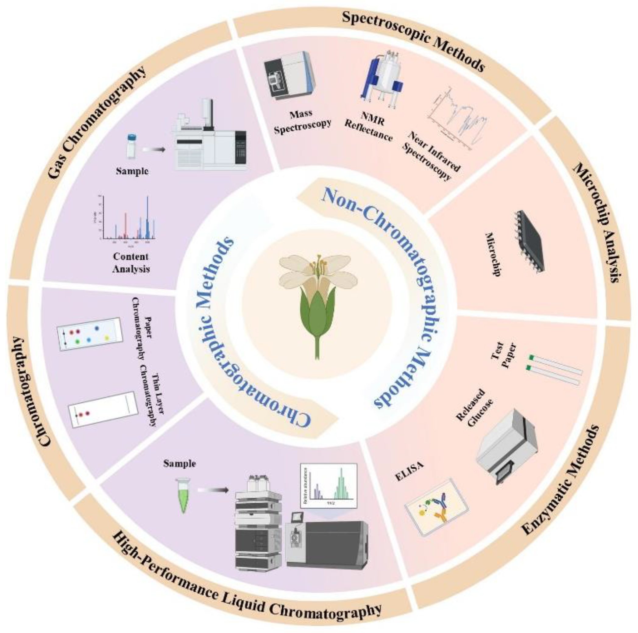
Table 2.
The profiles of glucosinolates detected in cruciferous plants.
| Glucosinolate types | Chemical Names | Common Names | Characterization Methods | |
|---|---|---|---|---|
| AliphaticGSLs | Methyl GSL | Glucocapparin | MS, NMR | |
| 1-Methylethyl GSL | Glucputranjivin | UV, IR, MS, NMR | ||
| (1R)-Methyl-2-hydroxyethyl GSL | Glucosisymbrin | MS, NMR | ||
| 3-Methoxycarbonyl-propyl GSL | Glucoerypestrin | NMR | ||
| Ethyl GSL | Glucolepidiin | Thiourea-type | ||
| 4-Oxoheptyl GSL | Glucocapanglin | Deducted from IR and 5-oxooctanoic acid | ||
| 5-Oxoheptyl GSL | Gluconorcappasalin | Thiourea-type, IR compared to GSL | ||
| 5-Oxooctyl GSL | Glucocappasalin | UV, IR of GSL and desGSL | ||
| 2-Hydroxy-2-methylpropyl GSL | Glucoconringiin | MS, NMR | ||
| (1S)-1-Methylpropyl GSL | Glucocochlearin | MS, NMR of GSL and desGSL | ||
| (1R)-1-(Hydroxymethyl)-propyl GSL | Glucosisaustricin | MS, NMR of desGSL | ||
| (2S)-2-Methylbutyl GSL | Glucojiaputin | UV, IR, MS, NMR of GSL and des GSL | ||
| (2S)-2-Hydroxy-2-methylbutyl GSL | Glucocleomin | NMR of desGSL | ||
| 3-(Methylsulfanyl)propyl GSL | Glucoibervirin | MS, NMR of GSL | ||
| 4-Oxoheptyl GSL | Glucocapangulin | Deduction from IR,5-oxooctanoic acid | ||
| 4-(Methylsulfanyl)butyl GSL | Glucoerucin | UV, IR, MS NMR of GSL | ||
| 5-(Methylsulfanyl)pentyl GSL | Glucoberteroin | UV, IR, MS, NMR of GSL; UV, MS, NMR of desGSL | ||
| 6-(Methylsulfanyl)hexyl GSL | Glucolesquerellin | UV, IR, MS, NMR of GSL | ||
| (R)-11-(Methylsulfinyl)-propyl glucosinolate | Glucoiberin | MS, NMR, X-Ray of GSL; UV | ||
| (R/S)-4-(Methylsulfinyl)-butyl glucosinolate | Glucoraphanin | MS, NMR of GSL, UV | ||
| (R/S)-5-(Methylsulfinyl)pentyl GSL | Glucoalyssin | MS, NMR of GSL | ||
| (R/S)-6-(Methylsulfinyl)-hexyl GSL | Glucohesperin | UV, IR, MS, NMR of GSL | ||
| (R/S)-8-(Methylsulfinyl)-octyl GSL | Glucohirsutin | UV, IR, MS, NMR of GSL; | ||
| (R/S)-9-(Methylsulfinyl)-nonyl GSL | Glucoarabin | UV, IR, MS, NMR of GSL; | ||
| (R/S)-10-(Methylsulfinyl)decyl GSL | Glucocamelinin | MS, NMR of GSL | ||
| 3-(Methylsulfonyl)-propyl GSL | Glucocheirolin | MS of GSL; NMR of desGSL | ||
| 4-(Methylsulfonyl)butyl GSL | Glucoerysolin | MS of GSL; MS, NMR of desGSL | ||
| (R/S, 3E)-4-(Methylsulfiny1)-but-3-enyl GSL | Glucoraphenin | MS, NMR of GSL; UV,NMR of desGSL | ||
| (R)-4-(Cystein-S-yl) butyl GSL | Glucorucolamine | MS, NMR of desGSL | ||
| 4-(β-D-Glucopyranosyl-disulfanyl)-butyl GSL | Diglucothiobeinin | MS of GSL; MS, NMR of desGSL | ||
| 6-Benzoyl-4 (methylsulfanyl)butyl GSL | 6'-Benzoyl-glucoerucin | UV, MS, NMR of desGSL | ||
| 6'-Benzoyl-4(methylsulfinyl)butyl GSL | 6'-Benzoyl-glucopharanin | UV, MS, NMR of desGSL | ||
| (R/S,3E)-6-Sinapoyl-4 (methylsulfinyl)but-3-enyl GSL | 6'-Sinapoyl-glucoraphenin | UV, IR, MS, NMR of desGSL | ||
| Allyl glucosinolate | Sinigrin | MS, NMR, X-Ray of GSL; UV | ||
| But-3-enyl GSL | Gluconapin | MS, NMR of GSL; UV, | ||
| Pent-4-enyl GSL | Glucobrassicanapin | MS of GSL | ||
| (2S)-2-Hydroxypent 4-enylGSL | Gluconapoleiferin | MS of GSL | ||
| (2R)-2-Hydroxybut 3-enylGSL | Progoitrin | MS, NMR of GSL; UV, MS, NMR of desGSL | ||
| (2S)-2-Hydroxybut 3-enylGSL | Epiprogoitrin | MS, NMR of GSL; UV, MS, NMR of desGSL | ||
| Glucosinolate types | Chemical Names | Common Names | Characterization Methods | |
| Aromatic GSLs | (1R)-2-Bezoyloxt-1-methylethyl GSL | Glucobenzosisymbrin | UV, IR of ITC | |
| (1R)-1-(Benzoyloxymethyl) propyl GSL | Glucobenzsisaustricin | Thiourease-type, IR compared to GSL | ||
| Benzyl GSL | Glucotropaeolin | MS, NMR of GSL; UV, MS, NMR of desGSL | ||
| 3-Hydroxybenzyl GSL | Glucolepigramin | MS of GSL; MS, NMR of desGSL | ||
| 3-Methoxybenzyl GSL | Glucolimnanthin | MS, NMR of GSL; UV, MS, NMR of desGSL | ||
| 4-Hydroxybenzyl GSL | Glucosinalbin | UV, MS, NMR of GSL and desGSL | ||
| 4-Methoxybenzyl GSL | Glucoaubrietin | MS, NMR of desGSL | ||
| 3,4-Dihydroxybenzyl GSL | Glucomatronalin | MS of GSL | ||
| 4-Hydorxy3-methoxybenzyl GSL | 3-Methoxysinalbin | UV, MS, NMR of desGSL | ||
| 3-Hydroxy-4-methoxybenzyl GSL | Glucobretschneiderin | UV, IR, MS, NMR of GSL | ||
| 4-Hydorxy-3,5-dimethoxybenzyl GSL | 3,5-Dimethoxy-sinalbin | UV, MS, NMR of desGSL | ||
| 2-Phenylethyl GSL | Gluconasturtiin | NMR of GSL; UV, MS, NMR of desGSL | ||
| 2-hydroxy-2-phenylethyl GSL | Glucobarbarin | MS, NMR of GSL and desGSL | ||
| (2R)-2-Hydroxy-2-phenylethyl GSL | Epiglucobarbarin | MS, NMR of GSL and desGSL | ||
| 2-(4-Methoxy-phenyl) ethyl GSL | Glucoarmoracin | NMR of GSL, MS, NMR of desGSL | ||
| (2R)-2-Hydroxy-2-(4-hydroxyphenyl) ethyl GSL | p-Hydroxy-epiglucobarbarin | MS, NMR of GSL; UV, MS, NMR of desGSL | ||
| (2S)-2-Hydroxy-2(4-hydroxyphenyl) ethyl GSL | p-Hydroxy-glucobarbarin | UV, MS, NMR of desGSL | ||
| 4-(4'-O-Acetyl-α-c-4-rhamnopyranosyloxy)-benzyl GSL | 4-Acetyl-glucomoringin | MS of GSL and ITC | ||
| 6'-Isoferuloyl-2 phenylethyl GSL | 6'-Isoferuloyl gluconasturtiin | MS of GSL, UV | ||
| 6'-Isoferuloyl-(2R) 2-hydroxy-2phenylethyl GSL | 6'-Isoferuloyl epiglucobarbarin | MS, NMR of GSL; UV | ||
| 6'-Isoferuloyl-(2S) 2-hydroxy-2phenylethyl GSL | 6'-Isoferuloyl glucobarbarin | MS, NMR of GSL; UV, MS, NMR of desGSL | ||
| 4-(α-L-Rhamnopyranosyloxy) benzyl GSL | Glucomorinigin | MS, NMR of GSL and desGSL | ||
| Indolic GSLs | 4-Methoxyindol-3-yl GSL | Glucorapassicin A | UV, IR, MS, NMR of synthesized GSL | |
| Indol-3-ymethyl GSL | Glucobrassicin | UV, IR, MS, NMR of GSL and desGSL | ||
| 4-Hydroxyindol-3-ylmethyl GSL | 4-Hydroxy-glucobrassicin | MS of GSL; UV, MS, NMR of desGSL | ||
| 4-Methoxyindol-3-ylmethyl GSL | 4-Methoxy-glucobrassicin | UV, MS, MS, NMR of GSL and desGSL | ||
| 1-Methoxyindol-3-ylmethyl GSL | Neoglucobrassicin | UV, IR MS, NMR of GSL; MS, NMR of desGSL | ||
| 1,4-Dimethoxyindol-3-ymethyl GSL | 1,4-Dimethoxy-glucobrassicin | UV, MS, NMR of desGSL | ||
| 1-Acetylindol-3-ymethyl GSL | N-Acetyl-glucobrassicin | MS of desGSL | ||
| 1-Sulfoindol-3-ylmethyl GSL | N-Sulfo-glucobrassicin | UV, IR, MS, NMR of GSL | ||
| 6'-Isoferuloylindol-3-ylmethyl GSL | 6'-Isoferuloyl-glucobrassicin | MS of GSL; UV, MS, NMR of desGSL | ||
Disclaimer/Publisher’s Note: The statements, opinions and data contained in all publications are solely those of the individual author(s) and contributor(s) and not of MDPI and/or the editor(s). MDPI and/or the editor(s) disclaim responsibility for any injury to people or property resulting from any ideas, methods, instructions or products referred to in the content. |
© 2024 by the authors. Licensee MDPI, Basel, Switzerland. This article is an open access article distributed under the terms and conditions of the Creative Commons Attribution (CC BY) license (https://creativecommons.org/licenses/by/4.0/).
Copyright: This open access article is published under a Creative Commons CC BY 4.0 license, which permit the free download, distribution, and reuse, provided that the author and preprint are cited in any reuse.


