
Submitted:
26 October 2024
Posted:
29 October 2024
You are already at the latest version
Abstract
Biomaterials have assumed a decisive role in modern medicine by enabling significant advancements in medical care practices. These materials are designed to interact with biological systems, offering substantial solutions for various medical needs. In this research, bioceramic materials consisting of a bioactive hydroxyapatite-based matrix with Ti nanoparticles, were processed as promising materials. These bioceramics were obtained using mechanical milling, uniaxial pressing, and sintering as powder processing techniques. This study evaluates the effect of Ti additions on the structural, electrochemical, and mechanical properties of the hydroxyapatite ceramic material. Titanium additions were about 1, 2 and 3 wt%. The experimental results demonstrate that the biocomposite's structure has two hexagonal phases: one corresponding to the hydroxyapatite matrix and the other to the Ti as a reinforced phase. The biomaterials' microstructure is completely fine and homogeneous. The biomaterial reinforced with 1wt. % Ti exhibits the best mechanical behavior. In this context, Electrochemical tests reveal that bio-ceramics can achieve stability through an ion adsorption mechanism when exposed to a physiological electrolyte. Bioceramics, particularly those containing 1%Ti, develop their bioactivity through the formation a high-density hydroxide film during a porous sealing process at potentials around −782.71 mV, with an ionic charge transfer of 0.43x10⁻⁹ A/cm2. Finally, this biofilm behaves as a capacitor Cc = 0.18 nF/cm2, resulting in lower ionic charge transfer resistance (Rct = 1.526x10⁶ Ω-cm2) at the interface. This mechanism promotes the material’s biocompatibility for bone integration as an implant material.
Keywords:
hydroxyapatite
; titanium
; biomaterial
; bone prosthesis
; mechanical properties
1. Introduction
Nowadays most bone implants are made of metallic materials because they have good mechanical properties and some are resistant to corrosion, examples of these are titanium-vanadium-cobalt alloys, stainless steel and cobalt-chromium alloys [1,2,3,4]. But there are several disadvantages to using a metallic implant, such as the high density of these, and that being bioinert materials do not promote biological processes, so there is no osseointegration of the implant with the bone tissue. Hydroxyapatite constitutes approximately 98% of the bones of vertebrates. It can be obtained from human or animal bones, by transforming natural materials (such as mineral skeletons of corals), or it can be artificially synthesized [5,6,7]. In recent years, the use of hydroxyapatite (HPa) as a bone implant has been studied, since it is a bioactive material that favors osseointegration and, being a porous material, allows biological processes to take place [8,9]. However, it also has disadvantages since, being a ceramic material, it is a brittle material and therefore has low mechanical properties compared to those of natural bone. To overcome the disadvantages of this ceramic material, metallic reinforcements have been used to improve the mechanical properties, thus obtaining composite biomaterials. For example, attempts have been made to strengthen it with nickel-based superalloys, with titanium-vanadium-cobalt alloys and even with some iron-based alloys [10,11,12,13,14]. However, metals in addition to being heavier than a common bone, can lose their chemical inertness over time, resulting in discomfort and health risks for the individual. Titanium, is an ideal metal in this context due to its low density 4.5 g/cm3, near to that of HPa (3.16 g/cm3), high mechanical strength and corrosion resistance [15,16]. In addition to being biocompatible and lightweight, it offers significant toughness, which improves the mechanical properties of artificial hydroxyapatite. As a result, arises the biomaterial Hydroxyapatite-Titanium, which has the potential to significantly increase life expectancy and improve the quality of life of people through the manufacture of more effective and functional bone prostheses [17]. Currently, there are several materials for the manufacture of prostheses and implants, titanium being one of the most widely used due to its high biocompatibility. However, sometimes the osseointegration process, known as the direct connection between the implant and the bone, is not fully completed, which can result in difficulties when the prosthesis is subjected to stress [18,19]. Within the market there is the composite; Hydroxyapatite/Titanium Dioxide which offers slightly different mechanical properties. Although, it is harder and more resistant to compression, its tenacity is lower, which makes it prone to tensile fractures, thus limiting its main use to coatings [20]. Also, there is the Hydroxyapatite/Zinc compound, noted for its antimicrobial properties that can be further enhanced with the use of copper or silver, however, it does not match the performance of titanium. This is due to its low mechanical performance which limits its application to dental environments and smaller sized coatings [21]. In general, hydroxyapatite-titanium alloys contain 70-80% hydroxyapatite and 20-30% titanium nanoparticles [22]. This composition provides a good combination of mechanical and biological properties. However, some alloys contain higher or lower amounts of each material. For example, while some alloys contain 50% hydroxyapatite and 50% titanium nanoparticles, some others contain up to 90% hydroxyapatite and at least 10% titanium nanoparticles. It should be emphasized that taking into account the different processes by which this material can be manufactured, which are generally complicated and costly, here we propose its manufacture through powder techniques, which is a simple process, non-polluting and with high reproducibility potential and, in turn, allows a significant improvement in the mechanical properties of the resulting material due to a good integration of its components. Therefore, the objective of this work is to study the effect on the chemical and mechanical properties of minimal titanium additions (0%, 1%, 2% and 3% by weight) in HPa, as well as to establish the processing conditions of the bioceramics through powder routes.
2. Materials and Methods
The powders used for the preparation of the biomaterials were hydroxyapatite (5 μm, 99.9%, Sigma-Aldrich) and titanium (<100nm, 99.9%, SkySpring Nanomaterials, Inc.). The added amounts of titanium were 0%, 1%, 2% and 3% by weight. Grinding of the initial Hydroxyapatite + Titanium powders is carried out in a high energy ball mill of the planetary type at 300 rpm for 6 hours, during this stage zirconia grinding elements with diameters of 3mm were used, the grinding was done dry and 1 ml of isopropyl alcohol was added as a control agent. The powder weight:ball weight ratio was 1:12. The grinding container is made of stainless steel and has a capacity of 250 ml. From the powders obtained from the milling process, cylindrical pellets are produced by uniaxial pressing, using a tool grade die and pressures of 200 kg/cm². The sintering of the pellets is carried out in an electric furnace at a temperature of 1,150°C and the samples are kept for 1 hour. The atmosphere inside the furnace was vacuum and the heating ramp followed was 10°C/min, at the end of the cycle the furnace is turned off and the samples are left to cool inside the furnace. After the sintering, physical characterization of the composites was carried out to determine their density according to the Archimedes' principle. Next the grinding stage, the particle size distribution was determined using a Mastersizer 2000 equipment. Fracture toughness was determined using the indentation fracture method employing the equation proposed by Evans [23]. The ultrasonic method determined Young’s modulus, following ASTM standards [24], using a Grindosonic A-360 Japanese manufacturing equipment. Microhardness was evaluated in agreement with the ASTM E384–16 standard [25]. In this case, twelve measurements were performed at different sample locations and the average value of the indentations is reported; these measurements were performed with a microhardness tester (Wilson Instruments Model S400, USA). The compressive strength was evaluated at a Universal Material Tester WP 300 Gunt following the standard ASTM E384–1 [26]. Structure crystalline was determined by DRX (Siemens, Germany, D-5000). The microstructural characteristics of the composites was observed by SEM (Jeol 6300, Japan).
Electrochemical tests were conducted in a physiological environment, specifically using a saline solution of sodium chloride (0.9% NaCl), which is commonly used as medical applications to restore electrolyte-salts lost due to dehydration. Samples of the Bio-ceramic materials were studied in their as-received condition, and were placed in an electrochemical cell, as illustrated in Figure 1. This electrochemical cell has a three-electrode configuration of arrangement. At the top, two electrodes are positioned; one is an Ag/AgCl Reference Electrode (RE) (saturated with KCl), which connected in parallel with a with a platinum wire and a 33 μF/10 mV as capacitor element. This connection mitigates distortion caused by high-frequencies interference, a very common problem in ceramic materials. The other electrode is related to a graphite bar, which serves as the counter electrode (CE). Whereas, at the bottom of this cell, the test sample (HPa with varying titanium contents; 1%, 2% and 3%) acts as the working electrode (WE), with an exposed area of 0.8cm2 to the test solution. So, measurements were achieved using a Prince Applied Research® Parstat 4000 potentiostat/galvanostat (serial number 14181860), connected to a personal Computer (PC) and controlled by Versa Studio software for data acquisition. The experimental sequence illustrated in Figure 2, began with an open-circuit potential (OCP) test of the ceramic material in the saline solution. The OCP behavior was monitored for 10,000 seconds without applying a current pulse, allowing the system to reach a thermodynamic equilibrium state in terms of corrosion potential. Following the OCP test, an alternate current (AC) as disturbance with a small amplitude is applied to the system. The Electrochemical Impedance Spectroscopy (EIS) technique is used with a frequency sweep ranging from 1MHz to 10mHz, with an amplitude of 10 mV and 10 data points per decade. The impedance data was analyzed and fitted to an appropriate equivalent electrical circuit (EEC) model to interpret the electrochemical behavior of the system. Subsequently, a direct current (DC) potential is applied through the anodic polarization technique, to obtain more information about the reactivity and passivation capacity of the sample. The applied potential ranges from -250 mV to 1600 mV/ocp at a sweep rate of 1.66 mV/s. Electrochemical measurements were conducted after immersion times of 5, 48, 168, and 504 hours.
3. Results
3.1. Particle Size
Figure 3 shows the particle size distribution obtained for each study sample after the grinding stage. For samples with 0 and 1% added titanium, the particle sizes obtained are similar and range from 2.2 to 3.5 microns approximately. The average particle size for both samples is 2.37 microns. For the case of the samples with 2 and 3% titanium, the particle size is finer and ranges between 0.85 and 1.7 microns, the average size being 1.28 microns. Therefore, by adding more titanium particles, the final size is considerably reduced. This may be due to the fact that during milling, titanium, being a more ductile metal in the first stages of milling, is slightly laminated, which causes it to form particles with acute angles, which, as milling progresses, act as blades, fracturing the fragile particles of the hydroxyapatite. In general there is a good distribution of particle sizes, which causes a good number of contacts between them and as a consequence a good densification process can be expected during sintering of the samples.
3.2. Structure
Figure 4 shows the X-ray diffraction pattern obtained for the sample with 3% titanium. It is worth mentioning that this analysis was only performed on this sample, since the low titanium content in the other samples would not be easy to detect. This pattern clearly shows that the sample is made up of 2 phases, the 1st and most abundant corresponds to hydroxyapatite and the 2nd, which is seen in much less proportion due to the intensity of its peaks, corresponds to titanium. Both phases have a compact hexagonal structure. Due to the similarity in the crystal structures of the constituents of the sample, some diffraction peaks interpose. This pattern helps to verify the formation of a bioceramic consisting of 2 phases, a ceramic matrix corresponding to hydroxyapatite and a second metallic reinforcement phase corresponding to titanium.
3.3. Microstructure
Figure 5 shows the microstructure observed by optical microscopy of the samples fabricated here and compares it with that of cortical bone. The microstructures of biomaterials are very fine, homogeneous and have similar characteristics. The presence of porosity in the samples is not evident, which is indicative that the pore size in the samples is extremely small. With respect to the microstructure of the cortical bone, there are no major differences between what is being presented here, both microstructures being very homogeneous, although in the microstructure of the bone the presence of porosity is evident.
3.4. Morphology
Figure 6 shows the morphology of the sample with 3% titanium that was fractured after sintering. The observation was made with the aid of a scanning electron microscope. In this figure, particles with large sizes close to 10 microns are observed, the most important thing is that there are also very fine particles homogeneously distributed. Another observation is the rounded shape of the particles which is due to the mechanical milling to which the powders were exposed, in general the morphology of the particles is equiaxial. The titanium particles are not visible due to their fine size.
3.5. Energy Dispersive Spectroscopy
Figure 7 shows the general microstructure of the sample with 3% titanium, as well as the energy dispersive X-ray analysis performed on the sample. The spectrum resulting from the analysis shows the chemical constituent elements of the sample; Ca, P and O chemical elements that correspond to hydroxyapatite. The presence of titanium, which is the reinforcing material used, is also noticeable. With this analysis, the formation of the biomaterial consisting of HPa/Ti is verified once again. With regard to microstructure sample exhibited particles with multimodal grain sizes oscillating from very small to about 50 microns. The porosity is not evident, neither titanium particles, this is indicative of the very small size of both pores and titanium reinforcements.
3.6. Mechanical Properties
3.6.1. Open Porosity
Figure 8 shows the porosity in each of the samples. This graph shows an increasing value of the porosity as the titanium content in the sample increases. Observing the porosity value in cortical bone, it is 20% and is lower than the porosity of the samples prepared here. García Barea in his thesis work [27], states that porosity in a biomaterial is important, since it is the source for the growth of bone tissue in grafts. Similarly, the presence of porosity favors the dispersal of stresses when the material is working under loads. Considering this, the optimum porosity values should be between 20 and 30%, which are the values obtained in all the samples here processed.
3.6.2. Hardness
The results of the microhardness measurements on the different study samples are presented also in Figure 8. This graph shows in general that the hardness of the samples tends to increase as the metal content of the composite increases. However, at 2% titanium content there is a decrease in the hardness value of this sample. This relatively strange behavior may be due to measurement errors. However, considering the standard deviations present in each measurement, it is observed that the hardness value in the sample with 2% falls within the values of the samples with 1 and 3% titanium. As in the elastic modulus measurements in the hardness measurements, the value of the cortical bone hardness is lower than those of the samples with a metallic reinforcement, which is indicative precisely of the function of the metallic phase to reinforce the composite. Especially when comparing the hardness value of the bone with that of the sample without metal reinforcement, where it is observed that the differences in values between them are not significant. This suggests that the hardness values for bioceramics with a higher amount of the metallic phase combined with the porosity values, allow obtaining a material with adequate porosity with the possibility of promoting the formation of tissue surrounding the implant and thus promoting the biological processes of bone regeneration in a material that does not present extreme stiffness mainly in the sample with 1% titanium.
3.6.3. Elastic Modulus
Figure 9 shows the results of the elastic modulus measurements in the different samples prepared, as well as the value of this property in cortical bone for comparative purposes. The graph shows that as the metal content in the HPa increases, the elastic modulus also increases. Nevertheless, the magnitude of E for the samples with 0 and 1% is very similar and slightly higher than that of cortical bone. On the other hand, when the titanium content increases to 2 or 3%, the value of elastic modulus increases even more, presenting very similar values for these two samples. The elastic modulus of cortical bone in all cases is lower than that of titanium. Which means that the bone is less stiff than any of the composites prepared here. Both HPa and Ti have a compact hexagonal structure, which has the main characteristic of being a rigid structure, with few sliding systems, hence the high value of the elastic modulus of the materials obtained. The presence of the metallic phase in the HPa matrix is the cause of the higher values of the elastic modulus in the composites, due to the fact that the metal, being subjected to the action of stresses, first has a good elastic behavior which transforms into plastic with increasing stresses.
3.6.4. Compression Strength
The results of compressive strength measurements on all samples are also shown in Figure 9. This figure shows that the value of the compressive strength of the bone and the samples with 2 and 3% titanium is very similar, being slightly lower in the samples with the metallic phase. Likewise, the sample without reinforcement has a considerable decrease in its compressive strength value compared to the previous ones. Observing the compressive strength value reached by the sample with 1% titanium, we can see that this value is much higher than that of the other samples. Therefore, according to this data and the results obtained in the measurements of the other mechanical properties, 1% Ti added as reinforcement material in HPa is the optimum amount to achieve the best mechanical properties in the resulting composite. Titanium in quantities of 1% in HPa, acts as a reinforcing material because it is capable of absorbing the stresses applied to the material, this is due to its ductile metallic characteristic, which allows a good degree of elastic-plastic deformation before fracturing.
3.6.5. Fracture Toughness
Figure 10 shows the values obtained from the fracture toughness measurements for each study sample. As expected, the fracture strength of HPa increases as the additions of the metallic phase in HPa increase. This is due to the ductile nature of titanium compared to the brittleness of HPa. In the case of the pure HPa sample, the fracture toughness value was the lowest. On the other hand, the samples with the highest Ti content have the highest toughness value, improving this value by 30% with respect to that of cortical bone. For the sample with 1% Ti, the fracture toughness improves by 20% with respect to the value of the bone. The reinforcement mechanism proposed here by many authors is the deflection of cracks when they grow and their advance is interrupted by the presence of metallic particles, which stop or deviate their trajectory, thus requiring greater efforts so that the crack can continue to grow [28,29,30].
3.7. Electrochemical Characterization
3.7.1. Potentiodynamic Polarization Curves (TAFEL)
Figure 11 illustrates the electrochemical behavior at open circuit potential (OCP) in relation to the activation potential E (mV) of the ceramic surface immersed in a saline solution of 0.9% NaCl, which simulates human saline solution. Data were collected over an immersion period ranging from 5 to 504 hours without applying an external current signal in the presence of an Ag/AgCl reference electrode. Based on previously investigated properties such as microstructure and mechanical characteristics, hydroxyapatite bioceramic reinforced with 1% titanium (Ti) exhibited the best structural and mechanical performance. Consequently, Figure 9 focuses only on the electrochemical curves at OCP for bioceramics containing 1%Ti. The curves labeled 1 and 2 represent the OCP transients after 5 and 48 hours of immersion in NaCl, respectively. During the 10,000-second evaluation period, these curves exhibit a highly unstable active trend, fluctuating between −670 and −650 mV. The notable changes in surface cause the potential value to vary sharply, probably can be attributed to the penetration of chlorides into the pores of the ceramic matrix, causing a surface hydration reaction. Specifically, hydroxide compounds progressively obstruct the ceramic matrix’s pores. This suggests that surface activation process is driven by ion transfer, which is the predominant process in the initial hours of immersion.
Similarly, for 168h (curve 3) and 504h (curve 4), a stable of the corrosion potential (Ecorr) is observed, indicating that the surface undergoes no significant changes. This suggests a uniform blockage of porosity, and possible surface activation, with a stable potential of around −685 mV, a more negative value. This behavior indicates that, over the extended immersion period, the OCP becomes more stable, suggesting that hydroxyapatite with 1% Titanium (HPa/Ti) will not experience significant surface changes under physiological conditions. In other words, the surface remains unactivated and unaltered, and it does not decompose rapidly in the studied media. In this OCP study, it’s observed that the potential becomes increasingly negative behavior with longer immersion times. This suggests that the surface of the bioceramic exhibits an electron transfer mechanism, facilitating a release process that leads to surface oxidation and the formation of a protective coating. Such behavior is promising for medical applications, where the material will be in continuous contact with bodily fluids. Additional insights can be drawn from the potentiodynamic polarization curves in the anodic branch and electrochemical impedance data, which are illustrated in Figure 10 and Figure 11, respectively, for each immersion time of the bioceramic with 1% Titanium as reinforcement particles.
Figure 12 presents the anodic polarization curves, illustrating the electrochemical processes involved, including the formation of a passive surface. The graph depicts the relationship between the potential of the test sample (bioceramic) in mV and the current density in A/cm2. All curves indicate a predominant passivity due to the formation of a passive oxide layer; however, significant changes in the curves are observed as the immersion time in NaCl increases. After 5h, the corrosion potential Ecorr is −856.32 mV with a current density of 1.92 nA/cm2, whereas after 504h, this value shifts to −782.71 mV with a current density of 0.35 nA/cm2. Also, abrupt changes in the slope are observed, indicating a rapid increase in current with respect to potential. This behavior suggests that, prior to surface passivation, there is a significant dissolution of the surface due to ion exchange during the initial 5h of immersion of the hydroxyapatite material, resulting in a current density around 1e-6 A/cm2. In contrast, after 504h of immersion, the tendency of increase in a current is much less pronounced, indicating the formation of a passive film. In this state, the current exchange is barely noticeable, measuring only 1e-8 A/cm2, with an activation potential close to -200 mV. This represents a difference of -600 mV in the protection against corrosion of the bio-ceramic material in this saline medium of NaCl solution.
However, at higher potentials, the curves reveal that transpassivation of the system occurs regardless of exposure time, suggesting that the passive layer becomes unstable and may fracture, potentially leading to the dissolution of the base metal. These tests suggest that when hydroxyapatite (Ca₁₀(PO₄)₆(OH)₂) material reinforced with 1% Ti is immersed in a physiological solution of NaCl (0.9% serum), its passivation mechanism is due to the presence of ions in the medium. As the hydration process advances during the exposure time, a highly stable layer forms on the surface, which prevents degradation in the corrosive environment. Additionally, Cl- and Na+ ions interact with the surface, and, in the presence of small amounts of Ca2 and PO4-3 phosphates, they react with OH- groups to form well-defined hydroxide compounds within the pores of the ceramic structure. With prolonged exposure time, the surface area increases, promoting the development of a thin layer of secondary calcium phosphate that contributes to its passivation.
In general, these graphs demonstrate a decrease in the corrosion current density (icorr) from 1x10⁻⁶ to 1x10⁻⁸ A/cm2, as well as a reduction in equilibrium potential from −856.32 mV to −782.71 mV is observed. In relation to the breakdown potential of the calcium phosphate film, values shift from −875 mV after 5 h, -455 mV after 38 h, and -425 mV at 168h and finally -272 mV after 504 h of immersion under direct current (DC) conditions. The last is characterized by a lower surface dissolution slope as both potential and immersion time in NaCl increase. These data suggest the formation of an activated surface layer that acts a physical barrier, that limits direct interaction between the surface of the ceramic material and the aggressive ions in the medium, particularly, Cl- ions. As this protective layer stabilizes over time, the flow of Ca²⁺ and PO₄³⁻ ions from the hydroxyapatite into the electrolyte decreases, resulting in a reduced rate and slope of material dissolution. This is particularly significant in NaCl solutions, where chlorides could enhance corrosion or dissolution in the absence of this protective layer. This passivation mechanism is essential in biomedical applications, as it protects hydroxyapatite coatings or implants in contact with body fluids, facilitating the material’s biocompatibility with surrounding bone and ensuring greater stability and durability in physiological environments.
3.7.2. Electrochemical Impedance Measurements (EIS)
This study is complemented by Electrochemical Impedance Spectroscopy (EIS) measurements, a technique for studying the electrochemical behavior of ceramic materials such as hydroxyapatite (HPa) in electrolyte solutions such as NaCl. This technique allows for more detailed information about the processes and mechanisms that occur at the interface between the material and the test solution, including the surface activation process, ion charge transfer resistance, corrosion mechanisms, passivation, and ion transport properties through passive layers or coatings. In this sense, the impedance diagrams shown in Figure 13 are plotted in the form of Nyquist, and Bode graphs are displayed in Figure 14, which indicate the different processes of ionic charge transfer and diffusion processes at the material-electrolyte interface. Impedance measures the resistance to the passage of an alternating current over a wide frequency range from 1Mhz to 1 mHz, in which capacitance is measured. This is observed by two-time constants, one at a high frequency that relates to surface processes that can be the presence of pores or the presence of a surface layer, and a second-time constant relates to the charge transfer on the electrode interface (HPa/1%Ti test sample) and the electrolyte ions. The EIS curves in the Nyquist representation shown in Figure 13 indicate a progressive charge transfer on the bioceramic interface from the start of the tests for 5h to 504h of immersion in NaCl, showing a time constant at high frequencies.
This time constant is related to the electrochemical phenomena that occur at the interface, being that for 5h of immersion, there is a semicircle amplitude of almost 1 kΩ-cm2 and a Rcoating value of 3.4 kΩ-cm2, which progressively decreases from 800 Ω-cm2 to 400 Ω-cm2, which indicates that the Cl- ions, Na+ and OH- penetrate the bioceramic matrix through its porous structure, curves labeled 1, 2, 3 and 4.
In the meantime, the amplitude of a second-time constant decreases with increasing immersion time, leading to a better consolidation of the porous surface by hydration products and a much higher resistivity to load transfer (Rct) value than its initial value, which cannot be shown. This is related to the ease or difficulty of the electrochemical processes in the hydroxyapatite/solution interface. Nyquist's graphs generally show a vast second semicircle at high frequencies, representing charge transfer (Rct) across the surface of the hydroxyapatite.
The diameter of these semicircles correlates with the load transfer resistance, Rct; the more significant the diameter, the greater the stability of the material against a corrosive attack due to the presence of a hydrated film that protects it. The Bode diagrams in Figure 12 allow us to understand and observe the mechanisms of charge transfer as a function of frequency, providing detailed information on the kinetics of the reactions and the processes of charge and mass transport. In this case, the absolute impedance Z's magnitude and the sinusoidal signal's offset angle F applied to the interface over a measured frequency range are plotted.
In this sense, two-time constants in the entire frequency range evaluated (1MHz to 1mHz) are very noticeable. The high-frequency response is associated with the HPa/1%Ti sample electrode interface and the NaCl electrolyte, which shows a resistive behavior to charge transfer, showing a constant impedance magnitude with a phase angle close to zero. However, bio-ceramics develop a biofilm when immersed in a physiological solution of NaCl, which affects the charge transfer mechanism by having a dielectric barrier. Meanwhile, at intermediate frequencies, a second-time constant is observed that reflects the purely capacitive mechanisms since the porous film formed allows the hydration process by OH- compounds with Na+ and Cl- ions, which indicates the ease of charge accumulation in the interface, which reaches a phase angle of 60° in the first hours of immersion. This pore hydration promotes the interface's consolidation and improves the biofilm's surface properties, allowing the easy transport of ionic species. This causes the dielectric constant of the interface to change; a change in the dielectric constant of the interface due to the process of hydration of pores and imperfections increases the capacitance of the protective film. This is evident in Figure 14 by increasing the phase angle from 60° to 75° for the 504-hour dive time, making a system dominated by a more capacitive behavior by approaching 90°. It is important to note that capacitance C is directly related to the dielectric constant C = εA/d, where ε is the dielectric constant, A is the area, and d is the thickness of the film. Finally, the low-frequency region impedance reflects slower processes, such as the diffusion of ionic species across the interface, as the film surface is consolidated by pore hydration, the absolute impedance value |Z| is higher as a function of time, indicating its high bio-film integrity by increasing the immersion time in NaCl.
To analyze this observed mechanism of pore hydration, to explain the presence of a bio-film formed during contact with the physiological medium NaCl, and the alteration of the dielectric constant, it is necessary to propose a model of electrical layers, to represent and study the electrical mechanisms that occur at the interface of the HPa/1%Ti sample immersed in a physiological solution of NaCl for 504 hours of immersion, to validate its integrity as a bio-material with the possibility of functioning as an implant. In Figure 15, an electrical model or equivalent circuit is proposed to simulate the mechanisms of the interface using a combination of resistors and capacitances, connected in series-parallel, depending on the surface conditions.
The electrochemical parameters associated with this passive surface can be obtained by modeling the experimental data with an equivalent electrical circuit (EEC), such as the one proposed in Figure 15. The arrangement of this circuit is simple: You have a resistor, Rs, which represents the resistance of the solution that is connected in series with the resistor, Rc, (which represents the resistance of the biofilm formed connected in parallel to a constant phase element, CPE (Q, which relates the properties of the passive surface). These are connected in parallel to a resistor that characterizes the charge transfer through the passive surface/electrolytic interface, which is connected in series to a second constant-phase element, CPE. In this circuit, a CPE is used instead of a pure capacitor to account for the non-ideal behavior of the passive surface, which is defined in the impedance representation as:
Where: Yo is the constant CPE, n is the exponent CPE indicating surface heterogeneity or roughness, j2 = 1 is an imaginary number, and w is the angular frequency in rad/s. Depending on n, CPE can represent a resistance (ZCPE = R, n= 0), capacitance (ZCPE = C, n= 1), Warburg impedance (ZCPE = W, n= 0.5), or inductance (ZCPE = L, n= 1). The correct equation to convert Yo into Capacitance is given by, where C is the capacitance and is the angular frequency at which Zre is maximum. The values of the electrochemical parameters obtained from the Nyquist diagrams are illustrated in Table 1.
The results show that the Rc decreases slightly with the advance of the immersion time, corresponding to the increase of the phase angle to values close to 90° (Figure 12). In contrast, the amplitude of the semicircles of Figure 11 decreases due to the entry of ions into the pores of the film formed on the surface of the HPa/1%Ti bio-ceramic. As can be seen from the previous results, hydroxyapatite has a porous structure in the nanometric order. These pores allow the transport of ionic species present in the test's electrolyte, and this mechanism helps the material develop bioactivity to form a bio-film. There are also sites where hydrated compounds enter through the physical adsorption mechanism (Van der Waals bonds) and the chemical interaction mechanism (formation of bonds with functional groups originating from the surface). When these pores are filled, a double electrical layer is formed, which acts as a capacitor that stores charge and allows charge transfer when a potential difference or an electric field is applied to develop a chemical reaction. In this sense, it is observed that the charge stored in the interface is measured by the application of a disturbance of a sinusoidal signal in a frequency sweep at different immersion times in the presence of Cl-, OH-, and Cl- ions, this capacitance varies from 5.27 nF/cm2 in the first hours of immersion to 0.18 nF/cm2 at 504h. This bioactivity of the film formed on the hydroxyapatite is observed by the increasingly positive value of the activation potential, which ranges from -856.32 to -782.71 mV at the end of the electrochemical test. These same kinetic results in Table 1 indicate that once the interface reaches a complete saturation of the pores or ionic sites, the charge transfer is relatively low, as noted in the Icorr current density in the order of nA/cm2, which facilitates ion transport in the medium that interacts with this interface. Once the HPa/Ti1% interface stabilizes at 0.9%NaCl, it reaches current or ion flow resistance values according to absolute impedance, 1.526x10⁶ A/cm2.
Finally, these results indicate that the ion adsorption and charge transfer mechanism through the pores of the 1%Ti bio-ceramic HPa as reinforcement can show more excellent electrochemical stability and resistance to corrosion attack as the exposure time in a physiological medium such as NaCl increases. Adsorption of Na, OH-, PO4-, and Ca ions promotes film bioactivity, as indicated by the potential for open-loop activation without interface disturbance. In medical applications as an implant, this ability facilitates the successful formation of a biofilm on the bone that allows cellular osseointegration by providing active sites for the formation of apatite, which are anchored by the presence of Ti particles, which, in addition to providing mechanical resistance, serves as an anchor of sites to the bone. In medical applications as an implant, this ability facilitates the successful formation of a biofilm on the bone that allows cellular osseointegration by providing active sites for the formation of apatite, which are anchored by the presence of Ti particles, which, in addition to providing mechanical resistance, serves as an anchor of sites to the bone. The electrochemical tests of this research indicate the ability of HPa/1%Ti bio-ceramic to adsorb ions at active sites called pores due to its porous structure, that the hydration mechanism is crucial to induce the formation of an active bio-film, that as a biomedical implant in devices with surgical grade titanium or stainless steel, it causes the formation of new bone tissue at the implant interface, facilitating osteoconductivity.
5. Conclusions
- ○
- Through the proposed methodology, hydroxyapatite biomaterials reinforced with titanium nanoparticles were successfully fabricated.
- ○
- The resulting biomaterial is constituted by two hexagonal compacted crystalline phases, one corresponding the hydroxyapatite ceramic matrix and a second phase that corresponds to the reinforced titanium metal.
- ○
- From the results obtained in the mechanical properties measurements, it is concluded that the biomaterial reinforced with 1 wt. % Ti, presents the best mechanical behavior.
- ○
- Electrochemical tests (OCP, anodic polarization, and EIS) show significant results in which the bioceramic is stabilized by the mechanism of chemical and physical adsorption of ions during its exposure for prolonged times (504h, 21d) in the physiological medium of 0.9%NaCl. Thus, developing bioactivity through a film formed by hydroxide compounds due to a surface sealing of the nanometric porous structure of hydroxyapatite, this phenomenon occurs at potentials close to -782.71 mV with an ionic charge transfer of about 0.43x10⁻⁹ A/cm2. This biofilm is a capacitor that stores a low ionic charge of 0.18 nF/cm2 and allows a charge transfer of 1.526x10⁶ W-cm2 to develop its bioactivity.
- ○
- Finally, in practical applications such as insertion in a physiological medium, the biofilm plays a crucial role. It is firmly anchored to the bone and facilitates cellular osseointegration by providing biocompatibility. The Ti particles, on the other hand, contribute to mechanical strength and serve as anchoring sites to the bone.
Author Contributions
Conceptualization, ERR and HHH; methodology, DRPH, CACA, HHH, JGMH; validation, DRPH, CACA, HHH, JGMH; formal analysis, JACR, ENAM, ERR; investigation, DRPH, CACA, HHH, JGMH; resources, ERR, HHH, JGMH; data curation, HHH, JGMH, JACR, ENAM, ERR; writing—original draft preparation, DRPH, SMEG; writing—review and editing, HHH, JGMH, JACR, ENAM, ERR; visualization, HHH, ERR; supervision, JACR, ENAM; project administration, HHH, ERR; funding acquisition, JGMH, ERR. All authors have read and agreed to the published version of the manuscript.
Funding
This research received no external funding.
Institutional Review Board Statement
Not applicable.
Informed Consent Statement
Not applicable.
Data Availability Statement
Not applicable.
Acknowledgments
Authors thank the Universidad Politécnica de Victoria and Universidad Autonóma del Estado de México, campus Valle de México, for the facilities granted to work in their laboratories.
Conflicts of Interest
The authors declare no conflict of interest.
References
- Ríos-Puerta, K.; Gutiérrez-Flores, O.D. Revista Politécnica, 2022, 18, 24–39. [CrossRef]
- Black, J.; Hastings, G. Handbook of biomaterial properties, 1st ed; Springer Science & Business Media, London, UL, 1998.
- Chen, Q.; Thouas, G.A. Metallic implant biomaterials. Mat Sci Eng R: Rep, 2015, 87, 1–57. [Google Scholar] [CrossRef]
- Manam, N.S.; Harun, W.S.W.; Shri, D.N.A.; Ghani, S.A.C.; Kurniawan, T.; Ismail, M.H.; Ibrahim, M.H.I. Study of corrosion in biocompatible metals for implants: A review. J Alloys Compd., 2017, 701, 698–715. [Google Scholar] [CrossRef]
- García-Garduño, M.V.; Reyes-Gasga, J. La hidroxiapatita, su importancia en los tejidos mineralizados y su aplicación bio-médica. TIP Rev Esp Cienc Quim Biol. 2006, 9, 90–95. [Google Scholar]
- Smith Deanne, K. Calcium phosphate apatites in nature. In Hydroxyapatite and related materials. eds. Browns, P.W. & Constantz, B., CRC Press, London. 2000, 46-52.
- Zapanta Legeros, R. Biological and synthetic apatites. In Hydroxyapatite and related materials. Eds. Browns, P.W. & Constantz, B. CRC Press, London, UK. 2000, 1-28.
- Fernández, J.; Gilemany, J.M.; Gaona, M. La proyección térmica en la obtención de recubrimientos biocompatibles ventajas de la proyección térmica por alta velocidad (HVOF) sobre la proyección térmica por plasma atmosférico (APS). Biomecánica. 2005, 13. [CrossRef]
- Pineda, V.; Olivares, O.; Yépez, J.; González, A. Parámetros histológicos de la regeneración ósea guiada con hidroxiapatita FOULA en ratas BIOU: Wistar. Revista Odontológica Mexicana Órgano Oficial de la Facultad De Odontología UNAM., 2020, 23, 149–155. [Google Scholar] [CrossRef]
- Grinschpun, L.S.; Oldani, C.R. Obtención de compuesto de Ti – HA por sinterizado a baja temperatura. Revista de la Facultad de Ciencias Exactas, Físicas y Naturales, Argentina 2021, 8, 55–60. Available online: https://revistas.unc.edu.ar/index.php/FCEFyN/article/view/32233.
- Nomura, N.; Sakamoto, K.; Takahashi, K.; Kato, S.; Abe, Y.; Doi, H.; Tsutsumi, Y.; Kobayashi, M.; Kobayashi, E.; Kim, W.-J.; et al. Fabrication and Mechanical Properties of Porous Ti/HA Composites for Bone Fixation Devices. Mater. Trans., 2010, 51, 1449–1454. [Google Scholar] [CrossRef]
- Martínez Ibáñez, I. Estudio de la viabilidad de adicionar magnesio a aleaciones pulvimetalúrgicas de titanio, para la obtención de productos densos y/o porosos. Universidad Politécnica de Valéncia, Spai. 2021. Available online: http://hdl.handle.net/10251/174753.
- Barrabés, M. Optimización de las aleaciones de NiTi porosas para aplicaciones biomédicas. Universidad Politécnica de Catalunya, Spain. 2005. Available online: http://hdl.handle.net/2099.1/3183.
- Montañez, N.; Peña, D.; Cardozo, R.; Faria, M. Corrosión de nitinol bajo tensiones de fuerza en fluido fisiológico simulado con y sin fluoruros. Revista Facultad de Odontología. 2016, 28, 54–70. [Google Scholar] [CrossRef]
- Shackelford, J.F.; Han, Y.; Kim, S.; Kwon, S. CRC Materials Science and Engineering Handbook. 4th ed. CRC Press, Boca Raton., USA. 2015.
- Sánchez, A. Titanio Como biomaterial. Azul Web. 2014. Available online: https://www.azulweb.net/titanio-como-biomaterial/ (accessed on 27 May 2024).
- Sáenz-Ramírez, A. Biomateriales. Revista Tecnología En Marcha, 2004, 17, 34–45. Available online: https://revistas.tec.ac.cr/index.php/tec_marcha/article/view/1432.
- Fendi, F.; Abdullah, B.; Suryani, S.; Raya, I.; Nilawati-Usman, A.; Tahir, D. Hydroxyapatite derived from fish waste as a biomaterial for tissue engineering scaffold and its reinforcement. AIP Conf. Proc. 2719, 020040 (2023), 8–9 September 2021, Palu, Indonesia. [CrossRef]
- Hartatiek, J.; Utomo, L.; Noerjannah, I.; Rohmah, N.Z. ; Yudyanto, Physical and mechanical properties of hydroxyap-atite/polyethylene glycol nanocomposites. Mater Today Proc, 2021, 44, 3263–3267. [Google Scholar] [CrossRef]
- Avinashi, S.K.; Hussain, A.; Kumar, K.; Yadav, B.C.; Gautam, C. Synthesis and structural characterizations of HAp–NaOH–Al2O3 composites for liquid petroleum gas sensing applications, Oxford Open Materials Science, 2021, 1. [CrossRef]
- Dragomir, L.; Antoniac, A.; Manescu, V.; Robu, A.; Dinu, M.; Pana, I.; Cotrut, C.M.; Kamel, E.; Antoniac, I.; Rau, J.V.; et al. Preparation and characterization of hydroxyapatite coating by magnetron sputtering on Mg–Zn–Ag alloys for orthopaedic trauma implants, Ceram Int. 2023, 49, 26274–26288. [CrossRef]
- Youness, R.A.; Taha, M.A.; Ibrahim, M.A. Effect of sintering temperatures on the in vitro bioactivity, molecular structure and mechanical properties of titanium/carbonated hydroxyapatite nanobiocomposites. J. Mol. Struct. 2017, 1150, 188–195. [Google Scholar] [CrossRef]
- Evans, A.G.; Charles, E.A. Fracture Toughness Determination by Indentation. J Ame Ceram Soc. 1976, 59, 371–372. [Google Scholar] [CrossRef]
- American Society for Testing Materials, ASTM E1876-22, Standard Test Method for Dynamic Young's Modulus, Shear Modulus, and Poisson's Ratio by Impulse Excitation of Vibration, 2022.
- American Society for Testing Materials, ASTM E384 – 16, Standard Test Method for Microindentation Hardness of Materials, 2016.
- American Society for Testing Materials, ASTM E9 – 89a, Standard Test Methods of Compression Testing of Metallic Materials at Room Temperature. (Reapproved 2000).
- García-Barea, E. Undergraduate Thesis, Universidad Carlos III de Madrid, Spain, 2015.
- Ighodaro, O.L.; Okoli, O.I. Fracture Toughness Enhancement for Alumina Systems: A Review. Int. J. Appl. Ceram. Technol. 2008, 5, 313–323. [Google Scholar] [CrossRef]
- Banerjee, T.; Dey, S.; Sekhar, A.P. Design of Alumina Reinforced Aluminium Alloy Composites with Improved Tri-bo-Mechanical Properties: A Machine Learning Approach. Trans. Indian Inst. Met. 2020, 73, 3059–3069. [Google Scholar] [CrossRef]
- Rocha Rangel, E. Fracture Toughness Determinations by Means of Indentation Fracture, In Nanocomposites with Unique Properties and Applications in Medicine and Industry, Ed. John Cuppoletti. IntechOpen, London, UK, 2011, 1-18. Available online: https://www.intechopen.com/chapters/16971.
Figure 1.
Design of the electrochemical cell of three-electrode configuration used for the corrosion testing of hydroxyapatite ceramic material.
Figure 1.
Design of the electrochemical cell of three-electrode configuration used for the corrosion testing of hydroxyapatite ceramic material.
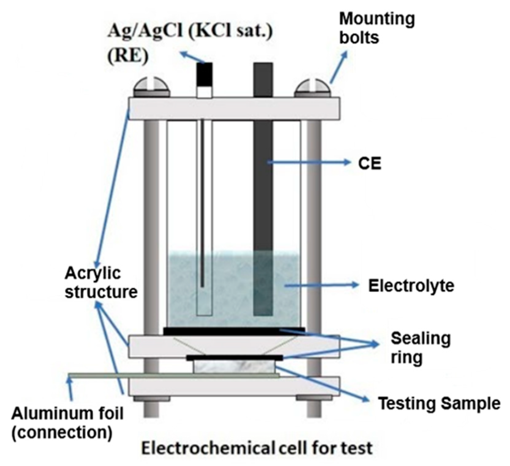
Figure 2.
Experimental sequence of electrochemical test for corrosion evaluation of hydroxyapatite ceramic material in a 0.9% NaCl solution, the setup includes OCP monitoring, EIS and anodic polarization test.
Figure 2.
Experimental sequence of electrochemical test for corrosion evaluation of hydroxyapatite ceramic material in a 0.9% NaCl solution, the setup includes OCP monitoring, EIS and anodic polarization test.
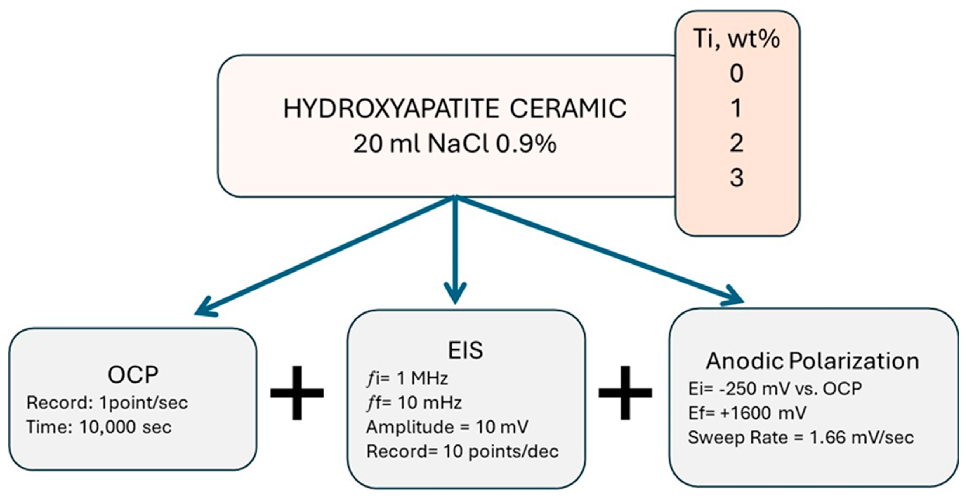
Figure 3.
Particle size distribution obtained in the different samples after the milling stage.
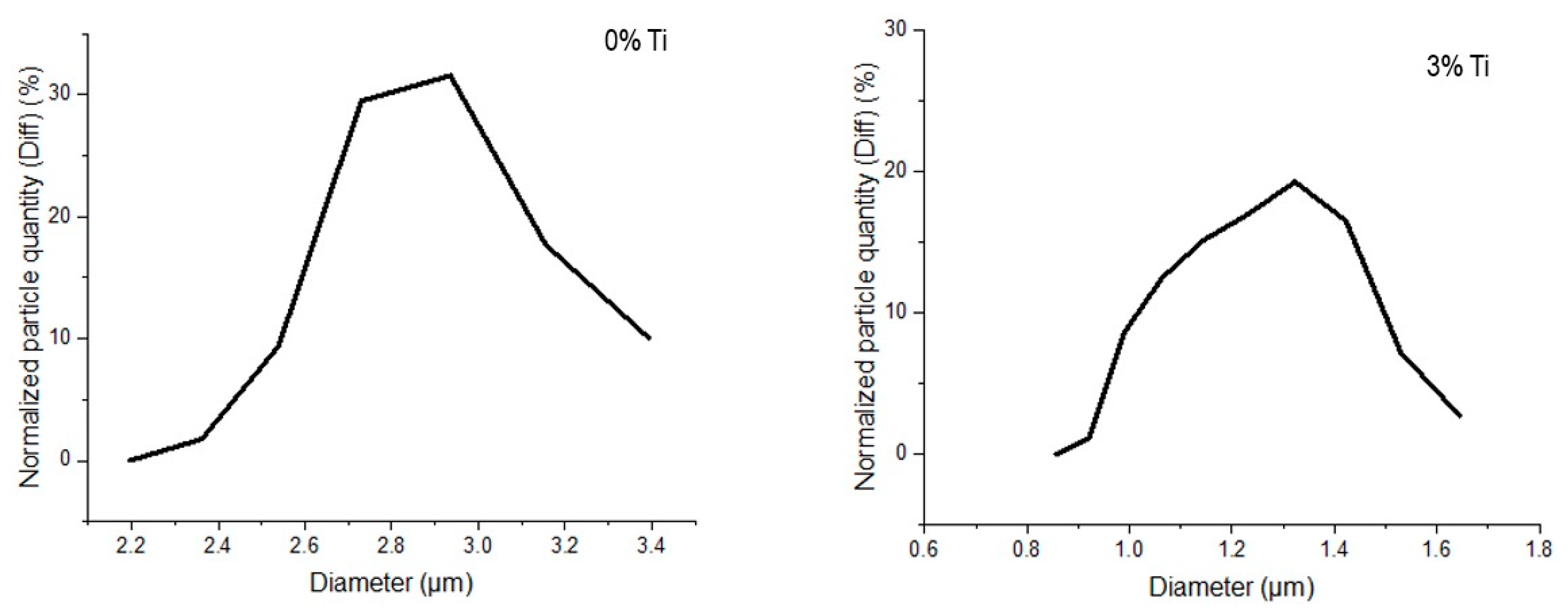
Figure 4.
X-ray diffraction pattern of the sample with 3% titanium.
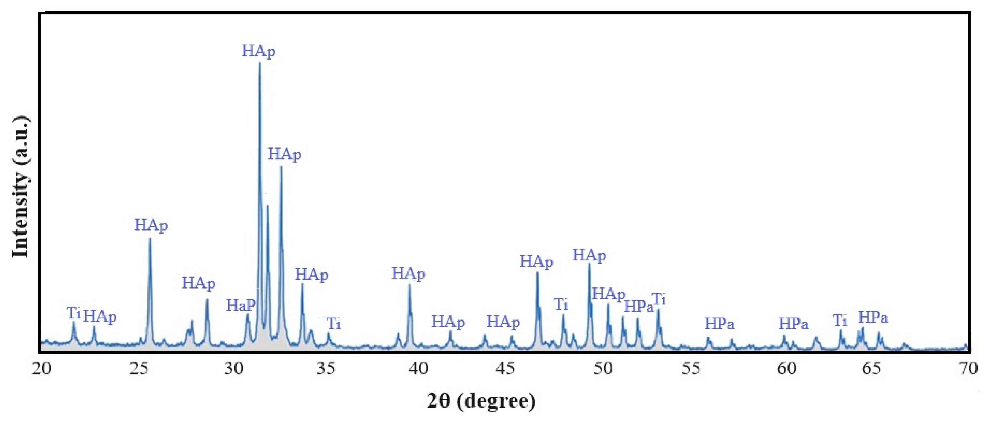
Figure 5.
Microstructure of study samples taken under an optical microscope.
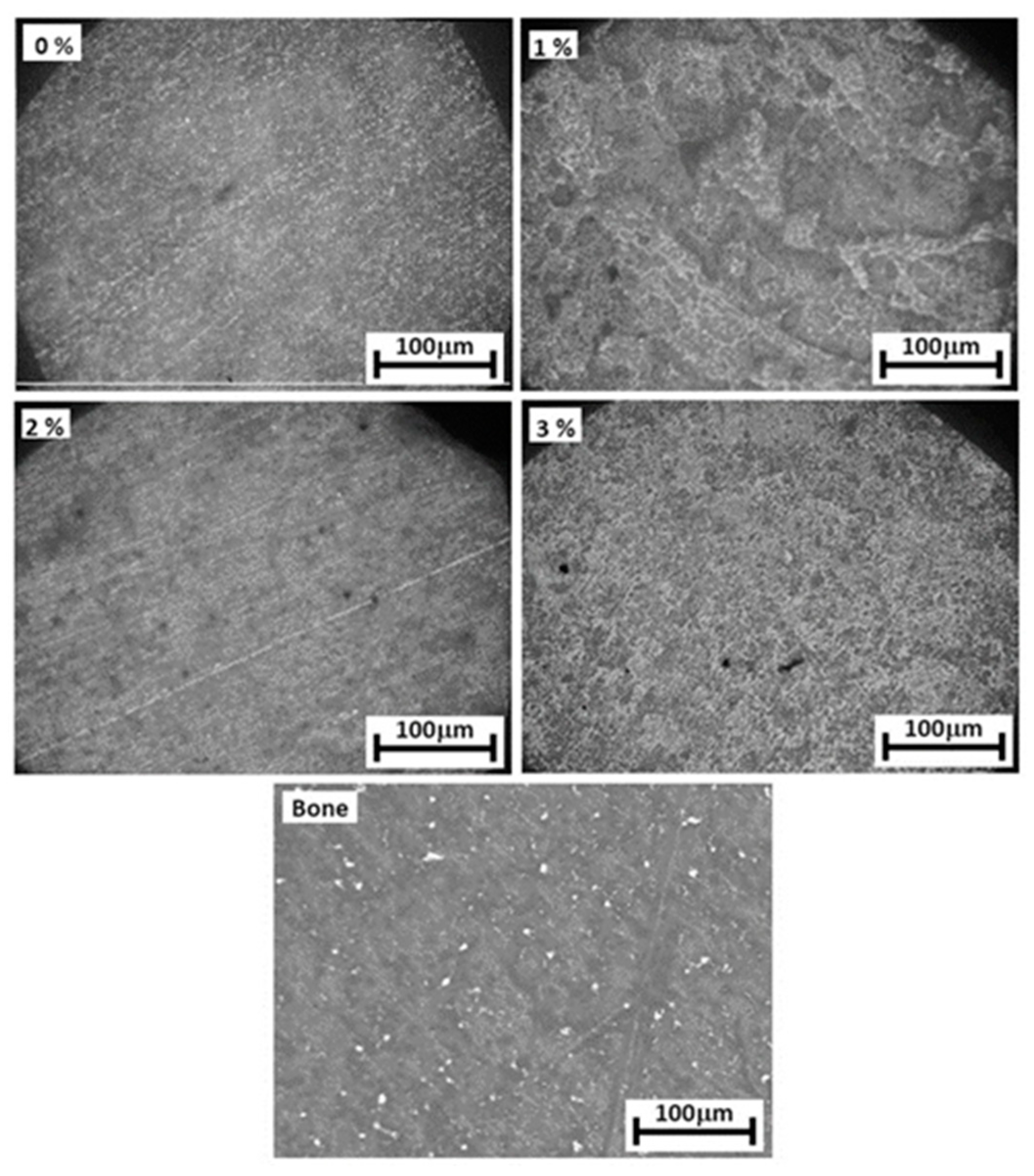
Figure 6.
Particle morphology of the fractured sample and after sintering.

Figure 7.
EDS analysis performed on 3% titanium sample.
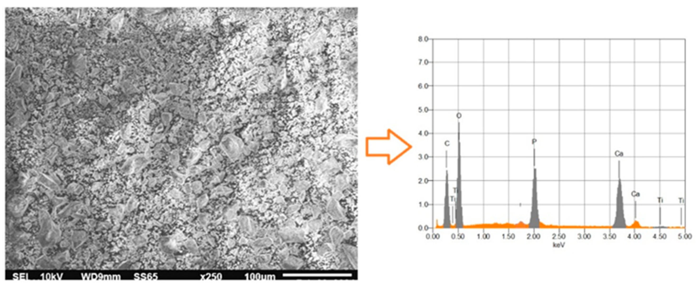
Figure 8.
Porosity and microhardness of HPa as a function of titanium content.
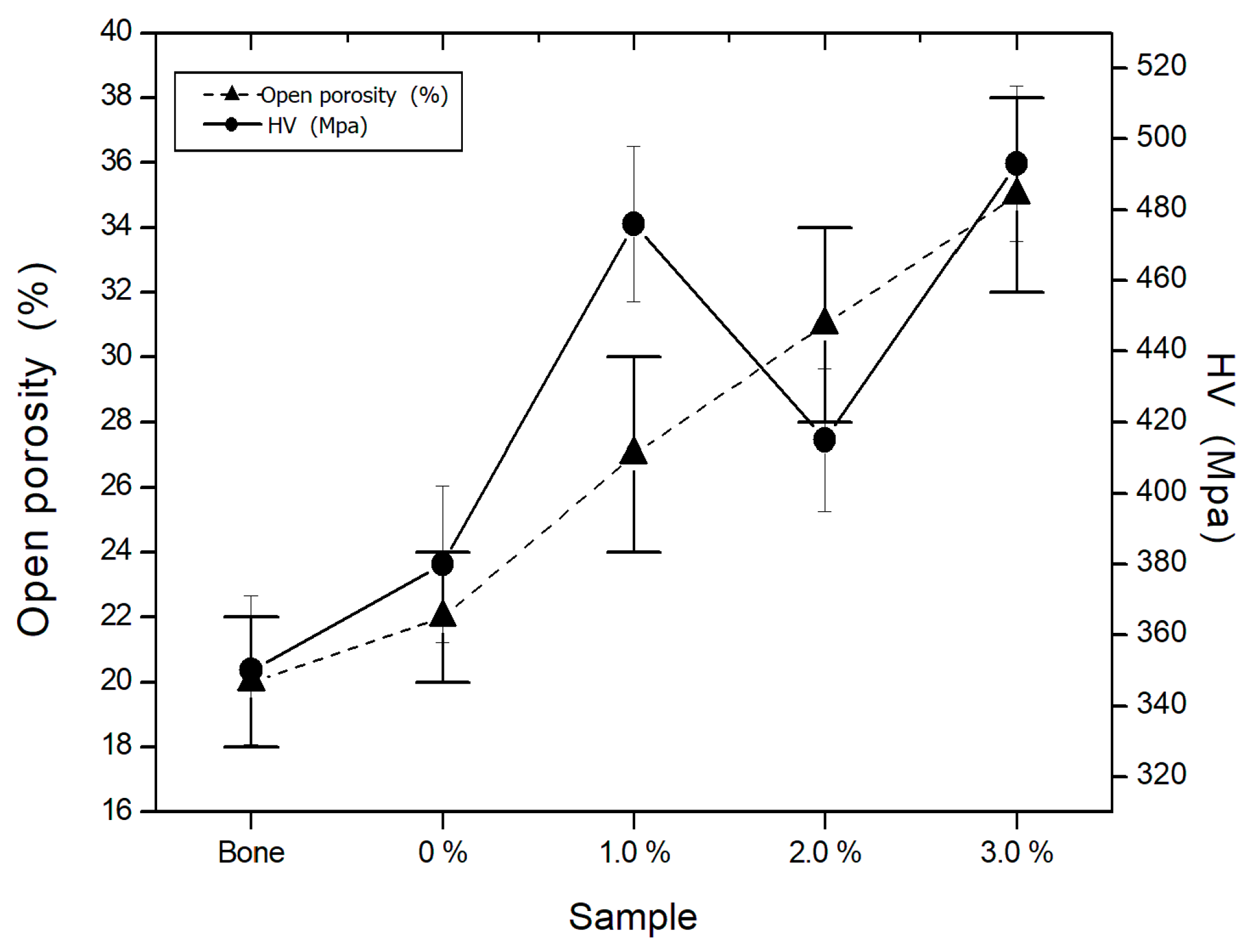
Figure 9.
Elastic modulus and compressive strength of HPa as a function of titanium content.
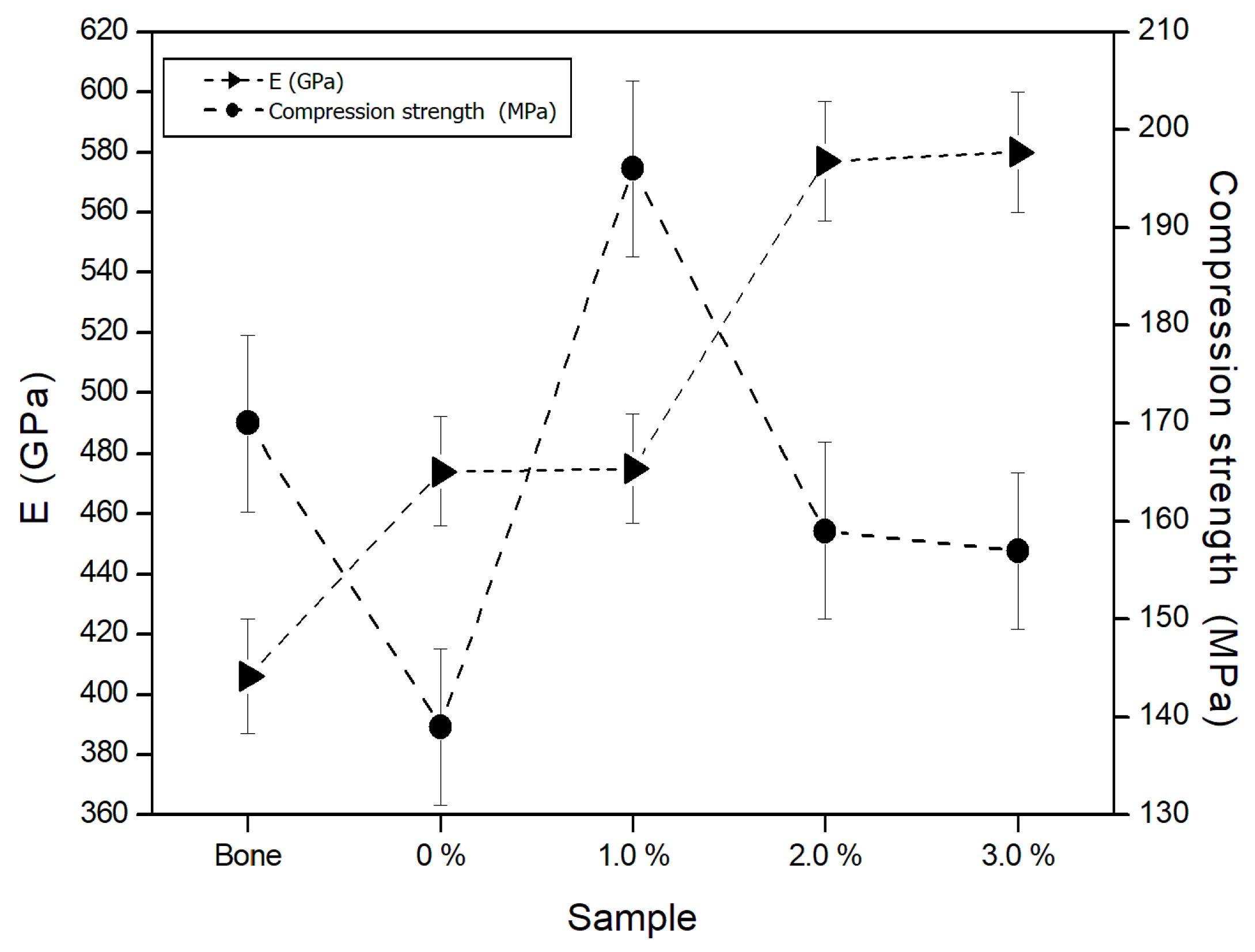
Figure 10.
Fracture toughness of HPa as a function of titanium content.
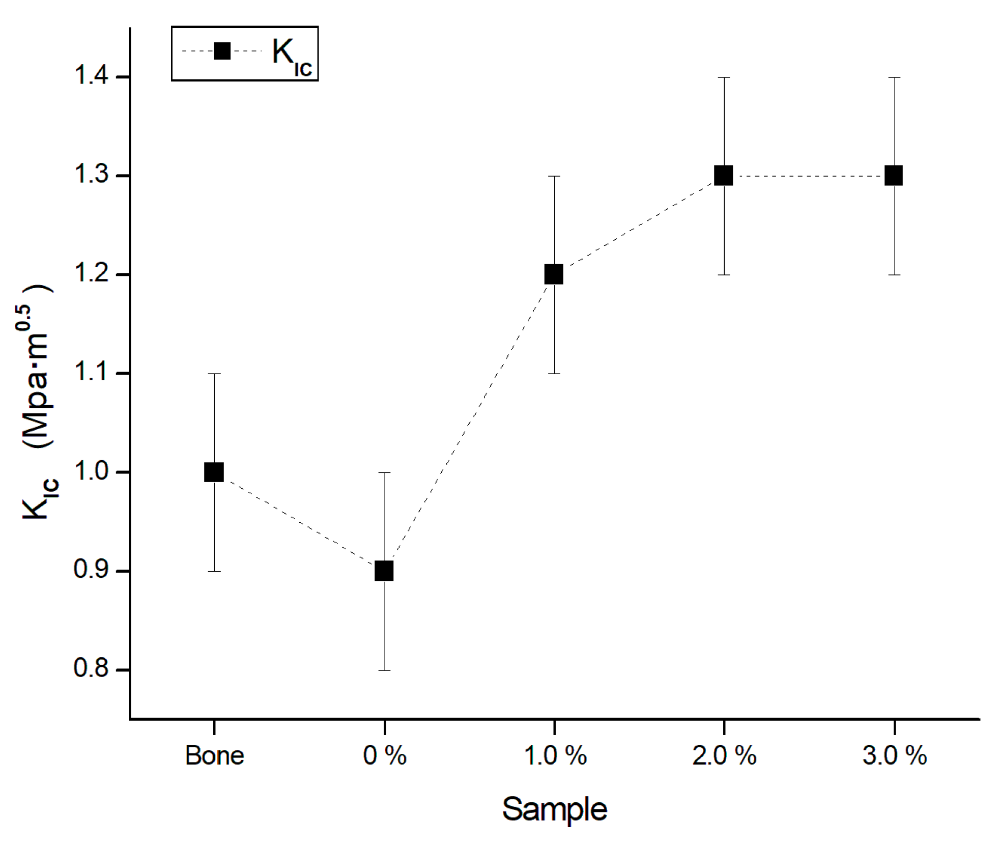
Figure 11.
Open circuit potential transients of the bioceramic material HPa/Ti 1% sample after continuous exposure to a 0.9%NaCl solution for 504 hours.
Figure 11.
Open circuit potential transients of the bioceramic material HPa/Ti 1% sample after continuous exposure to a 0.9%NaCl solution for 504 hours.
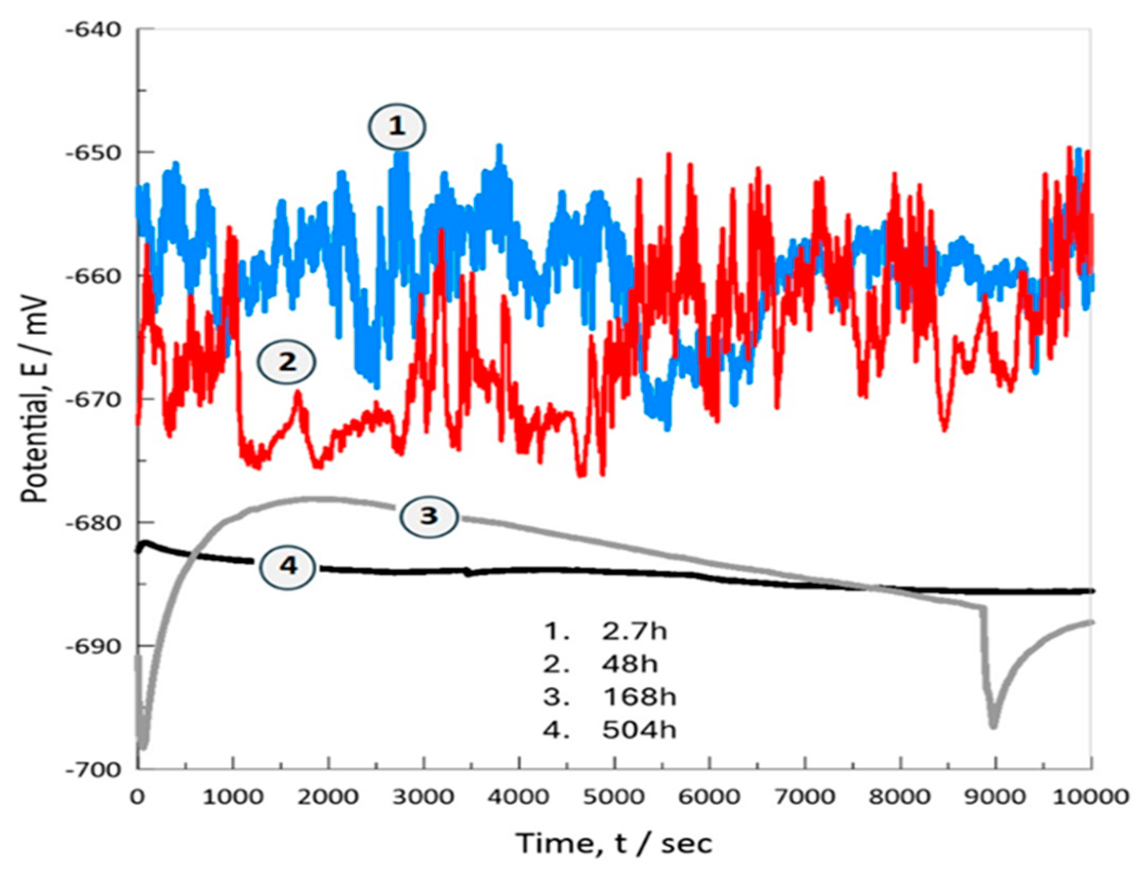
Figure 12.
Potentiodynamic polarization curves in the anodic branch.
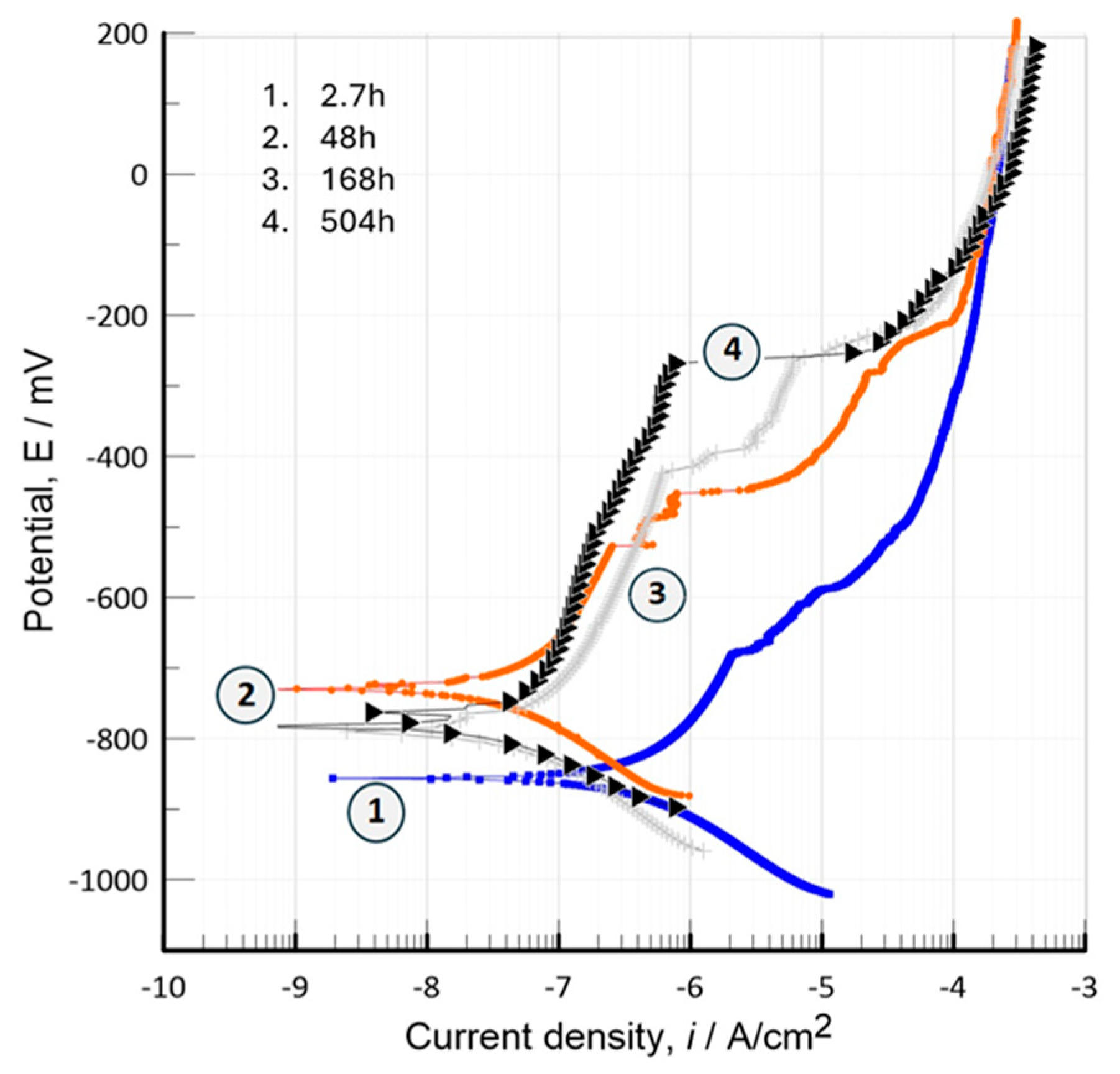
Figure 13.
EIS response in Nyquist form of a Bio-ceramic HPa/Ti1% sample during exposure in 0.9% NaCl solution at different periods of time, 5, 48, 168 y 504 h.
Figure 13.
EIS response in Nyquist form of a Bio-ceramic HPa/Ti1% sample during exposure in 0.9% NaCl solution at different periods of time, 5, 48, 168 y 504 h.
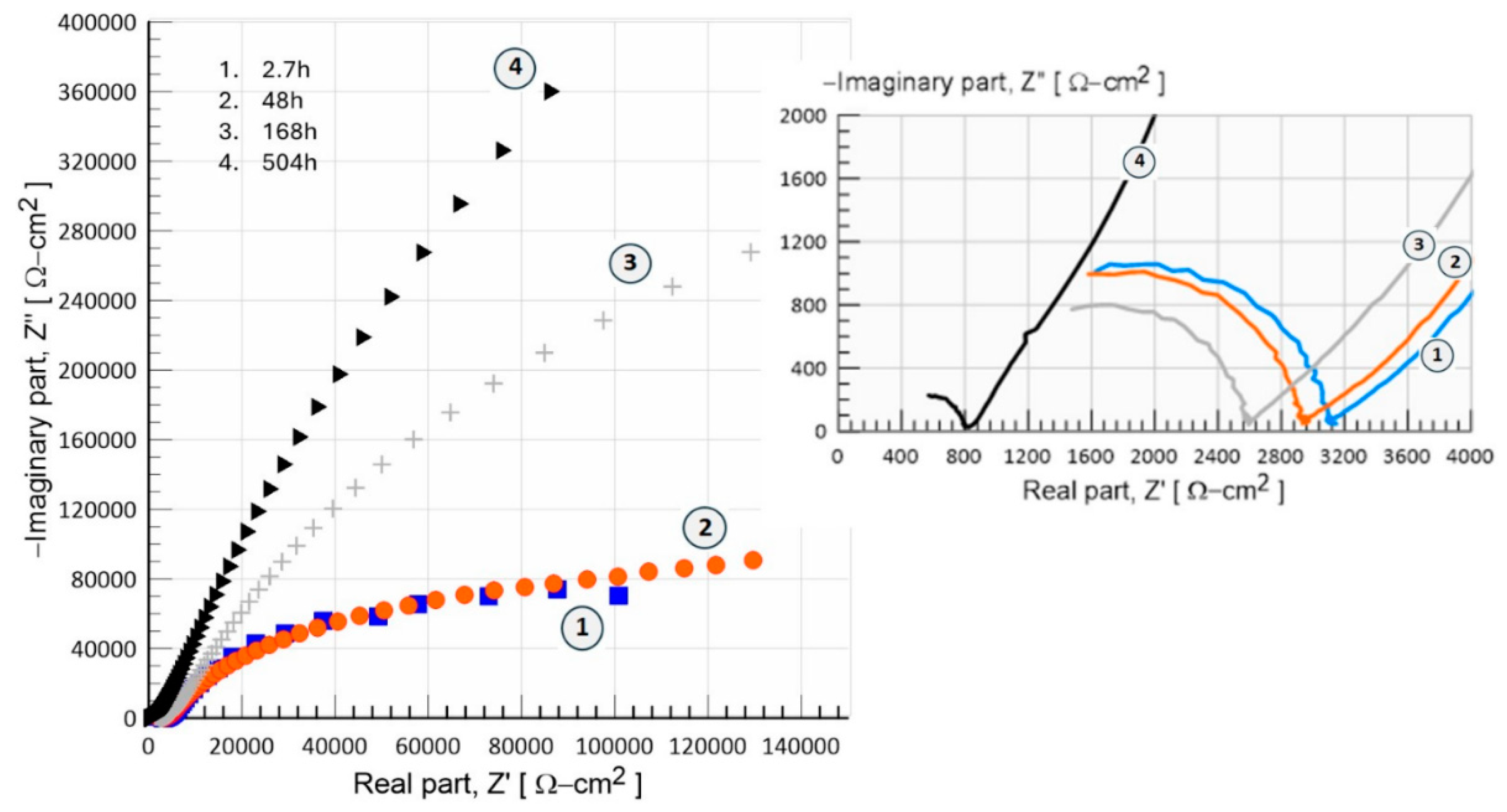
Figure 14.
EIS response in Bode form of a Bio-ceramic HPa/Ti1% sample during exposure in 0.9% NaCl solution at different periods of time, 5, 48, 168 y 504 h.
Figure 14.
EIS response in Bode form of a Bio-ceramic HPa/Ti1% sample during exposure in 0.9% NaCl solution at different periods of time, 5, 48, 168 y 504 h.
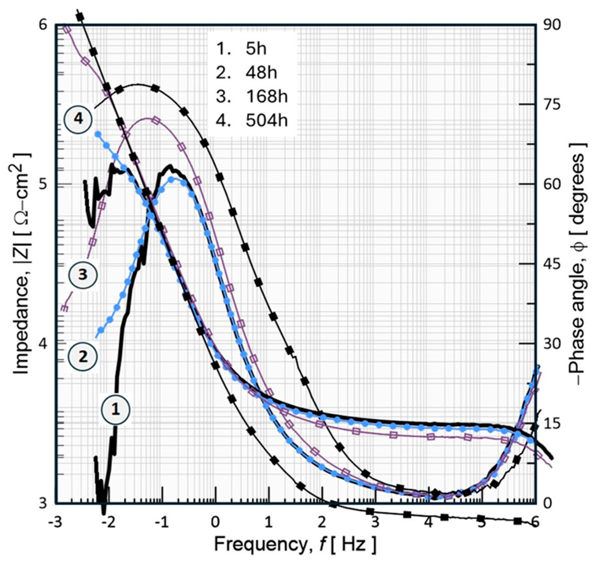
Figure 15.
Equivalent electrical circuit (EEC) used to model the impedance behavior of bioceramic material exposed for 504h in 0.9% NaCl. Rs- Solution resistance, Cc- coating capacitance, Rct-charge transfer resistance, and Q element phase constant.
Figure 15.
Equivalent electrical circuit (EEC) used to model the impedance behavior of bioceramic material exposed for 504h in 0.9% NaCl. Rs- Solution resistance, Cc- coating capacitance, Rct-charge transfer resistance, and Q element phase constant.
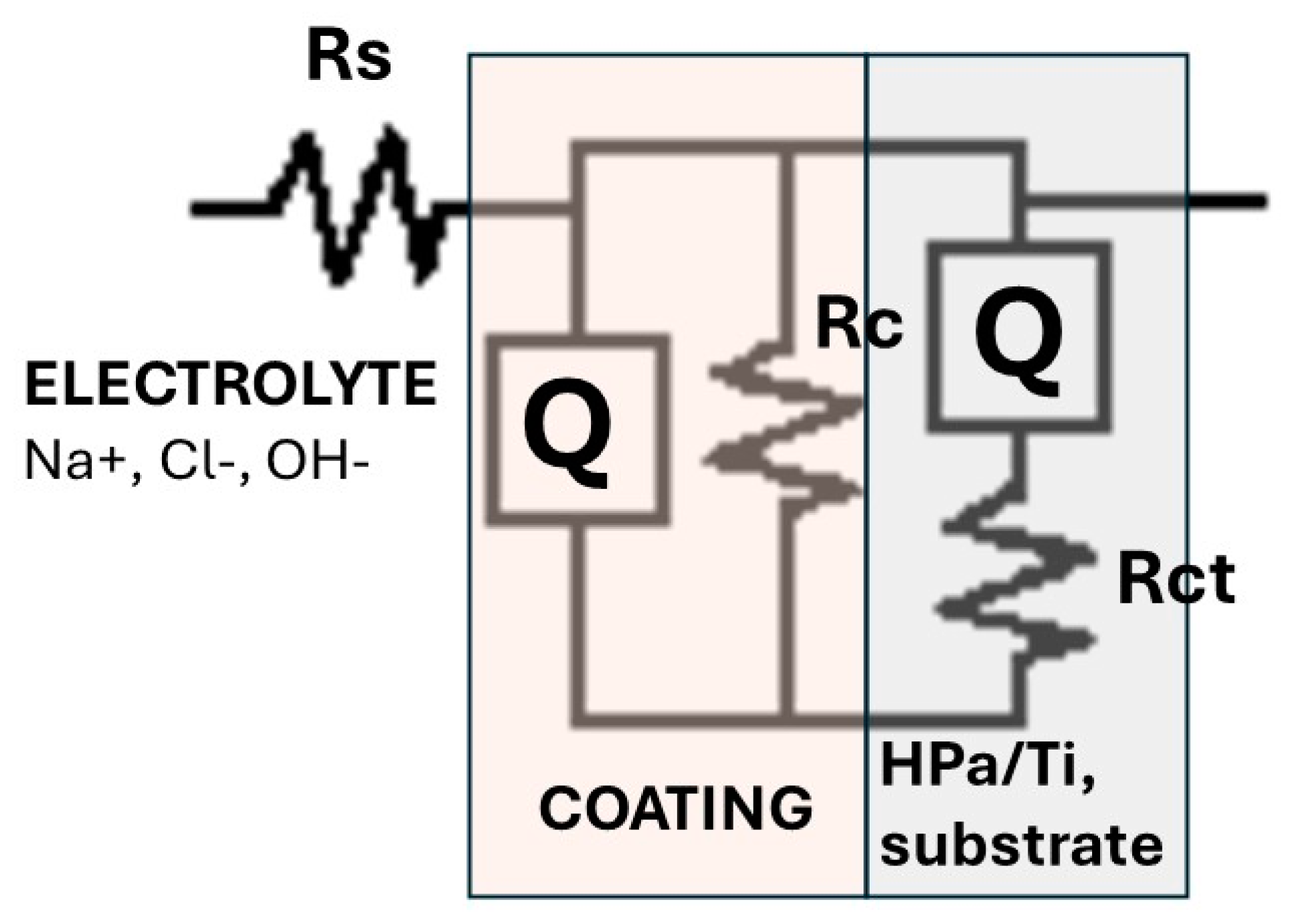

Table 1.
Electrochemical parameters obtained during the fitting procedure of EIS data with the equivalent electrical circuit ECC, proposed in Figure 15.
Table 1.
Electrochemical parameters obtained during the fitting procedure of EIS data with the equivalent electrical circuit ECC, proposed in Figure 15.
| Inmersión Time, [h] | Capacitance-Cc, [nF/cm2] | Coating Resistance, Rc [Ω-cm2] |
Charge Transfer Resistance, Rct [Ω-cm2] |
Ecorr / [mV] | Icorr [nA/cm2] |
|---|---|---|---|---|---|
| 5h | 5.27 | 3447 | 2.745x10⁵ | −856.32 | 2.40 |
| 48h | 5.11 | 3254 | 2.939 x10⁵ | −730.27 | 0.77 |
| 168 | 3.80 | 2963 | 3.15x10⁵ | −789.46 | 3.06 |
| 504 | 0.18 | 810 | 1.526x10⁶ | −782.71 | 0.43 |
Disclaimer/Publisher’s Note: The statements, opinions and data contained in all publications are solely those of the individual author(s) and contributor(s) and not of MDPI and/or the editor(s). MDPI and/or the editor(s) disclaim responsibility for any injury to people or property resulting from any ideas, methods, instructions or products referred to in the content. |
© 2024 by the authors. Licensee MDPI, Basel, Switzerland. This article is an open access article distributed under the terms and conditions of the Creative Commons Attribution (CC BY) license (http://creativecommons.org/licenses/by/4.0/).
Copyright: This open access article is published under a Creative Commons CC BY 4.0 license, which permit the free download, distribution, and reuse, provided that the author and preprint are cited in any reuse.


