Submitted:
25 October 2024
Posted:
29 October 2024
You are already at the latest version
Abstract
Background/Objectives: To develop a novel method for isolating proteins associated with Long Non-coding RNAs (LncRNAs) using high-throughput Liquid Chromatography Mass Spectrometry (LC-MS) and proteomics approaches for identifying cancer-associated proteins involved in post-translational modifications (PTMs) such as glycosylation, phosphorylation, citrullination, methylation, and acetylation. Additionally, the focus is on identifying and quantifying differentially modulated or regulated proteins associated with LncRNAs to discover novel molecular signatures or biomarkers.
Methods: Identification, quantification, and characterization of LncRNAs associated pro-teins by using Label free Quantification (LFQ) of proteins by Nono LC-Mass Spectrometry (LC-MS)., use Employ bioinformatics, biostatistics analysis for find out the differentially expressed, exclusive protein or modulated., find outInvestigate the mechanisms and pathways involving these unique or specific proteins analysis of unique/specific proteins and Validation of these unique/specific proteins by Immunohistochemistry (IHC) and western blotting/ enzyme-linked immunosorbent assay (ELISA) for to achieve better out-come.
Conclusions: Identified and validated novel proteins associated with LncRNAs could be early detective molecular biomarkers of cancer and it could be useful in clinical practice for diagnosis, prognosis of liver, colorectal and other several types of cancer types.
Keywords:
Long non-coding RNAs (LncRNAs)
; Protein and Pathways analysis
; Liver and Colorectal Cancer
; Liquid Chromatography Mass Spectrometry (LC-MS)
; Novel molecular signature/Biomarkers
; Diagnosis and Prognosis
1. Introduction
Long non-coding RNAs (LncRNAs) represent a diverse and functionally significant class of RNA molecules that do not encode proteins but play critical roles in regulating gene expression and cellular functions [1]. The isolation, identification and quantification of proteins which are associated with lncRNAs are difficult to understand [2,3]. Unlike proteins, which can often be studied using antibody-based techniques, lncRNAs require different investigative methods due to their distinct characteristics and interactions. LncRNAs can act as scaffolds to bring together various molecular components to form functional complexes essential for chromatin remodeling, transcription regulation, and RNA processing [4]. Their unique regulatory roles and involvement in diverse biological processes highlight the need for specialized techniques to study these molecules comprehensively, which begins with their identification and characterization. High-throughput sequencing technologies, such as RNA-seq, have been instrumental in identifying lncRNAs across different tissues and disease conditions, providing a comprehensive view of the transcriptome and revealing novel lncRNAs along with their expression patterns [5]. Bioinformatics tools predict the secondary structure of lncRNAs and identify potential binding sites for proteins, RNA, and DNA, while experimental techniques like RNA immunoprecipitation and crosslinking immunoprecipitation validate these interactions in vivo [6]. One such innovative approach involves pulling out lncRNAs using complementary oligonucleotide probes, which act as primers. These probes hybridize specifically the lncRNA of interest, allowing for its selective enrichment from a complex RNA mixture [7]. Once isolated, the lncRNA can be tagged with biotin, facilitating subsequent purification steps and enhancing the detection and analysis of the lncRNA and its associated proteins or other interacting molecules [8]. A second primer, complementary to a different region of the lncRNA, can further amplify or label the RNA, allowing for a highly specific and efficient way to study lncRNA interactions within the cell [9].These advanced techniques are particularly valuable in cancer research. Dysregulated lncRNAs are involved in regulating genes that control cell proliferation, apoptosis, metastasis, and help in identifying proteins that are hallmarks of cancer, such as proteins involved in oncogenic signaling pathways or tumor suppressor functions. One such example is lncRNA MALAT1, which interacts with proteins that promote metastasis in lung cancer [10]. Understanding these interactions is very crucial for developing these targeted therapies. By isolating and studying lncRNAs i.e., HOTAIR, researchers can identify chromatin-modifying complexes involved in cancer and design drugs that can disrupt these interactions to inhibit cancer-promoting effects [11]. The therapeutic potential of lncRNAs extends beyond cancer. LncRNAs are involved in various diseases, including cardiovascular diseases, neurological disorders, and metabolic diseases. For example, the lncRNA ANRIL is implicated in atherosclerosis and cardiovascular risk, while NEAT1 is associated with neurodegenerative diseases [1,12]. Targeting these specific lncRNAs could provide a novel therapeutic strategy for these various conditions. Moreover, the specificity and distinct expression patterns of lncRNAs make them attractive candidates for non-invasive cancer biomarkers. For example, the lncRNA PCA3, specific to prostate cancer, can be detected in urine, providing a non-invasive diagnostic tool [13,14,15].High-throughput sequencing technologies and bioinformatics tools have advanced our understanding of lncRNAs' regulatory roles and interactions. Techniques like RIP, CLIP, and CRISPR/Cas9-mediated genome editing have enabled researchers to map the interactome of lncRNAs and study their molecular functions comprehensively [16]. These methods have revealed the essential roles of lncRNAs in development, differentiation, and various diseases.
In short, lncRNAs are critical regulators of gene expression and cellular functions, with unique roles distinguishing them from protein-coding genes. Advanced techniques for studying lncRNAs, such as oligonucleotide probes, biotin tagging, RIP, CLIP, and CRISPR/Cas9-mediated genome editing, have greatly enhanced our understanding of their biology. These techniques are particularly valuable in cancer research, where lncRNAs play crucial roles in oncogenesis and tumor progression. Identifying lncRNA-associated proteins and understanding their interactions can reveal novel therapeutic targets and biomarkers, which can help pave the way for innovative treatments for cancer and other diseases. In this article, we describe a new method for the isolation, identification, quantification, and characterization of lncRNA-associated proteins using high-throughput nano liquid chromatography-mass spectrometry (LC-MS). Additionally, we validate the identified differentially expressed or modulated proteins that could be exclusively associated with lncRNAs using commercially available antibodies through western blotting and immunohistochemistry (IHC)/enzyme-linked immunosorbent assay (ELISA). This approach aims to uncover novel molecular signatures that could be useful in the diagnosis and prognosis of liver, colorectal, and other associated liver cancers/diseases, such as chronic liver disease (CLD), acute-on-chronic liver failure (ACLF), nonalcoholic fatty liver disease (NAFLD), severe alcoholic hepatitis (SAH), and other human diseases [3,13,15,17].
2. Method:
2.1. Isolation and Identification of LncRNAs Associated Proteins
The identification of LncRNAs associated proteins is crucial for the understanding of molecular mechanisms of their function and regulation[2,18]. The RNA Antisense Purification with Mass Spectrometry (RAP-MS) technique revolutionizes our ability to pinpoint direct RNA-protein interactions within living cells[19]. This method provides a detailed roadmap for isolating proteins directly associated with lncRNAs[19]. Whether utilizing whole-cell lysate or nuclear extract, the process culminates in a comprehensive roster of proteins intricately engaged with the target RNA within the dynamic milieu of cellular environments. Central to this method is the strategic design of 90-nucleotide oligos, engineered to cover the entire sequence of the lncRNA, with careful consideration given to avoid off-target effects through LC-MS analysis of the data. The incorporation of a 5’ biotin standard modification facilitates the efficient pull-down of lncRNA-associated proteins. Other studies, such as those focusing on isolating protein complexes associated with lncRNAs from HeLa cells, underscore the versatility and applicability of this approach[20].
Furthermore, advancements such as the HyPR-MS method [21] have enhanced the efficiency and scope of RNA-protein interactome analysis. By leveraging predictive RNA secondary structure modeling through M-fold software, HyPR-MS streamlines capture oligonucleotide design, optimizing experimental outcomes while minimizing design time [21,22]. Expanding the horizon of RNA interrogation, Engreitz et al. introduced RNA antisense purification to map RNA-RNA interactions (RAP-RNA), providing systematic means to enrich specific RNAs and identify interacting molecules [23]. This technique, exemplified in the study of xist lncRNA localization during X-chromosome inactivation [24], offers invaluable insights into lncRNA-mediated chromatin regulation[24]. Complementary methods such as Chromatin Isolation by RNA Purification (ChIRP) and Capture Hybridization Analysis of RNA Targets (CHART) provide high- throughput avenues for elucidating RNA-bound proteins and genomic binding sites of specific lncRNAs [25]. A pool of short complementary DNA oligonucleotide probes inspired by RNA FISH, CHART adapts an RNase H mapping assay, offering nuanced approaches tailored to different experimental contexts. Additionally, Reversible Cross-Linked Immune Precipitation (ReCLIP) emerges as a powerful tool [26,27], preserving loose protein associations intact to identify lncRNA-associated proteins. By leveraging cell-permeable, thiol-cleavable crosslinkers and in-cell crosslinking, ReCLIP captures endogenous protein-protein interactions with remarkable fidelity, offering a glimpse into the dynamic landscape of RNA-protein interactions within living cells[25,27]. Interaction with protein complexes is a common mechanism for the functions of lncRNAs. Thus, identifying proteins associated with lncRNAs is critical for the understanding of molecular mechanisms and functions of lncRNAs. Immunoprecipitation is commonly used for the isolation of protein complexes associated with a protein of interest. However, this method is not available for lncRNAs because antibodies generally do not recognize RNAs.[28,29] [30] Proteins that have higher MS counts in cells transfected with lncRNA-MS2 than controlled MS2 plasmid are selected as potential candidates. Knockdown of these protein candidates with short hairpin RNAs (shRNAs) is used to confirm whether any of the candidates are required for the functions of lncRNA of interest. Further validation of the binding of protein candidates to lncRNA of interest is required to confirm their association [2,3].
2.2. Identified Pathways Associated with lncRNAs:
LncRNAs associated proteins are emerging as significant players in the diagnosis and therapy of various cancers [27]. The extensive discovery and reporting of lncRNA-associated proteins highlight their diverse expression patterns and tumor-specificity across different cancer types [22,24,29]. Table 1 lists several key lncRNA-associated proteins such as MALAT1, UCA1, HULC, HOTTIP, CCAT1, CCAT2, and H19, detailing various methods used for their isolation and identification, which serve as biomarkers for treating liver and colorectal cancer patients [31,32,33,34]. These proteins also facilitate further mechanistic and pathway studies of lncRNAs, enhancing our understanding of cancer pathophysiology.
A study by [52] focused on Metastasis Associated Lung Adenocarcinoma Transcript 1 (MALAT-1) and its potential role as a diagnostic or prognostic biomarker for ovarian cancer. MALAT-1 expression was found to be significantly upregulated in ovarian cancer tissues compared to normal tissues. The findings suggest that MALAT-1 could serve as a therapeutic target due to its involvement in cancer progression, highlighting its importance in tumor growth and metastasis [29]. Explored Urothelial Cancer Associated 1 (UCA1) in gastric cancer, demonstrating that UCA1 interacts with miR-145 and MYO6, forming a regulatory axis that affects cancer cell proliferation and apoptosis, suggesting new therapeutic approaches targeting regulatory axis[53]. Examined the impact of metformin on cancer risk in type 2 diabetes patients, suggesting that metformin might reduce cancer risk, and highlighting its potential as a chemo-preventive agent and utilized techniques such as RAPID-SELEX and RNA compete to map lncRNA-protein interactions, providing insights into lncRNA cellular mechanisms and their disease-related perturbations, and further used MS2 trapping and SILAC-based display to identify lncRNA-bound proteomes, aiding in understanding lncRNA functions in cellular networks[54]. Another study focused on HULC, demonstrating its overexpression in various cancers and promoting tumor growth, suggesting its potential as a biomarker and therapeutic target[55]. Showed that HULC interacts with LDHA, promoting glycolysis in cancer cells, highlighting HULC's role in cancer metabolism[56]. Identified nearly 8000 cancer-specific lncRNAs, including PCA3 for prostate cancer, emphasizing their diagnostic and prognostic potential. A study suggested that M1 exosomes and HOTTIP polarize monocytes into an antitumor phenotype, suggesting a novel approach for immunotherapy[42] demonstrated that HOTAIR mediates gene silencing and enhances tumor progression, suggesting its use alongside existing therapies to sensitize tumors[43] and Another study showed that CCAT1 regulates miR-490-3p in ovarian cancer, indicating new therapeutic strategies [57]and the explored CCAT2's co-expressed genes, suggesting targeting CCAT2 pathways for cancer therapy. That found H19 enhances cancer cell proliferation and glycolysis by downregulating miRNA-519d-3p and upregulating LDHA, highlighting its potential as a therapeutic target[58] and its demonstrated that CCAT1-L inhibits EMT in gastric adenocarcinoma cells, suggesting its potential to prevent cancer metastasis. The identified CRNDE as a marker and therapeutic target against chemoresistance in gastric cancer [59], showed that FER1L4 regulates neural stem cell proliferation and differentiation, suggesting its therapeutic potential in neurodevelopmental disorders. Finally, it was demonstrated that PTENP inhibits cell proliferation and EMT while inducing apoptosis in cervical cancer cells, highlighting its potential as a therapeutic target as well[49]. These brief explanations of various studies are examples of LncRNAs associated proteins, and their further use in liver, colorectal and others several different types of cancer.
3. Discussion:
Liver and colorectal cancer are the third most common causes of cancer-related deaths. These cancers are highly aggressive and resistant to treatment. In liver cancer, including hepatocellular carcinoma (HCC), which accounts for 75% to 85% of cases, and intrahepatic cholangiocarcinoma, which makes up 10% to 15% of cases, shows the highest mortality rates in Asia and Africa. However, incidence and mortality rates are also increasing in Europe and the US [17,28,60]. Chronic liver disease (CLD), Acute-on-chronic liver failure (ACLF), Nonalcoholic fatty liver disease (NAFLD), severe alcoholic hepatitis (SAH), Drug and alcohol-induced liver injury and other human diseases promote liver and colorectal inflammation and increase oxidative stress, which accelerates oxidative cell death and promotes HCC. The HCC shares common altered metabolic pathways with liver cirrhosis, CLD, NAFLD and SAH suggesting the involvement of tumorigenesis promotion in dysregulated lipidaemia [34,38]. The identified lncRNA-associated proteins are involved in molecular pathways related to cancer pathogenesis and are directly implicated in liver inflammation and tumorigenesis, though their specific roles are not yet fully understood. But these could be involved in the post-transcriptional regulation of gene expression processes, molecules finding or signatures widely proposed as candidate biomarkers for diagnosis and prognosis of cancer including liver and colorectal cancer [61,62,63]. In this study, we highlight some of the emerging key players among lncRNA-associated proteins involved in liver and colorectal cancer, as well as other types of cancer. These proteins play specific roles in diagnosis and prognosis, potentially aiding in outcome prediction and treatment of cancer and we specially focus on HCC and CRC patients. [18,29].
4. Conclusions and Future Perspectives
We present this protocol for the isolation, identification, and characterization of LncRNAs associated proteins via LC-MS analysis, as there is no fixed method to their isolation and identification. This protocol includes the use of specific instrumentation software for LC-MS data analysis to identify novel molecular signatures or unique proteins related to liver and colorectal cancer, which could play a significant role in disease progression with clinical correlations [64].
This process is shown in Figure 1, Figure 2 and Figure 3, which illustrate the validation of lncRNA-associated proteins. Validation is crucial for confirming the relevance of identified molecules and their correlation with disease pathways, aiding in outcome prediction for various type cancers mainly liver and colorectal cancer. This information is vital for understanding the relationships between single genes and multiple proteins, single proteins and many amino acids, and how all these elements correlate within polypeptide chains [17,65].
The future of Reversible Cross-Linked Immune Precipitation (ReCLIP) in LncRNA research is relevant to significantly advance our understanding of RNA-protein interactions. Integrating ReCLIP with high-throughput LC-MS analysis will enable comprehensive mapping of the lncRNA-protein interactome, uncovering novel protein partners and their binding dynamics. This can help in the development of targeted interventions, such as small molecules or CRISPR-based therapies, to disrupt critical lncRNA-protein interactions involved in cancer and other diseases. ReCLIP's application in neurological, metabolic, cardiovascular, and immune-related disorders could lead to exclusive novel biomarkers and therapeutic targets.
Author Contributions
MSH initiated the writing of the manuscript, MKT given the idea about LncRNAs associated proteins. MSH made all figures and table, and MKT reviewed for improvement. PV help in written, drafted the manuscript with MSH and MKT. This manuscript has been seen and approved by all authors. Disclosure: All authors have declared no conflict of interest.
Funding
The work was supported by the NIH/NIGMS R16GM146696, ST-CECR-Project 2 (CPRIT RP230419), ARG-NTF-22-972518 Alzheimer's Association, and UTRGV SOM Startup funds to M.K.T.
Institutional Review Board Statement
NA, since this is reviewing article.
Informed Consent Statement
NA.
Acknowledgments
The work was supported by the NIH/NIGMS R16GM146696, ST-CECR-Project 2 (CPRIT RP230419), AARG-NTF Alzheimer's Association, and UTRGV SOM Startup funds. We thank our lab colleagues and Medicine and Oncology ISU staff for their support. BioRender for the images.
Conflicts of Interest
The authors declare no conflict of interest.
References
- Balasundaram, P.; Anilkumar, A.C. Myoclonic Epilepsy of Infancy. In StatPearls; StatPearls Publishing Copyright © 2024, StatPearls Publishing LLC.: Treasure Island, FL, USA, 2024. [Google Scholar]
- Li, M.; Xue, Y.; He, R.; Huang, Q. Isolation of Protein Complexes Associated with Long Non-coding RNAs. Methods Mol Biol 2021, 2372, 27–33. [Google Scholar] [CrossRef] [PubMed]
- Jiang, J.; Zhang, T.; Pan, Y.; Hu, Z.; Yuan, J.; Hu, X.; Zhang, L.; Zhang, Y. Characterization of Long Non-coding RNA Associated Proteins by RNA-Immunoprecipitation. Methods Mol Biol 2021, 2372, 19–26. [Google Scholar] [CrossRef] [PubMed]
- Rinn, J.L.; Chang, H.Y. Genome regulation by long noncoding RNAs. Annu Rev Biochem 2012, 81, 145–166. [Google Scholar] [CrossRef] [PubMed]
- Martinez-Nunez, R.T.; Sanford, J.R. Studying Isoform-Specific mRNA Recruitment to Polyribosomes with Frac-seq. Methods Mol Biol 2016, 1358, 99–108. [Google Scholar] [CrossRef]
- Roger, G.; Ducrocq, G.; Mesnier, J.; Sayah, N.; Abtan, J.; Ferrari, R.; Ford, I.; Fox, K.M.; Tardif, J.C.; Tendera, M.; et al. Chronic coronary syndromes without standard modifiable cardiovascular risk factors and outcomes: the CLARIFY registry. Eur Heart J 2024, 45, 2396–2406. [Google Scholar] [CrossRef]
- Qiu, B.; Simon, M.C. BODIPY 493/503 Staining of Neutral Lipid Droplets for Microscopy and Quantification by Flow Cytometry. Bio Protoc 2016, 6. [Google Scholar] [CrossRef]
- Lefaucheur, J.P.; Aleman, A.; Baeken, C.; Benninger, D.H.; Brunelin, J.; Di Lazzaro, V.; Filipović, S.R.; Grefkes, C.; Hasan, A.; Hummel, F.C.; et al. Evidence-based guidelines on the therapeutic use of repetitive transcranial magnetic stimulation (rTMS): An update (2014-2018). Clin Neurophysiol 2020, 131, 474–528. [Google Scholar] [CrossRef]
- Tran, T.H.; Hunger, S.P. The genomic landscape of pediatric acute lymphoblastic leukemia and precision medicine opportunities. Semin Cancer Biol 2022, 84, 144–152. [Google Scholar] [CrossRef]
- Genovese, G.; Carugo, A.; Tepper, J.; Robinson, F.S.; Li, L.; Svelto, M.; Nezi, L.; Corti, D.; Minelli, R.; Pettazzoni, P.; et al. Synthetic vulnerabilities of mesenchymal subpopulations in pancreatic cancer. Nature 2017, 542, 362–366. [Google Scholar] [CrossRef]
- Breit, S.N.; Brown, D.A.; Tsai, V.W.W. GDF15 analogs as obesity therapeutics. Cell Metab 2023, 35, 227–228. [Google Scholar] [CrossRef]
- Imamura, S.; Morioka, T.; Yamazaki, Y.; Numaguchi, R.; Urata, H.; Motoyama, K.; Mori, K.; Fukumoto, S.; Shoji, T.; Emoto, M.; et al. Response to comment on Imamura et al. Plasma polyunsaturated fatty acid profile and delta-5 desaturase activity are altered in patients with type 2 diabetes. Metabolism 2014;63(11):1432-8. Metabolism 2015, 64, e3–e4. [Google Scholar] [CrossRef] [PubMed]
- Talamantes, S.; Lisjak, M.; Gilglioni, E.H.; Llamoza-Torres, C.J.; Ramos-Molina, B.; Gurzov, E.N. Non-alcoholic fatty liver disease and diabetes mellitus as growing aetiologies of hepatocellular carcinoma. JHEP Rep 2023, 5, 100811. [Google Scholar] [CrossRef] [PubMed]
- Characterization of Long Non-coding RNA Associated Proteins by RNA-Immunoprecipitation Junjie Jiang.
- Guo, Y.; Zhao, J.; Bi, J.; Wu, Q.; Wang, X.; Lai, Q. Heterogeneous nuclear ribonucleoprotein K (hnRNP K) is a tissue biomarker for detection of early hepatocellular carcinoma in patients with cirrhosis. J Hematol Oncol 2012, 5, 37. [Google Scholar] [CrossRef] [PubMed]
- Gutschner, T.; Haemmerle, M.; Genovese, G.; Draetta, G.F.; Chin, L. Post-translational Regulation of Cas9 during G1 Enhances Homology-Directed Repair. Cell Rep 2016, 14, 1555–1566. [Google Scholar] [CrossRef] [PubMed]
- Isolation of Protein Complexes Associated with Long Non-coding RNAs.
- Galamb, O.; Barták, B.K.; Kalmár, A.; Nagy, Z.B.; Szigeti, K.A.; Tulassay, Z.; Igaz, P.; Molnár, B. Diagnostic and prognostic potential of tissue and circulating long non-coding RNAs in colorectal tumors. World J Gastroenterol 2019, 25, 5026–5048. [Google Scholar] [CrossRef]
- McHugh, C.A.; Chen, C.K.; Chow, A.; Surka, C.F.; Tran, C.; McDonel, P.; Pandya-Jones, A.; Blanco, M.; Burghard, C.; Moradian, A.; et al. The Xist lncRNA interacts directly with SHARP to silence transcription through HDAC3. Nature 2015, 521, 232–236. [Google Scholar] [CrossRef]
- Rodriguez-Frandsen, A.; Martin-Sancho, L.; Gounder, A.P.; Chang, M.W.; Liu, W.C.; De Jesus, P.D.; von Recum-Knepper, J.; Dutra, M.S.; Huffmaster, N.J.; Chavarria, M.; et al. Viral Determinants in H5N1 Influenza A Virus Enable Productive Infection of HeLa Cells. J Virol 2020, 94. [Google Scholar] [CrossRef]
- Spiniello, M.; Knoener, R.A.; Steinbrink, M.I.; Yang, B.; Cesnik, A.J.; Buxton, K.E.; Scalf, M.; Jarrard, D.F.; Smith, L.M. HyPR-MS for Multiplexed Discovery of MALAT1, NEAT1, and NORAD lncRNA Protein Interactomes. J Proteome Res 2018, 17, 3022–3038. [Google Scholar] [CrossRef]
- Razzaq, S.; Rauf, A.; Raza, A.; Akhtar, S.; Tabish, T.A.; Sandhu, M.A.; Zaman, M.; Ibrahim, I.M.; Shahnaz, G.; Rahdar, A.; et al. A Multifunctional Polymeric Micelle for Targeted Delivery of Paclitaxel by the Inhibition of the P-Glycoprotein Transporters. Nanomaterials (Basel) 2021, 11. [Google Scholar] [CrossRef]
- Engreitz, J.M.; Sirokman, K.; McDonel, P.; Shishkin, A.A.; Surka, C.; Russell, P.; Grossman, S.R.; Chow, A.Y.; Guttman, M.; Lander, E.S. RNA-RNA interactions enable specific targeting of noncoding RNAs to nascent Pre-mRNAs and chromatin sites. Cell 2014, 159, 188–199. [Google Scholar] [CrossRef]
- Engreitz, J.M.; Pandya-Jones, A.; McDonel, P.; Shishkin, A.; Sirokman, K.; Surka, C.; Kadri, S.; Xing, J.; Goren, A.; Lander, E.S.; et al. The Xist lncRNA exploits three-dimensional genome architecture to spread across the X chromosome. Science 2013, 341, 1237973. [Google Scholar] [CrossRef] [PubMed]
- Torres, M.; Becquet, D.; Guillen, S.; Boyer, B.; Moreno, M.; Blanchard, M.P.; Franc, J.L.; François-Bellan, A.M. RNA Pull-down Procedure to Identify RNA Targets of a Long Non-coding RNA. J Vis Exp 2018. [Google Scholar] [CrossRef]
- Smith, A.L.; Friedman, D.B.; Yu, H.; Carnahan, R.H.; Reynolds, A.B. ReCLIP (reversible cross-link immuno-precipitation): an efficient method for interrogation of labile protein complexes. PLoS One 2011, 6, e16206. [Google Scholar] [CrossRef] [PubMed]
- Smith, A.L.; Dohn, M.R.; Brown, M.V.; Reynolds, A.B. Association of Rho-associated protein kinase 1 with E-cadherin complexes is mediated by p120-catenin. Mol Biol Cell 2012, 23, 99–110. [Google Scholar] [CrossRef]
- Li, Y.; Wang, H.; Zhan, L.; Li, Q.; Li, Y.; Wu, G.; Wei, H.; Dong, X. LncRNA FER1L4 promotes differentiation and inhibits proliferation of NSCs via miR-874-3p/Ascl2. Am J Transl Res 2022, 14, 2256–2266. [Google Scholar]
- Rodriguez, P.D.; Paculova, H.; Kogut, S.; Heath, J.; Schjerven, H.; Frietze, S. Non-Coding RNA Signatures of B-Cell Acute Lymphoblastic Leukemia. Int J Mol Sci 2021, 22. [Google Scholar] [CrossRef]
- Bothos, E.; Hatzis, P.; Moulos, P. Interactive Analysis, Exploration, and Visualization of RNA-Seq Data with SeqCVIBE. Methods Protoc 2022, 5. [Google Scholar] [CrossRef]
- Liu, S.S.; Li, J.S.; Xue, M.; Wu, W.J.; Li, X.; Chen, W. LncRNA UCA1 Participates in De Novo Synthesis of Guanine Nucleotides in Bladder Cancer by Recruiting TWIST1 to Increase IMPDH1/2. Int J Biol Sci 2023, 19, 2599–2612. [Google Scholar] [CrossRef]
- Fu, S.; Wang, Y.; Li, H.; Chen, L.; Liu, Q. Regulatory Networks of LncRNA MALAT-1 in Cancer. Cancer Manag Res 2020, 12, 10181–10198. [Google Scholar] [CrossRef]
- Bhan, A.; Soleimani, M.; Mandal, S.S. Long Noncoding RNA and Cancer: A New Paradigm. Cancer Res 2017, 77, 3965–3981. [Google Scholar] [CrossRef]
- Wang, C.; Li, Y.; Yan, S.; Wang, H.; Shao, X.; Xiao, M.; Yang, B.; Qin, G.; Kong, R.; Chen, R.; et al. Interactome analysis reveals that lncRNA HULC promotes aerobic glycolysis through LDHA and PKM2. Nat Commun 2020, 11, 3162. [Google Scholar] [CrossRef] [PubMed]
- Pei, C.; Gong, X.; Zhang, Y. LncRNA MALAT-1 promotes growth and metastasis of epithelial ovarian cancer via sponging microrna-22. Am J Transl Res 2020, 12, 6977–6987. [Google Scholar] [PubMed]
- Song, W.; Wang, K.; Zou, S.B. UCA1 lncRNA in metastases and prognosis. Panminerva Med 2017, 59, 278–279. [Google Scholar] [CrossRef] [PubMed]
- Ferrè, F.; Colantoni, A.; Helmer-Citterich, M. Revealing protein-lncRNA interaction. Brief Bioinform 2016, 17, 106–116. [Google Scholar] [CrossRef] [PubMed]
- Rastad, H.; Samimisedeh, P.; Alan, M.S.; Afshar, E.J.; Ghalami, J.; Hashemnejad, M.; Alan, M.S. The role of lncRNA CERS6-AS1 in cancer and its molecular mechanisms: A systematic review and meta-analysis. Pathol Res Pract 2023, 241, 154245. [Google Scholar] [CrossRef]
- Yu, X.; Zheng, H.; Chan, M.T.; Wu, W.K. HULC: an oncogenic long non-coding RNA in human cancer. J Cell Mol Med 2017, 21, 410–417. [Google Scholar] [CrossRef]
- Ou, C.; Sun, Z.; He, X.; Li, X.; Fan, S.; Zheng, X.; Peng, Q.; Li, G.; Li, X.; Ma, J. Targeting YAP1/LINC00152/FSCN1 Signaling Axis Prevents the Progression of Colorectal Cancer. Adv Sci (Weinh) 2020, 7, 1901380. [Google Scholar] [CrossRef]
- Sun, J.Y.; Ni, M.M. Long non-coding RNA HEIH: a novel tumor activator in multiple cancers. Cancer Cell Int 2021, 21, 558. [Google Scholar] [CrossRef]
- Jiang, H.; Zhou, L.; Shen, N.; Ning, X.; Wu, D.; Jiang, K.; Huang, X. M1 macrophage-derived exosomes and their key molecule lncRNA HOTTIP suppress head and neck squamous cell carcinoma progression by upregulating the TLR5/NF-κB pathway. Cell Death Dis 2022, 13, 183. [Google Scholar] [CrossRef]
- Rajagopal, T.; Talluri, S.; Akshaya, R.L.; Dunna, N.R. HOTAIR LncRNA: A novel oncogenic propellant in human cancer. Clin Chim Acta 2020, 503, 1–18. [Google Scholar] [CrossRef]
- Mu, Y.; Li, N.; Cui, Y.L. The lncRNA CCAT1 upregulates TGFβR1 via sponging miR-490-3p to promote TGFβ1-induced EMT of ovarian cancer cells. Cancer Cell Int 2018, 18, 145. [Google Scholar] [CrossRef] [PubMed]
- Gao, P.; Sun, D.; Guo, H.; Wu, Z.; Chen, J. LncRNA CCAT2 promotes proliferation and suppresses apoptosis of colorectal cancer cells. J buon 2020, 25, 1840–1846. [Google Scholar] [PubMed]
- Hashemi, M.; Moosavi, M.S.; Abed, H.M.; Dehghani, M.; Aalipour, M.; Heydari, E.A.; Behroozaghdam, M.; Entezari, M.; Salimimoghadam, S.; Gunduz, E.S.; et al. Long non-coding RNA (lncRNA) H19 in human cancer: From proliferation and metastasis to therapy. Pharmacol Res 2022, 184, 106418. [Google Scholar] [CrossRef] [PubMed]
- Fang, H.; Liu, H.M.; Wu, W.H.; Liu, H.; Pan, Y.; Li, W.J. Upregulation of long noncoding RNA CCAT1-L promotes epithelial-mesenchymal transition in gastric adenocarcinoma. Onco Targets Ther 2018, 11, 5647–5655. [Google Scholar] [CrossRef] [PubMed]
- Zhang, F.; Wang, H.; Yu, J.; Yao, X.; Yang, S.; Li, W.; Xu, L.; Zhao, L. LncRNA CRNDE attenuates chemoresistance in gastric cancer via SRSF6-regulated alternative splicing of PICALM. Mol Cancer 2021, 20, 6. [Google Scholar] [CrossRef]
- Fan, Y.; Sheng, W.; Meng, Y.; Cao, Y.; Li, R. LncRNA PTENP1 inhibits cervical cancer progression by suppressing miR-106b. Artif Cells Nanomed Biotechnol 2020, 48, 393–407. [Google Scholar] [CrossRef]
- Jiang, J.; Azevedo-Pouly, A.C.; Redis, R.S.; Lee, E.J.; Gusev, Y.; Allard, D.; Sutaria, D.S.; Badawi, M.; Elgamal, O.A.; Lerner, M.R.; et al. Globally increased ultraconserved noncoding RNA expression in pancreatic adenocarcinoma. Oncotarget 2016, 7, 53165–53177. [Google Scholar] [CrossRef]
- Boyd, J.H.; Fjell, C.D.; Russell, J.A.; Sirounis, D.; Cirstea, M.S.; Walley, K.R. Increased Plasma PCSK9 Levels Are Associated with Reduced Endotoxin Clearance and the Development of Acute Organ Failures during Sepsis. J Innate Immun 2016, 8, 211–220. [Google Scholar] [CrossRef]
- Dhillon, R.S.; Yao, L.; Matey, V.; Chen, B.J.; Zhang, A.J.; Cao, Z.D.; Fu, S.J.; Brauner, C.J.; Wang, Y.S.; Richards, J.G. Interspecific differences in hypoxia-induced gill remodeling in carp. Physiol Biochem Zool 2013, 86, 727–739. [Google Scholar] [CrossRef]
- Compérat, E. Editorial for Cribriform architecture prostatic adenocarcinoma in needle biopsy is a strong independent predictor for lymph node metastases in radical prostatectomy (M. Downes et al.) and Ductal variant prostate carcinoma is associated with a significantly shorter metastasis-free survival (K. Chow et al.). Eur J Cancer 2021, 148, 430–431. [Google Scholar] [CrossRef]
- Lu, C.; Cai, R.; Grigg, J.C.; Ke, A. Using tRNA Scaffold to Assist RNA Crystallization. Methods Mol Biol 2021, 2323, 39–47. [Google Scholar] [CrossRef] [PubMed]
- Sun, Y.; Wu, L.; Zhong, Y.; Zhou, K.; Hou, Y.; Wang, Z.; Zhang, Z.; Xie, J.; Wang, C.; Chen, D.; et al. Single-cell landscape of the ecosystem in early-relapse hepatocellular carcinoma. Cell 2021, 184, 404–421.e416. [Google Scholar] [CrossRef] [PubMed]
- Zheng, L.; Sun, R.; Zhu, Y.; Li, Z.; She, X.; Jian, X.; Yu, F.; Deng, X.; Sai, B.; Wang, L.; et al. Lung microbiome alterations in NSCLC patients. Sci Rep 2021, 11, 11736. [Google Scholar] [CrossRef] [PubMed]
- Yang, M.; Li, S.; Huang, L.; Zhao, R.; Dai, E.; Jiang, X.; He, Y.; Lu, J.; Peng, L.; Liu, W.; et al. CTNND1 variants cause familial exudative vitreoretinopathy through the Wnt/cadherin axis. JCI Insight 2022, 7. [Google Scholar] [CrossRef]
- Shang, S.; Liu, J.; Hua, F. Protein acylation: mechanisms, biological functions and therapeutic targets. Signal Transduct Target Ther 2022, 7, 396. [Google Scholar] [CrossRef]
- Li, Y.; Xu, J.; Chen, W.; Wang, X.; Zhao, Z.; Li, Y.; Zhang, L.; Jiao, J.; Yang, Q.; Ding, Q.; et al. Hepatocyte CD36 modulates UBQLN1-mediated proteasomal degradation of autophagic SNARE proteins contributing to septic liver injury. Autophagy 2023, 19, 2504–2519. [Google Scholar] [CrossRef]
- Li, Z.; Liu, X.; Luo, N.; Pang, Y.; Hou, Y.; Jiang, G. Long non-coding RNA CERS6-AS1 plays a prognostic role in promoting the progression of gastric cancer. Bioengineered 2021, 12, 12931–12939. [Google Scholar] [CrossRef]
- Lamsisi, M.; Wakrim, L.; Bouziyane, A.; Benhessou, M.; Oudghiri, M.; Laraqui, A.; Elkarroumi, M.; Ennachit, M.; El Mzibri, M.; Ennaji, M.M. The Biological Significance of Long noncoding RNAs Dysregulation and their Mechanism of Regulating Signaling Pathways in Cervical Cancer. Int J Mol Cell Med 2021, 10, 75–101. [Google Scholar] [CrossRef]
- Ghafouri-Fard, S.; Khoshbakht, T.; Hussen, B.M.; Taheri, M.; Akbari Dilmaghani, N. A review on the role of PTENP1 in human disorders with an especial focus on tumor suppressor role of this lncRNA. Cancer Cell Int 2022, 22, 207. [Google Scholar] [CrossRef]
- Li, R.; Wang, X.; Zhu, C.; Wang, K. lncRNA PVT1: a novel oncogene in multiple cancers. Cell Mol Biol Lett 2022, 27, 84. [Google Scholar] [CrossRef]
- Loevenich, L.P.; Tschurtschenthaler, M.; Rokavec, M.; Silva, M.G.; Jesinghaus, M.; Kirchner, T.; Klauschen, F.; Saur, D.; Neumann, J.; Hermeking, H.; et al. SMAD4 Loss Induces c-MYC-Mediated NLE1 Upregulation to Support Protein Biosynthesis, Colorectal Cancer Growth, and Metastasis. Cancer Res 2022, 82, 4604–4623. [Google Scholar] [CrossRef] [PubMed]
- Frenkel-Pinter, M.; Haynes, J.W.; C, M.; Petrov, A.S.; Burcar, B.T.; Krishnamurthy, R.; Hud, N.V.; Leman, L.J.; Williams, L.D. Selective incorporation of proteinaceous over nonproteinaceous cationic amino acids in model prebiotic oligomerization reactions. Proc Natl Acad Sci U S A 2019, 116, 16338–16346. [Google Scholar] [CrossRef] [PubMed]
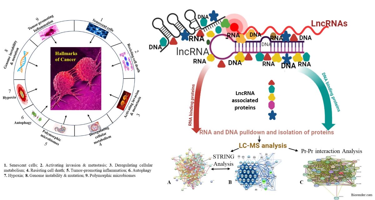
Figure 1.
Hallmarks of Cancerof and Long Non-coding RNAs (LncRNAs), How the identification of associated protein identify, quantification, and characterization by LC- Mass Spectrometry (LC-MS). And this is the flow work chart of differentially expressed significant proteins with bioinformatics of the novel molecules.
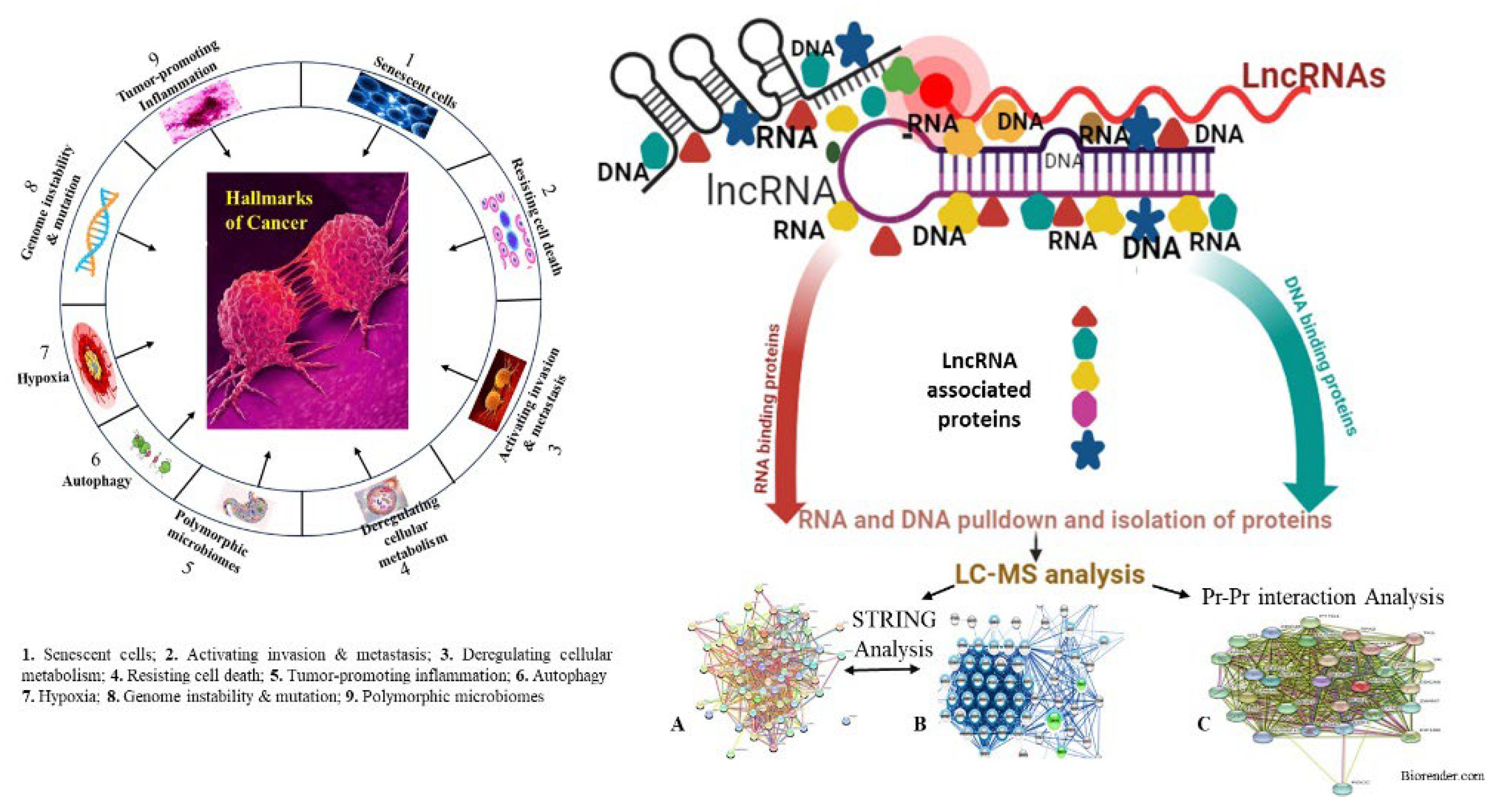
Figure 2.
Isolation of Long Non-coding RNAs (LncRNAs) associated protein then sample preparation for identification, quantification, and characterization of isolated/eluted protein by LC- Mass Spectrometry (LC-MS). Analysis of all MS data for identification of differentially expressed significant proteins after the validation of those proteins by Western Blotting and Immunohistochemistry (IHC) or the enzyme-linked immunosorbent assay (ELISA). Clinical correlation of identified and validated proteins that could be early detection biomarkers for diagnosis and prognosis of liver, colorectal and several types of cancer.
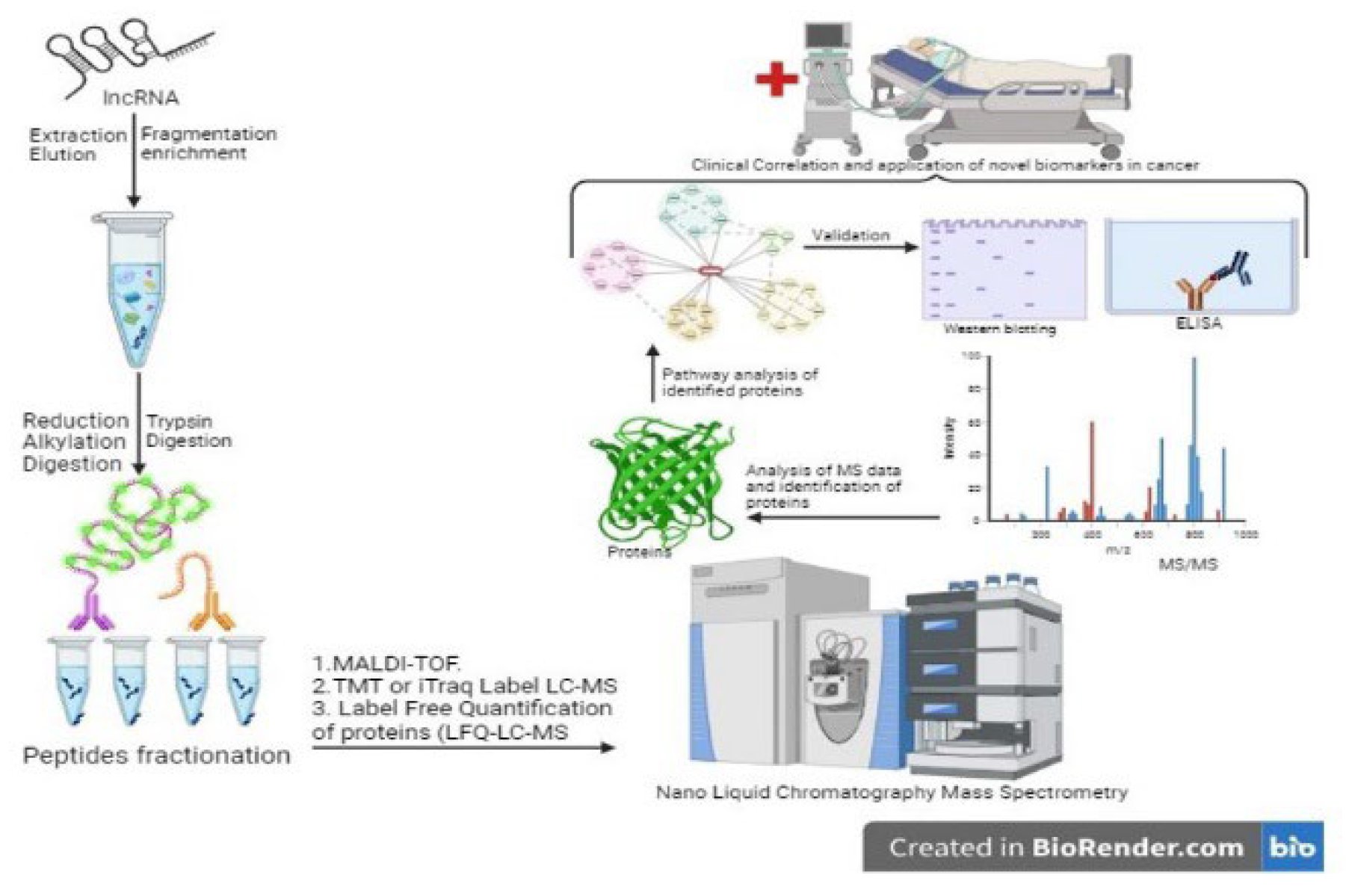
Figure 3.
Bioinformatics, Biostatistics and Regulatory analysis of all LC-MS data and find the novel LncRNAs associated proteins which are clinically relevant for the liver and colorectal cancer mechanisms or pathway that proteins will be validated by Western Blotting and Immunohistochemistry (IHC)or the enzyme-linked immunosorbent assay (ELISA). After validation of the above proteomics method, novel LncRNAs associated proteins could be helpful in early detection biomarker for diagnosis/prognosis, new therapies of anti-cancer drugs and further mechanistic study of liver, colorectal and other types of cancer.
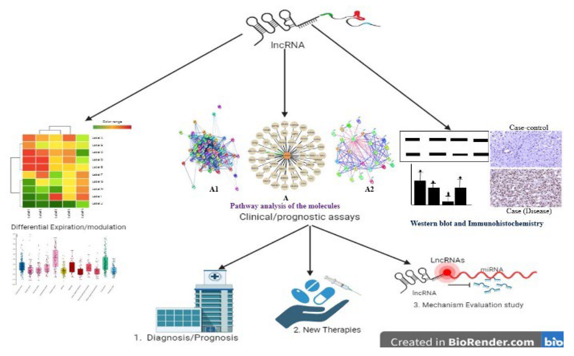

Table 1.
Identified lncRNA-associated proteins and utilized methods: The table includes serial numbers, study groups, lncRNA-associated proteins, methods used, and remarks/conclusions on these proteins and their further use in liver, colorectal, and other types of cancer.
Table 1.
Identified lncRNA-associated proteins and utilized methods: The table includes serial numbers, study groups, lncRNA-associated proteins, methods used, and remarks/conclusions on these proteins and their further use in liver, colorectal, and other types of cancer.
| S No. | Study | Target LncRNAs | Specific Method name (Cancer CRC & HCC) | Remarks/conclusions | Reference |
|---|---|---|---|---|---|
| 1 | Shijian Fu et al | MALAT-1 | Patient samples & ovarian cancer cell lines (SKOV3 & CAOV3) | MALAT-1 diagnostic or prognostic biomarker or therapeutic target in the treatment of many cancers. | [35] |
| 2 | An Yang et al | UCA1 | Via the miR-145/MYO6 axis | The UCA1/miR-145/MYO6 axis may serve as a potential therapeutic target for gastric cancer. | [36] |
| 3 | Enans M et al | T2D/ HCC | NAFLD/T2D-associated HCC | Metformin may reduce the risk of cancer in patients with T2D. The unadjusted odds ratio was 0.86 (95% CI 0.73 to 1.02). The unadjusted odds ratio for any exposure to metformin since 1993 was 0.79 (0.67 to 0.93) | [13,37] |
| 4 | Fabrizio Ferre et al | Revealing protein | RAPID-SELEX RNAcompete RNA Bin-n-Seq RNA-Ma | Better understanding of lncRNA cellular mechanisms and their disease-associated perturbations. | [37,38] |
| 5 | Fabrizio Ferre et al | LncRNA interaction | MS2 trapping SILAC-based Phage display Protein arrays | LncRNA-bound proteome, or if still uncharacterized protein domains and architectures are involved, network will be high | [37] |
| 6 | Arunoday Bhan et al | HULC | Tumorigenesis test in vitro and in vivo: RT-PCR, W. B | Potential implications in cancer diagnosis and therapy | [33,37] |
| 7 | Chunqing Wang et al | HULC | HULC interacts with the glycolytic enzyme LDHA | HULC promotes Warburg effect by orchestrating the enzymatic activities of glycolytic enzymes | [34,39] |
| 8 | Chunlin Ou et al | Linc00152 | Human Tissue Samples: | Targeting YAP1/LINC00152/FSCN1 Signaling Axis Prevents the Progression of Colorectal Cancer | [40] |
| 9 | Jie-yu Sun et al | HEIH | Non-coding RNAs | Nearly 8000 cancerspecifc lncRNAs have been nominated, PCA3 is prostate-specific, prognostic biomarker prostate cancer. | [41] |
| 10 | Huaili Jiang et al | HOTTIP | In silico analysis, Plasmid construction and transfection | Significantly, M1 exosomes and HOTTIP polarize circulating monocytes into the antitumor M1 phenotype, which may provide novel insight into HNSCC immunotherapy. | [42] |
| 11 | Taruna Rajagopal et al | HOTAIR | HOTAIR mediated gene silencing | It could be used in conjunction with current drugs to sensitize tumors to the existing therapies | [43] |
| 12 | Yang Mu et al | CCAT1 | RT-qPCR to level of miR-490-3p and CCAT1 | facilitate developing novel therapeutical therapies for treating ovarian cancer. | [44] |
| 13 | Peng Gao et al | CCAT2 | Dimethyl sulfoxide (DMSO) (Aldrich, St. Louis, | to explore genes co-expressed with lncRNA CCAT2 and functional molecular | [45] |
| 14 | Hashemi et al | H19 | Enhancing growth and cell cycle of cancers and by EMT induction | Increased proliferation Glycolysis induction miRNA-519d-3p down-regulation by H19 to increase LDHA expression | [46] |
| 15 | Hua Fang et al | CCAT1-L | Quantitative real-time PCR and Western blot, respectively. | inhibits epithelial–mesenchymal transition of gastric adenocarcinoma cells and thus suppresses the gastric adenocarcinoma metastasis. | [47] |
| 16 | Feifei Zhang1 | CRNDE | Chemosensitivity of GC in clinical samples and a PDX model. | Highlighting the significance of CRNDE as a potential prognostic marker and therapeutic target against chemoresistance in GC. | [48] |
| 17 | Yanping Li et al | FER1L4 | Cell was extracted from embryos of rat | FER1L4 modulates the proliferation and differentiation of NSCs via regulating Ascl2. | [28] |
| 18 | Yingrui Fan et al | PTENP | uciferase reporter assay and RNA-pulldown assay | Inhibit cell proliferation and EMT and induce cell apoptosisin cervical cancer cells. | [49] |
| 19 | Jinmai Jiang et al | T-UCRs | qPCR array to profile all 481 T-UCRs in pancreatic cancer specimens, pancreatic cancer cell lines | Expression of T-UCRs in both human and mouse PDAC and similar mechanism of upregulation in PDAC | [50] |
| 20 | Arunoday Bhan et al & John H Boyd | TUC338 | Plasma, treatment, and cell lines, MS2-MBP Protein Expression and Immobilization | The understanding of molecular mechanisms of lncRNAs. Inhibition of PCSK9 activity is an attractive target for treating the spectrum of sepsis and septic shock. |
[33,51] |
Disclaimer/Publisher’s Note: The statements, opinions and data contained in all publications are solely those of the individual author(s) and contributor(s) and not of MDPI and/or the editor(s). MDPI and/or the editor(s) disclaim responsibility for any injury to people or property resulting from any ideas, methods, instructions or products referred to in the content. |
© 2024 by the authors. Licensee MDPI, Basel, Switzerland. This article is an open access article distributed under the terms and conditions of the Creative Commons Attribution (CC BY) license (http://creativecommons.org/licenses/by/4.0/).
Copyright: This open access article is published under a Creative Commons CC BY 4.0 license, which permit the free download, distribution, and reuse, provided that the author and preprint are cited in any reuse.


