Submitted:
31 August 2024
Posted:
02 September 2024
You are already at the latest version
Abstract
Background and Purpose: Oxidative stress is an important factor in the pathogenesis of neurological problems. One of the compounds causing oxidative stress is diazinon. In the present project, the protective role of gallic acid compound in reducing oxidative stress in brain tissue of mice exposed to diazinon was investigated. Method: In this study, eight groups of six series of mice including the first group (control) of normal saline, the second group of diazinon (40 kg/mg) as IP, three groups of diazinon with gallic acid (50, 100, 200 kg/mg), was prescribed as a single dose and for 24 hours. The atropine + pralidoxime group was also evaluated as a standard treatment group. At 24 hours after the last dose, the animals were sacrificed and brain tissue was examined to assess oxidative stress parameters, including glutathione content, protein carbonyl content, lipid peroxidation, reactive oxygen species (ROS) and mitochondrial function in mitochondria isolated from brain tissue. Results: The results of this research showed that oxidative stress biomarkers increased significantly (P<0.001) in the diazinon group compared to the control group. Oxidative stress was significantly reduced in the groups receiving gallic acid at a dose of 200 and 100 kg/mg of diazinon compared to the diazinon group. Conclusion: The results of the present study show that diazinon causes damage to brain tissue by inducing oxidative stress. Therefore, according to the reduction of oxidative stress by gallic acid compound, it can be concluded that this compound can protect against brain toxicity caused by diazinon in animal model by stimulating antioxidant and radical scavenging pathways.
Keywords:
brain toxicity
; diazinon
; oxidative stress
1. Introduction
Diazinon is an organophosphate insecticide that inhibits the enzyme acetylcholinesterase and can cause severe acute poisoning, including headache, nausea, neurological effects and even death (36, 37). Acute poisoning may result in supranuclear palsy and opsoclonus due to cholinergic enhancement (37). Chronic exposure to diazinon has been associated with an increased risk of certain cancers, although the evidence is mixed (38). Exposure to this toxin can occur by dermal absorption, inhalation or ingestion, resulting in persistent neurological, skeletal and endocrine effects (36). Diagnosis of poisoning is based on history of exposure, symptoms such as miosis and fasciculation, response to treatment and reduction in blood cholinesterase activity, and in some cases permanent complications such as polyneuropathy and central nervous system symptoms may occur (39). The initial treatment approach for diazinon poisoning includes the use of atropine to counteract the effects of acetylcholine and pralidoxime chloride to restore blood cholinesterase levels (40). While pralidoxime has shown efficacy in the treatment of several organophosphorus poisonings, including diazinon, its efficacy varies in different combinations (39). Supportive measures such as intravenous fluids, oxygen and prompt gastric lavage are important (40). Studies have shown that exposure to diazinon reduces the activity of antioxidant enzymes, increases oxidative stress parameters and also causes blood abnormalities in mice (42).
Gallic acid (GA) is a phenolic compound that is widely distributed in various plants and foods and has significant medicinal properties. This compound has neuroprotective effects against neurodegeneration and oxidative stress (54). In addition, GA has potent anti-inflammatory activities, primarily through modulation of the MAPK and NF-kB signaling pathways, as well as reducing the release of inflammatory mediators (55). The strong antioxidant properties of GA contribute to its protective role in oxidative damage diseases, including cancer and cardiovascular disease (56). With specific effects attributed to its unique chemical structure, its cytotoxicity against cancer cells has been demonstrated (57). Despite its promising therapeutic potential, further clinical studies are needed to confirm its efficacy and safety in human application (56).
The aim of this study was to investigate the effect of gallic acid on the treatment of diazinon-induced toxicity in mouse tissues compared to standard treatment.
2. Material and methods
To evaluate the brain-protective effect of gallic acid on the brain mitochondria of exposed mice, six groups of mice were selected, each group consisting of six mice. The pralidoxime group received diazinon poison, pralidoxime and atropine, and the gallic acid group, which included three subgroups, received diazinon poison and gallic acid at concentrations of 50, 100 and 200 mg/kg. Mice were acutely poisoned with 0.5 LD50 of diazinon, and after 15 minutes treatments were started, including gallic acid, pralidoxime and atropine (standard treatment).
3-1-2 groups tested
Group 1. The control group, which received normal saline.
Group 2. The group that received diazinon at 40 mg/kg.
Groups 5, 3, 4 received different doses of gallic acid (50, 200 and 100 mg/kg) 15 minutes after diazinon administration.
Group 6 received standard treatment with atropine and pralidoxime 15 min after diazinon administration.
2-3 Ethical considerations
For two weeks prior to the experiments, the animals were kept in separate standard cages with a 12-hour light/12-hour dark cycle, access to food and water, and a temperature of 22 ± 2 degrees C for environmental adaptation. At the end of the treatment period, the animals were anaesthetized with ketamine/xylazine and operated on, and all ethical aspects of the study were followed. Brain tissue was then removed and used.
3. Results
The results obtained based on the method mentioned in the third chapter were determined as follows in this study.
1-4 The results of investigating the effect of gallic acid on mitochondrial glutathione content of mouse brain tissue exposed to diazinon poison
According to the analysis, the amount of glutathione in the Diazinon group has decreased significantly compared to the Control group (p<0.001).
The amount of glutathione in the gallic acid 100 and 200 mg/kg group increased significantly compared to the diazinon group (p<0.001). Also, the gallic acid 200 group had no significant difference with the atropine and pralidoxime group in glutathione content.
Gallic acid 50 groups did not have a significant increase in glutathione compared to diazinon group (Figure 1).
2-4 The results of investigating the effect of gallic acid on lipid peroxidation of mitochondria in rat brain tissue exposed to diazinon poison
According to the analysis, the amount of MDA in the Diazinon group increased significantly compared to the Control group (p<0.001).
The amount of MDA in the 100 and 200 mg/kg gallic acid group was significantly reduced compared to the diazinon group (p<0.001). Also, the gallic acid 100 and 200 group had no significant difference with the atropine and pralidoxime group in MDA content.
Gallic acid 50 groups did not have a significant increase in MDA compared to diazinon group (Figure 2).
3-4 The results of investigating the effect of gallic acid on the mitochondrial carbonyl protein content of rat brain tissue exposed to diazinon toxin.
According to the analysis, the amount of protein carbonyl in the Diazinon group has increased significantly compared to the Control group (p<0.001).
The amount of protein carbonyl in the gallic acid 100 and 200 mg/kg group was significantly reduced compared to the diazinon group (p<0.001). Also, the gallic acid 100 group had no significant difference with the atropine and pralidoxime groups in the carbonyl protein content.
Gallic acid 50 groups did not have a significant increase in the amount of protein carbonyl compared to the diazinon group (Figure 3).
4-4 The results of investigating the effect of gallic acid on the amount of mitochondrial ROS in the brain tissue of mice exposed to diazinon poison
According to the analysis, the amount of ROS in the Diazinon group increased significantly compared to the Control group (p<0.001).
The amount of ROS in the 100 and 200 mg/kg gallic acid group was significantly reduced compared to the diazinon group (p<0.001). Also, the gallic acid 100 group had no significant difference with the atropine and pyralidoxime groups in the carbonyl protein content.
Gallic acid 50 groups did not have a significant increase in glutathione compared to diazinon group (Figure 4).
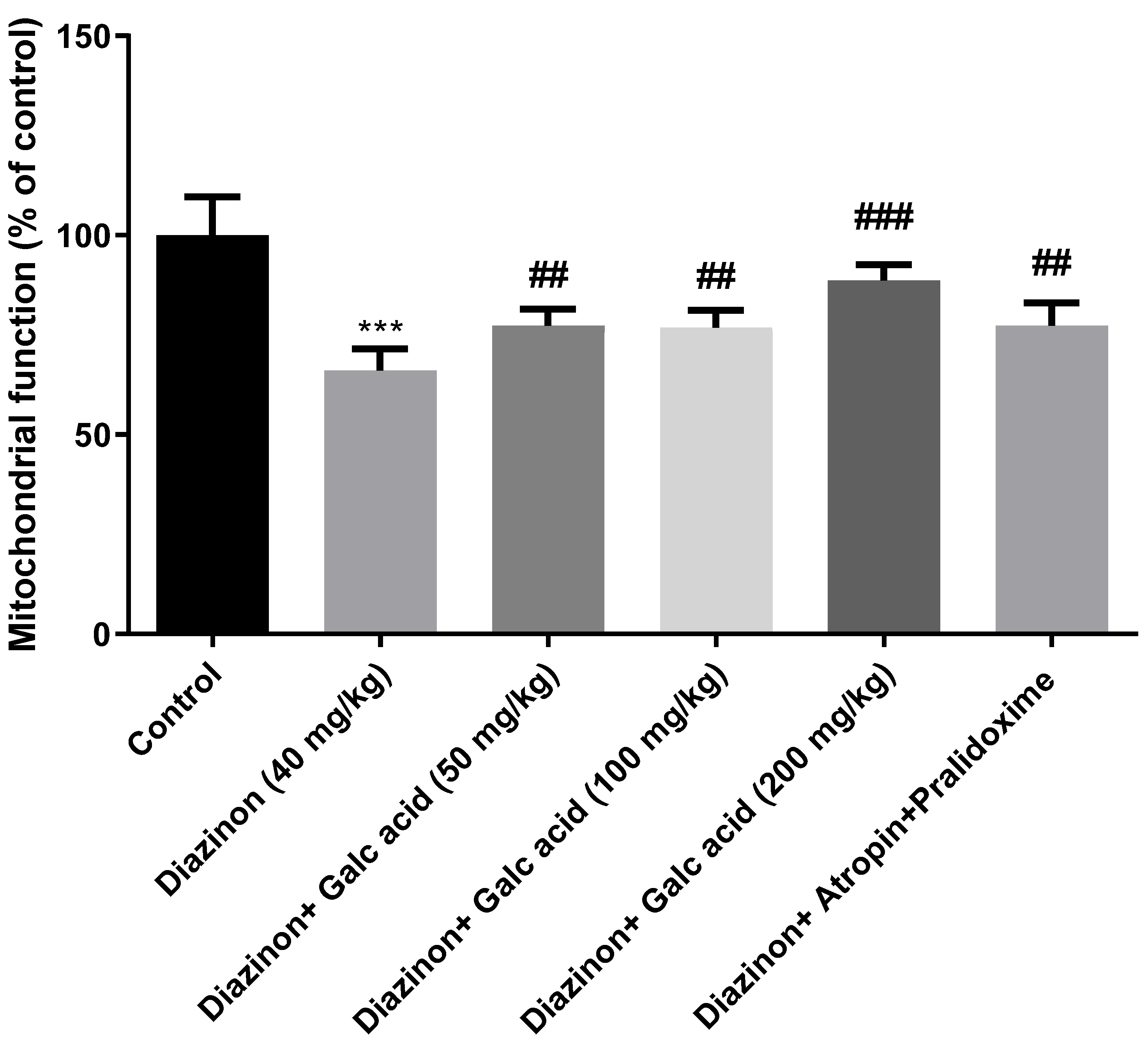
4-5 The results of investigating the effect of gallic acid on the mitochondrial function of mice brain tissue exposed to diazinon poison
The findings of this study showed that the mitochondrial function was significantly reduced in the group receiving diazinon, while after receiving doses of 200 and 100, 50 mg/kg of gallic acid, it was reduced compared to the control group. It is noteworthy that the greatest increase in mitochondrial function was related to the dose of 200 mg/kg. Figure 5
4. Discussion
The study was conducted to investigate the protective effect of gallic acid on the toxicity caused by diazinon in the mitochondria of mouse brain tissue.
According to the results of the present project, diazinon has significantly increased the oxidative stress parameters in the mitochondria of mouse brain tissue. These results are consistent with previous studies. Studies have shown that exposure to diazinon leads to an increase in lipid peroxidation and lactate dehydrogenase, a decrease in glutathione, and a change in the activity of antioxidant enzymes such as catalase and superoxide desmutase in the brain, heart, spleen, liver, and kidneys (71-71), (74). These effects are dose-dependent and vary between mouse strains, with Wistar rats showing higher sensitivity than Norway rats (71). Diazinon-induced oxidative stress can lead to tissue damage and organ dysfunction, especially in the kidneys (72). Exposure to diazinon significantly decreases the activity of antioxidant enzymes, increases markers of oxidative stress, and causes hematologic changes in mice (42). It also affects liver function and increases serum levels of ALT, AST, and ALP (75, 76). The use of antioxidants such as N-acetylcysteine may partially reduce diazinon-induced oxidative stress by scavenging reactive oxygen species and inducing glutathione synthesis (74). One of the primary pathways of diazinon in neurotoxicity involves inhibition of acetylcholinesterase (AChE), which leads to the accumulation of acetylcholine (ACh) in synapses, which disrupts normal neurotransmission and can lead to neurotoxicity (77, 78). However, the neurotoxic effects of diazinon go beyond cholinesterase inhibition, and oxidative stress and neuroinflammation have been implicated as important factors in its neurotoxicity (79, 80). Oxidative stress is a critical mechanism through which diazinon exerts its neuroprotective effects. Diazinon exposure has been shown to induce the production of reactive oxygen species (ROS), which lead to lipid peroxidation and damage to cellular components, including DNA and proteins (80, 81). This oxidative damage can trigger apoptotic pathways in neuronal cells, as studies show increased apoptosis in neuronal cell lines following diazinon exposure (82). In addition, alterations of neurotrophic factors and their signaling pathways have been observed, which may impair neurogenesis and neuronal survival (83).
5. Conclusion
In general, the results of the present study show that diazinon has the potential to cause damage in the mitochondria of the brain tissue by causing oxidative stress. In this study, the oxidative damage caused by diazinon was reduced by administering gallic acid, which is a compound with antioxidant properties.
References
- Aktar, W.; Sengupta, D.; Chowdhury, A. Impact of pesticides use in agriculture: their benefits and hazards. Interdiscip. Toxicol. 2009, 2, 1–12. [Google Scholar] [CrossRef] [PubMed]
- Jabbar ASA. Pesticide poisoning in humans. JPMA The Journal of the Pakistan Medical Association. 1992;42 10:251-5.
- Pratama, D.A.; Setiani, O.; Darundiati, Y.H. STUDI LITERATUR : PENGARUH PAPARAN PESTISIDA TERHADAP GANGGUAN KESEHATAN PETANI. J. Ris. Kesehat. Poltekkes Depkes Bdg. 2021, 13, 160–171. [Google Scholar] [CrossRef]
- Prajawahyudo, T.; Asiaka, F.K.P.; Ludang, E. Peranan keamanan pestisida di bidang pertanian bagi petani dan lingkungan. J. Socio Econ. Agric. 2022, 17, 1–9. [Google Scholar] [CrossRef]
- Pamungkas, OS. Bahaya paparan pestisida terhadap kesehatan manusia. Bioedukasi. 2017;14(1).
- Pawukir ES, Mariyono J. Hubungan Antara Penggunaan Pestisida Dan Dampak Kesehatan: Studi Kasus Di Dataran Tinggi Sumatra Barat (the Relationship Between Pesticides Use and Health Impact: a Case Study in Highlands of West Sumatera). Jurnal Manusia dan Lingkungan. 2002;9(3):126-36.
- Goel A, Aggarwal P. Pesticide poisoning. The National medical journal of India. 2022;20 4:182-91.
- Gupta RC, Mukherjee IRM, Malik JK, Doss RB, Dettbarn W-D, Milatovic D. Insecticides. Biomarkers in toxicology: Elsevier; 2019. p. 455-75.
- Araújo, M.F.; Castanheira, E.M.S.; Sousa, S.F. The Buzz on Insecticides: A Review of Uses, Molecular Structures, Targets, Adverse Effects, and Alternatives. Molecules 2023, 28, 3641. [Google Scholar] [CrossRef] [PubMed]
- Sarwar, M. The killer chemicals as controller of agriculture insect pests: The conventional insecticides. International Journal of Chemical and Biomolecular Science. 2015;1(3):141-7.
- Stehle, S.; Schulz, R. Global Insecticide Surface Water Contamination Assessment: BECT’s Contribution in the Last Five Decades. Bull. Environ. Contam. Toxicol. 2016, 96, 563–564. [Google Scholar] [CrossRef] [PubMed]
- Stehle, S.; Schulz, R. Agricultural insecticides threaten surface waters at the global scale. Proc. Natl. Acad. Sci. USA 2015, 112, 5750–5755. [Google Scholar] [CrossRef]
- Smith, J.J.; Herzig, V.; King, G.F.; Alewood, P.F. The insecticidal potential of venom peptides. Cell. Mol. Life Sci. 2013, 70, 3665–3693. [Google Scholar] [CrossRef]
- Alavanja, M.C.R.; Hofmann, J.N.; Lynch, C.F.; Hines, C.J.; Barry, K.H.; Barker, J.; Buckman, D.W.; Thomas, K.; Sandler, D.P.; Hoppin, J.A.; et al. Non-Hodgkin Lymphoma Risk and Insecticide, Fungicide and Fumigant Use in the Agricultural Health Study. PLOS ONE 2014, 9, e109332. [Google Scholar] [CrossRef]
- Baldi, I.; Gruber, A.; Rondeau, V.; Lebailly, P.; Brochard, P.; Fabrigoule, C. Neurobehavioral effects of long-term exposure to pesticides: results from the 4-year follow-up of the PHYTONER Study. Occup. Environ. Med. 2010, 68, 108–115. [Google Scholar] [CrossRef]
- Zhao, Y.; Yang, J.; Ren, J.; Hou, Y.; Han, Z.; Xiao, J.; Li, Y. Exposure Level of Neonicotinoid Insecticides in the Food Chain and the Evaluation of Their Human Health Impact and Environmental Risk: An Overview. Sustainability 2020, 12, 7523. [Google Scholar] [CrossRef]
- Viana, M.; Hughes, A.; Matthiopoulos, J.; Ranson, H.; Ferguson, H.M. Delayed mortality effects cut the malaria transmission potential of insecticide-resistant mosquitoes. Proc. Natl. Acad. Sci. 2016, 113, 8975–8980. [Google Scholar] [CrossRef]
- Chittrakul, J.; Sapbamrer, R.; Sirikul, W. Insecticide Exposure and Risk of Asthmatic Symptoms: A Systematic Review and Meta-Analysis. Toxics 2021, 9, 228. [Google Scholar] [CrossRef] [PubMed]
- Modak, S.; Ghosh, P.; Mandal, S.; Sasmal, D.; Kundu, S.; Sengupta, S.; Kanthal, S.; Sarkar, T. Organophosphate Pesticide: Environmental impact and toxicity to organisms. Int. J. Res. Agron. 2024, 7, 138–141. [Google Scholar] [CrossRef]
- Sidhu, G.K.; Singh, S.; Kumar, V.; Dhanjal, D.S.; Datta, S.; Singh, J. Toxicity, monitoring and biodegradation of organophosphate pesticides: A review. Crit. Rev. Environ. Sci. Technol. 2019, 49, 1135–1187. [Google Scholar] [CrossRef]
- Melo JS, editor Overview on Biosensors for Detection of Organophosphate Pesticides2017.
- Risal, P.; Lama, S.; Thapa, S.; Bhatta, R.; Karki, R.K. Cholinesterase and Liver Enzymes in Patients with Organophosphate Poisoning. J. Nobel Med Coll. 2019, 8, 33–37. [Google Scholar] [CrossRef]
- Chuang, C.-S.; Yang, K.-W.; Yen, C.-M.; Lin, C.-L.; Kao, C.-H. Risk of Seizures in Patients with Organophosphate Poisoning: A Nationwide Population-Based Study. Int. J. Environ. Res. Public Heal. 2019, 16, 3147. [Google Scholar] [CrossRef]
- Chowdhury, F.R.; Bari, S.; Alam, M.J.; Rahman, M.; Bhattacharjee, B.; Qayyum, J.A.; Mridha, S. Organophosphate poisoning presenting with muscular weakness and abdominal pain- a case report. BMC Res. Notes 2014, 7, 140–140. [Google Scholar] [CrossRef]
- Peter, J.V.; Sudarsan, T.; Moran, J. Clinical features of organophosphate poisoning: A review of different classification systems and approaches. Indian J. Crit. Care Med. 2014, 18, 735–745. [Google Scholar] [CrossRef]
- Peter, J.V.; Jerobin, J.; Nair, A.; Bennett, A.; Samuel, P.; Chrispal, A.; Abraham, O.C.; Mathews, K.P.; Fleming, J.J.; Oommen, A. Clinical profile and outcome of patients hospitalized with dimethyl and diethyl organophosphate poisoning. Clin. Toxicol. 2010, 48, 916–923. [Google Scholar] [CrossRef]
- Dündar ZD, Ergin M, Köylü R, Günaydın YK, Özer R, Cander B. Prognostic value of red cell distribution width in patients with organophosphate poisoning. 2015.
- E Ibrahim, A.; Ghantarchyan, H.; Le, T.; Bhagat, A.; Maknouni, B.; Arabian, S. A Rare Presentation of Severe Organophosphate Poisoning: A Case Report and Review of Literature. Cureus 2022, 14, e31497. [Google Scholar] [CrossRef] [PubMed]
- Narayan, R.; Abdulla, M.C.; Alungal, J. Transient Distal Renal Tubular Acidosis in Organophosphate Poisoning. Indian J. Crit. Care Med. 2017, 21, 170–171. [Google Scholar] [CrossRef] [PubMed]
- Alghafees, M.A.; Abdulmomen, A.; Eid, M.; Alhussin, G.I.; Alosaimi, M.Q.; Alduhaimi, G.S.; Albogami, M.T.; Alhelail, M. Poisoning-related emergency department visits: the experience of a Saudi high-volume toxicology center. Ann. Saudi Med. 2022, 42, 36–44. [Google Scholar] [CrossRef] [PubMed]
- Soliman, S.A.; Sovocool, G.W.; Curley, A.; Ahmed, N.S.; El-Fiki, S.; El-Sebae, A.-K. Two Acute Human Poisoning Cases Resulting from Exposure to Diazinon Transformation Products in Egypt. Arch. Environ. Heal. Int. J. 1982, 37, 207–212. [Google Scholar] [CrossRef]
- Dahlgren J, Takhar H, Ruffalo C, Zwass M. Health effects of diazinon on a family. Journal of Toxicology: Clinical Toxicology. 2004;42(5):579-91.
- Liang, T.-W.; Balcer, L.J.; Solomon, D.; Messé, S.R.; Galetta, S.L. Supranuclear gaze palsy and opsoclonus after Diazinon poisoning. J. Neurol. Neurosurg. Psychiatry 2003, 74, 677–679. [Google Scholar] [CrossRef]
- Sergi, CM. Diazinon—An Insecticide. Encyclopedia of Environmental Health. 2019.
- Namba T, Nolte CT, Jackrel J, Grob D. Poisoning due to organophosphate insecticides: acute and chronic manifestations. The American journal of medicine. 1971;50(4):475-92.
- Klemmer, H.W.; Reichert, E.R.; Yauger, W.L.; Haley, T.J. Five Cases of Intentional Ingestion of 25 Percent Diazinon with Treatment and Recovery. Clin. Toxicol. 1978, 12, 435–444. [Google Scholar] [CrossRef]
- Kamha AA, Al Omary IY, Zalabany HA, Hanssens Y, Adheir FS. Organophosphate poisoning in pregnancy: a case report. Basic & clinical pharmacology & toxicology. 2005;96(5):397-8.
- Ajibade, T.O.; Oyagbemi, A.A.; Omobowale, T.O.; Asenuga, E.R.; Afolabi, J.M.; Adedapo, A.A. Original article. Mitigation of diazinon-induced cardiovascular and renal dysfunction by gallic acid. Interdiscip. Toxicol. 2016, 9, 66–77. [Google Scholar] [CrossRef]
- Anbarkeh, F.R.; Nikravesh, M.R.; Jalali, M.; Soukhtanloo, M. The protective role of alpha-lipoic acid on the appearance of fibronectin and laminin in renal tubules following diazinon exposure: An experimental immunohistochemical study. Toxicology 2020, 444, 152583. [Google Scholar] [CrossRef]
- Karimani, A.; Ramezani, N.; Goli, A.A.; Shirazi, M.H.N.; Nourani, H.; Jafari, A.M. Subchronic neurotoxicity of diazinon in albino mice: Impact of oxidative stress, AChE activity, and gene expression disturbances in the cerebral cortex and hippocampus on mood, spatial learning, and memory function. Toxicol. Rep. 2021, 8, 1280–1288. [Google Scholar] [CrossRef]
- Afshari, S.; Sarailoo, M.; Asghariazar, V.; Safarzadeh, E.; Dadkhah, M. Persistent diazinon induced neurotoxicity: The effect on inhibitory avoidance memory performance, amyloid precursor proteins, and TNF-α levels in the prefrontal cortex of rats. Hum. Exp. Toxicol. 2024, 43. [Google Scholar] [CrossRef] [PubMed]
- Reshi M, Bhat A, Kaur P, editors. The Role of Antioxidants in Human Health 2013.
- Willcox, J.K.; Ash, S.L.; Catignani, G.L. Antioxidants and Prevention of Chronic Disease. Crit. Rev. Food Sci. Nutr. 2004, 44, 275–295. [Google Scholar] [CrossRef] [PubMed]
- Hamid A, Aiyelaagbe O, Usman L, Ameen O, Lawal A. Antioxidants: Its medicinal and pharmacological applications. African Journal of pure and applied chemistry. 2010;4(8):142-51.
- Halliwell, B. Antioxidants in human health and disease. Annual review of nutrition. 1996;16(1):33-50.
- Buelga, CS. Los polifenoles y la salud. ACTA/CL: revista de la Asociación de Científicos y Tecnólogos de Alimentos de Castilla y León. 2020(70):15-6.
- Kuhnert, N. Polyphenole: Vielseitige Pflanzeninhaltsstoffe: In Garten, Industrie, Medizin und Nahrung. Chemie in unserer Zeit. 2013;47(2):80-91.
- Quiñones M, Miguel M, Aleixandre A. Los polifenoles, compuestos de origen natural con efectos saludables sobre el sistema cardiovascular. Nutrición hospitalaria. 2012;27(1):76-89.
- Achat, S. Polyphénols de l’alimentation: extraction, pouvoir antioxydant et interactions avec des ions métalliques: Université d’Avignon; Université Abderrahmane Mira-Bejaïa (Bejaïa, Algérie); 2013.
- Daglia M, Di Lorenzo A, F Nabavi S, S Talas Z, M Nabavi S. Polyphenols: well beyond the antioxidant capacity: gallic acid and related compounds as neuroprotective agents: you are what you eat! Current pharmaceutical biotechnology. 2014;15(4):362-72.
- Bai, J.; Zhang, Y.; Tang, C.; Hou, Y.; Ai, X.; Chen, X.; Zhang, Y.; Wang, X.; Meng, X. Gallic acid: Pharmacological activities and molecular mechanisms involved in inflammation-related diseases. Biomed. Pharmacother. 2021, 133, 110985. [Google Scholar] [CrossRef]
- Gao, J.; Hu, J.; Hu, D.; Yang, X. A Role of Gallic Acid in Oxidative Damage Diseases: A Comprehensive Review. Nat. Prod. Commun. 2019, 14. [Google Scholar] [CrossRef]
- Inoue, M.; Suzuki, R.; Sakaguchi, N.; Li, Z.; Takeda, T.; Ogihara, Y.; Jiang, B.Y.; Chen, Y. Selective Induction of Cell Death in Cancer Cells by Gallic Acid. Biol. Pharm. Bull. 1995, 18, 1526–1530. [Google Scholar] [CrossRef] [PubMed]
- Dewick, P.M.; Haslam, E. Phenol biosynthesis in higher plants. Gallic acid. Biochem. J. 1969, 113, 537–542. [Google Scholar] [CrossRef]
- Pengelly, A. The constituents of medicinal plants: an introduction to the chemistry and therapeutics of herbal medicine: Routledge; 2020.
- Fiuza S, Gomes C, Teixeira L, Da Cruz MG, Cordeiro M, Milhazes N, et al. Phenolic acid derivatives with potential anticancer properties––a structure–activity relationship study. Part 1: Methyl, propyl and octyl esters of caffeic and gallic acids. Bioorganic & medicinal chemistry. 2004;12(13):3581-9.
- Kaur, M.; Velmurugan, B.; Rajamanickam, S.; Agarwal, R.; Agarwal, C. Gallic Acid, an Active Constituent of Grape Seed Extract, Exhibits Anti-proliferative, Pro-apoptotic and Anti-tumorigenic Effects Against Prostate Carcinoma Xenograft Growth in Nude Mice. Pharm. Res. 2009, 26, 2133–2140. [Google Scholar] [CrossRef]
- Choubey, S.; Varughese, L.R.; Kumar, V.; Beniwal, V. Medicinal Importance of Gallic Acid and Its Ester Derivatives: a Patent Review. Pharm. Pat. Anal. 2015, 4, 305–315. [Google Scholar] [CrossRef]
- Sarjit, A.; Wang, Y.; Dykes, G.A. Antimicrobial activity of gallic acid against thermophilic Campylobacter is strain specific and associated with a loss of calcium ions. Food Microbiol. 2015, 46, 227–233. [Google Scholar] [CrossRef]
- Hsieh, S.-C.; Wu, C.-C.; Hsu, S.-L.; Yen, J.-H. Molecular mechanisms of gallic acid-induced growth inhibition, apoptosis, and necrosis in hypertrophic scar fibroblasts. Life Sci. 2017, 179, 130–138. [Google Scholar] [CrossRef]
- Goldstein, A.; Aronow, L.; Kalman, S.M. Principles of Drug Action. The Basis of Pharmacology. The Basis of Pharmacology. J. Med. Chem. 1968, 13, 337–337. [Google Scholar] [CrossRef]
- Kaur, S.; Singla, N.; Dhawan, D.K. Neuro-protective potential of quercetin during chlorpyrifos induced neurotoxicity in rats. Drug Chem. Toxicol. 2019, 42, 220–230. [Google Scholar] [CrossRef]
- Abbassy M, Belal M, Nasr H, Mansy A. Role of vitamin E and rutin as potent inducers of chlorpyrifos degradation in the blood of treated male albino rats. Journal of Environmental Toxicology and Analytical Research. 2019;1(1):1.
- Adampourezare, M.; Sistani, P.; Nemati, H.H. Protective Effect of Dorema glabrum on Induced Oxidative Stress by Diazinon in Hippocampus of Rat. Int. J. Biochem. Res. Rev. 2019, 1–7. [Google Scholar] [CrossRef]
- Bameri, B.; Shaki, F.; Ahangar, N.; Ataee, R.; Samadi, M.; Mohammadi, H. Evidence for the Involvement of the Dopaminergic System in Seizure and Oxidative Damage Induced by Tramadol. Int. J. Toxicol. 2018, 37, 164–170. [Google Scholar] [CrossRef]
- Fathi, H.; Ebrahimzadeh, M.A.; Ziar, A.; Mohammadi, H. Oxidative damage induced by retching; antiemetic and neuroprotective role of Sambucus ebulus L. Cell Biol. Toxicol. 2015, 31, 231–239. [Google Scholar] [CrossRef] [PubMed]
- Jafari, M.; Salehi, M.; Ahmadi, S.; Asgari, A.; Abasnezhad, M.; Hajigholamali, M. The role of oxidative stress in diazinon-induced tissues toxicity in Wistar and Norway rats. Toxicol. Mech. Methods 2012, 22, 638–647. [Google Scholar] [CrossRef] [PubMed]
- Shah MD, Iqbal M. Diazinon-induced oxidative stress and renal dysfunction in rats. Food and chemical toxicology. 2010;48(12):3345-53.
- Salehi M, Jafari M, Saleh-Moqadam M, Asgari A. The comparison of the effect of diazinon and paraoxon on biomarkers of oxidative stress in rat serum. Zahedan Journal of Research in Medical Sciences. 2012;14(3).
- Izadi F, Jafari M, Bahdoran H, Asgari A, Divsalar A, Salehi M. The role of N-acetyl cysteine on reduction of diazinon-induced oxidative stress in rat liver and kidney. Journal of Rafsanjan University of Medical Sciences. 2014;12(11):895-906.
- Sherifa, K. Hepatic and renal biochemical responses to the toxicological interaction between acetylsalicylic acid and diazinon in albino rats. 2006.
- Abdel-Daim, M.M.; Taha, R.; Ghazy, E.W.; El-Sayed, Y.S. Synergistic ameliorative effects of sesame oil and alpha-lipoic acid against subacute diazinon toxicity in rats: hematological, biochemical, and antioxidant studies. Can. J. Physiol. Pharmacol. 2016, 94, 81–88. [Google Scholar] [CrossRef]
- Aronzon, C.M.; Marino, D.J.; Ronco, A.E.; Coll, C.S.P. Differential toxicity and uptake of Diazinon on embryo-larval development of Rhinella arenarum. Chemosphere 2014, 100, 50–56. [Google Scholar] [CrossRef]
- Pizzurro, D.M.; Dao, K.; Costa, L.G. Diazinon and diazoxon impair the ability of astrocytes to foster neurite outgrowth in primary hippocampal neurons. Toxicol. Appl. Pharmacol. 2013, 274, 372–382. [Google Scholar] [CrossRef]
- Lee, B.; Park, S.M.; Jeong, S.; Kim, K.; Jeung, E.-B. Combined Exposure to Diazinon and Nicotine Exerts a Synergistic Adverse Effect In Vitro and Disrupts Brain Development and Behaviors In Vivo. Int. J. Mol. Sci. 2021, 22, 7742. [Google Scholar] [CrossRef] [PubMed]
- Hameed, A.K.; Ahmed, J.A. The Neuro-protective Role of Vitamin B12 in Diazinon Poisoned Male Wistar Rats: Histopathological and Biochemical Evaluation. Egyptian Journal of Veterinary Sciences. 2021;52, 3: International Conference of Veterinary Research Division National Research Centre, Giza, Egypt 27th-29th 21), 20 September.
- Gao B, Bian X, Chi L, Tu P, Ru H, Lu K. Editor’s Highlight: Organophosphate diazinon altered quorum sensing, cell motility, stress response, and carbohydrate metabolism of gut microbiome. Toxicological Sciences. 2017;157(2):354-64.
- Sadri S, Bahrami F, Khazaei M, Hashemi M, Asgari A. Cannabinoid receptor agonist WIN-55,212-2 protects differentiated PC12 cells from organophosphorus-induced apoptosis. International journal of toxicology. 2010;29(2):201-8.
- Slotkin, T.A.; Seidler, F.J.; Fumagalli, F. Targeting of neurotrophic factors, their receptors, and signaling pathways in the developmental neurotoxicity of organophosphates in vivo and in vitro. Brain Res. Bull. 2008, 76, 424–438. [Google Scholar] [CrossRef] [PubMed]
- Wisudanti DD, Normasari R, Hidayati T. Hepatoprotective effects of soy flour (Glycine max (L.) Merr.) supplementation in diazinon-treated Wistar rats. 2021.
- Sinaei, N.; Jafari, E.; Najafi, A.; Karami-Mohajeri, S. Hepatic Oxidative Damages and Glucose Tolerance in Diabetic Rats Exposed to Repeated Oral Doses of Diazinon. Iran. J. Toxicol. 2022, 16, 221–228. [Google Scholar] [CrossRef]
- Oruc, E. Effects of diazinon on antioxidant defense system and lipid peroxidation in the liver of Cyprinus carpio (L.). Environ. Toxicol. 2011, 26, 571–578. [Google Scholar] [CrossRef]
- Esfandiarifar A, Azarbayjani M-A, Peeri M, Jameie SB. The Effect of Resistance Training and Berberine Chloride on the Apoptosis-Related UPR Signaling Pathway in the Hippocampus of Diazinon-Poisoned Rats Running title: Resistance Training and Berberine on the Hippocampus Apoptosis.
- Attabi, M.R.A.; Hussain, S.M.; Al-Okaily, B.N. Effect of Diazinon on Reproductive System of Adult Male Mice. J. Kerbala Agric. Sci. 2017, 4, 229–242. [Google Scholar] [CrossRef]
- Ebadimanas, G.; Najafi, G. The Regulation of Testosterone Levels and Improvement of Sperm DNA Maturity and In-Vitro Fertilization Outcome by Selenium Administration in Diazinon-treated Wistar Rats. J. Kermanshah Univ. Med Sci. 2020, 24. [Google Scholar] [CrossRef]
- Ghazy, E.; Mokh, A.; Abdelhady, D.; Goda, W.; Hashem, E. The role of thymoquinone in ameliorating the hepatoxic effect of diazinon in male rats. Slov. Veter- Res. 2019, 56, 745–744. [Google Scholar] [CrossRef]
- Sonei, A.; Fazelipour, S.; Kanaani, L.; Jahromy, M.H. Protective Effects of Berberis vulgaris on Diazinon-Induced Brain Damage in Young Male Mice. Prev. Nutr. Food Sci. 2020, 25, 65–70. [Google Scholar] [CrossRef]
- Roegge, C.S.; Timofeeva, O.A.; Seidler, F.J.; Slotkin, T.A.; Levin, E.D. Developmental diazinon neurotoxicity in rats: Later effects on emotional response. Brain Res. Bull. 2007, 75, 166–172. [Google Scholar] [CrossRef]
- Slotkin, T.A.; Ryde, I.T.; Levin, E.D.; Seidler, F.J. Developmental neurotoxicity of low dose diazinon exposure of neonatal rats: Effects on serotonin systems in adolescence and adulthood. Brain Res. Bull. 2007, 75, 640–647. [Google Scholar] [CrossRef]
- Slotkin TA, Tate CA, Ryde IT, Levin ED, Seidler FJ. Organophosphate insecticides target the serotonergic system in developing rat brain regions: disparate effects of diazinon and parathion at doses spanning the threshold for cholinesterase inhibition. Environmental health perspectives. 2006;114(10):1542-6.
- Gao, B.; Bian, X.; Mahbub, R.; Lu, K. Sex-Specific Effects of Organophosphate Diazinon on the Gut Microbiome and Its Metabolic Functions. Environ. Heal. Perspect. 2017, 125, 198–206. [Google Scholar] [CrossRef] [PubMed]
- Yang, Y.H.; Wang, Z.; Zheng, J.; Wang, R. Protective effects of gallic acid against spinal cord injury-induced oxidative stress. Mol. Med. Rep. 2015, 12, 3017–3024. [Google Scholar] [CrossRef] [PubMed]
- Mansouri, M.T.; Farbood, Y.; Sameri, M.J.; Sarkaki, A.; Naghizadeh, B.; Rafeirad, M. Neuroprotective effects of oral gallic acid against oxidative stress induced by 6-hydroxydopamine in rats. Food Chem. 2013, 138, 1028–1033. [Google Scholar] [CrossRef]
- Jin, L.; Piao, Z.H.; Sun, S.; Liu, B.; Kim, G.R.; Seok, Y.M.; Lin, M.Q.; Ryu, Y.; Choi, S.Y.; Kee, H.J.; et al. Gallic Acid Reduces Blood Pressure and Attenuates Oxidative Stress and Cardiac Hypertrophy in Spontaneously Hypertensive Rats. Sci. Rep. 2017, 7, 15607. [Google Scholar] [CrossRef]
- de Oliveira LS, Thomé GR, Lopes TF, Reichert KP, de Oliveira JS, da Silva Pereira A, et al. Effects of gallic acid on delta–aminolevulinic dehydratase activity and in the biochemical, histological and oxidative stress parameters in the liver and kidney of diabetic rats. Biomedicine & Pharmacotherapy. 2016;84:1291-9.
- Soumya, K.; James, J.; Archana, T.M.; Dhanya, A.T.; Shahid, A.P.; Sudheesh, S. Cytotoxic and antigenotoxic properties of phenolic compound isolated from the fruit of Terminalia chebula on HeLa cell. Beni-Suef Univ. J. Basic Appl. Sci. 2019, 8, 1–6. [Google Scholar] [CrossRef]
- Yang, D.J.; Moh, S.H.; Son, D.H.; You, S.; Kinyua, A.W.; Ko, C.M.; Song, M.; Yeo, J.; Choi, Y.-H.; Kim, K.W. Gallic Acid Promotes Wound Healing in Normal and Hyperglucidic Conditions. Molecules 2016, 21, 899. [Google Scholar] [CrossRef]
- Wen, L.; Tang, L.; Zhang, M.; Wang, C.; Li, S.; Wen, Y.; Tu, H.; Tian, H.; Wei, J.; Liang, P.; et al. Gallic Acid Alleviates Visceral Pain and Depression via Inhibition of P2X7 Receptor. Int. J. Mol. Sci. 2022, 23, 6159. [Google Scholar] [CrossRef]
- Karaman, A.; Aydın, H.; Geçkinli, B.; Çetinkaya, A.; Karaman, S. DNA damage is increased in lymphocytes of patients with metabolic syndrome. Mutat. Res. Toxicol. Environ. Mutagen. 2015, 782, 30–35. [Google Scholar] [CrossRef]
Figure 1.
Determining the effect of gallic acid on the mitochondrial glutathione content of rat brain tissue exposed to diazinon. * Significant difference with control group p<0.001***, p<0.01**, p<0.05 *. # Significant difference with diazinon group p<0.001##, p<0.01##, p<0.05#
Figure 1.
Determining the effect of gallic acid on the mitochondrial glutathione content of rat brain tissue exposed to diazinon. * Significant difference with control group p<0.001***, p<0.01**, p<0.05 *. # Significant difference with diazinon group p<0.001##, p<0.01##, p<0.05#
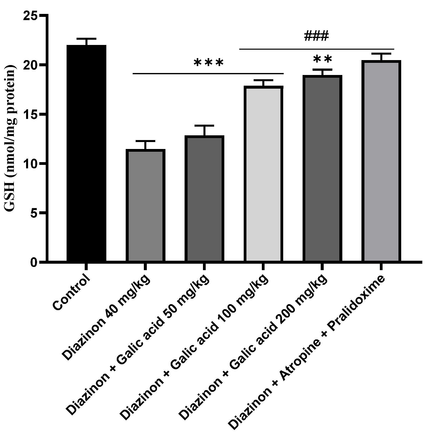
Figure 2.
Determining the effect of gallic acid on the mitochondrial MDA content of rat brain tissue exposed to diazinon. * Significant difference with control group p<0.001***, p<0.01**, p<0.05 *. # Significant difference with diazinon group p<0.001##, p<0.01##, p<0.05#
Figure 2.
Determining the effect of gallic acid on the mitochondrial MDA content of rat brain tissue exposed to diazinon. * Significant difference with control group p<0.001***, p<0.01**, p<0.05 *. # Significant difference with diazinon group p<0.001##, p<0.01##, p<0.05#
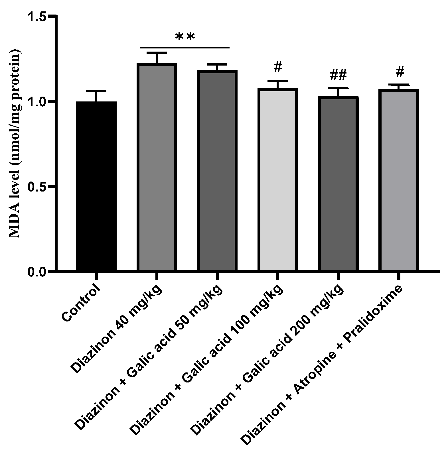
Figure 3.
Determining the effect of gallic acid on the mitochondrial carbonyl protein content of rat brain tissue exposed to diazinon. * Significant difference with control group p<0.001***, p<0.01**, p<0.05 *. # Significant difference with diazinon group p<0.001##, p<0.01##, p<0.05#
Figure 3.
Determining the effect of gallic acid on the mitochondrial carbonyl protein content of rat brain tissue exposed to diazinon. * Significant difference with control group p<0.001***, p<0.01**, p<0.05 *. # Significant difference with diazinon group p<0.001##, p<0.01##, p<0.05#
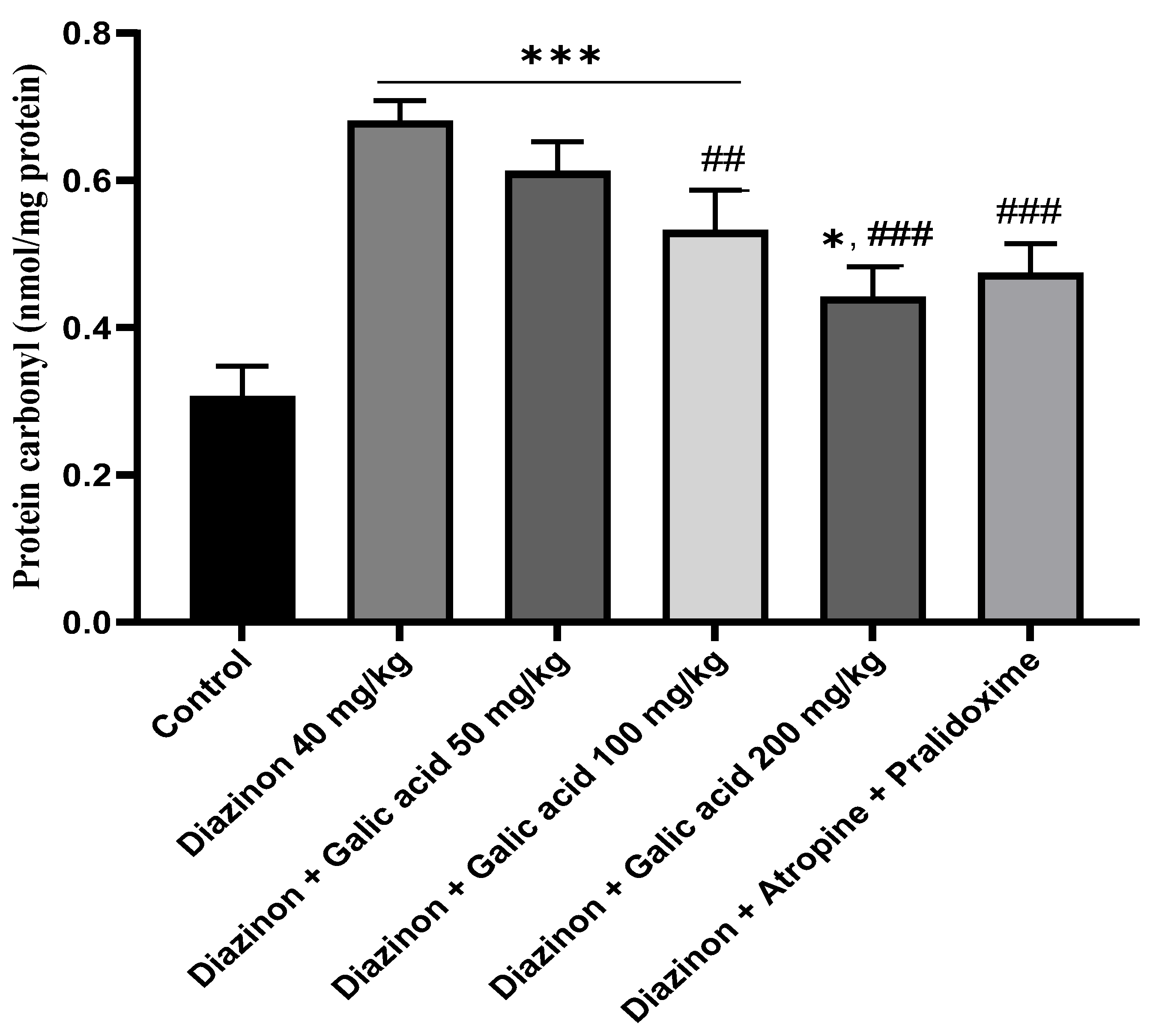
Figure 4.
Determining the effect of gallic acid on the amount of mitochondrial ROS in the brain tissue of rats exposed to diazinon. * Significant difference with control group p<0.001***, p<0.01**, p<0.05 *. # Significant difference with diazinon group p<0.001##, p<0.01##, p<0.05#
Figure 4.
Determining the effect of gallic acid on the amount of mitochondrial ROS in the brain tissue of rats exposed to diazinon. * Significant difference with control group p<0.001***, p<0.01**, p<0.05 *. # Significant difference with diazinon group p<0.001##, p<0.01##, p<0.05#
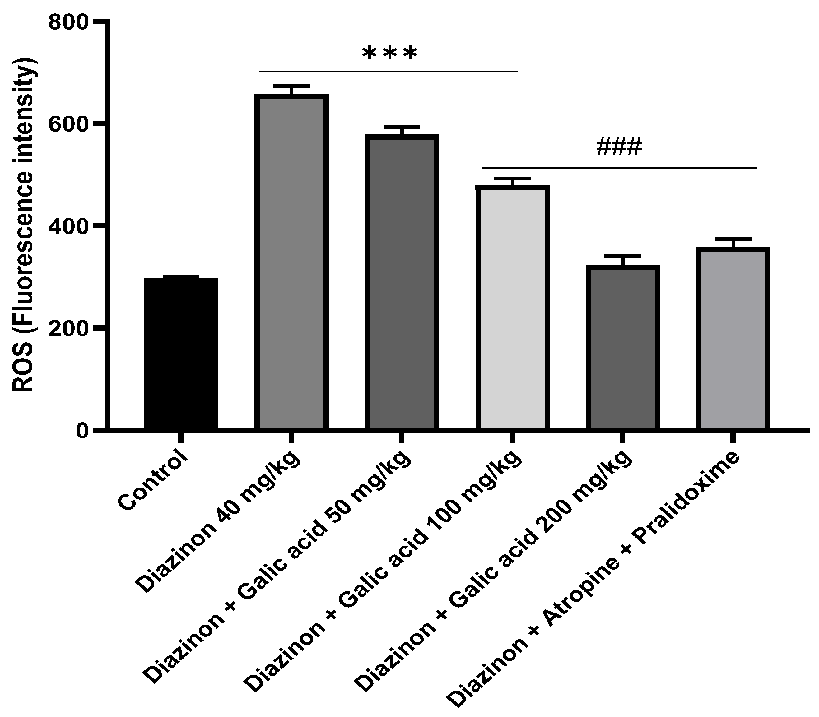
Figure 5.
Determining the effect of gallic acid on the mitochondrial function of rat brain tissue exposed to diazinon. * Significant difference with control group p<0.001***, p<0.01**, p<0.05 *. # Significant difference with diazinon group p<0.001##, p<0.01##, p<0.05#
Figure 5.
Determining the effect of gallic acid on the mitochondrial function of rat brain tissue exposed to diazinon. * Significant difference with control group p<0.001***, p<0.01**, p<0.05 *. # Significant difference with diazinon group p<0.001##, p<0.01##, p<0.05#
Disclaimer/Publisher’s Note: The statements, opinions and data contained in all publications are solely those of the individual author(s) and contributor(s) and not of MDPI and/or the editor(s). MDPI and/or the editor(s) disclaim responsibility for any injury to people or property resulting from any ideas, methods, instructions or products referred to in the content. |
© 2024 by the authors. Licensee MDPI, Basel, Switzerland. This article is an open access article distributed under the terms and conditions of the Creative Commons Attribution (CC BY) license (http://creativecommons.org/licenses/by/4.0/).
Copyright: This open access article is published under a Creative Commons CC BY 4.0 license, which permit the free download, distribution, and reuse, provided that the author and preprint are cited in any reuse.


