
Submitted:
19 November 2025
Posted:
20 November 2025
You are already at the latest version
Abstract
Anthocyanins are well known as colorants of flowers, but many other invisible functions might have been more important during the evolution of complex biosynthesis networks in plants. The number of anthocyanin decorating enzymes, the subtle control of structural genes by numerous transcription factors, and routes of intracellular anthocyanin transport are reviewed here. Various ecological functions of anthocyanins hold the key to understanding evolutionary trajectories that lead to the success of these pigments. Proposed functions include carbon sinks that prevent excessive sugar levels, sun blockers protecting the photosynthesis apparatus, antioxidants scavenging reactive oxygen species, providing camouflage, and attraction of pollinators and seed dispersers. Anthocyanins clearly fulfill different functions in different plant organs. It is currently believed that protective functions in leaves gave rise to the biosynthesis network and that flower and fruit coloration evolved later. Despite decades of research on the anthocyanin biosynthesis, there is still substantial potential for fundamental discoveries.
Keywords:
anthocyanins
; flavonoids
; photoprotection
; antioxidants
; pollination
; coloration
; transcriptional regulation
; intracellular transport
Background
Anthocyanins are well known as a phylogenetically widespread group of plant pigments providing colorful patterns to flowers and fruits. Research on anthocyanins has a rich history of interdisciplinary explorations dating back more than 100 years [1]. Anthocyanins can provide a range of different colors including red, orange, pink, purple, and blue. This turns anthocyanins into an economically relevant target in the engineering of horticultural plants [2,3]. Especially the rise of genome editing methods like CRISPR/Cas in plants [4] opens new avenues for targeted modification of the anthocyanin biosynthesis to achieve a desired flower color. However, such endeavors require a comprehensive understanding of all genes involved in the biosynthesis pathway. Due to the clearly visible phenotype, the anthocyanin biosynthesis quickly emerged as a model system for research on biosynthesis pathways and has even been described as a metamodel for understanding the genetic basis of evolutionary change [5,6]. First studies investigating genes of the anthocyanin biosynthesis date back to the 1950s, when Barbara McClintock studied transposons in maize [7] that generated diverse pigmentation patterns by disrupting anthocyanin biosynthesis genes. First biochemical studies of anthocyanins in flowers are even older - ranging at least back to 1835 [8]. Anthocyanins were also involved in classical genetic experiments performed by Gregor Mendel, as he worked on the flower color of peas [9]. However, it took almost 150 years until the underlying genes were discovered [10,11]. Anthocyanins are relevant for studies in many different fields, because they are present in almost all plant lineages. However, a recent, transcontinental study revealed that only about 56% of 926 analyzed animal-pollinated species from California, Southern Spain, and Southeastern Brazil have floral anthocyanins [12]. Given this distribution, it is not surprising that numerous studies described the loss of anthocyanin pigmentation in flowers due to mutations [13,14,15,16]. This lack of pigmentation in individual plants, at a species level, or at the genus level can be identified visibly and thus received substantial attention in ecological and evolutionary studies [16,17]. In many cases, not the anthocyanin biosynthesis genes themselves are mutated, but changes in their transcriptional regulators prevent an activation of the anthocyanin biosynthesis apparatus [16]. A noteworthy exception to the almost ubiquitous distribution of anthocyanins is the flowering plant order Caryophyllales, in which anthocyanins have been replaced by betalains, another pigment type with partial functional redundancy [18]. Anthocyanins and betalains appear mutually exclusive as some families within the Caryophyllales maintained their anthocyanin pigmentation, while others show only betalain pigmentation [18,19]. This complex pattern of pigment biosynthesis emergence and loss in the Caryophyllales provides an excellent system for evolutionary studies on biosynthesis pathways [20,21]. Recently, two additional major loss events have been reported at the family level in the Poaceae and Cucurbitaceae [22,23]. The Poaceae regained the ability to produce anthocyanins through independent evolution of an anthocyanin-related glutathione-S-transferase [22]. The Cucurbitaceae might represent the first example of anthocyanin replacement by carotenoids [23]. To fully understand why anthocyanins have been replaced by other pigments or why anthocyanin loss might be neutral or even advantageous under specific conditions, it is important to first clarify their biochemical and ecological functions of anthocyanins. This is a challenging undertaking given the biochemical diversity of anthocyanins facilitated by species-specific differences in the anthocyanin biosynthesis pathway and promiscuous enzymes that form a complex network of interconnected pathways.
In this review, we will summarize the existing knowledge about the biosynthesis of anthocyanins, the transcriptional control of this biosynthesis pathway, the intracellular transport of anthocyanins, and their diverse ecological functions.
Main Text
Biosynthesis of a Diverse Set of Anthocyanins
Anthocyanins comprise an aglycone that can be decorated by a range of sugar moieties, acids, and methyl groups (Figure 1). While the biosynthesis of the aglycone is well understood and largely conserved between plant species, the decoration is highly diverse and often differs between species or larger evolutionary lineages [24]. For example, cyanidin 3,7,3’-triglucosides appear to be specific to Epidendroideae [25]. Flavonoid patterns are even considered useful characters in phylogenetic studies [26]. A general dominance of glucose in the glycosylation of anthocyanins has been reported [27] and acetyl, malonyl, malyl, and succinyl appeared to be the most abundant acyl moieties [28]. The current knowledge about genes involved in the decoration of anthocyanins is limited to a small number of plant species. The huge diversity of flower colors and hues seen in other parts of plants suggest an enormous variety of different anthocyanin derivatives and combinations of these. Therefore, it is likely that most anthocyanin modification reactions have not been discovered and characterized yet.
Anthocyanin Biosynthesis as a Branch in the Flavonoid Biosynthesis
Anthocyanins are produced through one branch of the flavonoid biosynthesis pathway (Figure 2). Other branches of the flavonoid biosynthesis are competing for substrate with the anthocyanin biosynthesis and lead to biochemically different subclasses of flavonoids including flavones, flavonols, or proanthocyanidins. Phenylalanine is considered as the substrate of the anthocyanin biosynthesis. The general phenylpropanoid pathway comprising the three enzymes phenylalanine ammonium lyase (PAL), cinnamate 4-hydroxylase (C4H), and 4-coumarate:CoA ligase (4CL) processes phenylalanine and provides p-coumaroyl-CoA as substrate to the flavonoid biosynthesis. The enzymes naringenin-chalcone synthase (CHS), chalcone isomerase (CHI), flavanone 3-hydroxylase (F3H), dihydroflavonol 4-reductase (DFR), anthocyanidin synthase (ANS), and anthocyanin-related glutathione S-transferase (arGST) produce anthocyanidins which can be converted into anthocyanins through the addition of a sugar moiety by an UDP-dependent anthocyanidin-3-O-glucosyltransferase (3GT) [5,31,32]. The diversity of anthocyanins produced through this pathway is increased by two enzymes that can add additional hydroxyl groups to the B ring of the molecule: flavonoid 3’-hydroxylase (F3’H) and flavonoid 3’,5’-hydroxylase (F3’5’H) [33]. With increasing numbers of hydroxyl groups on the B-ring, anthocyanins are classified as pelargonidin, cyanidin, or delphinidin derivatives. This hydroxyl group difference has important functional consequences, because the color of the molecules ranges from orange-red (pelargonidin derivatives) to blue (delphinidin derivatives). Furthermore, the hydroxylation pattern influences the antioxidant capacity of the anthocyanin [34]. Since genes required for this anthocyanin core biosynthesis are well conserved across plant species, their identification based on orthology is straight forward [24,35] and resulted in a large number of studies reporting on just these genes in numerous species [16].
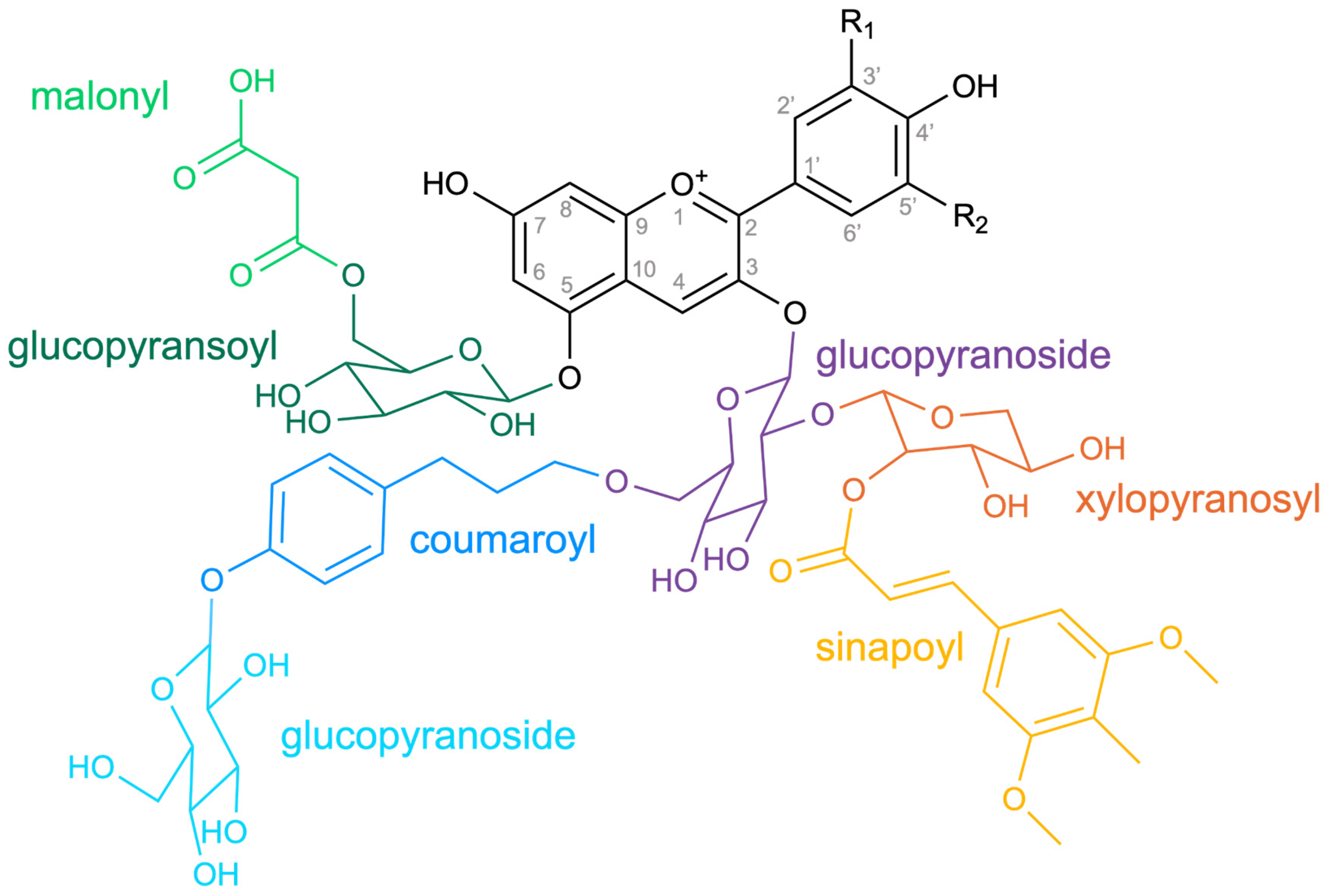
Figure 1.
A11 (cyanidin 3-O-[2-O(2-O-(sinapoyl)-β-D-xylopyranosyl)-6-O- (4-O-(β-D-glucopyranosyl)-p-coumaroyl-β-D-glucopyranoside] 5-O-[6-O-(malonyl) β-D-gluco-pyranoside]) is the major anthocyanin in the leaves and stems of Arabidopsis thaliana [29]. The IUPAC name was retrieved from PubChem [30]. The visualization of stereochemistry might deviate slightly due to spatial organization.
Enzyme Promiscuity Associated with the Anthocyanin Biosynthesis
Catalytic promiscuity is a feature of the specialized plant metabolism that can help to explain the plethora of different metabolites produced by a comparatively small number of enzymes and forms the basis for the evolution of novel enzymes [36]. One prominent example in the flavonoid biosynthesis is the first committed enzyme CHS, which catalyzes the production of naringenin chalcone, but also has the ability to produce other polyketides [37,38]. The binding of CHI-like (CHIL) proteins to CHS rectifies this promiscuity and ensures the efficient production of precursors for the following step in the flavonoid biosynthesis, but does not substantially increase the activity of CHS as initially assumed [37,38,39,40]. CHS duplication and subsequent loss of CHIL interaction ability resulted in the evolution of novel enzyme functions through neofunctionalization [37]. The interaction of CHS and CHIL appears conserved across all land plants and CHILs show species-specific affinity for CHS [37]. Another striking example of catalytic promiscuity is provided by members of the large 2-oxoglutarate-dependent dioxygenase family that are active in the flavonoid biosynthesis: F3H, flavonol synthase (FLS), and ANS [41,42,43,44,45,46]. While these enzymes predominantly catalyze different reactions, they can partially catalyze each other’s reaction with the extent of these side activities appearing species specific [41,42,43,44,45]. Gene duplication followed by neofunctionalization has been identified as an important factor contributing to the metabolic diversity across species [47]. In the Apiaceae, FNS I was identified as another member of this gene family that evolved through gene duplication from F3H [42,48]. However, F3H is assumed to have evolved from a promiscuous ancestor that harbored flavone synthase activity [44]. In summary, genes encoding promiscuous enzymes that undergo specialization after gene duplications appear to be a predominant theme in the evolution of the flavonoid biosynthesis.
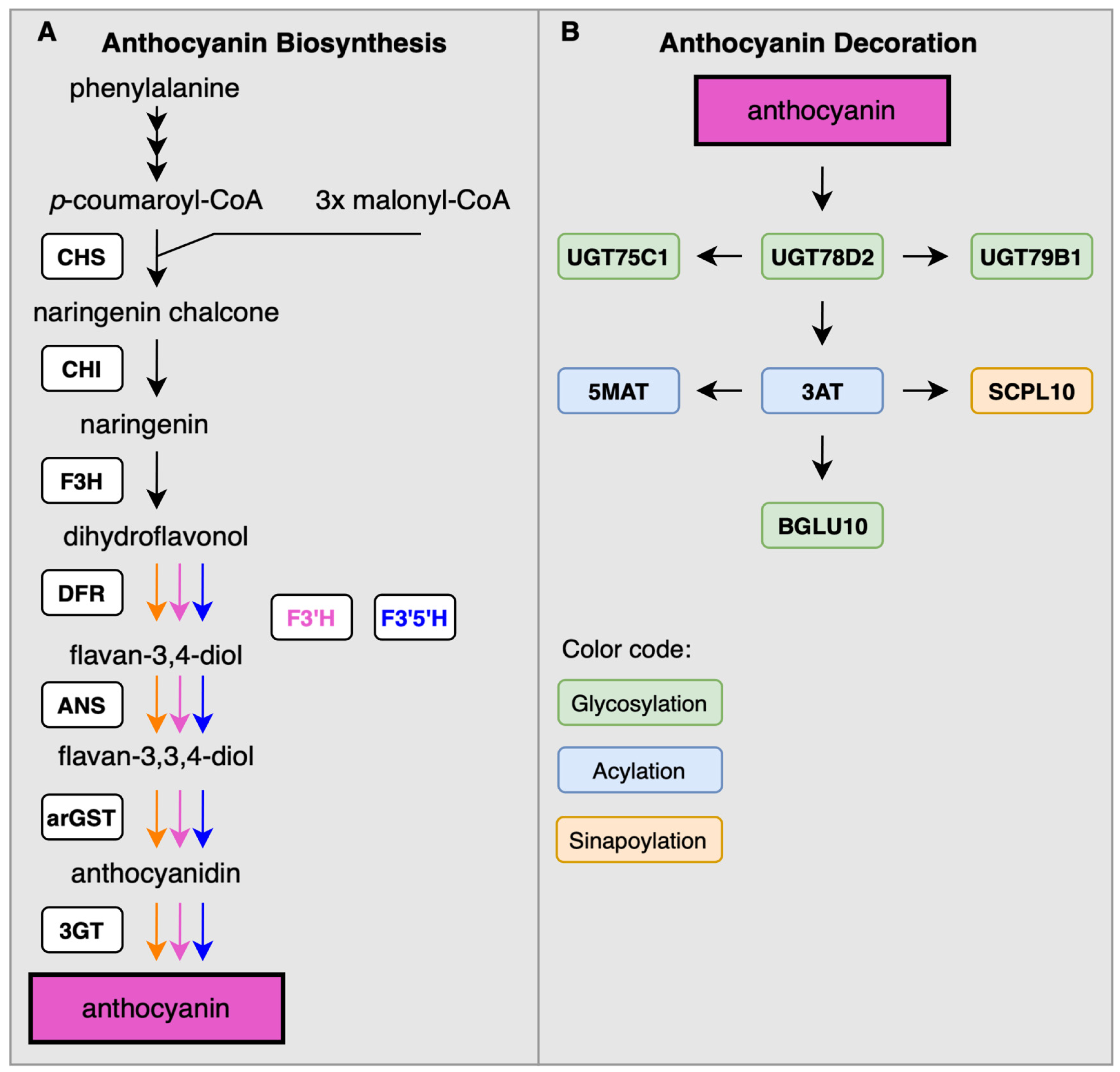
Figure 2.
Simplified illustration of the flavonoid biosynthesis with focus on the anthocyanin branch (A). Enzymes involved in the modification of anthocyanins as reported in A. thaliana (B). Displayed enzymes are chalcone synthase (CHS), chalcone isomerase (CHI), flavanone 3-hydroxylase (F3H), flavonoid 3’-hydroxylase (F3’H), flavonoid 3’,5’-hydroxylase (F3’5’H), dihydroflavonol 4-reductase (DFR), anthocyanidin synthase (ANS), anthocyanin-related glutathione S-transferase (arGST), UDP-dependent anthocyanidin-3-O-glycosyltransferase (3GT), UDP-glucosyltransferase 75C1 (UGT75C1), UDP-glucosyltransferase 78D2 (UGT78D2), UDP-glucosyltransferase 79B1 (UGT79B1), Malonyl-CoA:anthocyanidin 5-O-glucoside-6’’-O-malonyltransferase (5MAT), Coumaroyl-CoA:anthocyanidin 3-O-glucoside-6’’-O-coumaroyltransferase (3AT), sinapoyl-β-D-glucose:anthocyanin sinapoyl transferase 10 (SCPL10), and β-glucosidase 10 (BGLU10).
Diversity of Anthocyanin Decoration
Genes associated with the decoration of anthocyanins are less conserved and thus knowledge about them beyond A. thaliana is sparse in the literature. Studies in A. thaliana identified a range of genes that are associated with the anthocyanin decoration including UGT75C1/At4g14090 [49], UGT79B1/At5g54060 [50], 3AT1/At1g03940 and 3AT2/At1G03495 [51], BGLU10/At4g27830 [52], 5MAT/At3g29590 [51], and SCPL10/At2g23000 [53]. Glycosylation of anthocyanidins constitutes the final step of the anthocyanin biosynthesis. Glucosyltransferases can be classified based on the source of the sugar moiety: UDP-glucose or acyl-glucose. Only the UDP-dependent glycosyltransferases appear to be relevant enzymes for the transfer of other sugar moieties besides glucose. Usually, anthocyanidins are first glycosylated at the C3 position. Tohge et al. (2005) demonstrated that UGT78D2 (At5g17050) catalyzes this glycosylation of anthocyanidins at the C3 positions. Like many other UGTs, UGT78D2 also shows a high substrate promiscuity: In A. thaliana and Vitis vinifera, 3-O-glycosylation of flavonols is also catalyzed by UGT78D2 [49,54]. In Glycine max, GmUGT78K1 from black seed coat showed 3-O-glycosylation activity of anthocyanidins and flavonols [55]. Furthermore, UGT78G1 (GT83F) was identified as another enzyme that can perform 3-O-glycosylation of pelargonidin and cyanidin, while mostly acting on other flavonoids [56,57]. Interestingly, this enzyme has been reported to also catalyze deglycosylation of anthocyanidin 3-O-glycosides, i.e., the removal of a sugar moiety from an anthocyanin [57]. Following the 3-O-glycosylation, an additional 5-O-glycosylation is possible. Several studies reported 5-O-glycosyltransferases in a range of plant species. The best studied UDP-glycosyltransferase adding a glucose at the 5-position of the anthocyanin A-ring is the A. thaliana UGT75C1/AAt4g14090 [49]. UGT75C1 is responsible for glycosylation of anthocyanidin 3-O-β-D-glucosides leading to a 3-O-5-O-diglucoside product. Anthocyanidin 3-O-5-O-diglucosides have been reported to be more stable and soluble, responsible for bright-purple flower coloration [58] and form the basis for further complex modifications, e.g., sinapoylation or coumaroylation. Multiple UDP-dependent 5GTs were identified in Dahlia variabilis [59], Petunia hybrida [60], and Gentiana triflora [61]. Nakatsuka et al. (2008) showed that the Gt5GT accepts different anthocyanidin 3-O-beta-D-glucosides as substrates whereas the enzyme does not show activity using aglyconic anthocyanidins as substrates [61]. As reported for multiple UGTs, UGT75C1 also has a high promiscuity. SlUGT75C1 glycosylates abscisic acid (ABA) and IAA [62]. This example shows additional roles of UGT75C1 in fruit ripening and drought resistance in Solanum lycopersicum thus supporting a broad substrate promiscuity even beyond anthocyanins. Despite this high promiscuity, AtUGT75C1 does not appear to participate noticeably in flavonol glycosylation [63]. To the best of our knowledge, there are no reports about UDP-dependent enzymes catalyzing a 7-O-glycosylation of anthocyanins. However, DgAA7GT from Delphinium grandiflorum, an acyl-glucose-dependent glycosyltransferase, is proposed to glycosylate anthocyanidin 3-O-glycosides and anthocyanidin 3-O-malylglycosides [64]. In monocotyledones, AaAA7GT from Agapanthus africanus was found to 7-O-glycosylate anthocyanidin 3-O-glycosides, 3-O-galactosides and 3-O-rutinosides [65]. Both AA7GTs do not accept aglyconic anthocyanidins as substrates. Various galactosylated anthocyanidins were reported in different Actinidia species: cyanidin- and delphinidin 3-O-galactosides, as well as cyanidin- and delphinidin 3-[2-(xylosyl)galactosides], requiring the activity of galactosyltransferases [66]. In Vigna mungo, an UF3GaT was described, showing high UDP-dependent 3-O-galactosylation activity for different flavonols and anthocyanidins [67]. UCGalT1, also an UDP-dependent galactosyltransferase, was identified in purple carrots (Daucus carota) taproots and purple celery (Apium graveolens) [68, 69]. Recombinant expression of DcUCGalT1 as well as AgUCGalT1 in Escherichia coli led to the presence of 3-O-galactosidated anthocyanins. Both enzymes did not showed activity with either UDP-glucose or UDP-xylose as a sugar donor but had a high promiscuity regarding the sugar acceptor: Both UCGalT1 showed activity when incubated with flavonols (quercetin and kaempferol) and UDP-galactose, whereas DcUCGalT1 also catalyzed the galactose transfer of UDP-galactose to cyanidin, peonidin and pelargonidin [68, 69].
3-O-glycosylation is the prerequisite for multiple further modification steps. The addition of another sugar to the 3-O-glycosyl residue of the anthocyanin leads to formation of disaccharide residues, e.g., the attac[68,69hment of rhamnose to 3-O-glycosylated anthocyanins results in anthocyanin-rutinosides [70]. Multiple anthocyanin rutinosides in different plant species, especially Petunia and Solanum, were detected [27]. Among others, 6-hydroxycyanidin 3-rutinoside, cyanidin 3-rutinoside, pelargonidin 3-rutinoside were found in Alstroemeria cultivars [71]. The first rhamnosyltranserases were described in Silene dioica and Petunia, catalyzing the addition of a rhamnosyl group to 3-O-glycosylated and 3,5-diglycosylated anthocyanins [72,73,74]. In 2013, Cs1,6RhaT, encoding for an anthocyanin 3-O-glycoside 1,6-rhamnosyltransferase, was discovered, catalyzing the formation of peonidin- and cyanidin rutinosides in Citrus species with a high promiscuity, showing affinity for flavanones, flavones, and flavonols [75]. Interestingly, the gene is only common to non-bitter Citrus species, whereas bitter-tasting Citrus species have the gene Cm1,2RhaT, encoding a 1,2-rhamnosyltransferase that rhamnosylates flavanone-7-O-glycosides [76]. In Lobelia erinus, two UDP-dependent rhamnosyltransferases were found (ABRT2 and ABRT4) to rhamnosylate anthocyanin 3-O-glycosides [77].
In contrast to UDP-dependent glycosylation, AAGTs are acyl-glucose-dependent anthocyanin glucosyltransferases belonging to the glycoside hydrolase family 1 (GH1) [64]. Multiple BGLUs are shown to be responsible for encoding GH1-type glycosyltransferases using already substituted flavonoids as a substrate in A. thaliana [52,78,79]. However, only AtBGLU10 seems to play a role in anthocyanin modification [52,78], whereas BGLU1, BGLU3, BGLU4, and BGLU6 prefer flavonols over anthocyanins as substrates [79,80].
The most common anthocyanins have a xylosyl-group attached to the 2’’-position of the 3-O-sugar. In A. thaliana, AtUGT79B1 is known to catalyze the sugar attachment to 3-O-glycosylated anthocyanins [50]. Different anthocyanins can act as sugar acceptors, i.e., cyanidin 3-O-glucoside, delphinidin 3-O-glucoside and pelargonidin 3-O-glucoside. Notably, no activity was observed with cyanidin 3-O-rhamnoside, whereas activity was detected when cyanidin 3-O-rhamnosyl(1→6)glucoside was used as the sugar donor [50]. The enzyme also has a high promiscuity as it shows a high activity with the flavonols kaempferol 3-O-glucoside and quercetin 3-O-glucoside. Nevertheless, UDP-xylose is the only accepted sugar donor [50].
Addition of acyl groups to anthocyanins is another common modification of anthocyanins. In crops alone, various acylated anthocyanins were reported in 23 species [81]. Acylation of anthocyanins leads to higher activity, increased stability, and altered polarity which enhances the antioxidant effect and enables the usage as dye in industry [81]. In A. thaliana, the majority of the known anthocyanin-related acyltransferases belong to the BAHD superfamily. For At3AT1 (At1g03940) and At3AT2 (At1G03495), different acyl donors (p-coumaroyl-CoA, feruloyl-CoA and caffeoyl-CoA) and acyl acceptors (cyanidin 3-glucoside, pelargonidin 3-glucoside, malvidin 3-glucoside, quercetin 3-glucoside, kaempferol 3-glucoside and kaempferol 7-glucoside) are known [51]. At5MAT (At3g29590) also has a high promiscuity, as it acylates multiple 3-O-glycosylated anthocyanins, i.e., cyanidin 3,5-diglucoside, cyanidin 3-coumaroylglucoside 5-glucoside, cyanidin 3-O-(xylosyl) 6’’-O-p-coumaroyl glucoside 5-O-glucoside (A3) and delphinidin 3-coumaroylrutinoside 5-glucoside [51]. However, SCPL10, belonging to the serine carboxypeptidase-like gene family, catalyzes a sinapoyl-glucose dependent acylation of anthocyanins in A. thaliana [53].
Transcriptional Regulation of Anthocyanin Biosynthesis
Crucial for the metabolic flux control through the flavonoid biosynthesis is the transcriptional regulation of enzyme-encoding genes (Figure 3). Many MYB transcription factors, members of the largest transcription factor family in plants, play crucial roles in this regulation [16,82,83,84]. Genes of the anthocyanin biosynthesis branch are activated by a complex of multiple transcription factors including a MYB, a bHLH, and a WD40 protein leading to the name MBW complex [85,86]. It is generally assumed that the MYB component is determining the target gene specificity of this complex, because different MYB proteins can be incorporated into this complex leading to variations in the set of target genes [16,87,88]. Although the idea of a single transcription factor complex regulating all anthocyanin biosynthetic genes appears straightforward, extensive gene duplications across various taxonomic levels have rendered the regulatory network highly complex. Initially, genes in the flavonoid biosynthesis have been classified as early (CHS, CHI, F3H) and late (DFR, ANS, UFGT) due to the assumption that the transcriptional control would be distinct. However, discoveries of several transcription factors contributing to the anthocyanin biosynthesis regulation question this system.
Lineage-Specific Differences in the MBW Complex
Since anthocyanin activating MYBs have been investigated in a large number of plant species, a pattern emerged that implicated different MYB subgroup 6 (SG6) members in the activation of the anthocyanin biosynthesis. They are generally interacting with bHLHs of the IIIf subgroup. Initial studies in Zea mays discovered R and B as two copies of an anthocyanin-regulating bHLH gene [89]. COLORLESS1 (C1) is a MYB transcription factor that was also identified as a crucial factor in the anthocyanin biosynthesis regulation of maize [90]. In A. thaliana the MYB75 (PAP1), MYB90 (PAP1), MYB113, and MYB114 are anthocyanin regulators and also very close homologs suggesting an evolutionary recent radiation [86,91]. Anthocyanin activating MYBs from monocot species like banana do not efficiently interact with the same bHLHs as MYBs from dicots like A. thaliana [92,93]. There is also an additional component, the WRKY protein TTG2, that can be associated with the MBW complex [94,95,96]. The current model suggests that TTG2 is activated by the MBW complex and that the addition of the WRKY component to the MBW complex increases the specificity for the vacuolar transport associated genes TT12 and TT13 [88,96]. PH3 is a petunia gene with a function similar to the Arabidopsis TTG2 [95] thus suggesting that the involvement of WRKY is conserved across plant species. Anthocyanin biosynthesis activating WRKYs have also been described in pear and apple, but at least apples MdWRKY40 is reported to be functionally divergent from PH3 and TTG2 [97,98].
There are also reports that anthocyanin biosynthesis activation is possible without the canonical MBW complex. A recent study in blueberry revealed that proanthocyanidin biosynthesis regulators like TT2/MYB123 (subgroup 5, SG5) can contribute to the anthocyanin regulation [99]. Several studies suggested that MYB5 orthologs are also able to activate the anthocyanin biosynthesis [100,101,102]. In Vitis, VvMYB5a and VvMYB5b can activate specific genes in the anthocyanin biosynthesis with VvMYB5b being able to slightly trigger the accumulation of anthocyanins when heterologously expressed in unpigmented petunia [100]. Additionally, it seems that only VvMYB5a and VvMYB5b, but not the MYB75/PAP1/AN2 ortholog VvMYBA1 are able to activate the HYDROXYLATION AT FIVE (HF2, F3’5’H) expression [100]. It is important to note that Vitis also harbors VvMYBA1 and VvMYBA2 as orthologs of PhAN2 which seem to be the dominant activator of most anthocyanin biosynthesis genes [100,103,104,105]. Studies in strawberries reported MYB5 as an anthocyanin regulator that is TTG1-independent, but forms a MBW complex with EGL3 (bHLH) and LWD1 (WD40) instead [101]. Reports in other species also associated MYB5 with anthocyanin biosynthesis: PhPH4, also belonging to the MYB5 lineage, regulates vacuolar acidification in petunia [106], and SmMYB5 activates anthocyanin biosynthesis in eggplant [102]. Additionally, MYB5a/NEGAN is responsible for the pigmentation pattern in Mimulus [107]. While studies in many species identified MYB5 orthologs as anthocyanin biosynthesis regulators CsMYB5a and CsMYB5e in Camellia sinensis activate proanthocyanidin biosynthesis, but not anthocyanin biosynthesis [108] and A. thaliana MYB5 seems not tightly connected to the anthocyanin biosynthesis. In summary, MYBs of the SG6 are important regulators of the anthocyanin biosynthesis, but some members of the SG5 lineage might also activate specific genes in the anthocyanin biosynthesis. This could represent a mechanism for fine-tuned activation of specific branches within the complex anthocyanin metabolism, but further systematic investigations are required.
Additional Transcription Factors Influence Anthocyanin Biosynthesis
A number of other transcription factors have been implicated in the regulation of anthocyanin biosynthesis genes including ethylene responsive factor 109 (ERF109), NAM/ATAF/CUC2 (NAC), MADS-box, basic leucine zipper (bZIP), B-box domain protein 22 (BBX), and phytochrome interacting factor 3 (PIF3). Many of these transcription factors activate genes encoding MBW components rather than the actual anthocyanin biosynthesis genes. In Malus domestica, ERF109 activates the anthocyanin biosynthesis in early stages of apple coloration by binding promoters of MdCHS, MdUFGT, and MdbHLH3 [109]. Another study in red-skinned pear reported the interaction of PyERF with the anthocyanin regulators PyMYB114 and PybHLH3 to co-regulate the anthocyanin biosynthesis [110]. NAC proteins have been identified as activators of the anthocyanin biosynthesis in Arabidopsis thaliana and Prunus persica [111,112]. NACs probably trigger anthocyanin pigmentation through up-regulation of the important anthocyanin biosynthesis activating MYB as this was observed for a BLOOD/PpNAC1 heterodimer in the coloration of blood-fleshed peach during fruit ripening [112]. Since the expression pattern of the SQUAMOSA-class MADS-box gene tapetum degeneration retardation 4 (TDR4) is tightly correlated with the anthocyanin pigmentation in bilberry fruits (Vaccinium myrtillus), it was postulated that this transcription factor directly or indirectly controls the anthocyanin biosynthesis genes [113]. In A. thaliana, the bZIP protein long hypocotyl 5 (HY5) binds to the promoter of MYB75/PAP1 and triggers the anthocyanin biosynthesis through up-regulation of this MYB gene [114]. This result is supported by a study in Malus domestica that identified MdHY5 as an activator of the anthocyanin regulator MdMYB10 [115]. Another bZIP protein, PybZIPa, was reported as an activator of the anthocyanin activating MYB and UFGT, but not orthologous to HY5 [116]. The A. thaliana BBX protein BBX22/LZF1 was identified as an activator of the anthocyanin-regulating MYB75/PAP1 [117]. HY5 was discovered as a factor contributing to the expression of BBX22/LZF1 [117]. The apple BBX protein MdCOL11, ortholog of AtBBX22, is interacting with MdHY5 to activate the anthocyanin regulator MdMYBA [118]. A study in rice identified that OsBBX14 and OsHY5 interact to activate the anthocyanin regulating MYB and bHLH gene [119]. A study in red pear revealed that PpBBX16 interacts with PpHY5 to activate the expression of anthocyanin biosynthesis genes [120]. In summary, this suggests that HY5 usually acts in a complex with a BBX protein. In A. thaliana, PIF3 is another light-responsive transcription factor that can activate the anthocyanin biosynthesis, but requires the simultaneous binding of HY5 [121]. Another positive regulator of the anthocyanin biosynthesis in A. thaliana, especially associated with up-regulation of the UFGT, is the phytochrome-interacting ankyrin repeat protein 2 (PIA2) [122]. MYB112 was reported to trigger anthocyanin biosynthesis and block flavonol biosynthesis under salt and high light stress conditions, by activating the anthocyanin activator PAP1 and repressing the flavonol activators MYB12/MYB111 [123]. Not all transcription factors of the anthocyanin biosynthesis are activators. A comprehensive review summarized the role of various factors that repress the expression of anthocyanin biosynthesis genes [84]. Examples for characterized MYB repressors connected to the anthocyanin biosynthesis are PhMYB27 in petunia [124], FaMYB1 in strawberry [125], CAPRICE (CPC) in Arabidopsis [126], SQUAMOSA PROMOTER BINDING PROTEIN-LIKE 9 (SPL9) group in Arabidopsis [127], MYBL2 in Arabidopsis [128], MYBC2-L1 and MYBC2-L3 in grapevine [129], MYBF2 in ginkgo [130], and MaMYB4 in banana [131]. Many studies about regulators of the anthocyanin biosynthesis have observed this role only in a single plant species. It was previously observed that changes in the transcriptional regulation explain a majority of evolutionary anthocyanin pigmentation shifts at the species and genus level [16]. This suggests a high flexibility of the transcriptional control of the anthocyanin biosynthesis during evolution. Therefore, it is feasible that at least some of the large number of reported regulators are species- or lineage-specific features of the anthocyanin biosynthesis. There is a need for systematic studies that explore these transcription factors across a wide range of different plant species.
Regulatory RNAs in the Anthocyanin Biosynthesis
Activity of the anthocyanin biosynthesis is also controlled by regulatory RNAs. There are microRNAs (miRNAs) that suppress target genes, small RNAs that suppress their target gene, and long non-coding RNAs (lncRNAs) that counteract the miRNAs by target mimicry, i.e., sequestering miRNAs without cleavage of the lncRNAs [132,133,134,135]. Expression patterns of certain lncRNAs were associated with the accumulation of anthocyanins, but evidence for the molecular mechanism is missing [136]. A small RNA originating from an inverted duplication at the SULF locus results in suppression of the 4’CGT involved in aurone biosynthesis, which causes an increased synthesis of anthocyanins [135]. Anthocyanin accumulation in Arabidopsis is increased by miR156 that targets anthocyanin repressors of the SPL9 group [127]. The authors demonstrated that SPL9 captures PAP1 proteins and prevents the formation of the MBW complex that is required for the activation of anthocyanin biosynthesis genes. A similar system comprising miR156a and SPL9 as negative regulators of the anthocyanin biosynthesis was reported as the mechanism explaining the coloring of the peel in red pear [137]. Two long non-coding natural antisense transcripts (lncNATs) have been reported as repressors of the anthocyanin biosynthesis activators DcMYB6 and DcMYB7 [138]. A regulatory system identified in Malus spectabilis under nitrogen starvation comprises miR858 as a repressor of MYB62-like, which in turn represses the anthocyanin biosynthesis, thus high expression of miR858 leads to anthocyanin accumulation [134]. This study also identified eTM858-1 and eTM858-2, two target mimics of miR858, which reduce the activity of miR858 on MYB62-like thus leading to a repression of the anthocyanin biosynthesis [134]. In sea buckthorn, LNC1 and LNC2 (TCONS_00694050 and TCONS_00438839) were reported as endogenous target mimics of miR156a and miR828a, respectively [133]. While miR156a targets the anthocyanin biosynthesis repressor SPL9, miR828a targets the anthocyanin biosynthesis activator MYB114 [133]. Consequently, a high abundance of LNC1 leads to reduced anthocyanin accumulation in the fruit, while higher abundance of LNC2 results in a higher anthocyanin accumulation [133]. In apple, MdLNC499 activates the expression of MdERF109, which encodes a transcriptional activator of the anthocyanin biosynthesis genes CHS, bHLH3, and UFGT [109].
A large proportion of knowledge about the anthocyanins biosynthesis originates from research on A. thaliana, which limits the potential for discoveries to regulatory mechanism present in this species. The anthocyanin biosynthesis is generally considered one of the best studied pathways in plants. It is widely believed that this pathway including its regulation is well conserved across plants, which might be reinforced by numerous recent studies only focusing on the well characterized genes of the core anthocyanidin biosynthesis in new plant species. In the light of recent discoveries summarized above, it appears highly likely that there are lineage-specific features of the anthocyanin biosynthesis that could be discovered through comparative investigations.
Transport of Anthocyanins
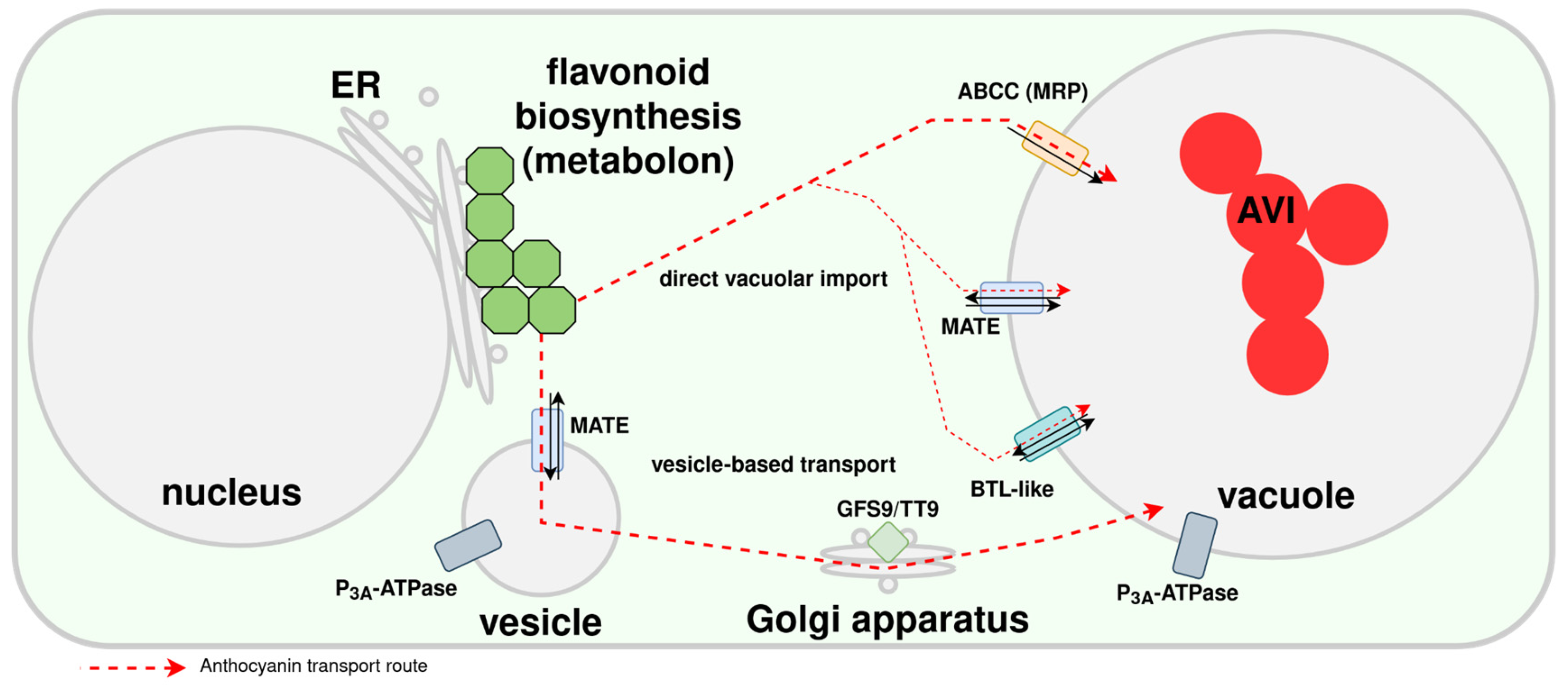
Anthocyanins are produced at the endoplasmatic side of the ER and require transportation into the central vacuole for long term storage [139] (Figure 4). Details regarding the intracellular transport of anthocyanins have eluded researchers as results of previous studies seem to contradict each other. Different ABCC proteins have been implicated in the transport of anthocyanins across a membrane [140,141,142]. However, it is not clear whether anthocyanins are imported into the ER and transferred to the vacuole or transported through the cytoplasm and then imported into the central vacuole [139]. The mechanism could differ between plant species or both routes could contribute to the anthocyanin transport. The discovery of TT9, a vesicle associated protein, as a crucial factor for proanthocyanidin pigmentation in A. thaliana [143] could suggest that anthocyanin transport is also occurring through vesicles. MATE (TT12) transporters have also been proposed as potential anthocyanin transporters [144]. They would require a proton gradient that could be maintained by AHA10/TT13 [145]. However, it was also suggested that MATEs are not transporting anthocyanins, but proanthocyanidin precursors [146]. Previously, it was postulated that TT19, a glutathione S-transferase (GST), serves a ‘ligandin’, an anthocyanin protection protein, during the transport through the cytoplasm [147,148,149]. However, a recent study demonstrated an enzymatic function of this anthocyanin-related GST (arGST) in the synthesis of cyanidin [32]. While this finding does not rule out an arGST function in the transport of anthocyanins, it provides an alternative explanation for previous observations that led to the postulation of the arGST function in the anthocyanin transport. Understanding the transport of anthocyanins is crucial as some modification reactions take place in the central vacuole, i.e., after the successful translocation [150]. Anthocyanin localization within the cell can influence the phenotypic appearance of the plant. Clusters of anthocyanins can be dispersed rapidly, altering the color of the plant organ [151]. Changes in the vacuolar acidity can quickly change the appearance of anthocyanins as these are depending on the pH [152]. While there is substantial research on the intracellular transport of anthocyanins, not much is known about the transport of anthocyanins between plant cells or even different organs [139]. Since all plant cells should be able to produce anthocyanins based on phenylalanine, transport between cells might not occur at a relevant level.
Cell-Specific Accumulation of Anthocyanins and Pigmentation Patterns
Pigmentation patterns mostly serve the attraction of pollinators by boosting the visibility of the flower to increase the number of visitation events. Such pigment cues are widespread across angiosperms and represent a general strategy to optimize pollinator foraging efficiency [153,154]. This can be achieved through high contrast patterns or special flower patterns [153,154,155]. These floral patterns are usually colored in a shade or color contrasting the rest of the flower and provide information for the pollinator on the location and orientation for the landing on the flower [156]. From there, spatially pigmented nectar guides can lead the way to the nectaries thus substantially increasing pollination success [155,157]. Anthocyanin pigmentation patterns can usually be attributed to the spatial expression of general flavonoid and particularly anthocyanin biosynthesis related genes. Anthocyanin biosynthesis genes are regulated by an ensemble of transcription factors that are active in very specific flower parts and tissues [155,158,159]. The anthocyanin biosynthesis promotion MYB is the most specific transcription factor in most plant species [16]. Different tissue types of the flower petal can contribute to specific spatial pigmentation of the tissue or the adjacent cells. This can be seen in Antirrhinum in the phenotype Venosa where the transcription factor VENOSA from the vascular tissues leads to red coloration of the adjacent adaxial epidermis [160]. In Petunia hybrida, the MADS-box gene DEFICIENS was reported as a cell layer-specific transcription factor in flowers that influences the pigmentation pattern through control of the expression of the major anthocyanin biosynthesis regulating MYB ANTHOCYANIN2 [161]. In Gossypium barbadense a MYB transcription factor is essential for the formation of one large spot at the base of the petal which attracts pollinators to the flowers more efficiently [162]. The petal spot formation in Gorteria diffusa is also controlled by an anthocyanin MYB that triggers the malonylation of anthocyanins specifically in this petal region [163].
Figure 4.
Simplified illustration of proposed anthocyanin transport routes from the endoplasmic reticulum (ER) to the central vacuole. Figure is based on a previous version published in Pucker & Selmar, 2022. Shown are ABCC (ATP-binding cassette (ABC) subfamily C), MATE (Multidrug And Toxin Extrusion transporter), BTL-like (bilitranslocase-like), GFS9/TT9 (Green Fluorescent Seed 9/Transparent Testa 9), and AVI (anthocyanin vacuolar inclusion). Green hexagons represent the anthocyanin biosynthesis enzymes at the ER.
Several plant species display intensely pigmented spots on their petals that appear to be randomly distributed. For example, in Mimulus lewisii, Mimulus guttatus, and Digitalis purpurea the lower petal contains an area with multiple spots contrasting the background [159,164,165]. This area is centered on the petal and forms nectar guides in Mimulus [159] and a putative landing site in Digitalis [165]. In Mimulus, it was shown that the basis of spot formation is an activator-repressor-relationship of two transcription factors (Figure 5), which is conforming with a reaction-diffusion model proposed by Turing and Gierer & Meinhardt [159,166,167]. This model assumes a relationship between an activator and a repressor, in which the activator (in this case the R2R3-MYB NEGAN) enhances its own expression and, in this case, upregulates the anthocyanin biosynthesis genes [159]. Through diffusion of the activator into adjacent cells, an anthocyanin-pigmented spot develops [159]. At the same time the activator upregulates a repressor gene (here: R3-MYB MlRTO) which competes with the activator for binding with the bHLH and WD40 partners [159]. Thereby it reduces the functionality of the activator and therefore the expression of anthocyanin biosynthesis genes [159]. Both regulators diffuse over cell boundaries. The repressor diffuses faster and represses the anthocyanin biosynthesis gene activity in cells adjacent to the cells where the activator is active [159]. The pigmented spot is confined to the specific boundaries being set by the diffusion- and degradation-properties of both transcription factors. White halos surrounding pigmented spots on otherwise differently pigmented backgrounds can be explained by the repressor activity in the spot-adjacent cells [155,159]. These examples of spatially restricted pigmentation illustrate the diversity of floral color patterns, which can arise through different molecular mechanisms. Another striking case presents itself in the Petunia hybrida ‘Red Star’ variety. Flowers of this variety display a white star-like pattern on a red petal background [168]. It was shown that spatial post transcriptional gene silencing of the PhCHS A mRNA is responsible for the lack of pigmentation around the center vein of the petals [168]. The resulting lack of CHS A enzymes in this tissue leads to the disruption of the early flavonoid pathway and a subsequent block of anthocyanin production.
Depending on their localization in certain cell layers (spongy palisade mesophyll, upper epidermis, lower epidermis), the same anthocyanins can lead to different colors of plant structures as recently observed in leaves of Tipularia discolor [25]. A large study investigating hundreds of plant species reported that mesophyll is the most frequent location of anthocyanins and only 24% of the studied species displayed anthocyanins in the epidermis [169]. The huge diversity of anthocyanin accumulation patterns reported is strong support for a broad range of different anthocyanin functions [169,170]. In contrast, anthocyanins have been reported to accumulate in the epidermis of A. thaliana plants under nitrogen starvation [171]. A study on various crucifers also reported high anthocyanin concentration in the epidermis with spread into the mesophyll occurring only at very high anthocyanin concentrations [172]. Understanding the localization of anthocyanins inside plants is a remaining challenge that can help to understand their physiological function and evolutionary relevance.
Ecological Functions of Anthocyanins
Functions of anthocyanins might differ depending on developmental state, plant organ, or environmental conditions. For example, anthocyanins in leaves primarily provide protection against high light intensities and other stress factors [1,173]. In flowers and fruits, anthocyanins are mainly involved in pollinators and seed disperser attraction, but also contribute to protection against environmental stresses [174,175,176,177,178,179]. Understanding the ecological functions of anthocyanins is crucial when generating hypotheses about evolutionary trajectories that lead to the anthocyanin biosynthesis as we see it today. For example, the loss of anthocyanins may coincide with changes in the pollination system or in seed dispersal syndrome.
Protective Functions of Anthocyanins in Photosynthetically Active Plant Organs
Anthocyanins protect green plant parts from excessive light intensities and oxidative stress. While the leaves of many plants turn red in response to high light intensities [180,181], other species show constant red leaf pigmentation [182,183,184]. The red color of anthocyanins is due to a strong absorbance of green and blue-green light which could damage cell components [185,186,187]. Anthocyanins can act as sunscreen shielding the leaf tissue from excess light that would otherwise be captured by chlorophyll b [182,188,189]. The maximum photosynthesis of leaves with anthocyanin pigmentation was increased compared to green leaves and photoinhibition was reduced [182,190]. The red undersurface of understory plants or floating leaves of aquatic plants is probably not backscattering red light as initially hypothesized, but helps the plants to cope with high-intensity sun flecks [182,191]. In addition to protection against excess light, anthocyanins have also been reported as antioxidants that quench reactive oxygen species (ROS) that could otherwise damage cell structures [192,193]. While ROS are mainly produced in chloroplasts, anthocyanins are transported into and stored in the central vacuole [139]. This brings up the question how anthocyanins can counteract ROS without physical proximity. Photooxidative stress leads to the inactivation of the ascorbate peroxidase, an enzyme that normally degrades H2O2 [193,194]. H2O2 is the ROS type that temporarily accumulates in plastids under photooxidative conditions and can move into the vacuole either through passive diffusion or through proteins located in the tonoplast [195,196,197]. The anthocyanin-filled vacuole would become a H2O2 sink which can explain the ROS scavenging through anthocyanins accumulated in the vacuole probably catalyzed by peroxidases [193,198,199]. This hypothesis aligned with the observation that ROS quenching by anthocyanins is substantially weaker in species like Rosa sp. and R. communis where anthocyanins are not located in the same cells as the plastid accumulating H2O2 [193]. A systematic study revealed that the photoprotective function of anthocyanins is especially important at low temperatures and high light intensities, when other photoprotection mechanisms are less effective [200]. Anthocyanin levels might also be regulated in response to ROS to ensure that excess ROS is scavenged [201]. The photoprotective role of anthocyanins is not restricted to leaves, but also extends to photosynthetically active stems, flowers, and fruits [178,202]. A protective function of anthocyanins could explain their presence in young leaves, seedlings, or developing fruits [203,204,205,206]. These emerging structures require a protective anthocyanin pigmentation, because light capture ability develops before CO2 assimilation capacity thus protection against photoinhibition is needed [207,208]. The de novo synthesis of anthocyanins in senescent leaves during autumn did appear less obvious and turned into an intensely studied field with many hypotheses arising to explain this phenomenon [188,209,210,211,212]. Anthocyanins can delay senescence and ensure functionality of leaves which improves the resorption of nitrogen and phosphorus in autumn [188,213,214]. Anthocyanin formation during autumn leading to red leaves is more pronounced in species that grow in areas with shorter vegetation periods and are more often experiencing cold snaps [212]. In light of the crucial role of anthocyanins in photoprotection, it has been proposed that this physiological function in photosynthetic tissues evolved prior to their role as visual cues for pollinators in floral tissues [182].
Importance of Anthocyanins in Drought and Salt Stress Response
Plants under drought stress caused by limited water availability or increased salt concentrations were often observed to turn red through the accumulation of anthocyanins. Studies exploring the transcriptomic or metabolomic changes report a generally increased activity of the flavonoid biosynthesis without specific explanations how the anthocyanin biosynthesis is increased [215,216,217,218]. Therefore, it is plausible that ROS scavenging is performed by anthocyanins and other flavonoids, i.e., the enrichment of anthocyanins is the consequence of generally increased activity of the flavonoid biosynthesis. A specific function of anthocyanins might be capturing excess light as photosynthesis cannot take place in the absence of water. In Arabidopsis, the two glycosyltransferases UGT79B2 and UGT79B3 were identified as important targets for activation of the anthocyanin biosynthesis under drought and salt stress conditions, but also in response to cold stress [219]. The importance of flavonols in response to salt stress was investigated based on flavonol deficient mutants that were more adversely affected than the wild type [220]. Given that the flavonol and anthocyanin biosynthesis branches are competing for shared substrates [221], this observation suggests that generally antioxidants, but not specifically anthocyanins, are needed under these stress conditions. An investigation of carrot cell cultures under salt stress discovered the importance of MATE in the increased anthocyanin accumulation [222], which could indicate that a generally increased transport across membranes to counteract the osmotic challenges increases anthocyanin transport as a side effect. Anthocyanins account for less than 1% of the osmotic potential in a plant cell which makes a role as compensating salts unlikely [211,223]. The sparsity of data about anthocyanin regulation in response to drought and salt stress suggests that their role in this context is rather minor and thus more challenging to resolve. This suggests that drought and salt stress response were not among the factors that contributed to the evolution of the anthocyanin biosynthesis. Although genetically engineered crops with increased anthocyanin content have been shown to have an enhanced resistance to salt and drought stress, further knowledge about regulation mechanisms in this process is required to enable effective applications [224]. It has been suggested that betalains, which replace anthocyanins in many Caryophyllales, might play a more important role in salt stress tolerance [225].
Cold Stress Response
When exposed to low temperatures, many plants turn red due to accumulation of anthocyanins. This color change is caused by transcriptional up-regulation of the genes involved in the anthocyanin biosynthesis [203]. Some evergreen species that maintain their leaves over the winter turn completely red due to intense anthocyanin accumulation [226,227]. Initially, it was postulated that anthocyanins could potentially turn light into heat thus increasing the plant temperature [228], but presence in tropical plants makes this hypothesized function of foliar anthocyanins unlikely [182,214,229]. Following the observation that light at low temperatures is particularly harmful for plants, it appears more likely that anthocyanins in evergreens protect against photoinhibition during winter [182,223,226,227]. While a likely anthocyanin function of anthocyanins under cold stress is protection [219,230] and potentially avoiding a sugar excess, the role of differently modified anthocyanins in cold response remains an open question.
Anthocyanin Accumulation as Sign of Nutritional Imbalance
An imbalance in the availability of sugar, nitrogen, phosphorus, and other nutrients can trigger the accumulation of foliar anthocyanins [231]. Magnesium, sulfur, boron, copper, and sometimes potassium deficiencies can also result in the blushing of plants [231,232,233,234,235,236,237], but results for some nutrients are restricted to individual species and limit general conclusions. While sugar and nitrogen are well investigated, little is known about most of the roles of most other elements. A positive impact of anthocyanins on performance under phosphorus starvation is suggested by an experiment with wheat seedlings [238]. ABI5 might be the interconnection of the anthocyanin boosting ABA signal, light signals, and the phosphor starvation signal in Arabidopsis [239]. Results regarding the impact of sulfur starvation on the anthocyanin biosynthesis are inconsistent and might indicate species-specific differences, which have recently been reviewed by Jezek et al. [231].
Sucrose and to a lower extent maltose can induce the anthocyanin biosynthesis when added to the growth media of Arabidopsis seedlings on agar plates (Figure 6)[240,241]. Signals indicating the increased sugar availability or the sugar availability itself could lead to anthocyanin accumulation [242,243]. There is no evidence for an osmotic effect of sucrose to explain this observation [241]. Instead, it appears that multiple signaling pathways are integrated, ultimately resulting in an activation of the anthocyanin biosynthesis. Previous studies investigated the impact of sucrose on the general flavonoid biosynthesis genes and the anthocyanin biosynthesis genes DFR, ANS, and UF3GT [241,244,245]. Additional investigations of the recently reported arGST [32] and various anthocyanin transporters [139] would be important in the future and might help to understand the whole picture. While studies often report the transcriptional up-regulation of individual structural genes in the anthocyanin biosynthesis, it seems more plausible that this is the consequence of transcription factor activation. Given the possibility for extensive decoration with sugars, anthocyanins could represent sugar sinks that would delay the onset of a sugar-promoted leaf senescence by preventing excessive sugar levels [246,247]. This aligns with the finding that anthocyanin accumulation during high light acclimation appears to depend mostly on the increased cellular sugar content [248] and the importance of arogenate-derived phenylalanine levels for anthocyanin accumulation [249]. Although stress-induced anthocyanins do not degrade immediately after the stress conditions are lifted [250], it is possible that individual sugar moieties might be released. However, based on data from Elatostema rugosum [251] and Zea mays [252] the amount of carbon stored in anthocyanins was reported as insufficient to account for all accumulating sugar [253]. A more comprehensive database could help to unravel the predominant function of anthocyanins and potential differences between plant species. SUC1 was proposed as a potential integrator of light, sugar, and ethylene signals in the control of the anthocyanin biosynthesis [254,255] with ethylene having a negative influence on anthocyanin formation [255,256]. However, a more recent study suggested an intracellular sucrose detection following a SUC1-mediated import as part of the sucrose-induced anthocyanin formation [257]. PAP1 expression is up-regulated in response to sucrose treatments (Figure 6), while other sugars do not have the same effect [240,258,259]. The circadian rhythms of the PAP1 expression and the close connection of its expression pattern to intracellular sugar levels suggest that PAP1 expression might be controlled in vivo by sugar levels [241,260]. PAP1 specifically up-regulates structural genes of the anthocyanin biosynthesis [258] thus other transcription factors must be involved in activating upstream genes [241]. In contrast to the PAP1 activation, the expression of its partner TT8 was reported to be repressed in the presence of sucrose through a signaling pathway comprising YDA-EIN3/EIL1 [256]. MYB30 was recently reported as an inhibitor of MYB75/PAP1 under low sucrose conditions [261]. The postulated regulatory model involves the ubiquitin E3 ligase RHA2b as a MYB75 target gene which triggers the degradation of MYB30 under high sucrose levels resulting in higher MYB5 activity and anthocyanin formation [261]. SnRK1 is activated by carbon- and energy-depleting stress and dissociates the MBW complex thus preventing the carbon-intensive biosynthesis of anthocyanins [262]. The MYB75 protein is degraded and TTG1 is exported from the nucleus [262]. An inhibition of the flavonol biosynthesis in Arabidopsis seedlings was observed in the presence of high sucrose levels [241], which aligns with the competition mitigation through almost mutually exclusive gene expression of the first committed genes in the anthocyanin and flavonol biosynthesis, respectively [221].
Anthocyanins increase the survival rate under nitrogen starvation [263]. On the molecular level, the decoration patterns of anthocyanins, i.e., the addition of sugar moieties and other functional groups, are altered under nitrogen deficiency [171,172,264]. This could be caused through differences in the activation of structural genes through stress-specific transcription factors. MYB90/PAP2 appears as the dominant anthocyanin activator under nitrogen starvation [265,266], while otherwise MYB75/PAP1 is considered the dominant anthocyanin activator [86,240]. Translocation of nitrogen into younger tissues and parallel breakdown of chlorophyll causes an initial reddening of blades, veins, and petioles of old leaves [267]. The pigmentation patterns caused by different nutritional imbalances can be distinct: while nitrogen starvation leads to anthocyanin accumulation along the veins, a phosphorus limitation would trigger a reddening between leaf veins [267,268]. Such differences in blush patterns representative for certain stress conditions might have the potential to be developed into biomarkers [231,269]. Of particular economic importance are nutritional imbalances influencing the accumulation of anthocyanins in fruits which can alter the commercial value depending on consumer preferences [270].
Pollinator Attraction
Anthocyanins are well known for their contribution to flower colors with a wide range of colors including orange, red, magenta, purple, blue, and black [5,271]. Flower color is a central visual signal for communication between plants and pollinators that should make the flower standout from the rest of the plant [272]. Evolutionary changes in flower colors, e.g., blue to red, are often associated with pollinator changes as different pollinator groups have different color perception and resulting preferences [273,274,275]. Many pollinators can generalize their foraging to an array of different flower colors [276]. A complete loss of anthocyanin pigmentation within a plant species or genus has been frequently observed [16] and might suggest a transition from animal to wind pollination or from color-attracted pollinators to nocturnal insects [277,278]. However, differences in flower color are not always associated with differences in pollinator attraction [279,280]. Some communication between plant and pollinator is not visible to humans with the naked eye. For example, flavonol patterns in the UV range can be perceived by animals with a compatible vision, but not by humans [281,282,283,284]. In rare cases, the flower color can change following a visitation or pollination event to redirect pollinators to the rewarding flowers [174,175,176,177]. The ‘flag hypothesis’ states that old flowers are retained for long-distance attraction of pollinators to the plant [174,176]. However, flower color change can also be independent of a visitation/pollination event as observed for Fuchsia excorticata [285], Pulmonaria collina [286], and Victoria cruziana [287]. Pollinator attraction might be a derived function of anthocyanins. This is further supported by the superior contrast provided to pollinators by aurones and carotenoids [288]. The widespread presence of anthocyanins in leaves and other green structures suggests that their role in pollinator attraction may be a secondary function that emerged later in evolution [289].
Seed Disperser Attraction
Many ripe fruits and berries like apples, pears, blackberries, blueberries, and grapes can be intensely pigmented by anthocyanins [290,291,292,293]. The fruit coloration can be a signal of ripe fruits and attract primarily frugivorous birds but also mammals by providing strong contrast to the background [228,294,295]. This attraction of birds seems to be important for the seed dispersal [228,296]. Additional fruit flags that support the attraction of seed dispersers have been described [228]. The accumulation of anthocyanins in fruits like pear is induced by light and involves characteristic WRKY transcription factors including PyWRKY26 [98] and PpWRKY44 [297]. HY5 is a central light-responsive regulator that activates the anthocyanin biosynthesis promoting MYB genes and genes of the anthocyanin biosynthesis directly [114]. It has been reported that the proteins BBX16 and BBX18 are interacting with HY5 in this light-dependent activation of the anthocyanin biosynthesis in pear [120,298]. To the best of our knowledge, there are no conclusive experiments showing the evolutionary benefit of light-induced formation of anthocyanins in fruits yet. It appears plausible that this is a derived property from the regulation of the anthocyanins in leaves, where light responsive formation of protective anthocyanins has obvious advantages, and could suggest that anthocyanins have also a protective function in fruits.
Herbivore Repellence and Pathogen Resistances
Plants are under constant thread by a range of different herbivore and parasite attacks directed against different plant structures and exposed to pathogen infections (Figure 7). Herbivores might target canopy, low branches, or seedlings while parasites might extract sugar and nitrogen from leaf veins or eat the leaf tissue [299]. As anthocyanins are usually not toxic to animals [300], their role can be considered as minor compared to many other defense compounds [299,301]. However, the biosynthesis of anthocyanins is tightly connected to the biosynthesis of proanthocyanidins, which have been reported as herbivore repellents [302]. A study investigating tropical trees observed lower herbivore damage on plants with high anthocyanin and tannin content [303]. Similarly, a feeding experiment observed that Helicoverpa zea and Trichoplusia ni larvae ate less of anthocyanin pigmented sectors of petunia flowers compared to white flower segments [304]. The authors further supported the defense role of anthocyanins by demonstrating that anthocyanin extracts can reduce the larva weight gains when added to their diet at approximately natural concentrations [304]. In light of the current knowledge about the coloration functions of anthocyanins, it appears more plausible that the pigments are involved in some kind of visual defense rather than direct repellence. A number of hypotheses have been formulated to explain the role of anthocyanins in defense against herbivores and parasites [299].
Aposematism is the bright coloration of an unpalatable/toxic or thorny plant to scare herbivores. Animals associate the color with an unpleasant experience which prevents or at least reduces future feeding attempts. This has been reported for multiple toxin-rich or unpalatable plant parts including fruits [305], seeds [306,307], flowers [308,309], and thorns [310]. Given that multiple independent events of aposematism have been described in the context of anthocyanin pigmentation, an evolutionary benefit is highly likely [299]. Since plants can recover better than animals from losing a part of their structures, an adaptive value is supported by the direct advantage of an individual plant that survives the encounter with a herbivore and lowers the chances of future feeding attempts. This could explain why aposematism spread faster in plants than in animals [299]. There are also anthocyanin-pigmented species like the carnivorous Drosera spatulata [311], where anthocyanins might have different functions. Ecological pleiotropy regarding the plant-insect interaction has been suggested in a recent study on green and red color morphs of the carnivorous plant species Sarracenia purpurea [312]. Nevertheless, deterrence of herbivores by pigmentation and other means has been proposed as an important first line of plant defense [313].
Bright coloration could also appear as mimicry i.e., non-toxic species showing bright colors to be mistaken with toxic plants occurring in the same habitat. It was postulated that mimicry in sessile plants would be less effective than in animals, because herbivores have more time to assess an individual and feeding attempts would not be detrimental for the herbivore [307]. Nevertheless, anthocyanin pigmentation could also contribute to cryptic mimicry by letting a plant structure appear dead or in a senescing stage [314,315]. The pigmentation could also generate the impression of defensive structures like thorns that are not actually sharp [316]. Since ants are often attracted by plants to defend against herbivores, some plant species display dark spots arranged in a way to mimic a column of ants that would pose a threat to herbivores [299,317]. Although the pigmentation composition of spots has not been tested, it is believed that they are anthocyanins [299]. Other reports describe aphid mimicry that makes a plant look infested in order to discourage grazers and insects from targeting this plant [317]. Immature pods of several legume species appear to mimic aposematic poisonous caterpillars due to their shape, size, and pigmentation pattern, which might protect these structures against herbivores and also provide an advantage to the entire plant [317]. Multiple functions of anthocyanins turn experimental investigations into a challenge. While high anthocyanin content in some plants could be the result of aposematism or mimicry, it might just indicate stress in other plant species. A study in Acer platanoides observed the highest anthocyanin production and thus red leaves in partially dead trees with the lowest defensive value [318]. Camouflage of plant structures due to pigmentation, especially dark pigmentation, has been postulated as another function of anthocyanins [170,271,319]. Variegation in understory plants in forests has the potential to disrupt leaf outlines and might protect against color-blind herbivores [319]. The co-occurence of anthocyanins and chlorophyll in seedlings of Pseudopanax crassifolius provides the plant with a brownish appearance that resembles the background colors [320]. Anthocyanins could mask the bright green color of chlorophyll-rich leaves thus making them less attractive to certain herbivores [299].
Bright anthocyanin pigmentation could serve as anti-camouflage, i.e., breaking the crypsis of herbivorous insects and making them more visible to predators or parasitoids [321,322]. Red color of young leaves has been explained as undermining the camouflage of herbivorous insects [322]. This has the potential to deter such herbivores as they might generally avoid surfaces of unsuitable color [299,322]. This could explain the color differences observed between the adaxial and abaxial side of leaves as well as all other plant structures, because herbivores adapted to the color of one leaf surface would be exposed on all other surfaces [321]. However, this hypothesis is somewhat weakened by a simulation that showed that a visually complex surface has higher potential of hiding insects [323].
Anthocyanins might serve an attraction function in the pollination or seed dispersal, while serving as herbivore defense against other species in the same plant. In Hypericum calycinum, dearomatized isoprenylated phloroglucinols, specific derivatives of aromatic polyketides, have an attraction function in the petals, but a repellent function in stamens and ovaries [324]. In Tipularia discolor, the same three anthocyanin derivatives stored in histologically different locations result in different leaf colors possibly associated with different functions [25]. It is also feasible that the function of anthocyanins in a plant changes over time [299]. Such synergistic effects could lead to evolutionary advantages thus making plants that use anthocyanins in different ways more likely [325].
Anthocyanins have been reported to contribute to resistances or at least tolerances against pathogens. Heterologous expression of the anthocyanin activator LEAF COLOUR from maize in apple resulted in an increased anthocyanin and proanthocyanidin level [326]. A stronger resistance against bacterial infection (Erwinia amylovora) and fungal infection (Venturia inaequalis) was observed [326]. High concentrations of anthocyanins in purple tomatoes conferred higher tolerance against Botrytis cinerea through perturbation of the ROS burst during infection resulting in an extended shelf life [327]. Anthocyanin-rich potatoes have been reported to show better performance against Pectobacterium carotovorum when compared to unpigmented potatoes [328]. In summary, multiple studies reported positive effects of high anthocyanin levels against pathogens, but the molecular mechanisms remained sometimes unknown. While an interference with ROS signals associated with infections has been identified as an important and potential universal mechanism [327], it remains feasible that the increased survival of anthocyanin-rich plants is also partially due to better stress resilience due to high levels of antioxidants or the simultaneously increased levels of proanthocyanidins.
Conclusions
Synthesizing knowledge about anthocyanin biosynthesis across different species, and gaining new insights through comparative analyses of plant lineages, represents a promising direction for future research. The following questions could be addressed using this ‘big data’ approach.
(1) How are different decorating reactions contributing to the diversity of anthocyanins? Correlating detected anthocyanins to the presence and activity of decorating enzymes could be a powerful approach if conducted across hundreds or thousands of species. Existing data sets allow this already at the genomics and transcriptomics level.
(2) How conserved is the transcriptional regulation of the anthocyanin biosynthesis across plant lineages? Large expression data sets are publicly available for hundreds of plant species which enable investigations of the regulatory networks controlling the anthocyanin biosynthesis.
(3) How widespread is independent evolution of steps in the anthocyanin biosynthesis? With the recent interest in the terrestrialization of plants and many genome sequencing efforts committed to early land plants, we can expect to gain a comprehensive understanding of the evolutionary trajectory that resulted in the anthocyanin biosynthesis. Generally, the rapid increase of available plant genome sequences has the potential to reveal cases of independent evolution through comprehensive phylogenies for all genes associated with the anthocyanin biosynthesis.
(4) Is there adaptive loss of anthocyanin pigmentation? There are numerous examples of anthocyanin loss within a species or at a genus level. However, reports about anthocyanin loss at the family level are currently restricted to families in the Caryophyllales, where anthocyanins have been replaced by betalains, Cucurbitaceae, where anthocyanins might have been substituted by carotenoids, and Poaceae. More data sets and especially high quality genome sequences facilitate systematic searches for adaptive loss at higher taxonomic levels.
(5) How can the anthocyanin biosynthesis be controlled through engineered regulation? Transcriptional regulation is the central level for controlling anthocyanin accumulation and integrating environmental signals. Simultaneously, the transcriptional control poses an elegant system for metabolic engineering in plants. First projects demonstrated the biotechnological potential, but a detailed understanding of nuances in the anthocyanin biosynthesis regulation and especially the role of negative regulators could lead to new targets for engineering via genome editing.
(6) What is the relative importance of different anthocyanin functions in a plant lineage? Due to the multifaceted role of anthocyanin functions, this task is similar to solving a system of differential equations. There are already numerous plausible hypotheses about the ecological functions of anthocyanins, but validation on a broad taxonomic level is needed. The relative importance of different anthocyanin functions might vary between plant lineages thus adding another layer of complexity.
(7) What is the molecular basis of pigmentation patterns? With the rapid spread of single cell RNA-seq methods, the control of the anthocyanin biosynthesis could be explored at the cell level. Cell-type specific differences in activity of the anthocyanin biosynthesis could be revealed.
Funding
Not applicable.
Ethics approval and consent to participate
Not applicable.
Consent for publication
Not applicable.
Availability of data and materials
Not applicable.
Competing interests
The authors declare that they do not have competing interests.
Authors’ contributions
NG, JMH, and BP conceptualized this work, wrote the initial draft, and performed final editing.
Acknowledgments
Many thanks to all current and previous members of the Plant Biotechnology and Bioinformatics group for discussions. Open Access funding enabled and organized by Project DEAL.
References
- Wheldale M. The anthocyanin pigments of plants. 1916.
- Gu K-D, Wang C-K, Hu D-G, Hao Y-J. How do anthocyanins paint our horticultural products? Sci Hortic. 2019;249:257–62. [CrossRef]
- Lozoya-Gloria E, Cuéllar-González F, Ochoa-Alejo N. Anthocyanin metabolic engineering of Euphorbia pulcherrima: advances and perspectives. Front Plant Sci. 2023;14.
- Cardi T, Murovec J, Bakhsh A, Boniecka J, Bruegmann T, Bull SE, et al. CRISPR/Cas-mediated plant genome editing: outstanding challenges a decade after implementation. Trends Plant Sci. 2023;28:1144–65. [CrossRef]
- Winkel-Shirley B. Flavonoid Biosynthesis. A Colorful Model for Genetics, Biochemistry, Cell Biology, and Biotechnology. Plant Physiol. 2001;126:485–93. [CrossRef]
- Kopp A. METAMODELS AND PHYLOGENETIC REPLICATION: A SYSTEMATIC APPROACH TO THE EVOLUTION OF DEVELOPMENTAL PATHWAYS. Evolution. 2009;63:2771–89. [CrossRef]
- McClintock B. The Origin and Behavior of Mutable Loci in Maize. Proc Natl Acad Sci U S A. 1950;36:344–55.
- Marquardt C. Die Farben der Blüthen. Habicht; 1835.
- Mendel G. Versuche uber pflanzen-hybriden. Vorgelegt Den Sitzungen. 1865.
- Hellens RP, Moreau C, Lin-Wang K, Schwinn KE, Thomson SJ, Fiers MWEJ, et al. Identification of Mendel’s White Flower Character. PLOS ONE. 2010;5:e13230. [CrossRef]
- Moreau C, Ambrose MJ, Turner L, Hill L, Ellis THN, Hofer JMI. The B gene of pea encodes a defective flavonoid 3’,5’-hydroxylase, and confers pink flower color. Plant Physiol. 2012;159:759–68. [CrossRef]
- Narbona E, Del Valle JC, Whittall JB, León-Osper M, Buide ML, Pulgar I, et al. Transcontinental patterns in floral pigment abundance among animal-pollinated species. Sci Rep. 2025;15:15927. [CrossRef]
- Ho WW, Smith SD. Molecular evolution of anthocyanin pigmentation genes following losses of flower color. BMC Evol Biol. 2016;16:98. [CrossRef]
- Del Valle JC, Alcalde-Eon C, Escribano-Bailón MT, Buide ML, Whittall JB, Narbona E. Stability of petal color polymorphism: the significance of anthocyanin accumulation in photosynthetic tissues. BMC Plant Biol. 2019;19:496. [CrossRef]
- Wong DCJ, Wang Z, Perkins J, Jin X, Marsh GE, John EG, et al. The road less taken: Dihydroflavonol 4-reductase inactivation and delphinidin anthocyanin loss underpins a natural intraspecific flower colour variation. Mol Ecol. 2024;:e17334. [CrossRef]
- Marin-Recinos MF, Pucker B. Genetic factors explaining anthocyanin pigmentation differences. BMC Plant Biol. 2024;24:627. [CrossRef]
- Smith SD, Goldberg EE. Tempo and mode of flower color evolution. Am J Bot. 2015;102:1014–25. [CrossRef]
- Timoneda A, Feng T, Sheehan H, Walker-Hale N, Pucker B, Lopez-Nieves S, et al. The evolution of betalain biosynthesis in Caryophyllales. New Phytol. 2019;224:71–85. [CrossRef]
- Mabry TJ, Turner BL. Chemical Investigations of the Batidaceae. TAXON. 1964;13:197–200. [CrossRef]
- Sheehan H, Feng T, Walker-Hale N, Lopez-Nieves S, Pucker B, Guo R, et al. Evolution of l-DOPA 4,5-dioxygenase activity allows for recurrent specialisation to betalain pigmentation in Caryophyllales. New Phytol. 2020;227:914–29. [CrossRef]
- Pucker B, Walker-Hale N, Dzurlic J, Yim WC, Cushman JC, Crum A, et al. Multiple mechanisms explain loss of anthocyanins from betalain-pigmented Caryophyllales, including repeated wholesale loss of a key anthocyanidin synthesis enzyme. New Phytol. 2024;241:471–89. [CrossRef]
- Khatun N, Jones A, Rahe A, Choudhary N, Pucker B. Evolutionary Rewiring of Anthocyanin Biosynthesis Pathway in Poaceae. 2025;:2025.09.21.677584. [CrossRef]
- Choudhary N, Hagedorn M, Pucker B. Large-scale Phylogenomics Reveals Systematic Loss of Anthocyanin Biosynthesis Genes at the Family Level in Cucurbitaceae. 2025;:2025.10.06.680802. [CrossRef]
- Pucker B, Reiher F, Schilbert HM. Automatic Identification of Players in the Flavonoid Biosynthesis with Application on the Biomedicinal Plant Croton tiglium. Plants. 2020;9:1103. [CrossRef]
- Hughes NM, Connors MK, Grace MH, Lila MA, Willans BN, Wommack AJ. The same anthocyanins served four different ways: Insights into anthocyanin structure-function relationships from the wintergreen orchid, Tipularia discolor. Plant Sci. 2021;303:110793. [CrossRef]
- Murai Y, Kokubugata G, Yokota M, Kitajima J, Iwashina T. Flavonoids and anthocyanins from six Cassytha taxa (Lauraceae) as taxonomic markers. Biochem Syst Ecol. 2008;36:745–8. [CrossRef]
- Andersen ØM, Jordheim M. The Anthocyanins. In: Flavonoids: Chemistry, Biochemistry and Applications. 2006.
- Saigo T, Wang T, Watanabe M, Tohge T. Diversity of anthocyanin and proanthocyanin biosynthesis in land plants. Curr Opin Plant Biol. 2020;55:93–9. [CrossRef]
- Bloor SJ, Abrahams S. The structure of the major anthocyanin in Arabidopsis thaliana. Phytochemistry. 2002;59:343–6. [CrossRef]
- Kim S, Chen J, Cheng T, Gindulyte A, He J, He S, et al. PubChem 2023 update. Nucleic Acids Res. 2023;51:D1373–80. [CrossRef]
- Grotewold E. The genetics and biochemistry of floral pigments. Annu Rev Plant Biol. 2006;57:761–80. [CrossRef]
- Eichenberger M, Schwander T, Hüppi S, Kreuzer J, Mittl PRE, Peccati F, et al. The catalytic role of glutathione transferases in heterologous anthocyanin biosynthesis. Nat Catal. 2023;6:927–38. [CrossRef]
- Seitz C, Ameres S, Forkmann G. Identification of the molecular basis for the functional difference between flavonoid 3’-hydroxylase and flavonoid 3’,5’-hydroxylase. FEBS Lett. 2007;581:3429–34. [CrossRef]
- Dudek A, Spiegel M, Strugała-Danak P, Gabrielska J. Analytical and Theoretical Studies of Antioxidant Properties of Chosen Anthocyanins; A Structure-Dependent Relationships. Int J Mol Sci. 2022;23:5432. [CrossRef]
- Rempel A, Choudhary N, Pucker B. KIPEs3: Automatic annotation of biosynthesis pathways. PLOS ONE. 2023;18:e0294342. [CrossRef]
- Moghe GD, Last RL. Something Old, Something New: Conserved Enzymes and the Evolution of Novelty in Plant Specialized Metabolism. Plant Physiol. 2015;169:1512–23. [CrossRef]
- Waki T, Mameda R, Nakano T, Yamada S, Terashita M, Ito K, et al. A conserved strategy of chalcone isomerase-like protein to rectify promiscuous chalcone synthase specificity. Nat Commun. 2020;11:870. [CrossRef]
- Ni R, Zhu T-T, Zhang X-S, Wang P-Y, Sun C-J, Qiao Y-N, et al. Identification and evolutionary analysis of chalcone isomerase-fold proteins in ferns. J Exp Bot. 2020;71:290–304. [CrossRef]
- Ban Z, Qin H, Mitchell AJ, Liu B, Zhang F, Weng J-K, et al. Noncatalytic chalcone isomerase-fold proteins in Humulus lupulus are auxiliary components in prenylated flavonoid biosynthesis. Proc Natl Acad Sci U S A. 2018;115:E5223–32. [CrossRef]
- Wolf-Saxon ER, Moorman CC, Castro A, Ruiz-Rivera A, Mallari JP, Burke JR. Regulatory ligand binding in plant chalcone isomerase-like (CHIL) proteins. J Biol Chem. 2023;299:104804. [CrossRef]
- Martens S, Forkmann G, Britsch L, Wellmann F, Matern U, Lukačin R. Divergent evolution of flavonoid 2-oxoglutarate-dependent dioxygenases in parsley 1. FEBS Lett. 2003;544:93–8. [CrossRef]
- Gebhardt YH, Witte S, Steuber H, Matern U, Martens S. Evolution of Flavone Synthase I from Parsley Flavanone 3β-Hydroxylase by Site-Directed Mutagenesis. Plant Physiol. 2007;144:1442–54. [CrossRef]
- Kawai Y, Ono E, Mizutani M. Evolution and diversity of the 2–oxoglutarate-dependent dioxygenase superfamily in plants. Plant J. 2014;78:328–43. [CrossRef]
- Li D-D, Ni R, Wang P-P, Zhang X-S, Wang P-Y, Zhu T-T, et al. Molecular Basis for Chemical Evolution of Flavones to Flavonols and Anthocyanins in Land Plants. Plant Physiol. 2020;184:1731–43. [CrossRef]
- Schilbert HM, Schöne M, Baier T, Busche M, Viehöver P, Weisshaar B, et al. Characterization of the Brassica napus Flavonol Synthase Gene Family Reveals Bifunctional Flavonol Synthases. Front Plant Sci. 2021;12.
- Busche M, Acatay C, Martens S, Weisshaar B, Stracke R. Functional Characterisation of Banana (Musa spp.) 2-Oxoglutarate-Dependent Dioxygenases Involved in Flavonoid Biosynthesis. Front Plant Sci. 2021;12. [CrossRef]
- Davies KM, Andre CM, Kulshrestha S, Zhou Y, Schwinn KE, Albert NW, et al. The evolution of flavonoid biosynthesis. Philos Trans R Soc B Biol Sci. 2024;379:20230361. [CrossRef]
- Pucker B, Iorizzo M. Apiaceae FNS I originated from F3H through tandem gene duplication. PLOS ONE. 2023;18:e0280155. [CrossRef]
- Tohge T, Nishiyama Y, Hirai MY, Yano M, Nakajima J, Awazuhara M, et al. Functional genomics by integrated analysis of metabolome and transcriptome of Arabidopsis plants over-expressing an MYB transcription factor. Plant J Cell Mol Biol. 2005;42:218–35. [CrossRef]
- Yonekura-Sakakibara K, Fukushima A, Nakabayashi R, Hanada K, Matsuda F, Sugawara S, et al. Two glycosyltransferases involved in anthocyanin modification delineated by transcriptome independent component analysis in Arabidopsis thaliana. Plant J. 2012;69:154–67. [CrossRef]
- Luo J, Nishiyama Y, Fuell C, Taguchi G, Elliott K, Hill L, et al. Convergent evolution in the BAHD family of acyl transferases: identification and characterization of anthocyanin acyl transferases from Arabidopsis thaliana. Plant J. 2007;50:678–95. [CrossRef]
- Kovinich N, Kayanja G, Chanoca A, Riedl K, Otegui MS, Grotewold E. Not all anthocyanins are born equal: distinct patterns induced by stress in Arabidopsis. Planta. 2014;240:931–40. [CrossRef]
- Fraser CM, Thompson MG, Shirley AM, Ralph J, Schoenherr JA, Sinlapadech T, et al. Related Arabidopsis Serine Carboxypeptidase-Like Sinapoylglucose Acyltransferases Display Distinct But Overlapping Substrate Specificities. Plant Physiol. 2007;144:1986–99. [CrossRef]
- Ford CM, Boss PK, Høj PB. Cloning and Characterization of Vitis viniferaUDP-Glucose:Flavonoid 3-O-Glucosyltransferase, a Homologue of the Enzyme Encoded by the Maize Bronze-1Locus That May Primarily Serve to Glucosylate Anthocyanidins in Vivo *. J Biol Chem. 1998;273:9224–33. [CrossRef]
- Kovinich N, Saleem A, Arnason JT, Miki B. Functional characterization of a UDP-glucose:flavonoid 3-O-glucosyltransferase from the seed coat of black soybean (Glycine max (L.) Merr.). Phytochemistry. 2010;71:1253–63. [CrossRef]
- Modolo LV, Blount JW, Achnine L, Naoumkina MA, Wang X, Dixon RA. A functional genomics approach to (iso)flavonoid glycosylation in the model legume Medicago truncatula. Plant Mol Biol. 2007;64:499–518. [CrossRef]
- Modolo LV, Li L, Pan H, Blount JW, Dixon RA, Wang X. Crystal Structures of Glycosyltransferase UGT78G1 Reveal the Molecular Basis for Glycosylation and Deglycosylation of (Iso)flavonoids. J Mol Biol. 2009;392:1292–302. [CrossRef]
- Jonsson LMV, Aarsman MEG, van Diepen J, de Vlaming P, Smit N, Schram AW. Properties and genetic control of anthocyanin 5-O-glucosyltransferase in flowers of Petunia hybrida. Planta. 1984;160:341–7. [CrossRef]
- Ogata J u. n., Sakamoto T, Yamaguchi M, Kawanobu S, Yoshitama K. Isolation and characterization of anthocyanin 5-O-glucosyltransferase from flowers of Dahlia variabilis. J Plant Physiol. 2001;158:709–14. [CrossRef]
- Yamazaki M, Yamagishi E, Gong Z, Fukuchi-Mizutani M, Fukui Y, Tanaka Y, et al. Two flavonoid glucosyltransferases from Petunia hybrida: molecular cloning, biochemical properties and developmentally regulated expression. Plant Mol Biol. 2002;48:401–11. [CrossRef]
- Nakatsuka T, Sato K, Takahashi H, Yamamura S, Nishihara M. Cloning and characterization of the UDP-glucose:anthocyanin 5-O-glucosyltransferase gene from blue-flowered gentian. J Exp Bot. 2008;59:1241–52. [CrossRef]
- Sun Y, Ji K, Liang B, Du Y, Jiang L, Wang J, et al. Suppressing ABA uridine diphosphate glucosyltransferase (SlUGT75C1) alters fruit ripening and the stress response in tomato. Plant J. 2017;91:574–89. [CrossRef]
- Yonekura-Sakakibara K, Tohge T, Matsuda F, Nakabayashi R, Takayama H, Niida R, et al. Comprehensive Flavonol Profiling and Transcriptome Coexpression Analysis Leading to Decoding Gene–Metabolite Correlations in Arabidopsis. Plant Cell. 2008;20:2160–76. [CrossRef]
- Matsuba Y, Sasaki N, Tera M, Okamura M, Abe Y, Okamoto E, et al. A Novel Glucosylation Reaction on Anthocyanins Catalyzed by Acyl-Glucose–Dependent Glucosyltransferase in the Petals of Carnation and Delphinium[C][W]. Plant Cell. 2010;22:3374–89. [CrossRef]
- Miyahara T, Takahashi M, Ozeki Y, Sasaki N. Isolation of an acyl-glucose-dependent anthocyanin 7-O-glucosyltransferase from the monocot Agapanthus africanus. J Plant Physiol. 2012;169:1321–6. [CrossRef]
- Montefiori M, Comeskey DJ, Wohlers M, McGhie TK. Characterization and Quantification of Anthocyanins in Red Kiwifruit (Actinidia spp.). J Agric Food Chem. 2009;57:6856–61. [CrossRef]
- Mato M, Ozeki Y, Itoh Y, Higeta D, Yoshitama K, Teramoto S, et al. Isolation and Characterization of a cDNA Clone of UDP-Galactose: Flavonoid 3-0-Galactosyltransferase (UF3GaT) Expressed in Vigna mungo Seedlings. Plant Cell Physiol. 1998;39:1145–55. [CrossRef]
- Xu Z-S, Ma J, Wang F, Ma H-Y, Wang Q-X, Xiong A-S. Identification and characterization of DcUCGalT1, a galactosyltransferase responsible for anthocyanin galactosylation in purple carrot (Daucus carota L.) taproots. Sci Rep. 2016;6:27356. [CrossRef]
- Feng K, Xu Z-S, Liu J-X, Li J-W, Wang F, Xiong A-S. Isolation, purification, and characterization of AgUCGalT1, a galactosyltransferase involved in anthocyanin galactosylation in purple celery (Apium graveolens L.). Planta. 2018;247:1363–75. [CrossRef]
- Harborne JB. Anthocyanins and their Sugar Components. In: Zechmeister L, editor. Fortschritte der Chemie Organischer Naturstoffe / Progress in the Chemistry of Organic Natural Products / Progrès dans la Chimie des Substances Organiques Naturelles. Vienna: Springer; 1962. p. 165–99. [CrossRef]
- Tatsuzawa F, Saito N, Murata N, Shinoda K, Shigihara A, Honda T. 6-Hydroxypelargonidin glycosides in the orange–red flowers of Alstroemeria. Phytochemistry. 2003;62:1239–42. [CrossRef]
- Kamsteeg J, Brederode J van, Nigtevecht G van. Identification, Properties and Genetic Control of UDP-ʟ-Rhamnose: Anthocyanidin 3-O-Glucoside, 6″-O-Rhamnosyltransferase Isolated from Retals of the Red Campion (Silene dioica). Z Für Naturforschung C. 1980;35:249–57. [CrossRef]
- Brugliera F, Holton TA, Stevenson TW, Farcy E, Lu C-Y, Cornish EC. Isolation and characterization of a cDNA clone corresponding to the Rt locus of Petunia hybrida. Plant J. 1994;5:81–92. [CrossRef]
- Kroon J, Souer E, De Graaff A, Xue Y, Mol J, Koes R. Cloning and structural analysis of the anthocyanin pigmentation locus Rt of Petunia hybrida: characterization of insertion sequences in two mutant alleles. Plant J. 1994;5:69–80. [CrossRef]
- Frydman A, Liberman R, Huhman DV, Carmeli-Weissberg M, Sapir-Mir M, Ophir R, et al. The molecular and enzymatic basis of bitter/non-bitter flavor of citrus fruit: evolution of branch-forming rhamnosyltransferases under domestication. Plant J. 2013;73:166–78. [CrossRef]
- Frydman A, Weisshaus O, Bar-Peled M, Huhman DV, Sumner LW, Marin FR, et al. Citrus fruit bitter flavors: isolation and functional characterization of the gene Cm1,2RhaT encoding a 1,2 rhamnosyltransferase, a key enzyme in the biosynthesis of the bitter flavonoids of citrus. Plant J Cell Mol Biol. 2004;40:88–100. [CrossRef]
- Hsu Y-H, Tagami T, Matsunaga K, Okuyama M, Suzuki T, Noda N, et al. Functional characterization of UDP-rhamnose-dependent rhamnosyltransferase involved in anthocyanin modification, a key enzyme determining blue coloration in Lobelia erinus. Plant J Cell Mol Biol. 2017;89:325–37. [CrossRef]
- Miyahara T, Sakiyama R, Ozeki Y, Sasaki N. Acyl-glucose-dependent glucosyltransferase catalyzes the final step of anthocyanin formation in Arabidopsis. J Plant Physiol. 2013;170:619–24. [CrossRef]
- Frommann J-F, Pucker B, Sielmann LM, Müller C, Weisshaar B, Stracke R, et al. Metabolic fingerprinting reveals roles of Arabidopsis thaliana BGLU1, BGLU3 and BGLU4 in glycosylation of various flavonoids. 2024;:2024.01.30.577901. [CrossRef]
- Ishihara H, Tohge T, Viehöver P, Fernie AR, Weisshaar B, Stracke R. Natural variation in flavonol accumulation in Arabidopsis is determined by the flavonol glucosyltransferase BGLU6. J Exp Bot. 2016;67:1505–17. [CrossRef]
- Jokioja J, Yang B, Linderborg KM. Acylated anthocyanins: A review on their bioavailability and effects on postprandial carbohydrate metabolism and inflammation. Compr Rev Food Sci Food Saf. 2021;20:5570–615. [CrossRef]
- Jaakola L. New insights into the regulation of anthocyanin biosynthesis in fruits. Trends Plant Sci. 2013;18:477–83. [CrossRef]
- Albert NW, Davies KM, Lewis DH, Zhang H, Montefiori M, Brendolise C, et al. A conserved network of transcriptional activators and repressors regulates anthocyanin pigmentation in eudicots. Plant Cell. 2014;26:962–80. [CrossRef]
- LaFountain AM, Yuan Y-W. Repressors of anthocyanin biosynthesis. New Phytol. 2021;231:933–49. [CrossRef]
- Ramsay NA, Glover BJ. MYB-bHLH-WD40 protein complex and the evolution of cellular diversity. Trends Plant Sci. 2005;10:63–70. [CrossRef]
- Gonzalez A, Zhao M, Leavitt JM, Lloyd AM. Regulation of the anthocyanin biosynthetic pathway by the TTG1/bHLH/Myb transcriptional complex in Arabidopsis seedlings. Plant J. 2008;53:814–27. [CrossRef]
- Hichri I, Barrieu F, Bogs J, Kappel C, Delrot S, Lauvergeat V. Recent advances in the transcriptional regulation of the flavonoid biosynthetic pathway. J Exp Bot. 2011;62:2465–83. [CrossRef]
- Gonzalez A, Brown M, Hatlestad G, Akhavan N, Smith T, Hembd A, et al. TTG2 controls the developmental regulation of seed coat tannins in Arabidopsis by regulating vacuolar transport steps in the proanthocyanidin pathway. Dev Biol. 2016;419:54–63. [CrossRef]
- Chandler VL, Radicella JP, Robbins TP, Chen J, Turks D. Two regulatory genes of the maize anthocyanin pathway are homologous: isolation of B utilizing R genomic sequences. Plant Cell. 1989;1:1175–83. [CrossRef]
- Cone KC, Burr FA, Burr B. Molecular analysis of the maize anthocyanin regulatory locus C1. Proc Natl Acad Sci U S A. 1986;83:9631–5. [CrossRef]
- Stracke R, Werber M, Weisshaar B. The R2R3-MYB gene family in Arabidopsis thaliana. Curr Opin Plant Biol. 2001;4:447–56. [CrossRef]
- Lloyd AM, Walbot V, Davis RW. Arabidopsis and Nicotiana Anthocyanin Production Activated by Maize Regulators R and C1. Science. 1992;258:1773–5. [CrossRef]
- Busche M, Pucker B, Weisshaar B, Stracke R. Three R2R3-MYB transcription factors from banana (Musa acuminata) activate structural anthocyanin biosynthesis genes as part of an MBW complex. BMC Res Notes. 2023;16:103. [CrossRef]
- Pesch M, Dartan B, Birkenbihl R, Somssich IE, Hülskamp M. Arabidopsis TTG2 Regulates TRY Expression through Enhancement of Activator Complex-Triggered Activation. Plant Cell. 2014;26:4067–83. [CrossRef]
- Verweij W, Spelt CE, Bliek M, de Vries M, Wit N, Faraco M, et al. Functionally Similar WRKY Proteins Regulate Vacuolar Acidification in Petunia and Hair Development in Arabidopsis. Plant Cell. 2016;28:786–803. [CrossRef]
- Lloyd A, Brockman A, Aguirre L, Campbell A, Bean A, Cantero A, et al. Advances in the MYB–bHLH–WD Repeat (MBW) Pigment Regulatory Model: Addition of a WRKY Factor and Co-option of an Anthocyanin MYB for Betalain Regulation. Plant Cell Physiol. 2017;58:1431–41. [CrossRef]
- An J-P, Zhang X-W, You C-X, Bi S-Q, Wang X-F, Hao Y-J. MdWRKY40 promotes wounding-induced anthocyanin biosynthesis in association with MdMYB1 and undergoes MdBT2-mediated degradation. New Phytol. 2019;224:380–95. [CrossRef]
- Li C, Wu J, Hu K-D, Wei S-W, Sun H-Y, Hu L-Y, et al. PyWRKY26 and PybHLH3 cotargeted the PyMYB114 promoter to regulate anthocyanin biosynthesis and transport in red-skinned pears. Hortic Res. 2020;7:1–12. [CrossRef]
- Karppinen K, Lafferty DJ, Albert NW, Mikkola N, McGhie T, Allan AC, et al. MYBA and MYBPA transcription factors co-regulate anthocyanin biosynthesis in blue-coloured berries. New Phytol. 2021;232:1350–67. [CrossRef]
- Cavallini E, Zenoni S, Finezzo L, Guzzo F, Zamboni A, Avesani L, et al. Functional Diversification of Grapevine MYB5a and MYB5b in the Control of Flavonoid Biosynthesis in a Petunia Anthocyanin Regulatory Mutant. Plant Cell Physiol. 2014;55:517–34. [CrossRef]
- Jiang L, Yue M, Liu Y, Zhang N, Lin Y, Zhang Y, et al. A novel R2R3-MYB transcription factor FaMYB5 positively regulates anthocyanin and proanthocyanidin biosynthesis in cultivated strawberries (Fragaria × ananassa). Plant Biotechnol J. 2023;21:1140–58. [CrossRef]
- Li S, Dong Y, Li D, Shi S, Zhao N, Liao J, et al. Eggplant transcription factor SmMYB5 integrates jasmonate and light signaling during anthocyanin biosynthesis. Plant Physiol. 2024;194:1139–65. [CrossRef]
- Walker AR, Lee E, Robinson SP. Two new grape cultivars, bud sports of Cabernet Sauvignon bearing pale-coloured berries, are the result of deletion of two regulatory genes of the berry colour locus. Plant Mol Biol. 2006;62:623–35. [CrossRef]
- Azuma A, Kobayashi S, Yakushui H, Yamada M, Mitani N, Sato A. VvmybA1 genotype determines grape skin color. VITIS - J Grapevine Res. 2007;46:154–154. [CrossRef]
- Jiu S, Guan L, Leng X, Zhang K, Haider MS, Yu X, et al. The role of VvMYBA2r and VvMYBA2w alleles of the MYBA2 locus in the regulation of anthocyanin biosynthesis for molecular breeding of grape (Vitis spp.) skin coloration. Plant Biotechnol J. 2021;19:1216–39. [CrossRef]
- Quattrocchio F, Verweij W, Kroon A, Spelt C, Mol J, Koes R. PH4 of Petunia is an R2R3 MYB protein that activates vacuolar acidification through interactions with basic-helix-loop-helix transcription factors of the anthocyanin pathway. Plant Cell. 2006;18:1274–91. [CrossRef]
- Zheng X, Om K, Stanton KA, Thomas D, Cheng PA, Eggert A, et al. The regulatory network for petal anthocyanin pigmentation is shaped by the MYB5a/NEGAN transcription factor in Mimulus. Genetics. 2021;217. [CrossRef]
- Jiang X, Huang K, Zheng G, Hou H, Wang P, Jiang H, et al. CsMYB5a and CsMYB5e from Camellia sinensis differentially regulate anthocyanin and proanthocyanidin biosynthesis. Plant Sci. 2018;270:209–20. [CrossRef]
- Ma H, Yang T, Li Y, Zhang J, Wu T, Song T, et al. The long noncoding RNA MdLNC499 bridges MdWRKY1 and MdERF109 function to regulate early-stage light-induced anthocyanin accumulation in apple fruit. Plant Cell. 2021;33:3309–30. [CrossRef]
- Yao G, Ming M, Allan AC, Gu C, Li L, Wu X, et al. Map-based cloning of the pear gene MYB114 identifies an interaction with other transcription factors to coordinately regulate fruit anthocyanin biosynthesis. Plant J. 2017;92:437–51. [CrossRef]
- Morishita T, Kojima Y, Maruta T, Nishizawa-Yokoi A, Yabuta Y, Shigeoka S. Arabidopsis NAC transcription factor, ANAC078, regulates flavonoid biosynthesis under high-light. Plant Cell Physiol. 2009;50:2210–22. [CrossRef]
- Zhou H, Lin-Wang K, Wang H, Gu C, Dare AP, Espley RV, et al. Molecular genetics of blood-fleshed peach reveals activation of anthocyanin biosynthesis by NAC transcription factors. Plant J. 2015;82:105–21. [CrossRef]
- Jaakola L, Poole M, Jones MO, Kämäräinen-Karppinen T, Koskimäki JJ, Hohtola A, et al. A SQUAMOSA MADS Box Gene Involved in the Regulation of Anthocyanin Accumulation in Bilberry Fruits. Plant Physiol. 2010;153:1619–29. [CrossRef]
- Shin DH, Choi M, Kim K, Bang G, Cho M, Choi S-B, et al. HY5 regulates anthocyanin biosynthesis by inducing the transcriptional activation of the MYB75/PAP1 transcription factor in Arabidopsis. FEBS Lett. 2013;587:1543–7. [CrossRef]
- An J-P, Qu F-J, Yao J-F, Wang X-N, You C-X, Wang X-F, et al. The bZIP transcription factor MdHY5 regulates anthocyanin accumulation and nitrate assimilation in apple. Hortic Res. 2017;4:17023. [CrossRef]
- Liu H, Su J, Zhu Y, Yao G, Allan AC, Ampomah-Dwamena C, et al. The involvement of PybZIPa in light-induced anthocyanin accumulation via the activation of PyUFGT through binding to tandem G-boxes in its promoter. Hortic Res. 2019;6:134. [CrossRef]
- Chang CJ, Li Y-H, Chen L-T, Chen W-C, Hsieh W-P, Shin J, et al. LZF1, a HY5-regulated transcriptional factor, functions in Arabidopsis de-etiolation. Plant J Cell Mol Biol. 2008;54:205–19. [CrossRef]
- Bai S, Saito T, Honda C, Hatsuyama Y, Ito A, Moriguchi T. An apple B-box protein, MdCOL11, is involved in UV-B- and temperature-induced anthocyanin biosynthesis. Planta. 2014;240:1051–62. [CrossRef]
- Kim D-H, Park S, Lee J-Y, Ha S-H, Lee J-G, Lim S-H. A Rice B-Box Protein, OsBBX14, Finely Regulates Anthocyanin Biosynthesis in Rice. Int J Mol Sci. 2018;19:2190. [CrossRef]
- Bai S, Tao R, Tang Y, Yin L, Ma Y, Ni J, et al. BBX16, a B-box protein, positively regulates light-induced anthocyanin accumulation by activating MYB10 in red pear. Plant Biotechnol J. 2019;17:1985–97. [CrossRef]
- Shin J, Park E, Choi G. PIF3 regulates anthocyanin biosynthesis in an HY5-dependent manner with both factors directly binding anthocyanin biosynthetic gene promoters in Arabidopsis. Plant J. 2007;49:981–94. [CrossRef]
- Yoo J, Shin DH, Cho M-H, Kim T-L, Bhoo SH, Hahn T-R. An ankyrin repeat protein is involved in anthocyanin biosynthesis in Arabidopsis. Physiol Plant. 2011;142:314–25. [CrossRef]
- Lotkowska ME, Tohge T, Fernie AR, Xue G-P, Balazadeh S, Mueller-Roeber B. The Arabidopsis Transcription Factor MYB112 Promotes Anthocyanin Formation during Salinity and under High Light Stress. Plant Physiol. 2015;169:1862–80. [CrossRef]
- Mur LA. Characterization of Members of the myb Gene Family of Transcription Factors from Petunia hybrida. PhD Thesis. 1995.
- Aharoni A, De Vos CHR, Wein M, Sun Z, Greco R, Kroon A, et al. The strawberry FaMYB1 transcription factor suppresses anthocyanin and flavonol accumulation in transgenic tobacco. Plant J. 2001;28:319–32. [CrossRef]
- Zhu H-F, Fitzsimmons K, Khandelwal A, Kranz RG. CPC, a single-repeat R3 MYB, is a negative regulator of anthocyanin biosynthesis in Arabidopsis. Mol Plant. 2009;2:790–802. [CrossRef]
- Gou J-Y, Felippes FF, Liu C-J, Weigel D, Wang J-W. Negative Regulation of Anthocyanin Biosynthesis in Arabidopsis by a miR156-Targeted SPL Transcription Factor. Plant Cell. 2011;23:1512–22. [CrossRef]
- Dubos C, Le Gourrierec J, Baudry A, Huep G, Lanet E, Debeaujon I, et al. MYBL2 is a new regulator of flavonoid biosynthesis in Arabidopsis thaliana. Plant J Cell Mol Biol. 2008;55:940–53. [CrossRef]
- Cavallini E, Matus JT, Finezzo L, Zenoni S, Loyola R, Guzzo F, et al. The Phenylpropanoid Pathway Is Controlled at Different Branches by a Set of R2R3-MYB C2 Repressors in Grapevine. Plant Physiol. 2015;167:1448–70. [CrossRef]
- Xu F, Ning Y, Zhang W, Liao Y, Li L, Cheng H, et al. An R2R3-MYB transcription factor as a negative regulator of the flavonoid biosynthesis pathway in Ginkgo biloba. Funct Integr Genomics. 2014;14:177–89. [CrossRef]
- Deng G-M, Zhang S, Yang Q-S, Gao H-J, Sheng O, Bi F-C, et al. MaMYB4, an R2R3-MYB Repressor Transcription Factor, Negatively Regulates the Biosynthesis of Anthocyanin in Banana. Front Plant Sci. 2021;11.
- Franco-Zorrilla JM, Valli A, Todesco M, Mateos I, Puga MI, Rubio-Somoza I, et al. Target mimicry provides a new mechanism for regulation of microRNA activity. Nat Genet. 2007;39:1033–7. [CrossRef]
- Zhang G, Chen D, Zhang T, Duan A, Zhang J, He C. Transcriptomic and functional analyses unveil the role of long non-coding RNAs in anthocyanin biosynthesis during sea buckthorn fruit ripening. DNA Res. 2018;25:465–76. [CrossRef]
- Meng J, Wang H, Chi R, Qiao Y, Wei J, Zhang Y, et al. The eTM–miR858–MYB62-like module regulates anthocyanin biosynthesis under low-nitrogen conditions in Malus spectabilis. New Phytol. 2023;238:2524–44. [CrossRef]
- Bradley D, Xu P, Mohorianu I-I, Whibley A, Field D, Tavares H, et al. Evolution of flower color pattern through selection on regulatory small RNAs. Science. 2017;358:925–8. [CrossRef]
- Zhang M, Zhang X, Wang H, Ye M, Liu Y, Song Z, et al. Identification and Analysis of Long Non-Coding RNAs Related to UV-B-Induced Anthocyanin Biosynthesis During Blood-Fleshed Peach (Prunus persica) Ripening. Front Genet. 2022;13.
- Liu H, Shu Q, Lin-Wang K, Allan AC, Espley RV, Su J, et al. The PyPIF5-PymiR156a-PySPL9-PyMYB114/MYB10 module regulates light-induced anthocyanin biosynthesis in red pear. Mol Hortic. 2021;1:14. [CrossRef]
- Chialva C, Blein T, Crespi M, Lijavetzky D. Insights into long non-coding RNA regulation of anthocyanin carrot root pigmentation. Sci Rep. 2021;11:4093. [CrossRef]
- Pucker B, Selmar D. Biochemistry and Molecular Basis of Intracellular Flavonoid Transport in Plants. Plants Basel Switz. 2022;11:963. [CrossRef]
- Goodman CD, Casati P, Walbot V. A Multidrug Resistance–Associated Protein Involved in Anthocyanin Transport in Zea mays. Plant Cell. 2004;16:1812–26. [CrossRef]
- Francisco RM, Regalado A, Ageorges A, Burla BJ, Bassin B, Eisenach C, et al. ABCC1, an ATP binding cassette protein from grape berry, transports anthocyanidin 3-O-Glucosides. Plant Cell. 2013;25:1840–54. [CrossRef]
- Behrens CE, Smith KE, Iancu CV, Choe J, Dean JV. Transport of Anthocyanins and other Flavonoids by the Arabidopsis ATP-Binding Cassette Transporter AtABCC2. Sci Rep. 2019;9:437. [CrossRef]
- Ichino T, Fuji K, Ueda H, Takahashi H, Koumoto Y, Takagi J, et al. GFS9/TT9 contributes to intracellular membrane trafficking and flavonoid accumulation in Arabidopsis thaliana. Plant J Cell Mol Biol. 2014;80:410–23. [CrossRef]
- Marinova K, Pourcel L, Weder B, Schwarz M, Barron D, Routaboul J-M, et al. The Arabidopsis MATE transporter TT12 acts as a vacuolar flavonoid/H+ -antiporter active in proanthocyanidin-accumulating cells of the seed coat. Plant Cell. 2007;19:2023–38. [CrossRef]
- Appelhagen I, Nordholt N, Seidel T, Spelt K, Koes R, Quattrochio F, et al. TRANSPARENT TESTA 13 is a tonoplast P3A-ATPase required for vacuolar deposition of proanthocyanidins in Arabidopsis thaliana seeds. Plant J. 2015;82:840–9. [CrossRef]
- Zhao J, Dixon RA. MATE Transporters Facilitate Vacuolar Uptake of Epicatechin 3′-O-Glucoside for Proanthocyanidin Biosynthesis in Medicago truncatula and Arabidopsis. Plant Cell. 2009;21:2323–40. [CrossRef]
- Mueller LA, Goodman CD, Silady RA, Walbot V. AN9, a Petunia Glutathione S-Transferase Required for Anthocyanin Sequestration, Is a Flavonoid-Binding Protein. Plant Physiol. 2000;123:1561–70. [CrossRef]
- Kitamura S, Shikazono N, Tanaka A. TRANSPARENT TESTA 19 is involved in the accumulation of both anthocyanins and proanthocyanidins in Arabidopsis. Plant J Cell Mol Biol. 2004;37:104–14. [CrossRef]
- Sun Y, Li H, Huang J-R. Arabidopsis TT19 Functions as a Carrier to Transport Anthocyanin from the Cytosol to Tonoplasts. Mol Plant. 2012;5:387–400. [CrossRef]
- Nozue M, Yamada K, Nakamura T, Kubo H, Kondo M, Nishimura M. Expression of a Vacuolar Protein (VP24) in Anthocyanin-Producing Cells of Sweet Potato in Suspension Culture. Plant Physiol. 1997;115:1065–72. [CrossRef]
- Irani NG, Grotewold E. Light-induced morphological alteration in anthocyanin-accumulating vacuoles of maize cells. BMC Plant Biol. 2005;5:7. [CrossRef]
- Verweij W, Spelt C, Di Sansebastiano G-P, Vermeer J, Reale L, Ferranti F, et al. An H+ P-ATPase on the tonoplast determines vacuolar pH and flower colour. Nat Cell Biol. 2008;10:1456–62. [CrossRef]
- Doody E, Moyroud E. Evolution of petal patterning: blooming floral diversity at the microscale. New Phytol. 2025;247:2538–56. [CrossRef]
- Hempel de Ibarra N, Holtze S, Bäucker C, Sprau P, Vorobyev M. The role of colour patterns for the recognition of flowers by bees. Philos Trans R Soc B Biol Sci. 2022;377:20210284. [CrossRef]
- Davies KM, Albert NW, Schwinn KE, Davies KM, Albert NW, Schwinn KE. From landing lights to mimicry: the molecular regulation of flower colouration and mechanisms for pigmentation patterning. Funct Plant Biol. 2012;39:619–38. [CrossRef]
- Richter R, Dietz A, Foster J, Spaethe J, Stöckl A. Flower patterns improve foraging efficiency in bumblebees by guiding approach flight and landing. Funct Ecol. 2023;37:763–77. [CrossRef]
- Owen CR, Bradshaw HD. Induced mutations affecting pollinator choice in Mimulus lewisii (Phrymaceae). Arthropod-Plant Interact. 2011;5:235–44. [CrossRef]
- Albert NW, Davies KM, Schwinn KE. Gene regulation networks generate diverse pigmentation patterns in plants. Plant Signal Behav. 2014;9:e29526. [CrossRef]
- Ding B, Patterson EL, Holalu SV, Li J, Johnson GA, Stanley LE, et al. Two MYB Proteins in a Self-Organizing Activator-Inhibitor System Produce Spotted Pigmentation Patterns. Curr Biol CB. 2020;30:802-814.e8. [CrossRef]
- Shang Y, Venail J, Mackay S, Bailey PC, Schwinn KE, Jameson PE, et al. The molecular basis for venation patterning of pigmentation and its effect on pollinator attraction in flowers of Antirrhinum. New Phytol. 2011;189:602–15. [CrossRef]
- Chopy M, Cavallini-Speisser Q, Chambrier P, Morel P, Just J, Hugouvieux V, et al. Cell layer–specific expression of the homeotic MADS-box transcription factor PhDEF contributes to modular petal morphogenesis in petunia. Plant Cell. 2024;36:324–45. [CrossRef]
- Abid MA, Wei Y, Meng Z, Wang Y, Ye Y, Wang Y, et al. Increasing floral visitation and hybrid seed production mediated by beauty mark in Gossypium hirsutum. Plant Biotechnol J. 2022;20:1274–84. [CrossRef]
- Fattorini R, Khojayori FN, Mellers G, Moyroud E, Herrero E, Kellenberger RT, et al. Complex petal spot formation in the Beetle Daisy (Gorteria diffusa) relies on spot-specific accumulation of malonylated anthocyanin regulated by paralogous GdMYBSG6 transcription factors. New Phytol. 2024;n/a n/a. [CrossRef]
- Glover BJ, Walker RH, Moyroud E, Brockington SF. How to spot a flower. New Phytol. 2013;197:687–9. [CrossRef]
- Horz JM, Wolff K, Friedhoff R, Pucker B. Genome sequence of the ornamental plant Digitalis purpurea reveals the molecular basis of flower color and morphology variation. 2024;:2024.02.14.580303. [CrossRef]
- Turing AM. The chemical basis of morphogenesis. Philos Trans R Soc Lond B Biol Sci. 1952;237:37–72. [CrossRef]
- Gierer A, Meinhardt H. A theory of biological pattern formation. Kybernetik. 1972;12:30–9. [CrossRef]
- Koseki M, Goto K, Masuta C, Kanazawa A. The Star-type Color Pattern in Petunia hybrida ‘Red Star’ Flowers is Induced by Sequence-Specific Degradation of Chalcone Synthase RNA. Plant Cell Physiol. 2005;46:1879–83. [CrossRef]
- Lee DW, Collins TM. Phylogenetic and Ontogenetic Influences on the Distribution of Anthocyanins and Betacyanins in Leaves of Tropical Plants. Int J Plant Sci. 2001;162:1141–53. [CrossRef]
- Gould KS. Nature’s Swiss Army Knife: The Diverse Protective Roles of Anthocyanins in Leaves. J Biomed Biotechnol. 2004;2004:314–20. [CrossRef]
- Shi M-Z, Xie D-Y. Features of anthocyanin biosynthesis in pap1-D and wild-type Arabidopsis thaliana plants grown in different light intensity and culture media conditions. Planta. 2010;231:1385–400. [CrossRef]
- Mark Hodges D, Nozzolillo C. Anthocyanin and Anthocyanoplast Content of Cruciferous Seedlings Subjected to Mineral Nutrient Deficiencies. J Plant Physiol. 1996;147:749–54. [CrossRef]
- Landi M, Tattini M, Gould KS. Multiple functional roles of anthocyanins in plant-environment interactions. Environ Exp Bot. 2015;119:4–17. [CrossRef]
- Weiss MR. Floral colour changes as cues for pollinators. Nature. 1991;354:227–9. [CrossRef]
- Willmer P, Stanley DA, Steijven K, Matthews IM, Nuttman CV. Bidirectional flower color and shape changes allow a second opportunity for pollination. Curr Biol CB. 2009;19:919–23. [CrossRef]
- Ruxton GD, Schaefer HM. Floral colour change as a potential signal to pollinators. Curr Opin Plant Biol. 2016;32:96–100. [CrossRef]
- Garcia JE, Hannah L, Shrestha M, Burd M, Dyer AG. Fly pollination drives convergence of flower coloration. New Phytol. 2022;233:52–61. [CrossRef]
- Dellinger AS, Meier L, Smith S, Sinnott-Armstrong M. Does the abiotic environment influence the distribution of flower and fruit colors? Am J Bot. 2025;n/a n/a:e70044. [CrossRef]
- Narbona E, Arista M, Whittall JB, Camargo MGG, Shrestha M. Editorial: The Role of Flower Color in Angiosperm Evolution. Front Plant Sci. 2021;12:736998. [CrossRef]
- Albert NW, Lewis DH, Zhang H, Irving LJ, Jameson PE, Davies KM. Light-induced vegetative anthocyanin pigmentation in Petunia. J Exp Bot. 2009;60:2191–202. [CrossRef]
- Araguirang GE, Richter AS. Activation of anthocyanin biosynthesis in high light – what is the initial signal? New Phytol. 2022;236:2037–43. [CrossRef]
- Gould KS, Kuhn DN, Lee DW, Oberbauer SF. Why leaves are sometimes red. Nature. 1995;378:241–2. [CrossRef]
- Chalker-Scott L. Environmental Significance of Anthocyanins in Plant Stress Responses. Photochem Photobiol. 1999;70:1–9. [CrossRef]
- Logan BA, Stafstrom WC, Walsh MJL, Reblin JS, Gould KS. Examining the photoprotection hypothesis for adaxial foliar anthocyanin accumulation by revisiting comparisons of green- and red-leafed varieties of coleus (Solenostemon scutellarioides). Photosynth Res. 2015;124:267–74. [CrossRef]
- Smillie RM, Hetherington SE. Photoabatement by Anthocyanin Shields Photosynthetic Systems from Light Stress. Photosynthetica. 1999;36:451–63. [CrossRef]
- Neill S, Gould KS. Optical properties of leaves in relation to anthocyanin concentration and distribution. Can J Bot. 2000;77:1777–82. [CrossRef]
- Landi M, Agati G, Fini A, Guidi L, Sebastiani F, Tattini M. Unveiling the shade nature of cyanic leaves: A view from the “blue absorbing side” of anthocyanins. Plant Cell Environ. 2021;44:1119–29. [CrossRef]
- Feild TS, Lee DW, Holbrook NM. Why Leaves Turn Red in Autumn. The Role of Anthocyanins in Senescing Leaves of Red-Osier Dogwood. Plant Physiol. 2001;127:566–74.
- Agati G, Guidi L, Landi M, Tattini M. Anthocyanins in photoprotection: knowing the actors in play to solve this complex ecophysiological issue. New Phytol. 2021;232:2228–35. [CrossRef]
- Manetas Y, Petropoulou Y, Psaras GK, Drinia A. Exposed red (anthocyanic) leaves of Quercus coccifera display shade characteristics. Funct Plant Biol FPB. 2003;30:265–70. [CrossRef]
- Hughes NM, Vogelmann TC, Smith WK. Optical effects of abaxial anthocyanin on absorption of red wavelengths by understorey species: revisiting the back-scatter hypothesis. J Exp Bot. 2008;59:3435–42. [CrossRef]
- Gould KS, McKelvie J, Markham KR. Do anthocyanins function as antioxidants in leaves? Imaging of H2O2 in red and green leaves after mechanical injury. Plant Cell Environ. 2002;25:1261–9. [CrossRef]
- Kytridis V-P, Manetas Y. Mesophyll versus epidermal anthocyanins as potential in vivo antioxidants: evidence linking the putative antioxidant role to the proximity of oxy-radical source. J Exp Bot. 2006;57:2203–10. [CrossRef]
- Yabuta Y, Motoki T, Yoshimura K, Takeda T, Ishikawa T, Shigeoka S. Thylakoid membrane-bound ascorbate peroxidase is a limiting factor of antioxidative systems under photo-oxidative stress. Plant J. 2002;32:915–25. [CrossRef]
- Nakano Y, Asada K. Spinach chloroplasts scavenge hydrogen peroxide on illumination. Plant Cell Physiol. 1980;21:1295–307. [CrossRef]
- Bienert GP, Schjoerring JK, Jahn TP. Membrane transport of hydrogen peroxide. Biochim Biophys Acta. 2006;1758:994–1003. [CrossRef]
- Agati G, Brunetti C, Di Ferdinando M, Ferrini F, Pollastri S, Tattini M. Functional roles of flavonoids in photoprotection: new evidence, lessons from the past. Plant Physiol Biochem PPB. 2013;72:35–45. [CrossRef]
- Yamasaki H, Sakihama Y, Ikehara N. Flavonoid-Peroxidase Reaction as a Detoxification Mechanism of Plant Cells against H2O2. Plant Physiol. 1997;115:1405–12. [CrossRef]
- Gould KS, Neill SO, Vogelmann TC. A unified explanation for anthocyanins in leaves? In: Advances in Botanical Research. Academic Press; 2002. p. 167–92. [CrossRef]
- Gould KS, Jay-Allemand C, Logan BA, Baissac Y, Bidel LPR. When are foliar anthocyanins useful to plants? Re-evaluation of the photoprotection hypothesis using Arabidopsis thaliana mutants that differ in anthocyanin accumulation. Environ Exp Bot. 2018;154:11–22. [CrossRef]
- Xu Z, Rothstein SJ. ROS-Induced anthocyanin production provides feedback protection by scavenging ROS and maintaining photosynthetic capacity in Arabidopsis. Plant Signal Behav. 2018;13:e1451708. [CrossRef]
- Gould KS, Dudle DA, Neufeld HS. Why some stems are red: cauline anthocyanins shield photosystem II against high light stress. J Exp Bot. 2010;61:2707–17. [CrossRef]
- Christie PJ, Alfenito MR, Walbot V. Impact of low-temperature stress on general phenylpropanoid and anthocyanin pathways: Enhancement of transcript abundance and anthocyanin pigmentation in maize seedlings. Planta. 1994;194:541–9. [CrossRef]
- Dodd IC, Critchley C, Woodall GS, Stewart GR. Photoinhibition in differently coloured juvenile leaves of Syzygium species. J Exp Bot. 1998;49:1437–45. [CrossRef]
- Merzlyak MN, Chivkunova OB. Light-stress-induced pigment changes and evidence for anthocyanin photoprotection in apples. J Photochem Photobiol B. 2000;55:155–63. [CrossRef]
- Chen Y, Huang Z, Tang L. Invisible red in young leaves: Anthocyanin likely plays a defensive role in some other way beyond visual warning. Flora. 2021;280:151833. [CrossRef]
- Krause GH, Virgo A, Winter K. High susceptibility to photoinhibition of young leaves of tropical forest trees. Planta. 1995;197:583–91. [CrossRef]
- Karageorgou P, Manetas Y. The importance of being red when young: anthocyanins and the protection of young leaves of Quercus coccifera from insect herbivory and excess light. Tree Physiol. 2006;26:613–21. [CrossRef]
- Matile Ph, Flach BM-P, Eller BM. Autumn Leaves of Ginkgo biloba L.: Optical Properties, Pigments and Optical Brighteners. Bot Acta. 1992;105:13–7. [CrossRef]
- Archetti M. Phylogenetic analysis reveals a scattered distribution of autumn colours. Ann Bot. 2009;103:703–13. [CrossRef]
- Archetti M, Döring TF, Hagen SB, Hughes NM, Leather SR, Lee DW, et al. Unravelling the evolution of autumn colours: an interdisciplinary approach. Trends Ecol Evol. 2009;24:166–73. [CrossRef]
- Renner SS, Zohner CM. The occurrence of red and yellow autumn leaves explained by regional differences in insolation and temperature. New Phytol. 2019;224:1464–71. [CrossRef]
- Hoch WA, Zeldin EL, McCown BH. Physiological significance of anthocyanins during autumnal leaf senescence. Tree Physiol. 2001;21:1–8. [CrossRef]
- Lee DW, O’Keefe J, Holbrook NM, Feild TS. Pigment dynamics and autumn leaf senescence in a New England deciduous forest, eastern USA. Ecol Res. 2003;18:677–94. [CrossRef]
- Nakabayashi R, Yonekura-Sakakibara K, Urano K, Suzuki M, Yamada Y, Nishizawa T, et al. Enhancement of oxidative and drought tolerance in Arabidopsis by overaccumulation of antioxidant flavonoids. Plant J. 2014;77:367–79. [CrossRef]
- Wang Y, Jiang W, Li C, Wang Z, Lu C, Cheng J, et al. Integrated transcriptomic and metabolomic analyses elucidate the mechanism of flavonoid biosynthesis in the regulation of mulberry seed germination under salt stress. BMC Plant Biol. 2024;24:132. [CrossRef]
- Feng X, Bai S, Zhou L, Song Y, Jia S, Guo Q, et al. Integrated Analysis of Transcriptome and Metabolome Provides Insights into Flavonoid Biosynthesis of Blueberry Leaves in Response to Drought Stress. Int J Mol Sci. 2024;25:11135. [CrossRef]
- Chen W, Miao Y, Ayyaz A, Huang Q, Hannan F, Zou H-X, et al. Anthocyanin accumulation enhances drought tolerance in purple-leaf Brassica napus: Transcriptomic, metabolomic, and physiological evidence. Ind Crops Prod. 2025;223:120149. [CrossRef]
- Li P, Li Y-J, Zhang F-J, Zhang G-Z, Jiang X-Y, Yu H-M, et al. The Arabidopsis UDP-glycosyltransferases UGT79B2 and UGT79B3, contribute to cold, salt and drought stress tolerance via modulating anthocyanin accumulation. Plant J. 2017;89:85–103. [CrossRef]
- Li B, Fan R, Guo S, Wang P, Zhu X, Fan Y, et al. The Arabidopsis MYB transcription factor, MYB111 modulates salt responses by regulating flavonoid biosynthesis. Environ Exp Bot. 2019;166:103807. [CrossRef]
- Choudhary N, Pucker B. Conserved amino acid residues and gene expression patterns associated with the substrate preferences of the competing enzymes FLS and DFR. PLOS ONE. 2024;19:e0305837. [CrossRef]
- Saad KR, Kumar G, Mudliar SN, Giridhar P, Shetty NP. Salt Stress-Induced Anthocyanin Biosynthesis Genes and MATE Transporter Involved in Anthocyanin Accumulation in Daucus carota Cell Culture. ACS Omega. 2021;6:24502–14. [CrossRef]
- Manetas Y. Why some leaves are anthocyanic and why most anthocyanic leaves are red? Flora. 2006;3:163–77. [CrossRef]
- Dabravolski SA, Isayenkov SV. The Role of Anthocyanins in Plant Tolerance to Drought and Salt Stresses. Plants. 2023;12:2558. [CrossRef]
- Dassanayake M, Larkin JC. Making Plants Break a Sweat: the Structure, Function, and Evolution of Plant Salt Glands. Front Plant Sci. 2017;8. [CrossRef]
- Oberbaueri SF, Starr G. The role of anthocyanins for photosynthesis of Alaskan arctic evergreens during snowmelt. In: Advances in Botanical Research. Academic Press; 2002. p. 129–45. [CrossRef]
- Hughes NM, Neufeld HS, Burkey KO. Functional role of anthocyanins in high-light winter leaves of the evergreen herb Galax urceolata. New Phytol. 2005;168:575–87. [CrossRef]
- Stiles EW. Fruit Flags: Two Hypotheses. Am Nat. 1982;120:500–9.
- Lee DW. Anthocyanins in autumn leaf senescence. In: Advances in Botanical Research. Academic Press; 2002. p. 147–65. [CrossRef]
- Naing AH, Ai TN, Lim KB, Lee IJ, Kim CK. Overexpression of Rosea1 From Snapdragon Enhances Anthocyanin Accumulation and Abiotic Stress Tolerance in Transgenic Tobacco. Front Plant Sci. 2018;9. [CrossRef]
- Jezek M, Allan AC, Jones JJ, Geilfus C-M. Why do plants blush when they are hungry? New Phytol. 2023;239:494–505. [CrossRef]
- Lawanson AO, Akindele BB, Fasalojo PB, Akpe BL. Time-course of anthocyanin formation during deficiencies of nitrogen, phosphorus and potassium in seedlings of zea mays Linn. var. E.S. 1. Z Für Pflanzenphysiol. 1972;66:251–3. [CrossRef]
- Hajiboland R, Farhanghi F. Remobilization of boron, photosynthesis, phenolic metabolism and anti-oxidant defense capacity in boron-deficient turnip (Brassica rapa L.) plants. Soil Sci Plant Nutr. 2010;56:427–37. [CrossRef]
- D’Hooghe P, Escamez S, Trouverie J, Avice J-C. Sulphur limitation provokes physiological and leaf proteome changes in oilseed rape that lead to perturbation of sulphur, carbon and oxidative metabolisms. BMC Plant Biol. 2013;13:23. [CrossRef]
- Tavares S, Vesentini D, Fernandes JC, Ferreira RB, Laureano O, Ricardo-Da-Silva JM, et al. Vitis vinifera secondary metabolism as affected by sulfate depletion: diagnosis through phenylpropanoid pathway genes and metabolites. Plant Physiol Biochem PPB. 2013;66:118–26. [CrossRef]
- Kobayashi NI, Saito T, Iwata N, Ohmae Y, Iwata R, Tanoi K, et al. Leaf senescence in rice due to magnesium deficiency mediated defect in transpiration rate before sugar accumulation and chlorosis. Physiol Plant. 2013;148:490–501. [CrossRef]
- Naya L, Paul S, Valdés-López O, Mendoza-Soto AB, Nova-Franco B, Sosa-Valencia G, et al. Regulation of copper homeostasis and biotic interactions by microRNA 398b in common bean. PloS One. 2014;9:e84416. [CrossRef]
- Kaur S, Kumari A, Sharma N, Pandey AK, Garg M. Physiological and molecular response of colored wheat seedlings against phosphate deficiency is linked to accumulation of distinct anthocyanins. Plant Physiol Biochem PPB. 2022;170:338–49. [CrossRef]
- Wang T-J, Huang S, Zhang A, Guo P, Liu Y, Xu C, et al. JMJ17-WRKY40 and HY5-ABI5 modules regulate the expression of ABA-responsive genes in Arabidopsis. New Phytol. 2021;230:567–84. [CrossRef]
- Teng S, Keurentjes J, Bentsink L, Koornneef M, Smeekens S. Sucrose-Specific Induction of Anthocyanin Biosynthesis in Arabidopsis Requires the MYB75/PAP1 Gene. Plant Physiol. 2005;139:1840–52. [CrossRef]
- Solfanelli C, Poggi A, Loreti E, Alpi A, Perata P. Sucrose-specific induction of the anthocyanin biosynthetic pathway in Arabidopsis. Plant Physiol. 2006;140:637–46. [CrossRef]
- Weiss D. Regulation of flower pigmentation and growth: Multiple signaling pathways control anthocyanin synthesis in expanding petals. Physiol Plant. 2000;110:152–7. [CrossRef]
- Lloyd JC, Zakhleniuk OV. Responses of primary and secondary metabolism to sugar accumulation revealed by microarray expression analysis of the Arabidopsis mutant, pho3. J Exp Bot. 2004;55:1221–30. [CrossRef]
- Loreti E, Povero G, Novi G, Solfanelli C, Alpi A, Perata P. Gibberellins, jasmonate and abscisic acid modulate the sucrose-induced expression of anthocyanin biosynthetic genes in Arabidopsis. New Phytol. 2008;179:1004–16. [CrossRef]
- Li Y, Van den Ende W, Rolland F. Sucrose induction of anthocyanin biosynthesis is mediated by DELLA. Mol Plant. 2014;7:570–2. [CrossRef]
- Waterman PG, Ross JA, McKey DB. Factors affecting levels of some phenolic compounds, digestibility, and nitrogen content of the mature leaves of Barteria fistulosa (Passifloraceae). J Chem Ecol. 1984;10:387–401. [CrossRef]
- Lo Piccolo E, Landi M, Pellegrini E, Agati G, Giordano C, Giordani T, et al. Multiple Consequences Induced by Epidermally-Located Anthocyanins in Young, Mature and Senescent Leaves of Prunus. Front Plant Sci. 2018;9. [CrossRef]
- Zirngibl M-E, Araguirang GE, Kitashova A, Jahnke K, Rolka T, Kühn C, et al. Triose phosphate export from chloroplasts and cellular sugar content regulate anthocyanin biosynthesis during high light acclimation. Plant Commun. 2023;4:100423. [CrossRef]
- Chen Q, Man C, Li D, Tan H, Xie Y, Huang J. Arogenate Dehydratase Isoforms Differentially Regulate Anthocyanin Biosynthesis in Arabidopsis thaliana. Mol Plant. 2016;9:1609–19. [CrossRef]
- Kumar V, Sharma SS. Nutrient Deficiency-dependent Anthocyanin Development in Spirodela Polyrhiza L. Schleid. Biol Plant. 1999;42:621–4. [CrossRef]
- Neill SO, Gould KS, Kilmartin PA, Mitchell KA, Markham KR. Antioxidant activities of red versus green leaves in Elatostema rugosum. Plant Cell Environ. 2002;25:539–47. [CrossRef]
- Mollier A, Pellerin S. Maize root system growth and development as influenced by phosphorus deficiency. J Exp Bot. 1999;50:487–97. [CrossRef]
- Henry A, Chopra S, Clark DG, Lynch JP. Responses to low phosphorus in high and low foliar anthocyanin coleus (Solenostemon scutellarioides) and maize (Zea mays). Funct Plant Biol. 2012;39:255–65. [CrossRef]
- Sivitz AB, Reinders A, Ward JM. Arabidopsis Sucrose Transporter AtSUC1 Is Important for Pollen Germination and Sucrose-Induced Anthocyanin Accumulation. Plant Physiol. 2008;147:92–100. [CrossRef]
- Jeong S-W, Das PK, Jeoung SC, Song J-Y, Lee HK, Kim Y-K, et al. Ethylene Suppression of Sugar-Induced Anthocyanin Pigmentation in Arabidopsis1. Plant Physiol. 2010;154:1514–31. [CrossRef]
- Meng L-S, Xu M-K, Wan W, Yu F, Li C, Wang J-Y, et al. Sucrose Signaling Regulates Anthocyanin Biosynthesis Through a MAPK Cascade in Arabidopsis thaliana. Genetics. 2018;210:607–19. [CrossRef]
- Lasin P, Weise A, Reinders A, Ward JM. Arabidopsis Sucrose Transporter AtSuc1 introns act as strong enhancers of expression. Plant Cell Physiol. 2020;61:1054–63. [CrossRef]
- Kranz HD, Denekamp M, Greco R, Jin H, Leyva A, Meissner RC, et al. Towards functional characterisation of the members of the R2R3-MYB gene family from Arabidopsis thaliana. Plant J Cell Mol Biol. 1998;16:263–76. [CrossRef]
- Broeckling BE, Watson RA, Steinwand B, Bush DR. Intronic Sequence Regulates Sugar-Dependent Expression of Arabidopsis thaliana Production of Anthocyanin Pigment-1/MYB75. PLOS ONE. 2016;11:e0156673. [CrossRef]
- Harmer SL, Hogenesch JB, Straume M, Chang HS, Han B, Zhu T, et al. Orchestrated transcription of key pathways in Arabidopsis by the circadian clock. Science. 2000;290:2110–3. [CrossRef]
- Zhou H, He J, Zhang Y, Zhao H, Sun X, Chen X, et al. RHA2b-mediated MYB30 degradation facilitates MYB75-regulated, sucrose-induced anthocyanin biosynthesis in Arabidopsis seedlings. Plant Commun. 2024;5:100744. [CrossRef]
- Broucke E, Dang TTV, Li Y, Hulsmans S, Van Leene J, De Jaeger G, et al. SnRK1 inhibits anthocyanin biosynthesis through both transcriptional regulation and direct phosphorylation and dissociation of the MYB/bHLH/TTG1 MBW complex. Plant J. 2023;115:1193–213. [CrossRef]
- Liang J, He J. Protective role of anthocyanins in plants under low nitrogen stress. Biochem Biophys Res Commun. 2018;498:946–53. [CrossRef]
- Zhou L-L, Shi M-Z, Xie D-Y. Regulation of anthocyanin biosynthesis by nitrogen in TTG1-GL3/TT8-PAP1-programmed red cells of Arabidopsis thaliana. Planta. 2012;236:825–37. [CrossRef]
- Lea US, Slimestad R, Smedvig P, Lillo C. Nitrogen deficiency enhances expression of specific MYB and bHLH transcription factors and accumulation of end products in the flavonoid pathway. Planta. 2007;225:1245–53. [CrossRef]
- Peng M, Bi Y-M, Zhu T, Rothstein SJ. Genome-wide analysis of Arabidopsis responsive transcriptome to nitrogen limitation and its regulation by the ubiquitin ligase gene NLA. Plant Mol Biol. 2007;65:775–97. [CrossRef]
- Koeslin-Findeklee F, Rizi VS, Becker MA, Parra-Londono S, Arif M, Balazadeh S, et al. Transcriptomic analysis of nitrogen starvation- and cultivar-specific leaf senescence in winter oilseed rape (Brassica napus L.). Plant Sci Int J Exp Plant Biol. 2015;233:174–85. [CrossRef]
- Barker A, Pilbeam D. Handbook of plant nutrition. 2015.
- Kalaji HM, Bąba W, Gediga K, Goltsev V, Samborska IA, Cetner MD, et al. Chlorophyll fluorescence as a tool for nutrient status identification in rapeseed plants. Photosynth Res. 2018;136:329–43. [CrossRef]
- Jezek M, Zörb C, Merkt N, Geilfus C-M. Anthocyanin Management in Fruits by Fertilization. J Agric Food Chem. 2018;66:753–64. [CrossRef]
- Wolff K, Pucker B. Dark side of anthocyanin pigmentation. Plant Biol. n/a n/a. [CrossRef]
- Miller R, Owens SJ, Rørslett B. Plants and colour: Flowers and pollination. Opt Laser Technol. 2011;43:282–94. [CrossRef]
- Rausher MD. Evolutionary Transitions in Floral Color. Int J Plant Sci. 2008;169:7–21. [CrossRef]
- Van der Niet T, Peakall R, Johnson SD. Pollinator-driven ecological speciation in plants: new evidence and future perspectives. Ann Bot. 2014;113:199–212. [CrossRef]
- Gervasi DDL, Schiestl FP. Real-time divergent evolution in plants driven by pollinators. Nat Commun. 2017;8:14691. [CrossRef]
- Lunau K, Maier EJ. Innate colour preferences of flower visitors. J Comp Physiol A. 1995;177:1–19. [CrossRef]
- Hoballah ME, Gübitz T, Stuurman J, Broger L, Barone M, Mandel T, et al. Single Gene–Mediated Shift in Pollinator Attraction in Petunia. Plant Cell. 2007;19:779–90. [CrossRef]
- Smith SD. Using phylogenetics to detect pollinator-mediated floral evolution. New Phytol. 2010;188:354–63. [CrossRef]
- Reverté S, Retana J, Gómez JM, Bosch J. Pollinators show flower colour preferences but flowers with similar colours do not attract similar pollinators. Ann Bot. 2016;118:249–57. [CrossRef]
- Narbona E, Perfectti F, González-Megías A, Navarro L, del Valle JC, Armas C, et al. Heat drastically alters floral color and pigment composition without affecting flower conspicuousness. Am J Bot. 2025;n/a n/a:e70096. [CrossRef]
- Sheehan H, Moser M, Klahre U, Esfeld K, Dell’Olivo A, Mandel T, et al. MYB-FL controls gain and loss of floral UV absorbance, a key trait affecting pollinator preference and reproductive isolation. Nat Genet. 2016;48:159–66. [CrossRef]
- Stoddard MC, Eyster HN, Hogan BG, Morris DH, Soucy ER, Inouye DW. Wild hummingbirds discriminate nonspectral colors. Proc Natl Acad Sci. 2020;117:15112–22. [CrossRef]
- Briscoe AD, Chittka L. THE EVOLUTION OF COLOR VISION IN INSECTS. Annu Rev Entomol. 2001;46 Volume 46, 2001:471–510. [CrossRef]
- Kelber A, Vorobyev M, Osorio D. Animal colour vision--behavioural tests and physiological concepts. Biol Rev Camb Philos Soc. 2003;78:81–118. [CrossRef]
- Delph LF, Lively CM. THE EVOLUTION OF FLORAL COLOR CHANGE: POLLINATOR ATTRACTION VERSUS PHYSIOLOGICAL CONSTRAINTS IN FUCHSIA EXCORTICATA. Evol Int J Org Evol. 1989;43:1252–62. [CrossRef]
- Oberrath R, Böhning-Gaese K. Floral color change and the attraction of insect pollinators in lungwort (Pulmonaria collina). Oecologia. 1999;121:383–91. [CrossRef]
- Nowak MS, Harder B, Meckoni SN, Friedhoff R, Wolff K, Pucker B. Genome sequence and RNA-seq analysis reveal genetic basis of flower coloration in the giant water lily Victoria cruziana. bioRxiv. 2024;:2024.06.15.599162. [CrossRef]
- Narbona E, del Valle JC, Arista M, Buide ML, Ortiz PL. Major Flower Pigments Originate Different Colour Signals to Pollinators. Front Ecol Evol. 2021;9. [CrossRef]
- Rudall PJ. Colourful cones: how did flower colour first evolve? J Exp Bot. 2020;71:759–67. [CrossRef]
- Albert NW, Iorizzo M, Mengist MF, Montanari S, Zalapa J, Maule A, et al. Vaccinium as a comparative system for understanding of complex flavonoid accumulation profiles and regulation in fruit. Plant Physiol. 2023;192:1696–710. [CrossRef]
- Schaefer HM, McGraw K, Catoni C. Birds use fruit colour as honest signal of dietary antioxidant rewards. Funct Ecol. 2008;22:303–10. [CrossRef]
- Schaefer HM, Ruxton GD. Plant-Animal Communication. Oxford University Press; 2011. [CrossRef]
- Steyn WJ. Prevalence and Functions of Anthocyanins in Fruits. In: Winefield C, Davies K, Gould K, editors. Anthocyanins: Biosynthesis, Functions, and Applications. New York, NY: Springer; 2009. p. 86–105. [CrossRef]
- Turcek F. Color preference in fruit- and seed-eating birds. Proc Int Ornithol Congr. 1963;13:285–92.
- Renoult JP, Valido A, Jordano P, Schaefer HM. Adaptation of flower and fruit colours to multiple, distinct mutualists. New Phytol. 2014;201:678–86. [CrossRef]
- Ridley HN. The Dispersal Of Plants Throughout The World. Kent: L. REEVE & CO., LTD; 1930.
- Alabd A, Ahmad M, Zhang X, Gao Y, Peng L, Zhang L, et al. Light-responsive transcription factor PpWRKY44 induces anthocyanin accumulation by regulating PpMYB10 expression in pear. Hortic Res. 2022;9:uhac199. [CrossRef]
- Bai S, Tao R, Yin L, Ni J, Yang Q, Yan X, et al. Two B-box proteins, PpBBX18 and PpBBX21, antagonistically regulate anthocyanin biosynthesis via competitive association with Pyrus pyrifolia ELONGATED HYPOCOTYL 5 in the peel of pear fruit. Plant J Cell Mol Biol. 2019;100:1208–23. [CrossRef]
- Lev-Yadun S, Gould KS. Role of Anthocyanins in Plant Defence. In: Winefield C, Davies K, Gould K, editors. Anthocyanins: Biosynthesis, Functions, and Applications. New York, NY: Springer; 2009. p. 22–8. [CrossRef]
- Costa-Arbulú C, Gianoli E, Gonzáles WL, Niemeyer HM. Feeding by the aphid Sipha flava produces a reddish spot on leaves of Sorghum halepense: an induced defense? J Chem Ecol. 2001;27:273–83. [CrossRef]
- Werlein H-D, Kütemeyer C, Schatton G, Hubbermann EM, Schwarz K. Influence of elderberry and blackcurrant concentrates on the growth of microorganisms. Food Control. 2005;16:729–33. [CrossRef]
- Barbehenn RV, Constabel CP. Tannins in plant-herbivore interactions. Phytochemistry. 2011;72:1551–65. [CrossRef]
- Gong W-C, Liu Y-H, Wang C-M, Chen Y-Q, Martin K, Meng L-Z. Why Are There so Many Plant Species That Transiently Flush Young Leaves Red in the Tropics? Front Plant Sci. 2020;11. [CrossRef]
- Johnson ET, Berhow MA, Dowd PF. Colored and White Sectors From Star-Patterned Petunia Flowers Display Differential Resistance to Corn Earworm and Cabbage Looper Larvae. J Chem Ecol. 2008;34:757–65. [CrossRef]
- Lev-Yadun S, Ne’eman G, Izhaki I. Unripe red fruits may be aposematic. Plant Signal Behav. 2009;4:836–41. [CrossRef]
- Cook AD, Atsatt PR, Simon CA. Doves and Dove Weed: Multiple Defenses against Avian Predation. BioScience. 1971;21:277–81. [CrossRef]
- Williamson GB. Plant mimicry: evolutionary constraints. Biol J Linn Soc. 1982;18:49–58. [CrossRef]
- Gerchman Y, Dodek I, Petichov R, Yerushalmi Y, Lerner A, Keasar T. Beyond pollinator attraction: extra-floral displays deter herbivores in a Mediterranean annual plant. Evol Ecol. 2012;26:499–512. [CrossRef]
- Lev-Yadun S. Visual-, Olfactory-, and Nectar-Taste-Based Flower Aposematism. Plants. 2024;13:391. [CrossRef]
- Lev-Yadun S. Aposematic (warning) Coloration Associated with Thorns in Higher Plants. J Theor Biol. 2001;210:385–8. [CrossRef]
- Ichiishi S, Nagamitsu T, Kondo Y, Iwashina T, Kondo K, Tagashira N. Effects of Macro-components and Sucrose in the Medium on in vitro Red-color Pigmentation in Dionaea muscipula Ellis and Drosera spathulata Laill. Plant Biotechnol. 1999;16:235–8. [CrossRef]
- Martin-Eberhardt SA, Weber MG, Gilbert KJ. Anthocyanin Impacts Multiple Plant-Insect Interactions in a Carnivorous Plant. Am Nat. 2025;205:502–15. [CrossRef]
- Lev-Yadun S. Avoiding rather than resisting herbivore attacks is often the first line of plant defence. Biol J Linn Soc. 2021;134:775–802. [CrossRef]
- Stone BC. Protective Coloration of Young Leaves in Certain Malaysian Palms. Biotropica. 1979;11:126. [CrossRef]
- Juniper B. Flamboyant flushes: a reinterpretation of non-green flush colours in leaves. Int Dendrol Soc Yearb. 1993;:49–57.
- Lev-Yadun S. Why do some thorny plants resemble green zebras? J Theor Biol. 2003;224:483–9. [CrossRef]
- Lev-Yadun S, Inbar M. Defensive ant, aphid and caterpillar mimicry in plants? Biol J Linn Soc. 2002;77:393–8. [CrossRef]
- Sinkkonen A. Red Reveals Branch Die-back in Norway Maple Acer platanoides. Ann Bot. 2008;102:361–6. [CrossRef]
- Givnish TJ. Leaf Mottling: Relation to Growth Form and Leaf Phenology and Possible Role as Camouflage. Funct Ecol. 1990;4:463–74. [CrossRef]
- Gould KS. Leaf Heteroblasty in Pseudopanax crassifolius: Functional Significance of Leaf Morphology and Anatomy. Ann Bot. 1993;71:61–70. [CrossRef]
- Lev-Yadun S, Dafni A, Flaishman MA, Inbar M, Izhaki I, Katzir G, et al. Plant coloration undermines herbivorous insect camouflage. BioEssays News Rev Mol Cell Dev Biol. 2004;26:1126–30. [CrossRef]
- Ide J-Y. Why do red/purple young leaves suffer less insect herbivory: tests of the warning signal hypothesis and the undermining of insect camouflage hypothesis. Arthropod-Plant Interact. 2022;16:567–81. [CrossRef]
- Schaefer HM, Rolshausen G. Plants on red alert: do insects pay attention? BioEssays News Rev Mol Cell Dev Biol. 2006;28:65–71. [CrossRef]
- Gronquist M, Bezzerides A, Attygalle A, Meinwald J, Eisner M, Eisner T. Attractive and defensive functions of the ultraviolet pigments of a flower (Hypericum calycinum). Proc Natl Acad Sci U S A. 2001;98:13745–50. [CrossRef]
- Armbruster WS. Can indirect selection and genetic context contribute to trait diversification? A transition-probability study of blossom-colour evolution in two genera. J Evol Biol. 2002;15:468–86. [CrossRef]
- Flachowsky H, Szankowski I, Fischer TC, Richter K, Peil A, Höfer M, et al. Transgenic apple plants overexpressing the Lc gene of maize show an altered growth habit and increased resistance to apple scab and fire blight. Planta. 2010;231:623–35. [CrossRef]
- Zhang Y, Butelli E, De Stefano R, Schoonbeek H, Magusin A, Pagliarani C, et al. Anthocyanins Double the Shelf Life of Tomatoes by Delaying Overripening and Reducing Susceptibility to Gray Mold. Curr Biol. 2013;23:1094–100. [CrossRef]
- Wegener CB, Jansen G. Soft-rot Resistance of Coloured Potato Cultivars (Solanum tuberosum L.): The Role of Anthocyanins. Potato Res. 2007;50:31–44. [CrossRef]
Figure 3.
Transcription factors influencing the activity of anthocyanin biosynthesis genes identified in different species. Multiple transcription factors are promoting the anthocyanins biosynthesis through the activation of the anthocyanin biosynthesis regulating MYB and the MBW complex (yellow). Displayed transcription factors are ETHYLENE RESPONSIVE FACTOR 109 (ERF109), BLOOD, NAM - ATAF1- CUC2 (NAC1), Vaccinium myrtillus TAPETUM DEGENERATION RETARDATION 4 (VmTDR4), phytochrome interacting factor 3 (PIF3), LONG HYPOCOTYL 5 (HY5), B-BOX DOMAIN PROTEIN 22 (BBX22), phytochrome-interacting ankyrin repeat protein 2 (PIA2), Pyrus pyrifolia bZIPa (PybZIPa). Repressors of the anthocyanin biosynthesis (red) are MYB4, Ginkgo biloba MYBF2, SQUAMOSA PROMOTER BINDING PROTEIN-LIKE 9 (SPL9), MYB27, Fragaria × ananassa MYB1, Arabidopsis thaliana CAPRICE (AtCPC), Vitis vinifera MYBC2, and MYBL2. Activations are shown by arrows, dashed arrows indicate involvement in early anthocyanin biosynthesis, and blunt-ended lines represent repression [109,112,113,114,115,116,117,121,122]. The myeoblastosis - basic helix-loop-helix – WD40 (MYB-bHLH-WD40, MBW) complex consists MYB75/PRODUCTION OF ANTHOCYANIN1 (PAP1), MYB90/PAP2, MYB113, MYB114, Vitis vinifera MYBA1, Petunia hybrida anthocyanin 2 (PhAN2), DEEP PURPLE (DPL), PURPLE HAZE (PHZ) or MYB5 and bHLH42/TRANSPARENT TESTA 8 (TT8), GLABRA 3 (GL3), ENHANCER OF GLABRA 3 (EGL3), myelocytomatosis 1 (MYC1), Petunia hybrida anthocyanin 1 (PhAN1) or JOHNANDFRANSCESCA 13 (JAF13) and TRANSPARENT TESTA GLABRA 1 (TTG1) and LIGHT-REGULATED WD1 (LWD1). Displayed structural genes are chalcone synthase (CHS), chalcone isomerase (CHI), flavanone 3-hydroxylase (F3H), flavonoid 3’-hydroxylase (F3’H), dihydroflavonol 4-reductase (DFR), anthocyanidin synthase (ANS), anthocyanin-related glutathione S-transferase (arGST) and UDP-dependent flavonoid-3-O-glycosyltransferase (UFGT). Transcription factors without a species prefix were identified in Arabidopsis thaliana or multiple species.
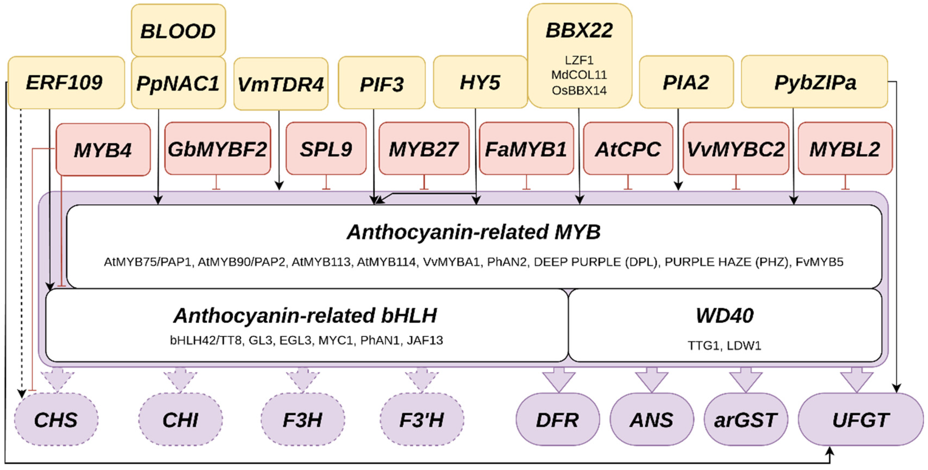
Figure 5.
Anthocyanin pigmentation patterns in petals. This cross-section shows that individual cells in specific layers can be colored by anthocyanins, while adjacent cells do not accumulate anthocyanins (A). Patterns on flowers emerge due to anthocyanin accumulation in restricted areas like a bull’s eye in the center of a flower or intensely pigmented spots on the petals (B). The formation of pigmented spots can be explained by an activator-repressor model as proposed by Ding et al., 2020 (C).
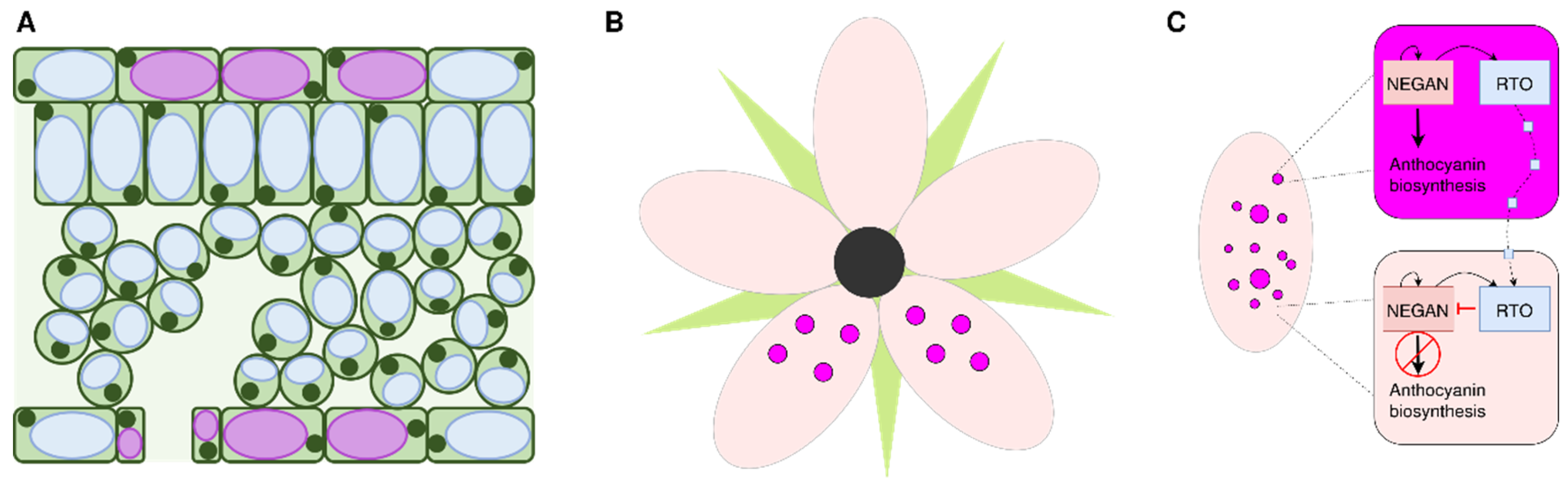
Figure 6.
Influence of sucrose on anthocyanin accumulation. Sucrose can either be taken up from the environment, e.g., via sucrose-containing medium (left), or produced through photosynthesis (top). High sucrose concentrations activate the transcription factor PRODUCTION OF ANTHOCYANIN1 (PAP1), which in turn induces general anthocyanin biosynthesis genes such as dihydroflavonol 4-reductase (DFR), anthocyanidin synthase (ANS), anthocyanin-related glutathione S-transferase (arGST), and anthocyanidin-3-O-glucosyltransferase (3GT), resulting in increased anthocyanin accumulation. The exact molecular regulatory mechanism leading to PAP1 activation through sucrose remains unclear.
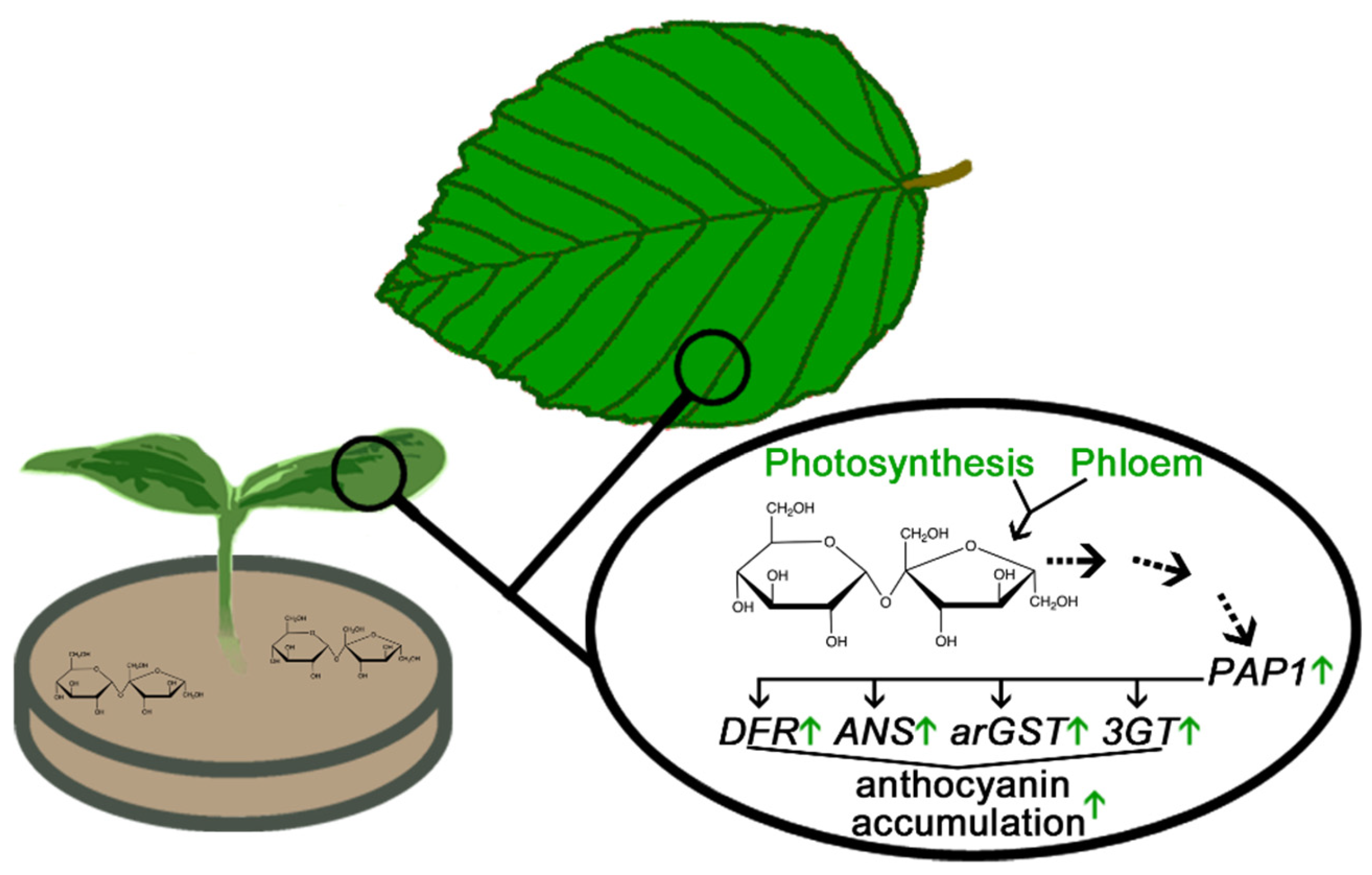
Figure 7.
Ecological functions of anthocyanins in plants. Aposematism: Bright coloration signals toxicity or unpalatability, deterring herbivores. Mimicry: Coloration imitates threat for herbivores (e.g., toxicity, unpalatability, or thorns). Pathogens: Anthocyanins have been showed to contribute to pathogen tolerance and resistance, possibly due to their interference with reactive oxygen species (ROS). The exact defense mechanism remains unclear. Camouflage: Dark pigmentation of plants is proposed to protect plants against color-blind herbivores. This part of this figure is based on Wolff & Pucker, 2025. Anti-camouflage: Bright coloration can break the crypsis of herbivores, exposing them to predators.
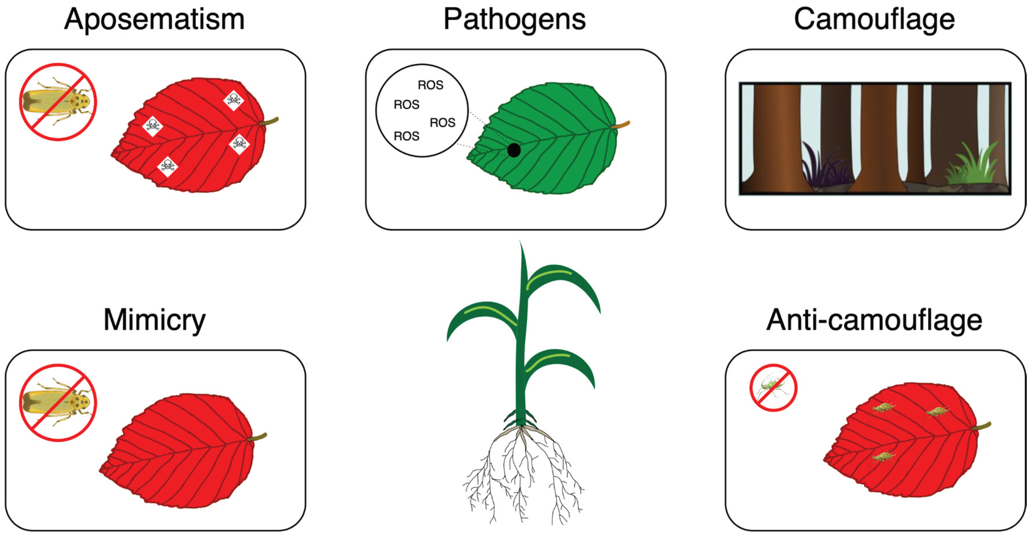
Disclaimer/Publisher’s Note: The statements, opinions and data contained in all publications are solely those of the individual author(s) and contributor(s) and not of MDPI and/or the editor(s). MDPI and/or the editor(s) disclaim responsibility for any injury to people or property resulting from any ideas, methods, instructions or products referred to in the content. |
© 2025 by the authors. Licensee MDPI, Basel, Switzerland. This article is an open access article distributed under the terms and conditions of the Creative Commons Attribution (CC BY) license (http://creativecommons.org/licenses/by/4.0/).
Copyright: This open access article is published under a Creative Commons CC BY 4.0 license, which permit the free download, distribution, and reuse, provided that the author and preprint are cited in any reuse.


