
Submitted:
22 August 2024
Posted:
23 August 2024
You are already at the latest version
Abstract
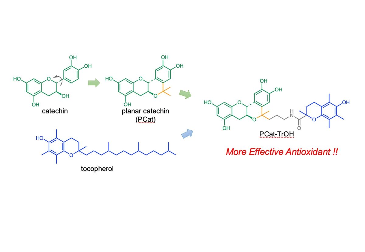
Planar catechin (PCat), a natural antioxidant with a fixed 3D catechin structure on a plane, exhibits radical-scavenging activity approximately five times stronger than the conventional catechin. We synthesized a compound, PCat–TrOH, by binding Trolox (TrOH), an α-tocopherol analog, to PCat to enhance its antioxidant effect against oxidative stress, such as lipid peroxidation. TrOH shows radical-scavenging activity about 6.5 times greater than PCat, and PCat–TrOH exhibited a similar level of radical-scavenging activity to TrOH. Additionally, PCat–TrOH demonstrated twice the radical-scavenging activity against reactive oxygen species compared to PCat or TrOH. This compound is also expected to exhibit an excellent antioxidant effect against lipid peroxidation caused by radical chain reactions, through interactions with vitamin C, similar to that in the case of α-tocopherol.
Keywords:
catechin
; trolox
; tocopherol
; antioxidant activity
1. Introduction
Oxidative stress is associated with the onset of and progression of various diseases such as cancer, diabetes, neurodegenerative diseases (Alzheimer’s disease, Parkinson’s, Huntington’s, amyotrophic lateral sclerosis), arteriosclerosis, and heart disease [1,2,3,4]. The mitochondria, the main source of reactive oxygen species (ROS), contribute to oxidative stress due to electron leakage in the electron transport chain during energy production [5]. Mitochondrial damage exacerbates this process, leading to the production of even more ROS. Excessive ROS production results in cell damage or cell death. Furthermore, ROS generated by inflamed cells or wounds cause oxidative damage to lipids and proteins in the cell membrane, as well as to enzymes and DNA, leading to various diseases [6]. Therefore, consuming substances that can eliminate ROS and reduce oxidative stress is effective in preventing the onset and progression of diseases associated with oxidative stress [7]. Various natural substances can eliminate ROS and suppress oxidative stress [8,9]. The consumption of red wine, known for its effectiveness in preventing coronary artery disease, has been shown to be due to the antioxidant effects of polyphenol compounds, such as resveratrol, catechin, epicatechin, and anthocyanin [10,11]. Among these, catechin is found not only in grapes but also green tea and apples. Catechin, with its catechol structure, reduces and eliminates ROS. It also has a chelating effect on metals, helping to eliminate metals involved in ROS generation [12]. In fact, catechin intake has been reported to have a preventative effect against diseases caused by oxidative stress [13].
Given that (+)-catechin has a structure where the chroman skeleton (AC ring) and catechol skeleton (B ring) are perpendicular to each other, we introduced an isopropyl group at the 3-position of the AC ring and the C6’-position of the C ring to synthesize a planar catechin (PCat) with a fixed 3D structure on a plane [14]. PCat exhibits radical-scavenging activity five times stronger than (+)-catechin and has been shown to possess powerful α-glucosidase inhibition, amyloid β aggregation inhibition, antitumor, and antiviral effects [15,16].
Tocopherol, a representative fat-soluble antioxidant, suppresses the oxidation of polyunsaturated fatty acids, abundant in biological membranes, into lipid peroxides through radical chain reactions [17]. Tocopherol eliminates free radicals generated during the lipid peroxidation process through reducing actions. Although tocopherol itself is oxidized in this process, vitamin C can reduce tocopherol, allowing it to regain its antioxidant effect [18]. The oxidized vitamin C is then regenerated by glutathione, NADH, and other substances, forming an antioxidant network in the body [19]. This defense mechanism effectively counters many oxidative stresses [20,21,22]. We hypothesized that if a single molecule could have multiple structures with radical-scavenging activity, an antioxidant network could form between these structures, exerting an even more powerful antioxidant effect.
In the present research, we aimed to form oxidative networks within a molecule to exert a more potent antioxidant effect. To achieve this, we synthesized a compound, PCat–TrOH, where PCat was conjugated with Trolox (TrOH), a water-soluble analog of α-tocopherol, and demonstrated its radical-scavenging activity (Figure 1).
2. Materials and Methods
2.1. General Methods
Unless otherwise noted, all commercially available reagents (Wako Chemicals, Tokyo, Japan; Tokyo Chemical Industry, Tokyo, Japan; Sigma-Aldrich, St. Louis, MO, USA) were used as received without purification. Solvents for chromatography were of industrial grade, and the process of all reactions were monitored by thin-layer chromatography that was on silica gel 60 F254 (0.25 nm, Merck). Column chromatography was performed on silica gel 60 (spherical, 63-210 μm, KANTO CHEMICAL). 1H and 13C NMR spectra were recorded with a JEOL JNMAL-400 (400 MHz) NMR spectrometer using CDCl3, DMSO-d6 as the solvent and tetramethylsilane as the internal reference. Mass spectra were measured using a JMS-700V (JEOL) mass spectrometer. The purity of all compounds was determined to be >95% using 1H and 13C NMR spectroscopy.
2.2. Synthesis of PCat-TrOH
2.2.1. Ethyl 3-((6aS,12aR)-2,3,8,10-tetrahydroxy-5-methyl-5,6a,7,12a-tetrahydroisochromeno [4,3-b]chromen-5-yl)propanoate (1)
Trimethylsilyl trifluoromethanesulfonate (TMSOTf, 5.42 mL, 30.0 mmol) was slowly added to a solution of (+)-catechin (6.0 g, 20.7 mmol) and ethyl 4-oxovalerate (7.14 mL 51.7 mmol) in dry tetrahydrofuran (THF) (200 mL) at –30 °C. After stirring for 20 h, the mixture was poured into water and extracted using diethyl ether (3 × 150 mL). The organic layer was washed with brine, dried over Na2SO4, and filtered. The solvent was evaporated, and the residue was purified using silica gel chromatography (4:1:0.1 toluene/acetone/methanol) to give 6.89 g (80.0%) of 1 as a white powder: m.p. 170 – 173 oC; 1H-NMR (400 MHz, DMSO-d6) δ 9.30 (s, 1H), 9.11 (s, 1H), 9.06 (s, 1H), 8.86 (s, 1H), 6.93 (s, 1H), 6.50 (s, 1H), 5.94 (d, J = 2.0Hz, 1H), 5.78 (d, J = 2.0Hz, 1H), 4.38 (d, J = 9.2Hz, 1H), 3.95 (q, J = 6.8Hz, 2H), 3.72 (m, 1H), 2.78 (dd, J = 5.6 and 15.2Hz, 1H), 2.28 (dd, J = 10.4 and 15.2Hz, 1H), 2.01 (t, J = 7.6Hz, 2H), 1.83 (t, J = 7.6Hz, 2H), 1.45 (s, 3H), 1.12 (t, J = 7.2Hz 3H); 13C NMR (100 MHz, DMSO-d6) δ 173.0, 156.7, 156.8, 155.2, 145.1, 144.4, 131.3, 124.7, 111.9, 111.4, 99.1, 95.6, 94.2, 76.1, 72.3, 65.9, 59.7, 28.7, 27.5, 27.2, 26.6, 14.1; HRMS (ESI+) m/z [M+] calcd for C22H24O8, 416.1471, found 416.1475.
2.2.2. Ethyl 3-((6aS,12aR)-2,3,8,10-tetrakis((tert-butyldimethylsilyl)oxy)-5-methyl-5,6a,7,12a-tetrahydroisochromeno[4,3-b]chromen-5-yl)propanoate (2)
To a solution of 1 (5.18 g, 12.4 mmol) in N,N-dimethylformamide (100 mL), tert-butyldimethylchlorosilane (11.25g, 74.6 mmol) and imidazole (5.08 g, 74.6 mmol) were added at 0°C under argon. After stirring at room temperature for 20 h, the mixture was diluted with ethyl acetate, washed with brine, dried over sodium sulfate (Na2SO4). The solvent was evaporated and the residue was subjected to silica gel column chromatography (1:3, n-hexane/ethyl acetate) to give 8.44g (78% yield) of 2 as a white solid: m.p. 104-106 oC;; 1H-NMR (400 MHz, CDCl3) δ7.07 (s, 1H), 6.57 (s, 1H), 6.42 (d, J = 2.0Hz, 1H), 6.29 (d, J = 2.2Hz, 1H), 4.55 (d, J = 8.8Hz 1H), 4.04 (q, J = 7.2Hz, 2H), 3.88 (m, 1H), 3.13 (m, 1H), 2.59 (m, 1H), 2.11 (m, 2H), 1.82 (m, 2H), 1.49 (s, 3H), 1.18 (t, J = 7.2Hz, 3H), 1.02 (s, 18H), 0.99 (s, 9H), 0.98 (s, 9H), 0.24 (s, 12H), 0.19(s, 6H), 0.180 (s, 6H); 13C NMR (100 MHz, CDCl3) δ 177.1, 157.3, 155.4, 154.4, 146.4, 145.3, 131.6, 127.5, 120.2, 119.7, 108.7, 103.8, 97.5, 85.2, 80.6, 73.5, 62.6, 37.8, 32.9, 31.3, 31.1, 26.7, 26.5, 25.1, 14.1, -0.8, -1.1; HRMS (ESI+) m/z [M+] calcd for C46H80O8Si4, 872.4930, found 872.4938.
2.2.3. 3-((6aS,12aR)-2,3,8,10-tetrakis((tert-butyldimethylsilyl)oxy)-5-methyl-5,6a,7,12a-tetrahydroisochromeno[4,3-b]chromen-5-yl)propan-1-ol (3)
A solution of 2 (6.81 g, 7.8 mmol) in dry THF (100 mL) was added dropwise to a solution of 1M lithium aluminum hydride (LiAlH4) in THF (11.7 mL, 11.7 mmol) and dry THF (20 mL) at 0 oC and then stirred for 20 h at room temperature. The mixture was cooled on ice, and saturated potassium sodium tartrate was added dropwise; subsequently, the mixture was filtered through a small pad of Celite and thoroughly washed with ethyl acetate. The organic solution was washed with brine, dried over Na2SO4, and filtered. The solvent was evaporated, and the residue was purified using silica gel chromatography (1:6 n-hexane/ethyl acetate) to give 3.24 g (50%) of 3 as a white powder: m.p. 144–147 oC; 1H-NMR (400 MHz, CDCl3) δ 7.10 (s, 1H), 6.48 (s, 1H), 6.09 (d, J = 2.2 Hz, 1H), 5.97 (d, J = 2.2 Hz, 1H), 4.51 (d, J = 9.6 Hz, 1H), 3.87 (m, 1H), 3.55 (m, 2H), 3.02 (dd, J = 6.0 and 15.6Hz, 1H), 2.51 (dd, J = 10.8 and 15.6 Hz, 1H), 1.91 (m, 2H), 1.53 (s, 3H), 1.28 (m, 2H), 1.01 (s, 18H), 0.99 (s, 9H), 0.98 (s, 9H), 0.29 (s, 12H), 0.19 (s, 6H), 0.18 (s, 6H); 13C NMR (100 MHz, CDCl3) δ 157.8, 153.3, 152.0, 141.6, 140.4, 129.4, 126.9, 118.5, 115.1, 107.3, 104.6, 96.1, 85.0, 84.2, 76.1, 60.5, 39.6, 33.3, 31.3, 31.1, 27.3, 26.7, 26.5, 24.4, -0.8, -1.1; HRMS (ESI+) m/z [M + H+] calcd for C44H79O7Si4, 831.4904, found 831.4910.
2.2.4. tert-butyl (tert-butoxycarbonyl)(3-((6aS,12aR)-2,3,8,10-tetrakis((tert-butyldimethylsilyl)oxy)-5-methyl-5,6a,7,12a-tetrahydroisochromeno[4,3-b]chromen-5-yl)propyl)carbamate (4)
To a stirred solution of 3 (5.99 g, 7.2 mmol), di-tert-butyl iminodicarboxylate (6.26g, 28.8 mmol), and triphenylphosphine (7.57 g, 28.8 mmol) in 100 mL of dry THF, 40 % diethyl azodicarboxylate in toluene (13.11 ml, 28.8 mmol) was slowly added under argon at 0 °C. After stirring at room temperature for 20 hours, the mixture was diluted with ethyl acetate, washed with brine and dried over Na2SO4. The solvent was evaporated and the residue was subjected to silica gel column chromatography (1:10, n-hexane/ethyl acetate) to give 5.57 g (75 % yield) of 4 as a white solid: m.p. 67–72 oC; 1H NMR (400 MHz, CDCl3) δ 7.08 (s, 1H), 6.46 (s, 1H), 6.10 (d, J = 2.2 Hz, 1H), 5.97 (d, J = 2.2 Hz, 1H), 4.61 (d, J = 9.2 Hz, 1H), 3.80 (m, 1H), 3.49 (m, 2H), 3.01 (dd, J = 6.0 and 15.8 Hz, 1H), 2.47 (dd, J = 10.4 and 15.8 Hz, 1H), 1.75 (m, 2H), 1.53 (m, 2H), 1.51 (s, 3H), 1.46 (s, 18H), 1.01 (s, 18H), 0.98 (s, 18), 0.25 (s, 12H), 0.18 (s, 12H); 13C NMR (100 MHz, CDCl3) δ 159.3, 156.6, 154.9, 150.5, 144.4, 140.3, 131.8, 127.6, 117.3, 115.6, 108.7, 106.1, 152.3, 83.9, 82.8, 80.5, 73.4, 44.6, 37.5, 33.0, 31.0, 30.9, 28.1, 27.3, 26.7, 26.5, 17.5, -0.8, -1.1; HRMS (ESI+) m/z [M + H+] calcd for C54H96NO10Si4, 1030.6112, found 1030.6121.
2.2.5. N-(3-((6aS,12aR)-2,3-bis((tert-butyldimethylsilyl)oxy)-8,10-dihydroxy-5-methyl-5,6a,7,12a-tetrahydroisochromeno[4,3-b]chromen-5-yl)propyl)-6-hydroxy-2,5,7,8-tetramethylchromane-2-carboxamide (5)
To a solution of 4 (2.10 g, 2.1 mmol) in dichloromethane (200 mL), trifluoroacetic acid (20 mL) was slowly added at 0 °C under argon. After stirring at room temperature for 2.5 hours, the solvent was evaporated. The residue containing the amine was dried in vacuum and then used in the next reaction. To a solution of TrOH (0.48 g, 1.9 mmol) in N,N-dimethylformamide (DMF: 20 mL) was added N,N-diisopropylethylamine (1.1 mL, 6.4 mmol) followed by 1-[Bis(dimethylamino)methylene]-1H-1,2,3-triazolo[4,5-b]pyridinium3-Oxide Hexafluorophosphate (HATU: 1.61 g, 4.2 mmol). The mixture was stirred at room temperature for 15 min, after which the time the dark clear mixture was treated with the solution of the amine and N,N-diisopropylethylamine (1.1 mL, 6.4 mmol) in DMF (20 mL). After stirring at room temperature for 24 hours, the mixture was diluted with ethyl acetate, washed with brine, dried over Na2SO4 and the solvent was evaporated. The residue was purified by silica gel column chromatography (1:1, n-hexane/ethyl acetate) to give 0.67 g (30 % yield) of 5 as a white solid: m.p. 167–170 oC; 1H NMR (400 MHz, DMSO-d6) δ 9.35 (s, 1H), 9.05 (s, 1H), 7.47 (s, 1H), 7.11 (m, 1H), 7.01 (s, 1H), 6.51 (s, 1H), 5.95 (d, J = 2.2 Hz, 1H), 5.80 (d, J = 2.2 Hz, 1H), 4.41 (d, J = 8.4 Hz, 1H), 3.74 (m, 1H), 2.98 (m, 2H), 2.83 (m, 1H), 2.42 (m, 2H), 2.30 (m, 1H), 2.11 (m, 2H), 2.10 (s, 3H), 2.08 (s, 3H), 2.00 (s, 3H), 1.71 (m, 2H), 1.55 (m, 2H), 0.98 (s, 9H), 0.94 (s, 9H), 0.21 (s, 6H), 0.18 (s, 6H); 13C NMR (100 MHz, CDCl3) δ 177.8, 157.7, 156.8, 156.0, 147.3, 146.8, 146.0, 145.7, 132.7, 129.3, 128.0, 121.5, 118.5, 117.7, 116.3, 114.0, 97.4, 96.3, 94.2, 92.4, 81.9, 80.8, 76.5, 42.5, 41.0, 36.2, 33.3, 31.1, 30.0, 28.3, 26.9, 22.3, 21.0, 16.8, 12.5, 11.8, -0.8; HRMS (ESI+) m/z [M + H+] calcd for C46H68NO9Si2, 834.4433, found 834.4439.
2.2.6. 6-hydroxy-2,5,7,8-tetramethyl-N-(3-((6aS,12aR)-2,3,8,10-tetrahydroxy-5-methyl-5,6a,7,12a-tetrahydroisochromeno[4,3-b]chromen-5-yl)propyl)chromane-2-carboxamide (PCat–TrOH)
To a solution of 5 (0.76 g, 0.72 mmol) in dry THF (200 mL), 1M tetrabutylammonium fluoride in THF(3.93 ml, 3.93 mmol) was slowly added at 0 °C under argon. After stirring at room temperature for 1.5 hours, the mixture was diluted with ethyl acetate, washed with brine, dried over Na2SO4. The solvent was evaporated, and the residue was purified by column chromatography (2:1:0.1, toluene/acetone/methanol to give 0.22g (50% yield) of PCat–TrOH as a white solid: m.p. 186–189 oC; 1H NMR (400 MHz, DMSO-d6) δ 9.29(s, 1H), 9.05 (s, 2H), 8.74(s, 1H), 7.48 (s, 1H), 7.16 (m, 1H), 6.90 (s, 1H), 6.38 (s, 1H), 5.94 (s, 1H), 5.78 (s, 1H), 4.34 (m, 1H), 3.69 (m, 1H), 2.95 (m, 2H), 2.80 (m, 1H), 2.42 (m, 2H), 2.31 (dd, J = 10.4 and 14.8 Hz, 1H), 2.14 (m, 2H), 2.09 (s, 3H), 2.07 (s, 3H), 1.99 (s, 3H), 1.68 (m, 2H), 1.51 (m, 2H), 1.34 (s, 3H); 13C NMR (100 MHz, CDCl3) δ 177.6, 157.8, 156.6, 155.6, 146.5, 148.8, 145.0, 144.0, 132.7, 129.0, 127.5, 121.0, 117.5, 116.0, 114.1, 113.0, 98.0, 96.8, 94.8, 92.4, 81.0, 79.2, 74.5, 42.0, 40.2, 36.0, 34.4, 32.4, 26.9, 22.3, 21.2, 16.8, 12.6, 11.5; HRMS (ESI+) m/z [M + H+] calcd for C34H40NO9, 606.2704, found 606.2709.
2.3. Antioxidant activity measurements
Galvinoxyl radical (GO•) was commercially obtained from Tokyo Chemical Industry Co., Ltd., (Tokyo, Japan), and GO• is the highest available purity and was used without further purification unless otherwise noted. Acetonitrile (MeCN) used as solvent was commercially obtained from Nacalai Tesque Inc. (Kyoto, Japan) and used as received. The rates of GO•-scavenging reactions using test samples in MeCN were determined by monitoring the absorbance change at 428 nm due to GO• (ε = 1.4 x 10⁵ M-1 cm-1) after mixing it in MeCN at a volumetric rate of 1:1 using a stopped-flow technique on a UNISOKU REP-1000-02 NM spectrophotometer (UNISOKU Co., Ltd., Osaka, Japan) at 298 K. The pseudo-first-order rate constants (kobs) were determined by a least-squares curve fitting using an UNITCOM SOLUTION-M06M-134-UHX (UNIT.COM INC., Osaka, Japan). The first-order plots of ln(A − A∞) vs. time (A and A∞ were denoted as the absorbance at the reaction time and the final absorbance, respectively) were linear with the correlation coefficient ρ > 0.999 until three or more half-lives. In all cases, the kobs values obtained from at least three independent measurements agreed within ±5% experimental error. In all cases, solutions were normally equilibrated with air.
2.4. Spectral titrations
Typically, a 5 μL of PCat–TrOH (2.63×10⁻⁴ M) in MeCN was added to GO• (7.77 x 10⁻⁶ M) in MeCN, and the absorbance at 428 nm due to GO• was plotted with respect to the concentration ratio, [PCat–TrOH]/[GO•]. The UV–vis spectral changes accompanying the reaction were monitored and measured using an Agilent 8453 photodiode array spectrophotometer thermostated with a Peltier temperature control at 298 K (Agilent Technologies, Santa Clara, CA, USA). In all cases, solutions were normally equilibrated with air.
3. Results
3.1. Chemistry
We designed and synthesized a compound, PCat–TrOH, in which TrOH was conjugated to PCat, creating a molecule with multiple radical-scavenging structures (Figure 2). The PCat site, where the 3D catechin structure was fixed, was obtained through the Oxa-Pictet Spengler reaction between (+)-catechin and ethyl levulinate. Specifically, 1 was synthesized by treating a tetrahydrofuran (THF) solution of catechin and ethyl levulinate with trimethylsilyl trifluoromethanesulfonate (TMSOTf). Next, the four hydroxyl groups were protected with tert-butyldimethylchlorosilyl (TBS) groups, after which the ester structure was reduced with lithium aluminum hydride (LiAlH4) to obtain alcohol (3). Then, 3 underwent a Mitsunobu reaction using (Boc)2NH as a pronucleophile in the presence of Ph3P and DEAD to obtain 4. Trifluoroacetic acid (TFA) was used to remove the BOC group together with the TBS groups on A ring, after which the amino group of 4 underwent a condensation reaction with the carboxyl group of TrOH using the condensing agent DIPEA/HATU, without purification, to obtain compound 5. Finally, TBAF was used for deprotection, resulting in PCAT–TrOH.
2.4. Radical Scavenging Activity
The radical-scavenging activities of PCat–TrOH, TrOH, PCat, and Cat were evaluated in acetonitrile using the galvinoxyl radical (GO•), a model compound for ROS. The strong absorption of GO• at 428 nm immediately disappeared upon reaction with the antioxidant. Therefore, the change in color of the GO• solution in the presence of the antioxidant was used to measure the radical-scavenging activity rates of PCat–TrOH, TrOH, PCat, and Cat. This spectral change was monitored by measuring the decrease in absorbance at 428 nm using the stopped-flow method (Figure 3). The concentrations of PCat–TrOH, TrOH, PCat, and Cat were set to over 10 times the concentration of GO• to measure the decrease in absorbance at 428 nm under pseudo-first-order reaction conditions, allowing us to obtain the pseudo-first order rate constant (kobs). The pseudo-first-order rate constant (kobs) increased linearly with increasing concentrations of PCat–TrOH, TrOH, PCat, and Cat (Figure 4). The slopes of the linear plots of kobs vs PCat–TrOH, TrOH, and PCat showed that the second-order rate constants (k) of the radical-scavenging activity were 9.6 × 10 2 M−1 s−1, 1.1 × 10³ M−1 s−1, and 1.7 × 10 2 M−1 s −1, respectively. We have also previously reported that the k of Cat under the same conditions was 2.6 × 10 1 M−1 s −1. The k value of PCat was approximately 6.5 times that of Cat, indicating that the planar structure increased its radical-scavenging activity. Meanwhile, TrOH was even more powerful than PCat, with a k value approximately 6.5 times greater than that of Cat. The k value of the newly synthesized compound PCat–TrOH, which binds TrOH to PCat, was approximately 5.6 times larger than that of planar catechin, exhibiting an almost equally powerful radical-scavenging activity as that of TrOH alone.
2.4. Spectral titrations
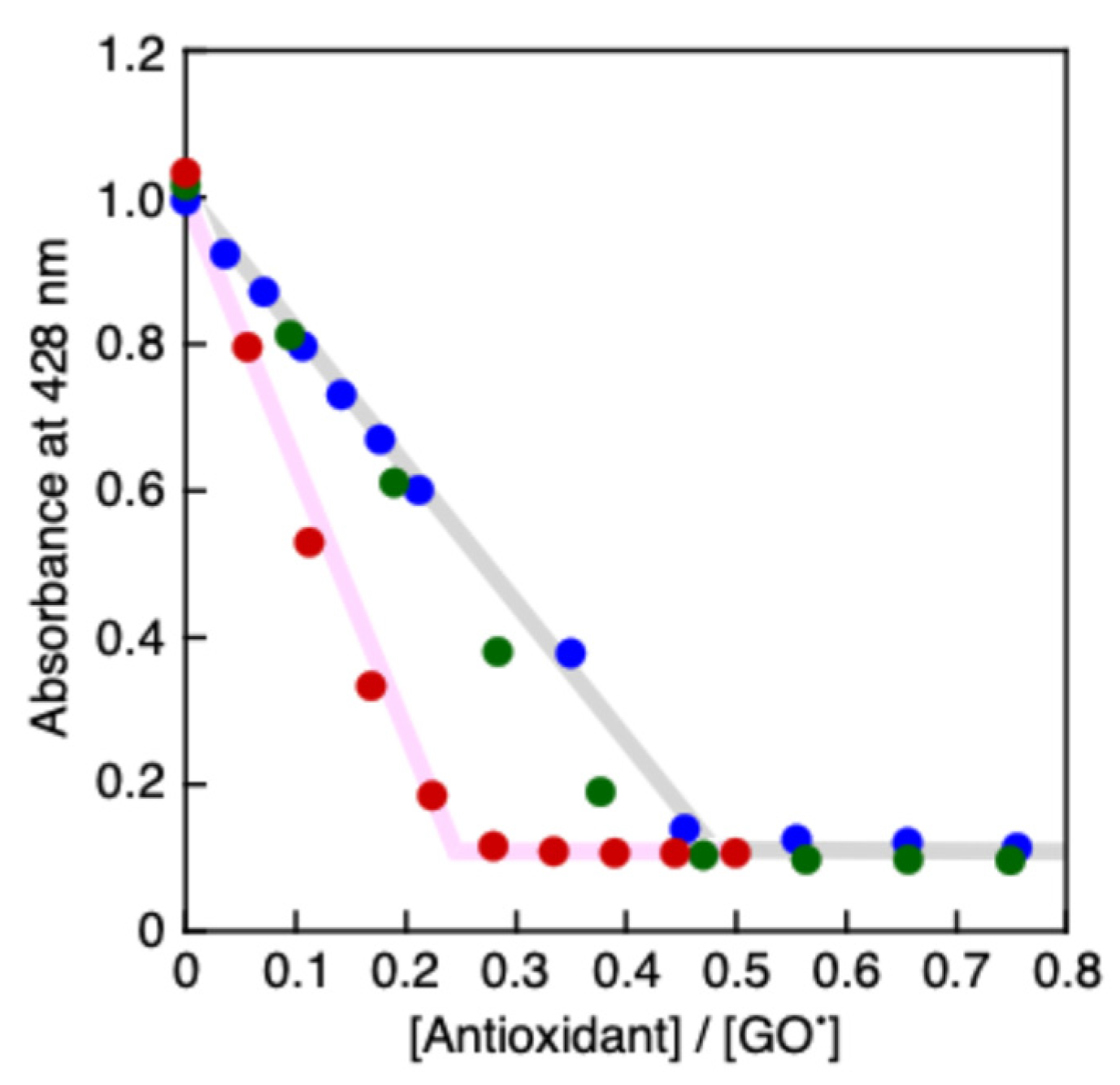
ROS are eliminated by one-electron reduction, so catechin eliminates two ROS and is oxidized to quinone. Additionally, α-tocopherol reduces two lipid peroxides and is itself reduced to tocopheryl quinone. Therefore, we sought to clarify the number of GO• radicals that could be eliminated by PCat–TrOH (antioxidant capacity) through titration (Figure 5), which we conducted by adding antioxidants to a GO• solution, plotting the reduced 428 nm absorbance of GO•, and finally obtaining the molar ratio of antioxidants to GO• when GO• was completely eliminated. As shown in Figure 6, in the spectral titration conducted in this study, the 428 nm absorbance of GO• was completely eliminated by 0.25 times the quantity of PCat–TrOH relative to GO•, indicating that the molar ratio of PCat–TrOH to GO• was 1:4. Meanwhile, the 428 nm absorbance was eliminated by adding 0.5 times the quantity of PCat or TrOH relative to GO•, indicating that their molar ratios were 1:2. Therefore, it was thought that PCat–TrOH eliminated four GO• radicals, compared to PCat or TrOH, which eliminated only two GO• radicals.
3. Discussions
PCat, with its 3D structure of catechin fixed in a planar direction, has a more powerful radical-scavenging activity with respect to GO• than catechin, while TrOH exhibits an even more powerful radical-scavenging activity, approximately 6.5 times that of PCat. The compound PCat-TrOH, newly synthesized in this study, had a k value approximately 5.6 times higher than that of planar catechin, indicating an antioxidant effect nearly as strong as TrOH. This result suggests that the radical-scavenging activity of PCat–TrOH may preferentially involve hydrogen transfer from TrOH. Meanwhile, titration results showed that four GO• radicals were eliminated for every PCat–TrOH molecule. This suggests that PCat–TrOH eliminates GO• through the reduction effects of both TrOH and PCat. The fact that TrOH exhibits a radical-scavenging activity approximately 6.5 times stronger than that of PCat, and that PCat–TrOH exhibits a similar level of antioxidant activity as TrOH, suggests that after TrOH in PCat–TrOH eliminates GO• through hydrogen transfer, the catechol-derived hydrogen of PCat is transferred to the oxidized TrOH, regenerating TrOH in its reduced form. Detailed analyses are currently underway to clarify the mechanism of radical elimination by this intramolecular network. The above results show that PCat–TrOH possesses excellent antioxidant properties, with powerful radical-scavenging activity nearly equivalent to that of TrOH and a radical-scavenging capacity double that of TrOH.
Vitamin E, particularly α-tocopherol, can trap lipid radicals and lipid peroxide radicals, which play an important role in lipid peroxidation chain reactions, and terminate free radical chain reactions [23]. These substances capture radicals through a hydrogen transfer reaction from the hydroxyl group on the aromatic ring of α-tocopherol, oxidizing themselves to an α-tocopherol radical [24]. Extracellular ascorbic acid (vitamin C) exerts its antioxidant effect in biological membranes, reducing the tocopheroxyl radicals produced and regenerating α-tocopherol [25]. This regenerative action allows α-tocopherol to exhibit a synergistic effect as an antioxidant, breaking free radical chains and providing excellent protection against oxidative stresses in the body. Following the elimination of ROS radicals through reduction, catechin is oxidized to radical catechin and subsequently converts to a two-electron oxidized quinone [26]. Ascorbic acid (vitamin C) also exhibits a reducing effect on these oxidized products, potentially regenerating catechin. Therefore, PCat–TrOH is thought to be regenerated by ascorbic acid reducing the radicals generated by the oxidation of PCat and TrOH during radical-scavenging activity. If PCat regenerates the oxidized form of TrOH during radical-scavenging activity, there is a possibility of forming an effective antioxidant network through the regeneration of the oxidized form of PCat to its reduced form with ascorbic acid. PCat–TrOH, which exhibits high antioxidant activity and capacity, is expected to provide an extremely high protective effect through the regenerative effect of extracellular ascorbic acid on lipid peroxidation chain reactions in biological membranes. The analysis of the antioxidant effect of PCat–TrOH against oxidative stress in cells and its mechanism is currently under investigation.
3. Conclusion
In the present study, we successfully synthesized PCat–TrOH by adding TrOH, known for its antioxidant effects, to planar catechin. This newly synthesized compound demonstrated even more potent radical-scavenging activity and capacity than planar catechin, as previously reported. PCat–TrOH may exhibit strong antioxidant effects through the formation of an intramolecular antioxidant network, involving hydrogen transfer reactions between the TrOH and PCat moieties. Detailed analysis to clarify this possibility, as well as biological tests on the antioxidant activity of PCat–TrOH using cells, are currently underway. Furthermore, the results of this study suggest that the addition of antioxidant compounds to planar catechin may further enhance antioxidant activity and promote the formation of intramolecular antioxidant networks. Consequently, we plan to synthesize compounds containing multiple antioxidants found in the body, such as lipoic acid, in future studies.
Author Contributions
Conceptualization, K.F.; methodology, K.F., I.N., and H.I.; investigation, W.S., Y.S., K.O., and K.F.; writing—original draft preparation, W.S. and I.N.; writing—review and editing, K.F.; funding acquisition, K.F. All authors have read and agreed to the published version of the manuscript.
Funding
This work was supported by Grant-in-Aid No. 22K06508 from the Ministry of Education, Culture, Sports, Science, and Technology (MEXT), Japan and MEXT Promotion of Development of a Joint Usage/Research System Project: Coalition of Universities for Research Excellence Program (CURE), Grant Number JPMXP1323015488.
Institutional Review Board Statement
Not applicable.
Informed Consent Statement
Not applicable.
Data Availability Statement
All data are contained in the article.
Conflicts of Interest
The authors declare no conflicts of interest.
References
- Feitosa, C. M.; da Silva Oliveira, G. L.; do Nascimento Cavalcante, A.; Morais Chaves, S. K.; Rai, M. , Determination of Parameters of Oxidative Stress in vitro Models of Neurodegenerative Diseases-A Review. Curr Clin Pharmacol 2018, (2), 100–109. [Google Scholar] [CrossRef] [PubMed]
- Teleanu, D. M.; Niculescu, A. G. ; Lungu, II; Radu, C. I.; Vladacenco, O.; Roza, E.; Costachescu, B.; Grumezescu, A. M.; Teleanu, R. I., An Overview of Oxidative Stress, Neuroinflammation, and Neurodegenerative Diseases. Int J Mol Sci, 2022; 23. [Google Scholar] [CrossRef]
- Batty, M.; Bennett, M. R.; Yu, E. , The Role of Oxidative Stress in Atherosclerosis. Cells, 2022; 11. [Google Scholar] [CrossRef]
- Chen, Y.; Guo, X.; Zeng, Y.; Mo, X.; Hong, S.; He, H.; Li, J.; Fatima, S.; Liu, Q. , Oxidative stress induces mitochondrial iron overload and ferroptotic cell death. Sci Rep 2023, (1), 15515. [Google Scholar] [CrossRef]
- Kowalczyk, P.; Sulejczak, D.; Kleczkowska, P.; Bukowska-Osko, I.; Kucia, M.; Popiel, M.; Wietrak, E.; Kramkowski, K.; Wrzosek, K.; Kaczynska, K. , Mitochondrial Oxidative Stress-A Causative Factor and Therapeutic Target in Many Diseases. Int J Mol Sci, 2021; 22. [Google Scholar] [CrossRef]
- Bhatti, J. S.; Bhatti, G. K.; Reddy, P. H. , Mitochondrial dysfunction and oxidative stress in metabolic disorders - A step towards mitochondria based therapeutic strategies. Biochim Biophys Acta Mol Basis Dis 2017, 1863, 1066–1077. [Google Scholar] [CrossRef] [PubMed]
- Forman, H. J.; Zhang, H. , Targeting oxidative stress in disease: promise and limitations of antioxidant therapy. Nat Rev Drug Discov 2021, 20, 689–709. [Google Scholar] [CrossRef] [PubMed]
- Jovanovic, S. V.; Simic, M. G. , Antioxidants in nutrition. Ann N Y Acad Sci 2000, 899, 326–34. [Google Scholar] [CrossRef] [PubMed]
- Cirillo, G.; Curcio, M.; Vittorio, O.; Iemma, F.; Restuccia, D.; Spizzirri, U. G.; Puoci, F.; Picci, N. , Polyphenol Conjugates and Human Health: A Perspective Review. Crit Rev Food Sci Nutr 2016, 56, 326–37. [Google Scholar] [CrossRef] [PubMed]
- Castaldo, L.; Narvaez, A.; Izzo, L.; Graziani, G.; Gaspari, A.; Minno, G. D.; Ritieni, A. , Red Wine Consumption and Cardiovascular Health. Molecules, 2019; 24. [Google Scholar] [CrossRef]
- Liberale, L.; Bonaventura, A.; Montecucco, F.; Dallegri, F.; Carbone, F. , Impact of Red Wine Consumption on Cardiovascular Health. Curr Med Chem 2019, 26, 3542–3566. [Google Scholar] [CrossRef] [PubMed]
- Bernatoniene, J.; Kopustinskiene, D. M. , The Role of Catechins in Cellular Responses to Oxidative Stress. Molecules, 2018; 23. [Google Scholar] [CrossRef]
- Rudrapal, M.; Khairnar, S. J.; Khan, J.; Dukhyil, A. B.; Ansari, M. A.; Alomary, M. N.; Alshabrmi, F. M.; Palai, S.; Deb, P. K.; Devi, R. , Dietary Polyphenols and Their Role in Oxidative Stress-Induced Human Diseases: Insights Into Protective Effects, Antioxidant Potentials and Mechanism(s) of Action. Front Pharmacol 2022, 13, 806470. [Google Scholar] [CrossRef] [PubMed]
- Fukuhara, K.; Nakanishi, I.; Kansui, H.; Sugiyama, E.; Kimura, M.; Shimada, T.; Urano, S.; Yamaguchi, K.; Miyata, N. , Enhanced radical-scavenging activity of a planar catechin analogue. J Am Chem Soc 2002, 124, 5952–3. [Google Scholar] [CrossRef] [PubMed]
- Mizuno, M.; Mori, K.; Misawa, T.; Takaki, T.; Demizu, Y.; Shibanuma, M.; Fukuhara, K. , Inhibition of beta-amyloid-induced neurotoxicity by planar analogues of procyanidin B3. Bioorg Med Chem Lett 2019, 29, 2659–2663. [Google Scholar] [CrossRef] [PubMed]
- Mizuno, M.; Mori, K.; Tsuchiya, K.; Takaki, T.; Misawa, T.; Demizu, Y.; Shibanuma, M.; Fukuhara, K. , Design, Synthesis, and Biological Activity of Conformationally Restricted Analogues of Silibinin. ACS Omega 2020, 5, 23164–23174. [Google Scholar] [CrossRef]
- Mustacich, D. J.; Bruno, R. S.; Traber, M. G. , Vitamin E. Vitam Horm 2007, 76, 1–21. [Google Scholar] [PubMed]
- Niki, E. , Interaction of ascorbate and alpha-tocopherol. Ann N Y Acad Sci 1987, 498, 186–99. [Google Scholar] [CrossRef] [PubMed]
- Sies, H.; Stahl, W.; Sundquist, A. R. , Antioxidant functions of vitamins. Vitamins E and C, beta-carotene, and other carotenoids. Ann N Y Acad Sci, 1992; 669, 7–20. [Google Scholar]
- Salvagno, M.; Sterchele, E. D.; Zaccarelli, M.; Mrakic-Sposta, S.; Welsby, I. J.; Balestra, C.; Taccone, F. S. , Oxidative Stress and Cerebral Vascular Tone: The Role of Reactive Oxygen and Nitrogen Species. Int J Mol Sci, 2024; 25. [Google Scholar] [CrossRef]
- La Torre, M. E.; Villano, I.; Monda, M.; Messina, A.; Cibelli, G.; Valenzano, A.; Pisanelli, D.; Panaro, M. A.; Tartaglia, N.; Ambrosi, A.; Carotenuto, M.; Monda, V.; Messina, G.; Porro, C. , Role of Vitamin E and the Orexin System in Neuroprotection. Brain Sci, 2021; 11. [Google Scholar]
- Engin, K. N. , Alpha-tocopherol: looking beyond an antioxidant. Mol Vis 2009, 15, 855–60. [Google Scholar] [PubMed]
- Mesa, T.; Munne-Bosch, S. , alpha-Tocopherol in chloroplasts: Nothing more than an antioxidant? Curr Opin Plant Biol 2023, 74, 102400. [Google Scholar] [CrossRef] [PubMed]
- Nakanishi, I.; Fukuhara, K.; Shimada, T.; Ohkubo, K.; Iizuka, Y.; Inami, K.; Mochizuki, M.; Urano, S.; Miyata, N.; Fukuzumi, S. , Effect of magnesiumu ion on kinetic stability and spin distribution of phenoxyl radical derived from a vitamin E analogue: mechanistic insight into antioxidative hydrogen-transfer reaction of vitamin E. J, Chem. Soc., Perkin Trans. 2002; 2, 1520–1524. [Google Scholar]
- Smirnoff, N. , Ascorbic acid metabolism and functions: A comparison of plants and mammals. Free Radic Biol Med 2018, 122, 116–129. [Google Scholar] [CrossRef] [PubMed]
- Nakanishi, I.; Fukuhara, K.; Ohkubo, K.; Shimada, T.; Kansui, H.; Kurihara, M.; Urano, S.; Fukuzumi, S.; Miyata, N. , Superoxide anion generation via electron-transfer oxidation of catechin dianion by molecular oxygen in an aprotic medium. Chemistry Letters 2001, 1152–1153. [Google Scholar] [CrossRef]
Figure 1.
Structures of (+)-catechin, PCat, α-tocopherol, TrOH, PCat–TrOH.
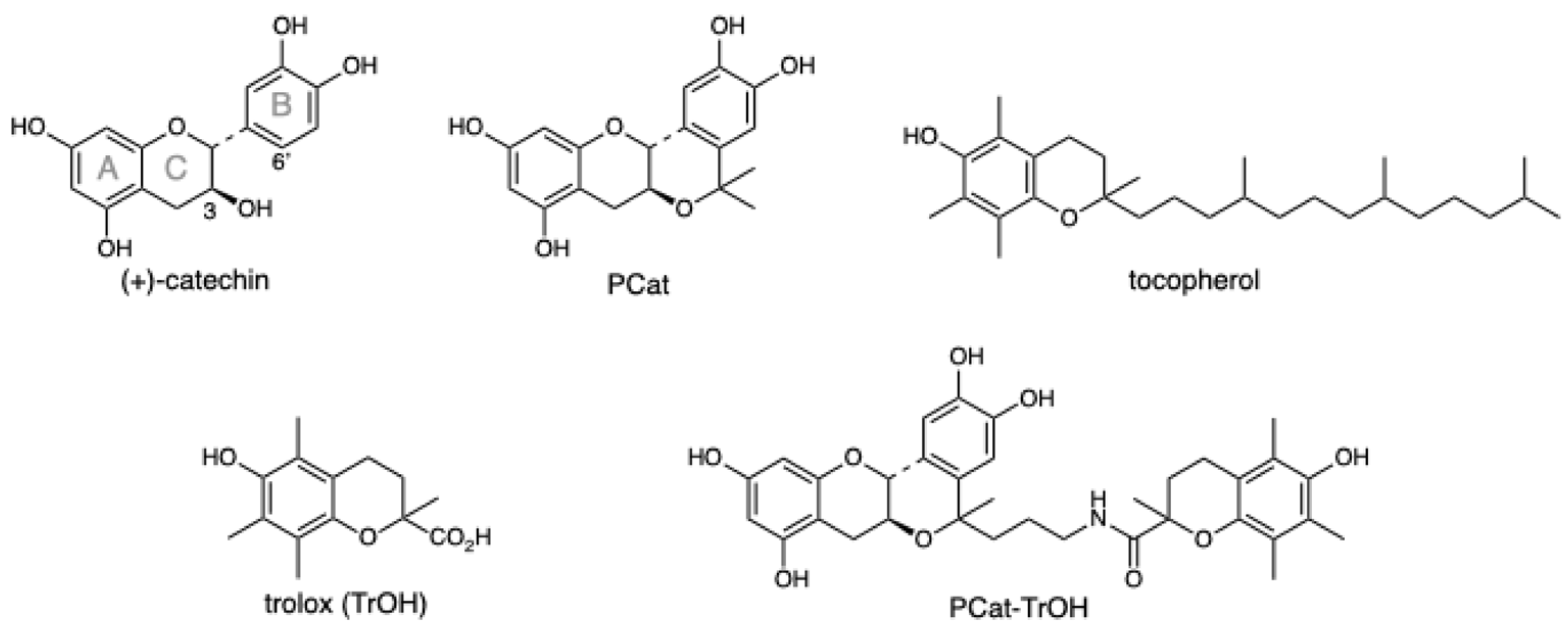
Figure 2.
Synthesis of PCat–TrOH. Regents and conditions: (a) ethyl levulinate, TMSOTf, THF, −30 °C; (b) tert-butyldimethylchlorosilane, imidazole, DMF, 0 °C → rt; (c) LiAlH4, THF, 0 °C → rt; (d) di-tert-butyliminodicarboxylate, Ph3P, DEAD,THF、0 °C→ rt; (e) TFA, CH2Cl2, 0 °C→ rt; (f) TrOH, DIPEA, HATU, DMF, 0 °C → rt; (g) TBAF, CH 2 Cl2, 0 °C → rt.
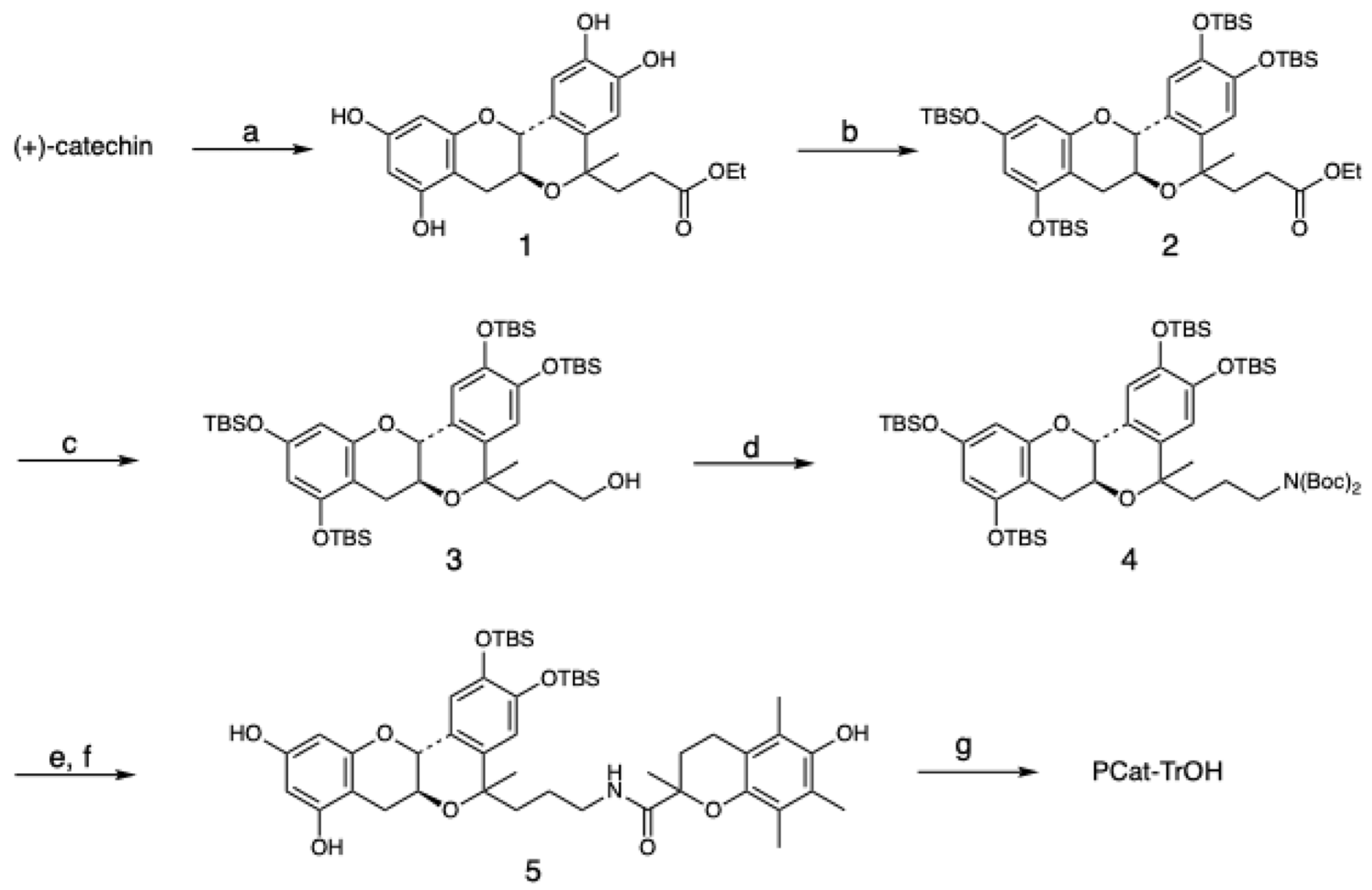
Figure 3.
Spectral change (interval: 1 s) observed during the reaction of PCat–TrOH (7.2 × 10⁻⁵ M) with GO• (5.6 × 10⁻⁶ M) in MeCN at 298 K. The arrow denotes the direction of absorbance changes. Inset: The first-order plot of the absorbance at 428 nm.
Figure 3.
Spectral change (interval: 1 s) observed during the reaction of PCat–TrOH (7.2 × 10⁻⁵ M) with GO• (5.6 × 10⁻⁶ M) in MeCN at 298 K. The arrow denotes the direction of absorbance changes. Inset: The first-order plot of the absorbance at 428 nm.
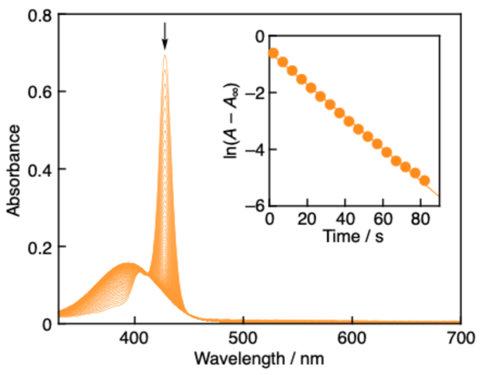
Figure 4.
Plots of pseudo-first-order rate constants (kobs) vs. concentrations of TrOH (green circles), PCat– TrOH (red circles), PCat (blue circles) and Cat (black circles) in MeCN at 298 K.
Figure 4.
Plots of pseudo-first-order rate constants (kobs) vs. concentrations of TrOH (green circles), PCat– TrOH (red circles), PCat (blue circles) and Cat (black circles) in MeCN at 298 K.
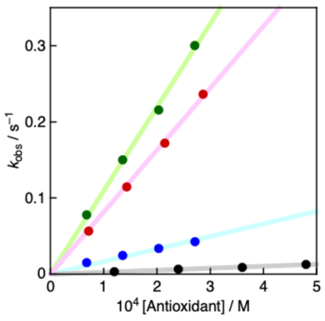
Figure 5.
Spectral change observed upon addition of PCat–TrOH (0, 4.4 × 10⁻⁷, 8.7 × 10⁻⁷, 1.3 × 10⁻⁶, 1.7 × 10⁻⁶, 2.2 × 10⁻⁶ M) to GO• (7.8 × 10⁻⁶ M) in MeCN at 298 K. The arrows denote the direction of absorbance changes.
Figure 5.
Spectral change observed upon addition of PCat–TrOH (0, 4.4 × 10⁻⁷, 8.7 × 10⁻⁷, 1.3 × 10⁻⁶, 1.7 × 10⁻⁶, 2.2 × 10⁻⁶ M) to GO• (7.8 × 10⁻⁶ M) in MeCN at 298 K. The arrows denote the direction of absorbance changes.
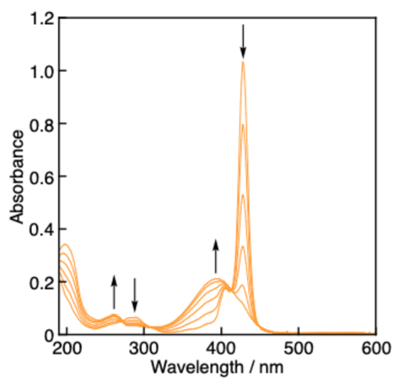
Figure 6.
Plots of the absorbance at 428 nm vs. the concentration ratios of antioxidants (TrOH (green circles), PCat–TrOH (red circles), and PCat (blue circles)) to GO•, [antioxidants]/[GO•], in MeCN.
Figure 6.
Plots of the absorbance at 428 nm vs. the concentration ratios of antioxidants (TrOH (green circles), PCat–TrOH (red circles), and PCat (blue circles)) to GO•, [antioxidants]/[GO•], in MeCN.
Disclaimer/Publisher’s Note: The statements, opinions and data contained in all publications are solely those of the individual author(s) and contributor(s) and not of MDPI and/or the editor(s). MDPI and/or the editor(s) disclaim responsibility for any injury to people or property resulting from any ideas, methods, instructions or products referred to in the content. |
© 2024 by the authors. Licensee MDPI, Basel, Switzerland. This article is an open access article distributed under the terms and conditions of the Creative Commons Attribution (CC BY) license (http://creativecommons.org/licenses/by/4.0/).
Copyright: This open access article is published under a Creative Commons CC BY 4.0 license, which permit the free download, distribution, and reuse, provided that the author and preprint are cited in any reuse.


