Submitted:
21 August 2024
Posted:
21 August 2024
Read the latest preprint version here
Abstract
Recently many medications are derived from natural substances that have a potential to fight diseases. These bioactive compounds such as curcumin (CUR) and nicotinamide (NIC) have an antioxidant, anti-inflammatory activity and boosting the immune system. Aim: This study aims to evaluate antibacterial and antimelanoma activity of CUR-NIC combination and their liposomal preparation. Method: Activity of CUR-NIC was investigated against skin infection bacteria, Staphylococcus aureus and Pseudomonas aeruginosa. The cytotoxicity assay for melanoma B16 cancer cell line using MTT and migration assays. Results: The CUR and CUR-NIC (1:1) combination and showed an inhibition zone against S. aureus of 18±0.8 mm and MIC 31.25 µg/ml compared to 16 ±0.85 and 62.5 µg/ml, respectively. The cytotoxicity assay for melanoma B16 cancer cell line revealed that the of CUR and NIC combination synergistically reduced cell proliferation with (CI< 1). Indeed, liposomal preparation showed cytotoxic effect of Lip-CUR (IC50= 31.4 ± 3.2), Lip-NIC (IC50=190 ± 7.3) and Lip-CUR-NIC (IC50 8.5 ± 0.3) while CUR showed IC50 of 9.8 ± 2.2µM and NIC (IC50 135.95 ±10.2 µM). Conclusion: synergistic effect when curcumin and nicotinamide are combined, potentially offering a more effective cancer treatment strategy.
Keywords:
Natural compounds
; melanoma
; skin infection
; synergism
; liposomes
1. Introduction
Bioactive compounds and vitamins can naturally be found in nutrients. They have anti-inflammatory, antioxidant, cardiovascular diseases, antimicrobial and anticancer activity. They have biological effects on health that support different functions such as regulatory and catalytic activities in the body [1-5].
Antibiotic resistance among microorganisms is a critical global public health concern. This situation occurs as bacteria develop defines mechanisms that reduce the efficacy of antibiotics' effectiveness, rendering them useless. The main cause of this issue, which hastens the development of antibiotic-resistant bacteria, is the excessive and incorrect use of antibiotics [6, 7]. These bacteria have the potential to spread to others and develop numerous antibiotic resistances over time, making it more challenging to treat illnesses they cause. The problem of antibiotic resistance is made worse by the slow rate at which new antibiotics are being developed to keep up with the appearance of resistant strains. As a result, the world may soon be entering a post-antibiotic era in which common infections and minor injuries may once again be fatal [8].
Melanoma is a type of skin cancer that arises from the pigment-producing cells called melanocytes [9]. Exposure to ultraviolet (UV) radiation from the sun or tanning beds is a significant risk factor for melanoma. Despite the success of chemotherapies, they result in systemic toxicity and the eventual emergence of multi-drug resistant (MDR) cancer cells [10]. Nanotechnology reaching new heights, many researchers are driven to create a safer and more efficient medication delivery approach for cancer treatment. Recently many approved anticancer medications are derived from natural substances [11, 12].
CUR possesses antioxidant and anti-inflammatory properties, which are highly relevant in the context of tumor initiation and progression. Oxidative stress and chronic inflammation are pivotal factors contributing to tumorigenesis. By counteracting reactive oxygen species and impeding inflammatory pathways, CUR can mitigate these processes and create an unfavourable situation for tumor growth [13, 14].
NIC exhibits distinct mechanisms of action that contribute to its potential as an anticancer agent, as supported by scientific evidence [15, 16]. One notable mechanism involves its involvement in DNA repair processes. Nicotinamide enhances the capacity for DNA repair by activating Poly-ADP-ribose Polymerase (PARP), an enzyme critical for the restoration of DNA damage [17, 18]. This particular property enables NIC to effectively sensitize cancer cells to DNA-damaging agents, including radiation and specific Chemotherapeutic Drugs [18]. This study aims to evaluate and analyze liposomes prepared by the ethanol injection method, loaded with CUR and NIC for potential as adjuvant treatment for skin infections and melanoma cancer.
2. Material and Methods
Nicotinamide (NIC) was obtained from Sigma-Aldrich (USA), Curcumin (CUR) was obtained from ICT (Japan). 1,2-dipalmitoyl-sn-glycero-3-phosphocholine (DPPC) was obtained from Avanti Polar Lipids, Inc. (Alabaster, Alabama, USA) and Cholesterol (CHO) was obtained from Carbosynth (UK). Phosphate Buffer Saline (PBS) was purchased from LONZA® (USA). PLC grade Methanol was from Sigma-Aldrich (USA). HPLC-grade Ethanol was from the carbon group- England. All chemicals and solvents were of high purity.
2.1. Liposomal Preparation
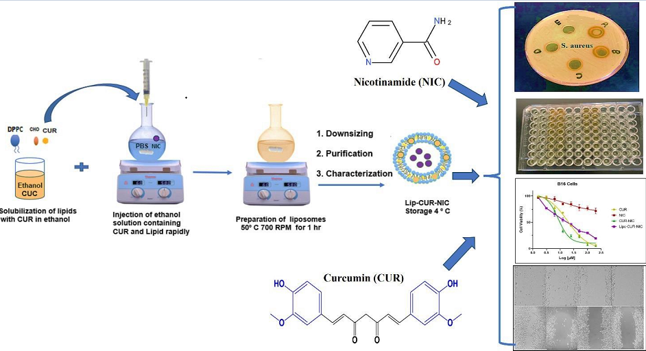
Liposomes of CUR and NIC were prepared using the conventional ethanol injection technique as described in our previous article (Fahdawi et.al. 2024). Briefly, 10 mg of DPPC, CHO 2.5 mg, and 2.0 mg CUR were dissolved ethanol, which was then warmed at 40 °C water bath. NIC 2mg/ml was dissolved PBS was heated using hot plate at ∼50 °C with string 700 rpm. Then the warm drug lipid ethanol mixture was injected rapidly into the PBS with heating and continuous stirring. After were prepped, the free of CUR and NIC were removed and washed and stored at 4°C [19].
The average particle size, Zeta Potential (charge) and Polydispersity Index (PDI) for liposomes were measured by DLS experiments using Zetasizer, Nano-ZS (Malvern Instruments Ltd., Malvern, UK). The Stability of the loaded liposomes was performed at 4 °C with storage times of one month. The EE % of CUR and NIC into liposomes was expressed as the percentage of drug complex encapsulated inside liposomes [20].
2.2. Screening of the Antibacterial Activity
2.2.1. Agar Well-Diffusion Method
The Antibacterial activity screening was performed by the agar well-diffusion method according to guidelines of the Clinical and Laboratory Standards Institute. Muller-Hinton Agar Plates were prepared according to the manufacturer instructions (Thermo Fisher Scientific, Waltham, MA, USA). Wells were made into the agar using a 6 mm diameter sterile borer. The selected bacteria were mainly S. aureus and p. aeruginosa (ATCC® 29213). Bacterial cultures were started in Muller-Hinton (MH) broth, and incubated overnight in the shaker at 37o C. Before use, the inoculum turbidity was standardized at OD520 nm = 0.1 (contrasted to 0.5 M McFarland). Bacteria were swabbed uniformly on the agar plates using sterile cotton applicators dipped into the standardized inoculum. Each well had 100 µL of the desired drug and liposomes concentration, while the control well contained the same volume of DMSO. Plates were then incubated at 37°C for 24 h, and the diameter of the inhibition zones was measured in millimeters (mm). Data are presented as the means for readings obtained from three different wells for each concentration.
2.2.2. Minimum Inhibitory Concentration (MIC)
If the bacteria were inhibited by the 1000 µg/mL at the primary screening, a determination for the MIC will take place using two-fold lower concentrations starting from 500, 250, 125, and 62.5 µg/ml. Five wells were made into the agar before streaking the plates with a standardized bacterial inoculum. Each well was filled with 100 µL of a selected drug, liposomes or with a control solvent which was used to solubilize the drugs and liposomes (DMSO or PBS). The plates were then incubated at 37°C for 24 h, and the inhibition zones were measured in mm.
2.2.3. S. aureus Susceptibility
The antibacterial activity of free Antibiotics and CUR and NIC loaded liposomes were evaluated by the broth Microdilution Method, according to guidelines of the Clinical and Laboratory Standards Institute [21], followed by turbidity evaluation. Briefly, the formulations were diluted in PBS to produce a serial dilution with concentrations ranging from: 0.1875 to 200 µg/mL of both drugs and their liposomes. Bacterial Suspensions were performed from a MSSA overnight culture diluted in broth media until reaching a value of 0.5 in a McFarland scale equivalent to 108 colony forming unit/ml by measuring the optical density at 600 nm. Bacterial Suspension was cultured in 96-well cell culture plates at 5 × 10⁵ bacterial density and incubated with the Antibiotics or the formulations, at 37 °C during 24 h. A negative control containing a Suspension of bacteria in broth, without treatment, and a sterile control containing broth only without bacteria, were included. Minimum Inhibitory Concentration (MIC: the lowest Antibiotic concentration able to prevent visible bacterial growth, resulting in the absence of turbidity) was determined Spectrophotometrically, at 570 nm in a microplate reader (Bio-Rad laboratories, Inc., Hercules, CA, USA).
2.3. Cell Culture
The B16 mouse cancer cell line was obtained from the American Type Culture Collection (ATCC, Manassas, VA, USA). These cells were cultivated as attached monolayers and preserved in DMEM medium (EuroClone, Italy), enriched with 10% (v/v) heat-inactivated Fetal Bovine Serum (FBS) (Euro Clone, Italy), 1% Penicillin-Streptomycin (Euro Clone, Italy), and 2 mM L-glutamine. Incubation of the cells was carried out at 37°C within a Tissue Culture Incubator (Memmert, Schwabach, Germany) containing 5% CO2.
2.3.1. Cell Viability Assay (MTT)
An MTT Assay was conducted to determine the IC50 of (Lip-CUR, Lip-NIC and their co-loaded liposome (Lip-CUR-NIC) prepared by the Ethanol Injection Technique on cancer cells. Roughly, (5 × 10³ cells/well) of B16 mouse Melanoma cancer cells placed into a 96-well late (Corning, USA). Cells were subjected to concentrations ranging from 3.125 to 200 μM.
Briefly, the different concentrations were prepared as follows; 200 μL of each Liposomal formulation (Lip-CUR, Lip-NIC and Lip-CUR-NIC) was mixed with 800 μL media to get a final volume of 1 ml Stock Solution (200 μM) for each formulation. Serial dilutions were made for each Stock Solution where 500 μL of each Stock Solution was diluted with 500 μL media. Then, the cells were cultured at 37 °C in a 5% CO2 Incubator for 72 hours. Afterward, the previous media was removed, and 100 μl of fresh media containing MTT Assay Salt (Bioworld, USA) was added to each well. Subsequently, the plates were incubated at 37 °C for 3 hours, followed by the addition of 50 μl of Solubilization Solution (DMSO) to each well to assess Viability. The optical absorbance of the Solution was measured at 560 nm using ELIZA Plate Reader (USA).
2.3.2. Cell Migration Assay
Melanoma B16 cell line was seeded in sterile 6-well cell culture plates at a density of 800,000 cells per well and then incubated for 24 hours at 37°C with 5% CO2. The following day, a vertical scratch was created at the center of the cell’s monolayer using a sterile 1,000 µl micropipette tip for either free drugs or nanoliposome’s treatment. Subsequently, each well was washed twice with sterile PBS. After 1 day, cells were exposed to CUR, NIC CUR-NIC and Lip-CUR-NIC at three different concentrations near IC50, as determined by the MTT test. Finally, images of the scratches were captured before and during cell treatment using a phase contrast microscope (model P. MICRO-001, Nikon) equipped with a 4× magnification objective. The Images J software was employed to calculate the wound closure area (µm2). DMSO and free media served as negative controls. The wound closure rate was observed on day 1 (before treatment) and day 2 after 48h of cell therapy [22]. Empty liposomes and media were employed as a negative control.
The wound closure (%) was calculated using eq
2.4. DPPH Assay
The free radical scavenging activities of four treatments Vit C, NIC, CUR, CUR-NIC prepared by the Ethanol Injection Technique, were determined employing the 2,2-diphenyl-1-picrylhydrazyl (DPPH) free radical scavenging technique, in accordance with the description provided by (Shirazi et al., 2014).
To prepare the Calibration Curve, a Stock Solution of Vitamin C was prepared by dissolving 1mg in 1mL Methanol to obtain a Standard Stock Solution of Vitamin C, CUR, NIC, and CUR-NIC. Serial dilutions were prepared by transferring 500 μl of each of the samples and mixing it with 500 μl Methanol. The DPPH Solution was prepared by dissolving 8 mg of DPPH in 10 ml Methanol. The obtained Solution had deep violet colour. In 96 well plate 100 μl of each sample were added to 100 μl of the freshly prepared DPPH Solution. Each was done in triplicate and incubated in dark place for 30 min. Controls were used, DPPH and CUR in Methanol. Absorbance was measured by an ELISA Plate Reader at 517 nm. The Calibration Curve of Vitamin C was used to calculate the (%) inhibition and the concentration of Vitamin C, and the prepared formulations required for the inhibition of % 50 of free radicals (IC50).
(%) Inhibition of Free Radical = (Ab Control – Ab Sample) / Ab Control *100 %
2.5. Statistical Analysis
The findings were expressed as the mean ± standard deviation from a minimum of three separate trials. GraphPad Prism 8 (GraphPad Software Inc., USA), CompuSyn.exe (Version 1) and Microsoft Office Excel (Microsoft, USA).
3. Results and Discussion
3.1. Liposomal Preparation and Characterization
The liposomal formula of Lip-CUR-NIC was developed and fully characterized using UV spectrophotometry and HPLC method of analysis which were published (Fahdawi et. al., 2024) and in this work Lip-CUR-NIC formula was biologically evaluated with pure compounds CUR, and NIC. The average particle size was less than 200 mm for all preparations (Figure 1A). PDI shows how uniform the particle size distribution (Figure 1B). is, with lower values indicating a narrow size range and higher values indicating a wide size range. A PDI of less than 0.4 is generally considered good for monodispersed nanoparticles (Bellone et al., 2015). Stability of loaded lipoamides was assessed for 30 days in terms of size distribution (Figure 1C). and zeta potential (Figure 1D, 1E) shows summary of average diameter, PDI, charge, EE% and LE%. All measured and evaluated characters were consistent with the previous article of the same for the same liposomal formulations (Fahdawi et. al., 2024). Invitro release of profile of CUR and NIC over 72 h was evaluated and showed slower release of CUR from Lip-CUR-NIC than NIC which is due to higher solubility of NIC in PBS then CUR.
3.2. Antioxidant Activity and DPPH Assay
A DPPH assay, which measures the free radical scavenging activity of CUR, NIC, CUR- NIC and Vit C as a standard (Figure 2A, 2B). The assay results are plotted as percentage inhibition of the DPPH radical against the concentration of the tested substances in mg/ml. NIC exhibited the lowest activity among the tested samples, with a more gradual increase in inhibition percentage as the concentration increases, while CUR demonstrated higher activity than NIC, with a steeper curve that approaches the activity level of Vit C at higher concentrations. The combination between CUR-NIC displayed an improved scavenging effect compared to NIC alone and slightly better than CUR alone, indicating a potential synergistic effect of the combination on radical scavenging activity. Vit C showed the highest radical scavenging activity, reaching close to 100% inhibition, which is expected as it is a known potent antioxidant used as a positive control.
3.3. Antibacterial Activity of CUR, NIC and lip- CUR-NC
CUR at various concentrations (1000 – 62.5 µg/ml) showed good bacterial growth inhibition of S. aureus bacteria while NIC at various concentrations did not result in bacterial growth inhibition of S. aureus. Treatment of S. Aureus bacteria with CUR and NIC mixture resulted in superior antibacterial activity compared to that of CUR alone, which may be explained by synergistic effect between both treatments. However, when NIC was administered with CUR, it enhanced the antibacterial activity of CUR. When a higher concentration of CUR-NIC Mix (100 µg/ml), it resulted in higher antibacterial effect of the lower concentration (50 µg/ml). these findings suggests that bacterial growth inhibition by CUR-NIC is concentration dependent (Figure 3A,3B).
Every liposomal formulations prepared were tested for antibacterial activity against normal flora S. epidermidis in order to check the safety of the treatment against normal bacteria found on top of our skin. It was found that CUR alone showed inhibition of bacterial growth.
After initial assessment of the antibacterial activity of both treatments CUR, NIC and CUR-NIC and their respective liposomal formulations against S. aureus and S. epidermidis bacterial strains (Figure 3C, 3D).
The results showed weak inhibition of bacterial growth with Lip-CUR against Vancomycin susceptible S. aureus strain which is promising finding that could be further enhanced by modification of preparation process of the liposomes. CUR at a concentration range of (0.1875 – 200 µg/ml) compared to Lip -CUR at the same concentrations, the results showed that CUR has an MIC value of 0.375 µg/ml, whereas Cur-Lip has lower MIC value of 0.1875 µg/ml. This indicates that liposomal formulation of CUR would enhance its antibacterial activity against S. aureus bacteria. Whereas NIC at a concentration range of (1.875 – 3000 µg/ml) compared to Lip-NIC at the same concentrations. The results showed that NIC has an MIC value of 1.5 mg/ml, and Lip-NIC has lower MIC value of 0.75 mg/ml. this indicates that incorporation of NIC into liposomal formulation would enhance its antibacterial activity against S. Aureus bacteria (Figure 4A, 4B).
As described by Harush-Frenkel in 2010, the positively charged nanoparticles resulted in increased side effects and toxicity. Results showed that the prepared liposomes had a negative average Zeta Potential which is favorable for the safety profile of the prepared liposomes (Harush-Frenkel et al., 2010).
According to various investigations, all studies have consistently shown that CUR possesses antimicrobial effects, and no contradictory results have been reported on this topic [23].
Inhibitors of bacterial resistance offer potential treatment options for patients with Antibiotic-resistant infections. The use of natural inhibitors could improve the re-treatment of patients who have previously received ineffective Antibiotics in clinics and help prevent the emergence of new strains of Antibiotic-resistant bacteria [24].
Teow and Ali (2015) conducted a study to investigate the combined Antibacterial effects of curcumin and eight different antibiotic groups. Their experiments using disc diffusion assays showed synergistic effects between Curcumin and the majority of Antibiotics against S. aureus. However, microdilution assays only revealed synergism with three Antibiotics (Ciprofloxacin, Gentamicin, and Amikacin). The other tested antibiotics showed no significant interaction, although there was no antagonism observed. Their results were similar to this study which could be attributed to the use of similar methods [25].
The antibacterial activity of CUR using the broth microdilution method, checkerboard dilution test, and time-kill assay. An antimicrobial activity of CUR was observed against all tested strains. In the checkerboard test, CUR significantly reduced the MIC of antibiotics such as Oxacillin, Ampicillin, Ciprofloxacin, and Norfloxacin that are used against methicillin-resistant Staphylococcus Aureus (MRSA) [26].
Similar to our study, Zhou et al. demonstrated that the combined treatment of Curcumin and Erythromycin effectively suppressed bacterial growth and alleviated bone infection. The combination showed stronger efficacy against MRSA-induced osteomyelitis in rats compared to monotherapy [27].
In a different study, Wang et al. utilized Curcumin as a natural Antibacterial and Antifungal Agent against various foodborne pathogens, including Staphylococcus Aureus, Escherichia coli, Yersinia enterocolitica, Bacillus cereus, and Aspergillus niger. They improved the stability and solubility of CUR by using Microcapsules. The study demonstrated a broad-spectrum inhibitory effect of CUR against all tested organisms using the Oxford Cup Method. The results also indicated that Curcumin had greater Antibacterial Activity against Gram-positive bacteria than Gram-negative bacteria, while its Antifungal Activity was significantly higher than its antibacterial activity [28].
Gunes et al. investigated the effect of curcumin on standard bacterial strains at high concentrations and demonstrated its strong antibacterial activity at high doses on animals. This study was conducted in Turkey, and the similarity in results could be attributed to the potential presence of the same bacterial strains and resistance genes [29].
In a study conducted by Shailendiran et al. in 2011, the antibacterial properties of curcumin and non-formulated CUR were examined against both a gram-positive bacterial strain (Cocci) and a gram-negative bacterial strain such as E. coli. The study applied the agar disc assay to observe the size of the inhibition zone over time. Results showed that after 10 hours, a clearly visible inhibition zone was observed, indicating inhibition of bacterial growth. However, this zone became less distinct after 24 hours for both curcumin and nanocurcumin-treated discs. These findings suggest that the tested samples exhibited bacteriostatic properties, inhibiting bacterial growth rather than killing the bacteria outright [30]. In another study conducted by Hu et al., the Antimicrobial Activity of Curcumin against S. Mutans was examined, and the inhibitory ability of Curcumin on purified Sortase A was evaluated using Western blot and Real-time PCR. The study revealed that Curcumin can effectively inhibit purified S. Mutans Sortase A at a concentration equivalent to half of the minimum inhibitory concentration (MIC), leading to a reduction in S. Mutans biofilm formation [31].
Furthermore, Lzui et al. demonstrated that Curcumin exhibited a dose-dependent inhibition of the growth of Prevotella intermedia, P. gingivalis, Treponema denticola, and Fusobacterium nucleatum. Even at very low concentrations, CUR significantly suppressed bacterial development [32].
3.4. Cytotoxicity study and Anticancer Activity
When looking at CUR, NIC, and their combination CUR-NIC, it was found that CUR-NIC had the most significant impact on reducing cancer cell viability, especially at moderate concentrations. This suggests that combining CUR and NIC might have a synergistic effect in fighting cancer, making it worth exploring further for potential cancer therapies.
For the Drug-loaded liposomes, the encapsulation of CUR in liposomes increases its IC50 to 50 ± 9.2 µM, suggesting reduced effectiveness compared to free CUR. NIC remains ineffective with an IC50 value still over 200 µM (Figure 5A).
Regarding drug-loaded liposomes, CUR’s IC50 was (31.4 ± 3.2 µM), and NIC’s (IC50 is 190 ± 7.3 µM). Both drugs showed improved effectiveness compared to their single-drug liposome counterparts but less than the free drug combination. Additionally, the combination of CUR and NIC exhibits a significant synergistic effect, markedly reducing the IC50 values for both drugs. The free combination offers the greatest enhancement of activity against B16 cells, whereas liposomal encapsulation, both as single and combination drugs, results in higher IC50 values compared to free drugs, suggesting that the encapsulation method might reduce drug availability to B16 cells (Figure 5A, 5B).
The IC50 values for CUR and NIC when administered alone and in combination to B16 cells were shown in (Figure 5C), for the free drugs alone, CUR has an IC50 value of 27.3 ± 2.3 µM, indicating moderate potency. NIC, however, has an IC50 value exceeding 200 µM, showing low effectiveness as a single agent against B16 cells. For free drugs in combination, the IC50 for CUR significantly decreases to 9.8 ± 2.2 µM, showing increased potency. Similarly, the IC50 for NIC drops to 135.95 ± 10.2 µM, indicating enhanced effectiveness when used with CUR.
The provided Isobologram represents a combination analysis for the CUR-NIC [1:1 plots three iso-effective combinations of Dose A (CUR) and Dose B (NIC) corresponding to Fraction Affected or inhibited (Fa) values of 0.5, 0.75, and 0.9. by the drug combination. The combination of doses required to achieve 50% effect shows that either drug can achieve this effect level alone at a certain dose.
The Isobologram suggests a synergistic interaction between CUR and NIC when combined at a 1:1 ratio, as the combination points fall below the line of additivity. This synergy implies that lower doses of each drug can be used in combination to achieve a high level of effect, potentially reducing side effects, and increasing treatment efficacy, allowing for dose reduction while maintaining or improving therapeutic outcomes.
Combination Index (CI) plot for the CUR and NIC Free Powder Combination, quantifies the interaction between two drugs, with CI<1 indicating synergy, CI=1 indicating an additive effect, and CI >1 indicating antagonism (Figure 6A, 6B, 6C, 6D, 6E).
The graph illustrates the Combination Index (CI) values at various Fraction Affected (Fa) levels, ranging from 0 (no effect) to 1 (complete effect). For the CUR+NIC combination, most CI values fall below 1 across the Fa range, indicating increasing synergy particularly at higher Fa levels. The Dose-effect Curve for CUR, NIC, and their Combination (CUR-NIC) is depicted in Figure 1. Fa is plotted against the dose, illustrating the drugs' impact on cell viability. CUR exhibits a steep dose-effect relationship, effectively inhibiting cell viability even at low doses, reaching near-complete effect (Fa close to 1) at low doses. In contrast, NIC shows a more gradual dose-effect curve, with lower inhibition of cell viability even at comparable doses, not achieving the same level of inhibition as CUR even at higher doses. CUR-NIC displays a dose-effect curve similar to CUR, suggesting the combination is as effective as CUR alone without NIC negatively impacting CUR's inhibitory effect. The dose reducing index (DRI) quantifies the dose reduction potential of a drug in combination therapy compared to its use alone. CUR exhibits a sharp increase in DRI with Fa, indicating significant dose reduction (up to 5 times or more) when used in combination, whereas data points for NIC are absent, suggesting NIC does not allow for dose reduction in the combination. The DRI curve for CUR suggests strong synergistic interaction, enabling marked dose reduction while maintaining Anticancer Efficacy. At the highest Fa level (0.97), the DRI for CUR exceeds 100 and for NIC surpasses 200, indicating doses required to achieve 97% inhibition of Cell Viability in combination. The CUR-NIC Combination shows a significantly lower IC50 of 10, a slope of 0.867, and a correlation coefficient of 0.981, indicating potentiated effect with high-quality Dose-response Curve fit. Moreover, when delivered via Lip-CUR and Lip-NIC, Lip-CUR demonstrates moderate anticancer activity with an IC50 of 20, while Lip-NIC shows cell viability with an IC50 above 50. However, Lip-CUR-NIC exhibits the sharpest decline in cancer cell viability among all treatments, suggesting the importance of exploring liposomal delivery for enhanced cancer treatment efficacy.
Wang et.al investigated curcumin-loaded MPEG-PLA micelles against melanoma both in vitro and in vivo. Their results revealed that the spherical curcumin/MPEG-PLA micelles disperse effectively in normal saline with sustained release and exhibit stronger cytotoxicity. Histochemical analysis for in vivo further confirms their efficacy in inducing melanoma cell apoptosis and inhibiting neovascularization in tumor tissues. They conclude that the curcumin/MPEG-PLA micelles show promise for clinical application in treating melanoma [33].
Fontes et. al explored the effect of CUR and disulfiram combination on B16 melanoma cells. The study explores the combined effect of CUR and disulfiram on melanoma cells. In vitro and in vivo experiments reveal synergistic effects at specific ratios, enhancing apoptosis and oxidative stress levels. The CUR-DSS combination shows promising potential for melanoma therapy, offering significant tumor growth inhibition compared to individual compounds [34].
3.5. Migration Test (Scratch Assay)
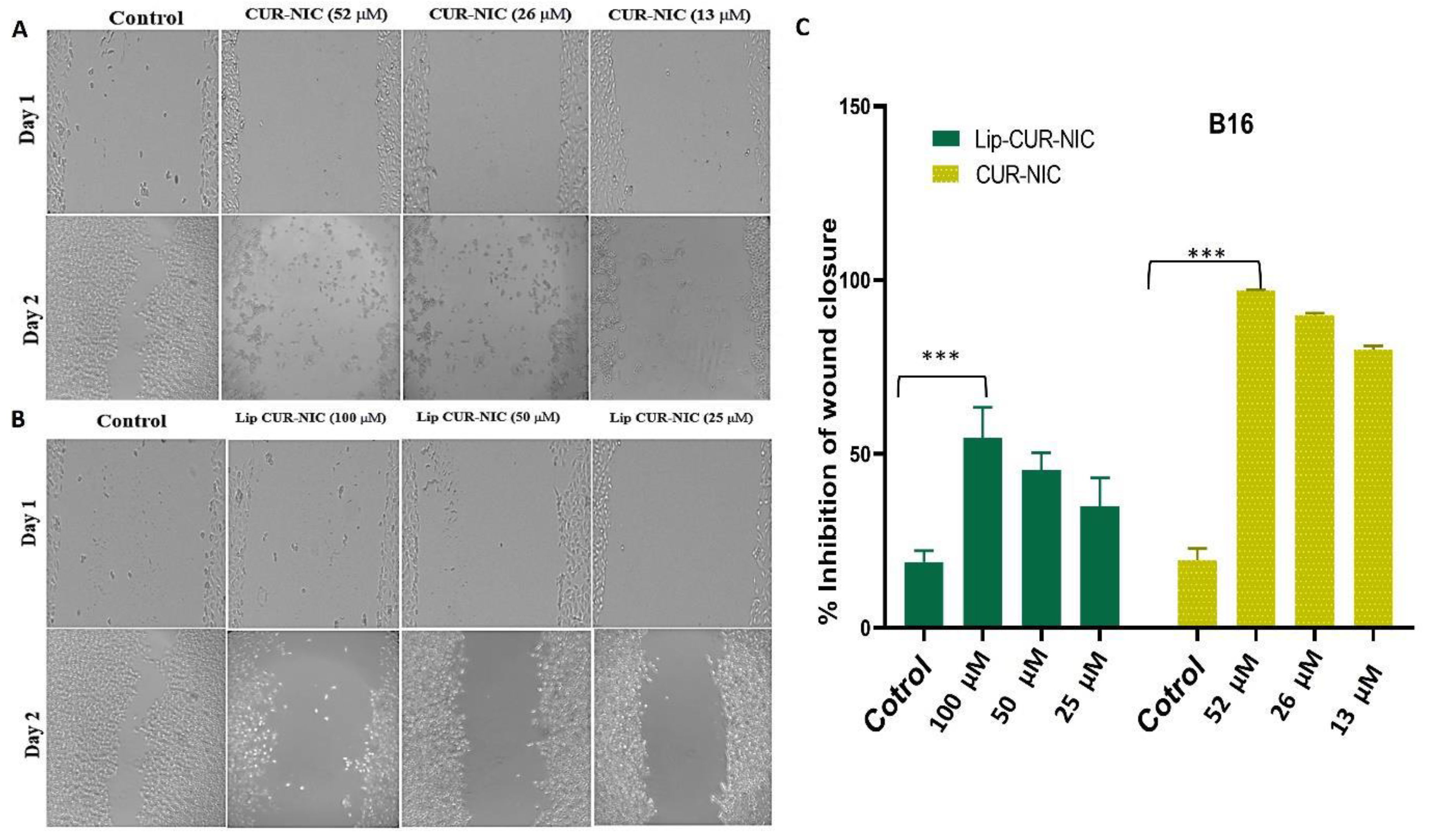
A Scratch Assay was conducted on B16 Melanoma cells over two days to assess cell migration following treatment with CUR-NIC and liposomes at different concentrations. For CUR-NIC (52 µM), the scratch area remained mostly clear by Day 2, indicating strong inhibition of cell migration. At 26 µM, moderate inhibition was observed, while at 13 µM, some cell migration occurred but less than in the control. These results demonstrate concentration-dependent inhibition of cell migration by CUR-NIC, with all concentrations effectively inhibiting migration, suggesting potential for preventing tumor metastasis. Similarly, Scratch Assay results for Lip-CUR-NIC at various concentrations were observed over two days. Control samples showed significant cell migration by day 2, while Lip-CUR-NIC (100 µM – displayed modest increase, indicating strong inhibition of migration. Lip-CUR-NIC showed moderate migration at (50 µM) and exhibited slight migration at (25 µM). These findings suggest that Lip-CUR-NIC effectively inhibits B16 Melanoma cell migration in a concentration-dependent manner, with higher concentrations providing greater inhibition (Figure 7A,7B). In contrast, CUR-NIC showed lower inhibition across all concentrations compared to the Lip-CUR-NIC, highlighting the potential advantages of liposomal encapsulation for therapeutic purposes. The control bars represent the baseline migration of untreated cells, providing a reference for comparison with treated samples (Figure 7C).
4. Conclusion
In conclusion, the CUR-NIC combination, demonstrated significant anticancer activity as evidenced by the MTT assay. This indicates a synergistic effect when curcumin and nicotinamide are combined, potentially offering a more effective cancer treatment strategy. The DPPH assay results revealed antioxidant properties of the CUR-NIC combination, which may contribute to its anticancer efficacy. Furthermore, the scratch assay results underscored the potential of liposomal CUR-NIC in inhibiting cancer cell migration, a crucial step in preventing metastasis. The in vitro release assay demonstrated that liposomal formulations provided a sustained release of curcumin and nicotinamide compared to the powder mix, suggesting improved therapeutic efficacy.
In summary, this study presents compelling evidence for the potential use curcumin and nicotinamide combination in melanoma therapy and skin infection with S. aureus. Furthermore, CUR, NIC, CUR-NIC and their liposomal declares further investigation as a potential strategy to combat S. aureus. Future research should focus on in vivo studies, mechanistic investigations, combination therapies, and clinical trials to validate the effectiveness, safety, and clinical translation potential of this formulation.
References
- Sorrenti, V.; Burò, I.; Consoli, V.; Vanella, L. Recent Advances in Health Benefits of Bioactive Compounds from Food Wastes and By-Products: Biochemical Aspects. Int. J. Mol. Sci. 2023, 24, 2019. [Google Scholar] [CrossRef] [PubMed]
- Nsairat, H.; Lafi, Z.; Al-Sulaibi, M.; Gharaibeh, L.; Alshaer, W. Impact of nanotechnology on the oral delivery of phyto-bioactive compounds. Food Chem. 2023, 424, 136438. [Google Scholar] [CrossRef]
- Lafi, Z.; et al. A review Echinomycin: A Journey of Challenges. Jordan Journal of Pharmaceutical Sciences 2023, 16, 640–654. [Google Scholar] [CrossRef]
- Hammad, H.M.; Imraish, A.; Al-Hussaini, M.; Zihlif, M.; Harb, A.A.; Abu Thiab, T.M.; Lafi, Z.; Nassar, Z.D.; Afifi, F.U. Ethanol Extract of Achillea fragrantissima Enhances Angiogenesis through Stimulation of VEGF Production. Endocrine, Metab. Immune Disord. - Drug Targets 2021, 21, 2035–2042. [Google Scholar] [CrossRef]
- Lafi, Z.M.; Irshaid, Y.M.; El-Khateeb, M.; Ajlouni, K.M.; Hyassat, D. Association of rs7041 and rs4588 Polymorphisms of the Vitamin D Binding Protein and the rs10741657 Polymorphism of CYP2R1 with Vitamin D Status Among Jordanian Patients. Genet. Test. Mol. Biomarkers 2015, 19, 629–636. [Google Scholar] [CrossRef] [PubMed]
- Watkins, R.R.; Bonomo, R.A. Overview: Global and Local Impact of Antibiotic Resistance. Infect. Dis. Clin. North Am. 2016, 30, 313–322. [Google Scholar] [CrossRef]
- Hussain, Y.; Alam, W.; Ullah, H.; Dacrema, M.; Daglia, M.; Khan, H.; Arciola, C.R. Antimicrobial Potential of Curcumin: Therapeutic Potential and Challenges to Clinical Applications. Antibiotics 2022, 11, 322. [Google Scholar] [CrossRef]
- Nwobodo, D.C.; Ugwu, M.C.; Anie, C.O.; Al-Ouqaili, M.T.S.; Ikem, J.C.; Chigozie, U.V.; Saki, M. Antibiotic resistance: The challenges and some emerging strategies for tackling a global menace. J. Clin. Lab. Anal. 2022, 36, e24655. [Google Scholar] [CrossRef]
- Mirzaei, H.; et al. Curcumin: A new candidate for melanoma therapy? International journal of cancer 2016, 139, 1683–1695. [Google Scholar] [CrossRef]
- Laikova, K.V.; Oberemok, V.V.; Krasnodubets, A.M.; Gal’chinsky, N.V.; Useinov, R.Z.; Novikov, I.A.; Temirova, Z.Z.; Gorlov, M.V.; Shved, N.A.; Kumeiko, V.V.; et al. Advances in the Understanding of Skin Cancer: Ultraviolet Radiation, Mutations, and Antisense Oligonucleotides as Anticancer Drugs. Molecules 2019, 24, 1516. [Google Scholar] [CrossRef]
- Zainab, L.; Hiba, T.; Hanan, A. An updated assessment on anticancer activity of screened medicinal plants in Jordan: Mini review. J. Pharmacogn. Phytochem. 2020, 9, 55–58. [Google Scholar] [CrossRef]
- Lafi, Z.; Aboalhaija, N.; Afifi, F. Ethnopharmacological importance of local flora in the traditional medicine of Jordan: (A mini review). Jordan J. Pharm. Sci. 2022, 15, 132–144. [Google Scholar] [CrossRef]
- El-Hack, M.E.A.; de Oliveira, M.C.; Attia, Y.A.; Kamal, M.; Almohmadi, N.H.; Youssef, I.M.; Khalifa, N.E.; Moustafa, M.; Al-Shehri, M.; Taha, A.E. The efficacy of polyphenols as an antioxidant agent: An updated review. Int. J. Biol. Macromol. 2023, 250, 126525. [Google Scholar] [CrossRef]
- Munef, A.; Lafi, Z.; Shalan, N. Investigating anti-cancer activity of dual-loaded liposomes with thymoquinone and vitamin C. Ther. Deliv. 2024, 15, 267–278. [Google Scholar] [CrossRef]
- Starr, P. Oral Nicotinamide Prevents Common Skin Cancers in High-Risk Patients, Reduces Costs. . 2015, 8, 13–4. [Google Scholar]
- Allen, N.C.; Martin, A.J.; Snaidr, V.A.; Eggins, R.; Chong, A.H.; Fernandéz-Peñas, P.; Gin, D.; Sidhu, S.; Paddon, V.L.; Banney, L.A.; et al. Nicotinamide for Skin-Cancer Chemoprevention in Transplant Recipients. New Engl. J. Med. 2023, 388, 804–812. [Google Scholar] [CrossRef] [PubMed]
- Surjana, D.; Halliday, G.M.; Damian, D.L. Role of Nicotinamide in DNA Damage, Mutagenesis, and DNA Repair. J. Nucleic Acids 2010, 2010, 1–13. [Google Scholar] [CrossRef]
- Salech, F.; et al. Nicotinamide, a poly [ADP-ribose] polymerase 1 (PARP-1) inhibitor, as an adjunctive therapy for the treatment of Alzheimer’s disease. Frontiers in Aging Neuroscience 2020, 12: p. 255.
- Lafi, Z.; et al. Aptamer-functionalized pH-sensitive liposomes for a selective delivery of echinomycin into cancer cells. RSC Advances 2021, 11, 29164–29177. [Google Scholar] [CrossRef]
- Alshaer, W.; Zraikat, M.; Amer, A.; Nsairat, H.; Lafi, Z.; Alqudah, D.A.; Al Qadi, E.; Alsheleh, T.; Odeh, F.; Alkaraki, A.; et al. Encapsulation of echinomycin in cyclodextrin inclusion complexes into liposomes: in vitro anti-proliferative and anti-invasive activity in glioblastoma. RSC Adv. 2019, 9, 30976–30988. [Google Scholar] [CrossRef]
- Weinstein, M.P.; Lewis, J.S. The Clinical and Laboratory Standards Institute Subcommittee on Antimicrobial Susceptibility Testing: Background, Organization, Functions, and Processes. J. Clin. Microbiol. 2020, 58. [Google Scholar] [CrossRef]
- Grimmig, R.; Babczyk, P.; Gillemot, P.; Schmitz, K.-P.; Schulze, M.; Tobiasch, E. Development and Evaluation of a Prototype Scratch Apparatus for Wound Assays Adjustable to Different Forces and Substrates. Appl. Sci. 2019, 9, 4414. [Google Scholar] [CrossRef]
- Shajari, M.; Rostamizadeh, K.; Shapouri, R.; Taghavi, L. Eco-friendly curcumin-loaded nanostructured lipid carrier as an efficient antibacterial for hospital wastewater treatment. Environ. Technol. Innov. 2020, 18, 100703. [Google Scholar] [CrossRef]
- Stavri, M.; Piddock, L.J.V.; Gibbons, S. Bacterial efflux pump inhibitors from natural sources. J. Antimicrob. Chemother. 2006, 59, 1247–1260. [Google Scholar] [CrossRef]
- Teow, S.-Y.; Ali, S.A. Synergistic antibacterial activity of Curcumin with antibiotics against Staphylococcus aureus. . 2015, 28, 2109–14. [Google Scholar] [PubMed]
- Mun, S.-H.; Joung, D.-K.; Kim, Y.-S.; Kang, O.-H.; Kim, S.-B.; Seo, Y.-S.; Kim, Y.-C.; Lee, D.-S.; Shin, D.-W.; Kweon, K.-T.; et al. Synergistic antibacterial effect of curcumin against methicillin-resistant Staphylococcus aureus. Phytomedicine 2013, 20, 714–718. [Google Scholar] [CrossRef] [PubMed]
- Zhou, Z.; Pan, C.; Lu, Y.; Gao, Y.; Liu, W.; Yin, P.; Yu, X. Combination of Erythromycin and Curcumin Alleviates Staphylococcus aureus Induced Osteomyelitis in Rats. Front. Cell. Infect. Microbiol. 2017, 7, 379. [Google Scholar] [CrossRef] [PubMed]
- Wang, Y.; Lu, Z.; Wu, H.; Lv, F. Study on the antibiotic activity of microcapsule curcumin against foodborne pathogens. Int. J. Food Microbiol. 2009, 136, 71–74. [Google Scholar] [CrossRef]
- Gunes, H.; et al. Antibacterial effects of curcumin: An in vitro minimum inhibitory concentration study. Toxicol Ind Health 2016, 32, 246–50. [Google Scholar] [CrossRef]
- Shailendiran, D.; Pawar, N.; Chanchal, A.; Pandey, R.P.; Bohidar, H.B.; Verma, A.K. Characterization and Antimicrobial Activity of Nanocurcumin and Curcumin. 2011 International Conference on Nanoscience, Technology and Societal Implications (NSTSI). LOCATION OF CONFERENCE, IndiaDATE OF CONFERENCE; pp. 1–7.
- Hu, P.; Huang, P.; Chen, M.W. Curcumin reduces Streptococcus mutans biofilm formation by inhibiting sortase A activity. Arch. Oral Biol. 2013, 58, 1343–1348. [Google Scholar] [CrossRef]
- Izui, S.; Sekine, S.; Maeda, K.; Kuboniwa, M.; Takada, A.; Amano, A.; Nagata, H. Antibacterial Activity of Curcumin Against Periodontopathic Bacteria. J. Periodontol. 2016, 87, 83–90. [Google Scholar] [CrossRef]
- Wang, B.; Liu, X.; Teng, Y.; Yu, T.; Chen, J.; Hu, Y.; Liu, N.; Zhang, L.; Shen, Y. Improving anti-melanoma effect of curcumin by biodegradable nanoparticles. Oncotarget 2017, 8, 108624–108642. [Google Scholar] [CrossRef] [PubMed]
- Fontes, S.S.; Nogueira, M.L.; Dias, R.B.; Rocha, C.A.G.; Soares, M.B.P.; Vannier-Santos, M.A.; Bezerra, D.P. Combination Therapy of Curcumin and Disulfiram Synergistically Inhibits the Growth of B16-F10 Melanoma Cells by Inducing Oxidative Stress. Biomolecules 2022, 12, 1600. [Google Scholar] [CrossRef] [PubMed]
Figure 1.
Characterization of the prepared liposomes A) Size Distribution B) Average Zeta Potential C) Stability Test for liposomal Hydrodynamic Diameter for one month D) Stability Test for liposomal Zeta Potential for one month E) Summary table for average size, zeta potential, encapsulation and loading efficiency F) In vitro release assay for CUR and NIC from Lip-CUR-NIC (mean ± SD, n = 3).
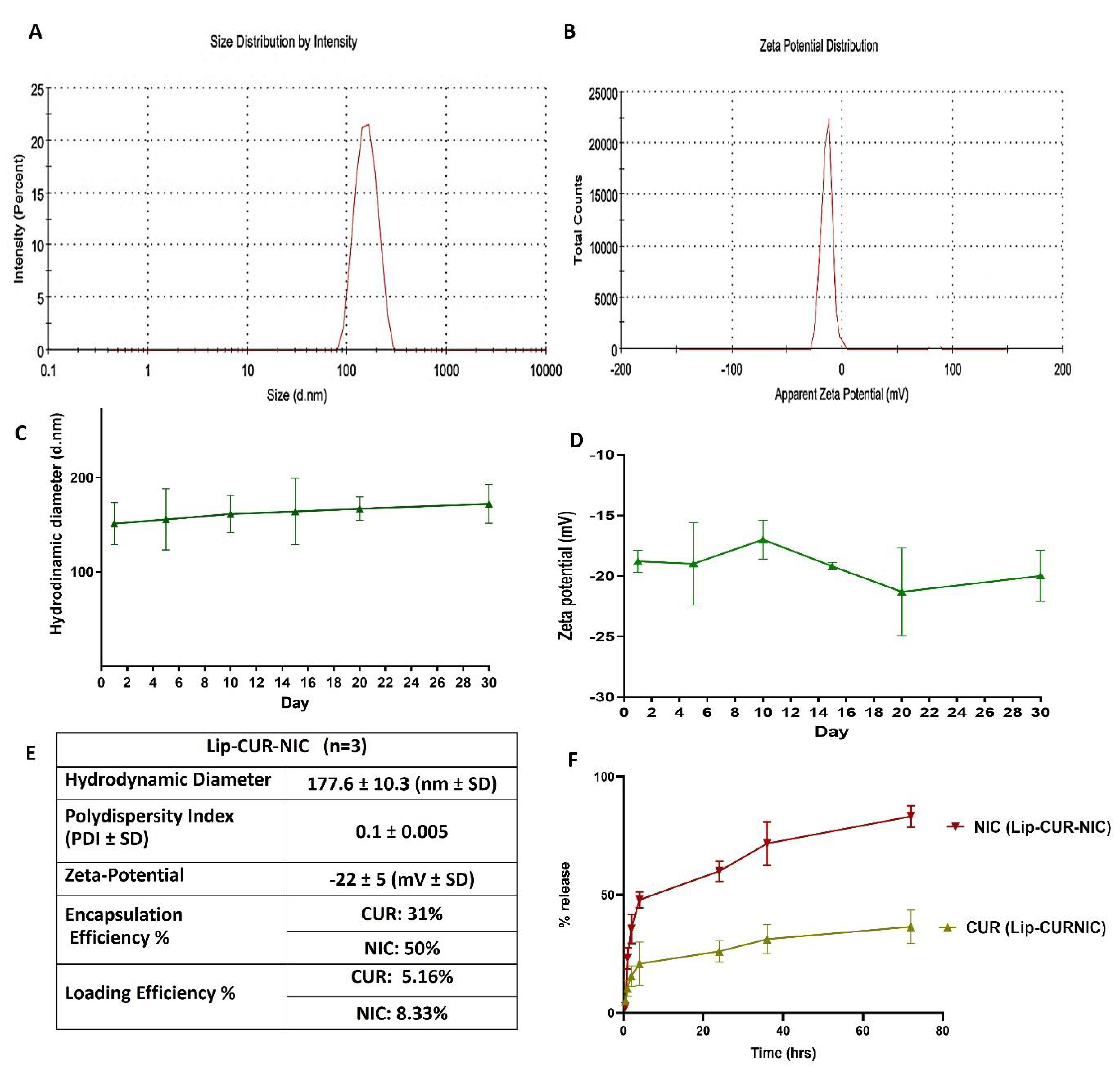
Figure 2.
A) Percent Inhibition of DPPH free radical using CUR, NIC and their combination B) IC50 of DPPH radical CUR, NIC, CUR-NIC and lip-CUR-NIC (mean ±SD, and n=3).
Figure 2.
A) Percent Inhibition of DPPH free radical using CUR, NIC and their combination B) IC50 of DPPH radical CUR, NIC, CUR-NIC and lip-CUR-NIC (mean ±SD, and n=3).
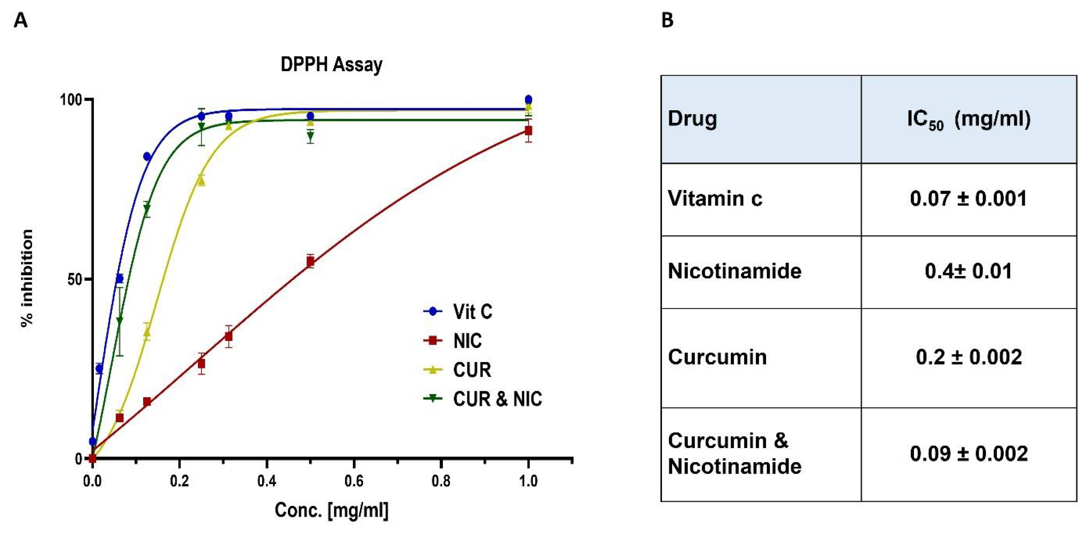
Figure 3.
Well-diffusion Method for A) CUR with 4 serial dilution concentrations (1000 – 62.5 μg/ml) against S. Aureus showing good inhibition zone. B) CUR-NIC combination with 6 serial dilution concentrations (1000 – 62.5 μg/ml) against S. Aureus C) Screening of CUR (A), NIC (B) and CUR-NIC (C, E) combination of two concentration against S. epidermises D) Screening of Lip-CUR, Lip-NIC, and Lip-CUR-NIC formulation against S. epidermises.
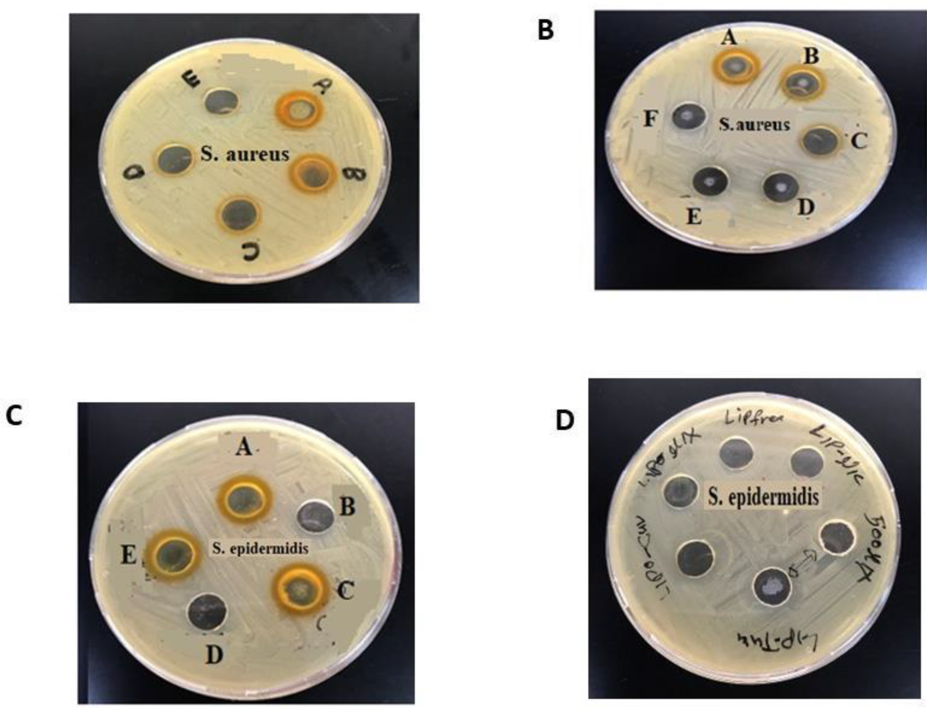
Figure 4.
A) Plate titter method for S. Aureus Susceptibility Test using CUR, Lip-CUR, CUR-NIC, Lip-CUR-NIC and NIC, at a concentration range of (1.875 – 3000 μg/ml) B) Table of inhibition zone and mic of the CUR, Lip-CUR, CUR-NIC, Lip-CUR-NIC and NIC B) Evaluation of inhibition zone and MIC of all treatments.
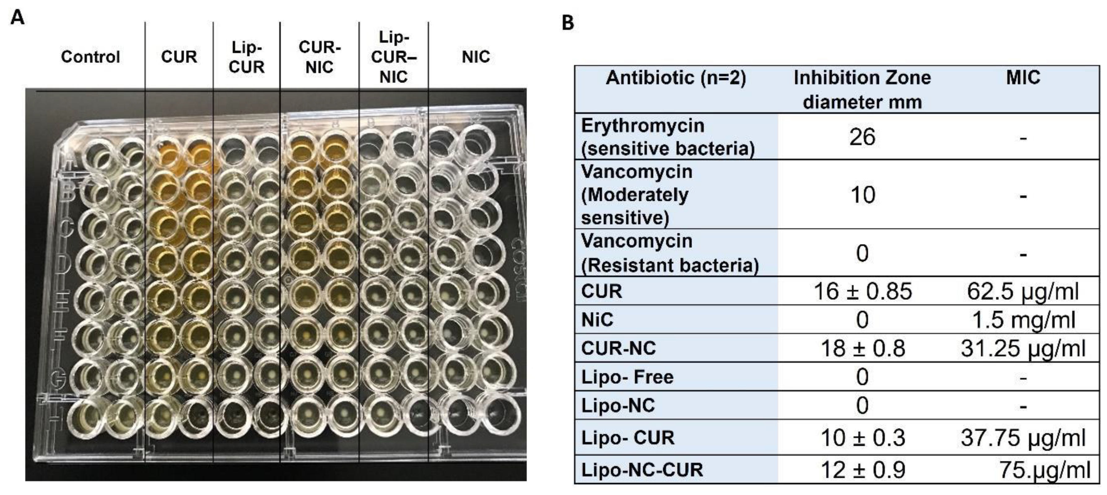
Figure 5.
(A)The Dose Response Curve for B16 cancer cells treated with Free CUR, NIC and their mixture (0.4-200 μM, n=3) (B) The Dose Response Curve for B16 cancer cells treated with Lip-CUR, Lip-NIC and Lip-CUR-NIC (0.4-200 μM, n=3) (C) Summary of IC50 values for single and combination of CUR & NIC against B16 cells.
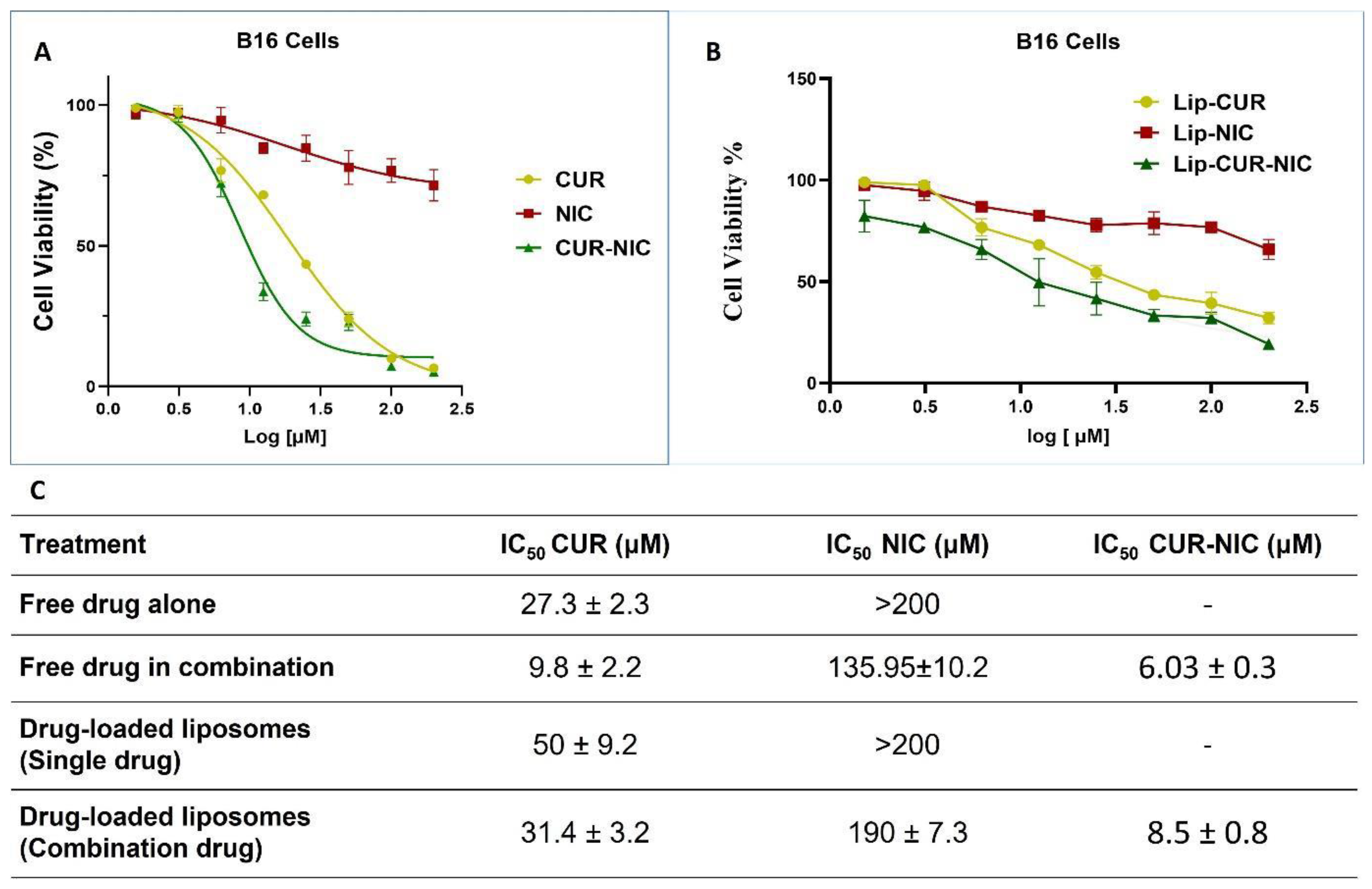
Figure 6.
(A) Isobologram for combination: CUR-NIC (CUR+NIC [1:1]) (B) Combination Index Plot of CUR and NIC Free Powder (C) Dose-reducing Index Curve of CUR and NIC combination (D) Dose-effect Curve of CUR and NIC combination (E) Summary of Combination Index (CI) and dose Reducing Index of (DRI) CUR-NIC.
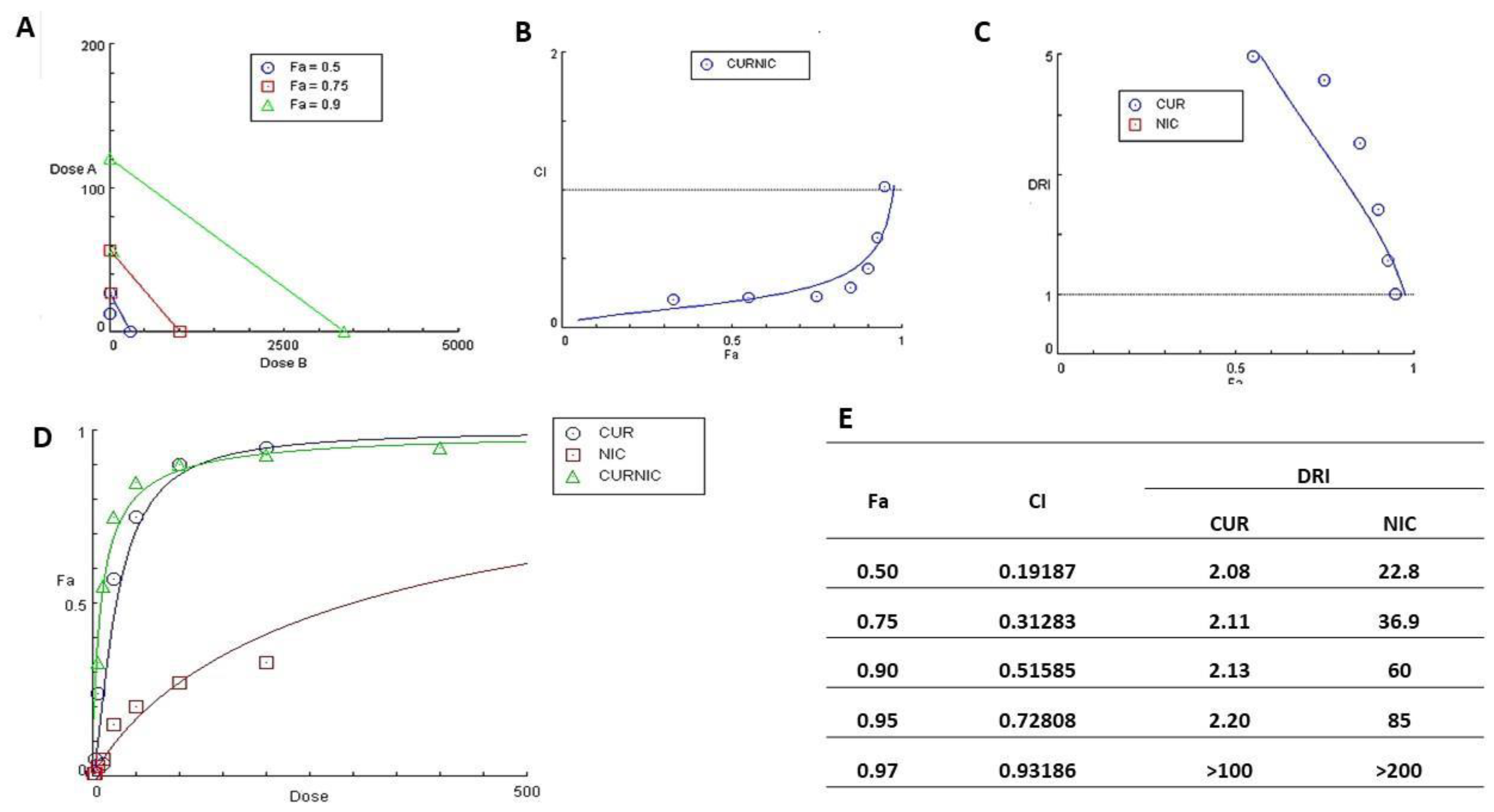
Figure 7.
Effect of CUR-NIC and Lip-CUR-NIC on cell migration and invasion capability. (A) The migration of B16 melanoma cells in the Matrigel surface after 48 h of scrape in different concentration (13 ,26, 52 μ M) of CUR-NIC combination groups. (B) show the migration of B16 melanoma cells in the matrigel surface after 48 h of scrape in different concentration (25, 50, 100 μM) of Lip-CUR-NIC. (C) The cells migration inhibited distance for all the formulations and free drugs were calculated. (Quantification of the wound scratch assay shown Data are expressed as a mean ± SD, for 24 hrs and n=2).
Disclaimer/Publisher’s Note: The statements, opinions and data contained in all publications are solely those of the individual author(s) and contributor(s) and not of MDPI and/or the editor(s). MDPI and/or the editor(s) disclaim responsibility for any injury to people or property resulting from any ideas, methods, instructions or products referred to in the content. |
© 2024 by the authors. Licensee MDPI, Basel, Switzerland. This article is an open access article distributed under the terms and conditions of the Creative Commons Attribution (CC BY) license (http://creativecommons.org/licenses/by/4.0/).
Copyright: This open access article is published under a Creative Commons CC BY 4.0 license, which permit the free download, distribution, and reuse, provided that the author and preprint are cited in any reuse.


