Submitted:
30 July 2024
Posted:
31 July 2024
You are already at the latest version
Abstract
Oxidative stress plays an essential role in the pathological processes that trigger various chronic liver diseases and contributes to the development of hepatocarcinogenesis. Antioxidants from natural sources trap free radicals by converting them into non-harmful products and minimize oxidative stress, playing a crucial function in the treatment of free radical-induced diseases. However, the activity of these antioxidants is limited due their poor bioavailability, which is why various nano-drug delivery systems have been developed to improve their stability, promote smooth release, and targeted delivery for liver cancer treatment. Specifically, this review aims to report recent scientific findings of novel nano-formulations containing natural antioxidant molecules targeted for the treatment of liver cancer.
Keywords:
antioxidants
; liver cancer
; nano-formulations
; drug delivery
; natural compounds
; hepatocellular carcinoma
; oxidative stress
1. Introduction
Liver cancer is a life-threatening illness and is one of the most aggressive and steadily increasing forms of cancer in the world [1]. Recent statistics indicate that it is the sixth-most common form of cancer and affects more men (fifth-most common form of cancer) than women (ninth-most common form of cancer) [2]. However, these findings refer to primary liver cancer, also known as primary hepatic cancer or primary hepatic malignancy, which starts and develops in the liver parenchyma or other structures within the liver, such as the bile duct, blood vessels, and immune cells [3,4]. In contrast, most liver cancers are secondary or metastatic, meaning they originate in other parts of the body (e.g., colon, breast, or lungs) and then spread to the liver. [5]. Among the sub-types of primary liver cancers, hepatocellular carcinoma (HCC), which forms from hepatocytes, is the most common (80–90%) [6,7], followed by cancer of the bile duct, namely intrahepatic cholangiocarcinoma (~6%), angiosarcoma and hemangiosarcoma (0.1–2.0%), two rare and very aggressive forms of cancer that originate from the endothelial layer of blood vessels and metastasize rapidly [8], and hepatoblastoma (~1%), a type of cancer that develops in immature liver cells and primarily affects children under the age of 15 years [9]. Risk-factors that increase the incidence of primary liver cancer include, in order, cirrhosis due to chronic viral infections (such as hepatitis B and hepatitis C) or excessive alcohol consumption, metabolic diseases (such as nonalcoholic steatohepatitis, nonalcoholic fatty liver diseases, obesity and diabetes) [10], hereditary liver diseases (such as hemochromatosis and Wilson’s disease) [11,12], exposure to aflatoxins ([13], iron accumulation ([14] and certain biliary tract diseases (e.g., fascioliasis, a zoonotic infectious disease caused by parasitic worms of the genus Fasciola) [15].
More and more studies have recently demonstrated a clear correlation between oxidative stress (OS) and the onset/development of liver cancer [16,17,18,19,20,21,22,23]. OS in defined as a non-physiological condition of the body in which there is a marked imbalance between the production of reactive species with oxidant properties and the functioning of endogenous defense systems with antioxidant capacity. These reactive species are generally classified into ROS (reactive oxygen species), RNS (reactive nitrogen species), RSS (reactive sulfur species) and RCS (reactive chlorine species). ROS are those produced the most and by far the most harmful. They include superoxide anion radical (O2•−), hydroxyl radical (OH•), hydroxyl ion (OH−), hydrogen peroxide (H2O2), singlet oxygen (1O2), and ozone (O3) [24]. They can be produced by exogenous sources (e.g., UV radiation, toxic substances, drugs), physiological changes (e.g., stress conditions, chronic inflammatory states, aging) or endogenous sources (e.g., byproducts of the mitochondrial respiratory chain, transmembrane enzymes of the NADPH oxidase family, myeloperoxidase in phagocytes) [25]. Under normal physiological conditions, OS is kept under control by the adequate presence of metabolic pathway cofactors with free radical scavenger properties (e.g., vit. C, vit. E, CoQ10) and by the activity of antioxidant enzymes such as catalase (CAT), superoxide dismutase (SOD), and glutathione reductase (GRS) [26]. In contrast, uncontrolled OS plays a pivotal role in carcinogenesis and liver cell progression; its action occurs through multiple pathways, including direct oxidative damage to DNA, instability of genetic material, interference with signaling pathways of tumor growth and programmed cell death, production of cytokines, chemokines and mediators of the immune system, activation of pathways involved in metastasis, dysfunction of cytoplasmic organelles, angiogenesis, and increased drug resistance [27]. The mechanisms by which OS intervenes in the onset and progression of liver cancer are depicted and briefly described in Figure 1.
In view of all the evidence demonstrating the involvement of OS in the development of liver cancer and given the latter’s crucial role as the primary organ of detoxification and regulator of homeostasis in the human body, the use of natural antioxidants is becoming increasingly important both in terms of prevention and therapeutic treatment of this disease [28,29,30,31]. The plant world offers the greatest resources in this regard, with its abundance of (poly)phenolic substances whose beneficial effects on health have been known since ancient times and whose use as medicinal herbs is part of traditional medicine in many countries [32]. Unfortunately, large-scale use of these compounds as drugs has not been very successful so far because of their poor oral bioavailability, mainly due to their poor solubility in water and rapid first-pass metabolism [33] . In addition, one must consider a certain chemical instability of these compounds, which are easily oxidized in air and light [34].
In this context, the use of nanotechnology offers a wide range of versatile and more effective solutions than older, nonspecific cancer therapies [35,36,37,38]. Nanoparticles (NPs), particles typically ranging in size from 1 to 200 nm (although slightly larger sizes are universally accepted for applications in nanomedicine), with their large surface area can greatly increase the solubility of natural antioxidants (entrapped in their core or chemically bound or adsorbed on the surface), protect them from biodegradation or physicochemical degradation, promote their passage through biological barriers, provide sustained and gradual release, and offer stimuli-responsive delivery systems [39]. In addition, the surface area of NPs, depending on the material used for their formation, can be exploited for the construction of tumor-specific delivery vehicles by linking to it (mostly via covalent bonding) targeting agents, thus limiting as much as possible off-target effects. Nonetheless, NPs’ surface area can be further functionalized/exploited to obtain drug delivery systems for combination therapy by joining natural antioxidants with standard anticancer drugs reducing dosage and side-effects of the latter due to synergistic effects [40,41].
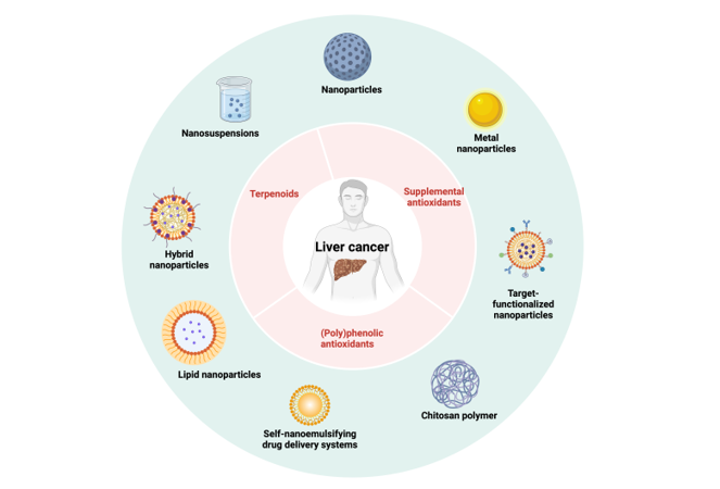
Several types of nanostructured materials have been intensively studied over the years for the possible treatment of liver cancer (more specifically for the treatment of HCC): NPs containing natural polymers as the basic component (e.g., chitosan, dextran); NPs constructed from synthetic polymers and/or copolymers [e.g., poly(ε-caprolactone), poly(α-hydroxybutyric acid), poly(lactide-co-glycolide)]; self-assembled nanostructures (e.g., liposomes, dendrimers, micelles); NPs obtained from biomacromolecules (e.g., albumin) [42]. The antioxidant profile of natural molecules has been improved mainly by techniques of loading onto the surface of nanostructures, entrapping in nanogels or in the core and/or core-shell spaces of hollow nanospheres [43,44,45,46]. In terms of biocompatibility and toxicity, carbon nanotubes, cationic NPs, metallic NPs, and silica-based NPs have been less successful for potential use against HCC. Carbon nanotubes have been associated with changes in liver cell morphology, platelet aggregation, toxicity due to the release of carbon residues, and, most importantly, the development of OS that could further exacerbate the pathological state of the liver [47,48]. Cationic NPs essentially cause blood clotting and hemolysis ([49]. Metallic NPs, particularly silver NPs, have also been associated with the development of OS with massive ROS production [50,51]. As for silica NPs, in addition to OS, several studies have shown increased biomarkers of liver damage [e.g., alanine amino transferase (ALT), aspartate amino transferase (AST), alkaline phosphatase (ALP)] and other hepatotoxic effects [52,53,54].
This review aims to present and discuss the research advances of the past decade pertaining to the development of nano-formulations of different structures and preparation techniques containing natural oxidants as bioactive components for the potential application in liver cancer treatment.
2. Nano-Formulations Containing (Poly)Phenolic Antioxidants
Plant-derived (poly)phenols are universally recognized by the scientific community as major OS effectors and are currently the subject of extensive research in the field of oncology [55,56]. Based on the chemical structure, these phytochemicals are classified into phenolic acids, stilbenes, flavonoids, coumarins, and lignans [57]. Among these, resveratrol (RES), a stilbene-type aflatoxin, has gained greater attention by virtue of its wide distribution in the plant kingdom (e.g., grapes, berries, legumes, peanuts, and various grasses) and, more importantly, because of its broad spectrum of pharmacological activities, which include (in addition to antioxidant) anti-inflammatory, anticarcinogenic, antiplatelet, cardioprotective, immunomodulatory, hyperlipidemic, neuroprotective, and vasorelaxant activities [58,59,60]. Naturally occurring RES (3,5,4′-trihydroxystilbene; Figure 2A) can be found as cis- or trans-isomer, with the latter being the more stable, abundant and therapeutically active of the two. Like all polyphenols, RES suffers from physicochemical instability and pharmacokinetic issues. Although it is well adsorbed by the intestinal mucosa (~75%), RES is extensively metabolized both at intestine and liver level to form inactive 3-O-glucuronide and 3-O-sulfate derivatives. In addition, RES binds to plasma proteins and low-density lipoproteins (LDL), resulting in only small amounts of free RES in the systemic circulation. As a result, RES has a very short plasma half-life (8–14 min) [61].
Under this scenario, nanotechnology has invested considerable resources and made efforts in the attempt to address problems [62,63]. Lian B. and co-workers were able to increase ~6-fold the bioavailability of RES (assessed in Sprague-Dawley rats) after intravenous administration of an intriguing nano-formulation composed of folic acid (FA) and human serum albumin (HSA), in which RES was encapsulated by a high-pressure fluid emulsification method (Figure 2) [64]. FA, employed as an imaging and cancer-targeting ligand because HCC cells (HepG2) overexpress FA-receptors on their surface, was first selectively activated at its terminal free -COOH group as a N-hydroxysuccinimide (NHS) ester moiety (DCC as a coupling reagent) and then conjugate to HSA under alkaline conditions (TEA in DMSO). The resulting FA-HSA conjugate (aqueous solution) was then mixed with RES (organic solution) and the resulting emulsion was homogenized by means of a high-pressure nanometer high-efficiency device. The final FA-HSA-RES-NPs showed spherical shape, proper size (~102 nm), high encapsulation efficiency (EE; ~98%, with RES amorphously encapsulated and in low crystallinity) and drug loading (DL; ~15%), superior cell uptake rate and antiproliferative activity in vitro than RES (IC50 = 110.8 µM vs. IC50 = 152.7 µM in HepG2 cells), and optimal tumor-targeting ability (detected in vivo by near-infrared imaging in tumor-bearing mice). In vitro drug release (DR) was claimed as slow and continuous [64]
The same authors previously developed a composed nano-formulation in which RES was encapsulated in a glycyrrhizic acid(GA)-HSA conjugate by means of a similar high-pressure homogenization emulsification method [65]. In that case, GA was employed as a targeting ligand instead of FA because GA receptors are highly present on the cell surface of hepatocytes, wherein GA accumulates and toward which it exerts hepatoprotective and detoxifying effects [66]. Indeed, several nanoscale drug delivery systems containing GA for the treatment of HCC have been the focus of recent research works or literature reviews [67,68,69]. Analogously to the work discussed above, the free -COOH groups of the targeting ligand were exploited for the conjugation with HSA under alkaline conditions and the resulting GA-HSA conjugates were loaded with RES as previously described. GA-HSA-RES-NPs were nearly spherical, ~108 nm in size and with EE and DL of 83.6% and 11.5%, respectively. RES was released from the nano-system slowly and continuously. The impact on the antiproliferative activity against HepG2 cells was slightly superior (1.5-fold) compared to the previous nano-formulation, i.e., GA-HSA-RES-NPs → IC50 = 62.5 µg/mL vs. RES → IC50 = 95.5 µg/mL. In vitro cell uptake and in vivo body distribution studies (near-infrared fluorescence imaging method in H22 tumor-bearing mice) confirmed the validity of this nano-formulation [65].
Noteworthy improvements on RES in terms of biological activity profile have been achieved also by Zhang D. et al. with the development of RES-loaded gold-based NPs (RES-AuNPs) [70]. AuNPs are quite exploited in cancer therapy as nanocarriers due to their unique properties including remarkable biocompatibility, low toxicity, high surface-to-volume ratio, and functional versatility (e.g., tunable surface plasmon resonance that can be exploited for localized photothermal therapy and ease of synthesis for eco-friendly chemistry applications) [71,72]. RES in this case was used both as a green reducing agent for the starting Au salt (i.e., chloroauric acid; HAuCl4) and bioactive component of the nano-formulation. RES-AuNPs showed higher antiproliferative activity than free RES (IC50 = 3.84 µg/mL vs. IC50 = 24.74 µg/mL in HepG2 cells) and lower toxicity against normal cells (IC50 = 66.7 µg/mL vs. IC50 = 8.3 µg/mL in L02 cells). Further in vitro studies performed on HepG2 cells showed the this nano-formulation has stronger effects than free RES on inducing apoptosis via down-regulation of pro-caspase-9, pro-caspse-8, PI3K and Akt, and up-regulation of caspase-8 and Bax, thus suggesting that the process might be mediated both via dysfunction of the mitochondrial-related pathway (the one mainly involved according to the uptake studies) and PI3K/Akt signaling pathway. Furthermore, in vivo studies (xenograft murine model) indicated that RES-AuNPs remarkably suppress tumor growth and angiogenesis and confirmed induction of apoptosis [70].
Another widely studied plant-derived polyphenolic derivative, second only to RES in terms of pharmacological importance and beneficial health effects, is curcumin (CUR; Figure 3) [73], a bright yellow-orange diarylethane-structured secondary metabolite found in the tuberous root (rhizome) of various turmeric species, particularly that of Curcuma longa L. [74]. The spectrum of pharmacological activities and pharmacokinetic limitations of CUR roughly overlaps that of RES [75], and the mechanisms of action (not fully elucidated) by which it exerts therapeutic effects on HCC cells are mainly the induction of apoptosis, inhibition of tumor cell invasion and metastasis, immunomodulation, and scavenging of free-radicals [76,77,78] .
Huang M. et al. used CUR as a bioactive component to fabricate a nano-system consisting of a poly(ethylene glycol) methyl ether block poly(lactide-co-glycolide (PEG-PLGA) core structure coated with galactosylated chitosan (GCS) [79]. PLGA is a hydrophobic copolymer well-known for its biocompatibility, biodegradability and non-toxicity which has also been approved by FDA for systemic administration [80]. PEG (also approved by FDA for formulations for human use) is a non-ionic hydrophilic polyether employed in this case as a hydrophilic modifier component of PLGA [81]. PEG-PLGA NPs were coated with GCS with the aim of obtaining a targeting delivery system as chitosan (CS) modified with galactose (G) is specifically recognized and internalized (via endocytosis) by asialoglycoprotein receptors (ASGPRs), which are cell surface glycoproteins highly expressed in liver tumor tissues [82]. The conjugate GCS was obtained by means of a standard amide bond formation via NHS-mediated chemistry and using EDCI as a coupling reagent between lactobionic acid (4-O-β-galactopyranosyl-d-gluconic acid) and CS (Figure 3). Then, a double emulsion-solvent evaporation approach was employed to afford the NPs assembly with functionalized CS and loaded with CUR, i.e., CUR-GCS@NPs (CUR-CS@NPs were also obtained for comparative studies). This surface functionalized nano-formulation (HD = ~ 109 nm with regular spherical shape, EE of ~94%, DL =4.56%) showed favorable biosafety profile (up to 500 µg/mL), efficient targeting properties (receptor-mediated internalization by HepG2 cells assessed both in vitro and in vivo plus accumulation within xenograft tumor tissue through enhanced permeability and retention effects) and stable slow DR capability. The antiproliferative activity against HepG2 cells was improved only to a minor extent compared to free CUR and CUR-CS@NPs, i.e., CUR-GCS@NPs → IC50 = 8.9 µg/mL vs. CUR-CS@NPs → IC50 = 10.8 µg/mL vs. CUR → IC50 = 12.0 µg/mL [79].
Anter H.M. et al. used the same nano-system for hepatocyte attachment (NPs coated with GCS) and preparation technique (double emulsion-solvent evaporation method) to obtain PLGA-based GCS@NPs loaded with apocynin (APO) in place of CUR as a phytochemical component [83]. APO (4′-hydroxy-3′-methoxyacetophenone, also known as acetovanillone; Figure 4) is a cell permeable phenolic derivative, discovered in the roots of Apocynum cannabinum and Picrorhiza kurroa, that acts as a potent, reversible and selective inhibitor of NADPH oxidase [84]. Because NADPH oxidase is an enzyme that reduces O2 to O2•−, APO is effective in preventing the formation of superoxide and thus combating OS and inflammation [85].
The morphological analyses carried out on APO-GCS@NPs showed spherical architecture with a smooth surface and a characteristic halo-like appearance traceable to the GCS coat of the NPs’ surface. Physical characterization studies showed an EE of ~34%, hydrodynamic diameter (HD) of ~224–232 nm (stable over six months), and a pH-dependent DR (31–60% after 72 h) supposedly governed by PLGA degradation. Noteworthy, the antiproliferative activity against HepG2 cells was increased ~5.6-fold compared to free APO (i.e., IC50 = 25.7 µg/mL vs. IC50 = 143.2 µg/mL, for APO-GCS@NPs and APO, respectively) [83].
A similar targeting strategy via ASGPRs was exploited by Huang Y. et al. to develop CUR-loaded NPs made up with bovine serum albumin (BSA) as a base component [86]. In this case, the galactosyl (G) targeting units were joint to BSA by reductive amination of 4-O-β-d-galactopyranosyl-d-glucopyranose using sodium cyanoborohydride. The base conjugate G-BSA was then transformed into CUR-loaded NPs by a desolvation method (i.e., slow addition of ethanol solution of CUR into aqueous solution of G-BSA under vigorous stirring) and using glutaraldehyde as a cross-linking agent to afford CUR-G-BSA-NPs with a spherical shape (~ 116 nm) and relatively narrow size distribution. In vitro studies evidenced a high DR rate and improved CUR bioavailability. EE and DL were set at ~55% and ~14%, respectively. Biological assessments performed on HepG2 cells confirmed for CUR-G-BSA-NPs effective receptor-mediated uptake, significant cytotoxicity (IC50 = 46 µg/L), induction of apoptosis via NF-κB-p65-mediated mechanism, wound healing properties, and inhibition of cell migration [86].
Instead, Zhao X. and co-workers sought to exploit the ability of CUR to exert a synergistic effect with Doxorubicin (Dox), one of the most used chemotherapeutic agents with limited effects in the management of HCC [87], by preparing lipid NPs loaded with both compounds using a high pressure microfluidics technique [88]. These Dox/CUR-NPs (spherical in shape with smooth surface and uniform particle size of ~ 89 nm; high EE assessed as 97.1% and 99.8% for Dox and CUR, respectively, together with sustained release profile) were comparatively studies in diethylnitrosamine(DEN)-induce HCC in mice together with CUR-NPs, Dox-NPs and blank lipid NPs after weekly intravenous administration. A significant reduction in liver/body weight ratio, hepatic nodule size, serum hepatotoxicity marker levels (ALT and AST), proliferation and angiogenesis marker levels (C-myc, PCNA and VEGF), and increase of the expression of proapoptotic markers (caspase-3 and Bax/Bcl-2 ratio) was observed for the co-delivery NPs compared to the others, therefore supporting the Dox/CUR synergistic effect in vivo. Cell-based assays also supported the Dox/CUR synergistic effect. Interestingly, the increase in cytotoxic effect by Dox/CUR-NPs towards free Dox was more pronounced against the drug-resistant strain (Bel-7402/5-FU cells; IC50 = 0.31 µg/mL vs. IC50 = 28.33 µg/mL) than against the drug-sensitive strain (Bel-7402 cells; IC50 = 0.15 µg/mL vs. IC50 = 0.61 µg/mL), indicating that CUR might reverse multidrug resistance through these pathways [88].
Barbinta-Patrascu M.-E. and co-workers used a turmeric aqueous extract for the green synthesis of Ag/AgCl-NPs which were in turn loaded into biomimetic membranes functionalized with a labeling agent (i.e., chlorophyll a) and a coating agent (i.e., CS) [89]. The resulting biohybrids underwent various biological assessments including the antiproliferative effects on HepG2 cells. Turmeric extract, rich in curcuminoids including CUR, demethoxy-CUR and bisdemethoxy-CUR, acted as a reducing agent to obtain the Ag-based NPs from AgNO3 providing at the same time the bioactive components. The functionalized biomimetic membranes were prepared by hydration of soybean thin film forming vesicular structures (liposomes). The eco-design of the final biohybrids entailed a bottom-up approach consisting of a strong stirring of the two components followed by ultrasound treatment in an ultrasonic bath. Biohybrids devoid of individual components were synthesized for comparative studies. Exhaustive morphological and structural analyses confirmed that all these biohybrids were in the nanoscale dimensions (~ 45 nm) and that Ag/AgCl-NPs were adsorbed on the biomimicking lipid bilayer. They also displayed good antioxidant activities (76–93%), hemocompatibility, and antiproliferative effects on HepG2 cells, in particular the complete nano-system (IC50 = 27.7 mg/mL) [89].
A green approach was also exploited by Andelwahab T.S. et al. to obtain FA-targeted AgNPs containing caffeic acid (CA; Figure 5) [90]. CA (3,4-hydroxycinnamic acid) is a very abundant phenolic acid, found in both free and functionalized forms (mainly esters with (-)-quinic acid or sugars, but also amides, glycosides or more complex structures such as dimers or trimers) in the plant kingdom (e.g., coffee beans, fruits, olives), that has been greatly associated with beneficial effects toward HCC [91]. The main pharmacokinetic issue with CA relies in the fact that human tissues and biological fluids lack specific enzymes to release it from the complex forms in which it is contained in foods and natural products.
In this research work, FA, upon activation of its terminal -COOH group as NHS ester using DCC as a coupling reagent, was chemically joint to CA (realistically through one of the phenolic moieties) and the resulting conjugate FA-CA was employed both as a reducing and capping agent for the formation of AgNPs. The final nano-system FA-CA@AgNPs (particle size 10–20 nm) showed good antiproliferative activity against HepG2 cells (IC50 = 7.7 µM) and the ability to induce apoptosis (affecting caspase-8, caspase-3, and TNF-α pathways) with or without IR radiation exposure, although the latter was evaluated in vivo in a mouse model of Erlich solid carcinoma [90].
Among the compounds of plant origin that are of greatest interest because of their marked antioxidant properties are undoubtedly flavonoids. Particularly (but not exclusively) investigated in the field of medical-pharmaceutical research are their potential anticancer properties and the preventive/beneficial role that these compounds are able to exert in the presence of diseases of different origin and nature [92]. Among these, quercetin (Q; 3,3′,4′,5-7-pentahydroxy-flavove. Figure 6) is one of the most studied both for the multiplicity of its pharmacological properties, including anti-inflammatory, immune-stimulating, risk-reducing cardiovascular diseases and metabolic disorders, and for its abundance in nature (berries, grapes, onions, citrus fruits, apples) where it is found more often as a glycoside of various sugars [93].
Bishayee K. et al. (2015) used Q as a reducing agent for the synthesis of AuNPs (starting from gold chloride) which were in turn capsulated with PLGA with the help of a stabilizer (1% polyoxyethylene-polyoxypropylene; F68) and conjugated at their surface to a fluorescent probe for biological assessments on HepG2 cells [94] . The latter indicated that Q-AuNPs (spherical shape with a smooth surface; diameter of ~114 nm) undergo considerable cellular uptake and accumulation within mitochondria and nuclei leading to cell cycle arrest at the sub-G stage and apoptosis via p53-ROS crosstalk pathway. Moreover, circular dichroism measurements showed the Q-AuNPs cause conformational changes in DNA and modulate proteins via epigenetic modifications such as downregulation of HDAC-Akt activities. More importantly, the cytotoxicity assays showed that Q-AuNPs preferentially kill cancer cells (LD50 = 24 µg/mL) compared to normal cells WRL-68 (LD50 = 75 µg/mL) [94]. Two years later (2017), Ren K.-W. and co-workers reproduced the exact same nano-formulation (spherical shape with a smooth surface; diameter of ~107 nm) for more in-depth biological studies both in vitro and in vivo by means of four human liver cancer lines (i.e., MHCC97H, Hep3B, HCCLM3 and Bel-7402) and a xenograft mouse model (i.e., MHCC97H cells injected subcutaneously into BALB/c nu/nu nude mice), respectively [95]. From these new studies emerged that Q-AuNPs suppress liver cancer progression and development affecting different signaling pathways, including inactivation of caspase/cytochrome c pathway, inhibition of telomerase reverse transcriptase (hTERT) through AP-2β expression, inhibition of COX-2 expression via suppression of the NF-κB nuclear translocation and its binding to COX-2 promoter, and suppression of the Akt/ERK1/2 pathway [95].
One of the forms in which Q is often found in nature is rutin (RU; Figure 7), also named quercetin-3-O-rutinoside, rutoside or sophorin, that is Q with the hydroxy group at position C-3 substituted with glucose and rhamnose. In the glycoside form, Q (as well as all flavonoids) is more soluble in water and therefore more bioavailable than the aglycone form, so it is also preferred for use in the preparation of nano-formulations [96].
Pandey P. et al. used RU as a bioactive component for the development of RU-loaded PLGA-based NPs obtained by double emulsion-solvent evaporation method [97]. The most interesting outcomes for this nano-formulation came from the in vivo preclinical and biochemical studies, performed against DEN-induced HCC in rats, in which it displayed significant improvements in hepatic and hematological parameters after oral administration including remarkable reduction of OS and levels of inflammatory markers, increase of the expression of antioxidant enzymes (e.g., GPx, GTS, MPO, CAT and SOD), down-regulation of inflammatory mediators (i.e., IL-1β, IL-6, TNF-α and NF-κB), and improvement on membrane-bound enzyme activity (Ca2+-ATPase, Na+/K+-ATPase, and Mg2+-ATPase) responsible for destruction of liver tissue. Moreover, the histopathological analysis of the liver tissue evidenced reduction of hepatic nodules and necrosis formation, infiltration of inflammatory cells, blood vessel inflammation, and reduction of liver damage markers such as ALT, AST, γ-glutamyl transferase (GGT), and ALP. RU-PLGA-NPs showed particle size of ~211 nm, DL = 6.39%, EE = 77.83%, and in vitro gastric stability at various pH media along with a biphasic pattern of DR (initial burst release in 4 h, then sustained release over 48 h with a maximal DR of 71%) [97].
A flavonoid with high structural similarity to Q is hesperetin (HP; 3′5,7-trihydroxy-4methoxy flavanone, Figure 8), which is abundant in citrus fruits. This potent antioxidant was loaded on the surface of AuNPs synthesized from Au salts using the polymer O-[2-(3-mercaptopropionylamino)ethyl]-O’-methyl polyethylene glycol (mPEG5000-SH) as both reducing and stabilizing agent [98]. The terminal –SH group of the polymer was the actual reducing function which also served to bind Au at the surface of the AuNPs. The latter were eventually capped with HP. This nano-formulation developed by Gokuladhas K. et al., namely HP-mPEG5000-S-AuNPs, underwent the similar biological assessments above-described for RU-PLGA-NPs, including in vivo preclinical and biochemical studies in DEN-induced HCC in rats, determination of the level of all inflammatory markers, antioxidant enzymes and ATPase activity related to liver damage along with the histological analysis of liver tissues in terms of number and size of nodules and tumor tissue growth. All these parameters were ameliorated by treatment with HP-mPEG5000-S-AuNPs to a significantly greater extent than by treatment with free HP [98].
A phenolic derivative with remarkable antioxidant properties, still little known in the Western world and in pharmaceutical research, is honokiol (HK; Figure 9). It is a biphenolic compound isolated from the root, bark and leaves of the Magnolia species, plants that have been widely used in traditional Chinese and Japanese medicine for a very long time. Recent studies highlighted the anticancer potential of HK both in vitro and in vivo ([99,100]. However, despite the low molecular weight, HK suffers from the same pharmacokinetic issues as its parent plant-derived phenolic derivatives, limiting its application as a therapeutic agent.
In order to overcome such issues, Tang P. et al. designed and synthesized a nano- drug delivery system in which HK was loaded into a polymer backbone, i.e., chitin (CH), functionalized with a second bioactive component, i.e., epigallocatechin-3-gallate (EGCG; Figure 10) [101]. CH is the most abundant naturally occurring biopolymer after cellulose. It is a major component of the exoskeleton of insects and other arthropods, the cell wall of fungi, and is present in other surface structures of many other invertebrates. Chemically, it is a polysaccharide consisting of N-acetyl-glucosamine units (specifically, poly(β-(1→4)-N-acetyl-d-glucosamine) [102].
The synthetic strategy entailed the initial deacetylation (50% NaOH) of CH to obtain free -NH2 groups which were in turn protected with phthalic anhydride. Then, phthaloyl-CH was functionalized with EGCG (to afford phthaloyl-CH-EGCG or phthaloyl-CE) by a grafting procedure consisting of a free radical reaction between the functional groups of the two components. An H2O2/ascorbic acid redox pair was employed to generate the free radicals. Eventually, the de-phthaloylation of the conjugate was performed using hydrazine monohydrate. The final CE-HK-NPs were prepared by an ionic cross-linking method. This nano-formulation turned out to be stable (~80 nm particles with spherical shape) and non-toxic, providing a sustained release of the bioactive component in vitro (80% after 24 h). Its antitumor activity was evaluated both in vitro (HepG2 cells) and in vivo (HepG2 tumor-bearing mice). The in vitro studies showed that CE-HK-NPs exert antiproliferative activity in the G2/M phase and decrease mitochondrial membrane potential. The in vivo studies evidenced a remarkable reduction of tumor growth (~84%) compared to free HK (~30%) after intertumoral injection thrice a week over 15-day treatment [101].
In the field of natural (poly)phenolic substances with antioxidant activity incorporated into nano-formulations for the treatment of HCC, the work of Kumar V. et al. unifies in a way all the features described so far both in terms of rational design, preparation/characterization techniques of the nano-platform for the delivery of the bioactive compound, and in vitro and in vivo experimental models suitable for the validation of the results. The nano-system in this case, obtained by a double emulsion-solvent evaporation method, consisting in a PLGA polymer matrix loaded with a coumarin derivative in the form of a glycoside, namely umbelliferone β-d-galactopyranoside (UFG; Figure 11), whose galactose sugar function serves as an active targeting element for ASGPRs [103]. UF was selected as a bioactive component because it is well-known that it exhibits (besides the antioxidant activity) antiproliferative activity in HCC cells via induction of apoptosis and cell cycle arrest [104].
UFG-PLGA-NPs displayed uniform size distribution (~187 nm), EE = 60–90%, and sustained in vitro DR (82.5% after 48 h). The in vivo assessments were performed on DEN-treated rats, one of most accepted experimental models for hepatocarcinogenesis, in which UFG-PLGA-NPs were able to reduce the liver/body weight ratio and the number of liver nodules together with a wide panel of standard parameters of liver damage, including AST, ALP, ALP serum levels, ROS generation, expression of pro-inflammatory cytokines and mitochondrial disfunction. In vitro studies revealed that UFG-PLGA-NPs inhibit cell proliferation of HuH-7 and HepG2 cells in a dose-dependent manner [103].
3. Nano-Formulations Containing Terpenoid Antioxidants
Terpenoids are another interesting class of naturally occurring compounds endowed with a wide range of pharmacological activities, including antioxidant, anti-inflammatory, immunomodulatory, and anticancer activities [105,106]. Most terpenoids are synthesized by plants in the chloroplasts (starting from the lipophilic 5-carbon unit isoprene via the photosynthesis-dependent 2-C-methyl-d-erythritol-4-phosphate pathway) toward which they mainly play a protective role against thermal and oxidative stress [107]. However, terpenoids can also be synthesized by bacteria and yeasts as part of primary and secondary metabolism. The antioxidant action of these compounds is due to the isoprene unit itself, which being endowed with a C=C unsaturation reacts and quenches ROS [108]. Based on the number of the isoprene building blocks, terpenoids are classified into monoterpenes (2 units), sesquiterpenes (3), diterpenes (4), triterpenes (6), tetraterpenes (8) and polyterpenes (> 8). From recent studies (including preclinical animal models), they have emerged as a promising group of phytochemicals for the chemopreventive and therapeutic treatment of HCC especially in view of the fact that they selectively kill liver cancer cells with a pleiotropic mode of action while sparing normal cells [109].
One of the most effective phytochemicals of the terpenoid class is andrographolide (AG; Figure 12), a major diterpene lactone isolated from Andrographis paniculata (Kalmegh). This plant is typical of the traditional system of Chinese and Indian medicine, where it is suggested as hepato-protective and hepato-stimulative with antioxidant effects against various hepatotoxins, and whose activities have been related to this bioactive terpenoid [110].
Das S. et al. used AG to prepare PLGA-based NPs loaded with this phytochemical ingredient that turned out to be ~5-fold more effective than free AG against arsenic (supplied via drinking water as NaAsO2 at a dose of 40 mg/L for 30 days) induced liver damage in mice after oral administration on alternate days [111]. AG-PLGA-NPs were obtained by emulsion-solvent evaporation method and showed an average particle size of ~66 nm, EE = 64%, and in vitro DR of 50% at day 8 and 100% at day 20. This nano-formulation remarkably decreased serum levels of liver damage markers (i.e., ALT, AST and ALP) as well as arsenic deposition in liver. In addition, it decreased the level of hepatic antioxidant enzymes such as SOD and CAT, and the level of GSH [111].
Khan M.W. and co-workers used oleanolic acid (OA) as a bioactive component to develop a pH-dependent stimuli-responsive nano-system co-loaded with the anticancer drug cisplatin (CDDP) [112]. OA ((3β)-3-hydroxy-olean-12-en-28-oic acid; Figure 13) is the most abundant pentacyclic triterpenoid in the plant kingdom widely known for its hepatoprotective, free radical scavenging and antitumor properties [113,114].
Previous studies have demonstrated that OA is able to attenuate Dox-induced multi-organ toxicity in HCC when co-delivered in liposomal formulation [115]. In Khan’s study, OA was shown to alleviate CDDP-induced hepatotoxicity, assessed by determination of ALT and AST levels and histochemical analysis, while exerting a synergistic effect with the chemotherapeutic agent, assessed by cell cycle analysis and apoptosis studies. In this nano-formulation CDDP was loaded as a prodrug (i.e., nitrate salt) into calcium carbonate (CC) cores through a water-in-oil microemulsion method. Specifically, two microemulsions were obtained before mixing, breaking and centrifugation: calcium emulsion (from CaCl2 and oil phase CO-520) and carbonate emulsion (from Na2CO3 and oil phase CO-520); CDDP was added to the aqueous phase of the carbonate emulsion. Then, the CDDP-CC cores underwent an outer lipid coating containing OA through emulsion-solvent evaporation method. The final CDDP/OA-LCC@NPs showed spherical shape (~206 nm), EE = ~64%, and a pH-dependent in vitro DR (i.e., 70% of CDDP at pH = 5.5 and 28% of CDDP at pH = 7.4). The biological assessments revealed that CDDP/OA-LCC@NPs induce apoptosis via downregulation of P13K/AKT/mTOR pathway and upregulation of p53 proapoptotic pathway, which are in accordance with the anticancer mechanisms previously determined for OA [116]. Moreover, the co-presence of OA and CDDP in the nano-formulation clearly helped CDDP to overcome the drug-resistance issue by downregulating proteins like XIAP and Bcl-2 via the NK-κB pathway [112].
OA was used as a bioactive component also by Bao X et al. to prepare OA-loaded NPs composed of PLGA-d-α-tocopheryl PEG1000 succinate (PLGA-TPGS) random copolymer as a biodegradable matrix [117]. TPGS is a versatile water-soluble PEG-derivative of vitamin E that finds application in drug delivery research as an excellent emulsifier, solubilizer, and bioavailability enhancer of hydrophobic drugs through multiple mechanisms including increasing drug permeability across the cell membrane and inhibiting the multidrug resistance transporter P-glycoprotein [118,119]. These OA-PLGA-TPGS-NPs, obtained by the ultrasonic emulsion-solvent evaporation method, showed spherical shape (~200 nm), DL = ~28%, EE = ~92%, and a biphasic DR pattern (initial burst followed by a sustained release). Biological studies evidenced efficient cellular uptake, remarkably higher cytotoxicity (IC50 = 0.9 µg/mL after 72 h) against HepG2 cells compared to free OA (IC50 = 48.9 µg/mL after 72 h) as well as compared to NPs lacking the TPGS (IC50 = 7.3 µg/mL) component and reference anticancer drug 5-FU (IC50 = 18.3 µg/mL), and better tumor-inhibiting effects in vivo (ascitic HCC strain HCa-F inoculated into mice) compared to the same standards (growth inhibition rate in volume = 53.6%) [117].
An uncommon terpenoid with very special characteristics is parthenolide (PLT; Figure 14), a sesquiterpene lactone contained in the medicinal plant feverfew (Tanacetum parthenium), which is used as an inhibitor of the nuclear transcription factor NF-κB [120]. Its structure contains two highly reactive functional groups, namely an epoxide ring and a lactone ring, which are susceptible to undergo nucleophilic attack by biomolecules such as DNA and proteins. Feverfew as an herbal product is widely used for its therapeutic properties, including the anti-inflammatory property presumably attributable to this terpenoid. However, the lack of water solubility and poor bioavailability limit the use of PLT as a drug.
This terpenoid was recently employed as a bioactive component together with tyrosol (TYR) by Baharani et al. for the development of hybrid NPs coated with CS and lecithin (L) [121]. TYR (2-(4-hydroxyphenyl)-ethanol) is a natural water-soluble phenolic compound found in several plant species and particularly in olive oil in the form of esters with fatty acids. Although it is not as potent as other natural antioxidants, its higher chemical stability and bioavailability than other poly(phenols) indicate that it may have an important overall effect in protecting cells from injury due to OS [122]. The final hybrid nano-system PLT/TYR-CSL@NPs (average size of ~38 nm; EE = 93% of PLT) was obtained by an auto-self-assembling method consisting of dropwise addition of the lipid phase solution (ethanol 96%) containing L and PLT to the aqueous phase solution (acetic acid 1%) containing CS and TYR. It showed a cancer-selective cytotoxicity in vitro assessed on HepG2 cells (IC50 = 95 µM) and normal fibroblast HFF cells (no significant inhibition), potent antioxidant activity (IC50 = 187 µg/mL and IC50 = 290 µg/mL in ABTS and DPPH test, respectively), and substantial apoptotic effects by up-regulating the expression of the apoptotic genes Bax and caspase-8 and down-regulating the expression of the anti-apoptotic gene Bcl-2 [121].
4. Nano-Formulations Containing Supplemental Antioxidants
Besides plant-derived antioxidants, there are numerous other natural antioxidants with similar therapeutic and/or preventive potential for managing liver cancer that can be exploited in nanomedicine, including, for example, certain vitamins such as vitamin C (Vit. C) and vitamin E (Vit. E). Vit. C is one of the most potent antioxidants in humans, where it is not synthesized due to the lack of the enzyme gluconolactone oxidase [123]. It is a water-soluble lactone with six carbon atoms that exists in nature in two enantiomeric forms of which only one (the enantiomer (5R)-5-[(1S)-1,2-dihydroxyethyl]-3,4-dihydroxyfuran-2(5H)-one; Figure 15) is considered Vit C. Vit. E (Figure 15), on the other hand, is lipid-soluble and consists of a group of eight tocopherols of which α-tocopherol is the most active form and the one considered Vit. E in the absence of further specification. It also exerts potent antioxidant activity and has a potent function in improving immune system, stress, and disease resistance [124].
Aljuhr S.A. et al. used these two vitamins as bioactive components for the coating of selenium NPs [125]. The rationale for the design of this nano-system relies upon the fact that selenium is another powerful antioxidant component and that in the form of SeNPs it is much more bioavailable, biocompatible, and biodegradable in vivo than the other forms in which it occurs in the diet, namely selenite (SeO3–2), selenate (SeO4–2), and organo-selenium compounds [126,127]. The Vit. E/C@SeNPs were obtained through a two-step process. First, Vit. C was used in an aqueous medium as both a reducing agent for Na2SeO3 and a coating agent for the resulting SeNPs. Second, the resulting Vit. C@SeNPs were further coated with Vit. E, which was dissolved in acetonitrile and added to the mixture. This nano-formulation showed efficacy in vivo in DEN/CCl4-induced HCC in rats with considerable reduction in liver function biomarkers (ALT, AST, ALP, total bilirubin and GGT), increase in GSH concentration and CAT activity, together with marked improvements in the histological features of liver tissue. qPCR analyses revealed also significant up-regulation of gene expression of various inflammatory and apoptotic genes. Vit. E/C@SeNPs (average core size of ~50 nm) showed also high antioxidant properties (~76% of radical scavenging capacity; DPPH test) and acceptable cytotoxicity (IC50 = 28 µg/mL assessed in normal MRC-5 cells by sulforhodamine B method) [125].
A different Vit. E component, specifically α-tocotrienol (Figure 16), was used by Tupal A. and co-workers for the development of solid lipid NPs (SLNPs) containing Precirol® ATO5 (glyceryl distearate) as a base component for the incorporation of this supplemental antioxidant [128].
Precirol® ATO5 is a high lipophilic glyceride that can be used as a powerful platform for controlled release of bioactive agents from dosage forms. SLNPs formed with this component have also shown high tumor tissue accumulation and biocompatibility [129]. These tumor-targeted SLNPs were obtained by means of a hot high shear homogenization method that entailed the solubilization of the bioactive compound in a liquid oil (miglyol) and its addition to the base component with the support of a proper surfactant (poloxamer 407). They turned out to be effective in enhancing the cytotoxicity of Dox as well as the cytotoxicity of free α-tocotrienol against HUH-7 HCC cells detected by apoptotic studies (18% → 37% and 21% → 33% of apoptotic cells, respectively) and cell cycle studies (arrest of HCC cells in sub-G1 phase up to 66%). These SLNPs showed average particle size of ~78 nm, efficient cellular internalization and ability to increase the cytotoxicity of α-tocotrienol in vitro (IC50 = 15 µM vs. IC50 = 10 µM). Furthermore, RT-PCR studies revealed a significant decrease in the expression of anti-apoptotic genes such as survivin and Bcl-2, and increase of proapoptotic genes including Bid and Bax [128].
A powerful natural antioxidant that can act directly as a ROS scavenger and indirectly regenerate the cellular Vit. C and Vit. E, discussed earlier, is coenzyme Q10 (CoQ10, also known as ubiquinone; Figure 17). This endogenous lipid-soluble and essential vitamin-like substance exerts several protective functions within the human body, including that of proton-electron transporter in the mitochondrial respiratory chain resulting in energy production for the cell, protection of membrane lipids from peroxidation, hepatoprotection from OS and xenobiotic-associated cellular damage. In spite of this huge protective potential, CoQ10 is endowed with poor bioavailability that limits its clinical applications especially if orally administered [130].
The protective effects of CoQ10 against hepatotoxicity and mitochondrial/lysosomal injury induced by the organophosphate pesticide dichlorvos in rats were evidenced by Eftekhari A. et al. through a nano-formulation in which CoQ10 was formulated by the precipitation method as naked nanocrystals and coarse suspensions [131]. The resulting CoQ10-NPs (mono-dispersed and with average diameter of ~54 nm) underwent exhaustive biological studies to evaluate fundamental parameters for maintenance of normal liver function, including ROS level, lipid peroxidation, cell viability, enzymatic liver damage markers, antioxidant enzyme activities, lysosome membrane integrity, cellular GSH content, and mitochondrial membrane potential. Overall, all these parameters were improved by nano-formulation compared with CoQ10 in non-particulate form, particularly the ROS formation, lipid peroxidation and cytotoxicity [131].
In the work of Quagliarello V. et al., CoQ10 was used to achieve both cardioprotection and hepatoprotection in human cardiomyocytes and hepatocytes during exposure to anthracyclines and human epidermal growth factor (HER2) inhibitors, in particular to Dox and monoclonal antibody Trastuzumab, respectively [132]. CoQ10 was first encapsulated in oil-in-water nano-emulsions (o/w NEs) in order to increase its oral bioavailability. Then, these o/w NEs were coated with a layer of CS that may provide selective biodistribution in cardiomyocytes and hepatocytes [133]. Then, CoQ10-CS@NPs were coated with a layer of hyaluronic acid (HA) with the aim of obtaining an active-targeting delivery nano-system as HA recognizes CD44 receptors that are overexpressed in heart and liver tissue [134]. CoQ10-CS/HA@NPs showed HD = ~ 122 nm relatively stable over six weeks, high loading ability, and increased cell viability (protection rate 47–53%) in both cell lines during treatments with Dox and Trastuzumab. Furthermore, under the same anticancer treatment conditions, this nano-formulation turned out to be effective in inhibiting lipid peroxidation, and inflammation via reduction of the expression of leukotriene B4, p65/NF-κB, and pro-inflammatory cytokines such as IL-1β and IL-6 [132].
Docosahexaenoic acid (DHA; Figure 18) is a natural omega-3 polyunsaturated fatty acid that has been found to possess remarkable antioxidant and anticancer property against several malignant neoplasms [135,136]. This highly lipophilic compound represents an important component of the human brain, cerebral cortex, retina, and skin. DHA shows affinity for LDL. Therefore, the latter can be envisaged as suitable nanocarriers for the delivery of this bioactive compound, which can be internalized into cells through endocytosis by LDL-specific receptors [137].
Moss L.R. and co-workers developed DHA-loaded LDL NPs that turned out to be selectively cytotoxic to liver cancer cells (i.e., TIB-75 → IC50 = 27.7 µM DHA) over normal hepatocytes (i.e., TIB-73 → IC50 = 91.4 µM DHA) and primary hepatocytes (IC50 = 148.8 µM DHA) by down-regulating ROS, causing lipid peroxidation and lysosome leaking, and promoting mitochondrial and nuclear damage. These NPs (average size of ~20 nm) were obtained by the reconstitution (core-loading) method, namely extraction with heptane of lyophilized LDL and addition of DHA to the LDL residue [138]. All the discussed nano-formulations, along with their physical characteristics and biological activity are listed in Table 1.
5. Conclusions
The nanomedicine-natural substance combination is the new frontier toward which all major pharmaceutical companies are moving. Nanomedicine, namely the medical application of the possibilities offered by nanotechnology, offers enormous potential in terms of the delivery of bioactive compounds that can be administered in the most appropriate formulation and site to achieve the greatest possible efficacy while containing the risks of adverse reactions. Depending on the matrix characteristics of NPs (from lipids in SLNPs to functionalizable organic polymers to real metals), it is possible not only to control the solubility and release-time of a bioactive compound, but also to obtain a stimuli-responsive delivery platform and/or effect a multiphase therapeutic treatment through the release of a single active ingredient in multiple stages or by allowing the simultaneous administration of two or more active compounds that can subsequently be released at different times from each other, going on to bind their specific targets with synergistic effects. These features prove to be crucial in the field of oncology (both in terms of therapy and early diagnosis through the identification of tumor biomarkers), where selectivity of action is essential. The use of natural substances, for its part, intersects well with the medicine of the future, which is increasingly geared toward preventing and curing disease through natural stimulation of the body’s functions and enhancement of its defense systems, and with the green approach in the use of natural resources and methods of synthesizing bioactive compounds in terms of eco-sustainability. This review presents a survey of the most significant scientific research conducted in the last decade that combines these two important themes (nanomedicine and natural substances) with specific reference to the treatment of liver cancer, still one of the leading causes of cancer death. In particular, synthetic methods for obtaining nano-formulations containing natural antioxidants of various origins and the results of their biological activity in vitro and in vivo were discussed, with the hope that they will inspire further research in this field.
Author Contributions
Writing—original draft preparation, N.M.; writing—review and editing, M.C., A.C. and F.C.; data curation, M.C. and F.C.; visualization, M.C., A.C., F.C. and N.M.; supervision, N.M. All authors have read and agreed to the published version of the manuscript.
Funding
This research received no external funding.
Informed Consent Statement
Not applicable.
Data Availability Statement
Not applicable.
Conflicts of Interest
The authors declare no conflict of interest.
References
- Oh, J.H.; Jun, D.W. The Latest Global Burden of Liver Cancer: A Past and Present Threat. Clin Mol Hepatol 2023, 29, 355–357. [Google Scholar] [CrossRef]
- Liver Cancer Statistics | World Cancer Research Fund International. WCRF International.
- Jiang, K.; Al-Diffalha, S.; Centeno, B.A. Primary Liver Cancers—Part 1: Histopathology, Differential Diagnoses, and Risk Stratification. Cancer Control 2018, 25, 107327481774462. [Google Scholar] [CrossRef]
- Jiang, K.; Centeno, B.A. Primary Liver Cancers, Part 2: Progression Pathways and Carcinogenesis. Cancer Control 2018, 25, 107327481774465. [Google Scholar] [CrossRef]
- Tsilimigras, D.I.; Brodt, P.; Clavien, P.-A.; Muschel, R.J.; D’Angelica, M.I.; Endo, I.; Parks, R.W.; Doyle, M.; De Santibañes, E.; Pawlik, T.M. Liver Metastases. Nat Rev Dis Primers 2021, 7, 27. [Google Scholar] [CrossRef]
- Balogh, J.; Victor, D.; Asham, E.H.; Burroughs, S.G.; Boktour, M.; Saharia, A.; Li, X.; Ghobrial, M.; Monsour, H. Hepatocellular Carcinoma: A Review. JHC 2016, Volume 3, 41–53. [Google Scholar] [CrossRef]
- Chidambaranathan-Reghupaty, S.; Fisher, P.B.; Sarkar, D. Hepatocellular Carcinoma (HCC): Epidemiology, Etiology and Molecular Classification. In Advances in Cancer Research; Elsevier, 2021; Vol. 149, pp. 1–61 ISBN 978-0-12-824030-4.
- Bragazzi, M.C.; Venere, R.; Ribichini, E.; Covotta, F.; Cardinale, V.; Alvaro, D. Intrahepatic Cholangiocarcinoma: Evolving Strategies in Management and Treatment. Digestive and Liver Disease 2024, 56, 383–393. [Google Scholar] [CrossRef]
- Pihlajoki, M.; Eloranta, K.; Nousiainen, R.; Väyrynen, V.; Soini, T.; Kyrönlahti, A.; Parkkila, S.; Kanerva, J.; Wilson, D.B.; Pakarinen, M.P.; et al. Biology of Childhood Hepatoblastoma and the Search for Novel Treatments. Advances in Biological Regulation 2024, 91, 100997. [Google Scholar] [CrossRef]
- Fattovich, G.; Stroffolini, T.; Zagni, I.; Donato, F. Hepatocellular Carcinoma in Cirrhosis: Incidence and Risk Factors. Gastroenterology 2004, 127, S35–S50. [Google Scholar] [CrossRef] [PubMed]
- Haider, M.B.; Al Sbihi, A.; Chaudhary, A.J.; Haider, S.M.; Edhi, A.I. Heredity Hemochromatosis: Temporal Trends, Sociodemographic Characteristics, and Independent Risk Factor of Hepatocellular Cancer – Nationwide Population-Based Study. World J Hepatol 2022, 14, 1804–1816. [Google Scholar] [CrossRef] [PubMed]
- Xu, R.; Hajdu, C.H. Wilson Disease and Hepatocellular Carcinoma. Gastroenterol Hepatol (N Y) 2008, 4, 438–439. [Google Scholar]
- Chu, Y.-J.; Yang, H.-I.; Wu, H.-C.; Lee, M.-H.; Liu, J.; Wang, L.-Y.; Lu, S.-N.; Jen, C.-L.; You, S.-L.; Santella, R.M.; et al. Aflatoxin B1 Exposure Increases the Risk of Hepatocellular Carcinoma Associated with Hepatitis C Virus Infection or Alcohol Consumption. European Journal of Cancer 2018, 94, 37–46. [Google Scholar] [CrossRef] [PubMed]
- Hino, K.; Yanatori, I.; Hara, Y.; Nishina, S. Iron and Liver Cancer: An Inseparable Connection. The FEBS Journal 2022, 289, 7810–7829. [Google Scholar] [CrossRef] [PubMed]
- Machicado, C.; Machicado, J.D.; Maco, V.; Terashima, A.; Marcos, L.A. Association of Fasciola Hepatica Infection with Liver Fibrosis, Cirrhosis, and Cancer: A Systematic Review. PLoS Negl Trop Dis 2016, 10, e0004962. [Google Scholar] [CrossRef] [PubMed]
- Wang, Z.; Li, Z.; Ye, Y.; Xie, L.; Li, W. Oxidative Stress and Liver Cancer: Etiology and Therapeutic Targets. Oxidative Medicine and Cellular Longevity 2016, 2016, 1–10. [Google Scholar] [CrossRef] [PubMed]
- Liver Pathophysiology: Therapies and Antioxidants; Muriel, P. , Ed.; Academic Press, an imprint of Elsevier: London, United Kingdom, 2017; ISBN 978-0-12-804274-8. [Google Scholar]
- Fu, Y.; Chung, F.-L. Oxidative Stress and Hepatocarcinogenesis. HR 2018, 4, 39. [Google Scholar] [CrossRef] [PubMed]
- Uchida, D.; Takaki, A.; Oyama, A.; Adachi, T.; Wada, N.; Onishi, H.; Okada, H. Oxidative Stress Management in Chronic Liver Diseases and Hepatocellular Carcinoma. Nutrients 2020, 12, 1576. [Google Scholar] [CrossRef]
- El Sayed, S.M. Oxidative Stress in Hepatocarcinogenesis and Role of Antioxidant Therapy. In Handbook of Oxidative Stress in Cancer: Mechanistic Aspects; Chakraborti, S., Ray, B.K., Roychoudhury, S., Eds.; Springer Nature Singapore: Singapore, 2022; pp. 821–838. ISBN 9789811594106. [Google Scholar]
- Li, Y.; Yu, Y.; Yang, L.; Wang, R. Insights into the Role of Oxidative Stress in Hepatocellular Carcinoma Development. Front. Biosci. (Landmark Ed) 2023, 28, 286. [Google Scholar] [CrossRef] [PubMed]
- Allameh, A.; Niayesh-Mehr, R.; Aliarab, A.; Sebastiani, G.; Pantopoulos, K. Oxidative Stress in Liver Pathophysiology and Disease. Antioxidants 2023, 12, 1653. [Google Scholar] [CrossRef] [PubMed]
- Brahma, M.K.; Gilglioni, E.H.; Zhou, L.; Trépo, E.; Chen, P.; Gurzov, E.N. Oxidative Stress in Obesity-Associated Hepatocellular Carcinoma: Sources, Signaling and Therapeutic Challenges. Oncogene 2021, 40, 5155–5167. [Google Scholar] [CrossRef]
- Sosa, V.; Moliné, T.; Somoza, R.; Paciucci, R.; Kondoh, H.; LLeonart, M.E. Oxidative Stress and Cancer: An Overview. Ageing Research Reviews 2013, 12, 376–390. [Google Scholar] [CrossRef]
- De Almeida, A.J.P.O.; De Oliveira, J.C.P.L.; Da Silva Pontes, L.V.; De Souza Júnior, J.F.; Gonçalves, T.A.F.; Dantas, S.H.; De Almeida Feitosa, M.S.; Silva, A.O.; De Medeiros, I.A. ROS: Basic Concepts, Sources, Cellular Signaling, and Its Implications in Aging Pathways. Oxidative Medicine and Cellular Longevity 2022, 2022, 1–23. [Google Scholar] [CrossRef] [PubMed]
- Morry, J.; Ngamcherdtrakul, W.; Yantasee, W. Oxidative Stress in Cancer and Fibrosis: Opportunity for Therapeutic Intervention with Antioxidant Compounds, Enzymes, and Nanoparticles. Redox Biology 2017, 11, 240–253. [Google Scholar] [CrossRef] [PubMed]
- Arfin, S.; Jha, N.K.; Jha, S.K.; Kesari, K.K.; Ruokolainen, J.; Roychoudhury, S.; Rathi, B.; Kumar, D. Oxidative Stress in Cancer Cell Metabolism. Antioxidants 2021, 10, 642. [Google Scholar] [CrossRef] [PubMed]
- Locatelli, C.; Jardim, J.K.B.; Zancanaro, V. Role of Antioxidants in the Treatment of Hepatocellular Carcinoma: Integrative Review. RSD 2021, 10, e46310112028. [Google Scholar] [CrossRef]
- Zhou, Y.; Li, Y.; Zhou, T.; Zheng, J.; Li, S.; Li, H.-B. Dietary Natural Products for Prevention and Treatment of Liver Cancer. Nutrients 2016, 8, 156. [Google Scholar] [CrossRef] [PubMed]
- Kim, D.B.; Lee, D.K.; Cheon, C.; Ribeiro, R.I.M.A.; Kim, B. Natural Products for Liver Cancer Treatment: From Traditional Medicine to Modern Drug Discovery. Nutrients 2022, 14, 4252. [Google Scholar] [CrossRef] [PubMed]
- Machado, I.F.; Miranda, R.G.; Dorta, D.J.; Rolo, A.P.; Palmeira, C.M. Targeting Oxidative Stress with Polyphenols to Fight Liver Diseases. Antioxidants 2023, 12, 1212. [Google Scholar] [CrossRef] [PubMed]
- Marrelli, M. Medicinal Plants. Plants 2021, 10, 1355. [Google Scholar] [CrossRef] [PubMed]
- Di Lorenzo, C.; Colombo, F.; Biella, S.; Stockley, C.; Restani, P. Polyphenols and Human Health: The Role of Bioavailability. Nutrients 2021, 13, 273. [Google Scholar] [CrossRef]
- Xiao, J. Recent Advances on the Stability of Dietary Polyphenols. eFood 2022, 3, e21. [Google Scholar] [CrossRef]
- Sanati, M.; Afshari, A.R.; Kesharwani, P.; Sukhorukov, V.N.; Sahebkar, A. Recent Trends in the Application of Nanoparticles in Cancer Therapy: The Involvement of Oxidative Stress. Journal of Controlled Release 2022, 348, 287–304. [Google Scholar] [CrossRef] [PubMed]
- Baig, B.; Halim, S.A.; Farrukh, A.; Greish, Y.; Amin, A. Current Status of Nanomaterial-Based Treatment for Hepatocellular Carcinoma. Biomedicine & Pharmacotherapy 2019, 116, 108852. [Google Scholar] [CrossRef]
- Escutia-Gutiérrez, R.; Sandoval-Rodríguez, A.; Zamudio-Ojeda, A.; Guevara-Martínez, S.J.; Armendáriz-Borunda, J. Advances of Nanotechnology in the Diagnosis and Treatment of Hepatocellular Carcinoma. JCM 2023, 12, 6867. [Google Scholar] [CrossRef] [PubMed]
- Ren, X.; Su, D.; Shi, D.; Xiang, X. The Improving Strategies and Applications of Nanotechnology-Based Drugs in Hepatocellular Carcinoma Treatment. Front. Bioeng. Biotechnol. 2023, 11, 1272850. [Google Scholar] [CrossRef]
- Khalil, I.; Yehye, W.A.; Etxeberria, A.E.; Alhadi, A.A.; Dezfooli, S.M.; Julkapli, N.B.M.; Basirun, W.J.; Seyfoddin, A. Nanoantioxidants: Recent Trends in Antioxidant Delivery Applications. Antioxidants 2019, 9, 24. [Google Scholar] [CrossRef] [PubMed]
- Gonda, A.; Zhao, N.; Shah, J.V.; Calvelli, H.R.; Kantamneni, H.; Francis, N.L.; Ganapathy, V. Engineering Tumor-Targeting Nanoparticles as Vehicles for Precision Nanomedicine. Med One 2019, 4, e190021. [Google Scholar] [CrossRef] [PubMed]
- Argenziano, M.; Arpicco, S.; Brusa, P.; Cavalli, R.; Chirio, D.; Dosio, F.; Gallarate, M.; Peira, E.; Stella, B.; Ugazio, E. Developing Actively Targeted Nanoparticles to Fight Cancer: Focus on Italian Research. Pharmaceutics 2021, 13, 1538. [Google Scholar] [CrossRef] [PubMed]
- Sezgin-Bayindir, Z.; Losada-Barreiro, S.; Fernández-Bravo, S.; Bravo-Díaz, C. Innovative Delivery and Release Systems for Antioxidants and Other Active Substances in the Treatment of Cancer. Pharmaceuticals 2023, 16, 1038. [Google Scholar] [CrossRef] [PubMed]
- Arriagada, F.; Günther, G.; Nos, J.; Nonell, S.; Olea-Azar, C.; Morales, J. Antioxidant Nanomaterial Based on Core–Shell Silica Nanospheres with Surface-Bound Caffeic Acid: A Promising Vehicle for Oxidation-Sensitive Drugs. Nanomaterials 2019, 9, 214. [Google Scholar] [CrossRef]
- Micale, N.; Citarella, A.; Molonia, M.S.; Speciale, A.; Cimino, F.; Saija, A.; Cristani, M. Hydrogels for the Delivery of Plant-Derived (Poly)Phenols. Molecules 2020, 25, 3254. [Google Scholar] [CrossRef]
- Taha, M.; Alhakamy, N.A.; Md, S.; Ahmad, M.Z.; Rizwanullah, Md.; Fatima, S.; Ahmed, N.; Alyazedi, F.M.; Karim, S.; Ahmad, J. Nanogels as Potential Delivery Vehicles in Improving the Therapeutic Efficacy of Phytopharmaceuticals. Polymers 2022, 14, 4141. [Google Scholar] [CrossRef] [PubMed]
- Stevanović, M.; Filipović, N. A Review of Recent Developments in Biopolymer Nano-Based Drug Delivery Systems with Antioxidative Properties: Insights into the Last Five Years. Pharmaceutics 2024, 16, 670. [Google Scholar] [CrossRef] [PubMed]
- Awasthi, K.K.; John, P.J.; Awasthi, A.; Awasthi, K. Multi Walled Carbon Nano Tubes Induced Hepatotoxicity in Swiss Albino Mice. Micron 2013, 44, 359–364. [Google Scholar] [CrossRef] [PubMed]
- Noonan, D.M.; Principi, E.; Girardello, R.; Bruno, A.; Manni, I.; Gini, E.; Pagani, A.; Grimaldi, A.; Ivaldi, F.; Congiu, T.; et al. Systemic Distribution of Single-Walled Carbon Nanotubes in a Novel Model: Alteration of Biochemical Parameters, Metabolic Functions, Liver Accumulation, and Inflammation in Vivo. IJN 2016, Volume 11, 4299–4316. [Google Scholar] [CrossRef]
- De La Harpe, K.; Kondiah, P.; Choonara, Y.; Marimuthu, T.; Du Toit, L.; Pillay, V. The Hemocompatibility of Nanoparticles: A Review of Cell–Nanoparticle Interactions and Hemostasis. Cells 2019, 8, 1209. [Google Scholar] [CrossRef] [PubMed]
- Kessler, A.; Hedberg, J.; Blomberg, E.; Odnevall, I. Reactive Oxygen Species Formed by Metal and Metal Oxide Nanoparticles in Physiological Media—A Review of Reactions of Importance to Nanotoxicity and Proposal for Categorization. Nanomaterials 2022, 12, 1922. [Google Scholar] [CrossRef] [PubMed]
- Min, Y.; Suminda, G.G.D.; Heo, Y.; Kim, M.; Ghosh, M.; Son, Y.-O. Metal-Based Nanoparticles and Their Relevant Consequences on Cytotoxicity Cascade and Induced Oxidative Stress. Antioxidants 2023, 12, 703. [Google Scholar] [CrossRef] [PubMed]
- Zhang, X.; Luan, J.; Chen, W.; Fan, J.; Nan, Y.; Wang, Y.; Liang, Y.; Meng, G.; Ju, D. Mesoporous Silica Nanoparticles Induced Hepatotoxicity via NLRP3 Inflammasome Activation and Caspase-1-Dependent Pyroptosis. Nanoscale 2018, 10, 9141–9152. [Google Scholar] [CrossRef]
- Aouey, B.; Boukholda, K.; Gargouri, B.; Bhatia, H.S.; Attaai, A.; Kebieche, M.; Bouchard, M.; Fetoui, H. Silica Nanoparticles Induce Hepatotoxicity by Triggering Oxidative Damage, Apoptosis, and Bax-Bcl2 Signaling Pathway. Biol Trace Elem Res 2022, 200, 1688–1698. [Google Scholar] [CrossRef]
- Abulikemu, A.; Zhao, X.; Xu, H.; Li, Y.; Ma, R.; Yao, Q.; Wang, J.; Sun, Z.; Li, Y.; Guo, C. Silica Nanoparticles Aggravated the Metabolic Associated Fatty Liver Disease through Disturbed Amino Acid and Lipid Metabolisms-Mediated Oxidative Stress. Redox Biology 2023, 59, 102569. [Google Scholar] [CrossRef]
- Bhosale, P.B.; Ha, S.E.; Vetrivel, P.; Kim, H.H.; Kim, S.M.; Kim, G.S. Functions of Polyphenols and Its Anticancer Properties in Biomedical Research: A Narrative Review. Transl Cancer Res TCR 2020, 9, 7619–7631. [Google Scholar] [CrossRef]
- Duthie, G.; Crozier, A. Plant-Derived Phenolic Antioxidants: Current Opinion in Clinical Nutrition and Metabolic Care 2000, 3, 447–451. 3. [CrossRef]
- Singla, R.K.; Dubey, A.K.; Garg, A.; Sharma, R.K.; Fiorino, M.; Ameen, S.M.; Haddad, M.A.; Al-Hiary, M. Natural Polyphenols: Chemical Classification, Definition of Classes, Subcategories, and Structures. j aoac int 2019, 102, 1397–1400. [Google Scholar] [CrossRef] [PubMed]
- Drago, L.; Ciprandi, G.; Brindisi, G.; Brunese, F.P.; Dinardo, G.; Gori, A.; Indolfi, C.; Naso, M.; Tondina, E.; Trincianti, C.; et al. Certainty and Uncertainty in the Biological Activities of Resveratrol. Food Frontiers 2024, 5, 849–854. [Google Scholar] [CrossRef]
- Farhan, M.; Rizvi, A. The Pharmacological Properties of Red Grape Polyphenol Resveratrol: Clinical Trials and Obstacles in Drug Development. Nutrients 2023, 15, 4486. [Google Scholar] [CrossRef] [PubMed]
- Micale, N.; Molonia, M.S.; Citarella, A.; Cimino, F.; Saija, A.; Cristani, M.; Speciale, A. Natural Product-Based Hybrids as Potential Candidates for the Treatment of Cancer: Focus on Curcumin and Resveratrol. Molecules 2021, 26, 4665. [Google Scholar] [CrossRef]
- Jarosova, V.; Vesely, O.; Doskocil, I.; Tomisova, K.; Marsik, P.; Jaimes, J.D.; Smejkal, K.; Kloucek, P.; Havlik, J. Metabolism of Cis- and Trans-Resveratrol and Dihydroresveratrol in an Intestinal Epithelial Model. Nutrients 2020, 12, 595. [Google Scholar] [CrossRef] [PubMed]
- Sarfraz, M.; Arafat, M.; Zaidi, S.H.H.; Eltaib, L.; Siddique, M.I.; Kamal, M.; Ali, A.; Asdaq, S.M.B.; Khan, A.; Aaghaz, S.; et al. Resveratrol-Laden Nano-Systems in the Cancer Environment: Views and Reviews. Cancers 2023, 15, 4499. [Google Scholar] [CrossRef] [PubMed]
- Annaji, M.; Poudel, I.; Boddu, S.H.S.; Arnold, R.D.; Tiwari, A.K.; Babu, R.J. Resveratrol-loaded Nanomedicines for Cancer Applications. Cancer Reports 2021, 4, e1353. [Google Scholar] [CrossRef]
- Lian, B.; Wu, M.; Feng, Z.; Deng, Y.; Zhong, C.; Zhao, X. Folate-Conjugated Human Serum Albumin-Encapsulated Resveratrol Nanoparticles: Preparation, Characterization, Bioavailability and Targeting of Liver Tumors. Artificial Cells, Nanomedicine, and Biotechnology 2019, 47, 154–165. [Google Scholar] [CrossRef]
- Wu, M.; Lian, B.; Deng, Y.; Feng, Z.; Zhong, C.; Wu, W.; Huang, Y.; Wang, L.; Zu, C.; Zhao, X. Resveratrol-Loaded Glycyrrhizic Acid-Conjugated Human Serum Albumin Nanoparticles Wrapping Resveratrol Nanoparticles: Preparation, Characterization, and Targeting Effect on Liver Tumors. J Biomater Appl 2017, 32, 191–205. [Google Scholar] [CrossRef] [PubMed]
- Li, J.; Cao, H.; Liu, P.; Cheng, G.; Sun, M. Glycyrrhizic Acid in the Treatment of Liver Diseases: Literature Review. BioMed Research International 2014, 2014, 1–15. [Google Scholar] [CrossRef] [PubMed]
- Huang, X.; Li, G.; Li, H.; Zhong, W.; Jiang, G.; Cai, J.; Xiong, Q.; Wu, C.; Su, K.; Huang, R.; et al. Glycyrrhetinic Acid as a Hepatocyte Targeting Ligand-Functionalized Platinum(IV) Complexes for Hepatocellular Carcinoma Therapy and Overcoming Multidrug Resistance. J. Med. Chem. 2024, 67, 8020–8042. [Google Scholar] [CrossRef] [PubMed]
- Zhang, Y.; Sheng, Z.; Xiao, J.; Li, Y.; Huang, J.; Jia, J.; Zeng, X.; Li, L. Advances in the Roles of Glycyrrhizic Acid in Cancer Therapy. Front. Pharmacol. 2023, 14, 1265172. [Google Scholar] [CrossRef]
- Lin, Y.; Zhang, Y.; Xiong, Z.; Wu, M.; Zeng, M.; Li, C.; Liu, F.; Liao, Y.; Liu, C.; Chen, J. Development of Glycyrrhetinic Acid Ligand-Functionalized Liposomes for Targeting Hepatocellular Carcinoma: Synthesis, Preparation, Characterization, and Evaluation. Arabian Journal of Chemistry 2023, 16, 105131. [Google Scholar] [CrossRef]
- Zhang, D.; Zhang, J.; Zeng, J.; Li, Z.; Zuo, H.; Huang, C.; Zhao, X. Nano-Gold Loaded with Resveratrol Enhance the Anti-Hepatoma Effect of Resveratrol In Vitro and In Vivo. j biomed nanotechnol 2019, 15, 288–300. [Google Scholar] [CrossRef] [PubMed]
- Sztandera, K.; Gorzkiewicz, M.; Klajnert-Maculewicz, B. Gold Nanoparticles in Cancer Treatment. Mol. Pharmaceutics 2019, 16, 1–23. [Google Scholar] [CrossRef] [PubMed]
- Vines, J.B.; Yoon, J.-H.; Ryu, N.-E.; Lim, D.-J.; Park, H. Gold Nanoparticles for Photothermal Cancer Therapy. Front. Chem. 2019, 7, 167. [Google Scholar] [CrossRef] [PubMed]
- Urošević, M.; Nikolić, L.; Gajić, I.; Nikolić, V.; Dinić, A.; Miljković, V. Curcumin: Biological Activities and Modern Pharmaceutical Forms. Antibiotics 2022, 11, 135. [Google Scholar] [CrossRef] [PubMed]
- Sharifi-Rad, J.; Rayess, Y.E.; Rizk, A.A.; Sadaka, C.; Zgheib, R.; Zam, W.; Sestito, S.; Rapposelli, S.; Neffe-Skocińska, K.; Zielińska, D.; et al. Turmeric and Its Major Compound Curcumin on Health: Bioactive Effects and Safety Profiles for Food, Pharmaceutical, Biotechnological and Medicinal Applications. Front. Pharmacol. 2020, 11, 01021. [Google Scholar] [CrossRef]
- Amalraj, A.; Pius, A.; Gopi, S.; Gopi, S. Biological Activities of Curcuminoids, Other Biomolecules from Turmeric and Their Derivatives – A Review. Journal of Traditional and Complementary Medicine 2017, 7, 205–233. [Google Scholar] [CrossRef] [PubMed]
- Shao, S.; Duan, W.; Xu, Q.; Li, X.; Han, L.; Li, W.; Zhang, D.; Wang, Z.; Lei, J. Curcumin Suppresses Hepatic Stellate Cell-Induced Hepatocarcinoma Angiogenesis and Invasion through Downregulating CTGF. Oxidative Medicine and Cellular Longevity 2019, 2019, 1–12. [Google Scholar] [CrossRef]
- Tian, S.; Liao, L.; Zhou, Q.; Huang, X.; Zheng, P.; Guo, Y.; Deng, T.; Tian, X. Curcumin Inhibits the Growth of Liver Cancer by Impairing Myeloid-derived Suppressor Cells in Murine Tumor Tissues. Oncol Lett 2021, 21, 286. [Google Scholar] [CrossRef] [PubMed]
- Wang, J.; Wang, C.; Bu, G. Curcumin Inhibits the Growth of Liver Cancer Stem Cells through the Phosphatidylinositol 3-Kinase/Protein Kinase B/Mammalian Target of Rapamycin Signaling Pathway. Exp Ther Med 2018. [Google Scholar] [CrossRef] [PubMed]
- Huang, M.; Liu, J.; Fan, Y.; Sun, J.; Cheng, J.-X.; Zhang, X.-F.; Zhai, B.-T.; Guo, D.-Y. Development of Curcumin-Loaded Galactosylated Chitosan-Coated Nanoparticles for Targeted Delivery of Hepatocellular Carcinoma. International Journal of Biological Macromolecules 2023, 253, 127219. [Google Scholar] [CrossRef] [PubMed]
- Alsaab, H.O.; Alharbi, F.D.; Alhibs, A.S.; Alanazi, N.B.; Alshehri, B.Y.; Saleh, M.A.; Alshehri, F.S.; Algarni, M.A.; Almugaiteeb, T.; Uddin, M.N.; et al. PLGA-Based Nanomedicine: History of Advancement and Development in Clinical Applications of Multiple Diseases. Pharmaceutics 2022, 14, 2728. [Google Scholar] [CrossRef] [PubMed]
- Shi, L.; Zhang, J.; Zhao, M.; Tang, S.; Cheng, X.; Zhang, W.; Li, W.; Liu, X.; Peng, H.; Wang, Q. Effects of Polyethylene Glycol on the Surface of Nanoparticles for Targeted Drug Delivery. Nanoscale 2021, 13, 10748–10764. [Google Scholar] [CrossRef] [PubMed]
- Shi, B.; Abrams, M.; Sepp-Lorenzino, L. Expression of Asialoglycoprotein Receptor 1 in Human Hepatocellular Carcinoma. J Histochem Cytochem. 2013, 61, 901–909. [Google Scholar] [CrossRef] [PubMed]
- Anter, H.M.; Aman, R.M.; Othman, D.I.A.; Elamin, K.M.; Hashim, I.I.A.; Meshali, M.M. Apocynin-Loaded PLGA Nanomedicine Tailored with Galactosylated Chitosan Intrigue Asialoglycoprotein Receptor in Hepatic Carcinoma: Prospective Targeted Therapy. International Journal of Pharmaceutics 2023, 631, 122536. [Google Scholar] [CrossRef] [PubMed]
- Savla, S.R.; Laddha, A.P.; Kulkarni, Y.A. Pharmacology of Apocynin: A Natural Acetophenone. Drug Metabolism Reviews 2021, 53, 542–562. [Google Scholar] [CrossRef]
- Boshtam, M.; Kouhpayeh, S.; Amini, F.; Azizi, Y.; Najaflu, M.; Shariati, L.; Khanahmad, H. Anti-Inflammatory Effects of Apocynin: A Narrative Review of the Evidence. All Life 2021, 14, 997–1010. [Google Scholar] [CrossRef]
- Huang, Y.; Hu, L.; Huang, S.; Xu, W.; Wan, J.; Wang, D.; Zheng, G.; Xia, Z. Curcumin-Loaded Galactosylated BSA Nanoparticles as Targeted Drug Delivery Carriers Inhibit Hepatocellular Carcinoma Cell Proliferation and Migration. IJN 2018, Volume 13, 8309–8323. [Google Scholar] [CrossRef]
- Guiu, B.; Assenat, E. Doxorubicin for the Treatment of Hepatocellular Carcinoma: GAME OVER! Ann Transl Med 2020, 8, 1693–1693. [Google Scholar] [CrossRef] [PubMed]
- Zhao, X.; Chen, Q.; Li, Y.; Tang, H.; Liu, W.; Yang, X. Doxorubicin and Curcumin Co-Delivery by Lipid Nanoparticles for Enhanced Treatment of Diethylnitrosamine-Induced Hepatocellular Carcinoma in Mice. European Journal of Pharmaceutics and Biopharmaceutics 2015, 93, 27–36. [Google Scholar] [CrossRef]
- Barbinta-Patrascu, M.-E.; Gorshkova, Y.; Ungureanu, C.; Badea, N.; Bokuchava, G.; Lazea-Stoyanova, A.; Bacalum, M.; Zhigunov, A.; Petrovic, S. Characterization and Antitumoral Activity of Biohybrids Based on Turmeric and Silver/Silver Chloride Nanoparticles. Materials 2021, 14, 4726. [Google Scholar] [CrossRef] [PubMed]
- Abdelwahab, T.; Abdelhamed, R.; Ali, E.; Mansour, N.; Abdalla, M. Evaluation of Silver Nanoparticles Caffeic Acid Complex Compound as New Potential Therapeutic Agent against Cancer Incidence in Mice. Asian Pac J Cancer Prev 2021, 22, 3189–3201. [Google Scholar] [CrossRef] [PubMed]
- Espíndola, K.M.M.; Ferreira, R.G.; Narvaez, L.E.M.; Silva Rosario, A.C.R.; Da Silva, A.H.M.; Silva, A.G.B.; Vieira, A.P.O.; Monteiro, M.C. Chemical and Pharmacological Aspects of Caffeic Acid and Its Activity in Hepatocarcinoma. Front. Oncol. 2019, 9, 541. [Google Scholar] [CrossRef]
- Ullah, A.; Munir, S.; Badshah, S.L.; Khan, N.; Ghani, L.; Poulson, B.G.; Emwas, A.-H.; Jaremko, M. Important Flavonoids and Their Role as a Therapeutic Agent. Molecules 2020, 25, 5243. [Google Scholar] [CrossRef] [PubMed]
- Anand David, A.; Arulmoli, R.; Parasuraman, S. Overviews of Biological Importance of Quercetin: A Bioactive Flavonoid. Phcog Rev 2016, 10, 84. [Google Scholar] [CrossRef]
- Bishayee, K.; Khuda-Bukhsh, A.R.; Huh, S.-O. PLGA-Loaded Gold-Nanoparticles Precipitated with Quercetin Downregulate HDAC-Akt Activities Controlling Proliferation and Activate P53-ROS Crosstalk to Induce Apoptosis in Hepatocarcinoma Cells. Molecules and Cells 2015, 38, 518–527. [Google Scholar] [CrossRef]
- Ren, K.-W.; Li, Y.-H.; Wu, G.; Ren, J.-Z.; Lu, H.-B.; Li, Z.-M.; Han, X.-W. Quercetin Nanoparticles Display Antitumor Activity via Proliferation Inhibition and Apoptosis Induction in Liver Cancer Cells. International Journal of Oncology 2017, 50, 1299–1311. [Google Scholar] [CrossRef] [PubMed]
- Xie, L.; Deng, Z.; Zhang, J.; Dong, H.; Wang, W.; Xing, B.; Liu, X. Comparison of Flavonoid O-Glycoside, C-Glycoside and Their Aglycones on Antioxidant Capacity and Metabolism during In Vitro Digestion and In Vivo. Foods 2022, 11, 882. [Google Scholar] [CrossRef] [PubMed]
- Pandey, P.; Rahman, M.; Bhatt, P.C.; Beg, S.; Paul, B.; Hafeez, A.; Al-Abbasi, F.A.; Nadeem, M.S.; Baothman, O.; Anwar, F.; et al. Implication of Nano-Antioxidant Therapy for Treatment of Hepatocellular Carcinoma Using PLGA Nanoparticles of Rutin. Nanomedicine (Lond.) 2018, 13, 849–870. [Google Scholar] [CrossRef] [PubMed]
- Gokuladhas, K.; Jayakumar, S.; Rajan, B.; Elamaran, R.; Pramila, C.S.; Gopikrishnan, M.; Tamilarasi, S.; Devaki, T. Exploring the Potential Role of Chemopreventive Agent, Hesperetin Conjugated Pegylated Gold Nanoparticles in Diethylnitrosamine-Induced Hepatocellular Carcinoma in Male Wistar Albino Rats. Ind J Clin Biochem 2016, 31, 171–184. [Google Scholar] [CrossRef] [PubMed]
- Ong, C.P.; Lee, W.L.; Tang, Y.Q.; Yap, W.H. Honokiol: A Review of Its Anticancer Potential and Mechanisms. Cancers 2019, 12, 48. [Google Scholar] [CrossRef] [PubMed]
- Banik, K.; Ranaware, A.M.; Deshpande, V.; Nalawade, S.P.; Padmavathi, G.; Bordoloi, D.; Sailo, B.L.; Shanmugam, M.K.; Fan, L.; Arfuso, F.; et al. Honokiol for Cancer Therapeutics: A Traditional Medicine That Can Modulate Multiple Oncogenic Targets. Pharmacological Research 2019, 144, 192–209. [Google Scholar] [CrossRef] [PubMed]
- Tang, P.; Sun, Q.; Yang, H.; Tang, B.; Pu, H.; Li, H. Honokiol Nanoparticles Based on Epigallocatechin Gallate Functionalized Chitin to Enhance Therapeutic Effects against Liver Cancer. International Journal of Pharmaceutics 2018, 545, 74–83. [Google Scholar] [CrossRef] [PubMed]
- Piekarska, K.; Sikora, M.; Owczarek, M.; Jóźwik-Pruska, J.; Wiśniewska-Wrona, M. Chitin and Chitosan as Polymers of the Future—Obtaining, Modification, Life Cycle Assessment and Main Directions of Application. Polymers 2023, 15, 793. [Google Scholar] [CrossRef]
- Kumar, V.; Bhatt, P.; Rahman, M.; Kaithwas, G.; Choudhry, H.; Al-Abbasi, F.; Anwar, F.; Verma, A. Fabrication, Optimization, and Characterization of Umbelliferone β-D-Galactopyranoside-Loaded PLGA Nanoparticles in Treatment of Hepatocellular Carcinoma: In Vitro and in Vivo Studies. IJN 2017, Volume 12, 6747–6758. [Google Scholar] [CrossRef]
- Yu, S.-M.; Hu, D.-H.; Zhang, J.-J. Umbelliferone Exhibits Anticancer Activity via the Induction of Apoptosis and Cell Cycle Arrest in HepG2 Hepatocellular Carcinoma Cells. Molecular Medicine Reports 2015, 12, 3869–3873. [Google Scholar] [CrossRef]
- Yang, W.; Chen, X.; Li, Y.; Guo, S.; Wang, Z.; Yu, X. Advances in Pharmacological Activities of Terpenoids. Natural Product Communications 2020, 15, 1934578X2090355. [Google Scholar] [CrossRef]
- Kamran, S.; Sinniah, A.; Abdulghani, M.A.M.; Alshawsh, M.A. Therapeutic Potential of Certain Terpenoids as Anticancer Agents: A Scoping Review. Cancers 2022, 14, 1100. [Google Scholar] [CrossRef] [PubMed]
- Srikanth, P.; Maxton, A.; Masih, S.A.; Sofo, A.; Khan, N.A. Isoprene: An Antioxidant to Guard Plants against Stress. IJPB 2024, 15, 161–174. [Google Scholar] [CrossRef]
- Pollastri, S.; Baccelli, I.; Loreto, F. Isoprene: An Antioxidant Itself or a Molecule with Multiple Regulatory Functions in Plants? Antioxidants 2021, 10, 684. [Google Scholar] [CrossRef] [PubMed]
- Thoppil, R.J. Terpenoids as Potential Chemopreventive and Therapeutic Agents in Liver Cancer. WJH 2011, 3, 228. [Google Scholar] [CrossRef] [PubMed]
- Trivedi, N.P.; Rawal, U.M.; Patel, B.P. Hepatoprotective Effect of Andrographolide Against Hexachlorocyclohexane-Induced Oxidative Injury. Integr Cancer Ther 2007, 6, 271–280. [Google Scholar] [CrossRef]
- Das, S.; Pradhan, G.K.; Das, S.; Nath, D.; Das Saha, K. Enhanced Protective Activity of Nano Formulated Andrographolide against Arsenic Induced Liver Damage. Chemico-Biological Interactions 2015, 242, 281–289. [Google Scholar] [CrossRef]
- Khan, M.W.; Zhao, P.; Khan, A.; Raza, F.; Raza, S.M.; Sarfraz, M.; Chen, Y.; Li, M.; Yang, T.; Ma, X.; et al. Synergism of Cisplatin-Oleanolic Acid Co-Loaded Calcium Carbonate Nanoparticles on Hepatocellular Carcinoma Cells for Enhanced Apoptosis and Reduced Hepatotoxicity. IJN 2019, Volume 14, 3753–3771. [Google Scholar] [CrossRef]
- Liu, J.; Lu, Y.; Wu, Q.; Xu, S.; Shi, F.; Klaassen, C.D. Oleanolic Acid Reprograms the Liver to Protect against Hepatotoxicants, but Is Hepatotoxic at High Doses. Liver International 2019, 39, 427–439. [Google Scholar] [CrossRef]
- Tang, Z.-Y.; Li, Y.; Tang, Y.-T.; Ma, X.-D.; Tang, Z.-Y. Anticancer Activity of Oleanolic Acid and Its Derivatives: Recent Advances in Evidence, Target Profiling and Mechanisms of Action. Biomedicine & Pharmacotherapy 2022, 145, 112397. [Google Scholar] [CrossRef]
- Sarfraz, M.; Afzal, A.; Raza, S.M.; Bashir, S.; Madni, A.; Khan, M.W.; Ma, X.; Xiang, G. Liposomal Co-Delivered Oleanolic Acid Attenuates Doxorubicin-Induced Multi-Organ Toxicity in Hepatocellular Carcinoma. Oncotarget 2017, 8, 47136–47153. [Google Scholar] [CrossRef] [PubMed]
- Žiberna, L.; Šamec, D.; Mocan, A.; Nabavi, S.; Bishayee, A.; Farooqi, A.; Sureda, A.; Nabavi, S. Oleanolic Acid Alters Multiple Cell Signaling Pathways: Implication in Cancer Prevention and Therapy. IJMS 2017, 18, 643. [Google Scholar] [CrossRef]
- Bao, X.; Gao, M.; Xu, H.; Liu, K.-X.; Zhang, C.-H.; Jiang, N.; Chu, Q.-C.; Guan, X.; Tian, Y. A Novel Oleanolic Acid-Loaded PLGA-TPGS Nanoparticle for Liver Cancer Treatment. Drug Development and Industrial Pharmacy 2015, 41, 1193–1203. [Google Scholar] [CrossRef] [PubMed]
- Kumbhar, P.S.; Nadaf, S.; Manjappa, A.S.; Jha, N.K.; Shinde, S.S.; Chopade, S.S.; Shete, A.S.; Disouza, J.I.; Sambamoorthy, U.; Kumar, S.A. D-ɑ-Tocopheryl Polyethylene Glycol Succinate: A Review of Multifarious Applications in Nanomedicines. OpenNano 2022, 6, 100036. [Google Scholar] [CrossRef]
- Dintaman, J.M.; Silverman, J.A. Inhibition of P-Glycoprotein by D-Alpha-Tocopheryl Polyethylene Glycol 1000 Succinate (TPGS). Pharmaceutical Research 1999, 16, 1550–1556. [Google Scholar] [CrossRef] [PubMed]
- Sztiller-Sikorska, M.; Czyz, M. Parthenolide as Cooperating Agent for Anti-Cancer Treatment of Various Malignancies. Pharmaceuticals 2020, 13, 194. [Google Scholar] [CrossRef]
- Bahrani, H.M.H.; Ghobeh, M.; Homayouni Tabrizi, M. The Anticancer, Anti-Oxidant, and Antibacterial Activities of Chitosan-Lecithin-Coated Parthenolide/Tyrosol Hybrid Nanoparticles. Journal of Biomaterials Science, Polymer Edition 2023, 34, 1603–1617. [Google Scholar] [CrossRef] [PubMed]
- Covas, M.I.; Miró-Casas, E.; Fitó, M.; Farré-Albadalejo, M.; Gimeno, E.; Marrugat, J.; De La Torre, R. Bioavailability of Tyrosol, an Antioxidant Phenolic Compound Present in Wine and Olive Oil, in Humans. Drugs Exp Clin Res 2003, 29, 203–206. [Google Scholar] [PubMed]
- Padayatty, S.J.; Katz, A.; Wang, Y.; Eck, P.; Kwon, O.; Lee, J.-H.; Chen, S.; Corpe, C.; Dutta, A.; Dutta, S.K.; et al. Vitamin C as an Antioxidant: Evaluation of Its Role in Disease Prevention. Journal of the American College of Nutrition 2003, 22, 18–35. [Google Scholar] [CrossRef]
- Traber, M.; Packer, L. Vitamin E: Beyond Antioxidant Function. The American Journal of Clinical Nutrition 1995, 62, 1501S–1509S. [Google Scholar] [CrossRef]
- Aljuhr, S.A.; Abdelaziz, G.; Essa, B.M.; Zaghary, W.A.; Sakr, T.M. Hepatoprotective, Antioxidant and Anti-Inflammatory Potentials of Vit-E/C@SeNPs in Rats: Synthesis, Characterization, Biochemical, Radio-Biodistribution, Molecular and Histopathological Studies. Bioorganic Chemistry 2021, 117, 105412. [Google Scholar] [CrossRef] [PubMed]
- Tinggi, U. Selenium: Its Role as Antioxidant in Human Health. Environ Health Prev Med 2008, 13, 102–108. [Google Scholar] [CrossRef]
- Sakr, T.M.; Korany, M.; Katti, K.V. Selenium Nanomaterials in Biomedicine—An Overview of New Opportunities in Nanomedicine of Selenium. Journal of Drug Delivery Science and Technology 2018, 46, 223–233. [Google Scholar] [CrossRef]
- Tupal, A.; Sabzichi, M.; Bazzaz, R.; Fathi Maroufi, N.; Mohammadi, M.; Pirouzpanah, S.M.; Ramezani, F. Application of Ɑ-Tocotrienol-Loaded Biocompatible Precirol in Attenuation of Doxorubicin Dose-Dependent Behavior in HUH-7 Hepatocarcinoma Cell Line. Nutrition and Cancer 2020, 72, 653–661. [Google Scholar] [CrossRef] [PubMed]
- German-Cortés, J.; Vilar-Hernández, M.; Rafael, D.; Abasolo, I.; Andrade, F. Solid Lipid Nanoparticles: Multitasking Nano-Carriers for Cancer Treatment. Pharmaceutics 2023, 15, 831. [Google Scholar] [CrossRef]
- Mantle, D.; Lopez-Lluch, G.; Hargreaves, I.P. Coenzyme Q10 Metabolism: A Review of Unresolved Issues. IJMS 2023, 24, 2585. [Google Scholar] [CrossRef] [PubMed]
- Eftekhari, A.; Ahmadian, E.; Azami, A.; Johari-Ahar, M.; Eghbal, M.A. Protective Effects of Coenzyme Q10 Nanoparticles on Dichlorvos-induced Hepatotoxicity and Mitochondrial/Lysosomal Injury. Environmental Toxicology 2018, 33, 167–177. [Google Scholar] [CrossRef] [PubMed]
- Quagliariello, V.; Vecchione, R.; De Capua, A.; Lagreca, E.; Iaffaioli, R.V.; Botti, G.; Netti, P.A.; Maurea, N. Nano-Encapsulation of Coenzyme Q10 in Secondary and Tertiary Nano-Emulsions for Enhanced Cardioprotection and Hepatoprotection in Human Cardiomyocytes and Hepatocytes During Exposure to Anthracyclines and Trastuzumab. IJN 2020, Volume 15, 4859–4876. [Google Scholar] [CrossRef]
- Sonin, D.; Pochkaeva, E.; Zhuravskii, S.; Postnov, V.; Korolev, D.; Vasina, L.; Kostina, D.; Mukhametdinova, D.; Zelinskaya, I.; Skorik, Y.; et al. Biological Safety and Biodistribution of Chitosan Nanoparticles. Nanomaterials 2020, 10, 810. [Google Scholar] [CrossRef]
- Jordan, A.R.; Racine, R.R.; Hennig, M.J.P.; Lokeshwar, V.B. The Role of CD44 in Disease Pathophysiology and Targeted Treatment. Front. Immunol. 2015, 6. [Google Scholar] [CrossRef]
- Newell, M.; Baker, K.; Postovit, L.; Field, C. A Critical Review on the Effect of Docosahexaenoic Acid (DHA) on Cancer Cell Cycle Progression. IJMS 2017, 18, 1784. [Google Scholar] [CrossRef] [PubMed]
- De Freitas Rodrigues, J.; Philippsen, H.K.; Dolabela, M.F.; Nagamachi, C.Y.; Pieczarka, J.C. The Potential of DHA as Cancer Therapy Strategies: A Narrative Review of In Vitro Cytotoxicity Trials. Nutrients 2023, 15, 2006. [Google Scholar] [CrossRef] [PubMed]
- Reynolds, L.; Mulik, R.S.; Wen, X.; Dilip, A.; Corbin, I.R. Low-Density Lipoprotein-Mediated Delivery of Docosahexaenoic Acid Selectively Kills Murine Liver Cancer Cells. Nanomedicine (Lond.) 2014, 9, 2123–2141. [Google Scholar] [CrossRef] [PubMed]
- Moss, L.R.; Mulik, R.S.; Van Treuren, T.; Kim, S.Y.; Corbin, I.R. Investigation into the Distinct Subcellular Effects of Docosahexaenoic Acid Loaded Low-Density Lipoprotein Nanoparticles in Normal and Malignant Murine Liver Cells. Biochimica et Biophysica Acta (BBA) - General Subjects 2016, 1860, 2363–2376. [Google Scholar] [CrossRef] [PubMed]
Figure 1.
Schematic representation of the molecular mechanisms and cascade pathways involved in liver damage and related to oxidative stress (OS). Intercellular communication that occurs during liver injury includes: sinusoidal endothelial cells (LSECs) capillarization; Kupffer cells (KCs) activation; hepatic stellate cells (HSCs) activation. These activated cells release warning signals, including pro-inflammatory cytokines and growth factors, which contribute to the progression and persistence of liver damage. HBV: hepatitis B virus; HCV: hepatitis C virus; NASH: nonalcoholic steatohepatitis; HCC: hepatocellular carcinoma; ECM: extracellular matrix; ET-1: endothelin-1; EVs: extracellular vesicles; ILs: interleukins; PDGF: platelet-derived growth factor; TGFβ: transforming growth factor β; TNFα: tumor necrosis factor α; TXA: thromboxane.
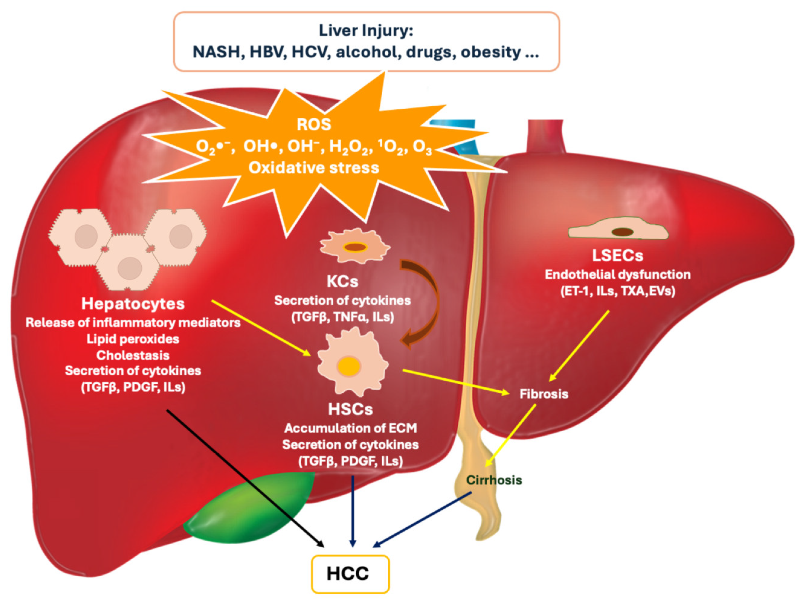
Figure 2.
Schematic representation of the targeted drug delivery system developed Lian B. et al. (A) Chemical structure of RES; (B) Synthesis of the FA-HSA conjugate; (C) Representation of the FA-HSA-RES-NPs obtained by the encapsulation of RES within FA-HAS using a high-pressure fluid nano-homogeneous emulsification method; (D) Proposed mechanism of active tumor-targeting.
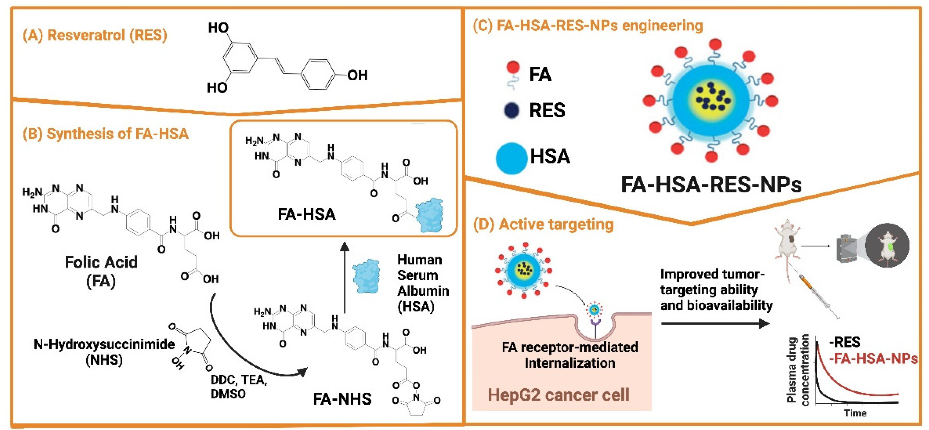
Figure 3.
Schematic representation of the synthesis of galactosylated chitosan (GCS) and its application in curcumin-loaded nanoparticles (CUR-GCS@NPs) developed by Huang M. et al. These GCS-coated NPs were designed to target the asialoglycoprotein receptors (ASGPRs) overexpressed on hepatocellular carcinoma cells, offering enhanced biocompatibility and potential therapeutic efficacy.
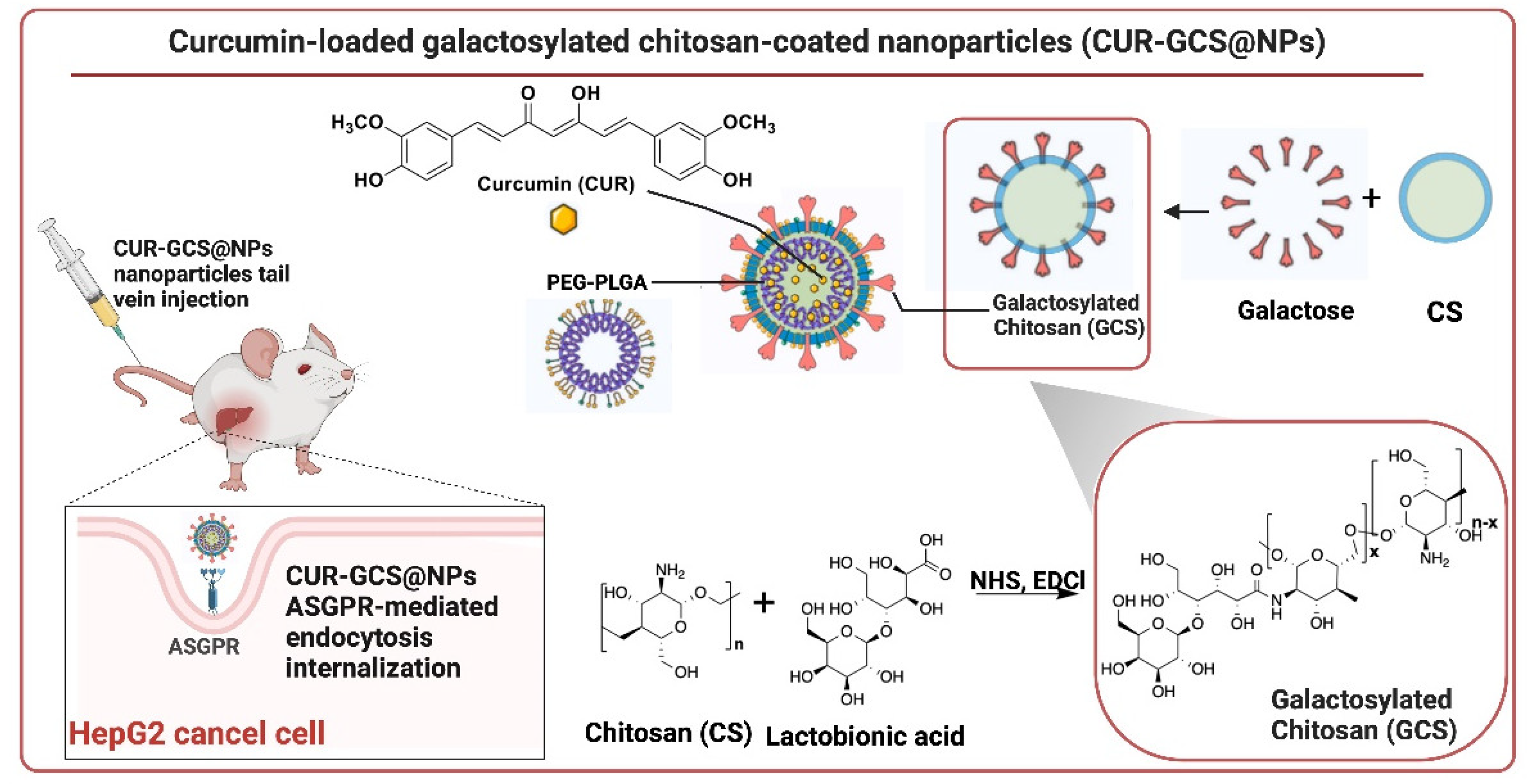
Figure 4.
Chemical structure of apocynin (APO).
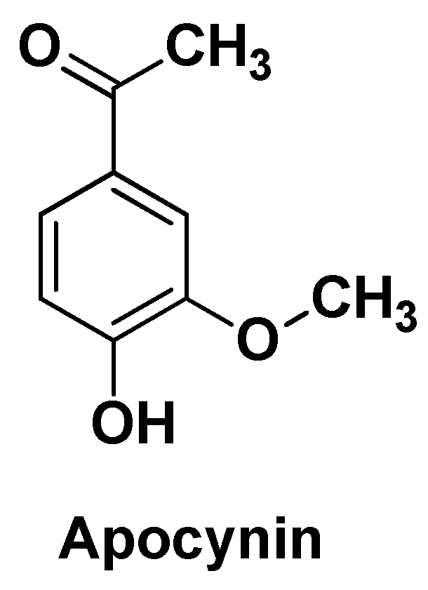
Figure 5.
Schematic representation of the tumor-targeting and IR sensitive nano-system (FA-CA@AgNPs) designed and developed by Abdelwahab T. et al. Caffeic acid (CA) was used both to chemically conjugate the targeting agent (folic acid; FA) and as a reducing agent to form AgNPs.
Figure 5.
Schematic representation of the tumor-targeting and IR sensitive nano-system (FA-CA@AgNPs) designed and developed by Abdelwahab T. et al. Caffeic acid (CA) was used both to chemically conjugate the targeting agent (folic acid; FA) and as a reducing agent to form AgNPs.
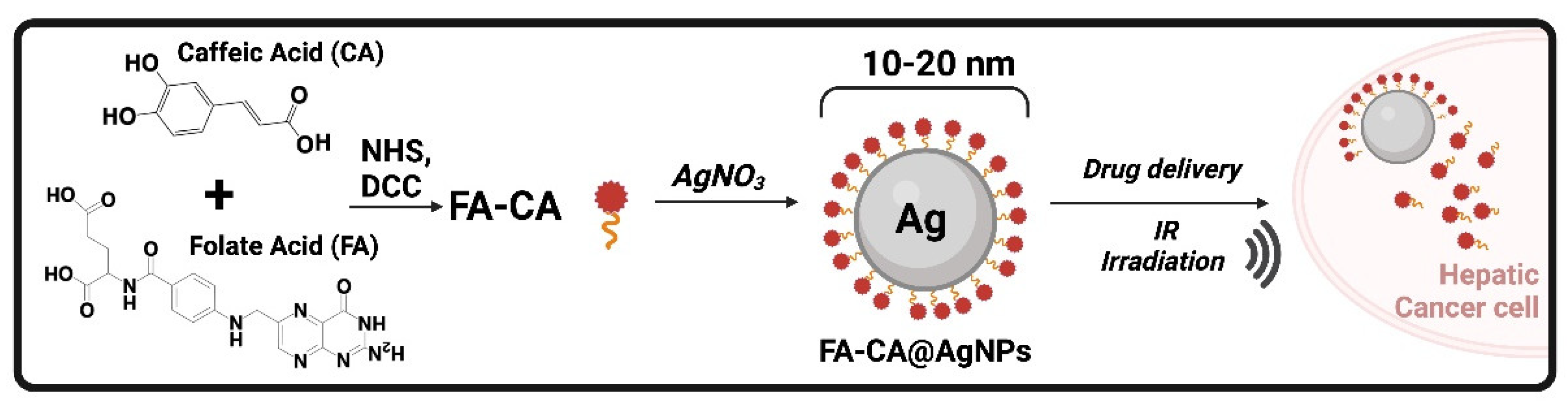
Figure 6.
Chemical structure of quercetin (Q).
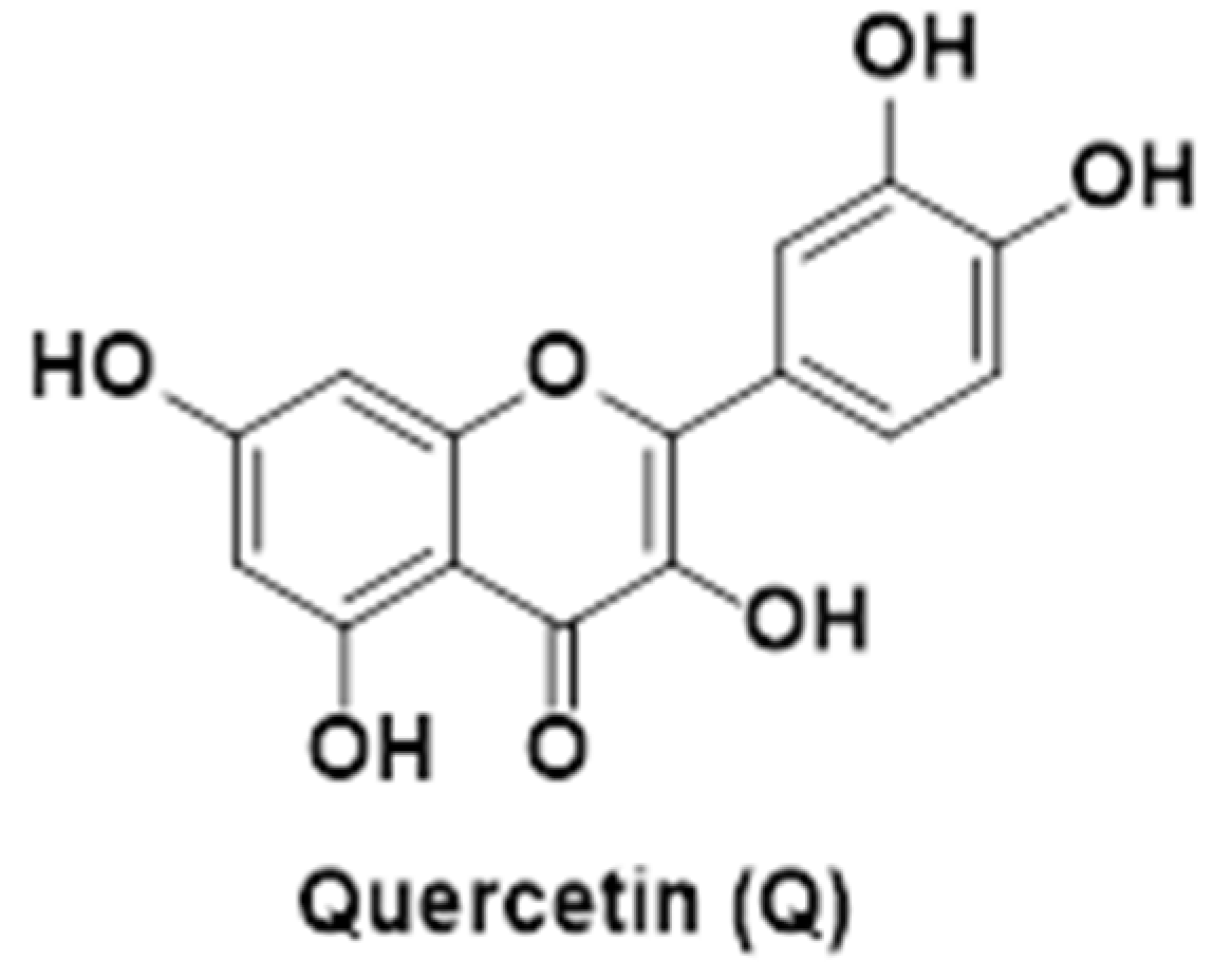
Figure 7.
Chemical structure of rutin (RU).
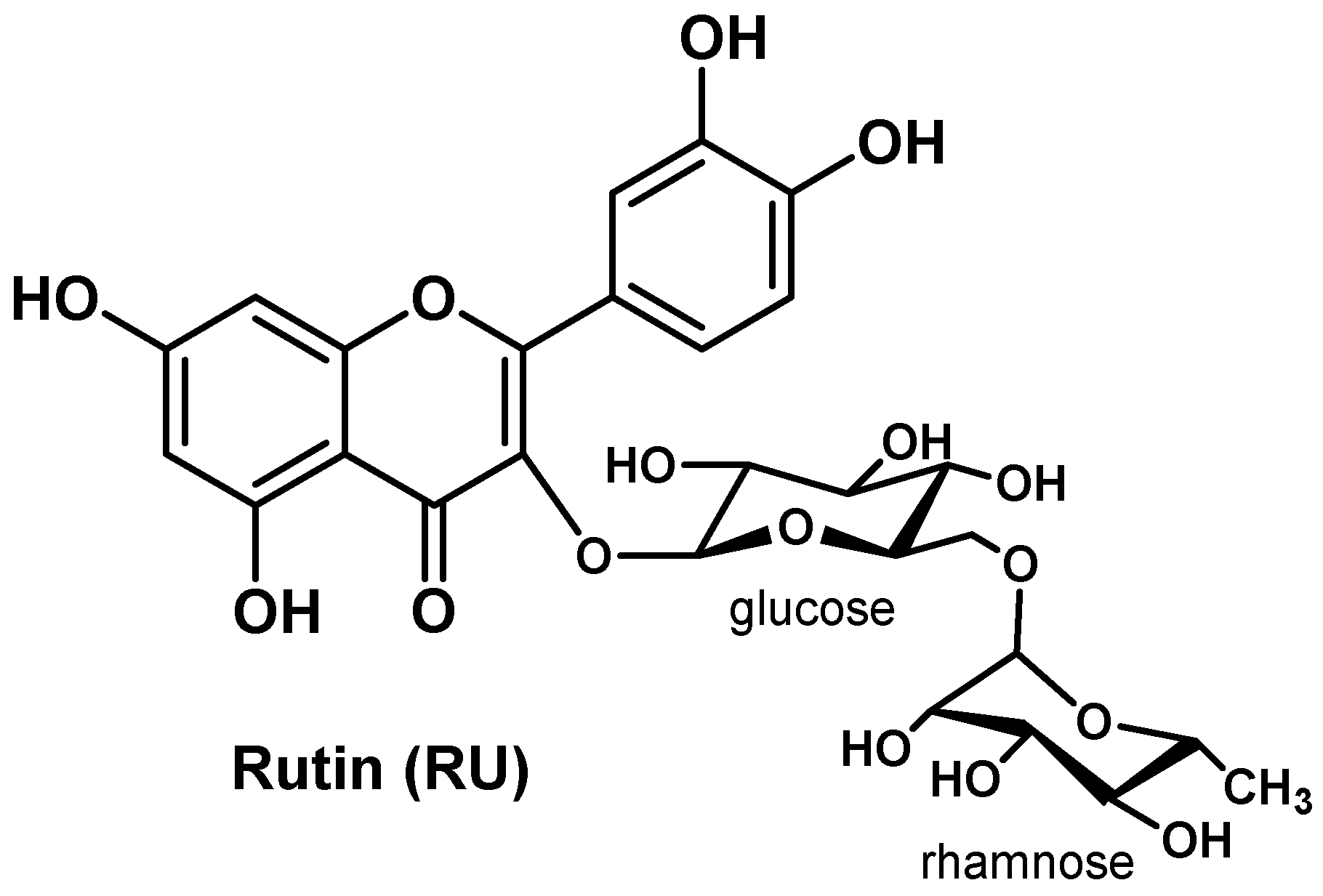
Figure 8.
Chemical structure of hesperetin (HP).
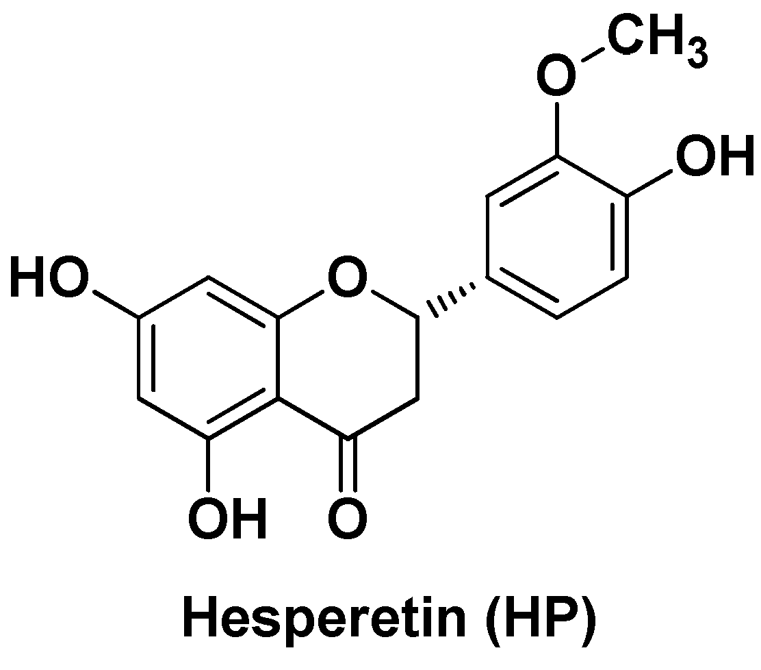
Figure 9.
Chemical structure of honokiol (HK).
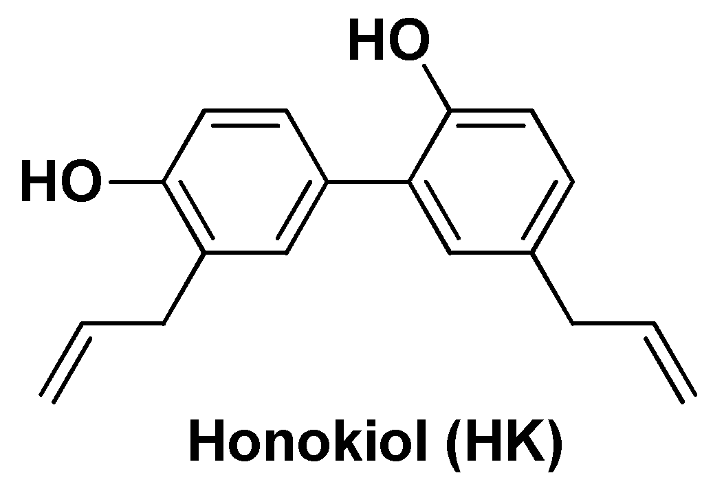
Figure 10.
Chemical structure of epigallocatechin gallate (EGCG).
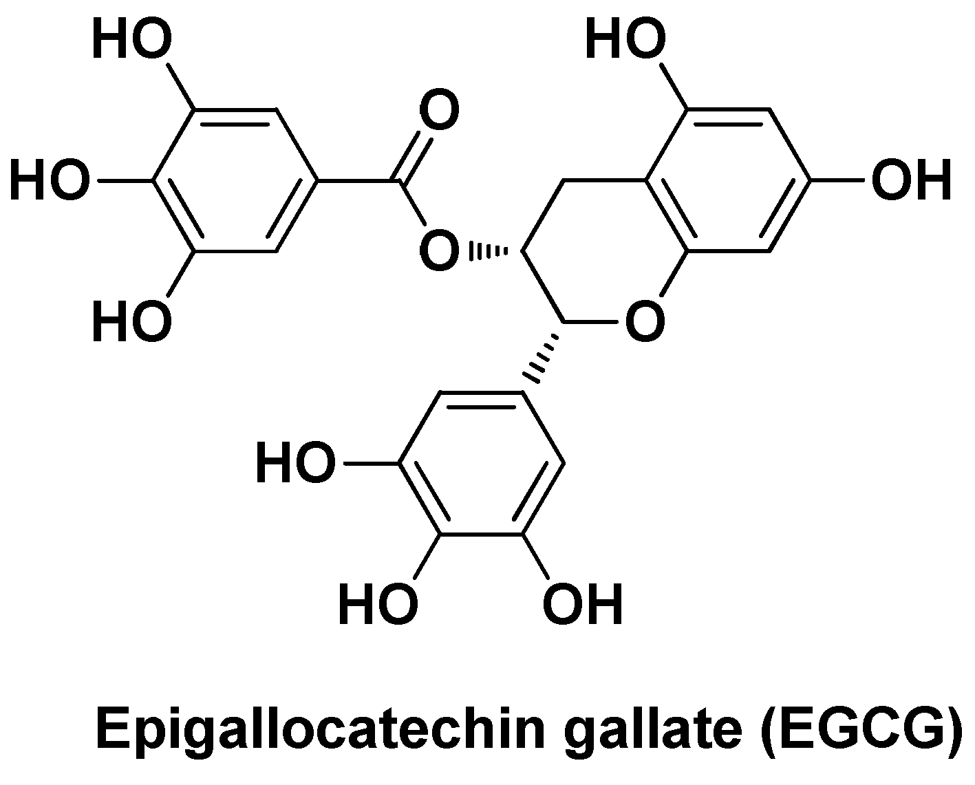
Figure 11.
Chemical structure of umbelliferone β-d-galactopyranoside (UFG).
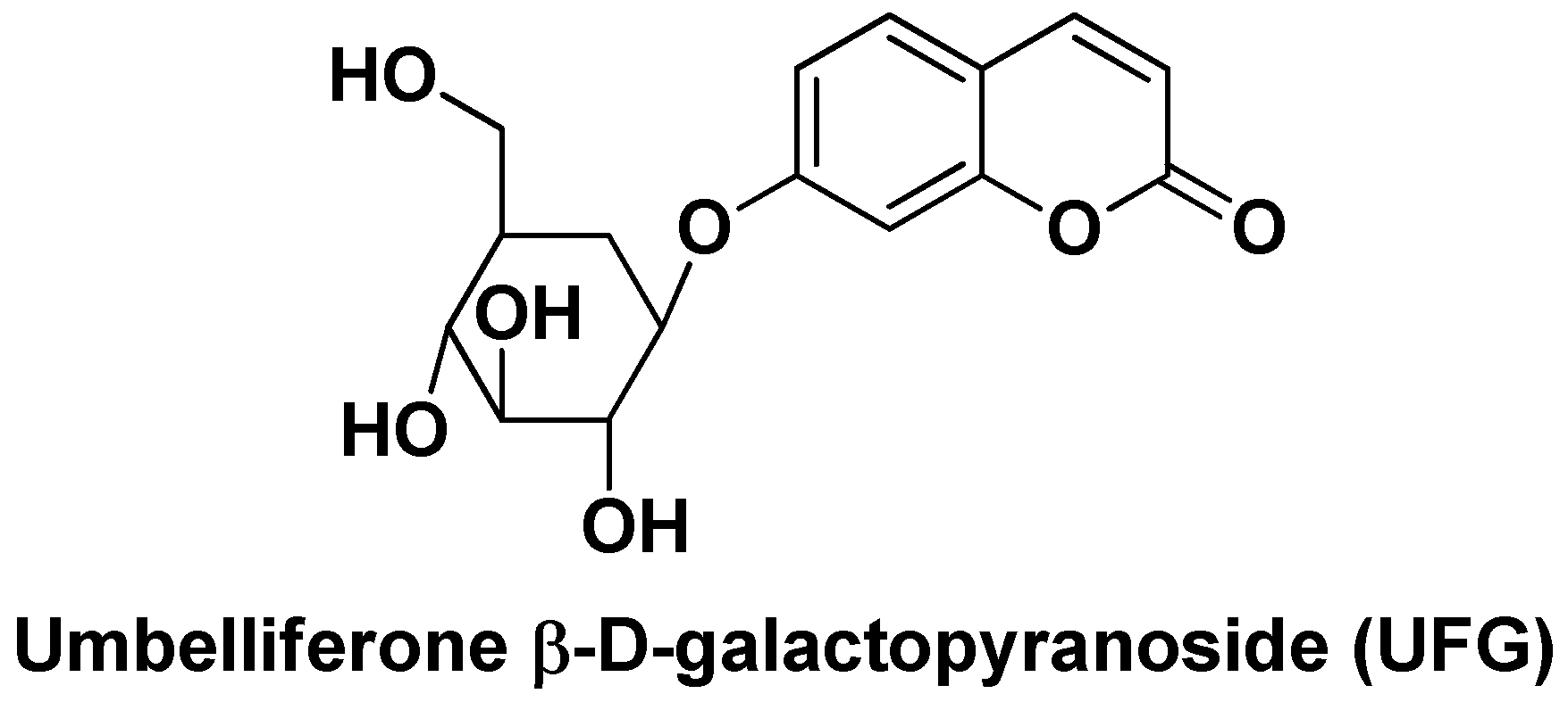
Figure 12.
Chemical structure of andrographolide (AG).
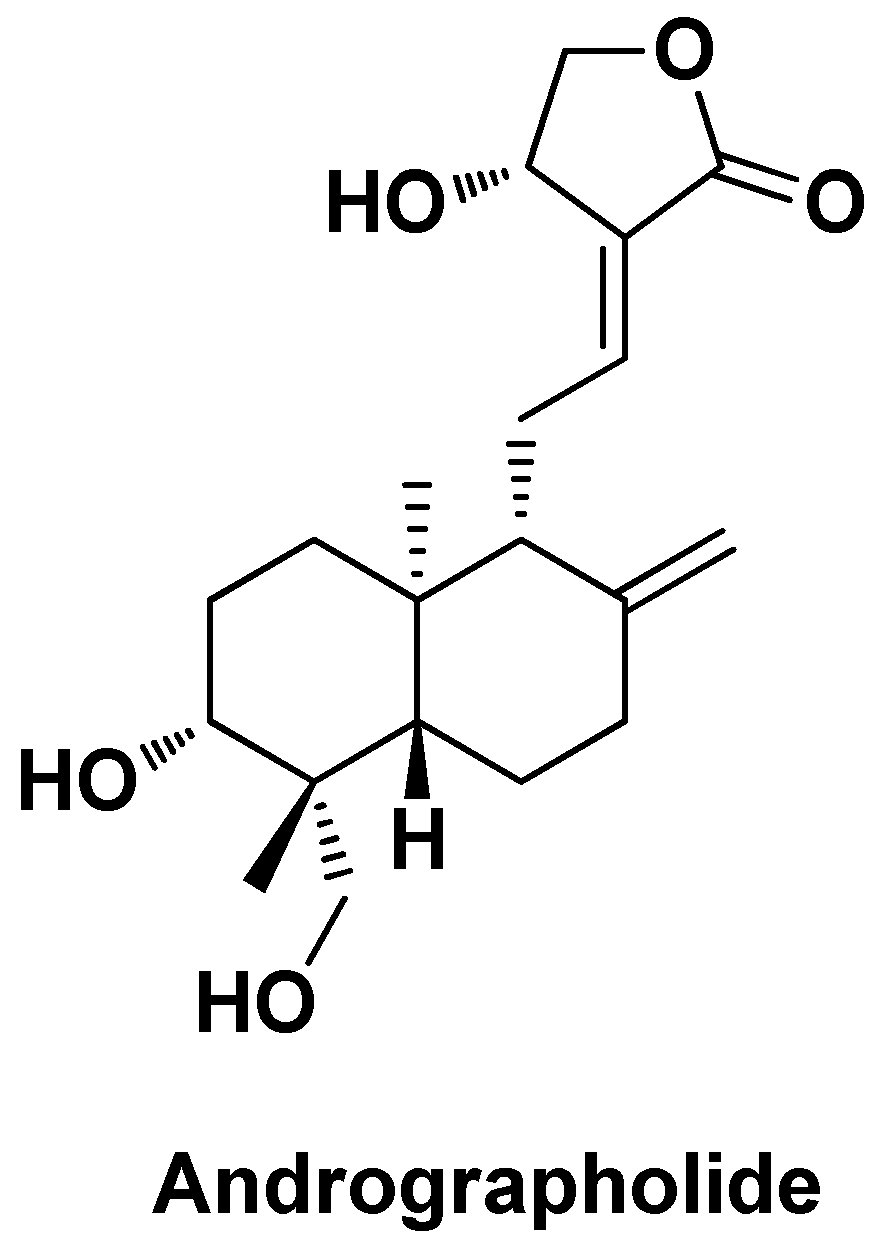
Figure 13.
Chemical structure of oleanolic acid (OA).
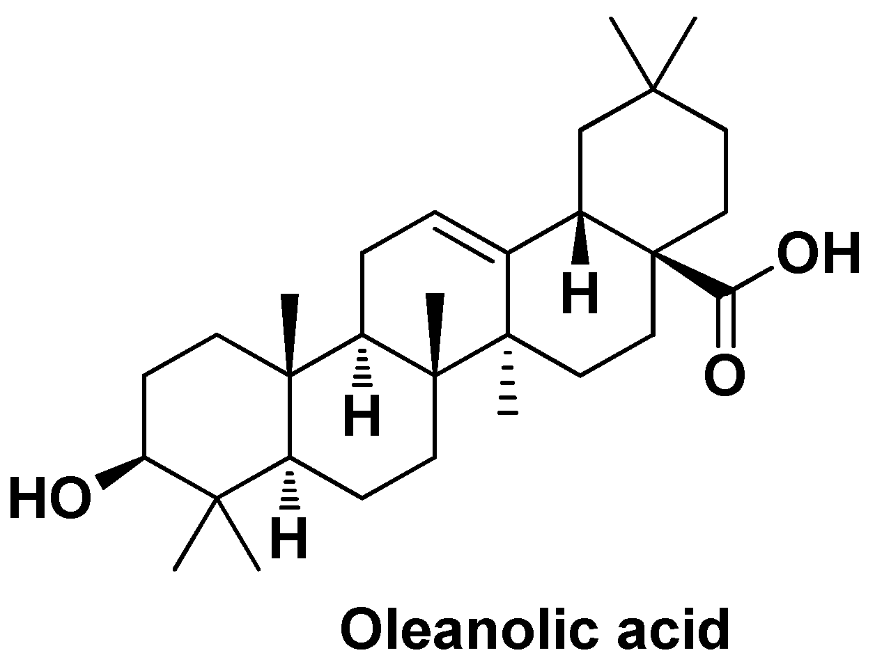
Figure 14.
Chemical structure of parthenolide (PLT).
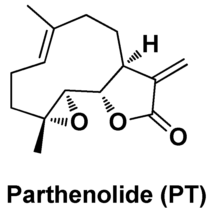
Figure 15.
Chemical structure of vitamin C (Vit. C) and vitamin E (Vit. E).
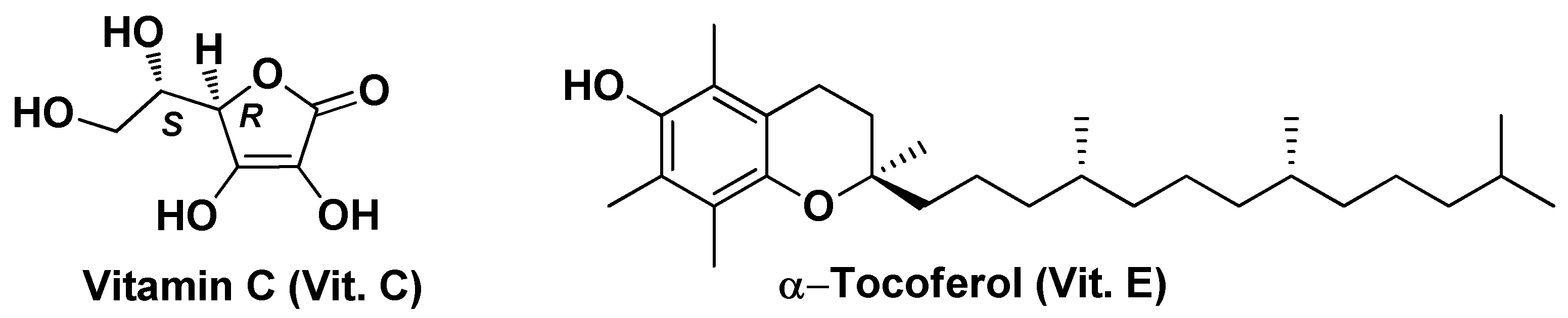
Figure 16.
Chemical structure of α-tocotrienol.
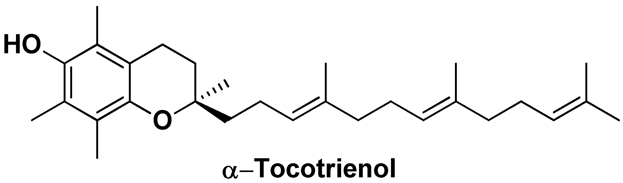
Figure 17.
Chemical structure of coenzyme Q10 (CoQ10).
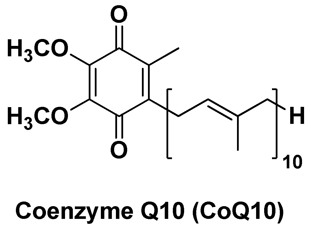
Figure 18.
Chemical structure of docosahexaenoic acid (DHA).
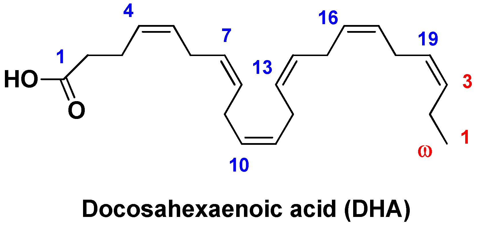

Table 1.
Nano-formulation’s characteristics and in vitro and/or in vivo biological activity in liver cancer.
Table 1.
Nano-formulation’s characteristics and in vitro and/or in vivo biological activity in liver cancer.
| Nano-formulation | API | Excipients | Physical characteristics | Biological Activity | Ref. | |||
|---|---|---|---|---|---|---|---|---|
| FA-HSA-RES-NPs | Resveratrol | Folic acid, human serum albumin | Spherical shape (~102 nm), high EE (~98%) and DL (~15%) | Enhanced cell uptake and antiproliferative activity in vitro and improved biodistribution | [64] | |||
| GA-HSA-RES-NPs | Resveratrol | Glycyrrhizic acid, human serum albumin | Nearly spherical (~108 nm), high EE (83.6%) and DL (11.5%) | Enhanced antiproliferative activity in HepG2 cells and better biodistribution via EPR effect and ASGPR-mediated endocytosis | [65] | |||
| RES-AuNPs | Resveratrol | Gold nanoparticles | Spherical shape (~39-1110 nm) and zeta potential (ζ) = -32.5 mV | Stronger apoptosis induction via down-regulation of pro-caspase-9, pro-caspse-8, PI3K and Akt and up-regulation of caspase-8 and Bax, tumor growth suppression. | [70] | |||
| CUR-GCS@NPs | Curcumin | PLGA-PEG, galactosylated chitosan | Particle size ~100 nm, EE = ~94%, DL = 4.56%, ζ = -9.82 mV, Polydispersity index (PDI) = 0.25 | Targeted delivery to liver tumor tissues, enhanced biocompatibility | [79] | |||
| APO-GCS@NPs | Apocynin | PEG-PLGA, galactosylated chitosan | Particle size 224-232 nm, EE = 34%, pH-dependent drug release (31-60% after 72h) | Significant improvement in antiproliferative activity against HepG2 cells | [83] | |||
| CUR-G-BSA-NPs | Curcumin | bovine serum albumin, galactosyl units | Spherical shape (~116 nm), high drug release rate | Better antiproliferative activity against HepG2 cells (~5,6-fold compared to the free drug) | [86] | |||
| Dox/CUR-NPs | Doxorubicin, Curcumin | Glyceril distearate, soybean lecithin, Polyoxyl 40 Hydrogenated Castor Oil, glycerin, triglycerides medium chain | Spherical shape (~89 nm), high EE (Dox = 97.1% and CUR = 99.8%), ζ = -14.3 mV, PDI = 0.22, sustained release profile (Dox = 55% and CUR = 40% after 48 h) | Synergistic antiproliferative effects in HCC models, modulation on apoptosis, proliferation-, angiogenesis-, MDR- and hypoxia-related mRNAs and proteins | [88] | |||
| FA-CA@AgNPs | Folic acidCaffeic Acid | Silver nanoparticles | Small particle size (10–20 nm) | Higher antiproliferative effects, apoptosis induction by caspase-8, caspase-3, and TNF-α pathways | [90] | |||
| Q-AuNPs | Quercetin | Gold nanoparticles | Spherical shape (~114 nm) | Higher cellular uptake and apoptosis induction via p53-ROS pathway | [94] | |||
| RU-PLGA-NPs | Rutin | PLGA | Particle size ~211 nm, high EE (77.83%) and DL (6.39%), sustained release (71% after 48h) | Over expression of GPx, GTS, MPO, CAT and SOD, down-regulation of IL-1β, IL-6, TNF-α and NF-κB, and improvement on membrane-bound enzyme activity (Ca2+-ATPase, Na+/K+-ATPase, and Mg2+-ATPase) | [97] | |||
| HP-mPEG5000-S-AuNPs | Hesperetin | mPEG5000-SH, gold nanoparticles | Spherical, triangular and pentagon in shape (110–120 nm), ζ = -4.38 mV, sustained release (80% after 72h) | High antioxidant activity, improved hepatic parameters in HCC models, inhibition of inflammatory markers, antioxidant enzymes and ATPase activity related to liver damage | [98]. | |||
| CE-HK-NPs | Honokiol | Chitin, epigallocatechin-3- gallate | Spherical shape (~80 nm), sustained release (80% after 24h) | Extended antiproliferative activity in vitro and reduction of tumor growth (~84%)after inter-tumoral injection (3 x week) | [101] | |||
| UFG-PLGA-NPs | Umbelliferone β-d-galactopyranoside | PLGA | Uniform size distribution (~187 nm), EE = 60–90%, sustained in vitro DR (82.5% after 48 h) | Reduced liver/body weight ratio and liver nodules in DEN-treated rats, inhibition of HCC cell proliferation in vitro. | [103] | |||
| AG-PLGA-NPs | Andrographolide | PLGA | Particle size ~66 nm, EE = 64% | Decreased serum levels of ALT, AST and ALP, arsenic deposition in liver, SOD, CAT and GSH | [111] | |||
| CDDP/OA-LCC@NPs | Oleanolic Acid, Cisplatin | Mono-methoxy polyethylene glycol 2000-distearoyl phosphatidylethanolamine (PEG-DSPE 2000) 1,2-dioleoyl-in-glycerol-3-phosphate (DOPA), dehydrogenated soya phosphatidylcholine (HSPC), Calcium carbonate | Particle size ~206 nm, EE = ~64%, pH-dependent drug release (70% of CDDP at pH = 5.5 and 28% of CDDP at pH = 7.4) | Induction of apoptosis via downregulation of P13K/AKT/mTOR pathway and upregulation of p53 proapoptotic pathway, inhibition of drug resistance by downregulating the proteins like XIAP and Bcl-2 via NK-κB pathway | [116] | |||
| OA-PLGA-TPGS-NPs | Oleanolic Acid | PLGA, d-α-tocopheryl PEG1000 succinate | Spherical shape (~200 nm), DL = ~28%, EE = ~92 % | Increased in vitro cytotoxicity against HepG2 cells compared to the free drug, and higher growth inhibition rate in volume | [117] | |||
| PLT/TYR-CSL@NPs | Parthenolide, Tyrosol | Chitosan, Lecithin | Particle size ~38 nm; PLT EE = 93% | Cancer-selective cytotoxicity in vitro assessed on HepG2 cells and potent antioxidant activity. Apoptotic effects by up-regulating the expression of the apoptotic genes Bax and caspase-8 and down-regulating the expression of the anti-apoptotic gene Bcl-2 | [121] | |||
| Vit. E/C@SeNPs | Vitamin C, Vitamin E | Selenium nanoparticles | Particle size ~50 nm, high antioxidant capacity (~76% DPPH scavenging), improved liver function markers | Higher antioxidant capacity (~76% DPPH scavenging), improved ALT, AST, ALP, total bilirubin and GGT, increased in GSH concentration and CAT activity | [125] | |||
| Precirol® ATO5 | α-Tocotrienol | Precirol ATO5 Glyceryl distearate, miglyol, poloxamer 407 | Particle size ~78 nm,ζ = -11 mV, PDI = 0.24 | Increased cytotoxicity in vitro (IC50 = 15 µM compared to IC50 = 10 µM of the free drug), decreased in the expression of the anti-apoptotic genes survivin and Bcl-2 and increased of theproapoptotic genes Bid and Bax | [128] | |||
| CoQ10-CS/HA@NPs | Coenzyme Q10 | Hyaluronic acid, Chitosan | Monodispersed, average diameter ~54 nm | Hepatoprotective effects against OS and xenobiotics, enhanced cellular antioxidant capacity | [131] | |||
| DHA-LDL-NPs | Docosahexaenoic acid | LDL | Particle size ~20 nm | Selective cytotoxicity against liver cancer cells, modulation of oxidative stress and mitochondrial damage | [138] | |||
Disclaimer/Publisher’s Note: The statements, opinions and data contained in all publications are solely those of the individual author(s) and contributor(s) and not of MDPI and/or the editor(s). MDPI and/or the editor(s) disclaim responsibility for any injury to people or property resulting from any ideas, methods, instructions or products referred to in the content. |
© 2024 by the authors. Licensee MDPI, Basel, Switzerland. This article is an open access article distributed under the terms and conditions of the Creative Commons Attribution (CC BY) license (http://creativecommons.org/licenses/by/4.0/).
Copyright: This open access article is published under a Creative Commons CC BY 4.0 license, which permit the free download, distribution, and reuse, provided that the author and preprint are cited in any reuse.


