Submitted:
16 July 2024
Posted:
17 July 2024
You are already at the latest version
Abstract
More than 700 anthocyanins are described so far, and many different sources are rich in these natural pigments. During the last few years, researchers have been focusing of strategies to recover and purify anthocyanins, from simple liquid-liquid extractions to more advanced supercritical CO2 extractions, however, with such a huge number of different sources, it’s extremely hard to get a simple and fast methodology that fits several sources, not only by the structural nature of the anthocyanins but also by the presence of specific groups of natural compounds. Using simple methodologies like solid-phase extraction resins represents a powerful economical and efficiency strategy. In this work, we tested several sources of structurally distinct anthocyanins, including purple basil, purple cabbage, purple sweet potato, butterfly pea flower, cornflower and wild pansy, in different resins to assess their efficiency in the obtention of anthocyanin-pure extracts. The results showed us that the combination of oasis HLB® resin followed by a cation-exchange SPE with DSC-MCAX® resulted in high-purity anthocyanin extracts for all the cases.
Keywords:
anthocyanins
; SPE
; hydrophilic-lipophilic balance
; cation-exchange chromatography
Introduction
Anthocyanins are a class of water-soluble pigments responsible for the red, purple, and blue colors in many fruits, vegetables, and flowers. These compounds belong to the flavonoid group and play crucial roles in plant physiology, including attracting pollinators, acting as antioxidants, and providing protection against ultraviolet radiation and pathogens. Due to their vibrant colors and health benefits, anthocyanins have garnered significant interest in the food, pharmaceutical, and cosmetic industries [1,2,3]. This interest has driven the need for efficient and effective purification methods to isolate these compounds from natural sources. The extraction and purification of anthocyanins from plant materials usually involve several steps, typically including the extraction, concentration, and purification. The most common strategy for the extraction of these compounds involves the use of solvents like methanol, ethanol, acetone or water [4]. Alternative methodologies such as the use of deep eutectic solvents, supercritical fluid extraction, pressurized liquid extraction, ultrasound assisted extraction or microwave extraction have been widely applied over the last years [5].
However, such methodologies promote the extraction of not only anthocyanins but also a large number of other compounds (soluble sugars, proteins, organic acids and other polyphenols) in the extraction process. This will be largely influenced by the food matrix and excessive impurities can significantly influence not only the physiological activities and stability but also the final product quality of anthocyanins, which is not conducive to the deep exploration and comprehensive utilization of anthocyanins [4]. Therefore, the separation and purification are an essential step for the obtention of anthocyanin-rich extracts from different sources.
Currently, purification methods for anthocyanins or anthocyanin extracts range from simple solvent extraction to more sophisticated column chromatography, membrane separation, high-speed counter-current chromatography and high-efficiency preparative liquid chromatography [5]. However, each method possesses limited applications due to their different drawbacks, like the difficulty to achieve large-scale production, the yield of purified anthocyanins, the equipment requirements and the time consumed.
Solid phase extraction (SPE) methodologies have attracted much research interest due to a balance of efficiency and cost. Several methods have been developed over the years with different resins [6,7,8,9,10]. And while some showed great results to a specific source of anthocyanins, others do not perform so well making it difficult to get a general methodology that do not depend on the food source. More recently, cation exchange chromatography has been applied to purify anthocyanins from some fruits and vegetables. Such option can offer a better controllability and high selectivity and be optimized for large-scale purification of anthocyanins [11]. Anthocyanins are converted to positively charged flavylium cations in an acidic environment because of their chemical and structure dependence on the pH. Then, the flavylium cations are retained on the resin with negative charge through ionic interactions. Due to the low pKa (4–5) of phenolic acids and the non-charged nature of other classes of compounds, they cannot be adsorbed by the resin and are flushed with the eluent, resulting in the separation of anthocyanins from the other non-charged compounds.
In a previous study, Liao et al. applied cation-exchange resin 001X7 to purify mulberry anthocyanins under optimized condition and the purity of anthocyanins reached 95% [11]. Besides, He et al. purified anthocyanin from fresh blueberries and red radishes using a novel cation-exchange/reversed-phase combination solid-phase extraction (SPE) technique and found the new method could efficiently remove non-anthocyanin phenolics and some anthocyanins can reach to 99% purity [12]. Also, anthocyanins from tulips were effectively separated from the other non-charged compounds utilizing a mixed-mode cation-exchange chromatography [13].
Nevertheless, to the best of our knowledge, the is no universal methodology that can be applied to any source of anthocyanins and get extracts with a high degree of purification. Therefore, this study focused on the obtention of a methodology suitable to purify anthocyanins from different sources including flowers, vegetables and leaves without the need of large extent of solvents, time consumption and in line with the recent trends that have mainly been focused on developing cost-effective and eco-friendly extraction/purification methodologies for selective recovery of targeted compounds.
2. Results and Discussion
2.1. Anthocyanin Extraction, Quantification and Characterization from the Different Extracts
All the samples were macerated in acidified water after to extract all the water-soluble compounds present in each one of them. Such strategy ensures that all the different anthocyanins are effectively extracted. Other approaches may also be applied including the using of some organic solvents such as ethanol or methanol, which usually enhances the yield of extraction in a shorter time period [14]. For instance, the optimum conditions for the extraction of anthocyanins and other polyphenols from the skin and seeds of Plinia cauliflora (Jabuticaba) were set as solvent mixture of methanol/water/acetic acid (80:20:0.5 v/v/v), with a solid–liquid ratio of 0.01 g/mL, with two hours of constant agitation [15]. On the other hand, a solvent mixture of ethanol (9.1% v/v) acidified with citric acid at pH 3, mixed and centrifuged with the powdered sample (~20 mesh), in a solid–liquid ratio of 50 g/L during 21.8 min at 47.1 °C was also claimed as the optimum conditions from the extraction of the anthocyanins from the same fruits [16]. The authors also observed that the total amount of anthocyanins extracted increase with temperature and decrease with higher ethanol concentrations and with long times of extraction.
Our objective was to apply the most greener approach possible, therefore acidified water (HCl 1M) was the only solvent utilized. A maceration of 4h showed to be effective in the extraction of the anthocyanins from the edible flowers extracts, while ultrasound-assisted extraction was necessary for the other sources. This may be explained by the fact that edible flowers are extremely sensitive as raw material, while purple vegetables and leaves are much more robust with making it necessary to apply more disruptive extraction techniques to ensure the extraction of phenolic compounds [17]. It is also important to notice that the acidic environment prevent the degradation of the anthocyanins stabilizing the cation flavylium structure that is less prone to degradation as well described in previous works [18,19]. Table 1 shows the total anthocyanin content in each extract in mg.g-1 DW of extract (Cy-3-glc equivalents). Based on the results, it is possible to notice that all sources have interesting amounts of anthocyanins. In fact, previous studies showed that high levels of anthocyanins can be found in the different food sources utilized herein [19,20,21,22,23,24]. The results showed that butterfly pea flower and purple cabbage showed the highest amount of anthocyanins, while cornflower and wild pansy contained the least quantity. It is important to notice that our results are based on the extracts and not on raw material fresh weight, which may result in a different comparable quantification. Nevertheless, such results are crucial to understand the potential enrichment of the purification treatments and is an indicator of the complexity of each matrix.
The LC-MS analysis allowed the identification of the anthocyanins in each extract. It is possible to see from Table 2 that the nature of each nature varies depending on the source varying from simple monoglucosides to complex polyglycosylated anthocyanins with several acylation patterns. The results indicates that 5 anthocyanins are present in the extract of cornflower, with molecular ions [M]+ at m/z 611, 697, 711, 725 and 549, respectively. The main anthocyanins in this source are derived from cyanidin with a 3,5-O-diglucoside structure.
For wild pansy, four anthocyanins including three delphinidin derivatives and one cyanidin derivative were identified ([M]+ at 773, 919, 903 and 757). all the anthocyanins were substituted by rutinoside at the C3 position. Delphinidin-3-(4″-p-coumaroyl)-O-rutinoside-5-O-glucoside was the most abundant anthocyanin in the wild pansy extract. In the case of butterfly pea flower, LC-MS analysis allowed the identification of 14 anthocyanins present in the purified extract. 9 anthocyanins are ternatins (A2, B1-B4, C2, C4, D1 and D2), one anthocyanin belong to preternatin A3 with a molecular ion [M]+ at m/z 1405, and the other 4 anthocyanins belong to delphinidin derivatives.
In contrast to the other sources, anthocyanins in the extract of butterfly pea flower possess a highly complex structure.
Twelve main anthocyanins were found in the extract of purple cabbage. Except for cyanidin-3-(6-sinapoyl)-O-glucoside-5-glucoside with a molecular ion [M]+ at m/z 1141, all the anthocyanins have a cyanidin-3-O-diglucoside-5-O-glucoside basic structure. Eleven anthocyanins were acylated by p-coumaric acid, ferulic acid and sinapic acid in the position-3 with molecular ions [M]+ at m/z 773, 1081, 1111, 1287, 1317, 919, 979, 817, 1125, 1155 and 1185.
In the case of purple basil, it was possible to identify 4 main anthocyanins (and their isomers), all of them derivatives of cyanidin with molecular ions [M]+ at m/z 919, 1081, 757 and 1065.
Finally in the case of purple sweet potato, four anthocyanins derived from peonidin and cyanidin were found with a 3-O-diglucoside-5-O-glucoside core basic structure with molecular ions [M]+ at m/z 787, 907, 1067 and 1097.
The diversity of anthocyanins found in the different extracts, with polyglycosylated and polyacylated anthocyanins with different substitution patterns allowed us to assess the universality of the purification methodologies applied herein. Table S1 shows the main polyphenols rather than anthocyanins identified in each extract.
2.2. Anthocyanins Purification Strategies with the Different SPE
The purification of anthocyanins is usually a complex procedure that involves the use of different techniques including liquid-liquid and solid-phase extraction. Such processes can be time consuming. Also, different works present different solutions based on the raw material and not necessarily a universal strategy that is efficient regardless of the anthocyanin source. Therefore, is it important to understand if a fast and reliable strategy can be applied to different sources, which from a technical perspective is a considerable advantage.
Our research findings are centered around two main parameters: the purity and recovery of anthocyanins in the methanol fraction. In all the solid phases the same protocol was applied after sample loading: 1) ethyl acetate, followed by 2) water and then 3) methanol to recover the anthocyanins. In the conventional extraction procedures such as liquid-liquid extraction, ethyl acetate is often used utilized to separate anthocyanins from other polyphenols that have a higher affinity to less polar solvents than water. Therefore, this solvent was used in the attempt to remove, at least, some of the non-anthocyanin compounds. Also, the weighted amounts of each resin were based on the volume occupied by each to match similar bed volumes and ensuring that the resins presented enough loading capacity for all the different extracts.
2.2.1. Non-Anthocyanin Fraction Removal with Ethyl Acetate
For the assessment of the removal efficiency of ethyl acetate, pools of 5 BV were collected and the total polyphenol content as well as the total anthocyanin content was assessed. From Figure 1 is possible to see in all cases that the removal of the non-anthocyanin fraction increases to a maximum that is dependent on the matrix and then starts to decrease until no polyphenols are detected in the quantification approach. Interestingly, for all the resins utilized, 5 BV are sufficient to remove the majority of the compounds that have a high affinity to ethyl acetate. Also, Oasis® HLB showed to be the resin with the lowest affinity to these type of non-anthocyanin compounds judging by the total polyphenol content comparison between them. On the other hand, Sephadex® LH-20 did not show a significant efficacy on the removal of such compounds using ethyl acetate. We also assessed the total anthocyanin amount after 40 BV of ethyl acetate compared to the loaded amount for each extract (Table 3). The results showed that the resins showed different retention capacity of the anthocyanins upon the application of ethyl acetate. Overall, the best performing resin was Oasis® HLB, with a small amount (1.0-1.3%) of anthocyanins from purple basil and purple sweet potato found in the eluted fraction. Toyopearl® HW-65S also showed an interesting performance in retaining the anthocyanins. On the other hand, Sephadex® LH-20 showed the worst performance with anthocyanins from all sources found in the ethyl acetate elute up to 11.4 ± 0.5% (wild pansy). Comparing between the different sources, as each possess structurally different anthocyanins, no defined pattern was found, however it is possible to see that each resin behaves in a specific way. C18 Silica resin performed better in the case of cornflower anthocyanins (p<0.05), with a similar efficiency as Toyopearl® HW-65S. In the case of Amberlite® XAD 7-HP, showed the least elution anthocyanin amount for purple cabbage (1.7 ± 0.7 %), however significant differences were only found between this source, purple basil and butterfly pea flower. Toyopearl® HW-65S did not show any significant differences among the different anthocyanin extract. On the other hand, Sephadex® LH-20, showed a higher performance in the case of purple basil and purple sweet potato but with no significant differences from the other sources except for wild pansy. Oasis® HLB showed no detectable amounts of anthocyanins in the ethyl acetate fraction for all the edible flowers and purple cabbage, while for purple basil and purple sweet potato values of 1.3 ± 0.4 % and 1.0 ± 0.2 % where found, respectively. Analyzing the results from the source perspective, overall purple cabbage showed the best results with values ranging from 1.7 to 9.4 % and two resins with no detectable amounts of anthocyanins.
2.2.2. Effect of Acidic Water Application in the Resins
Water is normally used as a washing procedure when purifying anthocyanins. Such strategy helps to elute hydrophilic compounds that have a higher affinity for water compared to the stationary phase. This usually involves sugars, amino acids, small acids, or water-soluble proteins [12,25,26,27]. Even though they are easily and readily eluted, these molecules are in a much higher concentration than that of the phytochemicals, therefore a sufficient amount of water is necessary to successfully remove them [25]. However, anthocyanins are also water-soluble, therefore understanding the retention capacity of the different resins when using water as eluent represents a key step for developing an efficient strategy. Also, the structure of the anthocyanins influences their higher or lower polarity determining the final strength of the system anthocyanin-solid phase-eluent interactions. On the other hand, acidification of solvents is crucial when extracting/purifying anthocyanins, once it promotes their flavylium cation form, promoting their overall stabilization. However, high concentrations of acids such hydrochloric acid (HCl), may lead to an acidic hydrolysis of, mainly, acylated anthocyanins. In an attempt to minimize such phenomenon, several authors utilized organic acids including acetic, tartaric or citric acid for their extraction/purification procedures [16,28]. In our case, we utilized a low amount of HCl 1M (1% v/v), to minimize the breakage of the different anthocyanin structures while ensuring a sufficiently acidic environment to promote the cation flavylium form (pH~2). Figure 2 shows the amount (%) of total anthocyanins detected after the different water BV applied in the resins. The most direct observations allow us to conclude that overall Sephadex® LH-20 and Amberlite® XAD 7-HP resins presented the least retention capacity of the anthocyanins upon the application of the water BV with significant losses of anthocyanins during the entire application of the water BV with cumulative losses between 45.4 ± 0.7 % to 64.7 ± 0.7 % in the case of Amberlite® XAD 7-HP and cumulative losses between 29.3% to 45.3% for Sephadex® LH-20 (Table S2). Such findings suggest that these resins may have a weaker interaction with anthocyanins. Interestingly, the highly complex anthocyanins from butterfly pea flower showed the highest elute amount when compared to the other sources. Amberlite® XAD 7-HP it is a macroporous, non-ionic, aliphatic acrylic resin supplied in the form of insoluble beads. Its macroporous structure, consisting of a continuous polymer phase, together with its high surface area and aliphatic nature, contribute to its adsorptive properties. This resin is able to adsorb non-polar compounds from aqueous systems and polar compounds from non-polar solvents due to its aliphatic nature. On the other hand, Sephadex® LH-20 is a crosslinked dextran-based resin used with organic solvent mobile phases for gel permeation, normal phase partition and adsorption chromatography of lipids, steroids, fatty acids, hormones, vitamins). It is also used in gel filtration chromatography, protein chromatography, gel filtration media, resin and separation media. Nevertheless, anthocyanins were found in all the earlier water fractions independently of the resin. In the case of silica C18 the values ranged from 1.8% to 5.6% at the pool of 10 BV. For Toyopearl® HW65-S immediately after 5 BV a small amount of anthocyanins was detected (1.1 – 1.4%), with a similar result at the pool of 10 BV. And finally, Oasis® HLB initial pool of 5 BV also presented a small amount of anthocyanins ranging from 1.8% to 2.2%. Interestingly, in the last two cases, the results suggests somehow that probably the loading capacity may have been surpassed even though the fabricant conditions were assessed. Therefore, Toyopearl® HW65-S and Oasis® HLB resins demonstrated the highest anthocyanin retention, which is crucial for the different technological and industrial applications, once yield should be a key parameter to consider not only from an economics but also from a sustainable perspective.
2.2.3. Recovery and Purity of Anthocyanins with Acidic Methanol
The recovery assessment was based on the amount of the methanolic phase until no anthocyanins were detected by the UHPLC methodology and having into account the losses of the previous steps. Consequently, the non-recovered anthocyanins were assumed to be retained in the resins. For the purity assessment, the chromatograms of each extract at 280 and 520 nm were superimposed, and the areas of the different peaks were determined.
Table 4 shows the efficacy of each resin in terms of recovery and purity of the different anthocyanins in each extract. It is possible to see that the recovery efficacy depended not only on the resin but also on the type of anthocyanins from each extract. In the case of C18 Silica resin, the values ranged from 46.1 ± 1.7% in the case of purple cabbage to 77.3 ± 10.4% in the of the butterfly pea flower. For Amberlite® XAD 7-HP, the values ranged from 33.2 ± 9.4% in the case of butterfly pea flower to 67.5 ± 1.8% in the case of purple sweet potato. Toyoperal®HW-65S ranged from 34.4 ± 8.9% in the case of purple cabbage and 73.9 ± 5.9% in the case of wild pansy. On the other hand, Sephadex® LH-20 recovery efficacy ranged from 37.0 ± 4.6% for butterfly pea flower to 68.4 ± 11.2% for purple basil. And finally, Oasis® HLB ranged from 79.9 ± 10.1% for wild pansy and 91.6 ± 2.6% for purple cabbage. From the point of view of the extract, it’s clear that Oasis® HLB showed the best performance, followed by C18 Silica. Interestingly, in a previous study with grape pomace extract, the authors tested the recovery rate of the anthocyanins in different materials (Amberlite® IRA 400 Cl-, Lewatit® TP 208 and TP 206, Chromosorb® G-HP, Amberlite® XAD 2, Zeocros® CA 150, Chemviron® Carbon, Oasis® HLB and Isolute C8), and verified the Oasis® HLB showed the best recovery rate and loading capacity (5.76 mg.cm-3) [29]. On the other hand, a study with chokeberry and purple corn anthocyanins showed values of 68.9% and 78.0% (respectively) for Oasis® HLB recovery rate, 91.4% and 87.8% for C18 Silica and 75.8% and 55.4% for Sephadex® LH-20 [12].
Nevertheless, in none of the cases a total recovery of the anthocyanins was achieved suggesting that there are always losses related with resin retention when using SPE techniques to purify anthocyanins, regardless of the type. In some cases, the recoveries were as low as 34%. Such details must have been considered when thinking about the best strategies to recover and purify these compounds.
Regarding the purity of the recovered anthocyanins, once again, the results showed that this parameter depended on both anthocyanin and resin type. Nevertheless, Oasis® HLB showed, once again the best results ranging from 61.6 ± 1.5% for purple cabbage to 74.9 ± 2.9% in the case of purple sweet potato, followed by Toyopearl® HW-65S with values ranging from 62.2 ± 4.5% for purple basil to 67.5 ± 2.4% for wild pansy. On the other hand, Sephadex® LH-20 fractions of the different extracts showed the least purity. None of the methodologies showed to be able to isolate and purify in a high degree the anthocyanins from the different sources. In fact, the study of He et al. showed that the purity of the fractions obtains from Oasis® HLB ranged from 57.7 to 67.9% for chokeberry and purple corn anthocyanins, respectively [12]. Such evidence supports the findings herein related to the type of anthocyanins where significant differences were found among between the different extract applied in Oasis® HLB resin.
2.3. Cation-Exchange Chromatography with Discovery® DSC-MCAX
Cation exchange chromatography is a type of ion exchange chromatography where positively charged ions (cations) are separated based on their affinity to a negatively charged resin. This technique involves passing a mixture through a column filled with a resin that binds cations, allowing other non-charged molecules to be washed away. The bound cations can then be eluted by changing the ionic strength or pH of the elution buffer. Under acidic conditions, this method can be particularly relevant for the isolation of anthocyanins. By exploiting their cationic nature, cation exchange chromatography may efficiently separate anthocyanins from other non-cationic compounds in a mixture, facilitating the purification of these pigments for further study or use in various applications, such as food coloring and pharmaceuticals. Previous studies have successfully applied this technique in different anthocyanins sources. A cation-exchange resin 001X7 was successfully applied to purify anthocyanins from mulberry, obtaining a degree of purification of about 95% [11]. In other study, a mixed mode cation-exchange/ reversed-phase resin (Oasis® MCX) was utilized to get high-purity extracts of anthocyanins from chokeberry [12]. Our group has also successfully applied cation-exchange chromatography with Discovery® DSC-MCAX to obtain high-purity extracts of pre-treated anthocyanins from different edible flowers with purities higher than 90% [30]. In fact, some compounds are extremely difficult to separate to separate from anthocyanins using traditional SPE approaches due to their structural similarity, such as flavonol glycosides. In our case, it is possible to see from Table S1 that most sources contain different classes of polyphenols, including the flavonol glycosides which may be the reason for not reaching a near 100% purity in the previous attempts. Therefore, we decided to use the Discovery® DSC-MCAX with both the full and pre-purified extracts from the best previous methodology (Oasis® HLB). This resin comprises a packed bed containing both octyl (C8) and benzene sulfonic acid (SCX) groups, enabling a dual retention mechanism based on reversed-phase and cation-exchange chromatography. Initially, the extracts are applied under acidic conditions, and non-anthocyanin compounds are eluted using acidic methanol. Subsequently, the system’s pH is raised to 6 using a phosphate buffer, and in the presence of methanol, the anthocyanins are eluted.
From Figure 3a is possible to see that the application of the full extract allowed to get extracts with purity ranging from 71.3 ± 9.2% for cornflower to 91.2 ± 2.0% to butterfly pea flower. Looking to the previous results, it is possible to see that in some sources the purity of Oasis® HLB showed values around 70%, therefore the results for Discovery® DSC-MCAX were unexpectedly low. However, in Figure 3a the results for the pretreated extracts with Oasis® HLB, showed a significant improvement in the purity for all the extracts with values ranging from 96.7 ± 1.4% for purple basil to 99.4 ± 0.4% for purple cabbage (and confirmed by the comparison between the chromatograms of the initial extracts and the purified ones at 280 and 520 nm – Figures S1–S6). Also, by comparing the individual extracts its clear the higher reproducibility on the case of the pre-treated methodology (as expressed by the shorter box-violin charter deviations). Such findings suggest that Discovery® DSC-MCAX alone does not works so well as if in combination with previous pre-treatments, in this case Oasis® HLB. Figure 3b, on the other hand, shows the recovery rate of the anthocyanins after elution with the mixture of phosphate buffer pH 6 and methanol. Once again, the results shows that the recovery was considerable higher in the pre-treated samples with values from 74.2 ± 6.7% for cornflower to 84.6 ± 2.5% for purple cabbage. Nevertheless, significant amounts of anthocyanins became irreversibly retained in the resin in all cases
3. Conclusion
Anthocyanins are natural compounds that can have several applications. However, to assess their role it is important to get high-purity extracts. The most traditional approaches show to have a general low efficiency and as shown herein, depending on the source of anthocyanins such methodologies may work better or worst. Therefore, finding a universal strategy to purify anthocyanins its crucial to all areas where these compounds may play a role. Our results showed that from the solid-phase resins normally utilized Oasis® HLB presented the best performance using the system ethyl acetate-water-methanol, however none resulted in high purity extracts. The cation-exchange chromatography exhibited the best results overall but again, without reaching the desired purity ranges. Only the combination of the two procedures was able to get high purity extracts rich in anthocyanins from the different sources, confirming that this two-step approach can be used as a universal methodology to purify anthocyanins regardless of the source and the type of these natural pigments.
4. Materials and Methods
4.1. Samples Preparation
4.1.1. Edible Flowers
Three species of edible flowers were utilized in this study: cornflower (centaurea cyanus), wild pansy (viola tricolor L.) and butterfly pea flower (clitoria ternatea).
To obtain the extracts of edible flowers, the petals were separated from the remaining parts of the flowers and submitted to freeze drying. Then, 200 mg of dried petals were taken from each flower and 20 ml of ultrapure MilliQ® water with 1% HCl 1M was added. The suspension was placed on a magnetic stirrer for 4 hours, followed by centrifugation at 10380 x g for 10 minutes at 4ºC.
The resulting solutions underwent a final filtration using a CHROMAFIL® PET-45/25 hydrophilic membrane with 0.45 μm pore size.
The final extracts where then freeze-dried and stored at -20°C, until use.
4.1.2. Purple Basil (Ocimum basilicum var. purpurascens), Purple Cabbage (Brassica oleracea) and Purple Sweet Potato (Ipomoea batatas L.)
The procedure for these three anthocyanin sources was as follows: 10g of each sample was cut in small pieces (including purple basil leaves) and submitted to freeze drying. Then, the freeze-dried samples were reduced to powder and 5g of each sample were added to 200 ml of ultrapure MilliQ® water with 1% HCl 1M. Then the different solutions were submitted to an ultrasonic bath extraction for 4h (200 W of heating power at 40 ºC). Following this step, the separation between the insoluble and soluble material was carried out using the same procedure mentioned in section 4.1.1. earlier for the cornflower and wild pansy samples: centrifugation and filtration. The final extracts where then freeze-dried and stored at -20°C, until use.
4.2. Solid-Phase Extractions Resins
4.2.1. C-18 Silica Resin
Sixteen grams of C18 Silica resin (LiChroprep® RP-18 40-63 μm) were weighed and transferred into a büchner funnel with a 0.7 μm membrane. Activation and packing were then carried out using 3 bed volumes (BV) of methanol with 1% HCl 1M and three BV of deionized water with HCl 1M. Then, about 3 mg of each extract were weighed and diluted in 10 mL of deionized water with 1% HCl 1M. The resulting solution was added to the funnel containing the C18 Silica resin. After the matrix has been absorbed, the purification and treatment proceeds as follows:
- 40 BV of ethyl acetate.
- 40 BV of deionized water with 1% HCl 1M.
Subsequently, the ethyl acetate and water fractions were recovered in pools of 5 BV. Of the resulting solutions an aliquot of 20 μL were analyzed by UHPLC. To elut the anthocyanins, methanol with 1% HCl 1M elution was applied and again pools of 5 BV were collected until no visible color was detected. Finally, an aliquot of 20 μL were analyzed by UHPLC to evaluate the degree of purification achieved.
4.2.2. Amberlite® XAD 7-HP Resin
Eleven grams of Amberlite® XAD 7-HP resin (20-60 mesh, 0.5mL/g pore volume) were weighed and transferred into a beaker with 50 mL of methanol. Activation was then carried for one with magnetic stirring. After that, the excess methanol was removed and an aqueous solution with 1% HCl 1M containing about 3 mg of each extract was added to the beaker. The beaker was placed back on the magnetic stirrer for one day.
The resulting solution was added to the a büchner funnel with a 0.7 μm membrane.
The purification treatment consisted:
- 40 BV of ethyl acetate.
- 40 BV of deionized water with 1% HCl 1M.
Subsequently, the ethyl acetate and water fractions were recovered in pools of 5 BV. Of the resulting solutions an aliquot of 20 μL were analyzed by UHPLC. To elut the anthocyanins, methanol with HCl 1M elution was applied and again pools of 5 BV were collected until no visible color was detected. Finally, an aliquot of 20 μL were analyzed by UHPLC to evaluate the degree of purification achieved.
4.2.3. Toyopearl® HW65-S Resin
Twelve grams of Toyopearl® HW65-S resin (20-40 μm particle size) were weighed and transferred into a büchner funnel with a 0.7 μm membrane. Activation and packing were then carried out using 3 bed volumes (BV) of methanol with 1% HCl 1M and three BV of deionized water with 1% HCl 1M. Then, about 3 mg of each extract were weighed and diluted in 10 mL of deionized water with 1% HCl 1M. The resulting solution was added to the a büchner funnel and after the matrix has been absorbed, the purification and treatment proceeds as follows:
- 40 BV of ethyl acetate.
- 40 BV of deionized water with 1% HCl 1M.
Subsequently, the ethyl acetate and water fractions were recovered in pools of 5 BV. Of the resulting solutions an aliquot of 20 μL were analyzed by UHPLC. To elut the anthocyanins, methanol with 1% HCl 1M elution was applied and again pools of 5 BV were collected until no visible color was detected. Finally, an aliquot of 20 μL were analyzed by UHPLC to evaluate the degree of purification achieved.
4.2.4. Sephadex® LH-20 Resin
Ten grams of Sephadex® LH-20 resin (25-100 μm bead size) were weighed and swollen in methanol for one hour for activation. Then the solution was transferred into a büchner funnel with a 0.7 μm membrane. About 3 mg of each extract were weighed and diluted in 10 mL of deionized water with 1% HCl 1M. The resulting solution was added to the a büchner funnel and after the matrix has been absorbed, the purification and treatment proceeds as follows:
- 40 BV of ethyl acetate.
- 40 BV of deionized water with 1% HCl 1M.
Subsequently, the ethyl acetate and water fractions were recovered in pools of 5 BV. Of the resulting solutions an aliquot of 20 μL were analyzed by UHPLC. To elut the anthocyanins, methanol with 1% HCl 1M elution was applied and again pools of 5 BV were collected until no visible color was detected. Finally, an aliquot of 20 μL were analyzed by UHPLC to evaluate the degree of purification achieved.
4.2.5. Oasis® HLB Resin
About 0.5 mg of each extract were dissolved in 5 mL of deionized water with 1% HCl 1M and loaded in Oasis® HLB 35cc Vac cartridges (60 μm pore size, bed weight 6g) previously activated with acidic methanol.
Subsequently, the purification treatments consisted in:
- 40 BV of ethyl acetate.
- 40 BV of deionized water with 1% HCl 1M
Subsequently, the ethyl acetate and water fractions were recovered in pools of 5 BV. Of the resulting solutions an aliquot of 20 μL were analyzed by UHPLC. To elut the anthocyanins, methanol with 1% HCl 1M elution was applied and again pools of 5 BV were collected until no visible color was detected. Finally, an aliquot of 20 μL were analyzed by UHPLC to evaluate the degree of purification achieved.
4.3. Cation-Exchange Extraction
About 20 mg of the anthocyanin extracts obtained from the Oasis® HLB resin were dissolved in 10 mL of 0.1% formic acid in water and applied in previously activated Discovery® DSC-MCAX cartidges (1g bed weight, volume 6 mL). The cartridges were activated with 3 BV of methanol with 1% HCl 1M and equilibrated with 3 BV of 0.1% formic acid in water. To remove the non-anthocyanin fraction, 20 BV of methanol with 1% HCl 1M was used, while 20 BV of a 1:1 solution of methanol and pH 6.00 buffer (HK2O4P 10 mM) was used to elute the anthocyanins from the cartridge.
For desalting, the resulting extracts were applied on Chromabond® C18 cartidges (45 μm, 1g bed weight, volume 6 mL). The salts were removed with 80 BV of deionized water (until the conductivity of the water sample matched the water alone), and the anthocyanins were recovered with methanol with 1% HCl 1M. Both the anthocyanin and non-anthocyanin fractions were freeze-dried and analysed on UHPLC to determine the degrees of purity.
4.4. UHPLC-DAD Analysis
For the quantification and evaluation of the purity, retention and recovery of anthocyanins from the different methodologies applied, UHPLC-DAD analysis were utilized as follows: a Thermo Scientific® Dionex UltiMate 3000 UHPLC (Thermo Fisher®, Waltham, MA, USA) was used; for the chromatographic separation, conditions were as follows: 1% formic acid/99% water as solvent A and 1% formic acid/99% as solvent B. The solvent gradient started with 1% B up to 13% B (0–2 min), then up to 18% B (2–15 min), up to 18.5% B (15–20 min), up to 18.8% B (20–35 min), up to 100% B (35–45 min). 100% B was then applied for 10 min (45–55 min), followed by the equilibration step under the initial conditions (55–60 min). The stationary phase utilized was a reversed-phase C18 Hypersil GOLDTM VANQUISH 150 × 2.1 mm with 1.9 μm particle size column with a steady flux of 0.3 mL.min−1.
4.5. HPLC-DAD-ESI-MS Analysis
For the characterization of the different extracts a HPLC-DAD-ESI-MS analysis were utilized as follows: a a ThermoFisher® LTQ XL iontrap quadrupole coupled with a Thermo® Finnigan Surveyor Plus HPLC System (Thermo Scientific, San Jose, USA) was used; for the chromatographic separation, conditions were as follows: 1% formic acid/99% water as solvent A and 1% formic acid/99% as solvent B. The solvent gradient started with 1% B up to 13% B (0–2 min), then up to 18% B (2–15 min), up to 18.5% B (15–20 min), up to 18.8% B (20–35 min), up to 100% B (35–45 min). 100% B was then applied for 10 min (45–55 min), followed by the equilibration step under the initial conditions (55–60 min). The stationary phase utilized was a reversed-phase C18 Hypersil GOLDTM VANQUISH 150 × 2.1 mm with 1.9 μm particle size column with a steady flux of 0.3 mL.min−1. Detection was carried out at 280 and 520 nm using a DAD. Double online detection was done by a photodiode spectrophotometer and mass spectrometry. The mass detector was a ThermoFisher® LTQ XL iontrap quadrupole equipped with an atmospheric pressure ionization (API) source, using an electrospray ionization (ESI) interface. The vaporizer and the capillary voltages were 5 kV and 4 V, respectively. The capillary temperature was set at 300 °C. Nitrogen was used both as sheath and auxiliary gas at flow rates of 40 and 15, respectively (in arbitrary units). Spectra were recorded in positive ion mode between m/z 150 and 1500.
4.5. Quantification of Anthocyanins
The quantification was based on the peaks integration from the chromatographic analysis using a standard curve with the same conditions of section 4.4. of cyanidin-3-O-glucoside in mg.g-1 of dry weight (DW) extract.
4.6. Total Polyphenol Content
To quantify the polyphenol content, the Folin-Ciocalteu method was utilized as described in literature and adjusted to a microscale. For the analysis of total phenolic content of Dianthus caryophyllus, 15 μL of each sample with a 2 mg.mL-1 concentration was mixed with 75 μL of the Folin-Ciocalteu reagent and 500 μL of water (dH2O). After stirring vortex in all samples and a 2-minute interval, 300 μL of 20% aqueous sodium carbonate (Na2CO3) were added, along with 610 μL of dH2O, and the samples were incubated for 30 minutes (40ºC). After the incubation period, 350 μL of each sample were added to the wells of a 96-well plate (n=8) and absorbance was read in a microplate reader at a wavelength of 760 nm, where the light absorption is at its maximum. To determine the concentration of phenolics present in each sample, a calibration standard curve with a range of concentrations of gallic acid (6,25-2000 μg.mL-1) was used. The results are presented in μg.ml-1.
4.7. Bed Volumes (BV) Definition
Each matrix was weighted to match similar bed volumes. The limitant factor was Oasis® HLB, thas was already pre-packed. Therefore the 1 BV corresponded to 21 mL of solvent. For DSC-MCAX®, 1 BV corresponded to 6 mL due to the fact the the solid-phase matrix was already pre-packed.
4.8. Statistical Analysis
Two-way ANOVA with comparisons between all groups with Tukey’s multiple comparisons test were performed using GraphPad Prism version 10.1.1 for MacOS, GraphPad Software, San Diego California USA, www.graphpad.com. Significant differences (p<0.05) were arranged as follows: letter for the different extracts applied in the same resin (a – CF; b – WP; c – BPF; d – PC; e – PB; f – PSP); numbers for the different resins loaded with the same extract (1 – C18 Silica; 2 – Amberlite® XAD 7-HP; 3 – Toyopearl® HW-65S; 4 – Sephadex® LH-20; Oasis® HLB). # - means significantly different from all other letters. $ - means significantly different from all other numbers.
Supplementary Materials
The following supporting information can be downloaded at the website of this paper posted on Preprints.org. Tables S1 and S2 and Figures S1–S6 can be found at the supplementary information file.
Author Contributions
Conceptualization, M.B. and H.O.; methodology, N.M. and H.O.; validation, N.M., V.d.F., M.B. and H.O.; investigation, W.T., L.d.L and H.O.; resources, N.M., V.d.F., M.B. and H.O.; data curation, M.B. and H.O.; writing—original draft preparation, W.T. and H.O.; writing—review and editing, N.M., V.d.F., M.B. and H.O; supervision, M. B. and H. O.; project administration, N.M. and H.O.; funding acquisition, N.M., V.d.F., M.B. and H.O. All authors have read and agreed to the published version of the manuscript.
Funding
This research was funded by Fundação para a Ciência e Tecnologia under the scope of the project “AnthoE.Flos” - 2022.01014.PTDC and the unit funding UIBD/50006/2020.
Acknowledgments
The authors would like to thank the following institutions: European Regional Development Fund (ERDF), through the NORTE 2020 (Programa Operacional Regional do Norte 2014/2020) for the AgriFood XXI I&D&I project (NORTE-01-0145-FEDER-000041). H.O. would like to also acknowledge its CEEC contract (2021.00002.CEECIND). W.T. would like to acknowledge the Chinese Science Council for her PhD scholarship (202208420012). L.d.L. would like to acknowledge the Erasmus+ Traineeship Project (2022-1-IT02-KA131-HED-000051930; Mobility of higher education students and staff supported by internal policy funds - Call 2022).
Conflicts of Interest
The authors declare no conflicts of interest.
References
- Mannino, G., et al. Anthocyanins: Biosynthesis, Distribution, Ecological Role, and Use of Biostimulants to Increase Their Content in Plant Foods—A Review. Agriculture, 2021. 11. [CrossRef]
- Krga, I. and D. Milenkovic, Anthocyanins: from sources and bioavailability to cardiovascular-health benefits and molecular mechanisms of action. Journal of Agricultural and Food Chemistry, 2019. 67 (7): p. 1771-1783. [CrossRef]
- Khoo, H.E., et al., Anthocyanidins and anthocyanins: colored pigments as food, pharmaceutical ingredients, and the potential health benefits. Food & Nutrition Research, 2017. 61(1): p. 1361779. [CrossRef]
- Tan, J., et al., Extraction and purification of anthocyanins: A review. Journal of Agriculture and Food Research, 2022. 8: p. 100306.
- Nunes, A.N., et al. Alternative Extraction and Downstream Purification Processes for Anthocyanins. Molecules, 2022. 27. [CrossRef]
- He, S., et al., Water Extraction of Anthocyanins from Black Rice and Purification Using Membrane Separation and Resin Adsorption. Journal of Food Processing and Preservation, 2017. 41(4): p. e13091. [CrossRef]
- Chen, Y., et al., Adsorption properties of macroporous adsorbent resins for separation of anthocyanins from mulberry. Food Chemistry, 2016. 194: p. 712-722.
- Xue, H., et al., Isolation and Purification of Anthocyanin from Blueberry Using Macroporous Resin Combined Sephadex LH-20 Techniques. Food Science and Technology Research, 2019. 25(1): p. 29-38.
- Mateus, N., et al., A New Class of Blue Anthocyanin-Derived Pigments Isolated from Red Wines. Journal of Agricultural and Food Chemistry, 2003. 51(7): p. 1919-1923.
- Ferreiro-González, M., et al., A New Solid Phase Extraction for the Determination of Anthocyanins in Grapes. Molecules, 2014. 19(12): p. 21398-21410.
- Liao, Z., et al., Recovery of value-added anthocyanins from mulberry by a cation exchange chromatography. Current Research in Food Science, 2022. 5: p. 1445-1451.
- He, J. and M.M. Giusti, High-purity isolation of anthocyanins mixtures from fruits and vegetables – A novel solid-phase extraction method using mixed mode cation-exchange chromatography. Journal of Chromatography A, 2011. 1218(44): p. 7914-7922.
- Shah, N.M. and J.M. Chapman, Rapid Separation of Anthocyanins and Flavonol Glycosides Utilising Discovery DSC-MCAX Solid Phase Extraction. Analyti X, 2009. 9.
- Tena, N. and A.G. Asuero, Up-To-Date Analysis of the Extraction Methods for Anthocyanins: Principles of the Techniques, Optimization, Technical Progress, and Industrial Application. Antioxidants (Basel), 2022. 11(2).
- Paludo, M.C., et al., Optimizing the extraction of anthocyanins from the skin and phenolic compounds from the seed of jabuticaba fruits (Myrciaria jabuticaba (Vell.) O. Berg) with ternary mixture experimental designs. Journal of the Brazilian Chemical Society, 2019. 30(7): p. 1506-1515.
- Albuquerque, B.R., et al., Anthocyanin-rich extract of jabuticaba epicarp as a natural colorant: Optimization of heat- and ultrasound-assisted extractions and application in a bakery product. Food Chemistry, 2020. 316.
- Xu, D.P., et al., Natural Antioxidants in Foods and Medicinal Plants: Extraction, Assessment and Resources. Int J Mol Sci, 2017. 18(1).
- Pina, F., J. Oliveira, and V. de Freitas, Anthocyanins and derivatives are more than flavylium cations. Tetrahedron, 2015. 71(20): p. 14.
- He, J., et al., Dietary polyglycosylated anthocyanins, the smart option? A comprehensive review on their health benefits and technological applications. Comprehensive Reviews in Food Science and Food Safety, 2022. 21(4): p. 3096-3128.
- Handayani, L., et al., Identification of the anthocyanin profile from butterfly pea (Clitoria ternatea L.) flowers under varying extraction conditions: Evaluating its potential as a natural blue food colorant and its application as a colorimetric indicator. South African Journal of Chemical Engineering, 2024. 49: p. 151-161.
- Teixeira, M., et al., Anthocyanin-rich edible flowers, current understanding of a potential new trend in dietary patterns. Trends in Food Science & Technology, 2023. 138: p. 708-725.
- Tan, S., et al., Physical character, total polyphenols, anthocyanin profile and antioxidant activity of red cabbage as affected by five processing methods. Food Research International, 2023. 169: p. 112929.
- Rodríguez-Mena, A., et al., Coloring potential of anthocyanins from purple sweet potato paste: Ultrasound-assisted extraction, enzymatic activity, color and its application in ice pops. Food Chemistry Advances, 2023. 3: p. 100358.
- da Silva, R.F.R., et al., Anthocyanin Profile of Elderberry Juice: A Natural-Based Bioactive Colouring Ingredient with Potential Food Application. Molecules, 2019. 24(13).
- Denev, P., et al., Solid-phase extraction of berries’ anthocyanins and evaluation of their antioxidative properties. Food Chemistry, 2010. 123(4): p. 1055-1061.
- Huopalahti, R., E.P. Järvenpää, and K. Katina, A NOVEL SOLID-PHASE EXTRACTION-HPLC METHOD FOR THE ANALYSIS OF ANTHOCYANIN AND ORGANIC ACID COMPOSITION OF FINNISH CRANBERRY. Journal of Liquid Chromatography & Related Technologies, 2000. 23(17): p. 2695-2701.
- Westfall, A., et al., Ex Vivo and In Vivo Assessment of the Penetration of Topically Applied Anthocyanins Utilizing ATR-FTIR/PLS Regression Models and HPLC-PDA-MS. Antioxidants (Basel), 2020. 9(6).
- Pérez-Magariño, S., M. Ortega-Heras, and E. Cano-Mozo, Optimization of a Solid-Phase Extraction Method Using Copolymer Sorbents for Isolation of Phenolic Compounds in Red Wines and Quantification by HPLC. Journal of Agricultural and Food Chemistry, 2008. 56(24): p. 11560-11570.
- Trikas, E.D., et al., Evaluation of Ion Exchange and Sorbing Materials for Their Adsorption/Desorption Performane towards Anthocyanins, Total Phenolics, and Sugars from a Grape Pomace Extract. Separations, 2017. 4(1): p. 9.
- Teixeira, M., et al. First Insights on the Bioaccessibility and Absorption of Anthocyanins from Edible Flowers: Wild Pansy, Cosmos, and Cornflower. Pharmaceuticals, 2024. 17. [CrossRef]
Figure 1.
Total polyphenol content (TPC) determination of the extracts collected with ethyl acetate in the different solid-phase resins. Pools of 5 bed volumes (BV) were made and the TPC determined in a non-cumulative way. For the values below the detection limit, 0 was considered. The results are present in μg.ml-1 (GA eq.) ± SD (n=3-8).
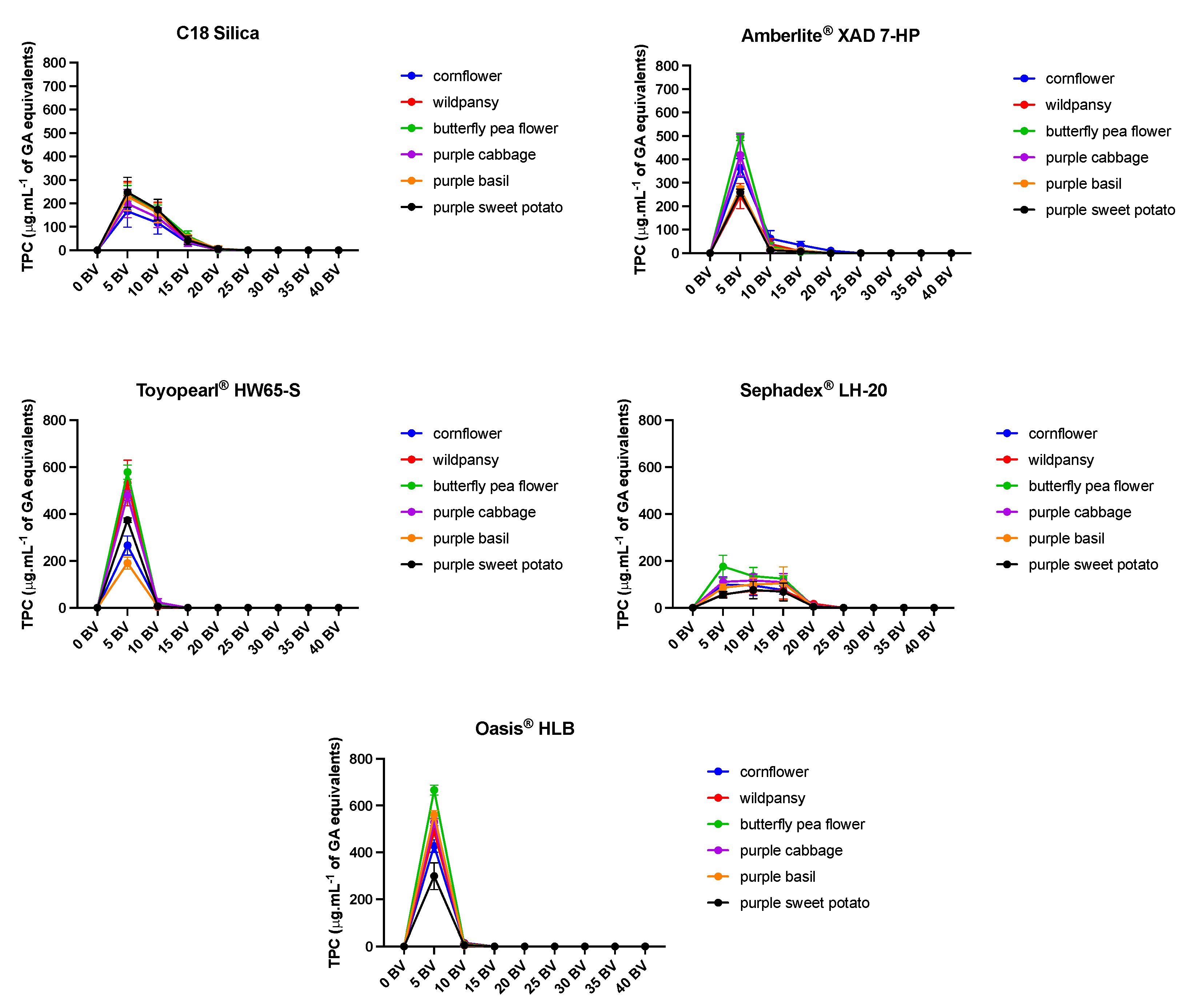
Figure 2.
Total anthocyanin content (%) determination of the extracts collected with water in the different solid-phase resins. Pools of 5 bed volumes (BV) were made and the TAC determined in a non-cumulative way. For the values below the detection limit, “n.d.” was considered. The results are present % ± SD of the TAC determined for the full extracts (n=3-8).
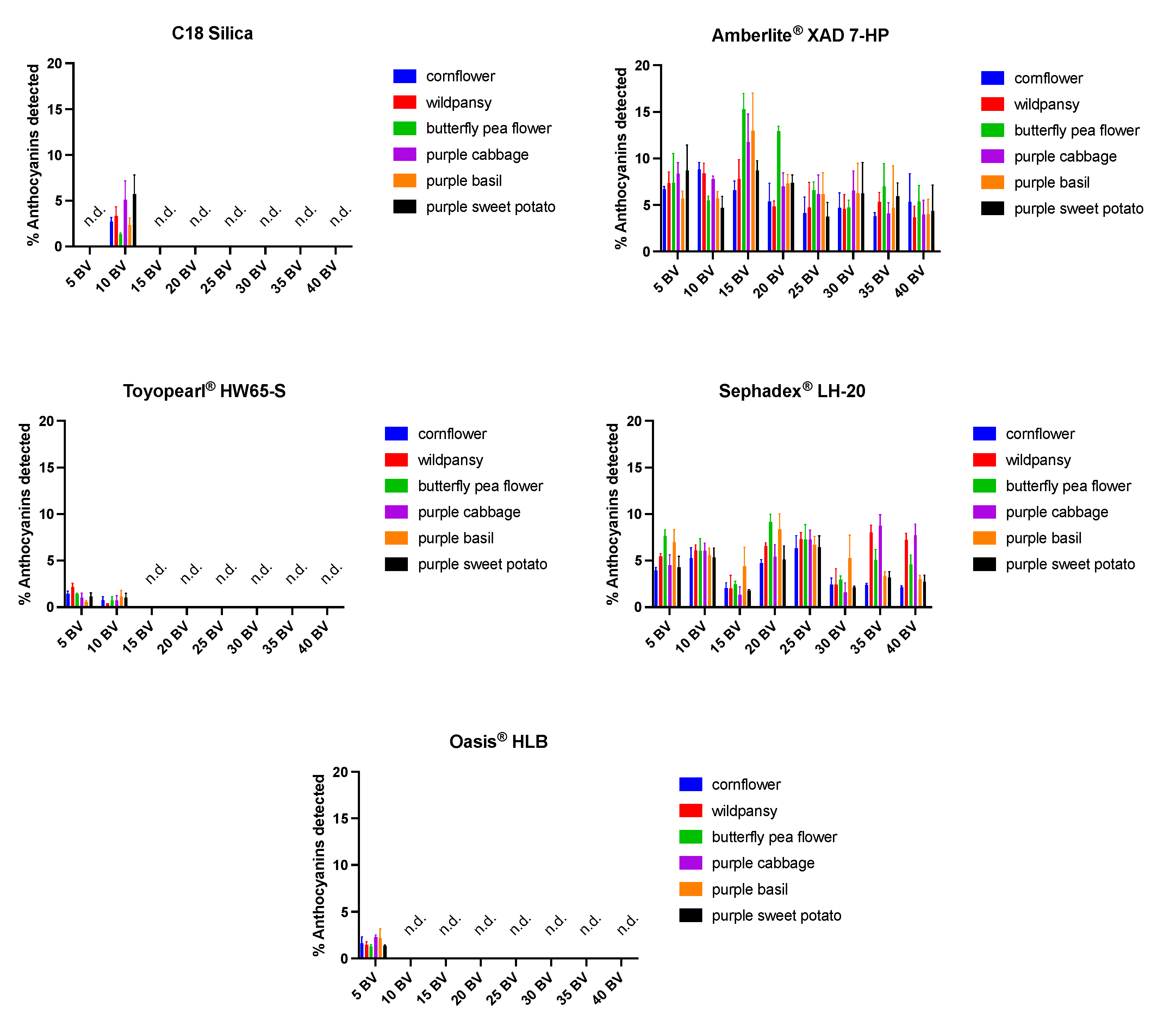
Figure 3.
a) Purity of the anthocyanin extracts after the application on the Discovery® DSC-MCAX resin. The full extract corresponds to the extract obtained after the initial water extraction. The Oasis® HLB correspond to the extract obtained from the application of the full extract on the Oasis® HLB resin. The results are presented as % ± SD of the purity by comparison of the chromatograms at 280 and 520 nm analyzed by UPHLC (n=4). b) recovery of the anthocyanins from the Discovery® DSC-MCAX resin. The Oasis® HLB correspond to the extract obtained from the application of the full extract on the Oasis® HLB resin. The results are presented as % ± SD of the recovered initial amount of anthocyanins applied in the resin (n=4).
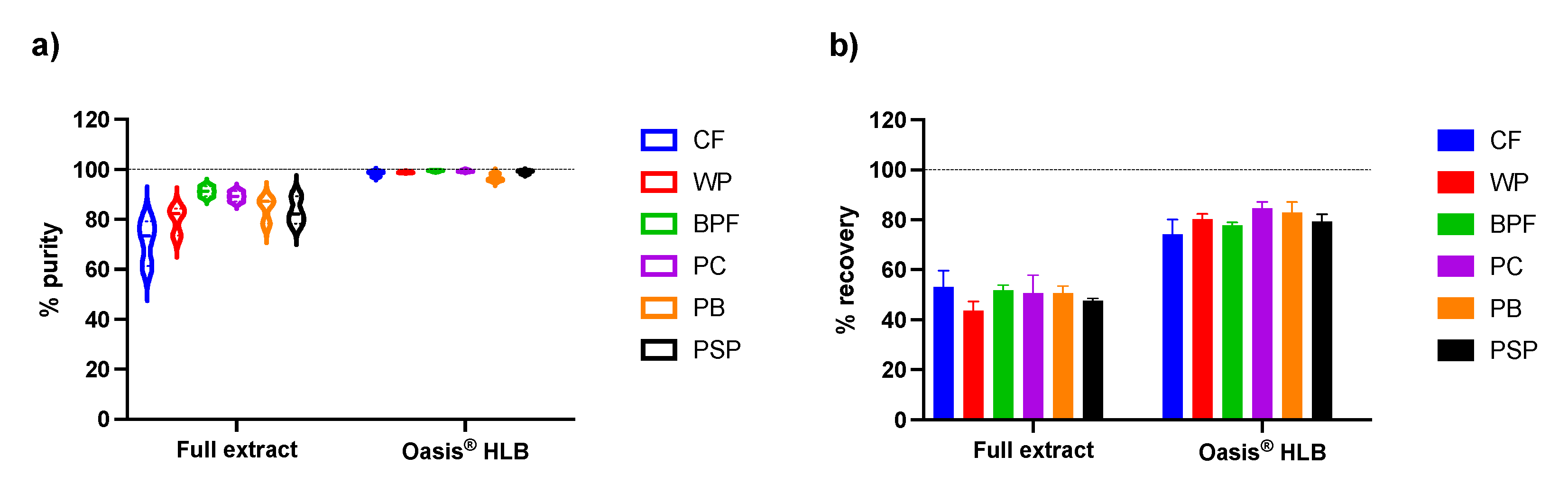

Table 1.
Total anthocyanin content on the different full extracts.
| Extract1 | Total Anthocyanin Content (mg.g-1 DW of extract) |
|---|---|
| cornflower | 67,4 ± 1,2 |
| wild pansy | 43,5 ± 2,3 |
| butterfly pea flower | 172,4 ± 4,3 |
| purple cabbage | 274,3 ± 2,1 |
| purple basil | 72,4 ± 1,4 |
| purple sweet potato | 87,3 ± 5,3 |
1 refers to the full extracts after water extraction and freeze drying.
Table 2.
LC-MS identification of the main anthocyanins in the different extracts on the positive mode.
Table 2.
LC-MS identification of the main anthocyanins in the different extracts on the positive mode.
| Extract | Rt (min) | [M+H]+ (m/z) | Main fragment ions | λmax (nm) | Tentative identification |
|---|---|---|---|---|---|
| cornflower | 8.20 | 611.39 | 449.26, 287.14 | 276, 512 | Cyanidin-3-O-glucoside-5-O-glucoside |
| 9.49 | 697.4 | 535.3, 449.49, 287.36 | 280, 512 | Cyanidin-3-(6-malonyl)-O-glucoside-5-O-glucoside | |
| 10.21 | 711.39 | 549.35, 449.5, 287.15 | 280, 516 | Cyanidin-3-(6-succinyl)-O-glucoside-5-O-glucoside | |
| 13.00 | 725.38 | 563.36, 449.5, 287.15 | 276, 516 | Cyanidin derivative | |
| 15.68 | 549.41 | 287.18 | 280, 516 | Cyanidin-3-(6-succinyl)-O-glucoside | |
| wild pansy | 7.50 | 773.38 | 611.34, 465.31, 303.18 | 276, 520 | Delphinidin-3-O-rutinoside-5-O-glucoside |
| 14.83 | 919.41 | 757.47, 465.47, 303.17 | 284, 528 | Delphinidin-3-(4″-p-coumaroyl)-O-rutinoside-5-O-glucoside | |
| 17.22 | 903.4 | 741.37, 449.3, 287.15 | 292, 524 | Cyanidin-3-(4′-cis-p-coumaroyl)-O-rutinoside-5-O-glucoside | |
| 20.01 | 757.42 | 611.3, 465.2, 303.15 | 284, 532 | Delphinidin-3-(4′-cis-p-coumaroyl)-O-rutinoside | |
| butterfly pea flower | 10.03 | 1183.37 | 935.47, 773.43, 611.44, 465.36, 303.19 | 184, 528 | ternatin C4 |
| 10.16 | 1405.44 | 1243.6, 1081.61, 935.46, 773.53 | 288, 536 | preternatin A3 | |
| 10.67 | 1491.36 | 1405.73, 1243.68 | 288, 540 | Ternatin C2 | |
| 12.5 | 1243.41 | 1081.48, 919.42, 773.46, 611.34, 465.36, 303.22 | 288, 540 | Delphinidin-3-O-glucoside-3′-(6-p-coumaroyl)-O-glucoside-5′- (6-p-coumaroyl)-O-diglucoside | |
| 13.4 | 1329.37 | 1081.56, 919.6, 773.56, 611.53, 465.4, 303.24 | 288, 540 | Ternatin B4 | |
| 15.06 | 1405.33 | 288, 540 | preternatin A3 | ||
| 15.38 | 1799.36 | 1713.7 | 290, 548 | Ternatin A2 | |
| 16.6 | 1343.37 | 1243.61, 1081.54, 919.60, 773.55, 611.42, | 288, 540 | Delphinidin-3-(6-succinyl)-O-glucoside-3′-(6-p-coumaroyl)-O-glucoside-5′- (6-p-coumaroyl)-O-diglucoside | |
| 16.73 | 1343.39 | 1081.49, 919.59, 773.55, 611.45, 465.32 | 288, 540 | Delphinidin-3-(6-succinyl)-O-glucoside-3′-(6-p-coumaroyl)-O-glucoside-5′- (6-p-coumaroyl)-O-diglucoside | |
| 17.05 | 1035.34 | 873.48, 773.42, 611.34, 465.35, 303.17 | 288, 540 | Delphinidin-3-O-glucoside-3′-(6-succinyl))-O-glucoside-5′- (6-p-coumaroyl)-O-glucoside | |
| 17.58 | 1637.34 | 1551.61, 1389.68 | 288, 548 | Ternatin B3 | |
| 18.19 | 1813.32 | 1713.64, 1551.87, | 288, 548 | N. D | |
| ·22.34 | 1637.34 | 1389.7 | 296, 548 | Ternatin B2 | |
| 23.16 | 1945.39 | 292, 552 | Ternatin B1 | ||
| 25.43 | 1475.31 | 1389.67, 1227.65, 919.67 | 292, 548 | Ternatin D2 | |
| 31.72 | 1783.36 | 1697.68, 1535.61 | 292, 552 | Ternatin D1 | |
| purple cabbage | 8.18 | 773.37 | 280, 516 | Cyanidin-3-O-diglucoside-5-glucoside | |
| 9.63 | 1141.39 | 979.42 | 280, 332, 528 | Cyanidin-3-(6-sinapoyl)-O-triglucoside-5-O-glucoside | |
| 12.21 | 1081.35 | 919.44, 757.52, 449.36, 287.13 | 280, 520 | Cyanidin-3-(6-p-coumaroyl)-O-triglucoside-5-O-glucoside | |
| 12.66 | 1111.38 | 949.44, 787.5, 611.5, 449.27, 287.12 | 280, 524 | Cyanidin-3-(6-feruloyl)-O-triglucoside-5-O-glucoside | |
| 15.6 | 1287.32 | 1125.5, 963.46, 449.29 | 288, 532 | Cyanidin-3-(6,6′-diferuloyl)-O-triglucoside-5-O-glucoside | |
| 16.23 | 1317.39 | 1155.5, 993.51, 611.35, 449.35 | 288, 324, 536 | Cyanidin-3-(6-feruloyl)(6′-sinapoyl)-O-triglucoside-5-O-glucoside | |
| 19.03 | 919.38 | 757.58, 595.44, 449.33, 287.12 | 280, 312, 520 | Cyanidin-3-(6-p-coumaroyl)-O-diglucoside-5-O-glucoside | |
| 19.6 | 979.37 | 817.41, 655.55, 449.29, 287.15 | 280, 324, 520 | Cyanidin-3-(6-sinapoyl)-O-diglucoside-5-O-glucoside | |
| 21.1 | 817.34 | 284, 328, 524 | Cyanidin-3-(6-sinapoyl)-O-glucoside-5-glucoside | ||
| 23.46 | 1125.35 | 963.42, 757.61, 595.56, 449.3, 287.13 | 296, 320, 532 | Cyanidin-3-(6-sinapoyl)(6′-p-coumaroyl)-O-diglucoside-5-O-glucoside | |
| 24.32 | 1155.32 | 993.43, 787.69, 449.36, 287.12 | 296, 328, 532 | Cyanidin-3-(6-feruloyl)(6′-sinapoyl)-O-diglucoside-5-O-glucoside | |
| 24.99 | 1185.36 | 1023.43, 817.49, 449.34, 287.12 | 300, 332, 532 | Cyanidin-3-(6,6′-sinapoyl)-O-diglucoside-5-O-glucoside | |
| purple basil | 11.04 | 919.36 | 757.43, 595.48, 449.4, 287.14 | 280, 524 | Cyanidin-3-(6-p-coumaroyl)(6‘-caffeoyl)-O-diglucoside isomer 1 |
| 12.44 | 919.37 | 757.48, 595.57, 449.47, 287.15 | 280, 520 | Cyanidin-3-(6-p-coumaroyl)(6‘-caffeoyl)-O-diglucoside isomer 2 | |
| 19.15 | 1081.32 | 919.43, 757.57, 595.46, 449.3, 287.13 | 284, 524 | Cyanidin-3-(6-p-coumaroyl)(6‘-caffeoyl)-O-diglucoside-5-O-glucoside isomer 1 | |
| 19.72 | 757.28 | 595.34, 449.38, 287.14 | 284, 520 | Cyanidin-3-(6-p-coumaroyl)-O-glucoside-5-O-glucoside | |
| 21.90 | 1081.34 | 919.4, 757.5, 595.46, 449.33, 287.14 | 288, 524 | Cyanidin-3-(6-p-coumaroyl)(6‘-caffeoyl)-O-diglucoside-5-O-glucoside isomer 2 | |
| 24.24 | 1065.41 | 903.44, 757.53, 595.47, 449.38, 287.14 | 284, 528 | Cyanidin-3-(6,6′-dip-coumaroyl)-O-diglucoside-5-O-glucoside isomer 1 | |
| 29.16 | 1065.37 | 903.54, 757.6, 595.49, 449.4, 287.14 | 284, 528 | Cyanidin-3-(6,6′-dip-coumaroyl)-O-diglucoside-5-O-glucoside isomer 2 | |
| purple sweet potato | 8.61 | 787.36 | 524 | Peonidin-3-O-diglucoside-5-O-glucoside | |
| 11.47 | 907.35 | 745.36, 463.29, 301.25 | 274, 520 | Peonidin-3-(6-p-hydroxybenzoyl)-O-diglucoside-5-O-glucoside | |
| 11.88 | 1067.39 | 905.4, 887.58, 605.55 | 273, 532 | Peonidin derivative isomer 1 | |
| 12.32 | 1097.35 | 935.43 | 532 | Cyanidin-3-(6,6′-dicaffeoyl)-O-diglucoside-5-glucoside | |
| 13.05 | 1067.35 | 905.46, 887.55, 605.45 | 273, 532 | Peonidin derivative isomer 2 |
Table 3.
Total anthocyanin content (TAC) determination of the extracts collected with ethyl acetate in the different solid-phase resins. The pools of 5 bed volumes (BV) were joint in a single 40 BV pool and the TAC was determined accordingly to the Materials and Methods section. For the values below the detection limit, “n.d.” was considered. The results are present % ± SD of the TAC determined for the full extracts (n=3-8). Significant differences (p<0.05) were arranged as follows: letter for the different extracts applied in the same resin (a – CF; b – WP; c – BPF; d – PC; e – PB; f – PSP; g – BE); numbers for the different resins loaded with the same extract (1 – C18 Silica; 2 – Amberlite® XAD 7-HP; 3 – Toyopearl® HW-65S; 4 – Sephadex® LH-20; Oasis® HLB). # - means significantly different from all other letters. $ - means significantly different from all other numbers.
| Resin | TAC (%) after 40 BV | ||||||
|---|---|---|---|---|---|---|---|
| CF1 | WP1 | BPF1 | PC1 | PB1 | PSP1 | ||
| C18 Silica | 2.5 ± 0.9# | 5.1 ± 0.73-5 | 7.7 ± 2.3 | 4.2 ± 0.4$ | 5.8 ± 0.5 | 8.8 ± 0.3a,b,d,e,,$ | |
| Amberlite® XAD 7-HP | 3.4 ± 0.3b,c,e | 5.8 ± 1.13-5 | 6.6 ± 1.2 | 1.7 ± 0.7$ | 4.2 ± 1.2 | 3.0 ± 0.5$ | |
| Toyopearl® HW-65S | 1.1 ± 0.4b,d,e | 2.4 ± 0.3$ | 1.5 ± 0.5$ | n.d.#,1,2,4 | 2.2 ± 0.91,2,4 | 0.8 ± 0.71,2,4 | |
| Sephadex® LH-20 | 7.8 ± 1.2d,$ | 11.4 ± 0.5#,$ | 7.2 ± 0.7 | 9.4 ± 0.3$ | 5.7 ± 0.7 | 6.4 ± 0.8$ | |
| Oasis® HLB | n.d.e-f,$ | n.d. e-f,$ | n.d. e-f,$ | n.d. e-f,1,2,4 | 1.3 ± 0.4a-d,1,2,4 | 1.0 ± 0.2 a-d | |
1 CF – cornflower; WP – wild pansy; BPF – butterfly pea flower; PC – purple cabbage; PB – purple basil; PSP – purple sweet potato;.
Table 4.
Anthocyanin recovery and purity (%) determination on the extracts collected with methanol in the different solid-phase resins. The results are present % ± SD of the TAC determined for the full extracts and subtracting the losses from the previous steps (n=3-8). Significant differences (p<0.05) were arranged as follows: letter for the different extracts applied in the same resin (a – CF; b – WP; c – BPF; d – PC; e – PB; f – PSP); numbers for the different resins loaded with the same extract (1 – C18 Silica; 2 – Amberlite® XAD 7-HP; 3 – Toyopearl® HW-65S; 4 – Sephadex® LH-20; Oasis® HLB). # - means significantly different from all other letters. $ - means significantly different from all other numbers.
| Resin | Anthocyanin Recovery (%) | |||||
|---|---|---|---|---|---|---|
| CF1 | WP1 | BPF1 | PC1 | PB1 | PSP1 | |
| C18 Silica | 72.6 ± 6.4c,2,3 | 62.3 ± 5.2 | 77.3 ± 10.4d,2,3,4 | 46.1 ± 1.7e,f,5 | 75.4 ± 2.52 | 76.7 ± 9.34 |
| Amberlite® XAD 7-HP | 49.1 ± 7.85 | 50.9 ± 6.83,5 | 33.2 ± 9.4d,f,3,5 | 58.1 ± 4.33,5 | 48.1 ± 15.84,5 | 67.5 ± 1.8 |
| Toyopearl® HW-65S | 48.0 ± 11.9b,5 | 73.9 ± 5.9d | 57.9 ± 4.0d,4,5 | 34.4 ± 8.9e,f,5 | 63.1 ± 15.75 | 59.9 ± 16.85 |
| Sephadex® LH-20 | 63.4 ± 14.5c,5 | 62.3 ± 13.1c | 37.0 ± 4.6e,5 | 45.0 ± 2.5e,5 | 68.4 ± 11.25 | 56.7 ± 8.95 |
| Oasis® HLB | 83.9 ± 4.9 | 79.9 ± 10.1 | 81.2 ± 4.3 | 91.6 ± 2.6 | 89.8 ± 2.2 | 85.9 ± 1.5 |
| Anthocyanin Purity (%) | ||||||
| CF1 | WP1 | BPF1 | PC1 | PB1 | PSP1 | |
| C18 Silica | 36.4 ± 1.9b,c,e,f,$ | 48.0 ± 3.0d,$ | 48.0 ± 1.4d,3,4,5 | 31.9 ± 1.2e,f,3,5 | 54.5 ± 2.24,5 | 57.1 ± 6.34,5 |
| Amberlite® XAD 7-HP | 47.3 ± 2.5d,f,$ | 38.6 ± 4.3c,e,f,$ | 38.6 ± 3.63,4,5 | 29.3 ± 0.9e,f,3,5 | 52.3 ± 2.73,4,5 | 58.9 ± 6.34,5 |
| Toyopearl® HW-65S | 66.4 ± 6.14 | 67.5 ± 2.44 | 67.5 ± 3.94,5 | 62.5 ± 4.34 | 62.2 ± 4.54,5 | 62.6 ± 2.74,5 |
| Sephadex® LH-20 | 15.3 ± 7.1#,5 | 29.0 ± 7.3d,5 | 29.0 ± 2.15 | 33.2 ± 2.15 | 39.8 ± 5.55 | 37.4 ± 2.15 |
| Oasis® HLB | 68.9 ± 4.7c | 72.4 ± 4.4d | 72.4 ± 3.5d | 61.6 ± 1.5e,f | 72.2 ± 2.1 | 74.9 ± 2.9 |
Disclaimer/Publisher’s Note: The statements, opinions and data contained in all publications are solely those of the individual author(s) and contributor(s) and not of MDPI and/or the editor(s). MDPI and/or the editor(s) disclaim responsibility for any injury to people or property resulting from any ideas, methods, instructions or products referred to in the content. |
© 2024 by the authors. Licensee MDPI, Basel, Switzerland. This article is an open access article distributed under the terms and conditions of the Creative Commons Attribution (CC BY) license (http://creativecommons.org/licenses/by/4.0/).
Copyright: This open access article is published under a Creative Commons CC BY 4.0 license, which permit the free download, distribution, and reuse, provided that the author and preprint are cited in any reuse.


