
Submitted:
23 October 2025
Posted:
24 October 2025
You are already at the latest version
Abstract
The interest in replacing artificial colorants, which can have mutagenic, carcinogenic, and teratogenic effect, has driven the search for safe natural pigments. In this context, this preliminary study aimed to explore the chromatographic identification of anthocyanins and anthocyanidins from Dioscorea trifida L. (purple sachapapa) and to evaluate a preliminary bioinorganic stabilization method using magnesium complexes. Samples were collected in Nuevo Oriente, District of Mazán, Loreto Region, and tubers were mechanically processed. Extraction was performed with HCl-acidified methanol. Pigment identification was carried out using paper chromatography with BAW and 1% HCl systems for anthocyanins, and formic acid and forestal systems for anthocyanidins, comparing Rf values and color with scientific literature. The identified anthocyanidins were cyanidin, peonidin, malvidin and pelargonidin, and their corresponding glucosides were cyanidin-3-rhamnosyl-glucoside, pelargonidin-3,5-diglucoside, malvidin-3,5-diglucoside, and peonidin-3,5-diglucoside. Stabilization was explored via a hexacoordinated complex [η6(anthocyanin)6Mg2+].6Cl-, whose formation is based on the stoichiometry pf the reactants and the sp3d2 structure of Mg2+, according to bioinorganic chemistry studies. Preliminary results suggest that this method can enhance anthocyanin stability and provide a foundation for future toxicity studies and applications in the natural colorant industry.
Keywords:
Dioscorea trifida L.
; anthocyanins
; anthocyanidins
; magnesium complexation
; bioinorganic stabilization
; natural food colorant
1. Introduction
Anthocyanins are water-soluble polyphenolic pigments belonging to the flavonoid family, responsible for the red, purple, and blue coloration in a wide variety of plant tissues [1]. These natural pigments have gained considerable attention in recent years, not only for their role as food colorants but also for their significant antioxidant, anti-inflammatory, and potential anticancer properties [2,3]. The growing consumer demand for natural food additives, together with increasing concerns about the safety of synthetic dyes–many of which have been associated with mutagenic and carcinogenic effects in laboratory studies [4,5], has intensified research efforts toward identifying, characterizing, and utilizing anthocyanins from diverse botanical sources. Unlike artificial dyes such as tartrazine and erythrosine, which have raised toxicological concerns, anthocyanins present a safe and health-promoting alternative for the food, pharmaceutical, and cosmetic industries [6].
Despite their numerous advantages, the industrial application of anthocyanins faces significant challenges due to their inherent chemical instability. Anthocyanin stability is profoundly affected by environmental factors including pH, temperature, light exposure, oxygen availability, and the presence of enzymes [2,7]. These pigments exist in various structural forms depending on pH: the red flavylium, cation predominates under acidic conditions, while neutral to alkaline pH values favor the formation of colorless or blue quinoidal bases and chalcones, leading to color fading [8]. Additionally, anthocyanins are susceptible to thermal degradation during food processing and storage, which limits their practical utility [9]. Consequently, developing effective stabilization strategies is crucial for expanding the commercial applications of these valuable natural compounds.
One promising approach to enhance anthocyanin stability involves the formation of metal-anthocyanin complexes (metalloanthocyanins). Anthocyanins containing ortho-dihydroxyl groups on the B-ring can chelate divalent and trivalent metal ions such as Mg2+, Fe2+, Fe3+, Al3+, Cu2+, and Sn2+, forming stable coordination complexes [10,11]. These metal complexes not only improve color stability across a broader pH range but also induce bathochromic shifts, often resulting in intensified and more vivid hues [12,13]. Magnesium ions, in particular, have been shown to form hexacoordinated complexes witch anthocyanins through coordination bonds involving oxygen atoms from hydroxyl and carbonyl groups, thereby protecting the chromophore from degradation [14,15]. This stabilization complex found in blue flowers, which demonstrates remarkable color stability [16].
Whitin the genus Dioscorea, several species are recognized for their colored tubers rich in bioactive compounds. Dioscorea alata L. (Purple yam) and Dioscorea trifida L. (Sachapapa or cush-cush yam) have received particular attention for their anthocyanin content. Previous phytochemical investigations using high-performance liquid chromatography couple with mass spectrometry (HPLC-DAD-ESI/MS) have identified multiple anthocyanin derivatives in D. alata, with cyanidin-based glycosides, particularly alatanin C (cyanidin 3-(6-sinapoyl gentiobioside)), as the predominant pigments [17,18]. Similarly, research on D. trifida L. from Peru has revealed the presence of at least 12 anthocyanin compounds derived from peonidin, cyanidin, and pelargonidin aglycones, many bearing acyl moieties that contribute to their stability and color properties [19]. However, comprehensive studies on the stabilization of anthocyanins from D. trifida through metal complexation remain limited, representing a significant gap in the current literature.
Give the promising bioactive profile of Dioscorea trifida L. anthocyanins and the need for effective stabilization methods, the present study was designed with a preliminary and exploratory approach witch two primary objectives: (a) to identify and characterize the anthocyanin components presents in purple sachapapa tubers using paper chromatography as an initial separation technique, employing BAW and HCl 1% systems for anthocyanins and formic/forestal systems for anthocyanidins; and (b) to stabilize the extracted anthocyanins metal-anthocyanin complexes that enhance both color intensity and chemical stability. By achieving these goals, this work aims to contribute valuable insights into the practical application of D. trifida L. anthocyanins as stable, natural colorants and functional ingredients with potential health benefits.
2. Results
2.1. Extraction Yield of Anthocyanins
A total of 9.0 f of dry anthocyanin pigment was obtained from 500 g of fresh Dioscorea trifida L. tuber, corresponding to yield of 6.0% (w/w) relative to the dry mass.
2.2. Chromatographic Characterization of Anthocyanins and Anthocyanidins
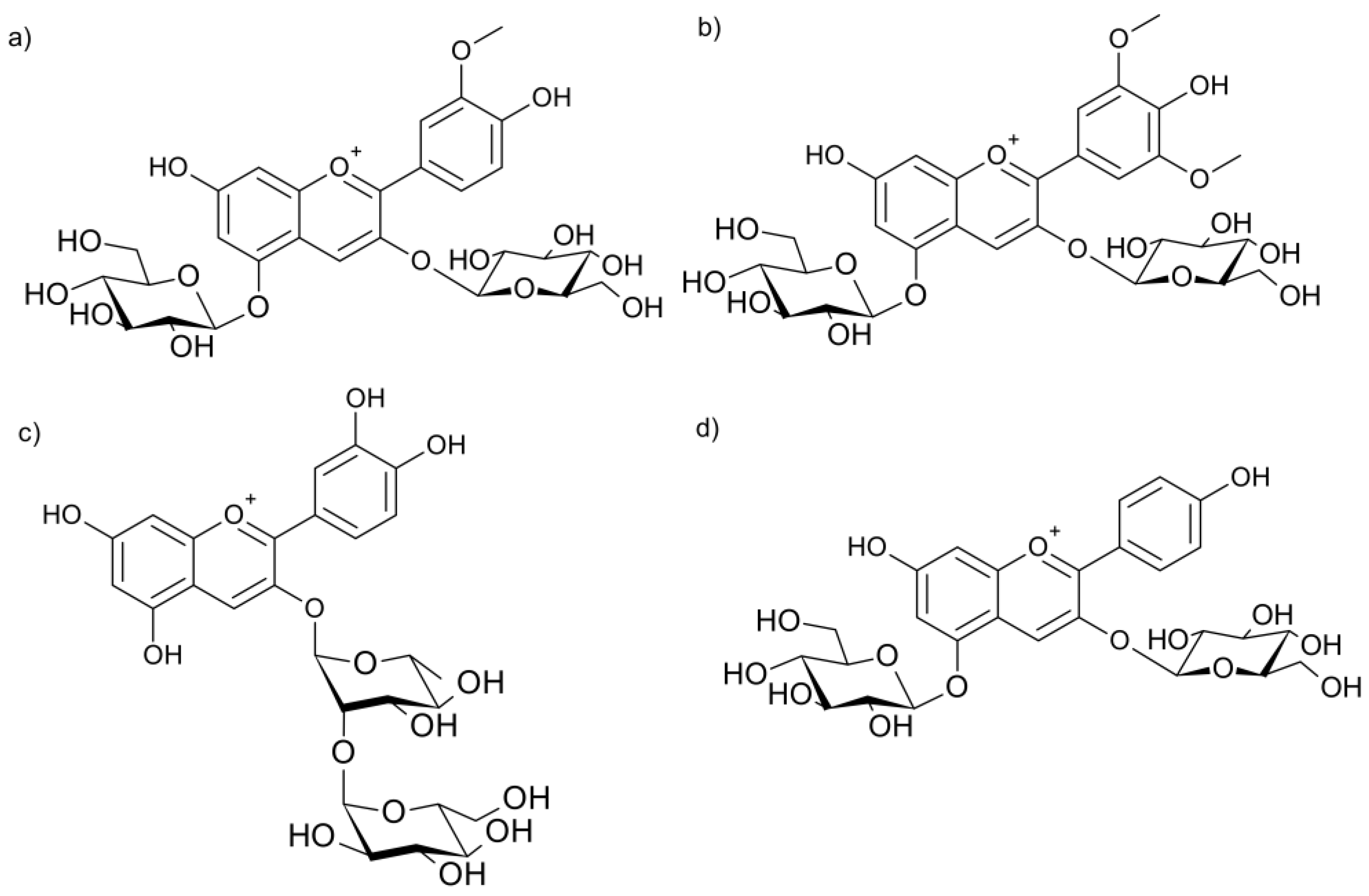
Paper chromatography of the concentrated methanolic extract produced four noticeable bands ranging from violet to pink for anthocyanins and anthocyanidins, respectively. The experimental Rf values and provisional identifications (based on comparison with bibliographic Rf ranges and color) are summarized in Table 1.
Figure 1.
Tentative structure of the main anthocyanins identified in Dioscorea trifida L. by paper chromatography: (a) Peonidin-3,5-diglucoside; (b) Malvidin-3,5-diglucoside; (c) Cyanidin-3-rhamnosyl-glucoside and (d) Pelargonidin-3,5-diglucoside.
Figure 1.
Tentative structure of the main anthocyanins identified in Dioscorea trifida L. by paper chromatography: (a) Peonidin-3,5-diglucoside; (b) Malvidin-3,5-diglucoside; (c) Cyanidin-3-rhamnosyl-glucoside and (d) Pelargonidin-3,5-diglucoside.
2.3. Formation and Characteristics of the Magnesium-Anthocyanin Complex
The coordination complex [η6(anthocyanin)6Mg(II)].6Cl- was obtained according to the stoichiometric principles of the reaction balance of magnesium oxide and hydrochloric acid (Figure 2). The resulting solid was an intense blue powder, soluble in water an ethanol, had a pH of 3.78, and produced 2.086g of the complex, which is consistent witch the calculated stoichiometric mass balance:
6 MgO + 6HCl → 6Mg2+ + 6Cl- + 3H2O + 3O=↑
6Mg2+ + 6Cl- + Anthocyanin → complex [η6(anthocyanin)6Mg(II)].6Cl-
2.4. Spectrophotometric Stability and Shelf-Life Projection
The absorbance of the Mg2+-anthocyanin complex at λmax showed a slight and gradual decrease from 2.000 at synthesis (month 0) to 1.976 after ten months of storage (Table 2 and Figure 3). The degradation trend was fitted by linear regression using the least-squares method, resulting in the equation A = 1.999 – 0.0025t with a correlation coefficient R2 = 0.998. The negative slope indicates a slows and consistent decline in absorbance over time, suggesting high pigment stability under ambient conditions.
A variation of 0.1 absorbance units was considered the degradation limit, corresponding to a projected shelf life of approximately 36 months for the stabilized pigment.
3. Discussion
The extraction yield of anthocyanin pigments is critical parameter for assessing the biotechnological potential of plant matrices as sustainable sources of natural colorants. In the present exploratory study, 9g of colorant were obtained from 150 g of dry Dioscorea trifida L. tubers samples, representing a yield of 6%, higher than that reported by Alberca [29], who extracted 253.584 mg of total anthocyanins per 100 g of sample without achieving isolation, resulting in crude dyes without exploring anthocyanin characterization. Likewise, interspecific comparison within the Dioscorea genus shows that the 5.22% yield reported for D. alata [30] is lower than that recorded for D. trifida L. in this work. Nevertheless, it is important to recognize that extraction yield depends not only on the endogenous anthocyanin concentration but also on methodological factors such as solvent polarity, contact time, extraction temperature, and degree of tissue fragmentation. Recent studies have demonstrated that ultrasound or microwave-assisted techniques can significantly enhance anthocyanin recovery efficiency in Dioscorea tubers while reducing both processing time and solvent consumption [31,32]. These variations reflect the high sensitivity of the process to experimental conditions and the intrinsic variability of the plant matrix.
A distinctive aspect of this study lies in the purification strategy applied. Whereas previous research was limited to obtaining crude extracts in paste or powder form without identifying or stabilizing the anthocyanins [33,34], the present work successfully isolated four major components using paper chromatography, a classical yet effective technique for pigment separation. Furthermore, this study incorporated a stabilization strategy through complexation with divalent metal ions (Mg2+), a methodological innovation that extends the temporal stability of the isolated colorant and opens new possibilities for developing colorants based on coordination complexes with biocompatible metals.
Closely related to the above, the identification of isolated anthocyanins and the understanding of pigment profile variability are essential for correlating yield with molecular composition. The anthocyanins identified from Dioscorea trifida L. were cyanidin-3-rhamnosylglucoside, peonidin-3,5-diglucoside, malvidin-3,5-diglucoside, and pelargonidin-3,5-diglucoside, as determined by paper chromatography using BAW (n-butanol-acetic acid-water) and 1% hydrochloric acid system, with characteristic Rfx100 values for each compound. Although traditional chromatographic techniques were employed, the results are consistent with structural patterns described for Dioscorea species, in which glycosylated derivatives of cyanidin, peonidin, malvidin and pelargonidin predominate.
Moreover, comparison with previous studies reveals remarkable intraspecific variability in pigment profiles. Samples of Dioscorea trifida L. from Venezuela yielded three anthocyanins derived from peonidin and malvidin [20], whereas plant material from the Alto Huallaga region (Perú) showed twelve anthocyanins derived from cyanidin, peonidin and pelargonidin [19], a qualitatively more diverse profile than found in this work. This heterogeneity has been widely documented in anthocyanin-rich plants, where genetic, environmental, and edaphic factors modulate both biosynthesis and accumulation of these pigments [35,36,37,38].
At the molecular level, anthocyanin biosynthesis is regulated by a complex network of transcription factors sensitive to light, temperature, water stress and mineral availability [39]. In D. alata L. light exposure induces the expression of the genes such as UDP-glycosyl transferase, flavanone-3-hydroxylase and anthocyanidin synthase, increasing pigment accumulation as a function of irradiance [40,41]. Similarly, various enzymes mediate post-biosynthetic modifications such as glycosylation, hydroxylation, acylation and methylation that determine the solubility, stability and toxicity of pigments [42].
In addition, soil pH exerts a marked influence: acidic soils favor the cationic flavylium form, whereas neutral and display distinct hues [35,43]. The presence of metal such Fe, Al and Mg in the soil can induce in planta formation of metalloanthocyanin complexes, thereby modifying color and stability [44]. These processes explain the chromatic variability observed among different populations of D. trifida L.
In other regions, even greater structural diversity has been reported. In Dioscorea trifida L. from the Philippines, eight anthocyanins were identified, four acylated with hydrocycinnamic acids and four uncharacterized [30]. Acylation, mediated by specific acyltransferases, enhances thermal and pH stability by sterically shielding the flavylium core through π-π interactions. In D. alata L. from Thailand, eleven anthocyanins were identified by, including glucosylated cyanidins and alatanin-type compounds, exclusive to this species [17]. This highlights the biosynthetic complexity of Dioscorea and the role of intrinsic genetic factors in determining glycosylation and substitution patterns [45,46].
From an analytical perspective, paper chromatography is useful for preparative isolation, but definitive structural confirmation requires advanced techniques such as MS/MS, NMR and UV-Vis spectroscopy, which allow determination of fragmentation patterns, substituent configurations and characteristic absorption maxima. The integration of these techniques has enabled the discovery of novel anthocyanins in Dioscorea, expanding our understanding of their chemical diversity and biotechnological potential [40].
In this context, anthocyanin stabilization plays a fundamental role, as molecular structure directly conditions color persistence and technological applicability. The characteristic coloration of flower and tuber, except in the Caryophyllales, is primarily due to anthocyanin accumulation in cellular vacuoles [21,47]. However, once extracted, their chemical stability is compromised by light, temperature, oxygen and pH [48], which limits their industrial use as natural colorants and drives the search for more efficient stabilization strategies.
Among the most widely explored strategies is the formation of inclusion complexes, in which a host molecule (polysaccharide or protein) encapsulates the anthocyanin via noncovalent interactions. Agents such as gelatin, carboxymethylcellulose, maltodextrin, gum arabic, tragacanth gum, citrus pectin and calcium alginate pearls have been employed [49,50,51,52,53,54,55]. This method reduces pigment exposure to oxygen and UV radiation, slowing degradation. Nonetheless, its effectiveness is temporally limited. Colorants from Morus nigra L. and Rubus fruticosus L. encapsuled with maltodextrin or gum arabic degraded completely within 9 to 33 days, depending on temperature [52]. Similarly, eggplant dye encapsulated with maltodextrin and tragacanth gum stored at 25ºC and 4ºC had degraded by 46% and 22.5% respectively, within 40 days [51], and carrot dye encapsulated with maltodextrin showed losses of up to 33% at 25ºC in 64 days [55]. Although combinations of maltodextrin with protein such as whey protein insolate (WPI) improve stability, they rarely exceed six months of shelf life [56].
Degradation causes include oxidation and quinone formation (particularly in alkaline media), rigidification of the molecular structure and hygroscopicity of the encapsulant. Therefore, inclusion complexes, though useful for temporary protection and controlled release, are insufficient for long-term industrial applications [56,57].
In addition to encapsulation methods using polysaccharides and proteins of animal or plant origin to mitigate anthocyanin degradation, other techniques have been explored. Freeze-drying offers limited protection, as ice sublimation generates porous structures that facilitate contact between the pigment and oxygen. Electrospray and electrospinning processes exhibit low yield and limited reproducibility. Likewise, although emulsions can provide good stability and protection against oxidation, they suffer from internal droplet coalescence, which causes anthocyanins release and promotes degradation. Liposomes, despite their ability to encapsulate hydrophilic compounds, offer low oxidative protection due to the presence of unsaturated fatty acids in their membrane. In ionic gelation, rapid diffusion limits its effectiveness for low-molecular-weight compounds. Finally, coacervation allows for encapsulation with good yields, although the particles obtained are sensitive to pH and ionic strength, which restricts their application in food matrices [58].
In contrast these methods, the present study implemented the formation of coordination complexes with Mg2+ ions, yielding a new, more stable chemical entity. The octahedral complex [η6(anthocyanin)6Mg(II)].6Cl- [59,60] was formed through coordination of Mg2+ with ortho-dihydroxyl groups on ring B, providing electronic stabilization without altering the π-conjugated system responsible for the intense blue color.
Analogous to other metal-coordination systems described in the literature, the behavior of Mg2+ in this complex shows notable similarly to that of Cr3+ ion in MOF-type materials. In particular, MIL-101 (Cr), first reported by Férey and later studied by Wee, Chinnappan and Ramakrishna (2023), exhibits an octahedral architecture in which each chromium ion acts as a coordination center surrounded by six terephthalic acid (H2BDC) ligands, forming a stable three-dimensional network with the formula Cr3(F/OH)(H2O)2O[(O2C)-C6H4-(CO2)]3.xH2O (x ~ 25) [61]. This type of organization, based on secondary building units, demonstrates the structures feasibility of stable metal-ligand octahedral assemblies. Similarly, in the present work, the Mg2+ ion behaves as a central node coordinated by six anthocyanin molecules, forming a hexacoordinated complex with biofunctional potential. Although MIL-101 (Cr) polymerizes into a microporous network, the underlying chemical principle of octahedral coordination is equivalent: metal-oxygen interactions stabilize the structure and modulate its physicochemical properties. This analogy with MOF systems not only reinforces the structural plausibility of the magnesium-anthocyanin complex but also suggests that such architectures could be adapted for developing bioactive, stable and biocompatible materials.
Anthocyanins derived from cyanidin, delphinidin and petunidin exhibit high chelating capacity, whereas those derived from pelargonidin, malvidin and peonidin show lower metal affinity [21,48]. In this study, the anthocyanin mixture suggests a heterogeneous complex dominated by cyanidin-type species. Reaction pH is critical: at values between 5 and 6, partial deprotonation and chelating capacity are optimized without compromising coloration [10]. At higher pH values, quinoidal and colorless forms predominate [57].
Natural examples of metalloanthocyanins include protocyanin from Centaurea cyanus, which contains Fe3+, Mg2+ and Ca2+ in an intense blue tetragonal architecture [62]; and cyanosalvianin from Salvia uliginosa, where six anthocyanins and two Mg2+ produce a brilliant blue [63]. These natural models inspired the strategy adopted here to create stable and safe colorants capable of replacing synthetic azo or diazo dyes.
The complex [η6(anthocyanin)6Mg(II)].6Cl- exhibited exceptional stability: after three years, absorbance decrease (λmax = 520) was only 0.100 units (<6%). In contrast, free or encapsulated anthocyanins lose more than 50% pf their initial absorbance within two months. Linear regression analysis (y = 1.999-0.0025t; R2 = 0.998) suggests first-order degradation kinetics and an estimated shelf life of 36 months under ambient conditions.
This stability is attributed to the electronic redistribution induced by Mg2+, which stabilizes the flavylium cationic form, provides steric protection against nucleophilic water attack, decreases water activity around the pigment, and lowers the redox potential of chelating phenolic groups [10,64]. Other metals ions, such as Al3+, Fe3+, Cu2+, Zn2+, have shown the ability to form stable complexes with anthocyanins, although they present limitations in terms of color and safety [13,65,66]. Aluminum complexes exhibit remarkable stability; however, their use in food applications is restricted due to toxicity concerns [67]. Iron complexes, while providing good durability, tend to produce brownish hues that reduce the visual appeal of products [65,68]. In contrast, preliminary studies suggest that Mg2+ may enhance the antioxidant stability of anthocyanins [69], although its specific effectiveness as a stabilizing agent in industrial applications still requires further validation.
Taken together, the results of this preliminary study open new research avenues aimed at developing natural colorants with functional, biocompatible and highly stable properties, with potential applications in the food, pharmaceutical and cosmetic industries. This work provides initial evidence that anthocyanins can behave as polydentate ligands in MOF-type coordination systems, thereby expanding their potential not only as pigments but also as bioinspired platforms for designing active materials. Future studies should address advance structural characterization of the complex, its thermodynamic behavior and toxicological evaluation to support its industrial application.
4. Materials and Methods
4.1. Reagents and Equipment
All solvents and reagents were of analytical grade and purchased from Merck (Darmstadt, Germany). Methanol, n-butanol, petroleum ether (boiling range according to the supplier), glacial acetic acid, formic acid, and concentrated hydrochloric acid (HCl) were used without further purification. Magnesium oxide (MgO) was employed in pigment stabilization assays, and bidistilled water was used for all aqueous preparations. Paper chromatography was performed on Whatman Nº 3 paper for pigment separation and Whatman Nº 1 paper for Rf value determination (rechromatography) using descending development chambers (“artesas”) under ambient laboratory conditions, with visualization under visible and ultraviolet light. The main equipment included a rotary evaporator for solvent concentration, a vacuum filtration system with Büchner funnel and kitasato flash, a UNICO 2800 spectrophotometer (USA), and a Baltalab digital potentiometer (Brazil).
4.2. Plant Material and Sample Collection
Tubers of Dioscorea trifida L. (purple “sachapapa”) were collected from cultivated plots belonging to the Kichwa native community of Nuevo Oriente, district of Mazán, province of Maynas, Loreto region, Peru (geographical coordinates: 3º13’33’’ S, 73º10’02’’ W; 108 ma.s.l.). A total of 1.0 kg of mature, healthy, and undamaged tubers from three plants was obtained. The plant material (stem/vine, leaves, and tuber) was taxonomically identified and certified at the Amazonian Herbarium (CIRNA-UNAP), confirming correspondence with D. trifida L. The collected tubers were transported in sealed plastic bags to the Phytochemistry and Natural Products Laboratory, Faculty of Chemical Engineering, National University of the Peruvian Amazon (UNAP), for processing.
4.3. Extraction and Concentration of Anthocyanins
Tubers of D. trifida L. were peeled and finely grated (approximately 0.5 kg, fresh weight), placed in 1 L beaker, and extracted with acidified methanol (methanol + drops of concentrated HCl) under magnetic stirring for 6 h at room temperature. The mixture was filtered, and the filtrate was concentrated under reduced pressure in a rotary evaporator at 35ºC and 120 rpm to prevent pigment degradation, yielding approximately 120 mL of concentrated extract. This concentrated fraction was used for (1) paper chromatography analysis of anthocyanins and anthocyanidins, (2) determination of total pigment yield (dry extract), and (3) stabilization assays with MgO and HCl.
4.4. Paper Chromatography and Identification of Anthocyanins and Anthocyanidins
Paper chromatography separations were performed on Whatman Nº3 paper for pigment separation and on Whatman Nº1 paper for Rf determination by descending rechromatography at room temperature. The solvent systems used were n-butanol-acetic acid-water (BAW, 4:1:5, upper phase) and 1% HCl for anthocyanins, and formic acid and HCl concentrated-acetic acid-water (Forestal, 3:3:10) for anthocyanidins (after acid hydrolysis under reflux for 20 min at 70ºC). Rf Values were calculated as Rf = (distance traveled by solute/ distance traveled by solvent) x 100. Identification of anthocyanins and anthocyanidins was achieved by comparing their Rf valued and coloration with reference data from Harborne (1984) [21], Carreño and Grau (1977) [20], Daravingas and Cain (1966) [23], Bate-Smith (1948) [22], Markakis and Jurd (1974) [25], Smith and Luh (1965) [26], Dekazos (1970) [27] and Lawanson and Osude (1972) [28].
4.5. Stabilization and Spectrophotometric Evaluation of the Magnesium Complex
To evaluate pigment stabilization, magnesium oxide (MgO) was used to form a coordination complex. The proposed structure, [η6(anthocyanin)6Mg(II)].6Cl-, was based on the principles of bioinorganic chemistry described by Vallet et al. (2009) [59] and Moeller (1961) [60], where Mg2+ acts a central cation with octahedral geometry through oxygen donor atoms of the anthocyanin molecules. The resulting complex was measured for solubility and pH. Spectrophotometric absorbance was measured at λ = 520 nm immediately after synthesis and after 4, 6, 8 and 10 months of storage to monitor stability, using a UNICO 2800 spectrophotometer. Data were extrapolated to 36 months using the least-squares method (y = a + mx), where a variation of 0.1 absorbance units in the first decimal place was considered the degradation threshold, defining the estimated shelf life.
4.6. Ethical and Environmental Considerations
Only mature tubers that had completed their natural growth cycle were used. Experimental procedures involved minimal reagent volumes, and the resulting residues were handled following standard laboratory safety practices to prevent any environmental impact.
5. Conclusions
In conclusion, this exploratory study indicates that the anthocyanins extracted from Dioscorea trifida L. could act as effective chelating ligands capable of forming a stable hexacoordinated complex with Mg2+, [η6(anthocyanin)6Mg(II)].6Cl-. The resulting compound exhibited intense coloration, high solubility and remarkable stability, maintaining its absorbance over time. These findings suggest that metal-ligand coordination provides a more durable and structurally robust stabilization mechanism than conventional encapsulation or copigmentation methods. The chemical stability achieved through magnesium coordination opens promising perspectives for the development of natural, food-grade colorants and bioactive material with potential applications in the food, pharmaceutical and cosmetic industries.
Author Contributions
conceptualization, S. Gallardo; methodology, S. Gallardo; formal analysis, S. Gallardo; investigation, S. Gallardo; data curation, S. Gallardo; writing (original draft preparation), S. Gallardo; writing (review and editing), S. Gallardo and H.E. Ricopa; visualization, S. Gallardo; supervision, S. Gallardo; project administration, S. Gallardo. All authors have read and agreed to the published version of the manuscript.
Funding
This research received no external funding.
Institutional Review Board Statement
Not applicable. The study did not involve humans or animals.
Informed Consent Statement
Not applicable.
Data Availability Statement
The data present in this study are available in the article.
Acknowledgments
The author wishes to thank Dr. Julio Arce Hidalgo and Dr. Cleto Jara Herrera for their valuable academic guidance and constructive discussions during the development of this research, and Eng. Hivelli Ericka Ricopa Cotrina for her insightful comments and support during the preparation the preparation of the manuscript.
Conflicts of Interest
The author declares no conflict of interest. The research was entirely self-funded by the author.
References
- Kong, J.-M.; Chia, L.-S.; Goh, N.-K.; Chia, T.-F.; Brouillard, R. Analysis and Biological Activities of Anthocyanins. Phytochemistry 2003, 64, 923–933. [Google Scholar] [CrossRef]
- Khoo, H.E.; Azlan, A.; Tang, S.T.; Lim, S.M. Anthocyanidins and Anthocyanins: Colored Pigments as Food, Pharmaceutical Ingredients, and the Potential Health Benefits. Food & Nutrition Research 2017, 61, 1361779. [Google Scholar] [CrossRef]
- Wallace, T.C.; Giusti, M.M. Anthocyanins. Advances in Nutrition 2015, 6, 620–622. [Google Scholar] [CrossRef]
- Kobylewski, S.; Jacobson, M.F. Toxicology of Food Dyes. International Journal of Occupational and Environmental Health 2012, 18, 220–246. [Google Scholar] [CrossRef] [PubMed]
- Amchova, P.; Kotolova, H.; Ruda-Kucerova, J. Health Safety Issues of Synthetic Food Colorants. Regulatory Toxicology and Pharmacology 2015, 73, 914–922. [Google Scholar] [CrossRef] [PubMed]
- Carocho, M.; Morales, P.; Ferreira, I.C.F.R. Natural Food Additives: Quo Vadis? Trends in Food Science & Technology 2015, 45, 284–295. [Google Scholar] [CrossRef]
- Castañeda-Ovando, A.; Pacheco-Hernández, Ma.D.L.; Páez-Hernández, Ma.E.; Rodríguez, J.A.; Galán-Vidal, C.A. Chemical Studies of Anthocyanins: A Review. Food Chemistry 2009, 113, 859–871. [Google Scholar] [CrossRef]
- Brouillard, R.; Dubois, J.-E. Mechanism of the Structural Transformations of Anthocyanins in Acidic Media. J. Am. Chem. Soc. 1977, 99, 1359–1364. [Google Scholar] [CrossRef]
- Patras, A.; Brunton, N.P.; O’Donnell, C.; Tiwari, B.K. Effect of Thermal Processing on Anthocyanin Stability in Foods; Mechanisms and Kinetics of Degradation. Trends in Food Science & Technology 2010, 21, 3–11. [Google Scholar] [CrossRef]
- Yoshida, K.; Mori, M.; Kondo, T. Blue Flower Color Development by Anthocyanins: From Chemical Structure to Cell Physiology. Nat. Prod. Rep. 2009, 26, 884. [Google Scholar] [CrossRef]
- Mazza, G.; Brouillard, R. The Mechanism of Co-Pigmentation of Anthocyanins in Aqueous Solutions. Phytochemistry 1990, 29, 1097–1102. [Google Scholar] [CrossRef]
- Takeda, K.; Hayashi, K. Metallo Anthocyanins. I. Reconstruction of Commelinin from Its Components, Awobanin, Flavocommelin and Magnesium. Proc. Jpn. Acad., Ser. B 1977, 53, 1–5. [Google Scholar] [CrossRef]
- Tang, P.; Giusti, M.M. Metal Chelates of Petunidin Derivatives Exhibit Enhanced Color and Stability. Foods 2020, 9, 1426. [Google Scholar] [CrossRef]
- Nissim-Levi, A.; Ovadia, R.; Forer, I.; Oren-Shamir, M. Increased Anthocyanin Accumulation in Ornamental Plants Due to Magnesium Treatment. The Journal of Horticultural Science and Biotechnology 2007, 82, 481–487. [Google Scholar] [CrossRef]
- Lin, Y.; Li, C.; Shi, L.; Wang, L. Anthocyanins: Modified New Technologies and Challenges. Foods 2023, 12, 1368. [Google Scholar] [CrossRef] [PubMed]
- Hondo, T.; Yoshida, K.; Nakagawa, A.; Kawai, T.; Tamura, H.; Goto, T. Structural Basis of Blue-Colour Development in Flower Petals from Commelina Communis. Nature 1992, 358, 515–518. [Google Scholar] [CrossRef]
- Srivichai, S.; Hongsprabhas, P. Profiling Anthocyanins in Thai Purple Yams (Dioscorea Alata L.). International Journal of Food Science 2020, 2020, 1–10. [Google Scholar] [CrossRef] [PubMed]
- Lim, S.; Xu, J.; Kim, J.; Chen, T.-Y.; Su, X.; Standard, J.; Carey, E.; Griffin, J.; Herndon, B.; Katz, B.; et al. Role of Anthocyanin-Enriched Purple-Fleshed Sweet Potato P40 in Colorectal Cancer Prevention. Mol. Nutr. Food Res. 2013, 57, 1908–1917. [Google Scholar] [CrossRef]
- Ramos-Escudero, F.; Santos-Buelga, C.; Pérez-Alonso, J.J.; Yáñez, J.A.; Dueñas, M. HPLC-DAD-ESI/MS Identification of Anthocyanins in Dioscorea Trifida L. Yam Tubers (Purple Sachapapa). Eur Food Res Technol 2010, 230, 745–752. [Google Scholar] [CrossRef]
- Carreno--Diaz, R.; Grau, N. Anthocyanin Pigments in Dioscorea Tryphida L. Journal of Food Science 1977, 42, 615–617. [Google Scholar] [CrossRef]
- Harborne, J.B. Phytochemical Methods; Springer Netherlands: Dordrecht, 1984; ISBN 978-94-010-8956-2. [Google Scholar]
- Bate-Smith, E.C. Paper Chromatography of Anthocyanins and Related Substances in Petal Extracts. Nature 1948, 161, 835–838. [Google Scholar] [CrossRef]
- Daravingas, G.; Cain, R.F. The Anthocyanin Pigments of Black Raspberries. Journal of Food Science 1966, 31, 927–936. [Google Scholar] [CrossRef]
- Asen, S. Anthocyanins in Bracts of Euphorbia Pulcherrima as Revealed by Paper Chromatographic and Spectrophotometric Methods. Plant Physiol. 1958, 33, 14–17. [Google Scholar] [CrossRef] [PubMed]
- Markakis, P.; Jurd, L. Anthocyanins and Their Stability in Foods. C R C Critical Reviews in Food Technology 1974, 4, 437–456. [Google Scholar] [CrossRef]
- Smith, R.M.; Luh, B.S. Anthocyanin Pigments in the Hybrid Grape Variety Rubired. Journal of Food Science 1965, 30, 995–1005. [Google Scholar] [CrossRef]
- Dekazos, E.D. Anthocianin Pigments in Red Tart Cherries. Journal of Food Science 1970, 35, 237–241. [Google Scholar] [CrossRef]
- Lawanson, A.O.; Osude, B.A. Identification of the Anthocyanin Pigments of Zea Mays Linn, Var. E.S.1. Zeitschrift für Pflanzenphysiologie 1972, 67, 460–463. [Google Scholar] [CrossRef]
- Alberca Laveriano, L.D.; Carrero Tejeda, S.A.; Flores Barraza, R.R. Favorable Conditions for the Extraction of Anthocyanins from Purple Papaya (Dioscerea Trifida L.) Using a SOXHLET Apparatus, Universidad Nacional del Callao: Callao, Perú, 2017.
- Moriya, C.; Hosoya, T.; Agawa, S.; Sugiyama, Y.; Kozone, I.; Shin-ya, K.; Terahara, N.; Kumazawa, S. New Acylated Anthocyanins from Purple Yam and Their Antioxidant Activity. Bioscience, Biotechnology, and Biochemistry 2015, 79, 1484–1492. [Google Scholar] [CrossRef] [PubMed]
- Zhong, M.; Huang, S.; Wang, H.; Huang, Y.; Xu, J.; Zhang, L. Optimization of Ultrasonic-Assisted Extraction of Pigment from Dioscorea Cirrhosa by Response Surface Methodology and Evaluation of Its Stability. RSC Adv. 2019, 9, 1576–1585. [Google Scholar] [CrossRef]
- Lucas-González, R.; Pateiro, M.; Domínguez-Valencia, R.; Carrillo, C.; Lorenzo, J.M. Optimization of Anthocyanin Extraction from Purple Sweet Potato Peel (Ipomea Batata) Using Sonotrode Ultrasound-Assisted Extraction. Foods 2025, 14, 2686. [Google Scholar] [CrossRef]
- Ochoa, S.; Osorio-Tobón, J.F. Isolation and Characterization of Starch from the Purple Yam (Dioscorea Alata) Anthocyanin Extraction Residue Obtained by Ultrasound-Assisted Extraction. Waste Biomass Valor 2024, 15, 379–389. [Google Scholar] [CrossRef]
- Santos, S.D.J.L.; Canto, H.K.F.; Da Silva, L.H.M.; Rodrigues, A.M.D.C. Characterization and Properties of Purple Yam (Dioscorea Trifida) Powder Obtained by Refractance Window Drying. Drying Technology 2022, 40, 1103–1113. [Google Scholar] [CrossRef]
- del Valle Leguizamón, G.; Gonzáles León, A.; Báez Sañudo, R. Grape Anthocyanins (Vitis Vinifera L.) and Their Relation to Color. Revista Fitotecnia Mexicana 2005, 28, 359–368. [Google Scholar]
- Beyerlein, P.; Pereira, H.D.S. Morphological Diversity and Identification Key for Landraces of the Amerindian Yam in Central Amazon. Pesq. agropec. bras. 2018, 53, 405–418. [Google Scholar] [CrossRef]
- Pérez Arévalo, J.; Ramírez Saavedra, R.; Adrianzen Julca, P.; Cobos Ruiz, M.; Castro Gómez, J. Diversidad Genética de Dioscorea Trifida “Sachapapa” de Cinco Cuencas Hidrográficas de La Amazonía Peruana. Cienc amaz (Iquitos) 2013, 3, 74. [Google Scholar] [CrossRef]
- Tuisima-Coral, L.L.; Guillén Huachua, W.F. Genetic Variability of Yam (Dioscorea Trifida) Genotypes in the Ucayali Region, Peru. Agron. Colomb. 2022, 40, 12–21. [Google Scholar] [CrossRef]
- Fedenko, V.S.; Shemet, S.A.; Landi, M. UV–Vis Spectroscopy and Colorimetric Models for Detecting Anthocyanin-Metal Complexes in Plants: An Overview of in Vitro and in Vivo Techniques. Journal of Plant Physiology 2017, 212, 13–28. [Google Scholar] [CrossRef]
- Zhang, P.; Xu, S.; Zhang, L.; Li, X.; Qi, J.; Weng, L.; Cai, S.; Wang, J. Metabolome and Transcriptome Profiling Reveals Light-Induced Anthocyanin Biosynthesis and Anthocyanin-Related Key Transcription Factors in Yam (Dioscorea Alata L.). BMC Plant Biol 2025, 25, 729. [Google Scholar] [CrossRef]
- Yin, J.M.; Yan, R.X.; Zhang, P.T.; Han, X.Y.; Wang, L. Anthocyanin Accumulation Rate and the Biosynthesis Related Gene Expression in Dioscorea Alata. Biologia plant. 2015, 59, 325–330. [Google Scholar] [CrossRef]
- Zhao, J.; Dixon, R.A. The ‘Ins’ and ‘Outs’ of Flavonoid Transport. Trends in Plant Science 2010, 15, 72–80. [Google Scholar] [CrossRef]
- Chen-Yi, H.; Murray, J.R.; Ohmann, S.M.; Tong, C.B.S. Anthocyanin Accumulation during Potato Tuber Development. J. Amer. Soc. Hort. Sci 1997, 122, 20–23. [Google Scholar]
- Dey, P.M.; Harborne, J.B. Plant Biochemistry; Elsevier, 1997; ISBN 978-0-12-214674-9.
- Chen, X.; Sun, J.; Shan, N.; Ali, A.; Luo, S.; Wang, S.; Zhu, Q.; Xiao, Y.; Li, Z.; Fang, Y.; et al. DaMYB75 and DaMYB56 Antagonistically Regulate Anthocyanin Biosynthesis by Binding to the DaANS Promoter in Dioscorea Alata. The Crop Journal 2025, S2214514125000881. [Google Scholar] [CrossRef]
- Wu, Z.-G.; Jiang, W.; Mantri, N.; Bao, X.-Q.; Chen, S.-L.; Tao, Z.-M. Transciptome Analysis Reveals Flavonoid Biosynthesis Regulation and Simple Sequence Repeats in Yam (Dioscorea Alata L.) Tubers. BMC Genomics 2015, 16, 346. [Google Scholar] [CrossRef]
- Aguilera Ortíz, Mi.; Reza Vargas, M. del C.; Chew Madinaveitia, R.G.; Meza Velázquez, J.A. Functional Properties of Anthocyanins. Biotecnia 2011, 13, 16–22. [Google Scholar]
- Starr, M.S.; Francis, F.J. Effect of Metallic Ions on Color and Pigment Content of Cranberry Juice Cocktail. Journal of Food Science 1973, 38, 1043–1046. [Google Scholar] [CrossRef]
- Colomé, F. de M.; García-Pinchi, R.; Carranza, J.; Alva, A. Obtaining Coloring from Dioscorea Trifida (Sachapapa Morada) by Atomization. Conoc. amaz 2010, 1, 77–83. [Google Scholar]
- Mendoza Sillerico, E.V.; Curi Borda, C.K.; Rojas Metrcado, V.J.; Alvarado Kirigin, J.A. Encapsulation, Characterization and Thermal Stability of Anthocyanins from Zea Mays L. (Purple Corn). Revista Boliviana de Química 2016, 33, 183–189. [Google Scholar]
- Arrazola, G.; Herazo, I.; Alvis, A. Obtaining and Evaluation of Stability of Eggplant Anthocyanins (Solanum Melongena L.) in Beverages. Inf. tecnol. 2014, 25, 43–52. [Google Scholar] [CrossRef]
- Niño Vega, E.M. Development and Stabilization of Food Colorants from Blackberry Extracts, University of Salamanca and Polytechnic Institute of Brangança: Braganza, 2019.
- Freitas da Silva, G.J.; Lessa Constant, P.B.; de Figueiredo, W.; Madeiros Moura, S. Formulation and Stability of Anthocyanin Dyes Extracted from Jabuticaba (Myrciaria Ssp.) Peels. Alim. Nutr. 2010, 21, 429–436. [Google Scholar]
- Acciarri, G. Stabilization of Natural Antioxidants through Encapsulation and Their Incorporation into Value-Added Dairy Products, Universidad Nacional del Litoral: Santa Fe, 2017.
- Ersus, S.; Yurdagel, U. Microencapsulation of Anthocyanin Pigments of Black Carrot (Daucus Carota L.) by Spray Drier. Journal of Food Engineering 2007, 80, 805–812. [Google Scholar] [CrossRef]
- Deng, W.; Li, X.; Ren, G.; Bu, Q.; Ruan, Y.; Feng, Y.; Li, B. Stability of Purple Corn Anthocyanin Encapsulated by Maltodextrin, and Its Combinations with Gum Arabic and Whey Protein Isolate. Foods 2023, 12, 2393. [Google Scholar] [CrossRef]
- Wu, H.; Oliveira, G.; Lila, M.A. Protein--binding Approaches for Improving Bioaccessibility and Bioavailability of Anthocyanins. Comp Rev Food Sci Food Safe 2023, 22, 333–354. [Google Scholar] [CrossRef] [PubMed]
- Mohammadalinejhad, S.; Kurek, M.A. Microencapsulation of Anthocyanins—Critical Review of Techniques and Wall Materials. Applied Sciences 2021, 11, 3936. [Google Scholar] [CrossRef]
- Vallet, M.; Faus, J.; García-España, E.; Moratal, J. Part III: Metals; Calcium and Magnesium. In Introduction to Bioinorganic Chemistry; Editorial Síntesis S. A: Madrid, España, 2003; p. 591.
- Moeller, T. Inorganic Chemistry; Editorial Reverté S. A: New York, U.S.A, 1961.
- Wee, M.G.V.; Chinnappan, A.; Ramakrishna, S. Elucidating Improvements to MIL-101(Cr)’s Porosity and Particle Size Distributions Based on Innovations and Fine-Tuning in Synthesis Procedures. Adv Materials Inter 2023, 10, 2300065. [Google Scholar] [CrossRef]
- Shiono, M.; Matsugaki, N.; Takeda, K. Structure of the Blue Cornflower Pigment. Nature 2005, 436, 791–791. [Google Scholar] [CrossRef]
- Mori, M.; Kondo, T.; Yoshida, K. Cyanosalvianin, a Supramolecular Blue Metalloanthocyanin, from Petals of Salvia Uliginosa. Phytochemistry 2008, 69, 3151–3158. [Google Scholar] [CrossRef]
- Adeagbo, A.I.; Azeez, A.A.; Ogunmola, O.O.; Sodamade, A.; Larayetan, R.A. The Anthocyanin Metal Interactions: An Overview. JCSN 2024, 49. [Google Scholar] [CrossRef]
- Marpaung, A.M.; Pustikarini, D. Spectrophotometric Change of Butterfly Pea (Clitoria Ternatea L.) Flower Extract in Various Metal Ion Solutions During Storage. Sci. Technol. Indones 2023, 8, 367–372. [Google Scholar] [CrossRef]
- Luna-Vital, D.; Cortez, R.; Ongkowijoyo, P.; Gonzalez De Mejia, E. Protection of Color and Chemical Degradation of Anthocyanin from Purple Corn ( Zea Mays L.) by Zinc Ions and Alginate through Chemical Interaction in a Beverage Model. Food Research International 2018, 105, 169–177. [Google Scholar] [CrossRef]
- European Food Safety Authority (EFSA) Safety of Aluminium from Dietary Intake – Scientific Opinion of the Panel on Food Additives, Flavourings, Processing Aids and Food Contact Materials (AFC). EFSA Journal 2008, 6. [CrossRef]
- Dai, H.; Forbes, A.; Guo, X.; He, L. Prediction of Anthocyanin Color Stability against Iron Co-Pigmentation by Surface-Enhanced Raman Spectroscopy. Foods 2022, 11, 3436. [Google Scholar] [CrossRef] [PubMed]
- Astuti, E.; Ahmad, A.; Dali, S. Effect of Mg (II) Metal Ion on Antioxidant Activities of Ethanol Extracts of Rambutan Peel (Nephelium Lappaceum). ICA 2019, 11, 46. [Google Scholar] [CrossRef]
Figure 2.
Theoretical structure of the coordination complex [η6(anthocyanin)6Mg(II)].6Cl-. The Mg2+ center has an octahedral coordination geometry through oxygen donor atoms of the 3’,4’-dihydroxyl groups (catechol residues) of six anthocyanin ligands. After oxidation, Mg2+ loses two 3s electrons, generating six accessible orbitales (3px, 3py, 3pz, 3dxy and 3dyz) that participate in coordination, while the 3dxz, 3dx2-y2 and 3dz2 orbitals remain non-bonding. The six anthocyanin molecules form the inner coordination sphere, while the chloride anions constitute the outer sphere, ensuring charge neutrality. The model was constructed in ChemSketch and represents a hypothetical configuration illustrating the possible coordination behavior of Mg2+ with anthocyanins.
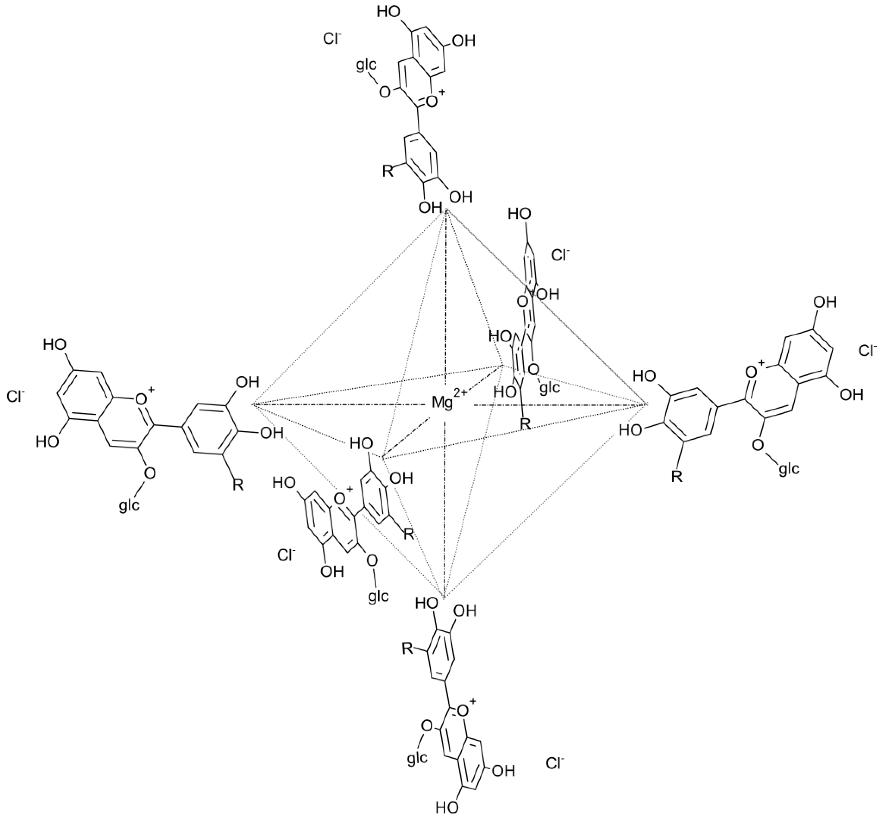
Figure 3.
Linear degradation profile of the Mg-anthocyanin complex based on absorbance (λ-max) versus storage time. The regression equation (y = 1.999 – 0.0025t; R2 = 0.998) was used to estimate the degradation rate and predict a shelf life of approximately 36 months, defined by a 0.1 absorbance unit variation threshold.
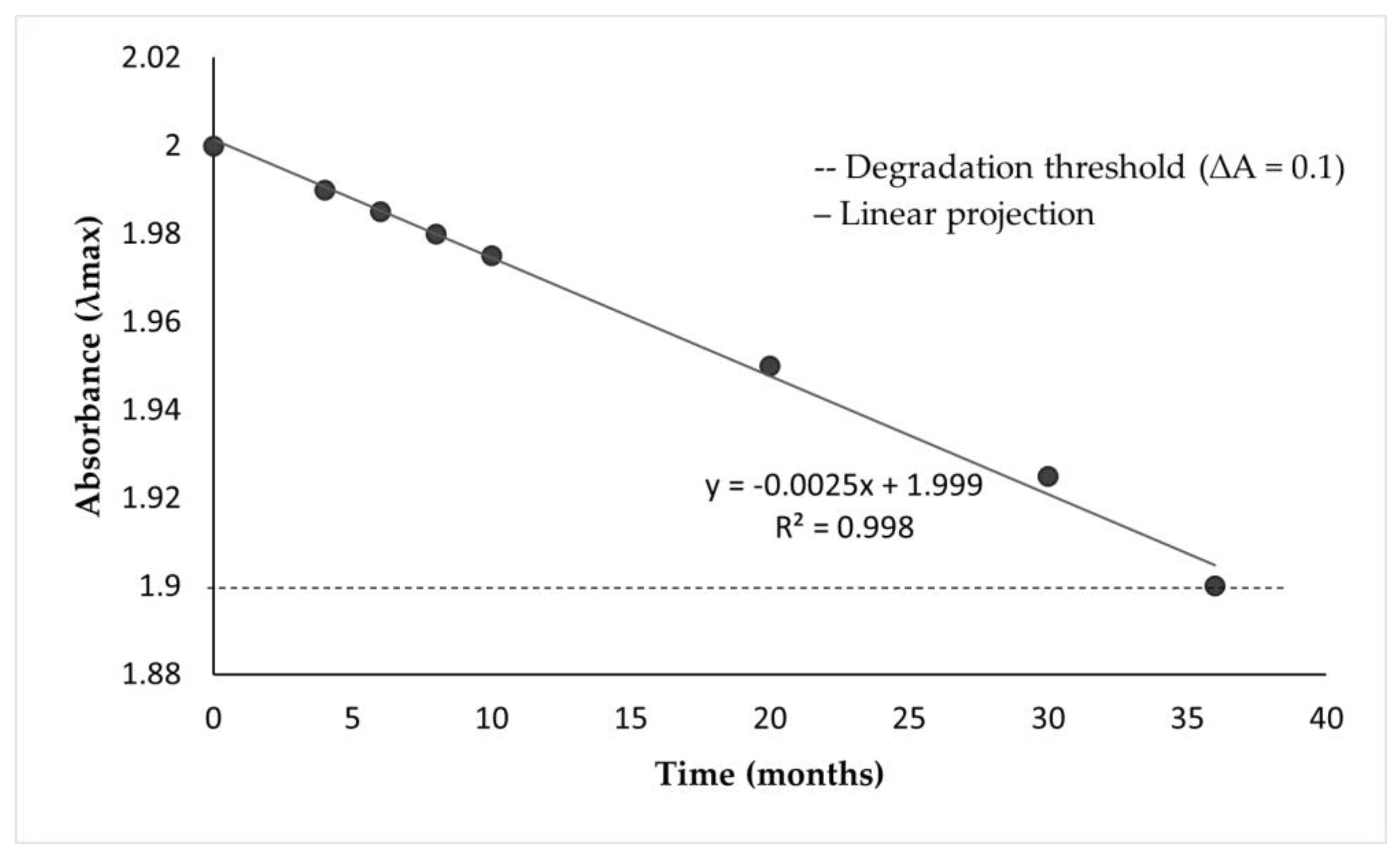

Table 1.
Experimental Rf values and tentative identification of anthocyanins and anthocyanidins from Dioscorea trifida L.
Table 1.
Experimental Rf values and tentative identification of anthocyanins and anthocyanidins from Dioscorea trifida L.
| Anthocyanins | Values Rf x100 | |||
|---|---|---|---|---|
| From D. trifida L. | Bibliographic source | |||
| BAW a | HCl 1% | BAW | HCl 1% | |
| Peonidin-3,5-diglucoside | 23 | 17 | 23 | 17 |
| Carreño and Grau (1977) [20] | Harborne (1984) [21] | |||
| Malvidin-3,5-diglucoside | 31 | 22 | 31 | 22 |
| Carreño and Grau (1977) [20] | Bate-Smith (1948) [22] | |||
| Cyanidin-3-rhamnosyl-glucoside | 32 | 19 | 32 | 19 |
| Daravingas and Cain (1966) [23] | Daravingas and Cain (1966) [23], Harborne (1984) [21] | |||
| Pelargonidin-3,5-diglucoside | 34 | 23 | 34 | 23 |
| Asen (1958) [24] | Harborne (1984) [21] | |||
| Anthocyanidins | Forestal b | Formic c | Forestal | Formic |
| Cyanidin | 49 | 22 | 49 | 22 |
| Harborne (1984) [21], Markakis and Jurd (1974) [25], Smith and Luh (1965) [26] and Dekazos (1970) [27] | Harborne (1984) [21], Markakis and Jurd (1974) [25], Smith and Luh (1965) [26] | |||
| Malvidin | 60 | 27 | 60 | 27 |
| Harborne (1984) [21], Markakis and Jurd (1974) [25], Smith and Luh (1965) [26] | ||||
| Peonidin | 63 | 30 | 63 | 30 |
| Harborne (1984) [21], Markakis and Jurd (1974) [25], Smith and Luh (1965) [26] | Harborne (1984) [21], Markakis and Jurd (1974) [25], Smith and Luh (1965) [26] and Dekazos (1970) [27] | |||
| Pelargonidin | 68 | 33 | 68 | 33 |
| Harborne (1984) [21], Markakis and Jurd (1974) [25] and Lawanson and Osude (1972) [28] | Harborne (1984) [21], Markakis and Jurd (1974) [25] | |||
a BAW: n-butanol-acetic acid-water, top layer (4:1:5). b Forestal: HCl concentrated-acetic acid-water (3:3:10). c Formic: HCl concentrated-formic acid-water (2:5:3). Note: identifications are provisional and based on comparison of experimental Rf values and color with the cited literature; no spectroscopic (MS, NRM) confirmation was performed.
Table 2.
Variation in measured and projected absorbance values of the Mg(II)-anthocyanin complex over time.
Table 2.
Variation in measured and projected absorbance values of the Mg(II)-anthocyanin complex over time.
| Time (months) | Measured absorbance | Projected absorbance |
|---|---|---|
| 0 | 2.000 | 2.000 |
| 4 | 1.990 | 1.990 |
| 6 | 1.980 | 1.985 |
| 8 | 1.978 | 1.980 |
| 10 | 1.976 | 1.975 |
| 20 | - | 1.950 |
| 30 | - | 1.925 |
| 36 | - | 1.900 |
Disclaimer/Publisher’s Note: The statements, opinions and data contained in all publications are solely those of the individual author(s) and contributor(s) and not of MDPI and/or the editor(s). MDPI and/or the editor(s) disclaim responsibility for any injury to people or property resulting from any ideas, methods, instructions or products referred to in the content. |
© 2025 by the authors. Licensee MDPI, Basel, Switzerland. This article is an open access article distributed under the terms and conditions of the Creative Commons Attribution (CC BY) license (http://creativecommons.org/licenses/by/4.0/).
Copyright: This open access article is published under a Creative Commons CC BY 4.0 license, which permit the free download, distribution, and reuse, provided that the author and preprint are cited in any reuse.


