
Submitted:
20 June 2024
Posted:
23 June 2024
You are already at the latest version
Abstract
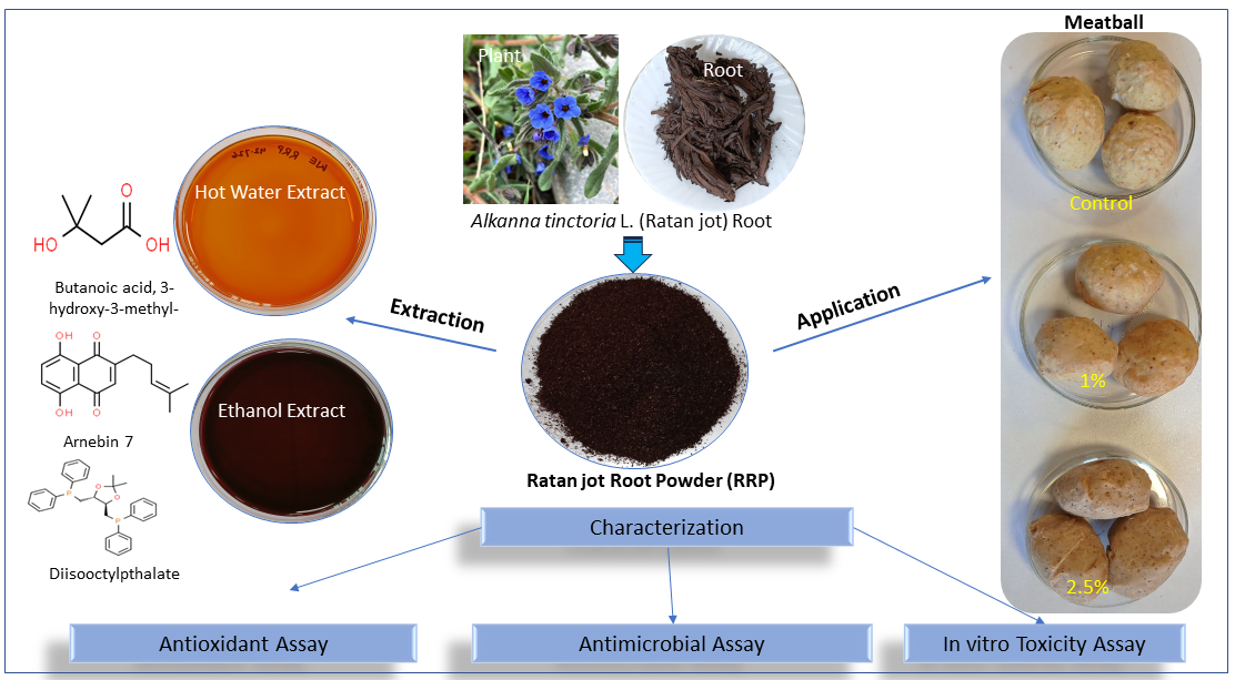
Natural and sustainable plant-based antioxidants and antimicrobials are highly desirable for improving food quality and safety. The present investigation assessed the antimicrobial and antioxidant properties of active components from Alkanna tinctoria (herb) roots, also known as Ratanjot root. Two methods were used to extract active components: microwave-assisted hot water (MAHW) and ethanol and air-dried. MAHW extract yielded 6.29% while the ethanol yielded 18.27%, suggesting superior Ratanjot root extract powder (RRP) solubility in ethanol over water. The ethanol extract showed significantly higher antioxidant activity than the MAHW extract. Gas chromatography and mass-spec analysis revealed three major phenolic compounds: Butanoic acid, 3-hydroxy-3-methyl-; Arnebin 7, and Diisooctylpthalate. The color attributes (L*, a*, b*, hab, C*ab) for the ethanolic and MAHW extracts revealed significant differences (P < 0.05) in all the above parameters for both types of extracts, except for yellowness (b*) and chroma (C*ab) values. The ethanol extract exhibited antimicrobial activity against 14 bacterial species, with a signifi-cantly higher inhibitory effect against Gram-positive bacteria (Listeria monocytogenes and Staphylococcus aureus) than the Gram-negative bacteria (Salmonella enterica serovar Typhimurium and Escherichia coli). The minimum inhibitory concentration (MIC) and minimum bactericidal concentration (MBC) were both 25 mg/ml for the Gram-negative bacteria, while the MIC and MBC concentrations varied for Gram-positive bacteria (0.049 - 0.098 mg/ml and 0.098 - 0.195 mg/ml) and the antimicrobial effect was bactericidal. The antimicrobial activities of RRP extract remained stable under broad temperature (37-100°C) and pH (2-6) conditions, as well as during refrigerated storage for 30 days. Application of RRP at 1% (10 mg/g) and 2.5% (25 mg/g) levels in a cooked chicken meatball model system prevented lipid oxidation and improved sensory attributes and retarded microbial growth during refrigerated (4°C) storage for 20 days. Furthermore, the RRP extract was non-toxic when tested with sheep erythrocytes and did not inhibit the growth of probiotics, Lacticaseibacillus casei, and Lactiplantibacillus plantarum. In conclusion, the study suggests that RRP possesses excellent antimicrobial and antioxidant activities, thus making it suitable for food preservation.
Keywords:
Alkanna tinctoria
; Ratanjot
; antimicrobial
; antioxidant
; toxicity
; probiotics
; sensory attributes
; food preservation
; food safety
; foodborne pathogens
1. Introduction
Synthetic antioxidants and antimicrobials have historically been employed to mitigate lipid oxidation, food spoilage, and foodborne infections. However, the ever-increasing consumer concerns regarding food safety coupled with the potential toxicity of these synthetic or chemical compounds like butylated hydroxyanisole (BHA), butylated hydroxytoluene (BHT), propyl gallate (PG), and nitrites have spurred a quest within the food industry for natural, sustainable alternatives [1,2,3]. Similarly, the emergence of antimicrobial resistance (AMR) in medicine has increased attention to discovering new plant, animal, or microbial antimicrobial compounds [4,5]. Natural products, especially plant components, are composed of a vast array of bioactive compounds like polyphenols, flavonoids, alkaloids, tannins, ketones, aldehydes, alcohols, and esters, with antioxidant and antimicrobial compounds [6,7], hence utilized in various industries [8,9]. In the food industry, plant extracts and essential oils are gaining popularity, as they are safe as perceived by consumers and received the GRAS (Generally Recognized As Safe) status by the United States Food and Drug Administration (FDA) due to their long history of use [10,11,12]. Further, around 75% of the world’s rural populace relies on plant materials mentioned in traditional systems of medicine [13].
Alkanna tinctoria, commonly known as dyer’s alkanet, alkanet, dyer’s bugloss, orchanet, or Ratanjot (in India), of the Boraginaceae family, is a perennial herb native to Europe and the Mediterranean region like Asia, including the Middle East[14,15,16]. The plant is characterized by its striking blue flowers and dark red roots [17,18] (Figure 1). The plant is most renowned for the vibrant red dye extracted from its roots, which has been treasured for centuries across various cultures and industries [14,19]. The natural red pigments in Ratanjot roots have a broader application in medicine, food, cosmetic, and textile industries [20,21].
In Ayurveda, the traditional Indian medicine, Ratanjot root is used to treat various ailments, including skin disease, digestive disorders, urinary calculi, menstruation abnormalities, and respiratory illness [14,22]. The Ratanjot root contains bioactive components known for their anti-inflammatory, antimicrobial, and wound-healing properties [23,24,25]. Moreover, Ratanjot root or alkanet extracts have shown anticancer activities [25,26] and are also used in histological staining of tissue samples [16,19].
In the food industry, Ratanjot (A. tinctoria) root is mainly used as a natural food coloring and flavoring agent to provide clean-label food products [15]. In Indian culinary practices, Ratanjot roots are used to give the captivating reddish hue and subtle earthy flavor to signature Kashmiri meat dishes like Rogan josh [22,27]. It is also used in sweets, beverages, and savory dishes to improve sensory attributes such as color and flavor [28].
However, very few studies have been conducted to understand the antioxidant, antimicrobial, and cytotoxicity effects of A. tinctoria leaves and root extracts [29,30]. The scarcity of research proving the antimicrobial and antioxidant properties of A. tinctoria roots might have arisen from limited research focus, overshadowed by its primary use as a dye in the textile industry. Moreover, there are no available scientific reports on the application of Alkanna tinctoria roots in the meat model system. The present study aimed to unveil and validate the in vitro antimicrobial and antioxidant activities of A. tinctoria or Ratanjot roots in a meat-model system for potential food industry application.
2. Materials and Methods
2.1. Chemicals and Media
Chemicals and analytical and molecular biology grade reagents were purchased from Sigma-Aldrich (USA) and Sisco Research Laboratories (SRL) (Mumbai, India). Media for bacterial culture were purchased from Hi-Media and SRL (Mumbai, India).
2.2. Bacterial Cultures and Growth Conditions
Staphylococcus aureus ATCC 25923, Listeria monocytogenes ATCC 19111, Listeria monocytogenes ATCC 13932, Salmonella enterica serovar Typhimurium ATCC 14028, Escherichia coli ATCC 25922, Lactiplantibacillus plantarum MTCC 2621 and Lacticaseibacillus casei MTCC 1423 and nine field isolates of Staphylococcus aureus isolated from poultry, goat and swine meat (Table 1) were obtained from the Department of Veterinary Public Health and Epidemiology, Banaras Hindu University, India. The foodborne pathogenic bacterial cultures and the lactobacilli were grown in brain heart infusion (BHI) broth and deMann-Rogosa-Sharpe (MRS) broth, respectively (Hi-Media) at 37ºC for 18-24 h and 48 h under aerobic conditions before being used in the study.
2.3. Preparation of Ethanol and Microwave-Assisted Hot Water Extracts of Ratanjot (Alkanna tinctoria) Root and Characterization
The dried Ratanjot roots (Figure 1) collected from the local market were identified by a botanist. The Ratanjot roots were further dried in a food drier (Ambay Biotech, India) at 40ºC overnight, chopped into small pieces, pulverized (500W Mixer Grinder, Philips), and sieved (using 60 mesh) to obtain fine powders, designated Ratanjot root powder (RRP). The RRP was stored in a sterile amber-colored glass bottle at room temperature (25 ± 1ºC) until use. The microwave-assisted hot water (MAHW) and ethanol extracts (EE) of RPP were prepared as described before[31,32] with slight modifications.
For the ethanol extraction method, 20 g RRP was extracted in 100 ml of 70% ethanol (v/v) by continuous shaking in a shaker incubator (Remi, India) for 24 h at 37°C. In the MAHW extraction method, 20 g of RRP was extracted with 100 ml of sterile distilled water in a mono-mode microwave oven (NEOS-GR, Milestone Sarl, Italy) equipped with a closed vessel system. The power setting was adjusted to 300 W and the temperature rose to 95°C within 10 min, extracting the water-soluble components. The samples were then cooled to room temperature. The entire content was centrifuged at 5000×g for 10 min (Remi, India), followed by filtration through Whatman filter paper (No. 1). In both methods, the extraction processes were repeated with the residue obtained, and the filtrates from both batches were concentrated using a rotary evaporator (HS-2005 VN, H.S. Scientific, Korea) at 40 ± 1°C overnight under reduced pressure.
The concentrated extracts were air-dried at room temperature and weighed to calculate the total extractable components (TEC). The dried crude extracts were diluted with sterile distilled water to obtain a concentration of 200 mg/ml and used as the stock preparation for antimicrobial and antioxidant activity assays. The TEC was calculated using the formula: TEC% = (Weight of extracted component/ Weight of RRP) × 100.
The pH values of the RRP extracts were recorded using a digital pH meter (pH Tutor, Eutech Instruments). The color of the RRP extracts was measured using a ColorQuest XE colorimeter (HunterLab, Reston, VA, USA). In brief, about 20 ml of each extract was placed in a 20 mm cell. The CIELAB parameters such as lightness (L*), redness (a*), yellowness (b*), hue angle (hab) and chroma (C*ab) were recorded in total transmittance mode, standard illuminant D65 and observer angle 10 degrees as per the manufacturer’s instruction manual (HunterLab, Reston, VA, USA).
2.4. Antioxidant Activity Analysis
2.4.1. Total Phenolic Content
The total phenolic content (TPC) of the RRP extract was estimated by the classical Folin-Ciocalteu (FC) method as before[33] with minor modifications. Briefly, 0.75 ml of 0.2N FC reagent was added to 100 µl of RRP extract at five different concentrations and the volume was made ten times with deionized distilled water. After mixing, the reaction was carried out for 5 min at room temperature. Thereafter, 0.4 ml of 7.5% w/v Na2CO3 solution was added, mixed gently, and incubated at room temperature in the dark for 30 min to observe visible deep blue color development. Absorbance was measured at 760 nm against a solvent blank using a UV-visible spectrophotometer (Spectra Max® M5, USA), and gallic acid at different concentrations was used as the standard. The TPC of the RRP extract was calculated from the standard curve plotted using different concentrations of gallic acid and expressed as gallic acid equivalent (GAE) in mg/g of RRP on a dry weight basis.
2.4.2. 2,2-Diphenyl-1-picrylhydrazyl (DPPH) Radical Scavenging Assay
The DPPH radical scavenging activity (RSA%) of RRP extract was assayed as before [34] with slight modification. A solution of 0.1 mM (w/v) DPPH was prepared by mixing 3.94 mg of DPPH in 100 ml methanol. About 1 ml of 0.1 mM DPPH methanol solution was added to equal volume (1 ml) of the RRP extract at different concentrations, mixed well, and incubated in the dark at room temperature for 30 min. The color change (from deep violet to light yellow) was measured spectrophotometrically at 517 nm against a solvent blank (DPPH methanolic solution without sample). BHT and BHA were used as positive controls or standards. The DPPH RSA (%) was calculated using the following formula:
DPPH RSA (%) = ((AB − AS)/AB) × 100, where AB = Absorbance of blank and AS= Absorbance of the reaction mixture with the sample. The half maximal inhibitory concentration (IC50) value was calculated by plotting a graph using the RSA (%) against different sample concentrations.
2.4.3. 2.2. Azino-bis (3-ethylbenzothiazoline-6-sulfonic acid) (ABTS.+) Radical Cation Decolorization Assay
The ABTS.+ radical scavenging activity was determined as before [35,40] with slight modifications. The ABTS.+ radical stock solution was prepared using an equal volume (1:1 v/v) of 7 mM ABTS.+ in water and 2.45 mM potassium persulfate, stored in the dark at room temperature for 16 h. The ABTS.+ radical stock solution was then diluted adequately with absolute ethanol to obtain an absorbance of 0.700 ± 0.05 at 734 nm. A 0.1 ml of the RRP extract at different concentrations was added to 3.9 ml of the diluted ABTS.+ radical solution, and the color change (from turquoise blue to colorless) was measured after 6 min at 734 nm using a UV-visible spectrophotometer (Spectra Max® M5, USA). The blank was prepared with the diluted ABTS.+ radical solution, whereas BHT and BHA were used as standards. The ABTS.+ RSA(%) was calculated using the following formula:
ABTS.+ RSA(%) = ((AB − AS)/AB) × 100, where AB = Absorbance of blank and AS= Absorbance of the reaction mixture with the sample. The IC50 value was calculated by plotting a graph using the ABTS.+RSA (%) against different concentrations of the sample.
2.4.4. Identification of Phytochemicals Using Gas Chromatography-Mass Spectrometry (GC-MS/MS)
For identification of phytochemicals, especially the phenolic compounds present in RRP extract, GC-MS/MS analysis was done using a gas chromatograph mass spectrometer (Shimadzu GCMSQP2010, Japan) interfaced with a turbo quadrupole mass spectrometer, fitted with an Rtx-5 fused silica capillary column. Methanol was used as the solvent to make dilutions, the total run time was ~ 70 min, and GC-MS solution software was used for the data analysis [36]. The phytochemical compounds in the RRP extract were identified by matching their mass spectra with standards from the National Institute of Standards and Technologies (NIST, USA). The RRP extract samples were analyzed in triplicate by GC-MS/MS.
2.5. Antimicrobial Activity Assays
2.5.1. Agar Well Diffusion Assay
The hot water and ethanol extracts of RRP were filter-sterilized by passing through syringe filters (0.45 µm) and were evaluated for antimicrobial activity against 14 foodborne bacteria (Table 1). The antimicrobial sensitivity testing of the extracts was done on Mueller-Hinton Agar (MHA) plates by agar well diffusion assay as described before[37] with slight modification. In brief, wells measuring 6 mm in diameter were dug on the MHA plates using a sterile cork borer. The MHA plates were swabbed with the standard inoculum (turbidity adjusted to 0.5 McFarland standard) of the test isolates. The RRP extract (100 µL) was transferred onto the corresponding wells. Autoclaved distilled water was used as a negative control. The plates were incubated at 37℃ for 24 h, and the zone of inhibition (ZOI) was reported in millimeters (mm).
2.5.2. Determination of Minimum Inhibitory Concentration (MIC) and Minimum Bactericidal Concentration (MBC)
The ethanol RRP extract exhibited strong antimicrobial activity; therefore, the MIC and MBC were determined against two Gram-positive (S. aureus ATCC 25923, L. monocytogenes ATCC 19111) and two Gram-negative bacteria (S. Typhimurium ATCC 14028 and E. coli ATCC 25922). In brief, 100 µL of sterile cation-adjusted Mueller Hinton broth (CAMHB) was added to each of the wells of a 96-well microtiter plate. To the first well of each row, 100 µL of ethanol extract (at a concentration of 200 mg/mL) was added and diluted serially in a two-fold dilution at concentrations ranging from 100–0.00038 mg/mL. The 0.5 McFarland standard suspension of a freshly grown log-phase bacterial culture of the test isolates was diluted 100X to a density of 106 cfu/mL (9.9 mL CAMHB + 0.1 mL 0.5 McFarland standard suspension). Then, 10 µL of the diluted inoculum was transferred to each well within 30 min of preparation. Two wells of the plate were used as broth control (without culture) and culture control (without antibiotic), respectively. The plates were covered with aluminum foil and incubated at 37℃ for 18-24 h. To each well, resazurin (0.015% w/v) was added (30 µL/ well), and further incubated for 2-4 h to observe color change. The lowest concentration of RRP extract that produced no color change (the blue color of resazurin remaining unchanged) was considered as the MIC value.
The MBC of the RRP extract was determined at the end of the incubation period by taking two 10 µL samples from each well showing no visible growth and plating the samples onto MHA plates. The resultant colonies were counted after overnight incubation at 37℃ for 18-24 h. The lowest dilution of the extract that produced at least 99.9% killing of the initial inoculum was considered the MBC value. The isolates treated with ciprofloxacin (512-0.25 mg/mL) were positive control.
The bactericidal or bacteriostatic activity was ascertained by calculating the MBC/MIC ratio or MIC index or tolerance level. Generally, a low MBC/MIC ratio (≤ 4) indicates that the antimicrobial action is bactericidal, i.e., it can kill the bacteria at concentrations closer to or equal to MIC. A high MBC/MIC ratio (≥ 4) indicates bacteriostatic, i.e., the antimicrobials can only inhibit the growth of bacteria but cannot kill them at concentrations closer to or equal to MIC. When the MBC/MIC ratio is very high (≥ 16), it indicates that the antimicrobial is tolerant, i.e., it has no bactericidal activity even at high concentrations [38,39].
2.5.3. Dose and Time-Dependent In-Vitro Growth Kinetics of S. aureus and L. monocytogenes Treated with RRP Extract
The growth kinetics of the bacterial cultures were evaluated against RRP extract (at 1X MIC and 1X MBC) by incubating the log-phase grown bacterial cultures (inoculum adjusted to 1×10⁷ cfu/mL) in CAMHB. In brief, the log phase culture of each isolate was prepared by inoculating a single colony in BHI broth and incubating at 37℃ for 4 h in a shaker incubator (REMI, CIS-24 Plus, Mumbai, India). After incubation, the culture was pelleted by centrifugation (2370xg for 5 min) and resuspended in sterile PBS to attain a turbidity equivalent to 0.5 McFarland standard, which was further diluted to a concentration equivalent to 1×10⁷ CFU/mL using sterile CAMHB. From this dilution, 50 µL of inoculum was transferred to respective tubes containing 1X MIC and 1X MBC of RRP extract and incubated at 37℃. The isolates treated with Ciprofloxacin (1X MIC and 1X MBC) served as treatment control, while the target bacterial isolate in CAMHB served as untreated control. The aliquots of 10 µL collected at each fixed time interval (0, 3 h, 6 h, 9 h, 12 h, 24 h) were serially diluted in sterile PBS, plated on MHA plates, incubated at 37℃ for 24 h, enumerated and reported log10 cfu/mL. To ascertain the dose and time-dependent growth kinetics, a line chart was prepared using the mean (n=6) bacterial colony counts (log10 cfu/ml) and time (hours) for each treatment.
2.5.4. Stability Assessment
The RRP extracts were exposed for 30 min to varying temperatures (37°C, 70°C and 100°C) and pH (2, 4, 6 and 8) conditions, and the refrigerated temperature (4 ± 1°C) for 0, 15, and 30 days). Then, the antimicrobial activity was evaluated against S. aureus ATCC 25923 and L. monocytogenes ATCC 19111 using an agar well diffusion assay as above.
2.6. Effect of RRP on Lactobacilli Growth
The effect of RRP extract was tested on beneficial probiotic lactobacilli (Lactiplantibacillus plantarum MTCC 2621 and Lacticaseibacillus casei MTCC 1423), which are generally present as commensals in the gut by plate count method [40]. Briefly, the lactobacilli probiotic bacteria (100 µl each, 107 CFU/ml) were inoculated in 100 µl of MRS broth medium containing RRP extract (1X MIC and 2X MIC for S. aureus) in a 96-well microtiter plate. The untreated bacterial cultures and MRS broth were used as positive and negative controls. After incubation at 37°C for 48 h, the cultures were diluted with MRS broth, and 10 µl from each was plated on MRS agar plates, and colonies were counted.
2.7. Effect of RRP on Red Blood Cells
An in vitro hemolysis assay using sheep red blood cells (RBCs) was conducted to ascertain cytotoxicity, if any, against mammalian cells as described before [41] with slight modification. Briefly, the aseptically collected sheep blood was washed several times with PBS until a clear supernatant was obtained, centrifuged at 1000x g for 15 min, and resuspended in 10% (v/v) PBS containing 10 mM Dithiothreitol (DTT). Different concentrations (1X, 2X, 3X, 5X and 10 X MIC for S. aureus) of 100 µl of RRP extract were added to individual tubes containing 100 µl of the sheep RBCs. Sheep RBCs (100 µl) with 100 µl each of 0.2% Triton X-100 and sterile PBS were used as positive control and negative control or blank, respectively. The reaction mixtures were incubated at 37°C for 1 h and then centrifuged at 1300x g for 10 min. The supernatants were transferred to a 96-well microtiter plate, and the hemoglobin released was measured spectrophotometrically (Spectra Max® M5, USA) at 540 nm. The hemolysis % was then calculated by using the following formula:
Hemolysis (%) = (Absorbance of Sample − Absorbance of PBS)/ (Absorbance of Triton X − Absorbance of PBS) × 100
2.8. Application of RRP in Meat-Model System
The application of RRP extract on food safety and quality was assessed using emulsion-based chicken meatballs containing RRP at 1% (T1) (10 mg/g, i.e., approximate 50X MBC for L. monocytogenes and 100X MBC for S. aureus) and 2.5% (T2) (25 mg/g, i.e., 1X MIC or MBC for S. Typhimurium and E. coli. The meatballs were prepared and cooked as described before [42] and stored at 4 ± 1°C for 20 days under aerobic packaging conditions. Every five days, samples were analyzed for sensory quality changes using an 8-point hedonic scale by trained panelists [43], oxidative changes by analyzing 2-thiobarbituric acid reactive substances (TBARS) [44], and the microbiological quality, including total viable counts (TVC), psychrophilic counts, Escherichia coli counts, and Staphylococcus aureus counts as outlined by American Public Health Association [45].
2.9. Statistical Analysis
The samples were analyzed thrice in duplicates (n=6), and the statistical analysis (ANOVA) was done using SPSS software (Version 20, IBM, USA). The data were presented as mean ± standard error (SE), and the statistical significance was determined at a 95% confidence interval level (P < 0.05). Graphs were generated using GraphPad Prism 9.3 (Boston, MA, USA).
3. Results and Discussion
3.1. Characteristics of Ethanol and Water Extracts of Ratanjot (A. tinctoria) Root
The total extractable components (TEC) of soluble fractions by ethanol was 18.27%, while the microwave-assisted hot water (MAHW) method yielded 6.29% (Table 2). This suggests that RRP was more soluble in alcohol and partially soluble in water. Using a water-infusion method, Guemmaz et al. [30] reported a similar phenolic compound yield 7.25% from A. tinctoria aerial parts. Alcohol-based phenolic compound extraction has been well documented in the literature. Using methanol, Gao et al. [46] obtained about 16.5% and 15.9% from the inner bark and outer bark of Port Oxford Cedar, respectively. Likewise, Ngo et al.[47] reported a 15.6% yield using absolute methanol from Salacia chinensis L root.
The mean pH values of ethanol and MAHW extracts were 2.92 and 3.15, respectively (Table 2). Zannou and Koca [23], reported similar pH values of A. tinctoria root extracts using natural deep eutectic solvents like sodium acetate:lactic acid (pH=3.41) and sodium acetate:formic acid (pH=3). The acidic nature of the A. tinctoria root extract might also contribute to their biological activities and be suitable for use in low-acid foods.
The mean values for the standard CIELAB color parameters for ethanol and MAHW extracts are summarized in Table 2, and the respective values are; L*(25.07 and 56.55), a*(45.13 and 12.02), b* (63.35 and 62.20), hab (2.61 and 41.70) and C*ab (77.97 and 63.72). In general, the values of L* range from 0-100 for black and white respectively: a*values correspond to negative values for greenness and positive values for redness, b* values correspond to negative values for bluish color and positive values for yellowness, hab correlate to tonality and C*ab correlate to color vividity. The ethanol extract had significantly (P < 0.05) lower L* and hab and higher a*values than the MAHW extract, indicating the ethanol extract has a dark red color compared to the light red and brighter color tone for water extract, thus making RRP extracts highly valued colorant for use in food, cosmetic, textile and pharmaceutical industries.
3.2. Antioxidant Activity of RRP
3.2.1. Total Phenolic Content
Plant phenolic compounds are known as free radical scavengers, and a plant extract’s total phenolic content (TPC) can be correlated to its antioxidant activity [48]. The mean TPC (mg GAE/g) of ethanol extract of RRP was 45.10 and 4.57 for MAHW extract (Figure 2). The difference in TPC of both the extracts might be because most of the phytochemicals, including phenolic compounds of RRP, were more soluble in alcohol and partially soluble in hot water.
These findings were partially corroborated by that of Zannou and Koca [23], who found that the total phenolic contents of 70% ethanol and 80% methanol extract of A. tinctoria root were 49.70 mg GAE/g and 33.10 mg GAE/g, respectively. However, in contrast to our results, these authors reported a higher total phenolic content, i.e., 86.80 mg GAE/ g of water extract of A. tinctoria root. Nevertheless, much higher values for mean total phenolic contents were obtained for A. tinctoria root extracts using natural deep eutectic solvents like sodium acetate: lactic acid (170.96 mg GAE/g) and sodium acetate: formic acid (175.97 mg GAE/g). Our findings partially agree with the reports of Kozłowska et al. [49], who found that the total phenolic content of the Angelica archangelica L. root extract was 20.35 mg GAE/g. Similarly, Gao et al.[46] also found that the mean total phenolic contents of bark and wood powders of Port Oxford Cedar ranged from 2.56-88.76 mg GAE/g. Another research similarly documented that the total phenolic contents of water extracts prepared from A. tinctoria aerial parts by decoction, maceration, and infusion were 173.28, 144.50 and 189.66 mg GAE/g, respectively. Another study also reported that the total phenolic content of the red radish (Raphanus sativus L.) root was 341.45 mg GAE/100g [50].
The differences in total phenolic contents observed by different researchers can be attributed to factors such as plant variety, plant parts, climate, geographical location, growth conditions, extraction method, and kind of solvents used [51].
3.3.2. 2,2-Diphenyl-1-Picrylhydrazyl (DPPH) Radical Scavenging Assay
The DPPH assay measures the electron-donating activity of a substance to scavenge or reduce DPPH free radicals and measures its antioxidant activity [52]. The data presented in Figure 2 shows a significant difference (P < 0.05) in mean IC50 values of ethanol extract (76.86 µg/ml) and MAHW extract (170.60 µg/ml) of RRP in scavenging DPPH free radicals as compared to the standard antioxidants like BHT (13.12 µg/ml) and BHA (6.99 µg/ml). However, BHA and BHT showed much lower IC50 concentrations than both extracts, which indicates their potency as standard antioxidants. Moreover, among both types of RRP extracts, 50% scavenging of the DPPH free radicals was more efficiently done by the ethanol extract than the MAHW extract. This might be because more phenolic compounds with DPPH radical scavenging activity were soluble in ethanol than water.
Our results are also in agreement with the findings of Zannou and Koca [23], who estimated the mean IC50 values for DPPH free radical scavenging activity of A. tinctoria root using 70% ethanol extract (75.24 mmol Trolox Equivalent or TE/g), 80% methanolic extract (74.07 mmol TE/g), water extract (97.39 mmol TE/g), extracts using natural deep eutectic solvents like sodium acetate: lactic acid (112.31 mmol TE/g) and sodium acetate: formic acid (130.91 mmol TE/g). Likewise, Guemmaz et al.[30] also reported that the mean IC50 values in the DPPH radical scavenging assay for water extracts prepared from A. tinctoria aerial parts by decoction, maceration, and infusion were 0.17 mg/ml, 1.006 mg/ml and 0.09 mg/ml, respectively. In another experiment, Gao et al.[46] also documented that the mean IC50 values of heartwood, sapwood, inner bark, and outer bark extracts of Port Oxford Cedar were 64.77 mg/ml, 29.03 mg/ml, 10.31mg/ml, 19.87 mg/ml, respectively. Contrastingly, Ngo et al. [47] reported a very high IC50 value (407.47 mmol TE/g) for acetone (50%) extracts of Salacia chinensis L. root in DPPH radical scavenging. The differences in results of the DPPH assay might be due to various factors, as mentioned earlier, regarding total phenolic contents.
3.2.3. 2.2-Azino-Bis (3-Ethylbenzothiazoline-6-Sulfonic Acid) (ABTS.+) Radical Cationdecolorization Assay
The ABTS assay is a simple, convenient, and efficient measure of total antioxidant capacity, which determines the ability of a test substance to neutralize the bluish-green ABTS free radical. The significant (P < 0.05) differences in IC50 concentrations for ethanol extract (37.95 µg/ml), MAHW extract of RRP (57.09 µg/ml), BHT (6.96 µg/ml), and BHA (5.06 µg/ml) in ABTS assay were evident (Figure 2). Significantly (P < 0.05), higher ABTS radical scavenging activity and lower IC50 concentrations were obtained for ethanol extract than the water extract of RRP, possibly due to better solubility and extractability of phenolic components in alcohol. Although more potent ABTS radical scavenging activities were observed for the standard chemical antioxidants like BHT and BHA, the observed IC50 concentrations of RRP extract in ABTS assay are highly desirable because of their natural origin and health benefits.
Like our findings, Sundaram et al. [53] reported that the IC50 for ABTS radical scavenging of Strobilanthes heyneanus root extracts from Southwest India was 33.92 µg/ml. These findings bear similarity with the reports of Kozłowska et al. [49], who reported that the IC50 value in ABTS assay for Angelica archangelica L. root extract was 86.54 µg/ml. In contrast to our results, a very high IC50 value (414.38 mmol TE/g) or lower ABTS radical scavenging activity for acetone (50%) extract of S. chinensis L. root was observed by Ngo et al.[47]. A recent study by Ganos et al. [54] found that the IC50 concentration of methanol extract of A. tinctoria aerial part was 366.88 mg TE/g. Similarly, the factors mentioned earlier might also contribute to the differences in results obtained for ABTS radical scavenging activity.
3.2.4. Identification of Phytochemicals Using Gas Chromatography-Mass Spectrometry (GC-MS/MS)
Understanding the phytochemical compounds in Ratanjot (A. tinctoria L.) root extract is essential for assessing its bioactivity and optimizing its application in the food and pharmaceutical industry[51]. The GC-MS/MS chromatography revealed the presence of three major phenolic compounds: (i) butanoic acid, 3-hydroxy-3-methyl-; (ii) Arnebin 7 and (iii) di-isooctylpthalate (Figure 3). Butanoic acid, 3-hydroxy-3-methyl- is also known as butyric acid, 3-hydroxy-3-methyl- and is soluble in alcohol. Butyric acid derivatives have been identified in marine red algae Rhodomela confervoides [55,56,57,58] and Mexican corn products [55] that contribute to potent DPPH and ABTS radical scavenging activities. Butyric acid contributes to overall health through anti-inflammatory and digestive functions [57]. Butyric acid also contributes flavor and aroma in dark chocolate [58] and coffee silver skin [57].
Arnebin 7, also known as deoxyshikonin, is insoluble in water but soluble in ethanol [59]. It is an enantiomer of alkannin and is grouped under hydroxynaphthoquinones [60], which are DNA topoisomerase inhibitors [25]. Alkannin and Shikonin are known for their antimicrobial [61] and healing properties. They are active ingredients in therapeutic wound healing ointments, like Hexiderm, Histoplastin red, etc. [25].
Diisooctylpthalate (DIOP), technically known as Pthalic acid, bis-isooctyl ester is insoluble in water but soluble in organic solvents. DIOP from marine brown algae Sargassum wightii was found to be an antimicrobial compound [62]. Similarly, DIOP was also identified as a phytochemical in Vernonia glabra root ethyl acetate extracts, which showed antitubercular activity against Mycobacterium tuberculosis [63].
3.3. Antimicrobial Activity of RRP
3.3.1. Antimicrobial Activity Spectrum of RRP
Antimicrobial activities of ethanol and MAHW extracts against several Gram-positive and Gram-negative bacterial pathogens were tested by agar well diffusion assay (Table 3). The ethanol extract was highly inhibitory against both Gram-positive S. aureus ATCC 25923, L. monocytogenes ATCC 19111, L. monocytogenes ATCC 13932, and some field isolates of S. aureus and Gram-negative bacteria S. Typhimurium ATCC 14028 and E. coli ATCC 25922. In contrast, the water extract had no antimicrobial activities. This might be because the phytochemicals responsible for the antimicrobial activity of RRP were soluble in ethanol, not in water. Other researchers also obtained similar results and observed that ethanol extract of A. tinctoria roots had a more potent antimicrobial effect against Gram-positive bacteria than Gram-negative bacteria and the water extracts had no or negligible effect. Interestingly, the Gram-positive bacteria were more sensitive than the Gram-negative bacteria, which agrees with other reports [25,29,61].
3.3.2. Determination of MIC and MBC
Next, we determined the MIC and MBC values of the ethanol extract. [Note that water extract was not used in subsequent experiments since it had no detectable antimicrobial activity.] The MIC of ethanol extract for S. aureus ATCC 25923 and L. monocytogenes ATCC 19111 was 0.049 and 0.098 mg/mL, respectively, while 25 mg/ml for Salmonella and E. coli. The MBC for S. aureus ATCC 25923 was 0.098 mg/ml, while 0.195 mg/ml for L. monocytogenes ATCC 19111. Furthermore, the MIC and MBC concentrations of ciprofloxacin against Gram-negative bacteria were 0.125 µg/ml and 0.250 µg/ml, respectively, whereas against Gram-positive bacteria were 0.250 µg/ml and 0.500 µg/ml, respectively.
The tolerance levels for RRP extract were 1 mg/ml and 2 mg/ml for Gram-negative and Gram-positive bacteria, respectively. Similarly, a tolerance level of 2 mg ciprofloxacin was recorded for all four target bacteria (Table 4). The ratio of MBC/MIC ratios is ≤ 4, suggesting that the RRP extract has bactericidal activity, similar to ciprofloxacin, i.e., RRP preparation can kill the target pathogens at concentrations closer to or equal to MIC[38,39,42].
However, slightly higher concentrations of MIC (6.25–25 mg/ml) and MBC (>100 mg/ml) were reported by Khan et al. [29] for ethanol, chloroform, hexane, and aqueous extracts of A. tinctoria leaves against multidrug-resistant (MDR) human pathogens. The difference between our results and the study of Khan et al. [29] suggests that the roots of A. tinctoria possess higher antimicrobial activities than the leaves. Another study reported that the MIC of the ethanol extract of A. tinctoria roots against B. subtilis and S. aureus was 2 µg/ml, whereas the MIC for E. coli and P. aeruginosa was 4 µg/ml [61].
3.3.3. Effect of RRP Extract on Growth Kinetics of S. aureus and L. monocytogenes
We also determined the growth kinetics of S. aureus and L. monocytogenes for 24 h after exposure to RRP extract (1X MIC and 1X MBC) (Figure 4). RRP extract (1X MIC) caused about a 5-log reduction of S. aureus within 6 h with a final reduction of 4 logs in 24 h, while 1X MBC caused about 5 log reductions in 6 h and was undetectable after 9 h. Ciprofloxacin (both 1 X MIC and MBC) used as a positive control showed similar growth kinetics as RRP extract against S. aureus (Figure 4A). A similar growth kinetics of L. monocytogenes was observed in the presence of RRP extract at dose levels of 1 X MIC and MBC (Figure 4B). Interestingly, ethanol extract of Erythrina caffra Thumb tree bark at 2X MIC killed several pathogens, including Micrococcus luteus, Proteus vulgaris, and S. aureus after 8 h of exposure [38]. Our data indicate that RRP extract is highly bactericidal, causing a 5-log reduction of both tested pathogens within 6 h.
3.3.4. Stability of RRP Extract under a Wide Range of Conditions
Considering the potential application of RRP in food, we tested its stability at different food-related environments, including a broad range of pH (2-8) and temperatures (4-100°C), and the antimicrobial activity against Listeria monocytogenes ATCC 19111 and Staphylococcus aureus ATCC 25923 (Figure 5). RRP was highly inhibitory to both pathogens when maintained at pH 2-6; however, it lost its activity at pH 8. RRP remained active at a broad temperature range, with maximum activity observed at 37°C, followed by a gradual and significant (P <0.05) decrease at 70°C and 100°C. Furthermore, RRP was held at 4°C for 30 days and maintained its inhibitory activity against both pathogens. These data demonstrate that RRP is highly stable at a broad range of pH and temperature associated with food product formulations and storage, thus suitable for food preservation[57]. Our findings bear similarity with the findings of both these studies and suggest that RRP has excellent thermostability at moderate to high temperatures, pH stability at acidic conditions (pH = 2-6), and refrigerated stability for up to 30 days.
3.4. Effect of RRP on Lactobacillus Growth
The effect of Ratanjot (Alkanna tinctoria) root extract (RRP) on probiotic lactobacilli growth were monitored since they are considered natural gut microflora. In the presence of RRP at 1X MIC and 2X MIC, the average counts of model probiotics, Lactiplantibacillus plantarum MTCC 2621 and Lacticaeibacillus casei MTCC 1423 were 9.3 log cfu/mL after 24 h exposure (Figure 6 AB). Probiotic lactobacilli confer pathogen colonization resistance and exhibit potent anti-inflammatory and immunomodulatory responses and gut health [65,66,67]. Our results indicate that RRP extract has no apparent inhibitory effect on tested probiotic lactobacilli; thus, the probiotics can potentially maintain homeostasis and gut health.
Selective inhibition against the potentially harmful foodborne pathogenic bacteria and maintenance of probiotic gut lactobacilli is a crucial property of plant phenolics and is linked with the host’s gut health [68]. The antioxidant dietary fibers, flavonoids, and phenolic compounds present in some plants act as prebiotics for stimulating the growth of probiotic gut lactobacilli [69,70], making them ideal ingredients of human functional foods [71], ruminant diets (Arowolo and He 2018) and poultry feed [72]. Likewise, the non-inhibitory and growth-stimulating effects of several plant extracts such as Aloysia citrodora leaves extract [73], Achillea millefolium L. or yarrow herb extract [69], Cannabis ativa L. or hemp seed extracts [68], and Sesbania grandiflora or agasti flower extract [74] on probiotic lactobacilli like Lactiplantibacillus plantarum, Lactobacillus acidophilus, Lacticaseibacillus rhamnosus, Bifidobacterium bifidum and probiotic yeast have been reported.
3.5. Effect of RRP on Red Blood Cells
In vitro, hemolysis assay is a simple and cost-effective alternative test to assess the safety of phytochemicals on mammalian cells [75], and this test is widely accepted for toxicity testing of molecules destined for food and/or medicine use [76]. The results show that RRP extracts at 1-3X MIC did not cause any hemolysis, while at 5X and 10 X MIC caused a meager 0.1 and 0.3% hemolysis when compared with Triton-X 100, a non-ionic detergent (Figure 7). Determining hemolysis when RBCs come in contact with any absorbed or injected substance represents a potential for assessing the safety or toxicity of the substance [77] (Afsar et al. 2016). If the degree of hemolysis caused by a plant extract is >30%, it is considered hazardous for RBCs [78]. The anti-hemolytic properties of plant extracts are attributed to their phenolic contents exerting antioxidant activity on RBC membrane rupture[79,80]. Similar anti-hemolytic activities of phytochemicals or extracts from several plants like Portulacaria afra [78], Albizia richardiana [79], mangrove plants [80], and Acacia hydaspica [77], have been reported.
3.6. Application of RRP as Food Preservative in Meat-Model System
Chicken meatballs were prepared to contain RRP at 1% (T1) (10 mg/g, i.e., approximate 50X MBC for LM and 100X MBC for SA) and 2.5% (T2) (25 mg/g, i.e., 1X MIC or MBC for ST and EC), cooked and stored at 4°C for 20 days (Figure 8A). Antioxidant activity (TBARS values) as measured by the production of malonaldehyde (mg/kg) indicated a slower rate of increase in the TBARS value for T1 and T2 samples, which were well within the acceptable limit (1 mg malonaldehyde/kg). In contrast, the control meatballs exceeded the permissible limit (1.43 mg malonaldehyde/kg) (Figure 8B).
Analysis of the microbial counts of the control chicken meatballs exceeded the permissible limits for TVC (3-4 log10 cfu/g) (Figure 8C), psychrophilic counts (4.1 log cfu/g) (Figure 8D), Escherichia coli count (1-2 log10 cfu/g) (Figure 8E) and Staphylococcus aureus counts (1-2 log10 cfu/g) (Figure 8F) as established by Food Safety and Standards Authority of India [81]. In contrast, microbial counts in both T1 and T2 meatball samples were within the acceptable limit and are perceived as microbiologically safe. Notably, the counts of psychrophilic bacteria, E. coli and S. aureus were undetectable in all samples on day 5; however, on day 10, control samples showed counts for all three groups, while undetectable in T1 and T2 samples (Figure 8D-F).
The sensory scores obtained for the chicken meatballs formulated with RRP (Table 5) revealed that both the T1 and T2 samples received better scores for color & appearance, flavor, texture & tenderness, juiciness, and overall acceptability than the control meatballs. The mean overall acceptability scores for the control, T1, and T2 samples ranged from 6.84-4.02, 6.89-5.06, 6.92-5.13 on day 0 and day 20 of refrigerated storage. The improvement in the sensory quality of treated meatballs could be attributed to the antioxidant, antimicrobial properties, and unique colorimetric characteristics of A. tinctoria roots. Similar improvements in sensory attributes of different meat products using plant parts are well documented by other researchers [2,42,82,83].
These results indicate that phenolic compounds and RRP’s potent antioxidant and antimicrobial activity were responsible for producing microbiologically safe ready-to-eat products. Our results agree with several other studies on using plant extracts and essential oils as food preservatives [42,74,84].
4. Conclusions
The current study contributes remarkably to the pre-existing, limited scientific reports on the antimicrobial and antioxidant activities of Ratanjot or A. tinctoria root extract. The ethanol extraction method was much superior to the hot water extraction, and the active components were identified as butanoic acid 3-hydroxy-3-methyl-, Arnebin 7, and di-isooctylpthalate. The extracts displayed potent antioxidant activity. It also had strong bactericidal activity against Gram-positive and Gram-negative bacterial pathogens, including Staphylococcus aureus, Listeria monocytogenes, Salmonella enterica, and Escherichia coli. RRP extract achieved a 5-log reduction in Staphylococcus aureus and Listeria monocytogenes growth within 6 h. Surprisingly, antimicrobial activity is much more robust against Gram-positive than Gram-negative bacteria. Interestingly, the extract did not inhibit the growth of common probiotic lactobacilli, implying its selective protection of probiotics known to exert health benefits to the host. Besides, the extract did not cause hemolysis, indicating it to be non-toxic, and its routine use in Asian cuisine suggests that the RRP is safe as a food preservative.
The incorporation of RRP at 1% (T1) (10 mg/g) and 2.5% (T2) (25 mg/g) levels in the meat-model system showed its effectiveness in retarding lipid oxidation and microbial counts under refrigerated storage for 20 days. The observed antioxidant and antimicrobial activities of Ratanjot (A. tinctoria) root extract could be attributed to the plethora of bioactive phytochemicals. The findings of the current study provide a scientific basis for the potential application of Ratanjot root extract in the food industry as a natural preservative for improving microbial quality and food safety, enhancing sensory qualities, retarding lipid oxidation, and extending shelf life.
Author Contributions
Conceptualization, A.K.D., P.D.; methodology, A.D., K.S. and D.B.; validation, A.D., K.S. and A.K.V.; formal analysis and investigation, A.D., N.I.T., A.K.B. and A.K.V; resources, N.I.T. and G.P.; data curation, S.N., O.P., N.I.T. and S.M.; writing—original draft preparation, A.K.D. and A.D.; writing—review and editing, S.B., A.K.B., P.K.N. and A.K.V.; visualization, N.I.T.; supervision, S.B. and A.K.D.; project administration, A.K.D., and P.K.N. All authors have read and agreed to the published version of the manuscript.
Funding
SB and AKD acknowledge the funding support provided by the West Bengal University of Animal and Fishery Sciences and ICAR-IVRI (ERS) for post-graduate studies. AKB acknowledges the funding support from the USDA National Institute of Food and Agriculture (Hatch accession no. 1016249).
Institutional Review Board Statement
The West Bengal University of Animal and Fishery Sciences has approved the sensory taste panel. Ref # LPT/Bel/2519
Informed Consent Statement
Not applicable
Data Availability Statement
All data are presented in the article, and further inquiries can be directed to the corresponding authors.
Conflicts of Interest
The authors declare no conflicts of interest.
References
- Das, A.; Bhattacharya, D.; Nanda, P. K.; Nath, S.; Das, A. K., Biopreservation in Meat and Meat Products. In Novel approaches in Biopreservation for Food and Clinical Purposes, 2024; pp 66-97. [CrossRef]
- Das, A. K.; Nanda, P. K.; Madane, P.; Biswas, S.; Das, A.; Zhang, W.; Lorenzo, J. M., A comprehensive review on antioxidant dietary fibre enriched meat-based functional foods. Trends Food Sci Technol 2020, 99, 323-336. [CrossRef]
- Pinto, L.; Tapia-Rodríguez, M. R.; Baruzzi, F.; Ayala-Zavala, J. F., Plant Antimicrobials for Food Quality and Safety: Recent Views and Future Challenges. Foods 2023, 12, (12). [CrossRef]
- Endale, H.; Mathewos, M.; Abdeta, D., Potential Causes of Spread of Antimicrobial Resistance and Preventive Measures in One Health Perspective-A Review. Infect. Drug Resist. 2023, 16, 7515-7545. [CrossRef]
- Salam, M. A.; Al-Amin, M. Y.; Salam, M. T.; Pawar, J. S.; Akhter, N.; Rabaan, A. A.; Alqumber, M. A. A., Antimicrobial Resistance: A Growing Serious Threat for Global Public Health. Healthcare (Switzerland) 2023, 11, (13). [CrossRef]
- Patani, A.; Patel, A.; Prajapati, D.; Khare, N.; Singh, S., Various Agriculture Crop Plant-Based Bioactive Compounds and Their Use in Nanomaterial Synthesis and Applications. In 2023; pp 223-241. [CrossRef]
- McClements, D. J.; Das, A. K.; Dhar, P.; Nanda, P. K.; Chatterjee, N., Nanoemulsion-based technologies for delivering natural plant-based antimicrobials in foods. Front. Sustain. Food Sys. 2021, 5, 643208. [CrossRef]
- Nguyen, T. L.; Ora, A.; Häkkinen, S. T.; Ritala, A.; Räisänen, R.; Kallioinen-Mänttäri, M.; Melin, K., Innovative extraction technologies of bioactive compounds from plant by-products for textile colorants and antimicrobial agents. Biomass Convers. Biorefin. 2023. [CrossRef]
- Yang, X.; Lan, W.; Sun, X., Antibacterial and antioxidant properties of phenolic acid grafted chitosan and its application in food preservation: A review. Food Chem. 2023, 428, 136788. [CrossRef]
- Gooderham, N. J.; Cohen, S. M.; Eisenbrand, G.; Fukushima, S.; Guengerich, F. P.; Hecht, S. S.; Rietjens, I. M. C. M.; Rosol, T. J.; Davidsen, J. M.; Harman, C. L.; Murray, I. J.; Taylor, V. S., FEMA GRAS assessment of natural flavor complexes: Clove, cinnamon leaf and West Indian bay leaf-derived flavoring ingredients. Food Chem. Toxicol. 2020, 145. [CrossRef]
- Singh, S.; Kumar Sharma, P.; Chaturvedi, S.; Kumar, P.; Deepak Nannaware, A.; Kalra, A.; Kumar Rout, P., Biocatalyst for the synthesis of natural flavouring compounds as food additives: Bridging the gap for a more sustainable industrial future. Food Chem. 2024, 435. [CrossRef]
- Burdock, G. A., Assessment of black cumin (Nigella sativa L.) as a food ingredient and putative therapeutic agent. Regulatory Toxicol. Pharmacol. 2022, 128. [CrossRef]
- Aliyu-Amoo, H.; Isa, H. I., Antimicrobial, antioxidant and anti-inflammatory activities of the root extracts and fractions of Terminalia avicennioides Guill. and Perr. Bull. Nat. Res. Center 2023, 47, (1). [CrossRef]
- Akbar, S., Alkanna tinctoria (L.) Tausch (Boraginaceae). In Handbook of 200 Medicinal Plants, Cham, 2020; pp 135-138.
- Adeel, S.; Kiran, S.; Alam, M.; Farooq, T.; Amin, N.; Gulzar, T., Alkanna tinctoria-based sustainable alkanin natural colorant for eco-dyeing of wool. Environ. Sci. Poll. Res. 2023, 30, (10), 27073-27080. [CrossRef]
- Alshamar, H. A.; Dapson, R. W., Using extract from alkanet (Alkanna tinctoria) as a source of both a red lipid stain and a blue counterstain for histology. Biotechnic Histochem. 2023, 98, (8), 554-560. [CrossRef]
- Gerardi, C.; Mita, G.; Grillo, E.; Giovinazzo, G.; Miceli, A.; De Leo, P., Alkanna tinctoria T. (Alkanets): In Vitro Culture and the Production of Alkannin and Other Secondary Metabolites. In In: Bajaj, Y.P.S. (eds) Medicinal and Aromatic Plants X. Biotechnology in Agriculture and Forestry, vol 41. Springer, Berlin, Heidelberg., 1998; pp 14-27.
- Jaradat, N. A.; Zaid, A. N.; Hussen, F.; Issa, L.; Altamimi, M.; Fuqaha, B.; Nawahda, A.; Assadi, M., Phytoconstituents, antioxidant, sun protection and skin anti-wrinkle effects using four solvents fractions of the root bark of the traditional plant Alkanna tinctoria (L.). Eur. J. Integ. Med. 2018, 21, 88-93. [CrossRef]
- Kusculu, N.; Eser, F., Applicability of alkanet (Alkanna tinctoria) extract for the histological staining of liver tissue. J. Indian Chem. Soc. 2022, 99, (4), 100409. [CrossRef]
- Michalak, M., Plant Extracts as Skin Care and Therapeutic Agents. Int. J. Mol. Sci. 2023, 24, (20), 15444. [CrossRef]
- Adeel, S.; Abrar, S.; Ozomay, M.; Fazal ur, R.; Hussaan, M.; Batool, F., Evolving role of plant pigments in the cosmetic industry. In Renewable Dyes and Pigments, 2024; pp 307-319.
- Rana, S.; Chauhan, P., Spices that heal: Review on untapped potential of lesser-known spices as immunity booster during COVID-19 pandemic. Annals Phytomed: Int. J. 2022, COVID-19 S, (3). [CrossRef]
- Zannou, O.; Koca, I., Optimization and stabilization of the antioxidant properties from Alkanet (Alkanna tinctoria) with natural deep eutectic solvents. Arabian J. Chem. 2020, 13, (8), 6437-6450. [CrossRef]
- Nikolova, M.; Aneva, I.; Zhelev, P.; Semerdjieva, I.; Zheljazkov, V. D.; Vladimirov, V.; Stoyanov, S.; Berkov, S.; Yankova-Tsvetkova, E., Metabolic Profiles, Genetic Diversity, and Genome Size of Bulgarian Population of Alkanna tinctoria. Plants 2022, 12, (1), 111. [CrossRef]
- MercİMek Takci, H. A.; Turkmen, F. U.; Anlas, F. C.; Ustun Alkan, F.; Bakirhan, P.; DemİR, C.; Sekeroglu, N., Antimicrobial Activity and Cytotoxicity of Alkanna Tinctoria (L.) Tausch Root Extracts. Karadeniz Fen Bilimleri Dergisi 2019, 9, (1), 176-185. [CrossRef]
- Rani, V.; B. C, R.; G. S, M.; Deshpande, S.; Venkatesan, J.; Appana Dalavi, P.; Prabhu, A., Cytotoxic and apoptotic efficacy of Alkanna tinctoria on glioma cells. Nat. Product Res. 2023, 37, (22), 3873-3877. [CrossRef]
- Sachan, A. K. R.; Kumar, S.; Kumari, K.; Singh, D.; Anupam Kr Sachan, C., Medicinal uses of spices used in our traditional culture: World wide. J. Medicinal Plants Studies 2018, 6, (3), 116-122.
- Malik, S.; Brudzyńska, P.; Khan, M. R.; Sytar, O.; Makhzoum, A.; Sionkowska, A., Natural Plant-Derived Compounds in Food and Cosmetics: A Paradigm of Shikonin and Its Derivatives. Materials 2023, 16, (12), 4377. [CrossRef]
- Khan, U. A.; Rahman, H.; Qasim, M.; Hussain, A.; Azizllah, A.; Murad, W.; Khan, Z.; Anees, M.; Adnan, M., Alkanna tinctoria leaves extracts: a prospective remedy against multidrug resistant human pathogenic bacteria. BMC Complement. Alternative Med. 2015, 15, (1), 127. [CrossRef]
- Guemmaz, T.; Arrar, L.; Baghiani, A., Total Phenolic Contents and Antioxidant Properties of Algerian Alkanna tinctoria aerial part Extracts. J. Drug Delivery Therapeutics 2020, 10, (5), 39-44. [CrossRef]
- Ngoc, P. C.; Leclercq, L.; Rossi, J. C.; Desvignes, I.; Hertzog, J.; Fabiano-Tixier, A. S.; Chemat, F.; Schmitt-Kopplin, P.; Cottet, H., Optimizing water-based extraction of bioactive principles of hawthorn: From experimental laboratory research to homemade preparations. Molecules 2019, 24, (23). [CrossRef]
- Nawaz, H.; Shad, M. A.; Rehman, N.; Andaleeb, H.; Ullah, N., Effect of solvent polarity on extraction yield and antioxidant properties of phytochemicals from bean (Phaseolus vulgaris) seeds. Brazilian J. Pharamaceutic Sci. 2020, 56. [CrossRef]
- Singleton, V. L.; Orthofer, R.; Lamuela-Raventós, R. M., Analysis of total phenols and other oxidation substrates and antioxidants by means of folin-ciocalteu reagent. In Methods in Enzymology, 1999; Vol. 299, pp 152-178. [CrossRef]
- Brand-Williams, W. M.; Cuvelier, M. E.; Berset, C., Use of free radical method to evaluate antioxidant activity. . LWT-Food Sci Technol 1995, 28, (1), 25-30. [CrossRef]
- Re, R.; Nicoletta, P.; Anna, P.; Ananth, P.; Min, Y.; Catherine, R.-E., Antioxidant activity applying an improved ABTS radical cation decolorization assay. Free Radical Biol. Med. 1999, 26, ((9-10)), 1231-1237.
- Al-Owaisi, M.; Al-Hadiwi, N.; Khan, S. A., GC-MS analysis, determination of total phenolics, flavonoid content and free radical scavenging activities of various crude extracts of Moringa peregrina (Forssk.) Fiori leaves. Asian Pacific J. Trop. Biomed. 2014, 4, (12), 964-970. [CrossRef]
- Cristina, P.; M, P.; Bazerque, P., Antibiotic assay by agarwell diffusion method. Acta Biol. Med. Exp. 1990, (5), 113-115.
- Olajuyigbe, O. O.; Afolayan, A. J., In Vitro Antibacterial and Time-Kill Evaluation of the Erythrina caffra Thunb. Extract against Bacteria Associated with Diarrhoea. Sci. World J. 2012, 2012, 1-8. [CrossRef]
- Gonzalez, N.; Sevillano, D.; Alou, L.; Cafini, F.; Gimenez, M. J.; Gomez-Lus, M. L.; Prieto, J.; Aguilar, L., Influence of the MBC/MIC ratio on the antibacterial activity of vancomycin versus linezolid against methicillin-resistant Staphylococcus aureus isolates in a pharmacodynamic model simulating serum and soft tissue interstitial fluid concentrations reported. J. Antimicrob. Chemother. 2013, 68, (10), 2291-2295. [CrossRef]
- Das, A.; Datta, S.; Mukherjee, S.; Bose, S.; Ghosh, S.; Dhar, P., Evaluation of antioxidative, antibacterial and probiotic growth stimulatory activities of Sesamum indicum honey containing phenolic compounds and lignans. LWT 2015, 61, (1), 244-250. [CrossRef]
- Ebbensgaard, A.; Mordhorst, H.; Overgaard, M. T.; Nielsen, C. G.; Aarestrup, F. M.; Hansen, E. B., Comparative evaluation of the antimicrobial activity of different antimicrobial peptides against a range of pathogenic bacteria. Plos One 2015, 10, (12). [CrossRef]
- Das, J. K.; Chatterjee, N.; Pal, S.; Nanda, P. K.; Das, A.; Das, L.; Dhar, P.; Das, A. K., Effect of bamboo essential oil on the oxidative stability, microbial attributes and sensory quality of chicken meatballs. Foods 2023, 12, (1), 218. [CrossRef]
- Koniecko, E. S., Handbook for meat chemists. 1979.
- Witte, V. C.; Krause, G. F.; Bailey, M. F., A new extraction method for determining 2-thiobarbituric acid values of pork and beef during storage. J. Food Sci. 1970, 35, 582-585. [CrossRef]
- Salfinger, Y.; Tortorello, M. L., Compendium of methods for the microbiological examination of foods. American Public Health Association: 2015. 0875530222.
- Gao, H.; Shupe, T. F.; Eberhardt, T. L.; Hse, C. Y., Antioxidant activity of extracts from the wood and bark of Port Orford cedar. J. Wood Sci. 2007, 53, 147-152. [CrossRef]
- Ngo, V. T.; Scarlett, C. J.; Bowyer, M. C.; Ngo, P. D.; Vuong, V. Q., Impact of Different Extraction Solvents on Bioactive Compounds and Antioxidant Capacity from the Root of Salacia chinensis L. J. Food Qual. 2017, 2017, 1-8. [CrossRef]
- Kainama, H.; Fatmawati, S.; Santoso, M.; Papilaya, P. M.; Ersam, T., The Relationship of Free Radical Scavenging and Total Phenolic and Flavonoid Contents of Garcinia lasoar PAM. Pharmaceutic. Chem. J. 2020, 53, (12), 1151-1157. [CrossRef]
- Kozłowska, M.; Ścibisz, I.; Przybył, J. L.; Laudy, A. E.; Majewska, E.; Tarnowska, K.; Małajowicz, J.; Ziarno, M., Antioxidant and Antibacterial Activity of Extracts from Selected Plant Material. Appl. Sci. 2022, 12, (19), 9871. [CrossRef]
- Goyeneche, R.; Roura, S.; Ponce, A.; Vega-Gálvez, A.; Quispe-Fuentes, I.; Uribe, E.; Di Scala, K., Chemical characterization and antioxidant capacity of red radish (Raphanus sativus L.) leaves and roots. J. Funct. Foods 2015, 16, 256-264. [CrossRef]
- Das, A.; Biswas, S.; Nanda, P. K.; Chatterjee, N.; Pal, S.; Dhar, P.; Verma, A. K.; Bhattacharya, D.; Koshy, R.; Das, A. K., Moringa pod derived antioxidant dietary fibre as a quality enhancer in goat meat nuggets. Sustain. Food Technol. 2024, 2, (1), 232-242. [CrossRef]
- Lang, Y.; Gao, N.; Zang, Z.; Meng, X.; Lin, Y.; Yang, S.; Yang, Y.; Jin, Z.; Li, B., Classification and antioxidant assays of polyphenols: a review. J. Future Foods 2024, 4, (3), 193-204. [CrossRef]
- Sundaram, V.; Sadhasivam, S.; Chandrasekaran, S.; Nanjian, R.; Pandian, A., Strobilanthes heyneanus root extract as a potential source for antioxidant and antimicrobial activity. Future J. Pharmaceutic. Sci. 2021, 7, (1). [CrossRef]
- Ganos, C.; Zengin, G.; Chinou, I.; Aligiannis, N.; Graikou, K., Phytochemical Profiling and Biological Assessment of the Aerial Parts from Three Mediterranean Alkanna Species (A. orientalis, A. tinctoria, A. kotschyana) in the Boraginaceae Family. Plants 2024, 13, (2), 278. [CrossRef]
- Zamora-Gasga, V. M.; Álvarez-Vidal, C.; Montalvo-González, E.; Loarca-Piña, G.; Vázquez-Landaverde, P. A.; Bello-Pérez, L. A.; Tovar, J.; Sáyago-Ayerdi, S. G., Gut metabolites associated with pH and antioxidant capacity during in vitro colonic fermentation of Mexican corn products. Cereal Chem. 2018, 95, (3), 399-410. [CrossRef]
- Li, K.; Li, X.-M.; Gloer, J. B.; Wang, B.-G., Naturally occurring ureidobromophenols with potent antioxidant activities from the marine red alga Rhodomela confervoides. Algal Res. 2021, 56, 102312. [CrossRef]
- Angeloni, S.; Scortichini, S.; Fiorini, D.; Sagratini, G.; Vittori, S.; Neiens, S. D.; Steinhaus, M.; Zheljazkov, V. D.; Maggi, F.; Caprioli, G., Characterization of Odor-Active Compounds, Polyphenols, and Fatty Acids in Coffee Silverskin. Molecules 2020, 25, (13), 2993. [CrossRef]
- Albak, F.; Tekin, A. R., Variation of total aroma and polyphenol content of dark chocolate during three phase of conching. J. Food Sci. Technol. 2016, 53, (1), 848-855. [CrossRef]
- Papageorgiou, V.; Assimopoulou, A.; Samanidou, V.; Papadoyannis, I., Analytical Methods for the Determination of Alkannins and Shikonins. Curr. Org. Chem. 2006, 10, (5), 583-622. [CrossRef]
- Kaur, K.; Sharma, R.; Singh, A.; Attri, S.; Arora, S.; Kaur, S.; Bedi, N., Pharmacological and analytical aspects of alkannin/shikonin and their derivatives: An update from 2008 to 2022. Chinese Herbal Med. 2022, 14, (4), 511-527. [CrossRef]
- Alwahibi, M. S.; Perveen, K., Chemical analysis by GC-MS and in vitro antibacterial activity of Alkanna tinctoria extracts against skin infection causing bacteria. Biomed. Res. 2017, 28, (18), 7946-7949.
- Sastry, V. M. V. S.; Rao, G. R. K., Dioctyl phthalate, and antibacterial compound from the marine brown alga ?Sargassum wightii. J. Appl. Phycol. 1995, 7, (2), 185-186. [CrossRef]
- Banda, G. C.; Monjerezi, M.; Sumani, J.; Mpeketula, P. M. G., Phytoconstituents and Antimycobacterial Activities of Root Extracts and Fractions from Vernonia glabra, (Steetz) Vatke. J. Chem. 2022, 2022, Article ID 7003809,. [CrossRef]
- Tung, N. H.; Du, G.-J.; Yuan, C.-S.; Shoyama, Y.; Wang, C.-Z., Isolation and chemopreventive evaluation of novel naphthoquinone compounds from Alkanna tinctoria. Anti-Cancer Drugs 2013, 24, (10), 1058-1068. [CrossRef]
- Drolia, R.; Amalaradjou, M. A. R.; Ryan, V.; Tenguria, S.; Liu, D.; Bai, X.; Xu, L.; Singh, A. K.; Cox, A. D.; Bernal-Crespo, V.; Schaber, J. A.; Applegate, B. M.; Vemulapalli, R.; Bhunia, A. K., Receptor-targeted engineered probiotics mitigate lethal Listeria infection. Nat. Commun. 2020, 11, (1), 6344. [CrossRef]
- Burkholder, K.; Bhunia, A., Salmonella enterica serovar Typhimurium adhesion and cytotoxicity during epithelial cell stress is reduced by Lactobacillus rhamnosus GG. Gut Pathog. 2009, 1, (1), 14. [CrossRef]
- Banerjee, P.; Merkel, G. J.; Bhunia, A. K., Lactobacillus delbrueckii ssp. bulgaricus B-30892 can inhibit cytotoxic effects and adhesion of pathogenic Clostridium difficile to Caco-2 cells. Gut Pathog. 2009, 1, (1), 8. [CrossRef]
- Frassinetti, S.; Gabriele, M.; Moccia, E.; Longo, V.; Di Gioia, D., Antimicrobial and antibiofilm activity of Cannabis sativa L. seeds extract against Staphylococcus aureus and growth effects on probiotic Lactobacillus spp. LWT 2020, 124. [CrossRef]
- Milutinović, M.; Dimitrijević-Branković, S.; Rajilić-Stojanović, M., Plant Extracts Rich in Polyphenols as Potent Modulators in the Growth of Probiotic and Pathogenic Intestinal Microorganisms. Front. Nutr. 2021, 8. [CrossRef]
- Holkem, A. T.; Silva, d. M. P.; Favaro-Trindade, C. S., Probiotics and plant extracts: a promising synergy and delivery systems. Crit. Rev. Food Sci. Nutr. 2023, 63, (28), 9561-9579. [CrossRef]
- Ziarno, M.; Kozłowska, M.; Ścibisz, I.; Kowalczyk, M.; Pawelec, S.; Stochmal, A.; Szleszyński, B., The Effect of Selected Herbal Extracts on Lactic Acid Bacteria Activity. Appl. Sci. 2021, 11, (9), 3898. [CrossRef]
- Zhou, Q.; Wang, S.-s.; Yang, G.; Zhao, W.; Li, H.-l., Development and evaluation of a herbal formulation with anti-pathogenic activities and probiotics stimulatory effects. J. Integrative Agriculture 2016, 15, (5), 1103-1111. [CrossRef]
- Gkalpinos, V. K.; Anagnostou, V. A.; Mitropoulou, G.; Kompoura, V.; Karapantzou, I.; Fasoulis, C. K.; Vasdekis, E. P.; Kourkoutas, Y.; Tzakos, A. G., Aloysia citrodora Extracts Cultivated in Greece as Antioxidants and Potent Regulators of Food Microbiota. Appl. Sci. 2023, 13, (6), 3663. [CrossRef]
- China, R.; Mukherjee, S.; Sen, S.; Bose, S.; Datta, S.; Koley, H.; Ghosh, S.; Dhar, P., Antimicrobial activity of Sesbania grandiflora flower polyphenol extracts on some pathogenic bacteria and growth stimulatory effect on the probiotic organism Lactobacillus acidophilus. Microbiol. Res. 2012, 167, (8), 500-506. [CrossRef]
- Selin, C.; Stietz, M. S.; Blanchard, J. E.; Gehrke, S. S.; Bernard, S.; Hall, D. G.; Brown, E. D.; Cardona, S. T., A Pipeline for Screening Small Molecules with Growth Inhibitory Activity against Burkholderia cenocepacia. Plos One 2015, 10, (6), e0128587. [CrossRef]
- Bowes, J.; Brown, A. J.; Hamon, J.; Jarolimek, W.; Sridhar, A.; Waldron, G.; Whitebread, S., Reducing safety-related drug attrition: the use of in vitro pharmacological profiling. Nat. Rev. Drug Discov. 2012, 11, (12), 909-922. [CrossRef]
- Afsar, T.; Razak, S.; Khan, M. R.; Mawash, S.; Almajwal, A.; Shabir, M.; Haq, I. U., Evaluation of antioxidant, anti-hemolytic and anticancer activity of various solvent extracts of Acacia hydaspica R. Parker aerial parts. BMC Complement. Alternative Med. 2016, 16, (1), 258. [CrossRef]
- Tabassum, S.; Ahmad, S.; Rehman Khan, K.; Tabassum, F.; Khursheed, A.; Zaman, Q.; Bukhari, N.; Alfagham, A.; Hatamleh, A.; Chen, Y., Phytochemical Profiling, Antioxidant, Anti-Inflammatory, Thrombolytic, Hemolytic Activity In Vitro and In Silico Potential of Portulacaria afra. Molecules 2022, 27, (8), 2377. [CrossRef]
- Islam, M. N.; Tasnim, H.; Arshad, L.; Haque, M. A.; Tareq, S. M.; Kamal, A. T. M. M.; Rahman, M. M.; Reza, A. S. M. A.; Chowdhury, K. A. A.; Tareq, A. M., Stem extract of Albizia richardiana exhibits potent antioxidant, cytotoxic, antimicrobial, anti-inflammatory and thrombolytic effects through in vitro approach. Clin. Phytosci. 2020, 6, (1). [CrossRef]
- Karim, M. A.; Islam, M. A.; Islam, M. M.; Rahman, M. S.; Sultana, S.; Biswas, S.; Hosen, M. J.; Mazumder, K.; Rahman, M. M.; Hasan, M. N., Evaluation of antioxidant, anti-hemolytic, cytotoxic effects and anti-bacterial activity of selected mangrove plants (Bruguiera gymnorrhiza and Heritiera littoralis) in Bangladesh. Clin. Phytosci. 2020, 6, (1), 8. [CrossRef]
- FSSAI, Version 1 (01.09.2023) APPENDIX B: Microbiological Requirements. In 2023.
- Chauhan, P.; Pradhan, S. R.; Das, A.; Nanda, P. K.; Bandyopadhyay, S.; Das, A. K., Inhibition of lipid and protein oxidation in raw ground pork by Terminalia arjuna fruit extract during refrigerated storage. Asian-Australasian J. Anim. Sci. 2019, 32, (2), 265.
- Madane, P.; Das, A. K.; Nanda, P. K.; Bandyopadhyay, S.; Jagtap, P.; Shewalkar, A.; Maity, B., Dragon fruit (Hylocereus undatus) peel as antioxidant dietary fibre on quality and lipid oxidation of chicken nuggets. J. Food Sci. Technol. 2020, 57, (4), 1449-1461. [CrossRef]
- Zwolan, A.; Pietrzak, D.; Adamczak, L.; Chmiel, M.; Kalisz, S.; Wirkowska-Wojdyła, M.; Florowski, T.; Oszmiański, J., Effects of Nigella sativa L. seed extracts on lipid oxidation and color of chicken meatballs during refrigerated storage. LWT 2020, 130, 109718. [CrossRef]
Figure 1.
Alkanna tinctoria (known as Ratanjot) plant and roots.
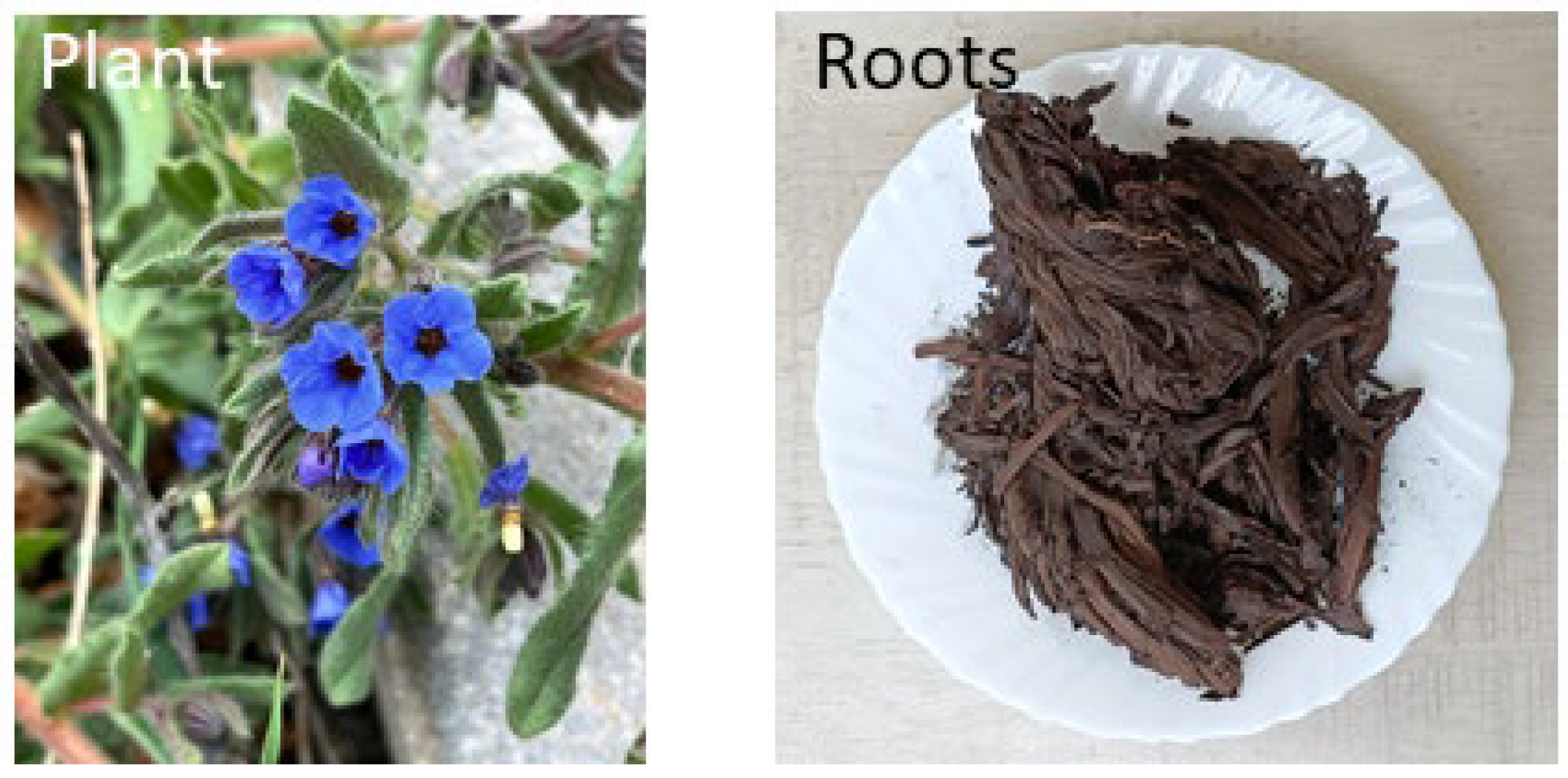
Figure 2.
Antioxidant activity of ethanol (EE) and water extract (MAHW) of Ratanjot (Alkanna tinctoria) root (RRP) extract. Bars are Average (n=6) ± SE bearing different superscript(s) (A, B, C, D) differ significantly at P < 0.05. TPC, total phenolic compound; ABTS, 2.2-Azino-bis (3-ethylbenzothiazoline-6-sulfonic acid); DPPH, 2,2-Diphenyl-1-picrylhydrazyl (DPPH); GAE, gallic acid equivalent.
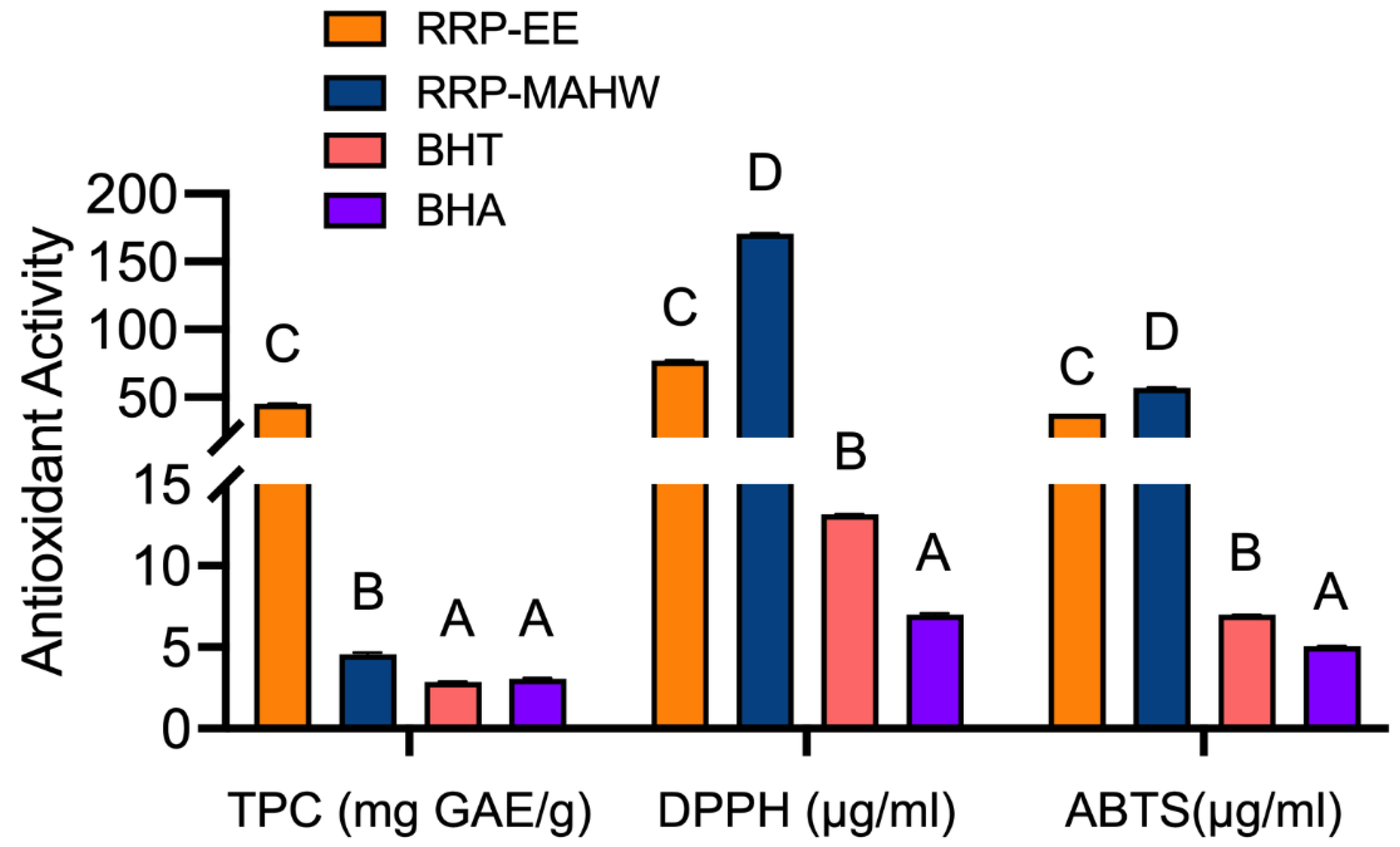
Figure 3.
Gas chromatography and mass spectrometry analysis of Alkanna tinctoria (known as Ratanjot) root ethanol extract produced three major components: Butanoic acid, 3-hydroxy-3-methyl-; Arnebin 7, and Diisooctylpthalate.
Figure 3.
Gas chromatography and mass spectrometry analysis of Alkanna tinctoria (known as Ratanjot) root ethanol extract produced three major components: Butanoic acid, 3-hydroxy-3-methyl-; Arnebin 7, and Diisooctylpthalate.
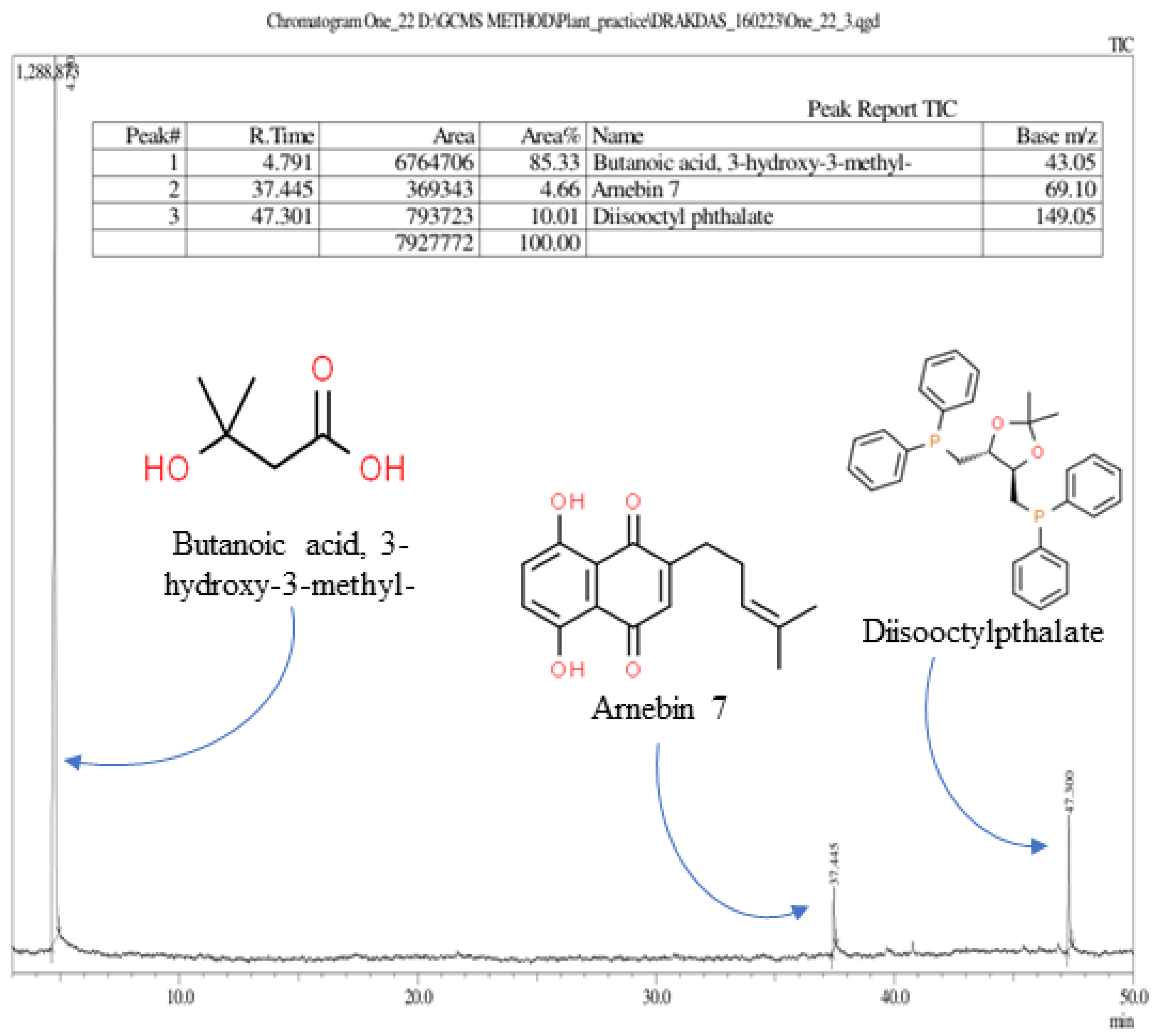
Figure 4.
Growth kinetics of (A) Staphylococcus aureus (SA) ATCC 25923 and (B) Listeria monocytogenes (LM) ATCC 19111after exposure to Ratanjot (Alkanna tinctoria) root extract (ethanol extract), RRP at 1X MIC and MBC for 24 h. Ciprofloxacin (1 X MIC and 1 X MBC) was used as a positive control.
Figure 4.
Growth kinetics of (A) Staphylococcus aureus (SA) ATCC 25923 and (B) Listeria monocytogenes (LM) ATCC 19111after exposure to Ratanjot (Alkanna tinctoria) root extract (ethanol extract), RRP at 1X MIC and MBC for 24 h. Ciprofloxacin (1 X MIC and 1 X MBC) was used as a positive control.
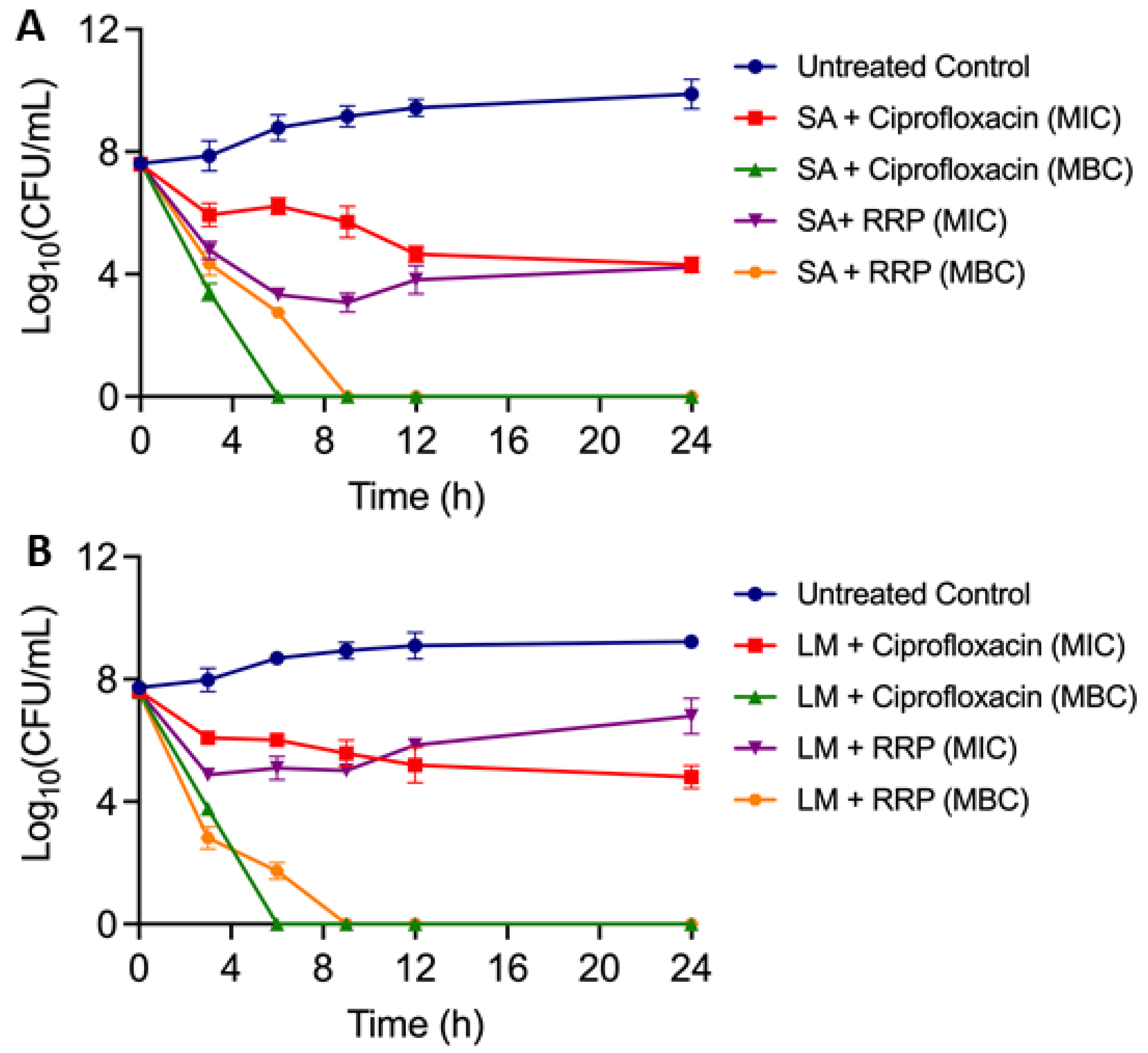
Figure 5.
Antimicrobial activity of Ratanjot (Alkanna tinctoria) root extract (RRP) exposed to various (A) pH range 2-8, (B) temperature range (37-100°C) and at 4C for 30 days against Listeria monocytogenes and Staphylococcus aureus analyzed by agar well-diffusion assay. Bars marked with different letters (a,b,c) are significantly different at P < 0.05.
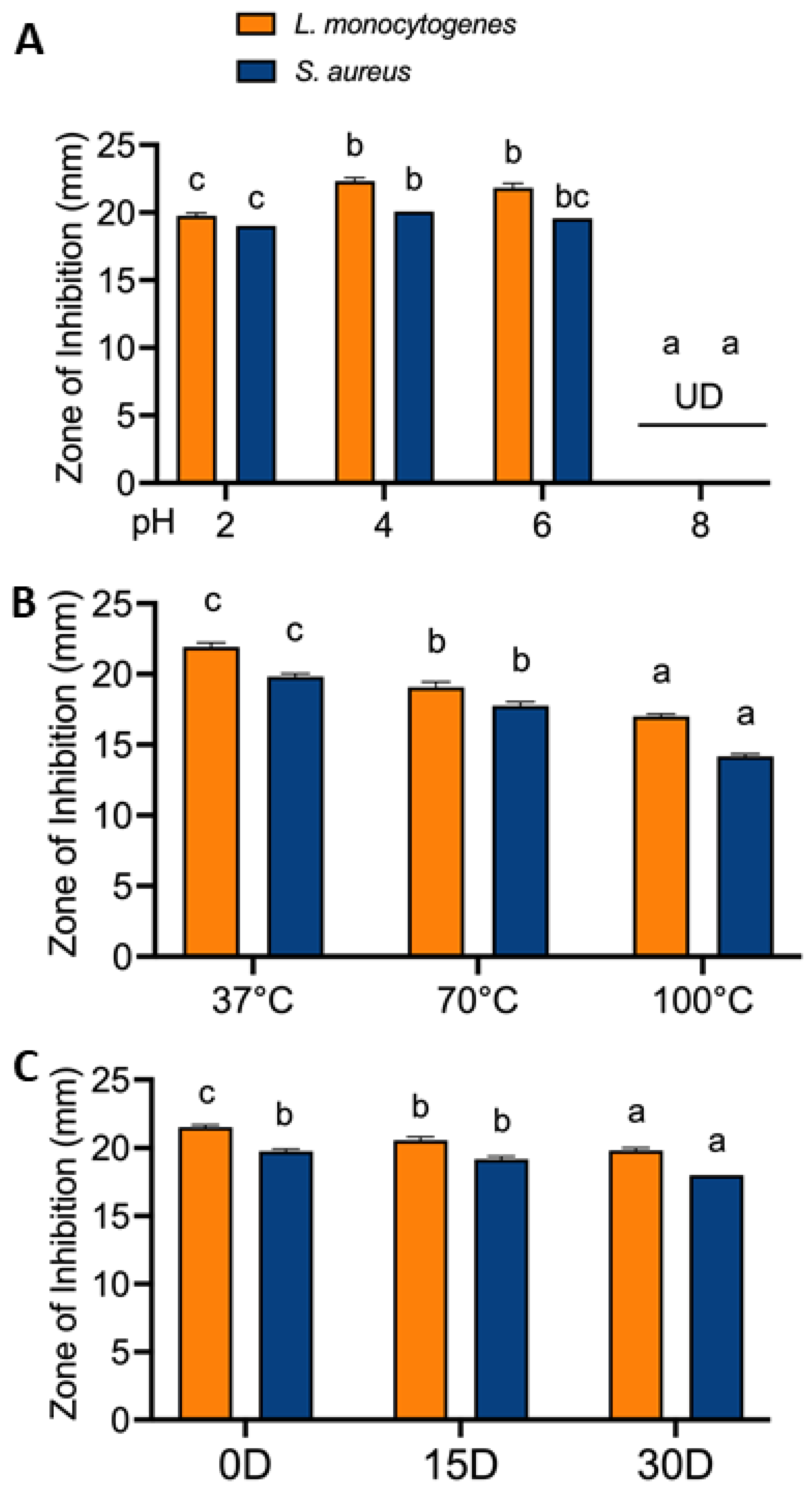
Figure 6.
Effect of Ratanjot (Alkanna tinctoria) root extract (RRP) at 1X and 2X MIC on Lactobacillus growth. (A) Lactiplantibacillus plantarum and Lacticaseibacillus casei counts (log 10 CFU/ml) after 24 h in MRS broth, (B) visual examination of turbidity of L. casei growth after 24 h.
Figure 6.
Effect of Ratanjot (Alkanna tinctoria) root extract (RRP) at 1X and 2X MIC on Lactobacillus growth. (A) Lactiplantibacillus plantarum and Lacticaseibacillus casei counts (log 10 CFU/ml) after 24 h in MRS broth, (B) visual examination of turbidity of L. casei growth after 24 h.
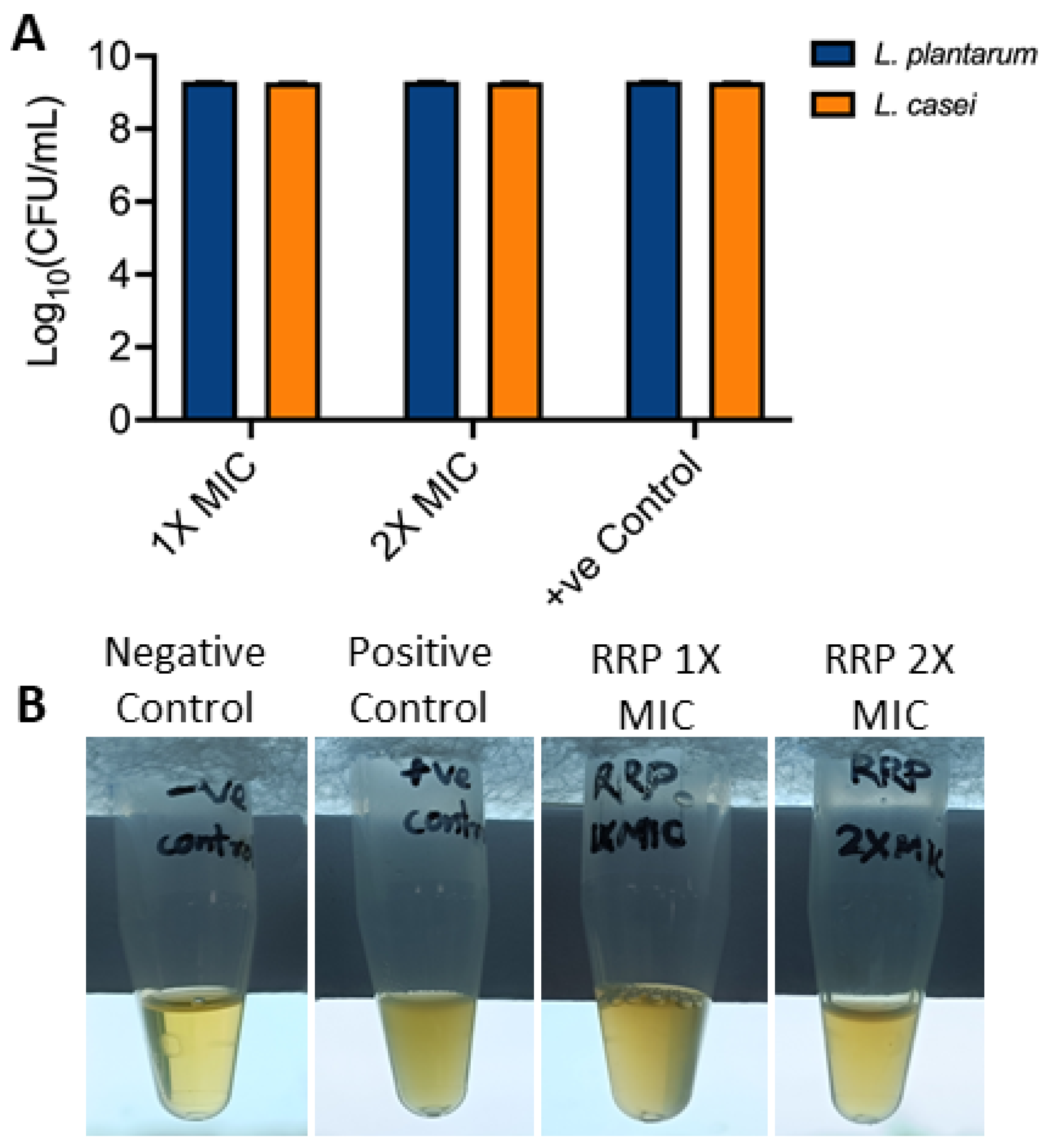
Figure 7.
In vitro hemolysis assay of Ratanjot (Alkanna tinctoria) root extract (RRP) at various concentrations (1-10XMIC). (A) Bar diagram showing percent hemolysis of sheep red blood cells. Data are mean (n=6) ± SE. *, P < 0.05. Triton X-100 (0.1%) used as a positive control for complete lysis. (B) Photographs showing the difference in hemolysis and no hemolysis of RBC.
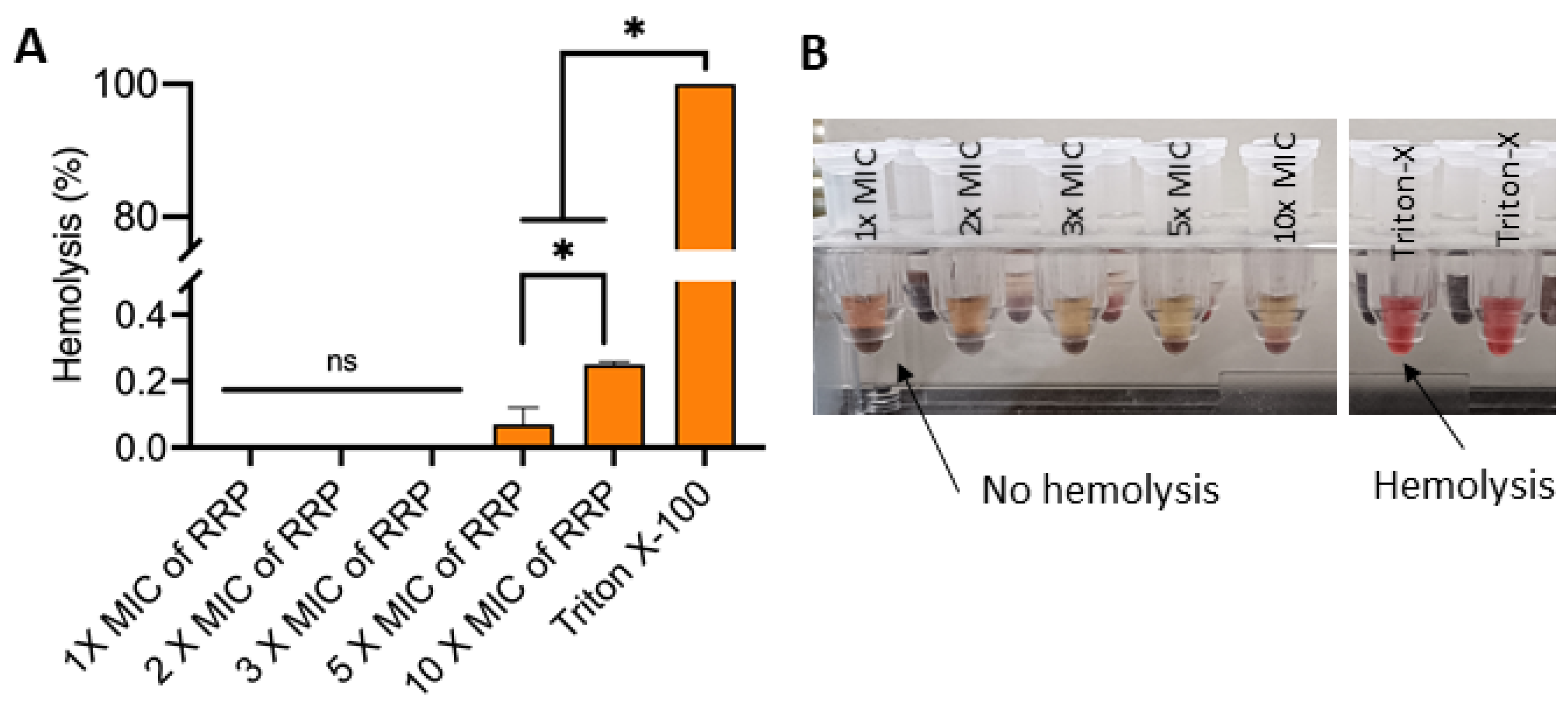
Figure 8.
Application of Ratanjot (Alkanna tinctoria) root extract (RRP) at 1% (T1) and 2% (T2) in chicken meatballs on antioxidant (TBARS) and antimicrobial activity stored at 4°C for 20 days. Bars marked with uppercase letters (A,B,C,D) signify differences in values on various days, and lowercase letters (a,b,c,d) signify differences within the same day for all three samples. P < 0.05.
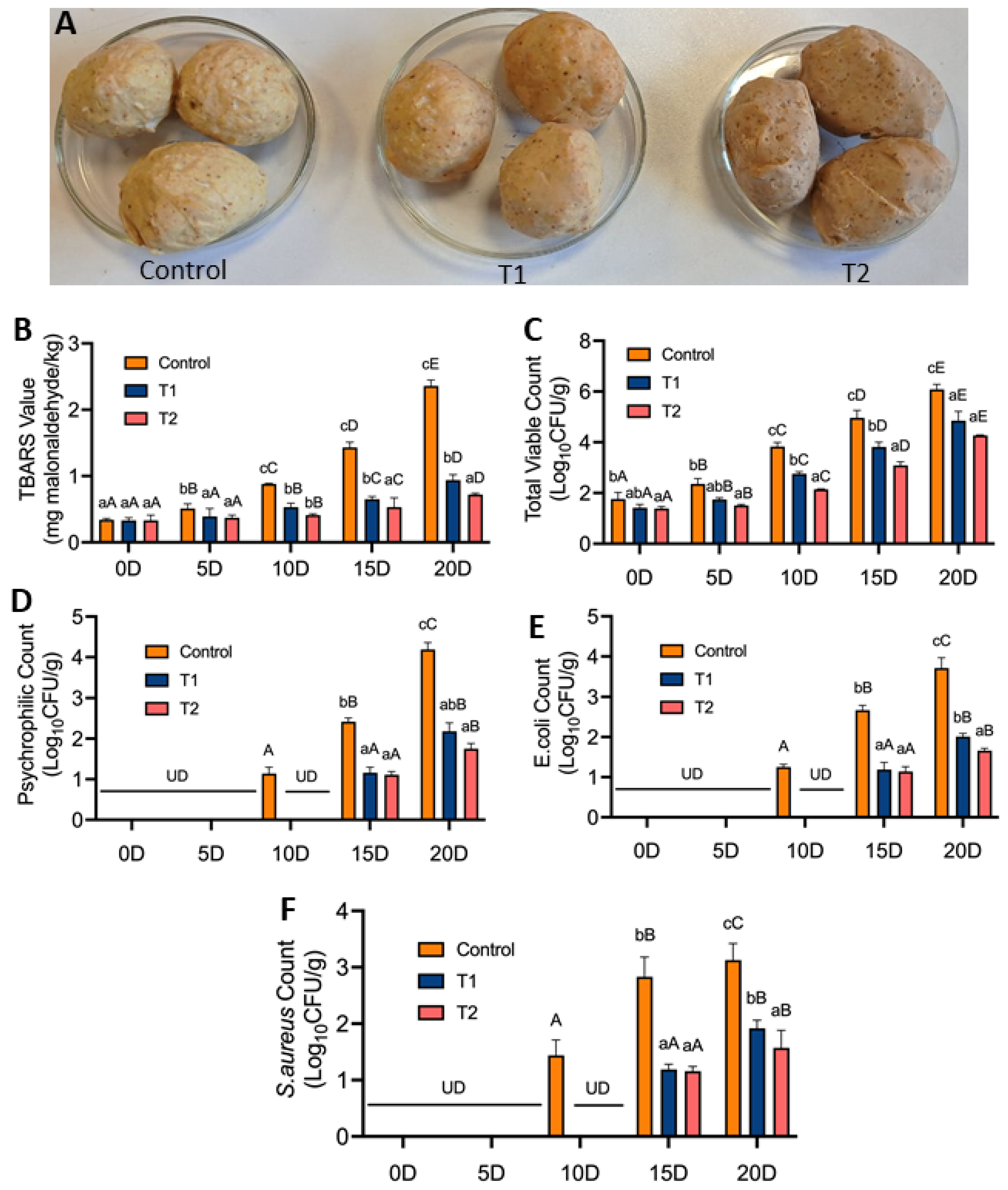

Table 1.
Bacterial cultures used in this study.
| Culture | Source |
| Staphylococcus aureus ATCC 25923 | American Type Culture Collection (ATCC), Manasas, VA, USA |
| Listeria monocytogenes ATCC 19111 | ATCC |
| Listeria monocytogenes ATCC 13932 | ATCC |
| Salmonella enterica serovar Typhimurium ATCC 14028 | ATCC |
| Escherichia coli ATCC 25922 | ATCC |
| Lactiplantibacillus plantarum MTCC 2621 | Microbial Type Culture Collection (MTCC) |
| Lacticaseibacillus casei MTCC 1423 | MTCC |
| Staphylococcus aureus strain MZP/SM/05 | Swine meat from Mirzapur, Uttar Pradesh, India |
| Staphylococcus aureus MZP/PM/38 | Poultry meat from Mirzapur, Uttar Pradesh, India |
| Staphylococcus aureus MZP/GM/02 | Goat meat from Mirzapur, Uttar Pradesh, India |
| Staphylococcus aureus ALD/PM/22 | Poultry meat from Allahabad, Uttar Pradesh, India |
| Staphylococcus aureus ALD/PM/23 | Poultry meat from Allahabad, Uttar Pradesh, India |
| Staphylococcus aureus LKO/GM/14 | Goat meat from Lucknow, Uttar Pradesh, India |
| Staphylococcus aureus LKO/PM/04 | Poultry meat from Lucknow, Uttar Pradesh, India |
| Staphylococcus aureus LKO/PM/08 | Poultry meat from Lucknow, Uttar Pradesh, India |
| Staphylococcus aureus LKO/GM/11 | Goat meat from Lucknow, Uttar Pradesh, India |
Table 2.
Characteristics of ethanol and microwave-assisted hot water (MAHW) extracts.
| Parameter | Ethanol extract ± SE* | MAHW extract ± SE* |
| Total phenolic Content (TPC)% | 18.27 ± 0.69b | 6.29 ± 0.30a |
| pH | 2.92 ± 0.02a | 3.15 ± 0.01b |
| CIELAB color parameters | ||
| L* | 25.07 ± 0.97a | 56.55 ± 0.79b |
| a* | 45.13 ± 0.44b | 12.02 ± 0.76a |
| b* | 63.35 ± 0.53 | 62.20 ± 0.65 |
| hab | 2.61 ± 0.08a | 41.70 ± 0.31b |
| C*ab | 77.97 ± 0.61 | 63.72 ± 0.38 |
*SE, Standard Error. Values labeled with superscripts (a,b) in the same row are significantly different at P < 0.05.
Table 3.
Antimicrobial activities of RRP extracts against some foodborne pathogens by agar well diffusion assay.
Table 3.
Antimicrobial activities of RRP extracts against some foodborne pathogens by agar well diffusion assay.
| Bacteria | Zone of Inhibition (mm) | |
| Ethanol Extract | Hot Water Extract | |
| Staphylococcus aureus ATCC 25923 | 19.75 ± 0.11ef | 0.00 ± 0.00 |
| Listeria monocytogenes ATCC 13932 | 21.17 ± 0.28gh | 0.00 ± 0.00 |
| Listeria monocytogenes ATCC 19111 | 21.92 ± 0.33h | 0.00 ± 0.00 |
| Salmonella Typhimurium ATCC 14028 | 9.92 ± 0.15b | 0.00 ± 0.00 |
| Escherichia coli ATCC 25922 | 8.58 ± 0.24a | 0.00 ± 0.00 |
| S. aureus MZP/SM/05 | 21.17 ± 0.40gh | 0.00 ± 0.00 |
| S. aureus MZP/PM/38 | 18.25 ± 0.11d | 0.00 ± 0.00 |
| S. aureus MZP/GM/02 | 20.08 ±0.27fg | 0.00 ± 0.00 |
| S. aureus ALD/PM/22 | 19.50 ± 0.26ef | 0.00 ± 0.00 |
| S. aureus ALD/PM/23 | 18.67 ± 0.25de | 0.00 ± 0.00 |
| S. aureus LKO/GM/14 | 27.50 ± 0.22i | 0.00 ± 0.00 |
| S. aureus LKO/PM/04 | 16.00 ± 0.13c | 0.00 ± 0.00 |
| S. aureus LKO/PM/08 | 20.00 ±0.37fg | 0.00 ± 0.00 |
| S. aureus LKO/GM/11 | 27.83 ± 0.21i | 0.00 ± 0.00 |
*Mean (n=6) ± SE bearing different superscript(s) (a, b, c, d, e, f, g, h) in a column differ significantly (P < 0.05).
Table 4.
Estimation of MIC, MBC, and tolerance to ethanol extract of Ratanjot root extract powder (RRP).
Table 4.
Estimation of MIC, MBC, and tolerance to ethanol extract of Ratanjot root extract powder (RRP).
| Bacteria | RRP ethanol Extract (mg/mL)* | Ciprofloxacin (µg/mL)* |
| MIC | ||
| Salmonella Typhimurium ATCC 14028 | 25.00 ± 0.00c | 0.125± 0.00a |
| Escherichia coli ATCC 25922 | 25.00 ± 0.00c | 0.125± 0.00a |
| Staphylococcus aureus ATCC 25923 | 0.049 ± 0.00a | 0.250± 0.00b |
| Listeria monocytogenes ATCC 19111 | 0.098 ± 0.00b | 0.250± 0.00b |
| MBC | ||
| Salmonella Typhimurium ATCC 14028 | 25.00 ± 0.00c | 0.25± 0.00a |
| Escherichia coli ATCC 25922 | 25.00 ± 0.00c | 0.25± 0.00a |
| Staphylococcus aureus ATCC 25923 | 0.098 ± 0.00a | 0.50± 0.00b |
| Listeria monocytogenes ATCC 19111 | 0.195 ± 0.00b | 0.50± 0.00b |
| Tolerance | ||
| Salmonella Typhimurium ATCC 14028 | 1.00 ± 0.00a | 2.00 ± 0.00 |
| Escherichia coli ATCC 25922 | 1.00 ± 0.00a | 2.00 ± 0.00 |
| Staphylococcus aureus ATCC 25923 | 2.00 ± 0.00b | 2.00 ± 0.00 |
| Listeria monocytogenes ATCC 19111 | 2.00 ± 0.00b | 2.00 ± 0.00 |
*Mean (n=6) ± SE bearing different superscript(s) (a, b, c) in a column differ significantly (P < 0.05). MIC, minimum inhibitory concentration; MBC, minimum bactericidal concentration.
Table 5.
Effect of Ratanjot root extract powder (RRP) on sensory attributes of aerobically packaged chicken meatballs during refrigerated storage (4 ± 1°C).
Table 5.
Effect of Ratanjot root extract powder (RRP) on sensory attributes of aerobically packaged chicken meatballs during refrigerated storage (4 ± 1°C).
| Sample | Refrigerated Storage Days* | ||||
| Day 0 | Day 5 | Day 10 | Day 15 | Day 20 | |
| Color and Appearance | |||||
| Control | 6.39±0.07aA | 6.28±0.04aB | 5.86±0.06aC | 5.44±0.12aD | 5.27±0.14aE |
| T1 | 7.41±0.05bA | 7.33±0.09bB | 7.16±0.05bC | 6.94±0.09bD | 6.38±0.21bE |
| T2 | 7.42±0.04bA | 7.37±0.04cB | 7. 24±0.11cC | 7.01±0.05cD | 6.63±0.13cE |
| Flavor | |||||
| Control | 6.37±0.09aA | 6.30±0.13aB | 5.86±0.06aC | 4.98±0.10aD | 4.04±0.06aE |
| T1 | 6.52±0.04bA | 6.50±0.20bA | 6.26±0.02bC | 6.07±0.17bD | 5.63±0.03bE |
| T2 | 6.76±0.06cA | 6.74±0.21cA | 6.33±0.04cB | 6.19±0.11cC | 5.81±0.04cD |
| Texture and Tenderness | |||||
| Control | 6.76±0.18aA | 6.68±0.13aB | 6.21±0.07aC | 5.69±0.05aD | 5.46±0.21aE |
| T1 | 6.82±0.05bA | 6.80±0.27bA | 6.64±0.06bB | 6.23±0.11bC | 6.09±0.18bD |
| T2 | 6.84±0.06bA | 6.82±0.12bA | 6.73±0.09cB | 6.44±0.04cC | 6.15±0.21cD |
| Juiciness | |||||
| Control | 6.25±0.10aA | 6.21±0.17aB | 5.69±0.38aC | 5.20±0.07aD | 5.04±0.04aE |
| T1 | 6.28±0.12bA | 6.26±0.15abA | 5.86±0.24bB | 5.41±0.10bC | 5.16±0.11bD |
| T2 | 6.30±0.17bA | 6.29±0.04bA | 5.98±0.21cB | 5.67±0.10cC | 5.23±0.10cD |
| Overall Acceptability | |||||
| Control | 6.84±0.09aA | 6.80±0.04aB | 5.68±0.21aC | 4.98±0.38aD | 4.02±0.50aE |
| T1 | 6.89±0.04bA | 6.86±0.08bA | 6.04±0.18bB | 5.65±0.17bC | 5.06±0.26bD |
| T2 | 6.92±0.07bA | 6.91±0.10cA | 6.17±0.23cB | 5.84±0.15cC | 5.13±0.14cD |
*Mean (n=6) ± SE bearing different superscript(s) in a column (a, b, c, d, e……) and in a row (A, B, C, D, E) differ significantly (P < 0.05). T1 contains 10 mg/g RRP and T2 contains 25 mg/ml.
Disclaimer/Publisher’s Note: The statements, opinions and data contained in all publications are solely those of the individual author(s) and contributor(s) and not of MDPI and/or the editor(s). MDPI and/or the editor(s) disclaim responsibility for any injury to people or property resulting from any ideas, methods, instructions or products referred to in the content. |
© 2024 by the authors. Licensee MDPI, Basel, Switzerland. This article is an open access article distributed under the terms and conditions of the Creative Commons Attribution (CC BY) license (http://creativecommons.org/licenses/by/4.0/).
Copyright: This open access article is published under a Creative Commons CC BY 4.0 license, which permit the free download, distribution, and reuse, provided that the author and preprint are cited in any reuse.


