Submitted:
19 June 2024
Posted:
20 June 2024
You are already at the latest version
Abstract
It's been a growing concern that many microbes are developing resistance against many antibiotics, lowering the effectiveness and progress expected from that treatment option. To overcome the re-sistance developed by microbes, metallic and non-metallic-based nanoparticle treatments are starting to present a very promising approach. Furthermore, polyurethanes are used in a wide range of biomedical applications due to their variety of physical-chemical, mechanical, and structural properties, and biotic and abiotic degradation. They are widely used in bio-imaging procedures when metallic-based filler particles are incorporated, making the final product radiopaque. It would be advantageous, however, if polyurethanes with intrinsic radiopacity could be produced in their synthesis, avoiding a series of disadvantages in the processing and final product and also presenting potential antimicrobial activities. This review’s objective was to study the radiopacifying charac-teristics of nanoparticles as well as the physical principles of radiopacity and the variety of medical applications of polyurethanes with nanoparticles. It was found in this study that the synthetization of radiopaque polyurethanes is not only possible but also that the efficiency of synthetization was improved when using atoms with high electron density as part of the back chainbone or grafted, making them great multipurpose materials.
Keywords:
nanoparticles
; radiopacity
; polyurethane
; biomedical materials
; structure vs. properties relationship
1. Introduction
Polyurethanes (PUs) are used in a wide range of applications due to their variety of physical-chemical, mechanical, and structural properties [1,2,3,4]. Lately, PU has been used for biomedical applications, mainly due to their new biobased macromolecular architectures, which allow the control of biotic and abiotic degradation [5,6]. Put this, medical-grade PU-based polymers can be commercially found under various trade names, such as Carbothane®, Pellethane®, and Tecoflex® from Lubrizol (US) [7] or Carbosil®, Bionate®, or Tecoflex® from DSM (Holland) [8]. According to IndustryARC [9] the medical polyurethane market size is forecast to reach US$ 5.2 billion by 2026, with a CAGR of 4.3% during 2021-2026.
Among the application characteristics, one of the most important is the visualization of the sample under a medical procedure. The radiopacity is one of the most important characteristics to visualize a sample, higher radiopacity means a better visualization inside the human body. Metals and ceramics are intrinsically radiopaque while polymers tend to be translucid due to the presence of low electronic density of atoms. Therefore, the radiopacity in polymers can be obtained in two distinct situations: the incorporation of a ceramic or metallic filler in the polymer or by synthesizing an intrinsic radiopaque polymer with high electron density reagents such as iodinated bisphenol A.
This review’s objective is to explain the properties and materials that induce radiopacity on polymers as well as the physical principles of radiopacity and the medical applications of these materials.
2. Principles and Effects of Radiopacity
2.1. Beer and Beer-Lambert Law
Beer’s and Beer-Lambert’s law states the light attenuation as it passes through some material. That is the reason why some materials are translucid or radiopaque under X-ray emission, for example. Beer’s law states that a material’s concentration and absorbance are directly proportional to each other and can be expressed by Equation (1) [10] where is , is a constant and is absorptivity. If put in the form of molar absorptivity.
Hence, Beer’s law can be treated as absorptivity () or molar absorptivity (), as Equation (2) shows.
The Beer-Lambert law relates the attenuation of light to the properties of the material through which the light is traveling. The relationship can be expressed by Equation (3): where A is absorbance, ε is the molar extinction coefficient (which depends on the nature of the chemical and the wavelength of the light used), l is the length of the path light must travel in the solution in centimeters, and c is the concentration of a given solution.
It is noteworthy to mention that sometimes and mistakenly the Beer-Lambert law is treated as the Beer’s law [10].
For X-ray measurement, it can be represented by equation 4 where is the intensity output, is the intensity input, is the linear attenuation coefficient, is the density and is the thickness of the material.
Physically, the Beer-Lambert law can be represented differently according to Figure 2A. Figure 2B shows the effect of the radiopacity of the chemical characteristics of the sample.
Figure 2A shows the physical representation of the Beer-Lambert law. When a wavelength with a certain energy is emitted and passes a sample, the length of a photon is half of the wavelength, and the radius is proportional to the square root of the wavelength [11]. Therefore, the higher the interaction of the photons and particles with the source, the higher the energy is lost as it passes through the sample and hence higher the radiopacity in the visualized image.
As demonstrated in Figure 2B above, the attenuation (absorption or scattering of the X-rays) of X-rays depends on sample characteristics. The attenuation is mainly dependent on the tissue type and medical device, electron density, and specific gravity of the material. For example, polymers mainly contain elements that have low electron density and low specific gravity, such as C, H, and O, which results in poor radiopacity. This is the reason why the bones present higher contrast compared to muscles.
Figure 3 presents an image with data taken from a didactic website. The website enables the insertion of tissue type, tissue thickness, and energy as input values and obtains the X-ray transmission value, tissue density of blood, and mass attenuation as output parameters.
2.2. Photoelectric, Compton, and Rayleigh Effects
The photoelectric effect (PEE), Compton, and Rayleigh effects (also known as coherent scattering) are the main X-ray interaction processes. Photoelectrons are emitted when electrons are released from a substance upon an incidence of an X-ray, for example. When the energy of the incident radiation approaches the electron binding energy in each of the atom’s many shells. When an electron at a higher energy level fills the hole left by the ejected electron, a characteristic X-ray with an energy equal to the difference between the binding energies of the two electrons is subsequently released (Figure 4) [13,14].
In general, photoelectrons can also be released from shells other than K and L, depending on the atomic number of the atom (occupation of other shells) (i.e., M and N). The low-energy characteristic X-rays are completely absorbed by the bodily tissues. The photoelectric interaction’s (mPE) linear attenuation coefficient is solely dependent on the energy of the input beam, the tissue-effective atomic number, and the tissue density [14,16]. Imaging creates a contrast between high- and low-density materials since the mPE for high atomic number materials will be larger than for less dense and low atomic number materials. For soft tissue with low attenuation capacity and bone with high attenuation, more contrast is obtained [17].
The photon in the Compton Effect (CE) is scattered by low binding energy electrons (outer-layer electrons), which only absorb a portion of the energy from the X-rays and let the photon enter the material at a lower energy and in a different direction. A photon of fixed energy can produce electrons of variable energy, ranging in value from zero to a maximum value because the energy transfer is dependent on the direction of the ejected electron, which is random [18], as demonstrated in Figure 4. In this case, it’s not the atomic number of the component atoms, but rather the electron density of the tissues, which determines the likelihood of Compton scattering. Moreover, it has a minor dependence on the X-ray incident energy. The PEE predominates over the CE at low-energy X-rays, but with high-energy X-rays, increasing the CE reduces the image contrast [16]. At extremely low photon energies, an elastic scattering phenomenon known as the Rayleigh scattering (RE) takes place. Only in mammography, which employs low photon energy, does this scattering become significant because diagnostic radiology uses photons above this range. In this instance, the atom’s electron cloud scatters the incident photons, slightly ionizing the surrounding area [16].
2.3. Radiopacity Measurement
When an external apparatus is inserted into the human body, the positioning is dependent on the radiopacity of the device aiming to easily detect the surrounding anatomical structures. In practical applications, the radiopacity can be determined qualitatively by a medical doctor who can approve or not the apparatus by the visual aspect or quantitatively via grayscale value using a photo-densitometer where the grayscale value is converted to absorbance values [19,20].
Considering dental materials, since pure aluminum’s radiopacity is extremely comparable to dentin’s, the International Standards Organization (ISO) emphasizes that any restorative material must exhibit radiopacity equal to or greater than pure aluminum of the same thickness [19]. Transmission densitometry is the gold standard and traditional typical method for measuring radiopacity. Using this method, the grayscale value of photographic images (films or digital) is calculated and compared to that of a standard aluminum wedge with 10 steps from 1 cm to 10 cm. The grayscale value is proportional to the ratio of the incident to transmitted X-ray radiation. The value is stated in relation to the thickness of aluminum which is equal. Since radiography images are two-dimensional, or depthless, projections [21], the various grayscale tones, ranging from white to black, correspond to the various anatomical features of the teeth [22].
In computed tomography, radiopacity is quantified using quantities known as Hounsfield Units (HU), named after the engineer who invented computed tomography, Godfrey Hounsfield. The linear attenuation coefficient of distilled water is used to standardize the HU of a substance. At standard temperature and pressure, water and air are assigned HU values of 0 and 1000, respectively [23]. The degree of a material’s attenuation capabilities can only be clearly appreciated from its X-ray pictures, despite the fact that these imaging modalities allow for quantitative measures of radiopacity [24,25]. Figure 5 shows the image of an arm in a radiographic image where it is observed the metal rods fixed in the arm.
3. Biomedical Applications of NPs
Inorganic NPs have unique physical, electrical, magnetic, and optical properties due to the intrinsic properties of the material [26]. Also, the detection through layers of skin and muscle in bioimaging diagnostics is possible, for example, Au NPs possess free electrons at their surface that continually oscillate at a frequency dependent on their size and shape, giving them specific photothermal properties [27]. Beyond imaging applications, it is noteworthy to mention that most metallic-based nanoparticles present antimicrobial properties that may be beneficial for several biomedical applications, be it in drug delivery methods, antimicrobial activities, or prevention of infections through contact with hospital devices.
3.1. Antimicrobial Activity of Metallic and Ceramic Nanoparticles and the Influence of Their Particle Characteristics
It’s been a growing concern that many microbes are developing resistance against many antibiotics. This, coupled with the abuse of drugs, as well as the release of antibiotics to the environment has impeded the progress expected from that treatment option [28,29]. Microbes with high mutative capacity and rapid morphological changes may lead to microbial resistance to antibiotics. To overcome the resistance developed by microbes, nanoparticle-based treatments are starting to present a very promising approach.
Nanoparticles (NPs) are substances with a diameter ranging from 1-100 nm (nm) [30,31,32]. According to the World Health Organization, in addition to their reduced size and selectivity for bacteria, metal-based nanoparticles have proved to be effective against pathogens listed as a priority. Metal-based nanoparticles are known to have non-specific bacterial toxicity mechanisms (they do not bind to a specific receptor in the bacterial cell) which not only makes the development of resistance by bacteria difficult but also broadens the spectrum of antibacterial activity [33]. Inorganic materials have been used to synthesize nanostructured particles for various drug delivery and imaging applications [26]. Figure 6 and Figure 7 scheme how metallic and ceramic nanoparticles act on the inactivation and/or destruction of different types of microbes.
Inorganic NPs can be engineered to have a wide variety of sizes, structures, and geometries [26]. For biomedical applications when modifying the size, shape, concentration, capping method, and/or surface chemistry of NPs, the properties of metal NPs are affected to the extent that it influences their behavior on microbial life forms, conditioning them to become better drug delivery agents or present better bactericidal or bacteriostatic behavior for example [34,35]. Table 1 shows the effect of the inorganic particles in microbial strains according to concentration and size.
The size of the nanoparticle has a direct influence on the antimicrobial activity of the substance. A vast variety of analyses on NPs suggests that a large surface area is imperative for microbial attachment and rapid penetration into the cell [59,60]. Furthermore, studies disclosed that nanoparticles with high surface area present the ability to deliver a large number of antibiotics or other substances, their variation in size and shape makes them ideal antimicrobial weapons [60,61,62]. As reported by Philip, D. [63], the NP shape is also directly important in determining the types and extent of interaction with the membrane or enzymes of microorganisms. For instance, ZnO nanoparticles of different shapes (plate, sphere, and pyramid) showed shape-dependent activity on a typical enzyme β-galactosidase (GAL) [64].
It has been observed, that NPs containing magnetic iron oxide, composed of magnetite (Fe3O4) or maghemite (Fe2O3), possess superparamagnetic properties at certain sizes and may be used as contrast agents, drug delivery vehicles, and thermal-based therapeutics [65].
3.2. Metallic Nanomaterials Characteristics on Bio-Imaging and Its Effects
As demonstrated by Beer-Lambert’s law, the radiopacity of a material depends on the atomic number of its elements, the material thickness, the intensity of the energy input, and its particle aspect (round, irregular, triangular, etc.). These conditions for radiopacity synergize with most metallic and ceramic intrinsic properties, making them uniquely qualified for applications such as diagnostics and imaging.
Marghalani et al. [66] investigated the effect of the filler characteristics on the radiopacity levels of nanocomposites for radiographic diagnosis using different particle size formulations (from 100–1500 nm). In general, the irregular-shape series showed more radiopacity than the spherical-shape ones. Another interesting feature is the decrease in the radiopacity values with increasing filler particle size for both formats. Finally, a higher filler amount promotes higher radiopacity. These characteristics have also been studied by Marghalani [66], where spherical nanofiller particles, mainly silica-based and irregular particles of ground glass melts (Ba–Al-B-silicate glass) had their radiopacity tested. The irregular filler-based series formulations were more radiopaque than the spherical series, this was attributed to the incorporation of a higher atomic number element; in this case, barium [67,68,69]. The barium element is found mainly in irregular shape series allowing this higher opacity in comparison to the spherical series.
This finding agrees with a study that showed a direct effect that occurs between the filler type and the radiopacity of the filling material [68]. The sharp edges of the irregularly shaped nanofillers of these nanocomposites may absorb the light resulting in a radiopaque characteristic [70,71]. Additionally, irregular edges and surfaces of these nanofiller series can yield a mismatch of the refractive indices between fillers and resin interface leading to high radiopacity of the nanocomposite. Besides radiopacity, the nanoparticles can be used for imaging as demonstrated in Figure 8, where an example of the use of nanoparticles for identification in the human body is presented.
Similar to “radiopaque particles”, bioimaging particles are used as contrast agents for in vitro and in vivo biological imaging in real-time, monitoring biological functions without affecting normal life processes such as respiration and movement.
It is noteworthy to mention that materials of high biocompatibility and low cytotoxicity are of great value when determining the uses of metallic-based nanoparticles. It has been noted that NP’s unique size-related characteristics and potential toxicity, show a display of biological effects in animal models and in cultured cells that differ from those observed with bulk material [73].
Since only a few of the NPs have decent biocompatibility and low cytotoxicity when utilizing them in unique niche applications that require properties unattainable by organic materials, metallic nanoparticles must be used carefully, especially in the case of heavy metals [65,74]. Beyond that, for some applications like endoscopy, materials must preferably have good synergy with the polymer they are incorporated with to avoid adverse results such as heterogeneous dispersions (which affect the physical and mechanical properties of polymers), loss of radiopacifying atoms, leaching of toxic radiopacifying additives, and systemic absorption by the host. For these reasons, biocompatible and low-toxicity polymers with intrinsic radiopacity would charm with the potential to attend these applications with lower drawbacks. Table 2 shows some examples of nanomaterials used for bioimaging applications.
4. Polymers with Intrinsic Radiopacity
Many polymers can be used in medical implants for permanent or temporary use. These polymers must attend specific functions, such as drug delivery, provide physical and structural support to vascular systems, or restore the normal function of joints in arthroplasty. Besides desirable characteristics such as ease of production, various mechanical properties, and biocompatibility, they also present a high potential for controlling properties to satisfy a wide range of requirements, which is a positive aspect. Common polymers include polyethylene, polytetrafluorethylene, poly(lactic acid), polypropylene, and polyurethane. Emonde et al. [20] pointed out radiopacity and polymers/fillers for different applications such as bone cement, joint replacements, bioresorbable stents, craniofacial implants, spinal implants, implant dentistry, and internal fixation systems. Figure 9 and Figure 10 compare the radiopacity of synthetic polymers in medical applications.
Polyurethanes particularly can be synthesized to be intrinsically radiopaque. This avoids problems in the manufacturing of the product by incorporation of particles in the polymer/particle mixture stage which would speed up the process, avoiding the lixiviation of the material and potentially lower the cost of production [109,110,111]. Since one of the main characteristics of radiopaque materials is the high electron density, one of the main alternatives (and more appropriate) is incorporating heavy atoms (such as barium or iodine) through physical blends or polymer salt complexes. Table 3 shows some studies of different polymers with their respective mechanical characteristics and contrast agent concentrations.
When synthesizing polyurethanes with heavy atoms the major role is to build blocks of polymers with intrinsic radiopacity without significant loss of mechanical and physical properties. There have been several studies where attempts at synthesizing intrinsically radiopaque polyurethanes were successful.
Dawlee and Jayabalan [110] synthesized intrinsically radiopaque polyurethanes with chain extender 4,4′-isopropylidenebis [2-(2,6-diiodophenoxy)ethanol] and claimed that the final product is cytocompatible with L929 mouse fibroblast cells. Furthermore, the toxicological studies and subcutaneous implantation of PTMGMDIBPA (polyurethane prepared with poly(tetramethylene ether) glycol polyol) indicated nontoxic and reasonable tissue compatibility of the iodinated polyurethane. Also, the sample was visible even after 12 weeks of implantation.
James et al. [111] produced an aliphatic polyurethane (Tecoflex 80A) with 5-iodine-containing molecule, N-(2,6-diiodocarboxyphenyl)-3,4,5-triiodo benzamide (DCPTB). The optimal reaction mechanism was obtained by incorporating 8% iodine (wt/wt) in the polymer. The radiopacity was equivalent to that of a 2 mm thick aluminum wedge but a reduced thermal stability compared to the starting polymer.
Dawlee and Jayabalan [112] developed segmented polyurethane elastomers with low iodine content exhibiting radiopacity and blood compatibility. The polyurethane was prepared aliphatic using chain extender 2,3-diiodo-2-butene-1,4-diol, polyol polytetramethylene glycol, and 4,4′-methylenebis(phenyl isocyanate) (MDI). The radiopacity was considered good at 3% iodine in aromatic PU and 10% in aliphatic PU. The PUs showed cytocompatibility with L929 fibroblast cells, non-hemolytic, and possessed good blood compatibility.
Kiran et al. [113] used an IBPA (Bisphenol-A with 4,4′-isopropylidinedi-(2,6-diiodophenol) as a chain extender for the preparation of a radiopaque PU. The authors obtained a highly radiopaque PU, in-vitro non-cytotoxic test using L929 mouse fibroblast cells and a desirable range of physical properties.
Qu et al. [114] synthesized radiopaque poly(ether urethane) with 15 wt% iodine-containing diol as a chain extender. The radiopacity did not alter after 6 weeks of exposition of an oxidative degradation treatment. The authors claimed that the thermal, mechanical, and cytotoxicity properties were satisfactory.
Kiran et al. [115] synthesized PU with an iodinated chain extender (IBPA) and three different diols (polypropylene glycol, polycaprolactone diol, and poly(hexamethylene carbonate) diol. The authors claimed that radiopacity was equivalent to the one containing 20 wt % barium sulfate, nontoxicity when exposed to L929 cells by direct contact and MTT assay (a colorimetric assay for assessing cell metabolic activity).
Kiran and Joseph [116] synthesized opaque polyurethane containing iodinated hydroquinone 2,2′-(2,5-diiodobenzene-1,4-diyl)bis(oxy)diethanol (DBD) ether as chain extender and obtained equivalent opacity to 15 wt % barium sulfate with polyurethane, optical transparency, hemocompatible and noncytotoxic to L929 fibroblast cell lines.
Sang et al. [117] synthesized radiopaque iodinated poly(ester urethane)s containing poly(ε-caprolactone) blocks and claimed a high radiopacity compared with an aluminum wedge of equivalent thickness. A prolonged enzymatic degradation test was also superior compared to the reference sample. Also, distinct mass loss and degradation occurred in the third month. [118].
Sang et al. [119] synthesized poly(lactic acid)-polyurethane for chemoembolization therapy and constated an X-ray visibility and drug-loaded beads as multifunctional embolic agents and noninvasive intraoperative location and postoperative examination.
It is feasible, as presented, to synthesize intrinsically radiopaque polyurethanes by different methods without compromising significantly their physical and mechanical properties.
5. Conclusion
This mini-review demonstrates the effect of the concentration, type, and format of different nanoparticles on the radiopacity of polyurethane. It was found that barium sulfate and iodinated compounds are the most clinically administered radiopaque agents.
One of the main characteristics is the cost-effectiveness and tailor-made properties that can be achieved compared to conventional materials. The synthetization of radiopaque polyurethanes is not only possible, as referenced here, but it has been observed that radiopaque PU’s are more efficiently synthesized with heavy atoms with a high electron density as part of the back chain bone or grafted. The synthetization of polyurethanes with biocompatible and low cytotoxicity high electron density composites or atoms produces a radiopaque and antimicrobial material without the downsides of incorporating nanoparticles in the processing steps of the product and enabling a multipurpose use on biomedical applications.
It is noteworthy to mention that physical blending is the most prevalent and economical method to induce radiopacity in polymers. However, the resultant mixture often lacks homogeneity and nanoparticle agglomeration which compromises the radiopacity. To minimize this drawback a higher concentration of the nanoparticle is required. Of course, surface-modifying agents can be used to improve dispersion. Also, higher radiopacity can be used if the contrast agent is soluble, which provides a homogeneous distribution between the phases and allows the use of a lower concentration of contrast agent than surface modification for the same level of radiopacity. The recommendation is that the amount of contrast agent must be as low as possible to provide acceptable radioactivity and mechanical properties. It is important to keep in mind that the radiopacity has to be coherent (visible enough) with the respective anatomical structures of different tissues.
Author Contributions
Conceptualization, H.L. Ornaghi Jr.; validation, H.L. Ornaghi Jr., J. Garavatti; formal analysis, H.L. Ornaghi Jr.; investigation, H.L. Ornaghi Jr.; writing—original draft preparation, H.L. Ornaghi Jr., J. Garavatti.; writing—review and editing, H.L. Ornaghi Jr., Garavatti; visualization, H.L. Ornaghi Jr., Garavatti; supervision, H.L. Ornaghi Jr.; project administration, H.L. Ornaghi Jr. All authors have read and agreed to the published version of the manuscript.
Funding
This research received no external funding.
Data Availability Statement
No data is available.
Conflicts of Interest
The authors declare no conflicts of interest.
References
- Das, A.; Mahanwar, P. A Brief Discussion on Advances in Polyurethane Applications. Advanced Industrial and Engineering Polymer Research 2020, 3 (3). [CrossRef]
- Wienen, D.; Gries, T.; Cooper, S.L.; Heath, D.E. An Overview of Polyurethane Biomaterials and Their Use in Drug Delivery. Journal of Controlled Release 2023, 363, 376–388. [Google Scholar] [CrossRef] [PubMed]
- Lucas Dall Agnol; Heitor Luiz Ornaghi; Monticeli, F.; Trindade, F.; Bianchi, O. Polyurethanes Synthetized with Polyols of Distinct Molar Masses: Use of the Artificial Neural Network for Prediction of Degree of Polymerization. SPE transactions/Polymer engineering and science 2021, 61 (6), 1810–1818. [CrossRef]
- Alves, F.C.; Vanessa; Monticeli, F.M.; Heitor Ornaghi; Barud, S.; Mulinari, D.R. Efficiency of Castor Oil–Based Polyurethane Foams for Oil Sorption S10 and S500: Influence of Porous Size and Statistical Analysis. Polymers and polymer composites/Polymers & polymer composites 2021, 29 (9_suppl), S1063–S1074. [CrossRef]
- Biobased Polyurethanes for Biomedical Applications. Bioactive Materials 2021, 6(4), 1083–1106. [CrossRef] [PubMed]
- Trhlíková, O.; Vlčková, V.; Abbrent, S.; Valešová, K.; Kanizsová, L.; Skleničková, K.; Paruzel, A.; Bujok, S.; Walterová, Z.; Innemanová, P.; et al. Microbial and Abiotic Degradation of Fully Aliphatic Polyurethane Foam Suitable for Biotechnologies. Polymer Degradation and Stability 2021, 194, 109764. [Google Scholar] [CrossRef]
- Specialty Chemicals—The Lubrizol Corporation. Lubrizol.com. Available online: https://www.lubrizol.com/ (accessed on 22 January 2024).
- Medical Polyurethanes. @Biomedical. Available online: https://www.dsm.com/biomedical/en_US/biomaterials-solutions/medical-polyurethanes.html (accessed on 22 January 2024).
- Medical Polyurethane Market Research Report: Market size, Industry outlook, Market Forecast, Demand Analysis, Market Share, Market Report 2021-2026. www.industryarc.com. Available online: https://www.industryarc.com/Report/18850/medical-polyurethane-market (accessed on 22 January 2024).
- Calloway, D. Beer-Lambert Law. Journal of Chemical Education 1997, 74(7), 744. [Google Scholar] [CrossRef]
- Liu, S.-L. Electromagnetic Fields, Size, and Copy of a Single Photon. arXiv (Cornell University) 2016. [CrossRef]
- Facebook, N. Interactive X-Ray Transmission Calculator For Radiologic Technologists (Beer’s Law Equation) • How Radiology Works. Available online: https://howradiologyworks.com/x-ray-transmission (accessed on 19 January 2024).
- Gaillard, F.; Goel, A. Photoelectric Effect. Radiopaedia.org 2014. [CrossRef]
- Maziar Montazerian; Geovanna, V. S. Gonçalves; Maria; Eunice; Glauber; Sousa, J.A.; Adrine Malek Khachatourian; Souza, S.; Silva; Marcus; Baino, F. Radiopaque Crystalline, Non-Crystalline and Nanostructured Bioceramics. Materials 2022, 15 (21), 7477–7477. [CrossRef]
- Nett, B. X-Ray Interactions, Illustrated Summary (Photoelectric, Compton, Coherent) For Radiologic Technologists And Radiographers • How Radiology Works. How Radiology Works. Available online: https://howradiologyworks.com/x-ray-interactions/ (accessed on 22 January 2024).
- Berger, M.; Yang, Q.; Maier, A. X-Ray Imaging. Lecture Notes in Computer Science 2018, 119–145. [Google Scholar] [CrossRef]
- Nett, B. PhD. X-ray attenuation of tissues for Radiologic Technologists. How Radiology Works. Available online: https://howradiologyworks.com/x-ray-attenuation-of-tissues/ (accessed on 22 January 2024).
- J. Seibert, J. Anthony; Boone, John, M. X-Ray Imaging Physics for Nuclear Medicine Technologists. Part 2: X-Ray Interactions and Image Formation; Issue 1; Journal of Nuclear Medicine Technology; 1850 Samuel Morse Drive, Reston, VA 20190, USA, March 2005; Vol. 33, pp. 3-18.
- Victor Manuel Ochoa-Rodríguez; Homero, J.; Roma, B.; Hernán Coaguila-Llerena; Mário Tanomaru-Filho; Andréa Gonçalves; Rubens Spin-Neto; Faria, G. Radiopacity of Endodontic Materials Using Two Models for Conversion to Millimeters of Aluminum. Brazilian Oral Research 2020, 34. [CrossRef]
- Crystal Kayaro Emonde; Eggers, M.-E.; Wichmann, M.; Christof Hurschler; Ettinger, M.; Berend Denkena. Radiopacity Enhancements in Polymeric Implant Biomaterials: A Comprehensive Literature Review. ACS Biomaterials Science & Engineering 2024. [CrossRef]
- Solmaz Valizadeh; Mohammad Amin Tavakoli; Zarabian, T.; Esmaeili, F. Diagnostic Accuracy of Digitized Conventional Radiographs by Camera and Scanner in Detection of Proximal Caries. PubMed 2009. [CrossRef]
- Paiva, M.A.A. de; Mangueira Leite, D.F.B.; Farias, I.A.P.; Costa, A. de P. C.; Sampaio, F.C. Dental Anatomical Features and Caries: A Relationship to Be Investigated. Dental Anatomy 2017. [CrossRef]
- Bryant, J.A.; Drage, N.A.; Richmond, S. CT Number Definition. Radiation Physics and Chemistry 2012, 81(4), 358–361. [Google Scholar] [CrossRef]
- Sneha, K.R.; Sailaja, G.S. Intrinsically Radiopaque Biomaterial Assortments: A Short Review on the Physical Principles, X-Ray Imageability, and State-of-The-Art Developments. Journal of Materials Chemistry B 2021, 9(41), 8569–8593. [Google Scholar] [CrossRef]
- Lusic, H.; Grinstaff, M.W. X-Ray-Computed Tomography Contrast Agents. Chemical Reviews 2012, 113(3), 1641–1666. [Google Scholar] [CrossRef] [PubMed]
- Mitchell, M.J.; Billingsley, M.M.; Haley, R.M.; Wechsler, M.E.; Peppas, N.A.; Langer, R. Engineering Precision Nanoparticles for Drug Delivery. Nature Reviews Drug Discovery 2020, 20(1), 1–24. [Google Scholar] [CrossRef] [PubMed]
- Wang, J.; Potocny, A.M.; Rosenthal, J.; Day, E.S. Gold Nanoshell-Linear Tetrapyrrole Conjugates for near Infrared-Activated Dual Photodynamic and Photothermal Therapies. ACS Omega 2019, 5(1), 926–940. [Google Scholar] [CrossRef] [PubMed]
- Laxminarayan, R.; Duse, A.; Wattal, C.; Zaidi, A.K.M.; Wertheim, H.F.L.; Sumpradit, N.; Vlieghe, E.; Hara, G.L.; Gould, I.M.; Goossens, H.; et al. Antibiotic Resistance—the Need for Global Solutions. The Lancet Infectious Diseases 2013, 13(12), 1057–1098. [Google Scholar] [CrossRef] [PubMed]
- Friedman, H.; Newton, C.; Klein, T.W. Microbial Infections, Immunomodulation, and Drugs of Abuse. Clinical Microbiology Reviews 2003, 16(2), 209–219. [Google Scholar] [CrossRef] [PubMed]
- Khan, I.; Saeed, K.; Khan, I. Nanoparticles: Properties, Applications and Toxicities. Arabian Journal of Chemistry 2017, 12(7), 908–931. [Google Scholar] [CrossRef]
- Song, X.; Bayati, P.; Gupta, M.; Elahinia, M.; Haghshenas, M. Fracture of Magnesium Matrix Nanocomposites—a Review. International Journal of Lightweight Materials and Manufacture 2021, 4(1), 67–98. [Google Scholar] [CrossRef]
- Vert, M.; Doi, Y.; Hellwich, K.-H.; Hess, M.; Hodge, P.; Kubisa, P.; Rinaudo, M.; Schué, F. Terminology for Biorelated Polymers and Applications (IUPAC Recommendations 2012). Pure and Applied Chemistry 2012, 84(2), 377–410. [Google Scholar] [CrossRef]
- Sánchez-López, E.; Gomes, D.; Esteruelas, G.; Bonilla, L.; Lopez-Machado, A.L.; Galindo, R.; Cano, A.; Espina, M.; Ettcheto, M.; Camins, A.; et al. Metal-Based Nanoparticles as Antimicrobial Agents: An Overview. Nanomaterials 2020, 10 (2). [CrossRef]
- Sharmin, S.; Md Rahaman, M.; Sarkar, C.; Atolani, O.; Islam, M.T.; Adeyemi, O.S. Nanoparticles as Antimicrobial and Antiviral Agents: A Literature-Based Perspective Study. Heliyon 2021, 7(3), e06456. [Google Scholar] [CrossRef]
- Nanoscale Materials in Targeted Drug Delivery, Theragnosis and Tissue Regeneration; Springer Nature, 2016. [CrossRef]
- Martínez-Castañón, G.A.; Niño-Martínez, N.; Martínez-Gutierrez, F.; Martínez-Mendoza, J.R.; Ruiz, F. Synthesis and Antibacterial Activity of Silver Nanoparticles with Different Sizes. Journal of Nanoparticle Research 2008, 10(8), 1343–1348. [Google Scholar] [CrossRef]
- Manikandan, V.; Velmurugan, P.; Park, J.-H.; Chang, W.-S.; Park, Y.-J.; Jayanthi, P.; Cho, M.; Oh, B.-T. Green Synthesis of Silver Oxide Nanoparticles and Its Antibacterial Activity against Dental Pathogens. 3 Biotech 2017, 7 (1). [CrossRef]
- Bera, R.K.; Mandal, S.M.; Raj, C.R. Antimicrobial Activity of Fluorescent Ag Nanoparticles. Letters in Applied Microbiology 2014, 58(6), 520–526. [Google Scholar] [CrossRef] [PubMed]
- Panáček, A.; Kolář, M.; Večeřová, R.; Prucek, R.; Soukupová, J.; Kryštof, V.; Hamal, P.; Zbořil, R.; Kvítek, L. Antifungal Activity of Silver Nanoparticles against Candida Spp. Biomaterials 2009, 30(31), 6333–6340. [Google Scholar] [CrossRef] [PubMed]
- Lara, H.H.; Ayala-Nuñez, N.V.; Ixtepan-Turrent, L.; Rodriguez-Padilla, C. Mode of Antiviral Action of Silver Nanoparticles against HIV-1. Journal of nanobiotechnology 2010, 8, 1. [Google Scholar] [CrossRef] [PubMed]
- Mori, Y.; Ono, T.; Miyahira, Y.; Nguyen, V.Q.; Matsui, T.; Ishihara, M. Antiviral Activity of Silver Nanoparticle/Chitosan Composites against H1N1 Influenza a Virus. Nanoscale Research Letters 2013, 8 (1). [CrossRef]
- Haggag, E.; Elshamy, A.; Rabeh, M.; Gabr, N.; Salem, M.; Youssif, K.; Samir, A.; Bin Muhsinah, A.; Alsayari, A.; Abdelmohsen, U.R. Antiviral Potential of Green Synthesized Silver Nanoparticles of Lampranthus Coccineus and Malephora Lutea. International Journal of Nanomedicine 2019, Volume 14, 6217–6229. [Google Scholar] [CrossRef]
- Kanhed, P.; Birla, S.; Gaikwad, S.; Gade, A.; Seabra, A.B.; Rubilar, O.; Duran, N.; Rai, M. In Vitro Antifungal Efficacy of Copper Nanoparticles against Selected Crop Pathogenic Fungi. Materials Letters 2014, 115, 13–17. [Google Scholar] [CrossRef]
- Hang, X.; Peng, H.; Song, H.; Qi, Z.; Miao, X.; Xu, W. Antiviral Activity of Cuprous Oxide Nanoparticles against Hepatitis c Virus in Vitro. Journal of Virological Methods 2015, 222, 150–157. [Google Scholar] [CrossRef] [PubMed]
- Shionoiri, N.; Sato, T.; Fujimori, Y.; Nakayama, T.; Nemoto, M.; Matsunaga, T.; Tanaka, T. Investigation of the Antiviral Properties of Copper Iodide Nanoparticles against Feline Calicivirus. Journal of Bioscience and Bioengineering 2012, 113(5), 580–586. [Google Scholar] [CrossRef] [PubMed]
- Mohamady Hussein, M.A.; Baños, F.G.D.; Grinholc, M.; Abo Dena, A.S.; El-Sherbiny, I.M.; Megahed, M. Exploring the Physicochemical and Antimicrobial Properties of Gold-Chitosan Hybrid Nanoparticles Composed of Varying Chitosan Amounts. International Journal of Biological Macromolecules 2020, 162, 1760–1769. [Google Scholar] [CrossRef] [PubMed]
- Wani, I.A.; Ahmad, T. Size and Shape Dependant Antifungal Activity of Gold Nanoparticles: A Case Study of Candida. Colloids and Surfaces B: Biointerfaces 2013, 101, 162–170. [Google Scholar] [CrossRef] [PubMed]
- Vijayakumar, S.; Ganesan, S. Gold Nanoparticles as an HIV Entry Inhibitor. Current HIV Research 2012, 10(8), 643–646. [Google Scholar] [CrossRef] [PubMed]
- Xie, Y.; He, Y.; Irwin, P.L.; Jin, T.; Shi, X. Antibacterial Activity and Mechanism of Action of Zinc Oxide Nanoparticles AgainstCampylobacter Jejuni. Applied and Environmental Microbiology 2011, 77(7), 2325–2331. [Google Scholar] [CrossRef] [PubMed]
- He, L.; Liu, Y.; Mustapha, A.; Lin, M. Antifungal Activity of Zinc Oxide Nanoparticles against Botrytis Cinerea and Penicillium Expansum. Microbiological Research 2011, 166(3), 207–215. [Google Scholar] [CrossRef] [PubMed]
- Green Synthesis of Zinc Oxide Nanoparticles Using Flower Extract of Nyctanthes Arbor-Tristis and Their Antifungal Activity. Journal of King Saud University—Science 2018, 30 (2), 168–175. [CrossRef]
- Ghaffari, H.; Tavakoli, A.; Moradi, A.; Tabarraei, A.; Bokharaei-Salim, F.; Zahmatkeshan, M.; Farahmand, M.; Javanmard, D.; Kiani, S.J.; Esghaei, M.; et al. Inhibition of H1N1 Influenza Virus Infection by Zinc Oxide Nanoparticles: Another Emerging Application of Nanomedicine. Journal of Biomedical Science 2019, 26 (1). [CrossRef]
- Mahdy, Saba A.; Qusay Jaffer Raheed; P. T. Kalaichelvan, Antimicrobial activity of zero-valent iron nanoparticles; Issue.1; International Journal of Modern Engineering Research; Jan-Feb 2012, Vol.2, pp. 578-581.
- Ismail, R.A.; Sulaiman, G.M.; Abdulrahman, S.A.; Marzoog, T.R. Antibacterial Activity of Magnetic Iron Oxide Nanoparticles Synthesized by Laser Ablation in Liquid. Materials Science and Engineering: C 2015, 53, 286–297. [Google Scholar] [CrossRef] [PubMed]
- Parveen, S.; Wani, A.H.; Shah, M.A.; Devi, H.S.; Bhat, M.Y.; Koka, J.A. Preparation, Characterization and Antifungal Activity of Iron Oxide Nanoparticles. Microbial Pathogenesis 2018, 115, 287–292. [Google Scholar] [CrossRef] [PubMed]
- Kumar, R.; Nayak, M.; Sahoo, G.C.; Pandey, K.; Sarkar, M.C.; Ansari, Y.; Das, V.N.R.; Topno, R.K.; Bhawna; Madhukar, M.; Das, P. Iron Oxide Nanoparticles Based Antiviral Activity of H1N1 Influenza a Virus. Journal of Infection and Chemotherapy 2019, 25 (5), 325–329. [CrossRef]
- Haghighi, Farnoosh; S. Roudbar, Mohammadi et al. Antifungal activity of TiO2 nanoparticles and EDTA on Candida albicans biofilms; n.1; Infection, Epidemiology and Microbiology, ; 2013; Volume 1, pp. 33-38.
- Webster, T.J.; Stout; Aninwene II, G.E.; Yang. Nano-BaSO4: A Novel Antimicrobial Additive to Pellethane. International Journal of Nanomedicine 2013, 1197. [CrossRef]
- Gurunathan, S.; Han, J.W.; Kwon, D.-N.; Kim, J.-H. Enhanced Antibacterial and Anti-Biofilm Activities of Silver Nanoparticles against Gram-Negative and Gram-Positive Bacteria. Nanoscale Research Letters 2014, 9 (1). [CrossRef]
- Ing, L.Y.; Zin, N.M.; Sarwar, A.; Katas, H. Antifungal Activity of Chitosan Nanoparticles and Correlation with Their Physical Properties. International Journal of Biomaterials 2012, 2012, 1–9. [Google Scholar] [CrossRef]
- Bhosale, S.V.; P.S. Ekambe; Bhoraskar, S.V.; Mathe, V.L. Effect of Surface Properties of NiFe2O4 Nanoparticles Synthesized by Dc Thermal Plasma Route on Antimicrobial Activity. 2018, 441, 724–733. [CrossRef]
- PREPARATION of BIO-NEMATICIDAL NANOPARTICLES of EUCALYPTUS OFFICINALIS for the CONTROL of CYST NEMATODE (HETERODERA SACCHARI). The Journal of Animal and Plant Sciences 2020, 30 (5). [CrossRef]
- Philip, D. Green Synthesis of Gold and Silver Nanoparticles Using Hibiscus Rosa Sinensis. Physica E: Low-dimensional Systems and Nanostructures 2010, 42 (5), 1417–1424. [CrossRef]
- Cha, S.-H.; Hong, J.; McGuffie, M.; Yeom, B.; VanEpps, J.S.; Kotov, N.A. Shape-Dependent Biomimetic Inhibition of Enzyme by Nanoparticles and Their Antibacterial Activity. ACS Nano 2015, 9(9), 9097–9105. [Google Scholar] [CrossRef] [PubMed]
- Arias, L.; Pessan, J.; Vieira, A.; Lima, T.; Delbem, A.; Monteiro, D. Iron Oxide Nanoparticles for Biomedical Applications: A Perspective on Synthesis, Drugs, Antimicrobial Activity, and Toxicity. Antibiotics 2018, 7(2), 46. [Google Scholar] [CrossRef] [PubMed]
- Marghalani, H.Y. Effect of Filler Characteristics on Radiopacity of Experimental Nanocomposite Series. Journal of Adhesion Science and Technology 2018, 32(14), 1599–1611. [Google Scholar] [CrossRef]
- Toyooka, H.; Taira, M.; K. Wakasa; Yamaki, M.; Fujita, M.; Wada, T. Radiopacity of 12 Visible-Light-Cured Dental Composite Resins. Journal of oral rehabilitation 1993, 20 (6), 615–622. [CrossRef]
- Watts, D.C. Radiopacity vs. Composition of Some Barium and Strontium Glass Composites. Journal of Dentistry 1987, 15(1), 38–43. [Google Scholar] [CrossRef]
- Moszner, N.; Klapdohr, S. Nanotechnology for Dental Composites. International Journal of Nanotechnology 2004, 1 (1/2), 130. [CrossRef]
- Beun, S.; Glorieux, T.; Devaux, J.; Vreven, J.; Leloup, G. Characterization of Nanofilled Compared to Universal and Microfilled Composites. Dental Materials 2007, 23(1), 51–59. [Google Scholar] [CrossRef] [PubMed]
- Kim, K.-H.; Ong, J.L.; Okuno, O. The Effect of Filler Loading and Morphology on the Mechanical Properties of Contemporary Composites. The Journal of Prosthetic Dentistry 2002, 87(6), 642–649. [Google Scholar] [CrossRef]
- Harish, V.; Tewari, D.; Gaur, M.; Yadav, A.B.; Swaroop, S.; Bechelany, M.; Barhoum, A. Review on Nanoparticles and Nanostructured Materials: Bioimaging, Biosensing, Drug Delivery, Tissue Engineering, Antimicrobial, and Agro-Food Applications. Nanomaterials 2022, 12(3), 457. [Google Scholar] [CrossRef]
- Kroll, A.; Dierker, C.; Rommel, C.; Hahn, D.; Wohlleben, W.; Schulze-Isfort, C.; Göbbert, C.; Voetz, M.; Hardinghaus, F.; Schnekenburger, J. Cytotoxicity Screening of 23 Engineered Nanomaterials Using a Test Matrix of Ten Cell Lines and Three Different Assays. Particle and Fibre Toxicology 2011, 8 (1). [CrossRef]
- Manshian, B.B.; Jiménez, J.; Himmelreich, U.; Soenen, S.J. Personalized Medicine and Follow-up of Therapeutic Delivery through Exploitation of Quantum Dot Toxicity. Biomaterials 2017, 127, 1–12. [Google Scholar] [CrossRef] [PubMed]
- Rhim, W.-K.; Kim, M.; Hartman, K.L.; Keon Wook Kang; Nam, J.-M. Radionuclide-Labeled Nanostructures for in Vivo Imaging of Cancer. Nano convergence 2015, 2 (1). [CrossRef]
- Xu, Y.; Wang, C.; Jiang, T.; Ran, G.; Song, Q. Cadmium Induced Aggregation of Orange–Red Emissive Carbon Dots with Enhanced Fluorescence for Intracellular Imaging. Journal of Hazardous Materials 2022, 427, 128092. [Google Scholar] [CrossRef] [PubMed]
- Zhao, W.; Yu, X.; Peng, S.; Luo, Y.; Li, J.; Lu, L. Construction of Nanomaterials as Contrast Agents or Probes for Glioma Imaging. Journal of Nanobiotechnology 2021, 19(1), 125. [Google Scholar] [CrossRef] [PubMed]
- Pratiwi, F.W.; Kuo, C.W.; Chen, B.-C.; Chen, P. Recent Advances in the Use of Fluorescent Nanoparticles for Bioimaging. Nanomedicine 2019, 14(13), 1759–1769. [Google Scholar] [CrossRef] [PubMed]
- Chen, S.; Wang, H.; Hong, Y.; Tang, B.Z. Fabrication of Fluorescent Nanoparticles Based on AIE Luminogens (AIE Dots) and Their Applications in Bioimaging. Materials Horizons 2016, 3(4), 283–293. [Google Scholar] [CrossRef]
- Caponetti, V.; Trzcinski, J.W.; Cantelli, A.; Tavano, R.; Papini, E.; Mancin, F.; Montalti, M. Self-Assembled Biocompatible Fluorescent Nanoparticles for Bioimaging. Frontiers in Chemistry 2019, 7. [Google Scholar] [CrossRef] [PubMed]
- Lin, J.; Huang, Y.; Huang, P. Graphene-Based Nanomaterials in Bioimaging. Biomedical Applications of Functionalized Nanomaterials 2018, 247–287. [Google Scholar] [CrossRef]
- Yadav, V.; Roy, S.; Singh, P.; Khan, Z.; Jaiswal, A. 2D MoS2 -Based Nanomaterials for Therapeutic, Bioimaging, and Biosensing Applications. Small 2018, 15(1), 1803706. [Google Scholar] [CrossRef]
- Zhao, W.; Li, A.; Zhang, A.; Zheng, Y.; Liu, J. Recent Advances in Functional-Polymer-Decorated Transition-Metal Nanomaterials for Bioimaging and Cancer Therapy. ChemMedChem 2018, 13(20), 2134–2149. [Google Scholar] [CrossRef] [PubMed]
- Yi, Z.; Luo, Z.; Qin, X.; Chen, Q.; Liu, X. Lanthanide-Activated Nanoparticles: A Toolbox for Bioimaging, Therapeutics, and Neuromodulation. Accounts of Chemical Research 2020, 53(11), 2692–2704. [Google Scholar] [CrossRef] [PubMed]
- Xu, Y.; Li, P.; Cheng, D.; Wu, C.; Lu, Q.; Yang, W.; Zhu, X.; Yin, P.; Liu, M.; Li, H.; et al. Group IV Nanodots: Synthesis, Surface Engineering and Application in Bioimaging and Biotherapy. Journal of materials chemistry. B 2020, 8(45), 10290–10308. [Google Scholar] [CrossRef] [PubMed]
- Esmaeili, Y.; Bidram, E.; Zarrabi, A.; Amini, A.; Cheng, C. Graphene Oxide and Its Derivatives as Promising In-Vitro Bio-Imaging Platforms. Scientific Reports 2020, 10 (1). [CrossRef]
- Das, A.; Gavel, P.K. Low Molecular Weight Self-Assembling Peptide-Based Materials for Cell Culture, Antimicrobial, Anti-Inflammatory, Wound Healing, Anticancer, Drug Delivery, Bioimaging and 3D Bioprinting Applications. 2020, 16 (44), 10065–10095. [CrossRef]
- Kjellson, F.; Almén, T.; Tanner, K.E.; McCarthy, I.D.; Lidgren, L. Bone Cement X-Ray Contrast Media: A Clinically Relevant Method of Measuring Their Efficacy. Journal of Biomedical Materials Research. Part B, Applied Biomaterials 2004, 70 (2), 354–361. [CrossRef]
- Wang, J.S.; Diaz, J.; A Sabokbar; N Athanasou; F Kjellson; Tanner, K.E.; McCarthy, I.D.; L Lidgren. In Vitro and in Vivo Biological Responses to a Novel Radiopacifying Agent for Bone Cement. Journal of the Royal Society interface 2005, 2 (2), 71–78. [CrossRef]
- Manero, J.M.; Ginebra, M.P.; Gil, F.J.; Planell, J.A.; Delgado, J.A.; Morejon, L.; Artola, A.; M Gurruchaga; I Goñi. Propagation of Fatigue Cracks in Acrylic Bone Cements Containing Different Radiopaque Agents. Proceedings of the Institution of Mechanical Engineers. Part H, Journal of engineering in medicine 2004, 218 (3), 167–172. [CrossRef]
- Lieberman, I.H.; Togawa, D.; Kayanja, M.M. Vertebroplasty and Kyphoplasty: Filler Materials. The Spine Journal 2005, 5(6), S305–S316. [Google Scholar] [CrossRef] [PubMed]
- Pepiol, A.; Teixidor, F.; Saralidze, K.; van der Marel, C.; Willems, P.; Voss, L.; Knetsch, M.L.W.; Vinas, C.; Koole, L.H. A Highly Radiopaque Vertebroplasty Cement Using Tetraiodinated O-Carborane Additive. Biomaterials 2011, 32(27), 6389–6398. [Google Scholar] [CrossRef] [PubMed]
- Bitsch, R.G.; Kretzer, J.P.; Vogt, S.; Büchner, H.; Thomsen, M.N.; Lehner, B. Increased Antibiotic Release and Equivalent Biomechanics of a Spacer Cement without Hard Radio Contrast Agents. Diagnostic Microbiology and Infectious Disease 2015, 83(2), 203–209. [Google Scholar] [CrossRef] [PubMed]
- Thomas Webster, T.J. Nanofunctionalized Zirconia and Barium Sulfate Particles as Bone Cement Additives. International Journal of Nanomedicine 2009, 1. [Google Scholar] [CrossRef]
- Park, J.-S.; Yim, K.H.; Jeong, S.; Lee, D.H.; Kim, D.G. A Novel High-Visibility Radiopaque Tantalum Marker for Biliary Self-Expandable Metal Stents. Gut and Liver 2019, 13(3), 366–372. [Google Scholar] [CrossRef] [PubMed]
- Chan, W.A.; Bini, T.B.; Venkatraman, S.S.; Boey, F.Y.C. Effect of Radio-Opaque Filler on Biodegradable Stent Properties. Journal of Biomedical Materials Research Part A 2006, 79A (1), 47–52. [Google Scholar] [CrossRef]
- Sung Yoon Choi; Hur, W.; Byeung Kyu Kim; Shasteen, C.; Myung Hun Kim; La Mee Choi; Seung Ho Lee; Chun Gwon Park; Park, M.; Hye Sook Min; Kim, S.; Tae Hyun Choi; Young Bin Choy. Bioabsorbable Bone Fixation Plates for X-Ray Imaging Diagnosis by a Radiopaque Layer of Barium Sulfate and Poly(Lactic-Co-Glycolic Acid). Journal of Biomedical Materials Research Part B: Applied Biomaterials 2014, 103 (3), 596–607. [CrossRef]
- Zaribaf, F.P.; Gill, H.S.; Pegg, E.C. Characterisation of the Physical, Chemical and Mechanical Properties of a Radiopaque Polyethylene. Journal of Biomaterials Applications 2020, 35(2), 215–223. [Google Scholar] [CrossRef] [PubMed]
- Wang, Q.; Yu, X.; Chen, X.; Gao, J.; Shi, D.; Shen, Y.; Tang, J.; He, J.; Li, A.; Yu, L.; et al. A Facile Composite Strategy to Prepare a Biodegradable Polymer Based Radiopaque Raw Material for “Visualizable” Biomedical Implants. ACS applied materials & interfaces 2022, 14 (21), 24197–24212. [CrossRef]
- Myriam Le Ferrec; Mellier, C.; Florian Boukhechba; Thomas Le Corroller; Daphné Guenoun; Franck Fayon; Valérie Montouillout; Christelle Despas; Alain Walcarius; Massiot, D.; François-Xavier Lefèvre; Robic, C.; Scimeca, J.-C.; Bouler, J.-M.; Bujoli, B. Design and Properties of a Novel Radiopaque Injectable Apatitic Calcium Phosphate Cement, Suitable for Image-Guided Implantation. Journal of Biomedical Materials Research Part B: Applied Biomaterials 2017, 106 (8), 2786–2795. [CrossRef]
- Leibundgut, G. A Novel, Radiopaque, Bioresorbable Tyrosine- Derived Polymer for Cardiovascular Scaffolds. Cardiac Interventions Today Europe 2018, 2 (2), pp. 66– 70.
- Dukic, W. Radiopacity of Composite Luting Cements Using a Digital Technique. Journal of Prosthodontics 2017, 28(2), e450–e459. [Google Scholar] [CrossRef]
- Wear Performance of Calcium Carbonate-Containing Knee Spacers. Materials 2017, 10(7), 805. [CrossRef] [PubMed]
- Deb, S.; Abdulghani, S.; Behiri, J.C. Radiopacity in Bone Cements Using an Organo-Bismuth Compound. Biomaterials 2002, 23(16), 3387–3393. [Google Scholar] [CrossRef] [PubMed]
- Roth, A.K.; Karlien Boon-Ceelen; Smelt, H.; Bert van Rietbergen; Willems, P.; Rhijn, van; Arts, J.J. Radiopaque UHMWPE Sublaminar Cables for Spinal Deformity Correction: Preclinical Mechanical and Radiopacifier Leaching Assessment. Journal of Biomedical Materials Research Part B 2017, 106 (2), 771–779. [CrossRef]
- Bogie, R.; Roth, A.K.; Faber, S.; Jong; T. Welting; Willems, P.; Arts, J.J.; L.W. van Rhijn. Novel Radiopaque Ultrahigh Molecular Weight Polyethylene Sublaminar Wires in a Growth-Guidance System for the Treatment of Early-Onset Scoliosis. Spine 2014, 39 (25), E1503–E1509. [CrossRef]
- Kozakiewicz, M.; Leszek Olbrzymek; Ludomir Stefanczyk; Marek Olszycki; Komorowski, P.; Walkowiak, B.; Konieczny, B.; Krasowski, M.; Jerzy Sokołowski. Radio-Opaque Polyethylene for Personalized Craniomaxillofacial Implants. Clinical oral investigations 2016, 21 (5), 1853–1859. [CrossRef]
- Wei Jen Chang; Yu Hwa Pan; Jy Jiunn Tzeng; Ting Lin Wu; Tsorng Harn Fong; Feng, S.-W.; Haw Ming Huang. Development and Testing of X-Ray Imaging-Enhanced Poly-L-Lactide Bone Screws. PloS one 2015, 10 (10), e0140354–e0140354. [CrossRef]
- Kim, G.B.; Guo, J.; Hu, J.; Shan, D.; Yang, J. Novel Applications of Urethane/Urea Chemistry in the Field of Biomaterials. Advances in Polyurethane Biomaterials 2016, 115–147. [Google Scholar] [CrossRef]
- Dawlee, S.; Jayabalan, M. Intrinsically Radiopaque Polyurethanes with Chain Extender 4,4′-Isopropylidenebis [2-(2,6-Diiodophenoxy)Ethanol] for Biomedical Applications. Journal of Biomaterials Applications 2014, 29(10), 1329–1342. [Google Scholar] [CrossRef] [PubMed]
- JAMES, N.; PHILIP, J.; JAYAKRISHNAN, A. Polyurethanes with Radiopaque Properties. Biomaterials 2006, 27(2), 160–166. [Google Scholar] [CrossRef]
- Dawlee, S.; Jayabalan, M. Development of Segmented Polyurethane Elastomers with Low Iodine Content Exhibiting Radiopacity and Blood Compatibility. Biomedical Materials 2011, 6(5), 055002. [Google Scholar] [CrossRef] [PubMed]
- Kiran, S.; James, N.R.; Joseph, R.; Jayakrishnan, A. Synthesis and Characterization of Iodinated Polyurethane with Inherent Radiopacity. Biomaterials 2009, 30(29), 5552–5559. [Google Scholar] [CrossRef] [PubMed]
- Qu, W.; Xia, W.; Feng, C.; Tuo, X.; Qiu, T. Synthesis and Characterization of Radiopaque Poly(Ether Urethane) with Iodine-Containing Diol as Chain Extender. Journal of Polymer Science Part A: Polymer Chemistry 2011, 49 (10), 2191–2198. [CrossRef]
- Kiran, S.; James, N.R.; Jayakrishnan, A.; Joseph, R. Polyurethane Thermoplastic Elastomers with Inherent adiopacity for Biomedical Applications. Journal of Biomedical Materials Research Part A 2012, 100A (12), 3472–3479. [Google Scholar] [CrossRef]
- Kiran, S.; Joseph, R. Synthesis and Characterization of a Noncytotoxic, X-Ray Opaque Polyurethane Containing Iodinated Hydroquinone Bis(2-Hydroxyethyl) Ether as Chain Extender for Biomedical Applications. Journal of Biomedical Materials Research Part A 2013, 102(9), 3207–3215. [Google Scholar] [CrossRef] [PubMed]
- Sang, L.; Wei, Z.; Liu, K.; Wang, X.; Song, K.; Wang, H.; Qi, M. Biodegradable Radiopaque Iodinated Poly(Ester Urethane)S Containing Poly(ε-Caprolactone) Blocks: Synthesis, Characterization, and Biocompatibility. Journal of Biomedical Materials Research Part A 2013, 102(4), 1121–1130. [Google Scholar] [CrossRef]
- Liaw, D.-J. The Relative Physical and Thermal Properties of Polyurethane Elastomers: Effect of Chain Extenders of Bisphenols, Diisocyanate, and Polyol Structures. Journal of applied polymer science 1997, 66(7), 1251–1265. [Google Scholar] [CrossRef]
- Sang, L.; Luo, D.; Wei, Z.; Qi, M. X-Ray Visible and Doxorubicin-Loaded Beads Based on Inherently Radiopaque Poly(Lactic Acid)-Polyurethane for Chemoembolization Therapy. Materials science & engineering. C, Biomimetic materials, sensors and systems 2017, 75, 1389–1398. [Google Scholar] [CrossRef]
Figure 1.
Different applications of polyurethane for the biomedical industry.
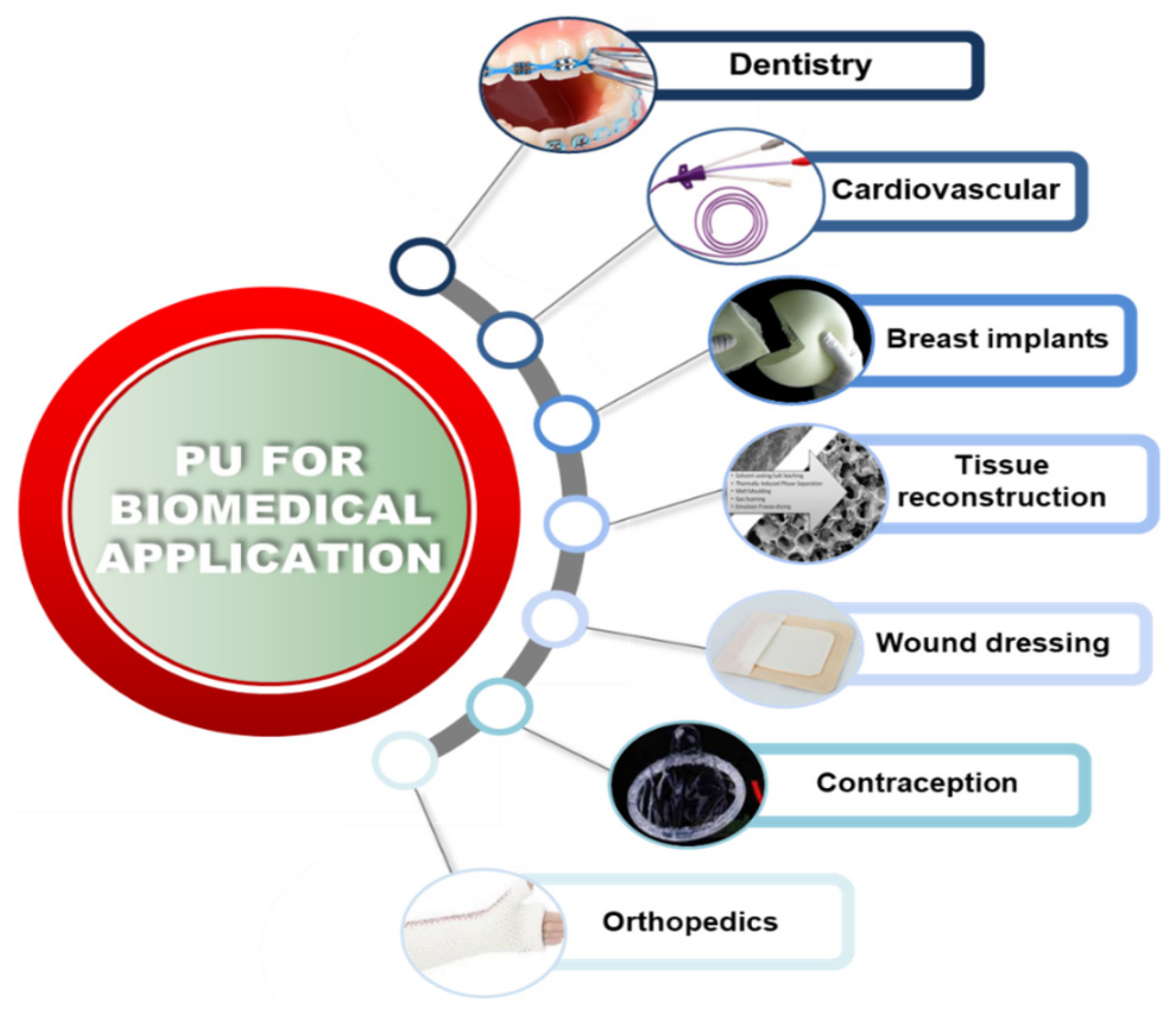
Figure 2.
A) Physical representations of the Beer-Lambert law and B) effect on the radiopacity of the sample. It shows the influence of the particle characteristic and the influence of the photon in the radiopacity of the sample.
Figure 2.
A) Physical representations of the Beer-Lambert law and B) effect on the radiopacity of the sample. It shows the influence of the particle characteristic and the influence of the photon in the radiopacity of the sample.
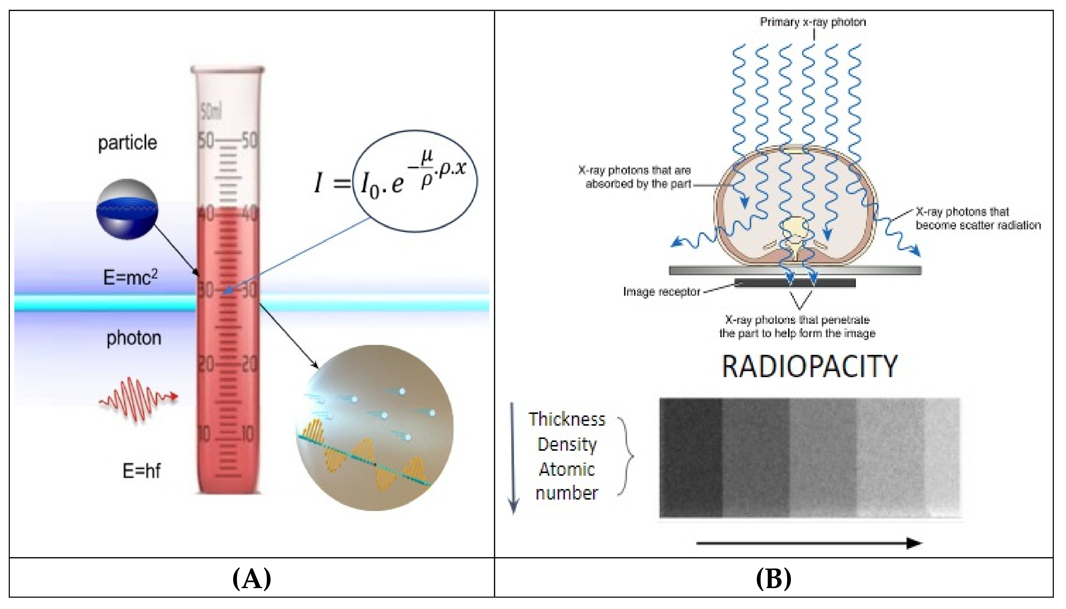
Figure 3.
Influence of the X-ray attenuation vs. energy emitted in the contrast between different materials. The figure was adapted from [12].
Figure 3.
Influence of the X-ray attenuation vs. energy emitted in the contrast between different materials. The figure was adapted from [12].
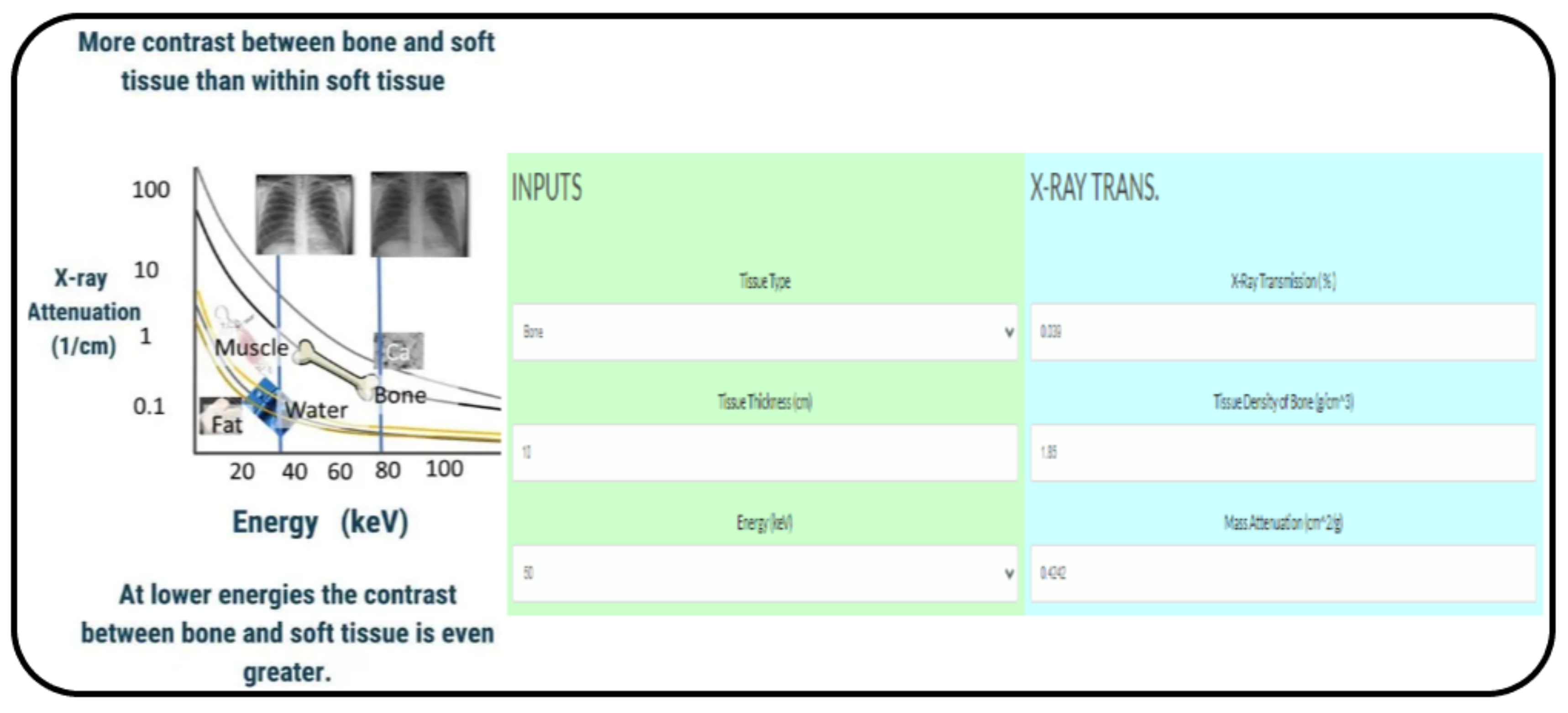
Figure 4.
Illustrative summary of x-ray and γ-ray interactions. Figure was obtained from [15].
Figure 4.
Illustrative summary of x-ray and γ-ray interactions. Figure was obtained from [15].
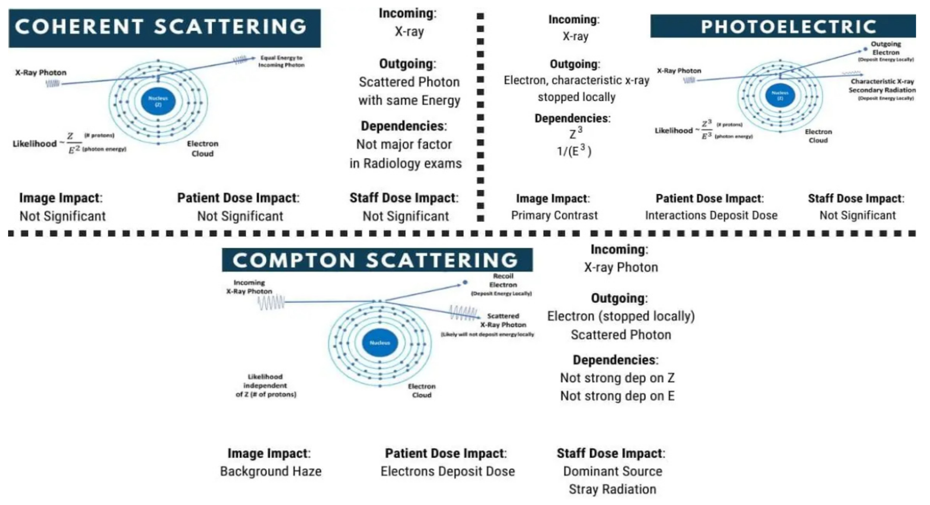
Figure 5.
Arm showing fractures in radiographic images, the corresponding color image after surgery, and the radiographic images after fixation of the bones using metal rods. The image was used under the Creative Commons License from [16].
Figure 5.
Arm showing fractures in radiographic images, the corresponding color image after surgery, and the radiographic images after fixation of the bones using metal rods. The image was used under the Creative Commons License from [16].
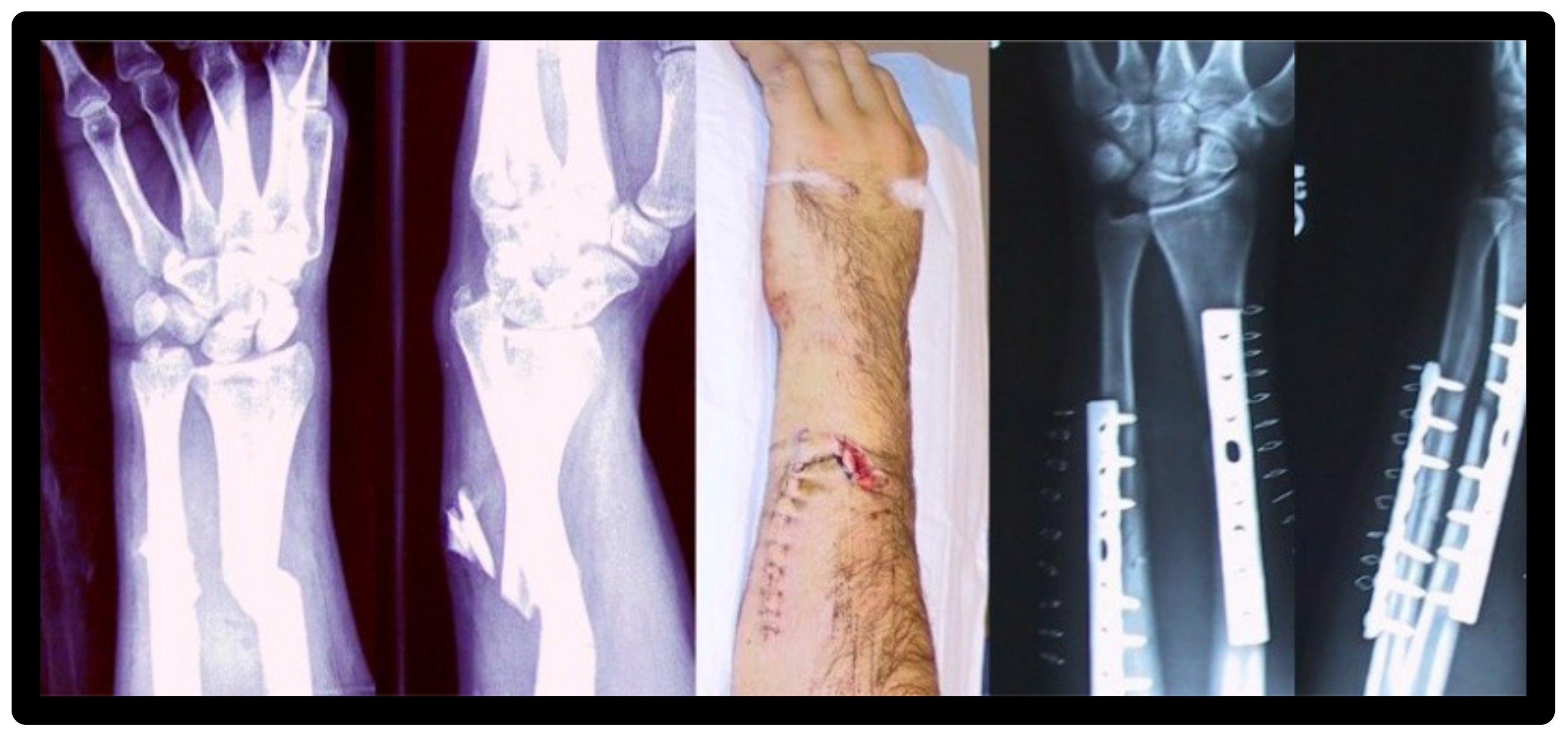
Figure 6.
Representation scheme of nanoparticles effects on microbes from [34].
Figure 6.
Representation scheme of nanoparticles effects on microbes from [34].
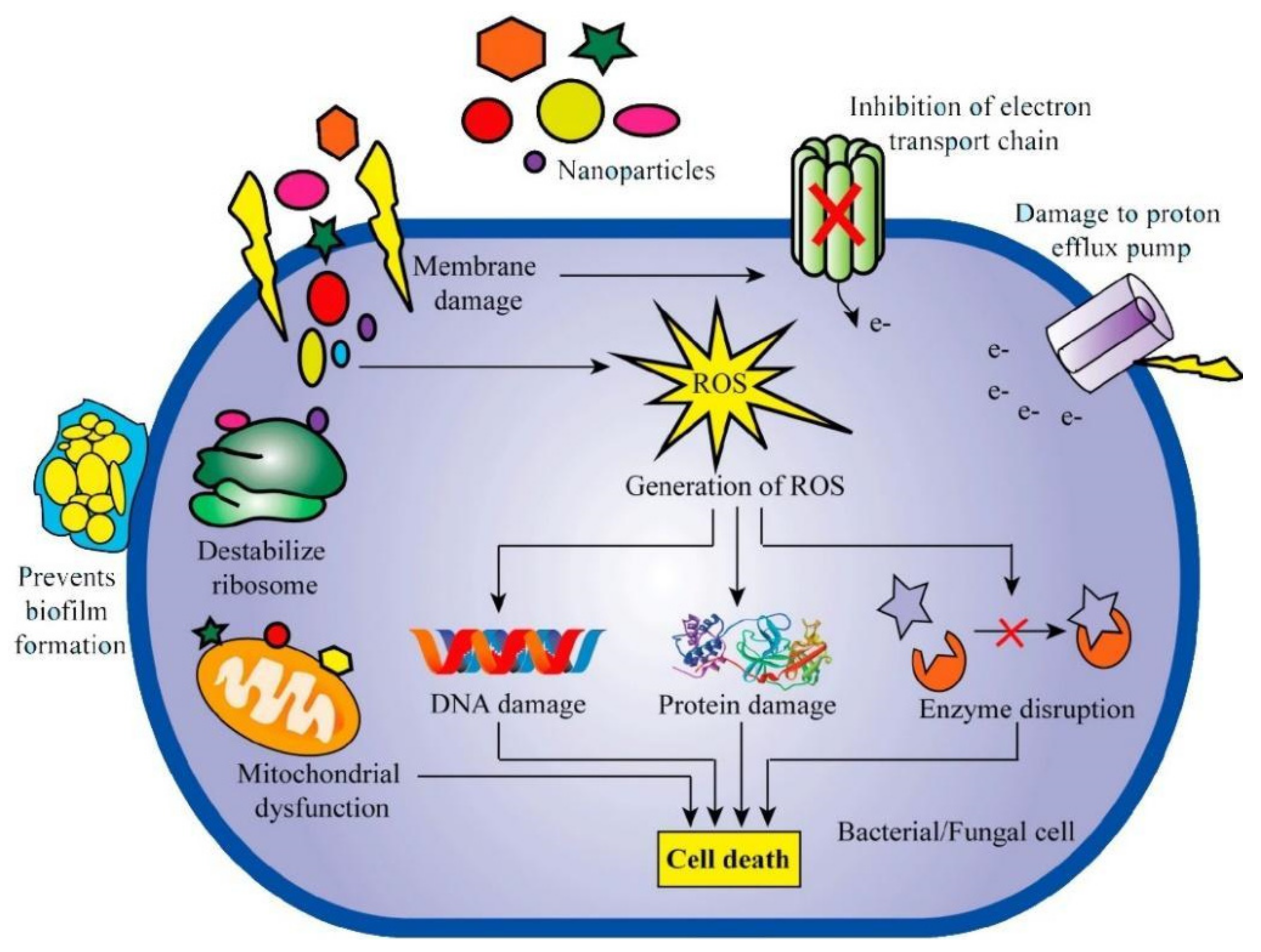
Figure 7.
Representation scheme of nanoparticles effects on the virus from [34].
Figure 7.
Representation scheme of nanoparticles effects on the virus from [34].
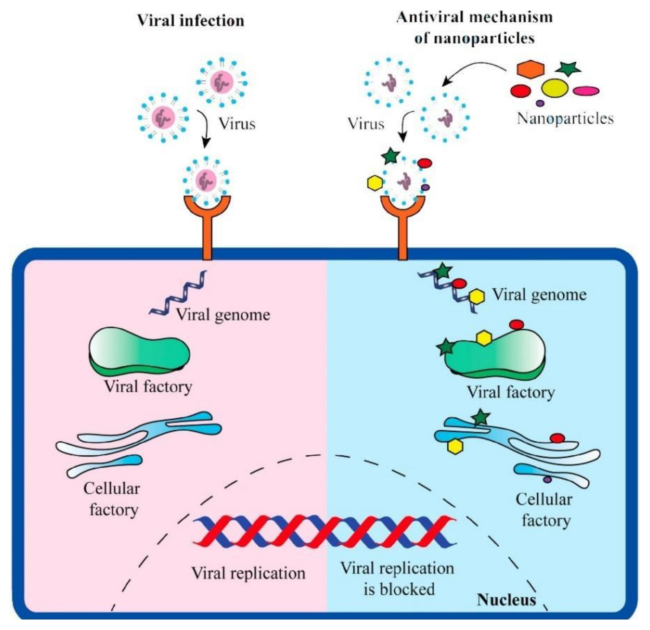
Figure 8.
Use of magnetic nanoparticles in tumor bioimaging and therapy. The Figure was used under the Creative Commons Attribution (CC BY) license [72].
Figure 8.
Use of magnetic nanoparticles in tumor bioimaging and therapy. The Figure was used under the Creative Commons Attribution (CC BY) license [72].
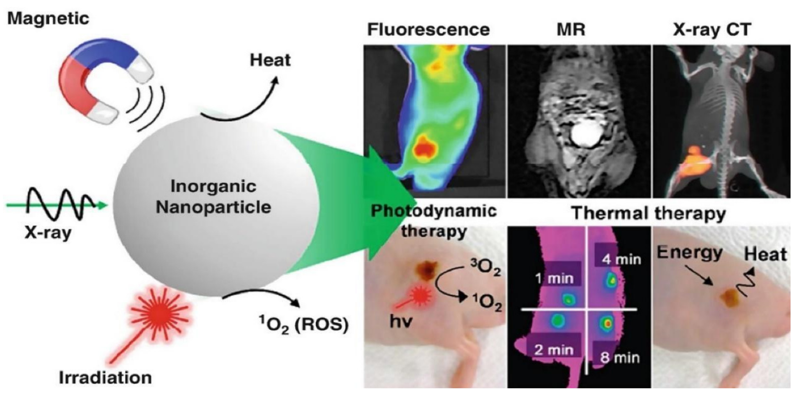
Figure 9.
(A) Digital radiograph of radiolucent UHMWPE cable (1), UHMWPE cable incorporated with Bi2O3 particles in different views (2−4), and a titanium cable (5−6) relative to an aluminum step wedge (B) radiograph of the radiopaque UHMWPE cable implanted in a sheep spine. The images were taken from [20] under CC-BY 4.0 license.
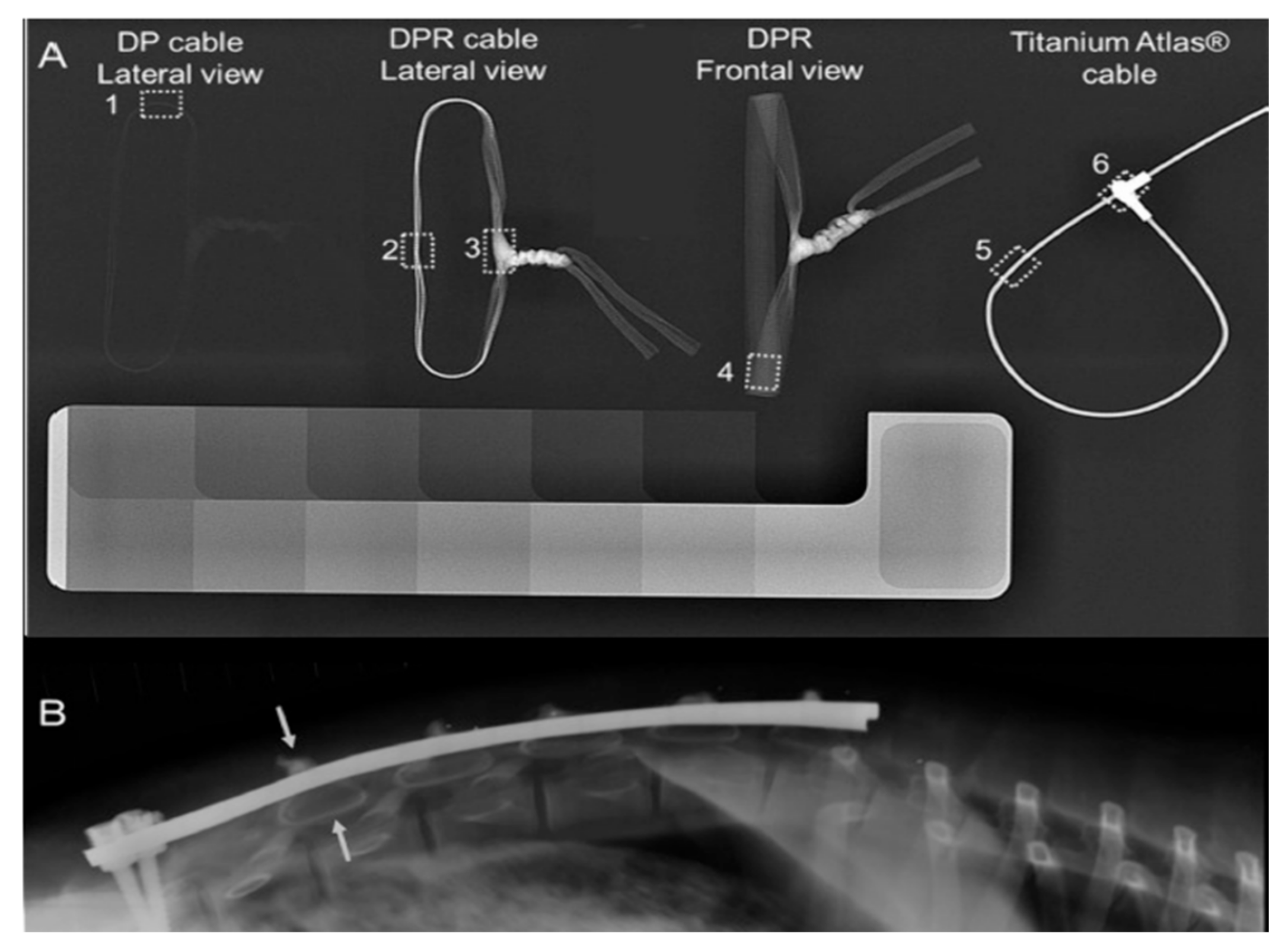
Figure 10.
Micro-CT images of radiolucent PLLA bone screws (a,c) and radiopaque PLLA + 20 wt %. Fe2O3 particles (b, d) at 2 and 4 weeks, respectively, were implanted in white rabbits. The images were taken from [20] under CC-BY 4.0 license.
Figure 10.
Micro-CT images of radiolucent PLLA bone screws (a,c) and radiopaque PLLA + 20 wt %. Fe2O3 particles (b, d) at 2 and 4 weeks, respectively, were implanted in white rabbits. The images were taken from [20] under CC-BY 4.0 license.
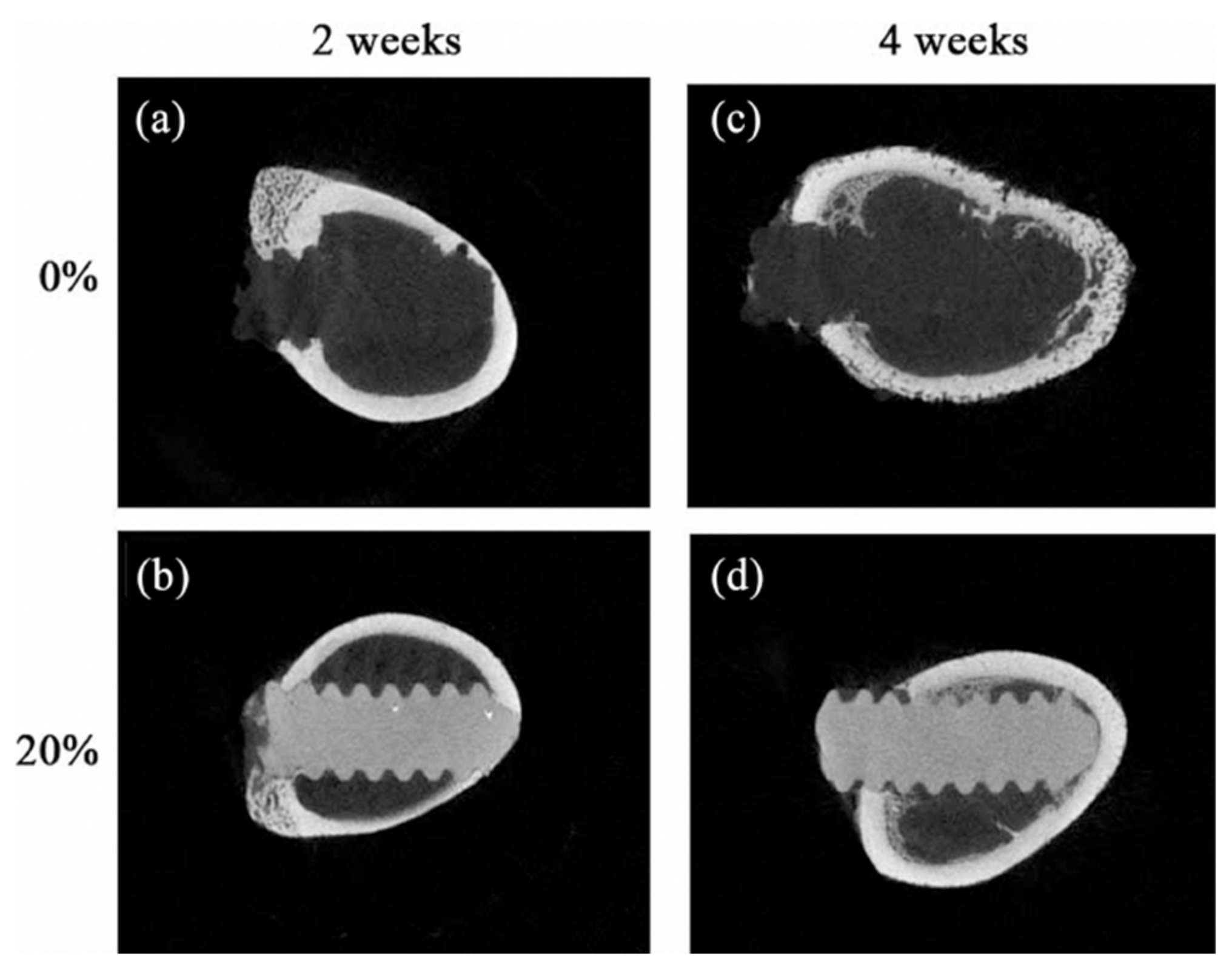

Table 1.
Inorganic nanoparticle activities in microbial strains according to concentration and size as referenced in [34].
Table 1.
Inorganic nanoparticle activities in microbial strains according to concentration and size as referenced in [34].
| Particle and Size | Test on microbial strains | Concentration | Mechanism of action | Ref |
|---|---|---|---|---|
| Silver nanoparticles (7 nm) |
E. coli (Gram-negative) S. aureus(Gram-positive) |
3.38 and 6.75 μg/mL | Damage DNA and disturb the synthesis of protein | [36] |
| Silver oxide nanoparticles (42.7 nm) |
Streptococcus mutans (Gram-positive) Lactobacillus acidophilus (Gram-positive) |
Streptococcus mutans: At conc 250 μg zone of inhibition (ZI) was 6 ± 0.8 mm, MBC was 22 ± 0.2% L. acidophilus: zone of inhibition 8 ± 0.4 mm, MBC 25 ± 0.5% |
Mechanism unclear | [37] |
| Fluorescent Ag nanoparticles (nAg-Fs), 1.5 nm |
Staphylococcus epidermidis NCIM2493, Bacillus megaterium (Gram-positive) Pseudomonas aeruginosa ATCC27853, Escherichia coli (Gram-negative) |
No cell growth was observed at conc. 2.0 μg/mL | Penetration of nAg-NPS into cell cytoplasm, leakage of cytoplasmic contents | [38] |
| SDS-stabilized silver nanoparticles (AgNPs), 25 nm |
Candida albicans (I, II) Candida tropicalis, Candida parapsilosis |
0.052 mg/L (C. albicans I) 0.1 mg/L (C. albicans II) 0.42 mg/L (C. tropicalis) 0.84 mg/L (C. parapsilosis) |
The surfactant activity of NPs disrupts the cell wall of yeast. | [39] |
| Silver nanoparticles (30–50 nm) | HIV-1 isolates | 0.44 to 0/91 mg/mL | Prevention of CD-4 dependent virion binding, fusion, infectivity, inhibition of post-entry stages of HIV-1 lifecycle. | [40] |
| Silver/chitosan nanoparticles (3.5, 6.5, 12.9 nm) | H1N1 influenza A | 100 μg of Ag NPs was added to 1 mg of chitosan | Inhibiting viral penetration into the host cell | [41] |
| Silver nanoparticles of Lampranthus coccineus (10.12–27.89 nm), Malephora lutea (8.91–14.48 nm). | HAV-10, HSV-1, CoxB4 |
L. coccineus: HAV-10- no activity, HSV-1- 520.6μg/mL, COxB4- no activity (aqueous nano extract) 11.7μg/mL, 36.36μg/mL, 12.74μg/mL (hexane nano extract) M. lutea: no activity for aqueous nano extract, HAV-10- 31.38μg/mL, HSV-1-no activity, COxB4- 29.04μg/mL (hexane nano extract) |
Not determined | [42] |
| Copper nanoparticles (3–10 nm) |
Phoma destructiva (DBT 66) Curvularia lunata (MTCC, 2030) Alternaria alternate (MTCC 6572) Fusarium oxysporum (MTCC 1755) |
Zone of inhibition (ZI) value for Phoma destructive: 22 ± 1 mm Curvularia lunata: 21 ± 0.5 mm Alternaria alternate: 18 ± 1 mm Fusarium oxysporum: 24 ± 0.5 mm |
Not clearly mentioned | [43] |
| Cuprous oxide nanoparticles (45.4 ± 68 nm) | Hepatitis C virus (HCV) | 2 μg/mL | Attachment and entry Inhibition of HCV infection | [44] |
| Copper Iodide nanoparticles (160 nm) | Feline Calicivirus | 10 ng/mL to 10 μg/mL | ROS generation and subsequent capsid protein oxidation | [45] |
| Gold-chitosan hybrid nanoparticles (16.9 nm) |
S. aureus (Gram positive) P. aeruginosa (Gram-negative) |
0.25 mg/mL | Mechanism still unclear | [46] |
| Gold nanoparticles (25 nm) | Candida sp | 16–32 μg/mL | Inhibition of H + ATPase leads to intracellular acidification and cell death | [47] |
| Gold nanoparticles (17 nm) | HIV-1 | 0.05–0.12 mg/mL | The mechanism of gold nanoparticles against HIV-1 is not clear but it inhibits the HIV-1 fusion | [48] |
| Zinc oxide nanoparticles (30 nm) | Camphylobacter jejuni (Gram-negative) | 0.05–0.025 mg/mL | Disruption of the cell membrane and oxidative stress in C. jejuni. | [49] |
| Zinc oxide nanoparticles (70 nm) |
Botrytis cinerea Penicillium expansum |
3–12 mol/Ll−1 | Inhibition of growth by affecting cellular functions | [50] |
| Zinc oxide nanoparticles (ZnO NPs), 70 ± 15 nm |
Botrytis cinerea, Penicillium expansum |
3 mmol/L | Deformation in fungal hyphae by affecting cellular function | [50] |
| Zinc oxide nanoparticles (12–32 nm) |
Alternaria alternata (ITCC 6531), Aspergillus niger (ITCC 7122), Botrytis cinerea (ITCC 6192), Fusarium oxysporum (ITCC 55), Penicillium expansum (ITCC 6755) |
64 μg/mL (A. alternata) 16 μg/mL (A. niger) 128 μg/mL (B. cinerea) 64 μg/mL (F. oxysporum) 128 μg/mL (P. expansum) |
Disruption of membrane structure and change in permeability. | [51] |
| Zinc oxide nanoparticles (16–20 nm) | H1N1 Influenza | 75 and 200 μg/mL | Suppress the proliferation of influenza virus at an inhibition rate of 52.2% | [52] |
| Zero-valent Iron (Fe°) nanoparticles, spherical (31.1 nm) |
Staphylococcus aureus (Gram-positive) E. coli (Gram-negative) |
MIC for both strains at 30 μg/mL and complete growth inhibition at 60 μg/mL | Oxidative stress generation via ROS and visible damage to bacterial protein and DNA. | [53] |
| Magnetic Iron oxide nanoparticles (50–110 nm) | S. aureus (Gram-positive) | DMF solution with 40 and 60 mJ laser fluencies showed the highest antibacterial activity | This could be due to stress generated by ROS disrupting the bacterial cell membrane. | [54] |
| Iron oxide nanoparticles (10–30 nm) | Trichothecium roseum, Cladosporium herbarum, Penicillium chrysogenum, Alternaria alternate and Aspergillus niger. | Varies between 0.063-0.016 mg/mL | Formation of ROS, damage of protein, and DNA by oxidative stress. | [55] |
| Nickel ferrite (NiFe2O4) nanoparticles (NFOTP) | Staphylococcus aureus NCIM 5021, Streptococcus pyogenes NCIM 5280 (Gram-positive) Escherichia coli NCIM 2345, Salmonella typhimurium NCIM 2501 (Gram-negative) |
Zone of inhibition for E. coli was seen but no numeric value is mentioned. | Higher negatively charged surface of E. coli, thin surface and formation of reactive oxidative species (ROS) and oxidative stress lead to cell death. | [56] |
| Iron oxide nanoparticles (10–15 nm) | A/Puerto Pico/8/1934H1N1 influenza virus strain (PR8-H1N1) | 1.1 pg | Inactivation of cell protein through the interaction of nanoparticles and –SH group (Proposed, not investigated yet) | [56] |
| TiO2 nanoparticles (70–100 nm) | Candida albicans | 5.14 μg/mL | Inhibition of fungal biofilms | [57] |
| BaSO4 nanoparticles (73 nm) |
Staphylococcus aureus; P. aeruginosa (Schroeter) ;Migula |
Nano BaSO4, 40% | Hypothesized the difficulty of preliminary steps on bacterial adhesion due to the nano roughness of the material | [58] |
Table 2.
Different nanomaterial types are used for bioimaging applications [72].
Table 2.
Different nanomaterial types are used for bioimaging applications [72].
| Nanomaterial | Functionalization | Cell Lines | Ref |
| Graphene-based nanosheets | Surface functionalization by bio-compatible targeting ligands and coatings | MDA-MB-468 (MCF-7) | [75] |
| Molybdenum disulfide nanosheets | Chitosan; PLGA, PEG functionalization | Breast cancer cells (MDA-MB-468), HeLa uterine cancer cells, human lung cancer cells | [76] |
| Transition metal nanoparticles decorated with polymers | Polymer functionalization | Mice bearing 4T1 breast cancer cell xenografts | [77] |
| Lanthanide-activated nanoparticles | Doping with lanthanide | Cancer cells xenografted in mice | [78] |
| Group IV quantum dots | Surface functionalization | Various cancer cell types | [79] |
| Graphene oxide nanosheets | Surface functionalization | Tumor cells | [80] |
| Peptide-based nanoparticles | Chemical functionalization | Peptide-treated HeLa cells preloaded with Hg2+ | [81] |
| Silver nanoparticles | Aptamer conjugation | Leukemia cells, neural stem cells, kidney tissue, renal carcinoma cells | [82] |
| Gold nanoprisms | Conjugation with polyethylene glycol | Gastrointestinal carcinoma cells (HT 29) | [83] |
| Gold nanorods | Encasing by mesoporous silica | Carcinoma cells | [84] |
| Magnetofluroscentnanoprobe | Surface functionalization | Human Breast Cancer (MCF-7), HeLa cells | [85] |
| Dye-loaded nanoemulsions | Lipids conjugation with polyethylene glycol | Human colon cancer (HCT116), HeLa cells | [86] |
| Cadmium telluride quantum dots | Capping by shells | Human bronchial epithelial cells | [87] |
Table 2. Nanomaterials used for bioimaging applications. The Table was used under the Creative Commons Attribution (CC BY) license [72].
Table 3.
Summary of Contrast Agents Incorporated into Synthetic Polymers for Implantable Devices—Radiopacity Enhancements in Polymeric Implant Biomaterials: A Comprehensive Literature Review | ACS Biomaterials Science & Engineering [20].
Table 3.
Summary of Contrast Agents Incorporated into Synthetic Polymers for Implantable Devices—Radiopacity Enhancements in Polymeric Implant Biomaterials: A Comprehensive Literature Review | ACS Biomaterials Science & Engineering [20].
| Contrast agent | Blending method | Polymer | Application | Content | Reported effects | Polymer biodegradable | Biological response | Ref |
|---|---|---|---|---|---|---|---|---|
| BaSO4 | Blended in the powder phase | PMMA | Bone cement | 9–15 wt % | Hard particles, third body wear, reduced tensile and flexural strength | NO | Osteoclast formation | [88] [89] [90] |
| Blended in the powder phase | PMMA | Vertebroplasty cement | 30 wt % | Hard particles, third body wear, lower viscosity | NO | Osteoclast formation | [91] [92] [93] |
|
| Twin-screw micro-compounding | PLLA | Bioresorbable stents | 5–20 wt % | Increased tensile modulus and strength, decreased elongation at break and ductility | YES | No adverse effects after 21 days | [94] [95] |
|
| Magnetic stirring in organic solvent | PLGA | Bioresorbable stent | 17.9 v/v % | Increased Young’s modulus, reduced elasticity, increased radial strength | YES | Na | [96] | |
| Solution mixing | PLGA | Bone fixation plate | 1:10 and 1:3 w/w PLGA:BaSO4 | Radiopaque up to 56 days, BaSO4 leaching < 0.5 mg/day; insufficient to induce cytotoxicity | YES | No adverse effects | [97] | |
| Lipiodol ultra fluid | Immersion in oil at elevated temperature | UHMWPE | TKA insert | 25 mL | Physical alteration–swelling, 54% reduction in surface radiopacity after 4 weeks | NO | Na | [98] |
| Iohexol(IHX) | Stirring | PLA | Bioresorbable implants | 40 wt % | Reduced tensile strength, elongation at break and increased tensile modulus, enhanced crystallinity, slower polymer degradation | YES | Thin fiber capsule | [99] |
| Blended in the powder phase | PMMA | Bone cement | 10 wt % | Better biocompatibility compared to conventional contrast agents | NO | Osteoclast formation | [90] | |
| Iodixanol(IDX) | Blended in the powder phase | PMMA | Bone cement | 10 wt % | Higher osteoclast formation than IHX | NO | Osteoclast formation | [90] |
| Iobitridol | Dissolved in liquid phase | CPC | Bone cement | 56 mg Ml^–1 | Rapid release of contrast, no significant change in mechanical properties, no effect on injectability, cohesion, or setting time | YES | No adverse effects | [100] |
| Iodinated diphenol | Polymerization reaction | PLA diol | Coronary stent | <1% of 1 mL of iodine contrast | Increased ultimate tensile strength and elongation at break, long-term radiopacity | YES | No adverse effects | [101] |
| Bismuth salicylate(BS) | Dissolved in liquid phase | PMMA | Vertebroplasty cement | 10 w/w | Reduced compressive and tensile strength, reduced strain, lower setting temperature, increased radiopacity, longer injection time, Better compatibility than BaSO4 | NO | Na | [102] [103] |
| Triphenyl bismuth(TPB) | Dissolved in liquid phase | PMMA | Bone cement | 10 wt % | Increased ultimate tensile strength, Young’s modulus and strain to failure, lower setting temperature, better homogeneity | NO | Na | [104] |
| Bismuth oxide Bi2O3 | Blended into fiber | UHMWPE | Sublaminar cables | 20 wt % | Decreased tensile strength, limited leaching below toxic levels | NO | No adverse effects | [105] [106] |
| Titanium dioxide TiO2 | Blending | PE | Orbital implant | 6% | Slight decrease in tensile strength and modulus, significant decrease in compressive strength and modulus, reduced hardness | NO | No adverse effects | [107] |
| Iron oxide Fe3O4 | Twin-screw extrusion | PLLA | Bone screws | 20 wt % | Reduced flexural strength, increased crystallinity, increased thermal stability | YES | Osteogenic effect, no adverse effects | [108] |
Disclaimer/Publisher’s Note: The statements, opinions and data contained in all publications are solely those of the individual author(s) and contributor(s) and not of MDPI and/or the editor(s). MDPI and/or the editor(s) disclaim responsibility for any injury to people or property resulting from any ideas, methods, instructions or products referred to in the content. |
© 2024 by the authors. Licensee MDPI, Basel, Switzerland. This article is an open access article distributed under the terms and conditions of the Creative Commons Attribution (CC BY) license (http://creativecommons.org/licenses/by/4.0/).
Copyright: This open access article is published under a Creative Commons CC BY 4.0 license, which permit the free download, distribution, and reuse, provided that the author and preprint are cited in any reuse.


