Submitted:
04 January 2025
Posted:
07 January 2025
You are already at the latest version
Abstract
EphB2 is a member of the Eph family tyrosine kinase receptors. EphB2 binds to ephrin-B1, ephrin-B2, and ephrin-B3, which are critical regulators of vascular and nervous development through controlling cell migration and axon guidance. EphB2 is overexpressed in tumors, including glioma, breast cancer, hepatocellular carcinoma, and malignant mesothelioma, and it functions as a tumor promoter. Therefore, the development of monoclonal antibodies (mAbs) against EphB2 is essential for tumor diagnosis and therapy for EphB2-positive tumors. In this study, we developed novel mAbs for human EphB2 using the Cell-Based Immunization and Screening (CBIS) method. One of the established anti-EphB2 mAbs, Eb2Mab-12 (mouse IgG1, kappa), reacted with EphB2-overexpressed Chinese hamster ovary-K1 (CHO/EphB2) and an endogenously EphB2-expressing cancer cell line (LS174T) by flow cytometry. Using flow cytometry, the dissociation constant (KD) values of Eb2Mab-12 for CHO/EphB2 and LS174T were determined as 1.7 × 10⁻⁹ M and 4.4 × 10⁻¹⁰ M, respectively. Furthermore, Eb2Mab-12 did not cross-react with other members of EphA and EphB receptors. These results indicated that Eb2Mab-12 possesses a high affinity and specificity for detecting EphB2 and could apply to tumor therapy.
Keywords:
EphB2
; monoclonal antibody
; Cell-Based Immunization and Screening
; flow cytometry
1. Introduction
The mammalian ephrin and Eph system includes eight cell surface ephrin ligands (five ephrin-As and three ephrin-Bs) and 14 receptor tyrosine kinases (nine EphA and five EphB receptors). [1,2,3,4,5,6] Eph receptors make the complex to ephrins with dimerization or oligomerization, which leads to the tyrosine phosphorylation of Eph receptor and ephrin-B [7]. The phosphorylated tyrosine recruits cytoplasmic effectors containing Src-homology 2 (SH2) domains, phosphotyrosine-binding (PTB) domains, and PDZ domains [8]. Therefore, the Eph receptor and ephrin complexes activate bidirectional (forward and reverse, respectively) signaling, which is essential for communication in the same or different types of cells [2,6,9]. Through bidirectional signaling, the Eph system regulates tissue development, homeostasis, and regeneration; the dysregulation causes many diseases including cancer.[3,4,10,11,12,13,14,15,16,17,18,19,20,21] Therefore, monoclonal antibody (mAb)-based tumor therapies have been developed in some Eph receptors [10,22,23,24,25,26,27,28,29].
The dysregulation of the Eph system is observed in both tumor cells and tumor microenvironment [19]. The Eph system plays distinct roles in tumor development and functions as both tumor promoters and suppressors in a cellular context-dependent manner [19]. EphB2 is overexpressed in several tumors, such as glioblastoma [30], breast cancer [31], hepatocellular carcinoma [32], and malignant mesothelioma [21], which is associated with poor clinical outcomes. In these tumors, EphB2 promotes the migration and invasion via forward signaling [33,34].
In contrast, the expression of EphB2 is downregulated in some tumors such as colorectal cancer [35]. In the intestinal epithelium, EphB receptors promote stem and progenitor proliferation [36]. The intestinal epithelial cell migration is deranged in mice lacking EphB2 and EphB3, and the absence of EphB signaling results in ~50% reduction in the number of proliferating cells [36]. Furthermore, EphB receptor expression is elevated in intestinal adenomas [37]. In contrast, EphB2 function as tumor suppressors by inhibiting the invasive growth. EphB signaling promotes adherens junction formation of colorectal cancer cells, suppressing cancer progression by inhibiting invasive growth [38]. EphB2 expression is lost during progression to carcinoma and initiation of invasive growth [39].
To evaluate the expression of EphB2 and targeting the EphB2-positive cancer cells, the development of monoclonal antibodies (mAbs) against EphB2 is essential. We have developed anti-receptor tyrosine kinase mAbs against human epidermal growth factor (EGFR) (clone EMab-17) [40], mouse EGFR (EMab-300) [41], HER2 (H2Mab-19) [42], mouse HER2 (H2Mab-304) [43], and HER3 (H3Mab-17) [44] using the Cell-Based Immunization and Screening (CBIS) method. The CBIS method includes immunizing antigen-overexpressed cells and high-throughput hybridoma screening using flow cytometry. In this study, novel anti-EphB2 mAbs were developed by the CBIS method.
2. Materials and Methods
2.1. Antibodies
OptiBuild™ RB545 mouse anti-human EphB2 mAb (clone 2H9; mouse IgG1, kappa) was purchased from BD Bioscience (Franklin Lakes, NJ). Alexa Fluor 488-conjugated anti-mouse IgG was purchased from Cell Signaling Technology, Inc. (Danvers, MA).
2.2. Preparation of cell lines
LS174T (human colorectal cancer), LN229 (human glioblastoma), Chinese hamster ovary (CHO)-K1, and P3X63Ag8U.1 (P3U1) were obtained from the American Type Culture Collection (Manassas, VA).
pCMV6-myc-DDK vector with EphB2 (Catalog No.: RC223882, Accession No.: NM_004442) was purchased from OriGene Technologies, Inc. (Rockville, MD). The EphB2 plasmids were transfected into CHO-K1 and LN229 cells using a Neon transfection system (Thermo Fisher Scientific Inc., Waltham, MA). Stable transfectants were established through cell sorting using the RB545-conjugated anti-human EphB2 (2H9) mAb and a cell sorter (SH800; Sony Corp., Tokyo, Japan), after which cultivation in a medium containing 0.5 mg/mL of G418 (Nacalai Tesque, Inc., Kyoto, Japan) was performed.
Other Eph receptor cDNAs including EphA1 (Catalog No.: RC213689, Accession No.: NM_005232), EphA4 (Catalog No.: RC211230, Accession No.: NM_004438), EphA5 (Catalog No.: RC213206, Accession No.: NM_004439), EphA6 (Catalog No.: RC223510, Accession No.: NM_001080448), EphA7 (Catalog No.: RC226293, Accession No.: NM_004440), EphA8 (Catalog No.: RC220352, Accession No.: NM_020526), EphA10 (Catalog No.: RC218374, Accession No.: NM_001099439) EphB1 (Catalog No.: RC214301, Accession No.: NM_004441), EphB6 (Catalog No.: RC229404, Accession No.: NM_004445), were purchased from OriGene Technologies, Inc. EphA2 (Catalog No.: HGY095959, Accession No.: NM_004431), EphA3 (Catalog No.: HGY053437, Accession No.: NM_005233), and EphB3 (Catalog No.: HGX039581, Accession No.: NM_004443) cDNAs were purchased from RIKEN DNA Bank (Ibaraki, Japan).
EphA2 and EphB3 cDNAs were cloned into a pCAGzeo vector (FUJIFILM Wako Pure Chemical Corporation, Osaka, Japan). EphB6 cDNA was cloned into a pCMV6 vector. EphA1 cDNA was cloned into a pCAGzeo-ssnPA vector. EphA3, EphA4, EphA5, EphA6, EphA7, EphA8, EphA10, and EphB1 cDNA was cloned into a pCAGzeo_ssnPA16 vector.
The plasmids were also transfected into CHO-K1 cells and stable transfectants were established by staining with an anti-EphA2 mAb (clone SHM16; BioLegend, San Diego, CA, USA), an anti-EphB3 mAb (clone 647354; R&D Systems Inc., Minneapolis, MN, USA), an anti-EphB6 mAb (clone T49-25; BioLegend), and an anti-PA tag mAb (clone NZ-1 for EphA2, EphA3, EphA4, EphA5, EphA6, EphA7, EphA8, EphA10, and EphB1), and sorted using SH800. After sorting, cultivation in a medium containing 0.5 mg/mL of Zeocin (InvivoGen, San Diego, CA, USA) or 0.5 mg/mL of G418 was conducted. These Eph receptors-overexpressed CHO-K1 (e.g., CHO/EphB2) clones were finally established. CHO/PA16-EphB4 was previously established [45].
2.3. Development of hybridomas
Two five-week-old BALB/cAJcl mice were purchased from CLEA Japan (Tokyo, Japan). To develop mAbs against EphB2, we intraperitoneally immunized the mice with LN229/EphB2 (1 × 10⁸ cells) plus Alhydrogel adjuvant 2% (InvivoGen). The harvested spleen cells were subsequently fused with P3U1 cells, using PEG1500 (Roche Diagnostics, Indianapolis, IN). The supernatants were subsequently screened using flow cytometry using CHO/EphB2 and CHO-K1.
2.4. Flow cytometric analysis
Cells were harvested after brief exposure to 0.25% trypsin and 1 mM ethylenediaminetetraacetic acid (EDTA, Nacalai Tesque, Inc.). The cells were washed with 0.1% bovine serum albumin (BSA) in PBS (blocking buffer) and treated with 0.01, 0.1, 1, and 10 μg/mL of primary mAbs for 30 min at 4°C. The cells were treated with Alexa Fluor 488-conjugated anti-mouse IgG (1:2,000). The cells were also suspended in 0.01, 0.1, 1, and 10 μg/mL of the RB545-conjugated anti-human EphB2 mAb (2H9). The fluorescence data were collected using the SA3800 Cell Analyzer (Sony Corp), and the data were analyzed using FlowJo (BD Biosciences).
2.5. Determination of dissociation constant (KD) by flow cytometry
CHO/EphB2 and LS174T cells were suspended in serially-diluted Eb2Mabs for 30 min at 4°C. The cells were treated with Alexa Fluor 488-conjugated anti-mouse IgG (1:200). The cells were also suspended in serially-diluted RB545-conjugated anti-human EphB2 mAb (2H9). The fluorescence data were collected using a FACSLyric (BD Biosciences). The KD was calculated by fitting saturation binding curves to the built-in, one-site binding models in GraphPad PRISM 6 (GraphPad Software, Inc., La Jolla, CA).
3. Results
3.1. Development of anti-EphB2 mAbs using the CBIS method
To develop anti-EphB2 mAb, mice were immunized with LN229/EphB2 cells (Figure 1A). The spleen was then excised from the mice, and splenocytes were fused with myeloma P3U1 cells (Figure 1B). The developed hybridomas were subsequently seeded into ten 96-well plates and cultivated for six days. The positive wells were screened by selecting CHO/EphB2-reactive and CHO-K1-non-reactive supernatants using flow cytometry (Figure 1C). We finally obtained 133 positive wells (13.9%) out of 956 wells. After the limiting dilution of the part of positive wells and several additional screenings, twelve clones were finally established (Figure 1D).
3.2. Flow cytometric analysis using anti-EphB2 mAbs
We next focus on eight mouse IgG1 clones (Eb2Mab-1, 2, 3, 4, 7, 8, 10, and 12) and purified the mAbs from supernatants (Table 1). We first investigated the specificity of Eb2Mabs and 2H9 (from BD Biosciences) in 14 Eph receptor tyrosine kinases (nine EphA and five EphB receptors)-expressed CHO-K1 cells. 2H9 exhibited the reactivity to not only CHO/EphB2, but also CHO/EphA4, CHO/EphB1, and CHO/EphB3 at 1 μg/mL (Supplementary Figure S1). In contrast, Eb2Mab-1, 2, 4, 7, and 12 recognized CHO/EphB2, but did not show the cross-reactivity even at 10 μg/mL. In contrast, Eb2Mab-3 exhibited the cross-reactivity to EphA3, EphB1, and EphB3. In addition, Eb2Mab-8 and 10 also exhibited the cross-reactivity to EphB3 (Supplementary Figures S2–S4 and Table 1). We next conducted flow cytometry using the Eb2Mabs and 2H9 against CHO/EphB2, CHO-K1, LN229/EphB2, and LN229 cells. Eb2Mabs recognized CHO/EphB2 cells dose-dependently at 10, 1, 0.1, and 0.01 μg/mL (Supplementary Figure S5). Among EphB2-specific Eb2Mabs (Eb2Mab-1, 2, 4, 7, and 12), Eb2Mab-12 showed the high reactivity (Figure 2A). 2H9 also recognized CHO/EphB2 cells dose-dependently at 10, 1, 0.1, and 0.01 μg/mL, which are less effective compared to Eb2Mab-12 (Figure 2A). Parental CHO-K1 cells were not recognized even at 10 μg/mL of Eb2Mab-12 and 2H9 (Figure 2B). The superior reactivity of Eb2Mab-12 compared to 2H9 was also observed in LN229/EphB2 and LN229 cells (Supplementary Figure S6). The weak expression of endogenous EphB2 in LN229 was previously confirmed by quantitative PCR and western blot analyses [46].
We next investigated the reactivity of Eb2Mabs and 2H9 against an endogenous EphB2-expressing colorectal cancer cell line, LS174T [47]. Eb2Mabs recognized LS174T cells dose-dependently at 10, 1, 0.1, and 0.01 μg/mL (Supplementary Figure S7). Among Eb2Mabs, Eb2Mab-12 also showed the high reactivity (Figure 3). In contrast, 2H9 could react with LS174T cells at more than 0.1 μg/mL (Figure 3). These results suggest that Eb2Mab-12 specifically recognizes EphB2, and is also helpful in detecting endogenous EphB2 by flow cytometry.
3.3. Determination of the binding affinity of Eb2Mabs and 2H9 using flow cytometry
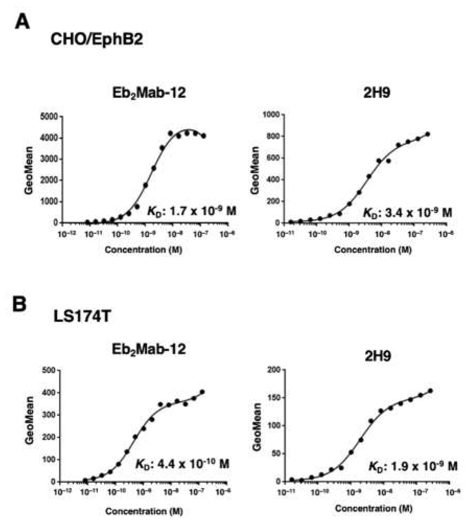
To determine the KD values of Eb2Mabs and 2H9, we conducted flow cytometry, and the geometric mean of the fluorescence intensity was plotted versus the concentration of mAbs. The KD values of Eb2Mab-12 and 2H9 for CHO/EphB2 were determined as 1.7 × 10⁻⁹ M and 3.4 × 10⁻⁹ M, respectively (Figure 4A). Although we also determined the KD values of other Eb2Mabs, Eb2Mab-12 exhibited the high affinity (Table 1). We next determined the KD values of Eb2Mab-12 and 2H9 for LS174T as 4.4 × 10⁻¹⁰ M and 1.9 × 10⁻⁹ M, respectively (Figure 4B). These results indicate that Eb2Mab-12 possesses the superior affinity to CHO/EphB2 and LS174T compared to that of 2H9.
Figure 4.
The binding affinity of Eb2Mab-12 and 2H9. CHO/EphB2 (A) and LS174T (B) cells were suspended in serially diluted Eb2Mab-12. The cells were treated with anti-mouse IgG conjugated with Alexa Fluor 488. The cells were also suspended in serially diluted 2H9 conjugated with RB545. The fluorescence data were subsequently collected using the FACSLyric, followed by the calculation of the KD using GraphPad PRISM 6.
4. Discussion
An anti-EphB2 mAb, clone 2H9, was extensively characterized and developed for tumor therapy as an antibody-drug conjugate [48]. The 2H9 was established by the immunization of mice with the EphB2 ectodomain produced by baculovirus expression system [48]. However, 2H9 showed the cross-reactivity to CHO/EphA4, CHO/EphB1, and CHO/EphB3 (Supplementary Figure S1). In this study, we established anti-EphB2 mAbs using the CBIS method (Figure 1). Among the established mAbs, Eb2Mab-12 exhibited the superior reactivity compared to 2H9 in CHO/EphB2 (Figure 2), LN229/EphB2 (Supplemental Figure S2), and LS174T (Figure 3) cells. Furthermore, Eb2Mab-12 possesses a higher affinity to CHO/EphB2 and LS174T than that of 2H9 (Figure 4). Importantly, Eb2Mab-12 did not show the cross-reactivity even at a high concentration (Supplementary Figures S2–S4 and Table 1). Therefore, Eb2Mab-12 is a highly-sensitive and specific anti-EphB2 monoclonal antibody for flow cytometry.
The 2H9 effectively blocked the interaction of EphB2 with the ligands and inhibited the autophosphorylation of EphB2 [48]. However, 2H9 did not affect the proliferation of EphB2-positive tumor cells [48]. The identification of the epitope is essential to assess the properties of Eb2Mab-12 and 2H9. We have developed the RIEDL insertion for epitope mapping (REMAP) and PA insertion for epitope mapping (PAMAP) methods to determine the conformational epitopes of mAbs. The epitopes of anti-EGFR mAb (EMab-134) [49] and anti-CD44 mAb (C44Mab-46) [50] could be determined using the REMAP method. Furthermore, the epitopes of anti-mouse CD39 mAb (C39Mab-1) could be determined using both REMAP and PAMAP methods [51]. Therefore, further studies are required to determine the epitope and biological activities of Eb2Mab-12.
We investigated the expression of EphB2 in more than 100 cell lines using flow cytometry. LS174T exhibited the highest expression (Figure 3). Since LS174T is a transplantable cancer cell line in BALB/c nude mice [52], in vivo antitumor effects of Eb2Mab-12 could be evaluated. To perform this, converting Eb2Mab-12 (mouse IgG1) to mouse IgG2a subclass is essential for enhancing effector activation ability. We previously produced recombinant mAbs, which were converted into the mouse IgG2a subclass from mouse IgG1. Furthermore, we produced defucosylated IgG2a mAbs using fucosyltransferase 8-deficient CHO-K1 cells to enhance the antibody-dependent cellular cytotoxicity and in vivo antitumor effect in mouse xenograft models [53]. Therefore, a class-switched and defucosylated type of Eb2Mab-12 could contribute to the treatment of EphB2-positive tumors in preclinical studies. We also determined the property of Eb2Mabs (Eb2Mab-3, 8, and 10) which showed the cross-reactivity (Table 1). Such mAbs may be useful when targeting multiple EphBs for mAb-based therapy.
We have developed cancer-specific mAbs (CasMabs) against HER2 (H2Mab-250) [54], podocalyxin (PcMab-6) [55], and podoplanin (LpMab-2) [56], and revealed that these mAbs react with cancer cells, but not normal cells in flow cytometry. The strategy used in this study is also applicable to select anti-EphB2 CasMabs from Eb2Mabs (Table 1) or clones from remaining positive wells. We have confirmed that EphB2 is detected in some normal epithelial cells and will screen the anti-EphB2 CasMabs. The unique property of H2Mab-250 could contribute to developing HER2-targeting chimeric antigen receptor (CAR)-T cells (now in a clinical phase I study in the US) [57]. Therefore, developing Eb2Mabs for CAR-T would be necessary for treating EphB2-positive tumors.
Supplementary Materials
The following supporting information can be downloaded at the website of this paper posted on Preprints.org, Figure S1: Flow cytometry of an anti-EphB2 mAb (clone 2H9) in Eph receptor-expressed CHO-K1 cells.; Figure S2: Flow cytometry of Eb2Mabs (10 µg/mL) in EphA1, EphA2, EphA3, EphA4, and EphA5-expressed CHO-K1 cells.; Figure S3: Flow cytometry of Eb2Mabs (10 µg/mL) in EphA6, EphA7, EphA8, and EphA10-expressed CHO-K1 cells.; Figure S4: Flow cytometry of of Eb2Mabs (10 µg/mL) in EphB1, EphB2, EphB3, EphB4, and EphB6-expressed CHO-K1 cells.; Figure S5: Flow cytometry of EphB2-expressed CHO-K1 cells using Eb2Mabs and 2H9. ; Figure S6: Flow cytometry of EphB2-expressed LN229 cells using Eb2Mab-3 and 2H9.; Figure S7: Flow cytometry of endogenous EphB2-expressing cells using Eb2Mabs and 2H9.
Author Contributions
R.U., H.Suzuki., M.H., H.Satofuka, and T.T. performed the experiments. M.K.K. and Y.K. designed the experiments. R.U. and H.Suzuki. analyzed the data. H.Suzuki. and Y.K. wrote the manuscript. All authors have read and agreed to the published version of the manuscript.
Funding
This research was supported in part by Japan Agency for Medical Research and Development (AMED) under Grant Numbers: JP23ama121008 (to Y.K.), 23bm1123027 (to Y.K.), and JP23ck0106730 (to Y.K.), and by the Japan Society for the Promotion of Science (JSPS) Grants-in-Aid for Scientific Research (KAKENHI) grant nos. 22K06995 (to H.S.) and 22K07224 (to Y.K.).
Institutional Review Board Statement
The animal study protocol was approved by the Animal Care and Use Committee of Tohoku University (Permit number: 2022MdA-001) for studies involving animals.
Informed Consent Statement
Not applicable.
Data Availability Statement
All related data and methods are presented in this paper. Additional inquiries should be addressed to the corresponding authors.
Conflicts of Interest
The authors declare no conflict of interest involving this article.
References
- Zhu, Y., S. A. Su, J. Shen, H. Ma, J. Le, Y. Xie, and M. Xiang. "Recent Advances of the Ephrin and Eph Family in Cardiovascular Development and Pathologies." iScience 27, no. 8 (2024): 110556. [CrossRef]
- Pasquale, E. B. "Eph Receptor Signaling Complexes in the Plasma Membrane." Trends Biochem Sci 49, no. 12 (2024): 1079-96. [CrossRef]
- "Eph Receptors and Ephrins in Cancer Progression." Nat Rev Cancer 24, no. 1 (2024): 5-27.
- Anderton, M., E. van der Meulen, M. J. Blumenthal, and G. Schäfer. "The Role of the Eph Receptor Family in Tumorigenesis." Cancers (Basel) 13, no. 2 (2021). [CrossRef]
- Liang, L. Y., O. Patel, P. W. Janes, J. M. Murphy, and I. S. Lucet. "Eph Receptor Signalling: From Catalytic to Non-Catalytic Functions." Oncogene 38, no. 39 (2019): 6567-84. [CrossRef]
- Pasquale, E. B. "Eph Receptors and Ephrins in Cancer: Bidirectional Signalling and Beyond." Nat Rev Cancer 10, no. 3 (2010): 165-80. [CrossRef]
- "Eph Receptor Signalling Casts a Wide Net on Cell Behaviour." Nat Rev Mol Cell Biol 6, no. 6 (2005): 462-75. [CrossRef]
- Arora, S., A. M. Scott, and P. W. Janes. "Eph Receptors in Cancer." Biomedicines 11, no. 2 (2023). [CrossRef]
- Lau, A., N. Le, C. Nguyen, and R. P. Kandpal. "Signals Transduced by Eph Receptors and Ephrin Ligands Converge on Map Kinase and Akt Pathways in Human Cancers." Cell Signal 104 (2023): 110579. [CrossRef]
- Toracchio, L., M. Carrabotta, C. Mancarella, A. Morrione, and K. Scotlandi. "Epha2 in Cancer: Molecular Complexity and Therapeutic Opportunities." Int J Mol Sci 25, no. 22 (2024). [CrossRef]
- Scarini, J. F., M. W. A. Gonçalves, R. A. de Lima-Souza, L. Lavareze, T. de Carvalho Kimura, C. C. Yang, A. Altemani, F. V. Mariano, H. P. Soares, G. C. Fillmore, and E. S. A. Egal. "Potential Role of the Eph/Ephrin System in Colorectal Cancer: Emerging Druggable Molecular Targets." Front Oncol 14 (2024): 1275330. [CrossRef]
- Guo, X., Y. Yang, J. Tang, and J. Xiang. "Ephs in Cancer Progression: Complexity and Context-Dependent Nature in Signaling, Angiogenesis and Immunity." Cell Commun Signal 22, no. 1 (2024): 299.
- Stergiou, I. E., S. P. Papadakos, A. Karyda, O. E. Tsitsilonis, M. A. Dimopoulos, and S. Theocharis. "Eph/Ephrin Signaling in Normal Hematopoiesis and Hematologic Malignancies: Deciphering Their Intricate Role and Unraveling Possible New Therapeutic Targets." Cancers (Basel) 15, no. 15 (2023). [CrossRef]
- Papadakos, S. P., N. Dedes, N. Gkolemi, N. Machairas, and S. Theocharis. "The Eph/Ephrin System in Pancreatic Ductal Adenocarcinoma (Pdac): From Pathogenesis to Treatment." Int J Mol Sci 24, no. 3 (2023). [CrossRef]
- Psilopatis, I., E. Souferi-Chronopoulou, K. Vrettou, C. Troungos, and S. Theocharis. "Eph/Ephrin-Targeting Treatment in Breast Cancer: A New Chapter in Breast Cancer Therapy." Int J Mol Sci 23, no. 23 (2022). [CrossRef]
- Psilopatis, I., A. Pergaris, K. Vrettou, G. Tsourouflis, and S. Theocharis. "The Eph/Ephrin System in Gynecological Cancers: Focusing on the Roots of Carcinogenesis for Better Patient Management." Int J Mol Sci 23, no. 6 (2022). [CrossRef]
- Psilopatis, I., I. Karniadakis, K. S. Danos, K. Vrettou, K. Michaelidou, K. Mavridis, S. Agelaki, and S. Theocharis. "May Eph/Ephrin Targeting Revolutionize Lung Cancer Treatment?" Int J Mol Sci 24, no. 1 (2022).
- Papadakos, S. P., L. Petrogiannopoulos, A. Pergaris, and S. Theocharis. "The Eph/Ephrin System in Colorectal Cancer." Int J Mol Sci 23, no. 5 (2022). [CrossRef]
- Liu, W., C. Yu, J. Li, and J. Fang. "The Roles of Ephb2 in Cancer." Front Cell Dev Biol 10 (2022): 788587.
- Chen, X., D. Yu, H. Zhou, X. Zhang, Y. Hu, R. Zhang, X. Gao, M. Lin, T. Guo, and K. Zhang. "The Role of Epha7 in Different Tumors." Clin Transl Oncol 24, no. 7 (2022): 1274-89. [CrossRef]
- Goparaju, C., J. S. Donington, T. Hsu, R. Harrington, N. Hirsch, and H. I. Pass. "Overexpression of Eph Receptor B2 in Malignant Mesothelioma Correlates with Oncogenic Behavior." J Thorac Oncol 8, no. 9 (2013): 1203-11. [CrossRef]
- Cha, J. H., L. C. Chan, Y. N. Wang, Y. Y. Chu, C. H. Wang, H. H. Lee, W. Xia, W. C. Shyu, S. P. Liu, J. Yao, C. W. Chang, F. R. Cheng, J. Liu, S. O. Lim, J. L. Hsu, W. H. Yang, G. N. Hortobagyi, C. Lin, L. Yang, D. Yu, L. B. Jeng, and M. C. Hung. "Ephrin Receptor A10 Monoclonal Antibodies and the Derived Chimeric Antigen Receptor T Cells Exert an Antitumor Response in Mouse Models of Triple-Negative Breast Cancer." J Biol Chem 298, no. 4 (2022): 101817. [CrossRef]
- Xiao, T., Y. Xiao, W. Wang, Y. Y. Tang, Z. Xiao, and M. Su. "Targeting Epha2 in Cancer." J Hematol Oncol 13, no. 1 (2020): 114. [CrossRef]
- Tang, F. H. F., D. Davis, W. Arap, R. Pasqualini, and F. I. Staquicini. "Eph Receptors as Cancer Targets for Antibody-Based Therapy." Adv Cancer Res 147 (2020): 303-17. [CrossRef]
- London, M., and E. Gallo. "Critical Role of Epha3 in Cancer and Current State of Epha3 Drug Therapeutics." Mol Biol Rep 47, no. 7 (2020): 5523-33.
- Janes, P. W., M. E. Vail, H. K. Gan, and A. M. Scott. "Antibody Targeting of Eph Receptors in Cancer." Pharmaceuticals (Basel) 13, no. 5 (2020). [CrossRef]
- Buckens, O. J., B. El Hassouni, E. Giovannetti, and G. J. Peters. "The Role of Eph Receptors in Cancer and How to Target Them: Novel Approaches in Cancer Treatment." Expert Opin Investig Drugs 29, no. 6 (2020): 567-82. [CrossRef]
- Saha, N., D. Robev, E. O. Mason, J. P. Himanen, and D. B. Nikolov. "Therapeutic Potential of Targeting the Eph/Ephrin Signaling Complex." Int J Biochem Cell Biol 105 (2018): 123-33. [CrossRef]
- Taki, S., H. Kamada, M. Inoue, K. Nagano, Y. Mukai, K. Higashisaka, Y. Yoshioka, Y. Tsutsumi, and S. Tsunoda. "A Novel Bispecific Antibody against Human Cd3 and Ephrin Receptor A10 for Breast Cancer Therapy." PLoS One 10, no. 12 (2015): e0144712. [CrossRef]
- Zhou, F., B. Wang, H. Wang, L. Hu, J. Zhang, T. Yu, X. Xu, W. Tian, C. Zhao, H. Zhu, and N. Liu. "Circmelk Promotes Glioblastoma Multiforme Cell Tumorigenesis through the Mir-593/Ephb2 Axis." Mol Ther Nucleic Acids 25 (2021): 25-36.
- Lam, S., E. Wiercinska, A. F. Teunisse, K. Lodder, P. ten Dijke, and A. G. Jochemsen. "Wild-Type P53 Inhibits Pro-Invasive Properties of Tgf-Β3 in Breast Cancer, in Part through Regulation of Ephb2, a New Tgf-Β Target Gene." Breast Cancer Res Treat 148, no. 1 (2014): 7-18.
- Leung, H. W., C. O. N. Leung, E. Y. Lau, K. P. S. Chung, E. H. Mok, M. M. L. Lei, R. W. H. Leung, M. Tong, V. W. Keng, C. Ma, Q. Zhao, I. O. L. Ng, S. Ma, and T. K. Lee. "Ephb2 Activates Β-Catenin to Enhance Cancer Stem Cell Properties and Drive Sorafenib Resistance in Hepatocellular Carcinoma." Cancer Res 81, no. 12 (2021): 3229-40.
- Nakada, M., J. A. Niska, H. Miyamori, W. S. McDonough, J. Wu, H. Sato, and M. E. Berens. "The Phosphorylation of Ephb2 Receptor Regulates Migration and Invasion of Human Glioma Cells." Cancer Res 64, no. 9 (2004): 3179-85. [CrossRef]
- Nakada, M., J. A. Niska, N. L. Tran, W. S. McDonough, and M. E. Berens. "Ephb2/R-Ras Signaling Regulates Glioma Cell Adhesion, Growth, and Invasion." Am J Pathol 167, no. 2 (2005): 565-76.
- Xi, H. Q., X. S. Wu, B. Wei, and L. Chen. "Eph Receptors and Ephrins as Targets for Cancer Therapy." J Cell Mol Med 16, no. 12 (2012): 2894-909. [CrossRef]
- Holmberg, J., M. Genander, M. M. Halford, C. Annerén, M. Sondell, M. J. Chumley, R. E. Silvany, M. Henkemeyer, and J. Frisén. "Ephb Receptors Coordinate Migration and Proliferation in the Intestinal Stem Cell Niche." Cell 125, no. 6 (2006): 1151-63. [CrossRef]
- Batlle, E., J. T. Henderson, H. Beghtel, M. M. van den Born, E. Sancho, G. Huls, J. Meeldijk, J. Robertson, M. van de Wetering, T. Pawson, and H. Clevers. "Beta-Catenin and Tcf Mediate Cell Positioning in the Intestinal Epithelium by Controlling the Expression of Ephb/Ephrinb." Cell 111, no. 2 (2002): 251-63.
- Cortina, C., S. Palomo-Ponce, M. Iglesias, J. L. Fernández-Masip, A. Vivancos, G. Whissell, M. Humà, N. Peiró, L. Gallego, S. Jonkheer, A. Davy, J. Lloreta, E. Sancho, and E. Batlle. "Ephb-Ephrin-B Interactions Suppress Colorectal Cancer Progression by Compartmentalizing Tumor Cells." Nat Genet 39, no. 11 (2007): 1376-83.
- Jubb, A. M., F. Zhong, S. Bheddah, H. I. Grabsch, G. D. Frantz, W. Mueller, V. Kavi, P. Quirke, P. Polakis, and H. Koeppen. "Ephb2 Is a Prognostic Factor in Colorectal Cancer." Clin Cancer Res 11, no. 14 (2005): 5181-7. [CrossRef]
- Takei, J., M. K. Kaneko, T. Ohishi, M. Kawada, H. Harada, and Y. Kato. "A Novel Anti-Egfr Monoclonal Antibody (Emab-17) Exerts Antitumor Activity against Oral Squamous Cell Carcinomas Via Antibody-Dependent Cellular Cytotoxicity and Complement-Dependent Cytotoxicity." Oncol Lett 19, no. 4 (2020): 2809-16. [CrossRef]
- Goto, N., H. Suzuki, T. Tanaka, K. Ishikawa, T. Ouchida, M. K. Kaneko, and Y. Kato. "Emab-300 Detects Mouse Epidermal Growth Factor Receptor-Expressing Cancer Cell Lines in Flow Cytometry." Antibodies (Basel) 12, no. 3 (2023). [CrossRef]
- Kato, Y., T. Ohishi, M. Sano, T. Asano, Y. Sayama, J. Takei, M. Kawada, and M. K. Kaneko. "H(2)Mab-19 Anti-Human Epidermal Growth Factor Receptor 2 Monoclonal Antibody Therapy Exerts Antitumor Activity in Pancreatic Cancer Xenograft Models." Monoclon Antib Immunodiagn Immunother 39, no. 3 (2020): 61-65. [CrossRef]
- Ouchida, Tsunenori, Hiroyuki Suzuki, Tomohiro Tanaka, Mika K. Kaneko, and Yukinari Kato. "Development of Highly Sensitive Anti-Mouse Her2 Monoclonal Antibodies for Flow Cytometry." International Journal of Translational Medicine 3, no. 3 (2023): 310-20. [CrossRef]
- Asano, T., T. Ohishi, J. Takei, T. Nakamura, R. Nanamiya, H. Hosono, T. Tanaka, M. Sano, H. Harada, M. Kawada, M. K. Kaneko, and Y. Kato. "Anti-Her3 Monoclonal Antibody Exerts Antitumor Activity in a Mouse Model of Colorectal Adenocarcinoma." Oncol Rep 46, no. 2 (2021). [CrossRef]
- Nanamiya, R., H. Suzuki, M. K. Kaneko, and Y. Kato. "Development of an Anti-Ephb4 Monoclonal Antibody for Multiple Applications against Breast Cancers." Monoclon Antib Immunodiagn Immunother 42, no. 5 (2023): 166-77.
- Qiu, W., S. Song, W. Chen, J. Zhang, H. Yang, and Y. Chen. "Hypoxia-Induced Ephb2 Promotes Invasive Potential of Glioblastoma." Int J Clin Exp Pathol 12, no. 2 (2019): 539-48.
- Genander, M., M. M. Halford, N. J. Xu, M. Eriksson, Z. Yu, Z. Qiu, A. Martling, G. Greicius, S. Thakar, T. Catchpole, M. J. Chumley, S. Zdunek, C. Wang, T. Holm, S. P. Goff, S. Pettersson, R. G. Pestell, M. Henkemeyer, and J. Frisén. "Dissociation of Ephb2 Signaling Pathways Mediating Progenitor Cell Proliferation and Tumor Suppression." Cell 139, no. 4 (2009): 679-92. [CrossRef]
- Mao, W., E. Luis, S. Ross, J. Silva, C. Tan, C. Crowley, C. Chui, G. Franz, P. Senter, H. Koeppen, and P. Polakis. "Ephb2 as a Therapeutic Antibody Drug Target for the Treatment of Colorectal Cancer." Cancer Res 64, no. 3 (2004): 781-8. [CrossRef]
- Sano, M., M. K. Kaneko, T. Aasano, and Y. Kato. "Epitope Mapping of an Antihuman Egfr Monoclonal Antibody (Emab-134) Using the Remap Method." Monoclon Antib Immunodiagn Immunother 40, no. 4 (2021): 191-95. [CrossRef]
- Asano, T., M. K. Kaneko, J. Takei, N. Tateyama, and Y. Kato. "Epitope Mapping of the Anti-Cd44 Monoclonal Antibody (C(44)Mab-46) Using the Remap Method." Monoclon Antib Immunodiagn Immunother 40, no. 4 (2021): 156-61. [CrossRef]
- Okada, Y., H. Suzuki, T. Tanaka, M. K. Kaneko, and Y. Kato. "Epitope Mapping of an Anti-Mouse Cd39 Monoclonal Antibody Using Pa Scanning and Riedl Scanning." Monoclon Antib Immunodiagn Immunother 43, no. 2 (2024): 44-52. [CrossRef]
- Sharkey, R. M., S. V. Govindan, T. M. Cardillo, J. Donnell, J. Xia, E. A. Rossi, C. H. Chang, and D. M. Goldenberg. "Selective and Concentrated Accretion of Sn-38 with a Ceacam5-Targeting Antibody-Drug Conjugate (Adc), Labetuzumab Govitecan (Immu-130)." Mol Cancer Ther 17, no. 1 (2018): 196-203.
- Li, G., H. Suzuki, T. Ohishi, T. Asano, T. Tanaka, M. Yanaka, T. Nakamura, T. Yoshikawa, M. Kawada, M. K. Kaneko, and Y. Kato. "Antitumor Activities of a Defucosylated Anti-Epcam Monoclonal Antibody in Colorectal Carcinoma Xenograft Models." Int J Mol Med 51, no. 2 (2023). [CrossRef]
- Kaneko, M. K., H. Suzuki, and Y. Kato. "Establishment of a Novel Cancer-Specific Anti-Her2 Monoclonal Antibody H(2)Mab-250/H(2)Casmab-2 for Breast Cancers." Monoclon Antib Immunodiagn Immunother 43, no. 2 (2024): 35-43.
- Suzuki, H., T. Ohishi, T. Tanaka, M. K. Kaneko, and Y. Kato. "A Cancer-Specific Monoclonal Antibody against Podocalyxin Exerted Antitumor Activities in Pancreatic Cancer Xenografts." Int J Mol Sci 25, no. 1 (2023). [CrossRef]
- Kato, Y., and M. K. Kaneko. "A Cancer-Specific Monoclonal Antibody Recognizes the Aberrantly Glycosylated Podoplanin." Sci Rep 4 (2014): 5924. [CrossRef]
- Hosking, Martin, Soheila Shirinbak, Kyla Omilusik, Shilpi Chandra, Angela Gentile, Stephanie Kennedy, Lorraine Loter, Samad Ibitokou, Chris Ecker, Nicholas Brookhouser, Lauren Fong, Loraine Campanati, Xu Yuan, Karina Palomares, Yijia Pan, Shohreh Sikaroodi, Mika K Kaneko, Tatsuo Maeda, Daisuke Nakayama, Betsy Rezner, Eigen Peralta, Peter Szabo, Laura Chow, Raedun Clarke, Ramzey Abujarour, Tom Lee, Susumu Yamamoto, Yukinari Kato, and Bahram Valamehr. "268 Development of Ft825/Ono-8250: An Off-the-Shelf Car-T Cell with Preferential Her2 Targeting and Engineered to Enable Multi-Antigen Targeting, Improve Trafficking, and Overcome Immunosuppression." Journal for ImmunoTherapy of Cancer 11, no. Suppl 1 (2023): A307-A07.
Figure 1.
The production of anti-EphB2 mAbs (A) LN229/EphB2 cells were immunized into two BALB/cAJcl mice. (B) The spleen cells were fused with P3U1 cells. (C) To select anti-EphB2 mAb-producing hybridomas, the supernatants were screened by flow cytometry using CHO-K1 and CHO/EphB2 cells. (D) After limiting dilution, anti-EphB2 mAbs were finally established.
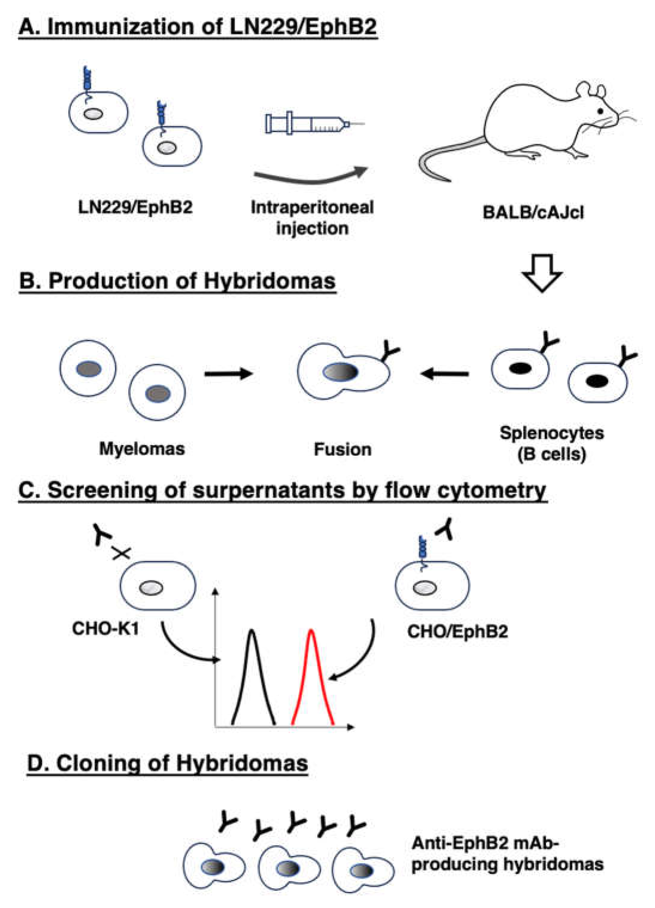
Figure 2.
Flow cytometry of EphB2-expressed CHO-K1 cells using Eb2Mab-12 and 2H9. CHO/EphB2 (A) and CHO-K1 (B) cells were treated with 0.01–10 µg/mL of Eb2Mab-12 or 2H9 conjugated with RB545 (Red line). The Eb2Mab-12 treated cells were further incubated with anti-mouse IgG conjugated with Alexa Fluor 488. The fluorescence data were subsequently collected using the SA3800 Cell Analyzer. The black line represents the negative control (blocking buffer).
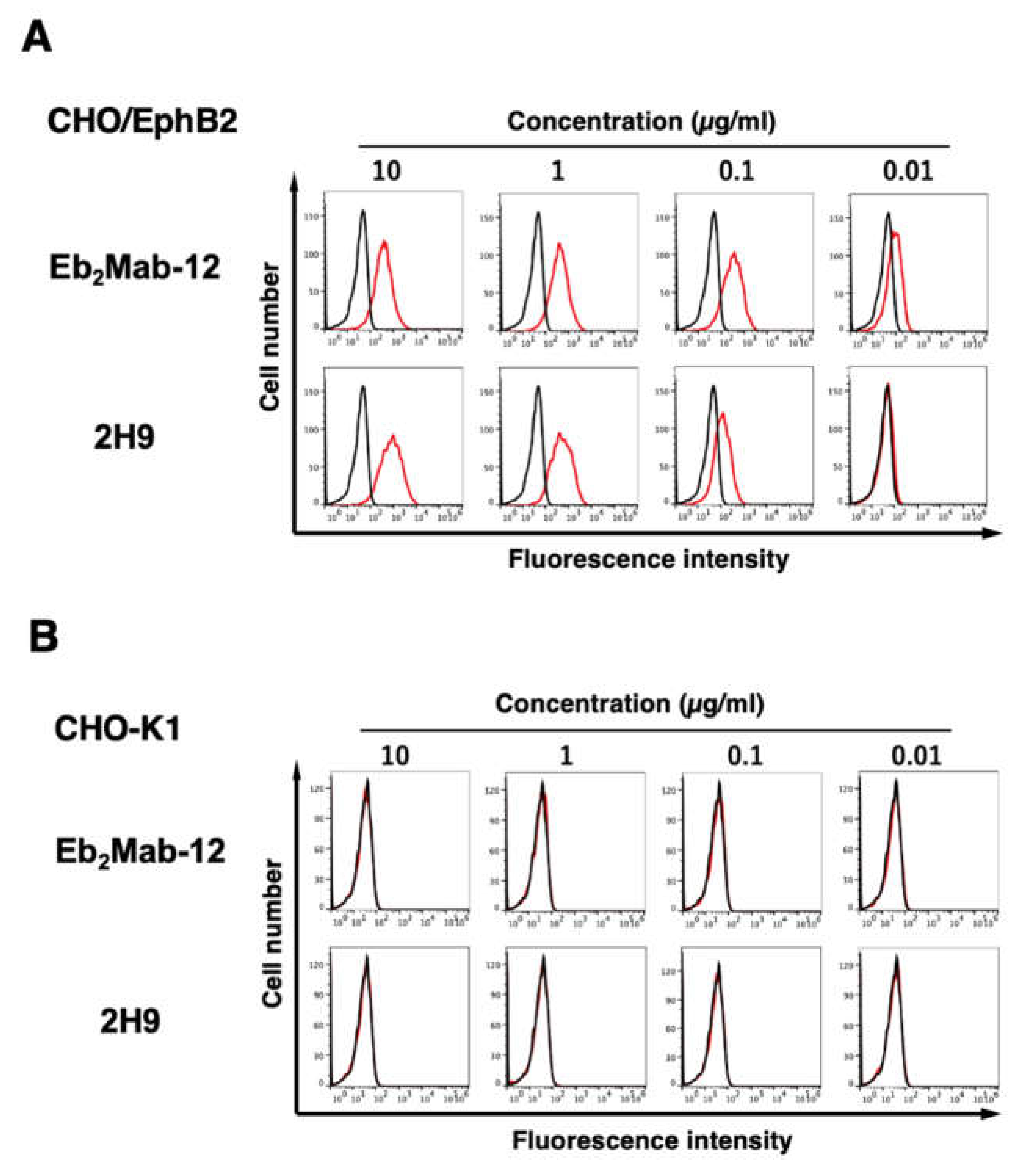
Figure 3.
Flow cytometry of endogenous EphB2-expressing cells using Eb2Mab-12 and 2H9. LS174T cells were treated with 0.01–10 µg/mL of Eb2Mab-12 or 2H9 conjugated with RB545 (Red line). The Eb2Mab-12 treated cells were further incubated with anti-mouse IgG conjugated with Alexa Fluor 488. The fluorescence data were subsequently collected using the SA3800 Cell Analyzer. The black line represents the negative control (blocking buffer).


Table 1.
Cross-reactivity and KD values of Eb2Mabs (IgG1 isotype) in flow cytometry.
| Isotype | Cross-reactivity | KD (× 10−9 M) | |
| Eb2Mab-1 | IgG1, kappa | − | 5.9 |
| Eb2Mab-2 | IgG1, kappa | − | 6.4 |
| Eb2Mab-3 | IgG1, kappa | EphA3, EphB1, EphB3 | 1.1 |
| Eb2Mab-4 | IgG1, kappa | 3.3 | |
| Eb2Mab-7 | IgG1, kappa | 5.2 | |
| Eb2Mab-8 | IgG1, kappa | EphB3 | 9.5 |
| Eb2Mab-10 | IgG1, kappa | EphB3 | 3.4 |
| Eb2Mab-12 | IgG1, kappa | − | 1.7* |
CHO/EphB2 was used to determine the KD. Cross-reactivity was determined by flow cytometry (Supplementary Figures S2–S4). Eb2Mab-5 is IgG3. Eb2Mab-6, Eb2Mab-9, and Eb2Mab-11 are IgM. *The data was presented in Figure 4A.
Disclaimer/Publisher’s Note: The statements, opinions and data contained in all publications are solely those of the individual author(s) and contributor(s) and not of MDPI and/or the editor(s). MDPI and/or the editor(s) disclaim responsibility for any injury to people or property resulting from any ideas, methods, instructions or products referred to in the content. |
© 2025 by the authors. Licensee MDPI, Basel, Switzerland. This article is an open access article distributed under the terms and conditions of the Creative Commons Attribution (CC BY) license (http://creativecommons.org/licenses/by/4.0/).
Copyright: This open access article is published under a Creative Commons CC BY 4.0 license, which permit the free download, distribution, and reuse, provided that the author and preprint are cited in any reuse.


