Preprint
Article

This version is not peer-reviewed.

Comparison of Methods for Isolating Exosomes from Plasma in Subjects with Normal and High-Fat Percentage

A peer-reviewed article of this preprint also exists.

Submitted:

24 January 2025

Posted:

24 January 2025

You are already at the latest version

Abstract

The adipose tissue is responsible for fat storage and is an important producer of extracellular vesicles (EVs). The biological content of exosomes, one kind of EVs, provides information such as immunometabolic alterations. This study aimed to compare three plasma exosome isolation methods and select the best. Plasma from 118 individuals, categorized by normal and high body fat percentage, and developing a 3T3-L1 cell line as an obesogenic model through continuous glucose exposure and a prolonged hypoxic microenvironment, were used. We perform three exo-some isolation methods: commercial kit (CK), size exclusion chromatography (SEC), and differ-ential centrifugation (DC), and characterized by DLS, cryo-TEM, TEM, and western blot CD9 and CD81 markers. In the DLS, cryo-TEM, and TEM analysis showed similar quality and mor-phology of exosomes. The CK and DC proved to comply with most of the advantages of an exo-some isolation method. Still, we emphasize the importance of selecting the appropriate method-ology depending on the specific research objectives. At the same time, no differences in exosome morphology, total protein, nor microRNAs concentration were observed between individuals categorized by body fat percentage, so we suggest that exosome cargo is the one that varies in in-dividuals with normal and high-fat percentages.

Keywords: 
;  ;  ;  

1. Introduction

Exosomes, ranging in size from 50 to 150 nm, represent a subtype of extracellular vesicles (EVs) due to their biogenesis, found in several bodily fluids, including plasma, serum, urine, seminal fluid, tears, breast milk, aqueous humor, saliva, and within diverse cell types, as well as in vitro cultures [1,2,3]. Exosomes work as carriers for biological material such as DNA, mRNA, microRNA, lipids, and proteins, thereby conveying information about the originating cells' state and potentially influencing the function of recipient cells [4,5,6,7,8]. Adipose tissue (AT) is currently recognized as an essential source of circulating EVs, also known as adipocyte-derived extracellular vesicles (AdEVs), which function as a bridge between adipocytes and cells in the stromal fraction of the AT as well as with cells from other systems [9,10]. AdEVs are filled with biological material that, in AT, play a role in metabolic alterations such as obesity, type 2 diabetes, and related illnesses and maintain the body’s homeostasis [9,11,12].
Even though is unclear how in fat depot accumulation AdEVs cargo is involved in obesity-related diseases and how these signaling molecules vary, exosomes are an alternative communication pathway that can influence cellular and tissue functions by exerting stimulatory or inhibitory effects, such as promoting cell proliferation, inducing apoptosis, modulating cytokine production, regulating immune responses [13]. Consequently, the significance of exosome content in developing AT dysfunction lies in their role as carriers of proteins that recruit macrophages to the AT, mainly TNFα and IL-6, which contribute to the onset of insulin resistance (IR) [4]. On the other hand, this scenario occurs within AT and distant organs, as exosomes can damage endothelial cells, compromise blood vessels, exacerbate liver fibrogenesis, and promote polarization to the M1 macrophage phenotype [9]. The increase of fat mass, which translates into the accumulation of triglycerides within adipocytes, triggers persistent cellular apoptosis. This process creates a hypoxic microenvironment characterized by chronic low-grade inflammation, leading to dysregulation in the secretion of cytokines, adipokines, and other factors essential for AT homeostasis [12,14].
To ensure precision and reproducibility, it is essential to compare other methods with the gold standard of exosome isolation, typically represented by differential centrifugation (DC) [15]. Recently, methods have been incorporated to reduce time-consuming procedures, the use of specialized equipment, the number of samples and supplies required. Depending on the source of exosomes, as well as the research goals, these methods include polymer precipitation, immunoaffinity capture, chromatography [16], size exclusion [17], and commercial kits [18,19]. Alternatively, to characterize exosomes, both optical and non-optical techniques are available, each chosen based on the specific information required for research purposes. Optical methods include dynamic light scattering (DLS), multi-angle light scattering, nanoparticle tracking analysis, flow cytometry, and surface plasmon resonance [20,21]. Non-optical approaches encompass scanning electron microscopy, transmission electron microscopy (TEM), cryogenic transmission electron microscopy (cryo-TEM), atomic force microscopy, Fourier transform infrared spectroscopy, and labeling of exosome membrane proteins such as CD9, CD63, CD81, and CD82 [20,22,23,24,25].
Based on the eight exosome isolation methods recently updated in the MISEV2023 guidelines, differential centrifugation (DC) in combination with other approaches demonstrates higher specificity, whereas precipitation methods alone offer superior recovery [26], therefore, this study aimed to compare three plasma exosome isolation techniques: precipitation using a commercial kit (CK, Invitrogen®), size exclusion chromatography (SEC), and differential centrifugation (DC). The isolated exosomes were characterized using dynamic light scattering (DLS), cryo-TEM, TEM, and western blot analysis targeting the CD9 and CD81 markers, to facilitate subsequent protein and molecular biology assays.
Furthermore, to facilitate the identification of exosomes exclusive to mature adipocytes in future studies, our second objective was to establish an obesogenic model using the 3T3-L1 cell line. This was achieved through prolonged glucose exposure and sustained hypoxia, enabling the cells to differentiate, proliferate, and maintain viability in an exosome-free medium.
Figure 1. Diagram illustrating exosome sources, isolation methods, and their association with metabolic diseases. Exosomes were isolated from plasma using three different methods: a commercial kit (CK, Invitrogen®), size exclusion chromatography (SEC), and differential centrifugation (DC). The cargo of exosomes is linked to metabolic diseases and the development of dysfunctional adipose tissue. This figure was created with BioRender.com.
Preprints 147145 g001

2. Materials and Methods

2.1. Samples

2.1.1. Plasma Sample

Blood collection was approved by the Comisión de Investigación y Ética del Antiguo Hospital Civil de Guadalajara “Fray Antonio Alcalde” O.P.D. HCG/CEI-0835/22, Nº. 130/22, after 118 participants aged 20 to 59 (71 women and 47 men) provided written informed consent and were classified by fat percentage; high was considered more than 25% in men and 35% in women, we included 59 individuals with normal and 59 with high-fat percentages. Ten milliliters of blood were obtained in EDTA-coated tubes and allowed to sit at room temperature for 30 minutes. The whole blood was centrifuged at 3000 g for 15 minutes at room temperature to separate plasma. The individual plasma samples were stored at 4º C until the experiments.

2.1.2. Cell Culture

A lecturer at the Universitat de Barcelona donated the embryonic mouse fibroblast cell line 3T3-L1. Cells were cultured on a polystyrene tissue culture flask t75 cm2. Cell growth started with basal media, DMEM (Dulbecco’s modified Eagles medium. Merck. Cat D6429) with 10% FBS (Merck. F2442-500ML) for one week. This time point was referred to as day 0 to begin adipogenic differentiation with MDI (DMEM with 10% FBS + 0.5 mM de IBMX (3-Isobutyl-1-methylxanthine) (SERVA. 26445.02. 500 mg), 1 μM dexamethasone (Dexa. MCE. Cat. HY-14648. 500 mg) + 10 μg/mL insulin (NeoBiotech. Cat. NB-58-0012. 5 mg/ml)) for four days at 37º C and 5% CO2. On day five, we used MDII (DMEM with 10% FBSdEVs (Depleted of exosomes by overnight ultracentrifugation at 100000 g 4º C) + 10 μg/mL insulin) for 43 days at 37º C and 5% CO2 (changing medium and flask, removal dear cells, each two days), on this day cells were stained with oil-red O (ORO) ((Sigma-Aldrich Cat. O0625)). On day 19, one subculture was subjected to hypoxia by anaerobiosis (BD GasPakTM EZ Lot. 581972) inside an airtight chamber (BD GasPakTM EZ. Anaerobe Container System. Ref 260678) for 2.5 hours and 24 hours [27].

2.2. Exosome Isolation and Characterization

2.2.1. Isolation by Differential Centrifugation (DC)

We used 2 mL of blood plasma. All centrifugations were realized at 4º C. We started by 300 g for 10 minutes (Heraeus Megafuge 2.0 R) discarded the pellet; 2000 g for 20 minutes (Heraeus Megafuge 2.0 R) discarded the pellet; 10000 g for 20 minutes (Beckman Coulter. Rotor JA-14. Jars NALGENE® 250 mL) discarded the pellet; 100000 g for 70 minutes (Beckman Colter. Rotor T70i. Beckman Centrifuge Tubes 26.3 mL), discarded supernatant and resuspend the pellet with 500 μL of 1X PBS, 100000 g for 70 minutes (Thermo Sorvall Discovery M150 SE Floor Micro-Ultracentrifuge 150K rpm. Rotor S150-AT Eppendorf Tubes PP 1 mL) and resuspend in 200 μL of 1X PBS [28].

2.2.2. Isolation by Size Exclusion Chromatography (SEC)

We used 1mL of blood plasma at room temperature. Bridely, we centrifugated at 2000 g for 10 minutes and used the supernatant for a second centrifugation at 10000 g for 30 minutes at 10º C; the supernatant was filtrated through a 0.22 μm filter (CORNING 45 mm diameter). Finally, pass the sample through each column (Econo-Pac® Chromatography Columns, Pkg of 50 #7321010 with Sepharose™ CL-2B) with 1X PBS at room temperature filtrated through a 0.22 μm filter. We collected 200 μL of 15 fractions. [17,29].

2.2.3. Isolation by Commercial KIT (CK)

The kit Total Exosome Isolation (from plasma) (Invitrogen Cat. No. 4404450) was used. All centrifugations were performed at room temperature. Starting with 500 μL of blood plasma, we centrifugated at 10000 g for 20 minutes, added 0.5 volumes of PBS 1X to the supernatant, mixed by vortex, added 0.2 volume of exosome precipitation reagent, pipetted up and down. This mix was incubated for 10 minutes at room temperature, then centrifugated at 10000 g for 5 minutes, and the supernatant was discarded. Finally, the pellet was resuspended in 50 μL of PBS 1X.

2.2.4. Characterization by Dynamic Light Scattering (DLS)

For this analysis, 500 μL of each sample was dissolved in NaCl. The equipment was set up at 25º C for 60 minutes (Antor Paar). Litesizer DLS 700, software Malverne Zetasizer Software v7.13 PSS0012-39).

2.2.5. Characterization by Cryo-TEM and TEM

Three μL of blood plasma were used for each electron microscopy. Transmission Electron Microscopy (FEI Technology, Model Tecnai Spirit BioTwin, software FEI TIA 4.15.) and cryo-TEM (Jeol JM-2011, Tokyo, Japan) were analyzed by technicians of the Instituto Nacional de Ciencias Médicas y Nutrición Salvador Zubirán, Ciudad de México-México and Serveis Científico-tècnics UAB, Barelona-España, respectively.

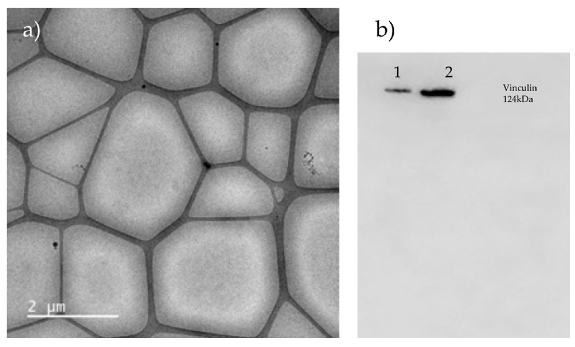
2.2.6. Characterization by Western Blot

Starting with exosome lysis buffer (25 mM Tris-HCl +120mM NaCl + 1% Triton-X100 + complete Mini, EDTA Free. Merck) by adding 20 μL to 150 μL of sample in ice-cold, then incubating in rotational agitation at 4º C for 1 hour, then centrifugate at 15000 g for 15 minutes at 4º C, the supernatant was measured in the spectrophotometer (NanoDrop 1000 UV Visible Spectrophotometer, Thermo Scientific). We charged 5 μg of total protein on 14% acrylamide gel. Exosome markers were quantified using mouse monoclonal CD9 (1:1000; antibodies.com. Anti-CD9 (MEM-61) (A86089)), CD81 (1:1000; antibodies.com. Anti-CD81 (M38) (A86719), and Vinculin (1:500 Invitrogen Ref. MA5-11690), we used anti-mouse HRP (1:5000; abcam. NA931V ECL Anti-mouse IgG), revealed with ECL (BioRad Clarity Western ECL Substrate, 500 ml #1705061) in the spectrophotometer (Li-cor, Odyssey XF, LI-COR Acquisition Software).
2.3. microRNA Isolation
Trizol reagent was used to isolate microRNA. 50 µL of isolated exosome was lysed in 200 µL Trizol + 1 µL (1 µg/µL glycogen RNA grade. Thermo Scientific #R0551). Subsequently, 40 µL of chloroform was used for phase separation and 100% isopropanol for microRNA precipitation. Finally, microRNA was eluted in 30 µl RNase-free water after being washed twice in 75% ethanol. The microRNA concentration was assessed using a Qubit 4.0.

2.4. Statistical and Image Analyses

For each method, one-way ANOVA, post hoc Tukey tests, and the Mann-Whitney U test compared the total protein and microRNAs concentration difference between average and high-fat percentages. P < 0.05 was considered significant. GraphPad Prism version 8.4.0 for macOS was used for data analysis and graphing. ImageJ2 version: 2.1.4.0/1.54f for image analyses.

3. Results

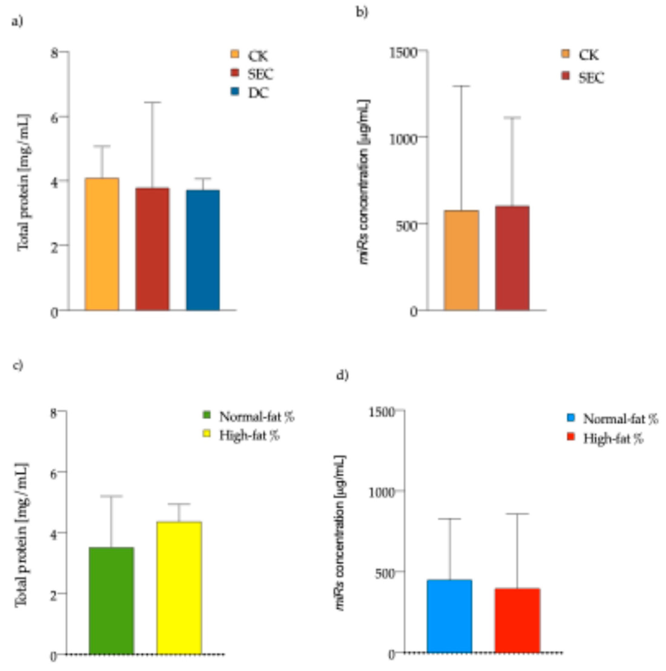
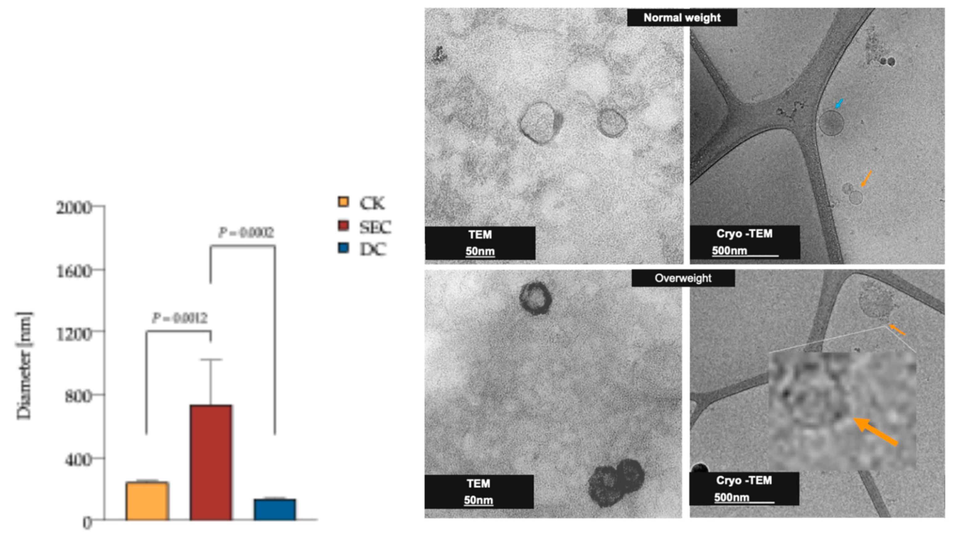
3.1. The Exosome Isolation Methods Showed Equal Performance in Total Protein and microRNA Concentration, While an Inverse Pattern Is Observed Among Individuals with High-Fat Mass Content

Exosomes were isolated according to the manufacturer’s recommended instructions for the CK, whereas DC and SEC were done for classical ultracentrifugation and column fraction separation, respectively. Exosome yield was determined by total protein concentration using Qubit Protein Assay (Invitrogen™). We observed that the precipitation-based Total Exosomes Isolation kit (Invitrogen) had the maximum yield, followed by SEC and DC (Figure 2a). After these results, we chose CK and SEC to isolate microRNAs from the exosome, with equal performance (Figure 2b). On the other hand, in total exosome proteins as shown in Figure 2c we distinguish an increasing pattern in individuals with high-fat percentages while microRNA concentration reduced (Figure 2d).

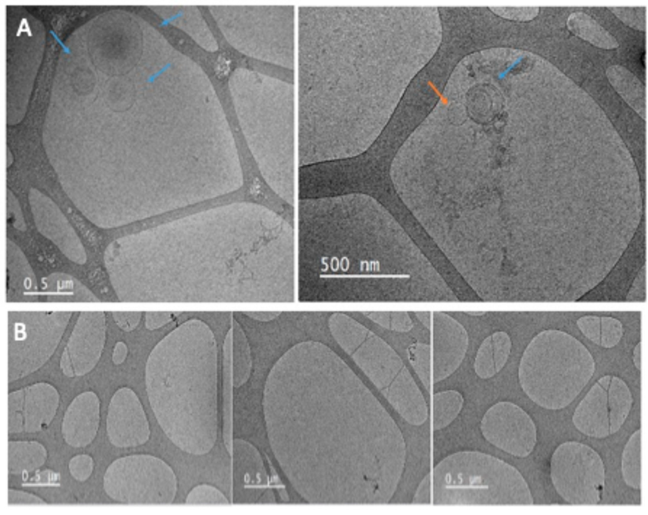
3.2. The Morphology and Quality of the Three Exosome Isolation Methods Were as Expected, with Inconsistencies in Purity

By DLS analysis, we observed that the diameter measure of the vesicles was compatible with exosome diameter by CK and DC (Figure 3). At the same time, the purity of the vesicle subpopulation differed between CK and DC with SEC. By TEM and cryo-TEM, we observed that the morphology and quality of exosomes were reliable between them. In the morphology exosome, no difference was observed between individuals with normal and high-fat percentages (Figure 3).
The exosomes' average diameter was 235.1 nm, 736.9 nm, and 138.2 nm for CK, SEC, and DC, respectively (n = 15 for each method, one-way ANOVA, and post hoc Tukey tests P < 0.05). TEM of exosomes isolated by CK (Invitrogen Cat. No. 4404450). Diameter of 53.9 nm, scale 50 nm. Cryo-TEM of exosomes isolated by SEC. Diameter of 84.3 nm (orange arrow), as well as microvesicles of the highest dimension (blue arrow) of 191.7 nm, scale 500 nm. Exosomes from plasma samples of individuals were classified by fat percentage (59 for each group). Abbreviations DLS: dynamic light scattering; CK: commercial kit; SEC: size exclusion chromatography; TEM: transmission electron microscopy.

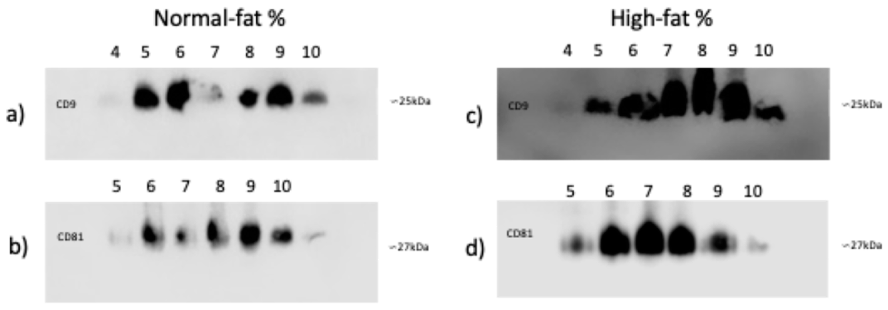
3.3. CD9 and CD81 Do Not Differ Between Normal and High-Fat Percentage Individuals in SEC Fractions

Since integrity and quality in microscopy images and total protein did not differ, we chose an exosome isolation technique that was more cost-effective and equipment-efficient for identifying CD9 and CD81 in plasma exosomes from individuals with normal and high-fat percentages. In individuals with normal-fat and high-fat percentages, there is no shift in CD9 marker fractions from 4 to 10 (Figure 4 a and c), nor in CD81 fractions from 5 to 10 (Figure 4 b and d).
Under our experience, we can resume some criteria for these three-exosome isolation methods in Table 1 below:

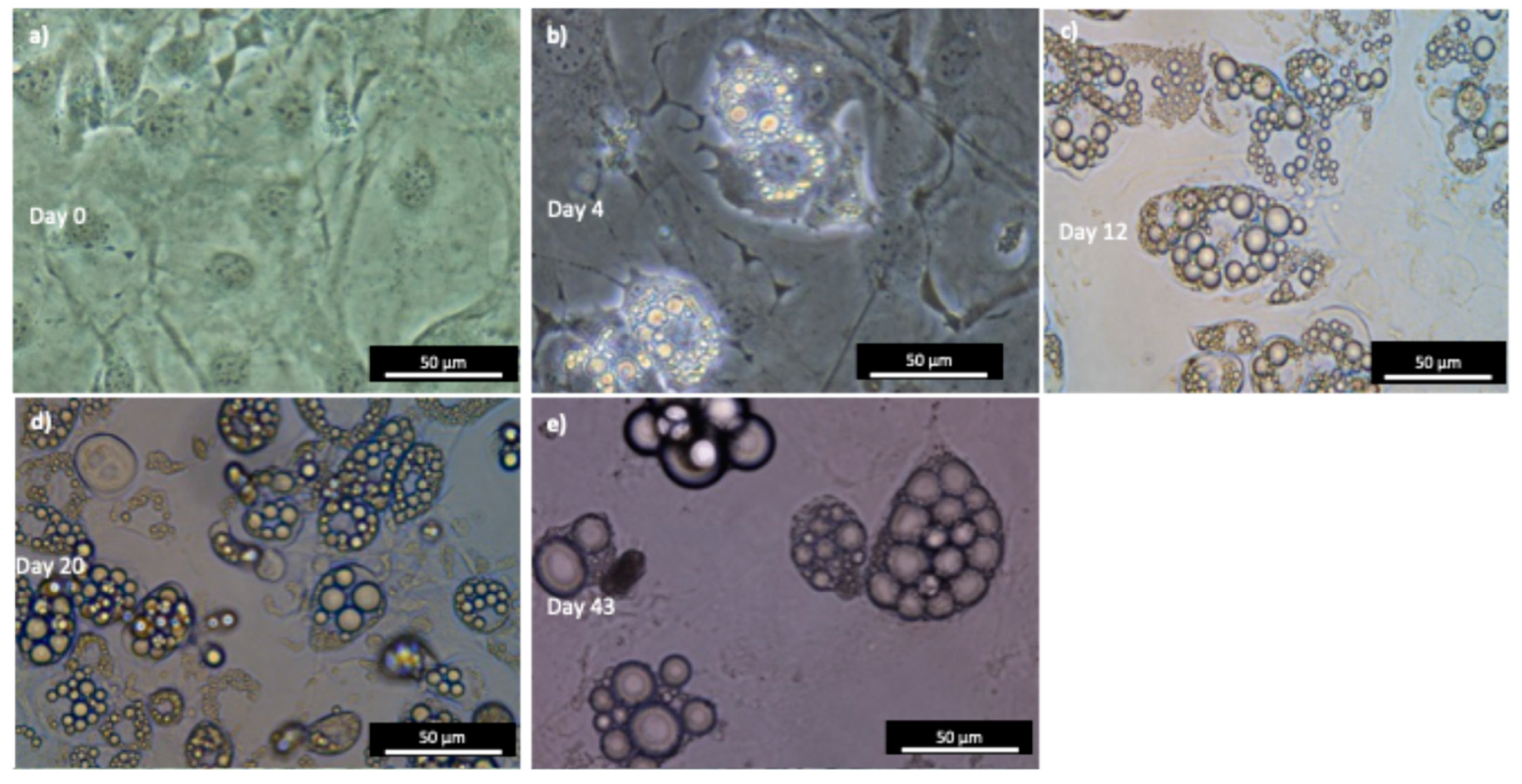
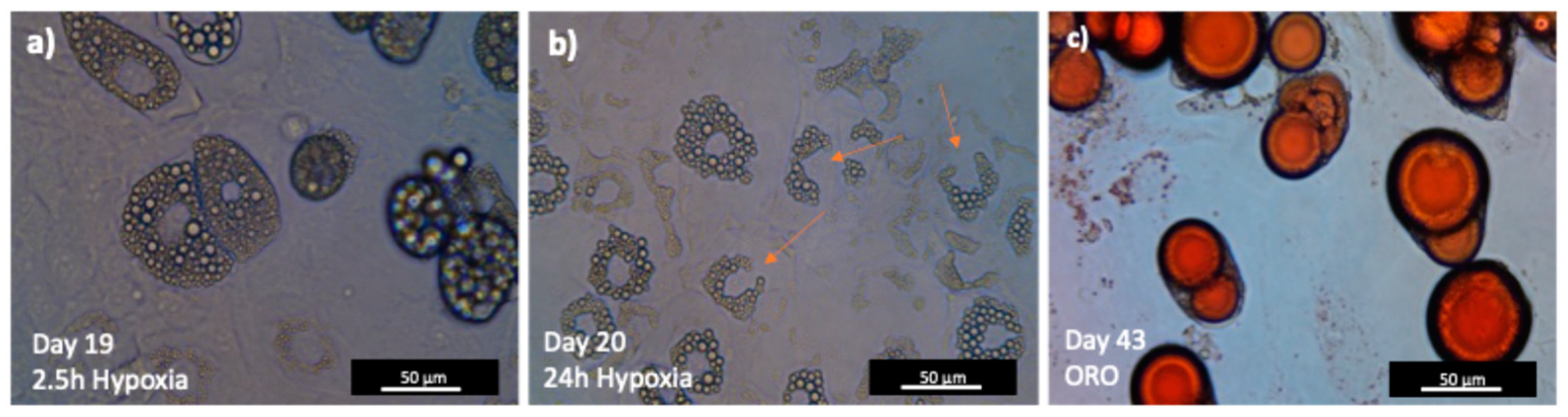
3.4. The Successful Differentiation of 3T3-L1 Cells Results in Acquiring a Mature Adipocyte Phenotype

In this study, we aimed to establish a foundation for future investigations demonstrating that adipocytes with obesogenic characteristics (hypoxia and sustained glucose exposure) produce exosomes. To this end, we utilized the differentiated 3T3-L1 cell line as a model. The fetal bovine serum (FBS) used in the culture medium was treated to deplete extracellular vesicles (FBSdEVs) through overnight ultracentrifugation (Figure A.1).
To ensure that FBSdEVs did not compromise cell growth and proliferation, cells were preconditioned with this medium for one week prior to differentiation. Cell density was monitored, revealing comparable growth rates between conditions: 570,000 cells/mL for untreated FBS and 500,000 cells/mL for FBSdEVs. This step was critical to confirm that the use of EV-depleted serum does not negatively impact the differentiation process or cell viability, providing a reliable baseline for future experiments.
To induce differentiation, 3T3-L1 cells were stimulated to acquire an adipocyte phenotype. During the first three days, cell proliferation ceased, and differentiation commenced in a subset of cells, as depicted in Figure 5a. Initially, on day 0, the average cell diameter measured 17.1 ± 1.4 µm, with no observable lipid droplets. By day 4, differentiation progressed, leading to a noticeable increase in cell size, with an average diameter of 47.2 ± 11.9 µm and the appearance of small lipid droplets measuring 4.1 ± 0.7 µm (Figure 5b).
As differentiation continued, the accumulation of triglycerides within lipid droplets resulted in a progressive enlargement of both cell and droplet size. By subsequent time points, lipid droplets reached diameters of up to 10 µm, as illustrated in Figure 5c (12.8 ± 2.3 µm), 5d (12.9 ± 2.4 µm), and 5e (23.0 ± 4.4 µm). These findings underscore the dynamic morphological changes associated with adipogenesis, including the substantial accumulation of intracellular lipids, a hallmark of mature adipocytes.
We developed an obesogenic model using the 3T3-L1 cell line, subjecting the cells to prolonged glucose exposure and continuous hypoxia to facilitate differentiation, growth, and maintenance in an exosome-free medium. Notably, on day 19 of culture, 3T3-L1 cells exposed to 2.5 hours of hypoxia retained a stable morphology (Figure 6a). However, after 24 hours of hypoxia, significant cell disintegration was observed, accompanied by a reduction in lipid droplet size, with an average droplet diameter of 2.9 ± 0.7 µm (Figure 6b).
To determine whether this morphological shift was caused by prolonged hypoxia rather than continuous glucose intake, cells were cultured in MDII differentiation medium for 43 days and stained with oil-red O (ORO) to visualize intracellular triglycerides. Over time, the lipid droplets exhibited changes in form and size, eventually expanding to fill nearly the entire cytoplasm, as shown in Figure 6c. These findings highlight the distinct cellular responses to sustained hypoxia and glucose exposure in this obesogenic model.

4. Discussion

It is estimated that the prevalence of obesity and overweight will be 1.7 billion by 2030 [30], so understanding the mechanism by which the AT performs under these conditions is critical to preventing, treating, and reversing the loss of homeostasis within this tissue. The adipocyte-derived extracellular vesicles play an essential role in developing associated obesity diseases, like cancer [31], among others, as suggested by Delgadillo et al, that AdEVs size can be related to liver and systemic IR [32].
Nowadays, the choice of method to isolate exosomes is wide-ranging, depending on the application of these vesicles. Ultracentrifugation, preceded by low-speed centrifugation, is the gold standard technique [33]. Still, the possibility of having the equipment and accessories for it is not available in all the laboratories. In the present study, we gauged material and equipment price, time-consuming procedure, vesicle yield, purity subpopulation, protein, and microRNAs concentration of three isolation methods to define the most suitable for further analyses. The results were: a) CK presented the highest total protein concentration in contrast with the other two techniques, b) there was a similarity in microRNAs concentration in exosomes isolated with CK and SEC, c) there was a likeness in exosome total protein and microRNAs concentration between normal and high fat-percentage individuals and d) FBS depleted of EVs allows differentiated correctly 3T3-L1 cells.
Even though total protein quantification is a suitable method for estimating the concentration of exosomes, it cannot distingue contaminating proteins like albumin that are not associated with them [34], so between the three methods, the commercial kit is the most reproducible, as mentioned by Caradec et al [35]. After comparing the concentration of total protein content of the exosomes using the three methods, we decided to evaluate the normal and high percentages of fat and the total microRNAs using the most straightforward method, which is less time-consuming and allows the analysis of a more significant number of samples. The precipitation by CK is an excellent alternative to nucleic acid subsequent analysis, even if we could not find a difference between the three methods, as Mohammad et al determined when comparing one commercial kit with ultracentrifugation [36]. In this context, without difference between individuals with normal and high-fat percentages, considering Wang et al.'s evaluation of the exosome proteome in obese and non-obese individuals with T2DM [37], we can propose that exosome cargo is linked to metabolic-associated disorders rather than fat content [38,39].
Many techniques have been used for exosome characterization [40], depending on the information the research looks for. In our case, we chose two that can be used for quality (protein marker, shape, and size) analyses: DLS, electron microscopy (TEM and cryo-TEM), and western blot (CD9 and CD81 markers). DLS allowed characterization of plasma exosome subpopulations according to their diameter, as shown by the CK and DC methods. In contrast, SEC appears to yield a higher presence of aggregated exosomes and other plasma particles of similar size, probably due to the intrinsic nature of its isolation mechanism [41]. Electron microscopy enables imaging of the single exosome, visualizing its size and morphology. Exosomes isolated by CK and DC demonstrated fewer microvesicle presence, while SEC presented a considerable microvesicle population, as mentioned by Davidson et al [42].
We chose two tetraspanins to verify that we isolated microvesicles compatible with exosomes (CD9 and CD81). We analyzed for normal and high-fat individuals, where there was no difference, only in the delay of fractions between markers, but not by fat condition. This pattern was shown in carcinoma exosomes [43] and circulating exosomes [44] isolated by SEC. Following our results, Sharif et al mention that SEC is a suitable method for further nucleic acid analyses, where they also found a distinct population of EVs [45]. Our decision to use the three exosome isolation methods and exosome characterization is supported in the last update of the International Society for Extracellular Vesicles (MISEV2023 [3]), as well as the advantages and disadvantages found according to our results (Table 1).
The adipogenic differentiation of 3T3-L1 allows hypertrophy in the AT by stimulating glucose uptake and storing in the triglycerides [46], suggesting that the excess and continuous intake plus the lack of energy expenditure is a delicate balance between the glucose concentration and the differentiation time as reported by Jackson et al [47], promoting fat accumulation in adipose tissue. Additionally, we corroborated that the morphology of cells subjected to 24 hours of hypoxia changes, as mentioned by Vogler M. et al., who demonstrated the increase in vinculin-positive focal cell contacts in hypoxia by microscopy imaging [48], as we did by western blot (Figure A2.b), and like Synowiec et al, did not detect a difference by western blot for vinculin [49]. The importance of isolating exosomes from 3T3-L1 cells has been demonstrated in coculture experiments involving healthy 3T3-L1 adipocytes and a human pancreatic cell line. Exosomes derived from 3T3-L1 cells were shown to enhance pancreatic cell survival, promote proliferation, and stimulate insulin secretion, simulating the physiological scenario observed in individuals with a normal fat percentage [50]. Similarly, it has been demonstrated that ADEVs obtained from the culture supernatant of mature 3T3-L1 adipocytes can reduce plasma clotting times [51].
After evaluating the three exosome isolation techniques, we identified specific limitations linked with each method. In the case of differential centrifugation (DC), a major drawback is the requirement for access to ultracentrifugation or mini-ultracentrifugation equipment, which is not available in all laboratories. Similarly, for size-exclusion chromatography (SEC), noteworthy challenges include the extended time required for sample processing and variability between protocols, particularly regarding the initial sample volume and the volume collected from each fraction.
Exosomes have reached significant interest due to their potential as biomarkers, as they transport proteins and microRNAs capable of predicting the diseases associated with obesity. Furthermore, they enable the evaluation of the functional status of adipose tissue and are emerging as promising therapeutic vesicles for restoring homeostasis in this tissue. Notably, by conducting a comprehensive immunometabolic assessment of an individual, exosome-based therapies could be tailored, paving the way for personalized medical approaches.

5. Conclusions

We highlight the importance of selecting the most suitable exosome isolation method based on specific research objectives. For studies prioritizing exosome integrity, we recommend using SEC for plasma samples, as it yields a higher concentration of intact exosomes. However, if the focus is on achieving greater purity in EVs subpopulations, DC or a commercial isolation kit are more appropriate methodologies. We suggest the DC and CK methods for further protein and molecular analysis.
Our results indicate that circulating exosomes do not differ in quantity between individuals with normal and high body fat percentages; however, their composition does vary. We recommend further investigation into the molecular cargo of circulating exosomes, as they may serve as indicators of adipose tissue status and could help elucidate these differences.

Author Contributions

Conceptualization, J.N., I.Á., and R.N.; methodology, J.N., and J.L.; formal analysis, J.N., I.Á., and R.N.; investigation, J.N., J.L., J.C., A.F., I.Á. and R.N.; resources, J.C., A.F., I.Á., and R.N.; data curation, J.N., I.Á. and R.N., A; writing—original draft preparation, J.N., I.Á. and R.N.; writing—review and editing, J.N., J.L., J.C., A.F., P.M., I.Á. and R.N.; visualization, J.N., J.L., J.C., A.F., P.M., I.Á. and R.N.; supervision, I.Á. and R.N.; project administration, I.Á. and R.N.; funding acquisition, J.C., A.F., I.Á. and R.N.

Funding

This research was funded by Doctoral scholarship number (CVU): 1103690, by CONAHCYT.

Institutional Review Board Statement

The study was conducted by the Declaration of Helsinki and approved by the Ethics Committee of Comisión de Investigación y Ética del Antiguo Hospital Civil de Guadalajara “Fray Antonio Alcalde” O.P.D. HCG/CEI-0835/22, Nº. 130/22 at 23-05-2022” for studies involving humans.

Informed Consent Statement

Informed consent was obtained from all subjects involved in the study.

Data Availability Statement

Information is available at a reasonable request to the corresponding author.

Conflicts of Interest

The authors declare no conflict of interest.

Appendix A

Figure A1. Cryo-TEM of EVs isolated by ultracentrifugation. a) Microvesicles diameter 227 ± 133 nm (scale 500 nm) isolated previously by overnight ultracentrifugation and b) FBS depleted EVs. Blue arrow shows vesicles in microscale and orange arrow in nanoscale.
Preprints 147145 g0a1
In our initial attempt to isolate exosomes from the supernatant of differentiated 3T3-L1 cells cultured with EV-depleted FBS, we employed a combination of two methods: DC coupled with SEC and ultrafiltration (passed by filter 0.5 μm, filter 0.22 μm, 10,000 x g 30 minutes 4º C, filter 0.15 μm) with SEC.
Various techniques are commonly utilized for exosome isolation from cell culture supernatants, each with distinct advantages. Sidhom et al [52] highlighted SEC as a viable alternative, but recommended an optimized combination of methods for better results. Following this guidance, we tested particle concentration (Centriprep YM-10, 10 kDa NMWL, Millipore) prior to SEC and DC, respectively, as well as DC prior to SEC, as described by Akbar et al [44]. However, despite these efforts, we were unable to isolate exosomes from the supernatant, as confirmed by cryo-TEM and western blot analyses for exosomal markers CD9 and CD81.
These findings suggest that factors such as the initial volume of the supernatant, ultracentrifugation time, and cell stimulation may play critical roles in successful exosome isolation, as proposed by Nakatani et al [51]. We recommend exploring commercial kits specifically designed for cell culture exosome isolation, optimizing ultracentrifugation parameters, or employing stimuli to enhance exosome secretion from cultured cells.
Figure A2. The combined isolation methods for the exosomes from 3T3-L1 cells: DC with SEC and ultrafiltration. a) Observation by Cryo-TEM without exosomes. b) Western blot shows Vinculin as a constitutive protein. Rail 1 = supernatant in normoxia. Rail 2 = supernatant in hypoxia.
Preprints 147145 g0a2

References

  1. Thery C, Amigorena S, Raposo G, Clayton A: Isolation and characterization of exosomes from cell culture supernatants and biological fluids. Curr Protoc Cell Biol 2006, Chapter 3:Unit 3 22.
  2. Abdulmalek O, Husain K, AlKhalifa H, Alturani M, Butler A, Moin A: Therapeutic Applications of Stem Cell-Derived Exosomes. International Journal of Molecular Sciences 2024, 25:3562.
  3. Welsh JA, Goberdhan DCI, O'Driscoll L, Buzas EI, Blenkiron C, Bussolati B, Cai H, Di Vizio D, Driedonks TAP, Erdbrugger U, et al.: Minimal information for studies of extracellular vesicles (MISEV2023): From basic to advanced approaches. J Extracell Vesicles 2024, 13:e12404. [CrossRef]
  4. Lei LM, Lin X, Xu F, Shan SK, Guo B, Li FX, Zheng MH, Wang Y, Xu QS, Yuan LQ: Exosomes and Obesity-Related Insulin Resistance. Front Cell Dev Biol 2021, 9:651996.
  5. Dance A: The body’s tiny cargo carriers. In Knowable magazine. Edited by; 2019.
  6. Jiang X, You L, Zhang Z, Cui X, Zhong H, Sun X, Ji C, Chi X: Biological Properties of Milk-Derived Extracellular Vesicles and Their Physiological Functions in Infant. Frontiers in Cell and Developmental Biology 2021, 9.
  7. Ragusa M, Barbagallo C, Statello L, Caltabiano R, Russo A, Puzzo L, Avitabile T, Longo A, Toro MD, Barbagallo D, et al.: miRNA profiling in vitreous humor, vitreal exosomes and serum from uveal melanoma patients: Pathological and diagnostic implications. Cancer Biol Ther 2015, 16:1387-1396. [CrossRef]
  8. Jan AT, Rahman S, Badierah R, Lee EJ, Mattar EH, Redwan EM, Choi I: Expedition into Exosome Biology: A Perspective of Progress from Discovery to Therapeutic Development. Cancers 2021, 13.
  9. Mei R, Qin W, Zheng Y, Wan Z, Liu L: Role of Adipose Tissue Derived Exosomes in Metabolic Disease. Frontiers in Endocrinology 2022, 13.
  10. Liu Y, Wang C, Wei M, Yang G, Yuan L: Multifaceted Roles of Adipose Tissue-Derived Exosomes in Physiological and Pathological Conditions. Front Physiol 2021, 12:669429.
  11. Ji C, Guo X: The clinical potential of circulating microRNAs in obesity. Nat Rev Endocrinol 2019, 15:731-743.
  12. Bond ST, Calkin AC, Drew BG: Adipose-Derived Extracellular Vesicles: Systemic Messengers and Metabolic Regulators in Health and Disease. Front Physiol 2022, 13:837001.
  13. Camino T, Lago-Baameiro N, Pardo M: Extracellular Vesicles as Carriers of Adipokines and Their Role in Obesity. Biomedicines 2023, 11:422.
  14. Fasshauer M, Bluher M: Adipokines in health and disease. Trends Pharmacol Sci 2015, 36:461-470.
  15. Zhao R, Zhao T, He Z, Cai R, Pang W: Composition, isolation, identification and function of adipose tissue-derived exosomes. Adipocyte 2021, 10:587-604. [CrossRef]
  16. Yang XX, Sun C, Wang L, Guo XL: New insight into isolation, identification techniques and medical applications of exosomes. Journal of Controlled Release 2019, 308:119-129.
  17. Muller L, Hong CS, Stolz DB, Watkins SC, Whiteside TL: Isolation of biologically-active exosomes from human plasma. J Immunol Methods 2014, 411:55-65.
  18. Cheng Y, Qu X, Dong Z, Zeng Q, Ma X, Jia Y, Li R, Jiang X, Williams C, Wang T, et al.: Comparison of serum exosome isolation methods on co-precipitated free microRNAs. PeerJ 2020, 8:e9434. [CrossRef]
  19. Chen J, Li P, Zhang T, Xu Z, Huang X, Wang R, Du L: Review on Strategies and Technologies for Exosome Isolation and Purification. Front Bioeng Biotechnol 2021, 9:811971.
  20. Rech J, Getinger-Panek A, Gałka S, Bednarek I: Origin and Composition of Exosomes as Crucial Factors in Designing Drug Delivery Systems. Applied Sciences 2022, 12:12259.
  21. Koritzinsky EH, Street JM, Star RA, Yuen PS: Quantification of Exosomes. J Cell Physiol 2017, 232:1587-1590.
  22. Whitehead C, Luwor R, Morokoff A, Kaye A, Stylli S: Cancer exosomes in cerebrospinal fluid. Translational Cancer Research 2017, 6.
  23. Deng FY, Miller J: A review on protein markers of exosome from different bio-resources and the antibodies used for characterization. Journal of Histotechnology 2019, 42:226-239.
  24. Chernyshev VS, Rachamadugu R, Tseng YH, Belnap DM, Jia Y, Branch KJ, Butterfield AE, Pease LF, 3rd, Bernard PS, Skliar M: Size and shape characterization of hydrated and desiccated exosomes. Anal Bioanal Chem 2015, 407:3285-3301.
  25. Li D, Luo H, Ruan H, Chen Z, Chen S, Wang B, Xie Y: Isolation and identification of exosomes from feline plasma, urine and adipose-derived mesenchymal stem cells. BMC Vet Res 2021, 17:272.
  26. Welsh JA, Goberdhan DCI, O'Driscoll L, Buzas EI, Blenkiron C, Bussolati B, Cai H, Di Vizio D, Driedonks TAP, Erdbrügger U, et al.: Minimal information for studies of extracellular vesicles (MISEV2023): From basic to advanced approaches. Journal of Extracellular Vesicles 2024, 13:e12404. [CrossRef]
  27. Etesami B, Ghaseminezhad S, Nowrouzi A, Rashidipour M, Yazdanparast R: Investigation of 3T3-L1 Cell Differentiation to Adipocyte, Affected by Aqueous Seed Extract of Phoenix Dactylifera L. Rep Biochem Mol Biol 2020, 9:14-25.
  28. Lobb RJ, Becker M, Wen SW, Wong CS, Wiegmans AP, Leimgruber A, Moller A: Optimized exosome isolation protocol for cell culture supernatant and human plasma. J Extracell Vesicles 2015, 4:27031.
  29. Paolo Fortina EL, Jason Y. Park, Larry J. Kricka: Acute Myeloid Leukemia - Methods and Protocols. In Springer Protocols. Edited by; 2017:258 - 266. vol Methods in Molecular Biology 1633.].
  30. Federation WO: World Obesity Atlas 2024. In World Obesity Federation. Edited by; 2024. vol 2024.].
  31. Zhou C, Huang YQ, Da MX, Jin WL, Zhou FH: Adipocyte-derived extracellular vesicles: bridging the communications between obesity and tumor microenvironment. Discov Oncol 2023, 14:92.
  32. Delgadillo-Velázquez J, Alday E, Aguirre-García MM, Canett-Romero R, Astiazaran-Garcia H: The association between the size of adipocyte-derived extracellular vesicles and fasting serum triglyceride-glucose index as proxy measures of adipose tissue insulin resistance in a rat model of early-stage obesity. Frontiers in Nutrition 2024, 11.
  33. Coughlan C, Bruce KD, Burgy O, Boyd TD, Michel CR, Garcia-Perez JE, Adame V, Anton P, Bettcher BM, Chial HJ, et al.: Exosome Isolation by Ultracentrifugation and Precipitation and Techniques for Downstream Analyses. Curr Protoc Cell Biol 2020, 88:e110. [CrossRef]
  34. Tang YT, Huang YY, Zheng L, Qin SH, Xu XP, An TX, Xu Y, Wu YS, Hu XM, Ping BH, et al.: Comparison of isolation methods of exosomes and exosomal RNA from cell culture medium and serum. Int J Mol Med 2017, 40:834-844. [CrossRef]
  35. Caradec J, Kharmate G, Hosseini-Beheshti E, Adomat H, Gleave M, Guns E: Reproducibility and efficiency of serum-derived exosome extraction methods. Clin Biochem 2014, 47:1286-1292.
  36. Aziz MA, Seo B, Hussaini HM, Hibma M, Rich AM: Comparing Two Methods for the Isolation of Exosomes. J Nucleic Acids 2022, 2022:8648373.
  37. Wang Y, Wu Y, Yang S, Chen Y: Comparison of Plasma Exosome Proteomes Between Obese and Non-Obese Patients with Type 2 Diabetes Mellitus. Diabetes Metab Syndr Obes 2023, 16:629-642.
  38. Kwan HY, Chen M, Xu K, Chen B: The impact of obesity on adipocyte-derived extracellular vesicles. Cell Mol Life Sci 2021.
  39. Son T, Jeong I, Park J, Jun W, Kim A, Kim O-K: Adipose tissue-derived exosomes contribute to obesity-associated liver diseases in long-term high-fat diet-fed mice, but not in short-term. Frontiers in Nutrition 2023, 10.
  40. Lai JJ, Chau ZL, Chen SY, Hill JJ, Korpany KV, Liang NW, Lin LH, Lin YH, Liu JK, Liu YC, et al.: Exosome Processing and Characterization Approaches for Research and Technology Development. Adv Sci (Weinh) 2022, 9:e2103222. [CrossRef]
  41. Dilsiz N: A comprehensive review on recent advances in exosome isolation and characterization: Toward clinical applications. Transl Oncol 2024, 50:102121.
  42. Davidson SM, Boulanger CM, Aikawa E, Badimon L, Barile L, Binder CJ, Brisson A, Buzas E, Emanueli C, Jansen F, et al.: Methods for the identification and characterization of extracellular vesicles in cardiovascular studies: from exosomes to microvesicles. Cardiovasc Res 2023, 119:45-63. [CrossRef]
  43. Sakha S, Muramatsu T, Ueda K, Inazawa J: Exosomal microRNA miR-1246 induces cell motility and invasion through the regulation of DENND2D in oral squamous cell carcinoma. Sci Rep 2016, 6:38750.
  44. Akbar N, Pinnick KE, Paget D, Choudhury RP: Isolation and Characterization of Human Adipocyte-Derived Extracellular Vesicles using Filtration and Ultracentrifugation. J Vis Exp 2021.
  45. Sharif S, Mozaffari-Jovin S, Alizadeh F, Mojarrad M, Baharvand H, Nouri M, Abbaszadegan MR: Isolation of plasma small extracellular vesicles by an optimized size-exclusion chromatography-based method for clinical applications. Journal of Drug Delivery Science and Technology 2023, 87:104796.
  46. Lee MJ, Wu Y, Fried SK: A modified protocol to maximize differentiation of human preadipocytes and improve metabolic phenotypes. Obesity (Silver Spring) 2012, 20:2334-2340.
  47. Jackson HC, Pheiffer C, Jack B, Africander D: Time- and glucose-dependent differentiation of 3T3-L1 adipocytes mimics dysfunctional adiposity. Biochem Biophys Res Commun 2023, 671:286-291.
  48. Vogler M, Vogel S, Krull S, Farhat K, Leisering P, Lutz S, Wuertz CM, Katschinski DM, Zieseniss A: Hypoxia modulates fibroblastic architecture, adhesion and migration: a role for HIF-1alpha in cofilin regulation and cytoplasmic actin distribution. PLoS One 2013, 8:e69128.
  49. Synowiec A, Brodaczewska K, Wcisło G, Majewska A, Borkowska A, Filipiak-Duliban A, Gawrylak A, Wilkus K, Piwocka K, Kominek A, et al.: Hypoxia, but Not Normoxia, Reduces Effects of Resveratrol on Cisplatin Treatment in A2780 Ovarian Cancer Cells: A Challenge for Resveratrol Use in Anticancer Adjuvant Cisplatin Therapy. International Journal of Molecular Sciences 2023, 24:5715. [CrossRef]
  50. Gesmundo I, Pardini B, Gargantini E, Gamba G, Birolo G, Fanciulli A, Banfi D, Congiusta N, Favaro E, Deregibus MC, et al.: Adipocyte-derived extracellular vesicles regulate survival and function of pancreatic beta cells. JCI Insight 2021, 6.
  51. Nakatani E, Naito Y, Ishibashi K, Ohkura N, Atsumi GI: Extracellular Vesicles Derived from 3T3-L1 Adipocytes Enhance Procoagulant Activity. Biol Pharm Bull 2022, 45:178-183.
  52. Sidhom K, Obi PO, Saleem A: A Review of Exosomal Isolation Methods: Is Size Exclusion Chromatography the Best Option? International Journal of Molecular Sciences 2020, 21:6466.
Figure 2. Exosomes total protein and microRNAs concentration. a) Protein concentration mean 4.09, 3.97, and 3.97 mg/mL for CK, SEC, and DC, respectively (One-way ANOVA and post hoc Tukey tests), and b) microRNAs concentration mean 579 and 603 mg/mL for CK and SEC, respectively (Mann-Whitney U test). Both were measured from exosomes n = 118, significance P < 0.05. c) Total protein concentration, and d) total microRNAs concentration (Qubit assay kits Invitrogen™). From plasma exosomes isolated by commercial kit. (Mann-Whitney U test, P < 0.05). Abbreviations CK: commercial kit; SEC: size exclusion chromatography; DC: differential centrifugation.
Preprints 147145 g002
Figure 3. DLS of plasma exosomes isolated by CK, SEC, and DC, and TEM and cryo-TEM exosomes photomicrography.
Preprints 147145 g003
Figure 4. CD9 and CD81 markers of exosomes isolated by SEC. a) and c) CD9 marker: the presence of exosomes of the exclusion fractions from 4 to 10. b) and d) CD81 marker, presence of exosomes of the exclusion fractions from 5 to 10. Exosomes were isolated from plasma samples of individuals that were classified by fat percentage a) and b) normal, c) and d) high (59 for each group). Abbreviation SEC: size exclusion chromatography.
Preprints 147145 g004
Figure 5. T3-L1 adipocyte differentiation in normoxia. The images illustrate the differentiation of 3T3-L1 cells through stimulation (from 0 to 43 days) with FBS-depleted EVs. a) Initially, cells exhibit a preadipocyte phenotype. b) to e) As differentiation progresses, their morphology changes, and they start accumulating lipid droplets internally. Phase-contrast images 50 µm (40X).
Preprints 147145 g005
Figure 6. Differentiated 3T3-L1 subjected to hypoxia. a) On Day 19, after 2.5 hours of hypoxia, no adipocyte morphological changes were observed. b) On day 20, after 24 hours of hypoxia, morphological changes were observed (orange arrows 107,000 cells/mL). c) Staining with oil-red O (ORO) next to day 43 in normoxia. The different patterns of lipid droplet accumulation can be seen in red (diameter 40.3 ± 7.3 µm). Phase-contrast images right 50 µm (40X).
Preprints 147145 g006
Table 1. Comparison between three methods for exosome isolation.
Isolation Method Advantage Disadvantage
Commercial Kit (CK) Fast procedure
Many samples at the same time (centrifugation rotor tubes capacity)
No expensive or complicated equipment
Easy technique
High yield
Exosome integrity is maintained
Relative economic
Kit stability
No high purity (for further proteomic analysis)
Size exclusion chromatography (SEC) Economical material
Non-destructive
High yield
Exosome integrity is maintained
More time-consuming procedure
Limit the sample’s quantity to work at the same time
Differential centrifugation (DC) Purity (exosome size)
Many samples at the same time (ultracentrifugation rotor tube capacity)
High yield
More time-consuming procedure
Expensive equipment
Pressure damages the exosome's integrity
Induce aggregation of exosome
Disclaimer/Publisher’s Note: The statements, opinions and data contained in all publications are solely those of the individual author(s) and contributor(s) and not of MDPI and/or the editor(s). MDPI and/or the editor(s) disclaim responsibility for any injury to people or property resulting from any ideas, methods, instructions or products referred to in the content.
Copyright: This open access article is published under a Creative Commons CC BY 4.0 license, which permit the free download, distribution, and reuse, provided that the author and preprint are cited in any reuse.
Prerpints.org logo

Preprints.org is a free preprint server supported by MDPI in Basel, Switzerland.

Subscribe

Disclaimer

Terms of Use

Privacy Policy

Privacy Settings

© 2025 MDPI (Basel, Switzerland) unless otherwise stated