
Submitted:
18 March 2024
Posted:
22 March 2024
You are already at the latest version
Abstract
Cardiovascular diseases are a broadly understood concept focusing on vascular and heart dys-function. Lack of physical exercise, type 2 diabetes, obesity, hypertension, dyslipidemia, throm-boembolism, kidney, and lung diseases all contribute to the development of heart and blood vessel dysfunction. Although effective and important, traditional treatment with diuretics, statins, beta–blockers, calcium inhibitors, ACE inhibitors, and anti–platelet drugs remain a second–line treatment after dietary intervention and lifestyle changes. Scientists worldwide are still looking for an herbal product that would be quite effective and free from side effects, either taken together or before the standard pharmacological intervention. Such herbal–originated medication therapy may include Morus alba L. (white mulberry), Elaeagnus rhamnoides (L.) A. Nelson (sea–buckthorn), Allium sativum L. (garlic), Convallaria majalis L. (lily of the valley), Leonurus cardiaca L. (mother-wort), and Crataegus spp. (hawthorn). Valuable herbal raw materials include leaves, fruits, seeds, and even thorns. This short review focuses on six herbs that can constitute an interesting and po-tential therapeutic option in the management of cardiovascular disorders.
Keywords:
white mulberry
; sea–buckthorn
; garlic
; lily of the valley
; motherwort
; hawthorn
1. Introduction
As in some cases, traditional pharmacological treatment may fall in the management of cardiovascular disorders, attention has focused on traditional herbal medications to replace pharmacological treatment, especially for those who are not qualified for the typical pharmacological intervention. In this review, we have focused on white mulberry, sea–buckthorn, garlic, lily of the valley, motherwort, and hawthorn suggesting that they could contribute to the treatment of cardiovascular disorders. This review highlights the importance of herbal–originated medication, with the division into different fractions including leaves, fruits, seeds, and even thorns, which differ among themselves in the content of active substances as supported by the latest research. The literature search on the subject encompassed various scientific databases, including PubMed, Scopus, Web of Science, and the Cochrane Library. We employed a wide range of keywords during this analysis, including, “metabolic diseases”, “hypertension”, and “oxidative stress” to capture the diverse uses of the 6 plants described (Figure 1), and their parts such as “leaves”, “fruits”, “seeds”, “thorns” in “animal studies” both “in vitro” and “in vivo”/ “ex vivo’’.
2. White Mulberry
Morus alba L. is a deciduous, medium–sized tree, belonging to the Moraceae family, was originated, and widely cultivated in Asian countries. White mulberry is highly adaptable to different soil climatic and topographical conditions and widely distributed from temperate to subtropical regions [1,2]. Mulberry leaves are a precious source of bioactive compounds, including flavonoids, phenolic acids, iminosugars, vitamins, organic acids, proteins, and macro– and micronutrients [3,4,5,6,7,8]. Because of rich phytochemistry, the leaves hold a wide spectrum of pharmacological activities such as antidiabetic, antibacterial, anticancer, hypolipidemic, antioxidant, antiatherogenic, and anti–inflammatory properties [2,3,5,6,7,8].
Furthermore, according to the studies, designed and conducted on animal model the leaves are also considered to be an effective alternative therapy for cardiovascular diseases (CVDs) including coronary heart disease, arterial hypertension, prevention and/or treatment of myocardial infarction, and stroke [1,9,10,11,12,13]. Madhumitha and Indhuleka [9], as well as Nade et al. [10], reported that the plant extract may exert a beneficial role in the treatment of myocardial infarction and hypertension through the regulation of antioxidant defensive mechanisms. Male Wistar albino rats with induced myocardial infarction treated with methanolic extract of white mulberry leaves experienced significantly increased activities of antioxidants such as superoxide dismutase (SOD), catalase (CAT), glutathione peroxidase (GPx) and glutathione–S–transferase (GSH) and notable inhibition of the lipid peroxidative process [9,10]. Moreover, the biochemical markers such as lactate dehydrogenase (LDH) and creatine kinase (CK) were importantly increased. Oral administration of mulberry extract significantly reduced heart rate, arterial pressure, pressure–rate index and heart weight [10]. In histopathological studies, the plant extract also contributed to the reconstruction of damaged fragments of cardiac muscle, as well as to the reduction of the area of myocardial necrosis and its severity [9,10]. According to the authors, phenolic compounds are responsible for the above–mentioned properties of white mulberry leaves [9,10]. In agreement with those findings stay Cao et al. [14], who explored the myocardial protective effects of the components of mulberry leaves in diabetic mice. Mulberry extract and iminosugar 1–deoxynojirimycin (1–DNJ) significantly improved myocardial oxidative stress and fibrosis in the heart of diabetic mice and consequently improved cardiac function [14]. Moreover, authors found that 1–DNJ crucially improved the level of oxidative stress and inhibited the TGF–β–Smad2/3 pathway, ultimately ameliorating myocardial fibrosis and cardiac dysfunction. Authors concluded that 1–DNJ may be the main component of plant extract that contributes to the protective effect on diabetic myocardium [14]. The mulberry extract is also a promising candidate for an antiplatelet and antithrombotic agent [12]. In the in vitro assays using isolated rat platelets, plant extract showed important dose–dependent inhibition of collagen–induced platelet aggregation, through reduction [Ca2+]i, ATP, and integrin αIIbβ3. Mulberry also attenuated serotonin secretion and thromboxane A2 formation. In addition, mulberry leaves at 100, 200, and 400 mg/kg significantly and dose–dependently attenuated thrombus formation in the rat arterio–venous shunt model in vivo [12]. Mulberry leaves may also become a useful drug for the treatment of metabolic syndrome and its related atherosclerotic lesion formation [15]. Yang et al. determined the consequence of mulberry leaf extract and its major component, neochlorogenic acid, on the proliferation and migration of rat aortic vascular smooth muscle cells (VSMCs, A7r5 cell line) under diabetic cultured conditions [15]. Their results indicate the anti–atherosclerotic effects of plant extract and neochlorogenic acid in reducing vascular smooth muscle cell migration and proliferation under diabetic cultured conditions via inhibition of focal adhesion kinase (FAK) and small GTPase proteins, phosphoinositide 3–kinases (PI3K)/protein kinase B (Akt), and Ras–related signaling [15]. Whereas Grassi et al. suggested that mulberry leaves are effective in improving the endothelial function and flavonoids, as fundamental vasoactive compounds, are responsible for the above–mentioned effect. The benefits of mulberry leaves on endothelial function were also seen by Carrizzo et al. [11]. In their vascular reactivity studies, authors demonstrated that white mulberry extract evokes endothelial vasorelaxation through a nitric oxide (NO)–dependent pathway. They indicate that the vascular complex PERK/HSP90/eNOS is crucial for mulberry extract vasorelaxant action. Their molecular analysis highlights an increase in endothelial nitric oxide synthase (eNOS) phosphorylation. Finally, they found that a single oral dose of white mulberry extract reduces blood pressure levels in vivo. The hypotensive effect started 2 days after the oral application of a single dose of plant extract and after 4 days, blood pressure levels returned to baseline, and the administration of a second dose evoked the same hypotensive effect previously observed [11]. Cardiovascular effects of Morus alba L. (white mulberry) are presented in Table 1.
3. Sea–Buckthorn
Elaeagnus rhamnoides (L.) A. Nelson is a shrub which can reach the size of a small tree and belongs to Elaeagnaceae family. In the folk medicine sea–buckthorn has been used in the treatment of ulcers, wounds, inflammation, oedema, and hypertension. Sea–buckthorn fruits (berries) and leaves constitute a well–known source of phytochemicals and were widely explored, contrary to seeds which were poorly studied as of yet. Sea–buckthorn fruits contain polyphenols (proanthocyanidins), flavonoids (flavonol glycosides isorhamnetin 3–O–hexoside–deoxyhexoside and isorhamnetin 3–O–hexoside), phenolic acids, vitamins (vitamin C), fatty acids and phytosterols. Moreover, fruits are a rich source of different secondary metabolites, i.e. triterpenes and triterpene derivates [17]. In another study 11 flavonols have been detected: six compounds derived from isorhamnetin, four compounds derived from quercetin, and kaempferol [18]. Leaf extract contains ellagitannins (mainly casuarinin, hippophaenin B, casuarictin, stachyurin, strictinin or their isomers), as well as ellagic acid and its glycosides [17]. Another constituent of leaf extract are flavonoids, including glycosides of isorhamnetin, kaempferol and quercetin [17]. Twig extract consist mainly of proanthocyanidins and catechin [17]. Seeds contain flavonoids, mostly glycosides of isorhamnetin, kaempferol, and quercetin. Smaller amounts of proanthocyanidins and catechin, triterpenoid saponins, and several unidentified polar and hydrophobic compounds were also detected [19].
Consumption of sea–buckthorn flavonoids is inversely associated with increased mortality to CVDs. These compounds may inhibit blood platelet activation by various mechanisms, including GPIIb/IIIa–mediated signaling pathway, inhibiting the expression of COX, and regulating of protein kinase C [17]. Triterpenes and their derivates present in sea–buckthorn fruits possess not only antioxidant properties but may also display anticoagulant attributes [20]. Sea–buckthorn fruits lower cholesterol concentration and inhibit blood platelet activation [20]. H–flavone extracted from fruits and leaves effectively inhibited macrophage foaming, inflammation and reduced the risk of vascular plaque formation by upregulating the expression of CTRP6, a member of C1q/TNF–related protein (CTRP) family. H–flavone may be used to treat and prevent atherosclerosis based on its substantial effects of anti–inflammatory and hypolipidemia [21]. In another study it was observed that the extract from leaves had stronger antiplatelet potential than the extract from twigs [17]. However, both leaf and twig extract possess anti–platelet and anticoagulant properties [17]. Authors concluded that the leaf extract can be used in the prevention and treatment of CVDs associated with hyperactivity of blood platelets [17]. Ethanolic seed and root extracts are better radical scavengers than leaf and stem extracts which correlated with the presence of phenolic compounds in the active fractions [22]. Sea–buckthorn seeds possess anticoagulant potential and antioxidant activity that is not impaired by thermal processing, but more research is needed in order to ascertain which compounds are responsible for these effects, especially in the in vivo model [19].
In diabetes mellitus type 2 sea–buckthorn berries suppressed hyperglycemia and decreased water intake of Zucker diabetic fatty rats [23]. In another study against induced oxidative stress in hyperlipidemic rats sea–buckthorn berries diminished the oxidative stress in the heart, liver, and kidney [24]. In young male Sprague Dawley (SD) rats supplementation with berries (7–28 mg/kg) significantly improved the tolerance of hyperlipidemia, prevented endothelial dysfunction of the aorta by enhancing the activity of antioxidant enzymes, attenuating the levels of inflammatory cytokines such as TNF–α and IL–6, and decreased the level of eNOS, ICAM–1, and LOX–1 expression [25]. Sea–buckthorn leaves induced protection against hexachlorocyclohexane–induced oxidative stress in rats [26]. In Wistar rats leaf–extract (at 100 mg/kg body weight per os) protected against hepatic damage caused by lead acetate in the drinking water [27]. In the liver of Sprague Dawley rats phenolic rich fraction from leaves prevented oxidative damage to major biomolecules and provided significant protection against carbon tetrachloride induced oxidative damage [28]. Leaf aqueous extracts (200 and 800 mg/kg body weight) enhanced exercise capacity and protected against oxidative damage caused by exhaustive exercise in Wistar male rats [29]. Cardiovascular effects of Elaeagnus rhamnoides (L.) A. Nelson (sea–buckthorn) are presented in Table 2.
4. Garlic
Allium sativum L. belongs to the Amaryllidaceae family. Phytochemicals in garlic have a positive effect on the progression of hypertension, dyslipidemia, and atherosclerosis; through the anti–inflammatory and antioxidant properties important in CVDs. The main bioactive chemical compounds contained in garlic are organosulfur compounds, which include alliin, allicin, E–ajoene, Z–ajoene, 2–vinyl–4H–1,3–dithiin, diallyl sulfide (DAS), diallyl disulfide (DADS), diallyl trisulfide (DATS) and allyl methyl sulfide (AMS) [30,31]. Alliin could become the precursor to all the bioactive compounds in garlic. Alliin is hydrolyzed by the enzyme alliinase to dehydroalanine and allyl sulfenic acid. As a result of biochemical changes, two molecules of allyl sulfenic acid combine to form allicin [32]. In subsequent spontaneous reactions, allicin can be spontaneously transformed into allyl polysulfides from thiosulfinates [33].
According to scientific reports, alliin is one of the compounds that lowers blood triglycerides and increases high–density lipoprotein (HDL) levels [34,35]. Alliin also prevents myocardial tissue lipid accumulation in rats with isoproterenol–induced myocardial ischemia. This is probably due to its free radicals scavenging effect. It also helps lower lipid levels by reducing or inhibiting the process of lipid peroxidation [36]. Allicin has a similar mechanism of action to alliin. This includes an anti–oxidative effect, which improves mitochondria’s functioning, affecting their integrity and longevity. It may be beneficial in CVDs associated with mitochondrial dysfunction. Allicin advantageously affects CVDs risk factors (dyslipidemia, obesity, atherosclerosis, oxidative stress, hypertension, diabetic cardiomyopathy, arrhythmias, and myocardial hypertrophy) by regulating many intracellular signaling pathways and thermogenic genes. Some reports also stated that allicin inhibits vascular calcification, which affects the elasticity of vessels, and thus the deposition of atherosclerotic plaques [37,38]. Furthermore, allicin improves endothelial progenitor cell migration [39]. Allicin decreased the blood pressure and plasma triglyceride levels in hypertensive rats [40]. 2–Vinyl–4H–1,3–dithiin is another compound of the metabolic pathway of allicin, with anti–proliferative and anti–immigration effects, which protects vascular smooth muscle cells against reactive oxygen species induced by angiotensin II [41]. Another compound, AMS reduces fetal gene expression and oxidative stress, which has a positive effect on myocardial fibrosis and hypertrophy. Besides AMS has a similar effect on the mitochondria as allicin, and improves the rate of oxygen consumption, production of reactive oxygen species, and membrane potential [42,43]. Tsai et al. [44] investigated that DATS increases the expression of proteins in the IGF1R survival signaling pathway, which contributes to the cardioprotective effect. DATS also induces an antioxidant effect on the processes in mitochondria, which protects the vascular endothelium damaged by hyperglycemia [45]. Lestari et al. [46] proved that organosulfur compounds in garlic inhibit the fatty acid synthase (FAS) enzyme and have the same binding site as statins. The FAS enzyme catalyzes the reaction of fatty acid synthesis, which inhibition reduces the level of lipids in the blood. In addition, phytochemicals contained in garlic may inhibit platelet aggregation by increasing cAMP and cGMP levels and preventing the GPIIb/IIIa receptor from binding to fibrinogen. This may be helpful in anticoagulant therapy in people with CVDs [47,48].
Garlic may be helpful in the prevention and treatment of many CVDs, however, evidence–based medicine research is needed to standardize dosages and specific uses in humans. The phytochemicals in garlic can affect other systems and processes in the human body that directly or indirectly affect the cardiovascular system. Cardiovascular effects of Allium sativum L. (garlic) are presented in Table 3.
5. Lily of the Valley
Convallaria majalis L. is a species of perennial rhizome belonging to the Asparagaceae family. It grows in areas with a temperate climate, stands out for its resistance and durability, and is commonly found in deciduous forests in the Northern Hemisphere [49]. Lily of the valley is a source of many bioactive compounds, such as glycosides [50]. This popular garden plant is also well known to be toxic for both humans and animals [51]. It is in use since the 16th century in herbal medicine, nowadays mostly as a component of herbal extracts [52]. Due to its health–promoting properties, it is used as a cardioprotective agent [53]. Lily of the valley, when used in appropriate doses, is an effective alternative in the treatment of cardiac dysfunction [54].
The plant extract contains many toxins, such as around 40 cardiac glycosides, convallarin, convallotoxin, convalloside, convallasaponin, cholestane glycoside, strophanthidin, cannogenol, sarmentogenin, dipindogenin, hydroxysarmentogenin, and saponins [55]. Cardiac glycosides are capable of inhibiting the sodium–potassium pump (Na+/K+–ATPase) [52]. Positive inotropic effects are caused by raising the level of sodium ions in cardiac myocytes, which leads to an increase in the level of calcium ions, and as a result, the force of contraction of the heart increases [56]. The primary active glycoside presenting structural similarity to digoxin is convallatoxin [54]. As shown in studies, cardiotonic steroids, including convallatoxin, in therapeutic doses have a positive impact on Na+/K+–ATPase [57]. Those digitalis–like properties are responsible for the positive inotropic effect. The transformation of convalloside, the basic metabolic glycoside, into convallatoxin and other cardiac glycosides, takes place in the plant [54]. Hoi et al. [58] confirmed the convallatoxin–induced positive inotropic effect. Moreover, convallatoxin has both vasoconstrictor and vasodilator effects. Furthermore, it affects cardiac stroke volume, pulse pressure, and cAMP activity [58]. Recent research focuses on Na+/K+–ATPase as a versatile signal transducer in which cardiac glycosides activate many signal transduction pathways and regulate important cellular processes such as cell growth, motility, and apoptosis. In addition, cardiac glycosides cause a positive inotropic effect with calcium at concentrations that do not interfere with the pumping activity of Na+/K+–ATPase. Cardiac glycosides may be beneficial in the treatment of cardiac diseases by activating the Na+/K+–ATPase signaling properties and their beneficially enhanced inotropy–to–toxicity ratio [53]. Convallamaroside, a steroidal saponin isolated from lily of the valley, may inhibit angiogenesis, and has anticancer activity [59]. Matsuo et al. [60] confirmed those findings by determining cytotoxic activity against tumor cells in many steroidal glycosides found in lily of the valley. Furthermore, lower mortality rates in patients with cancer receiving cardiac glycosides were noticed [61]. Moreover, lily of the valley was also found to inhibit lipoxygenase, the enzyme which takes part in arachidonic acid metabolism [62]. Cardiovascular effects of Convallaria majalis L. (lily of the valley) are presented in Table 4.
6. Motherwort
Leonurus cardiaca L. is an herbaceous perennial plant, belonging to the Lamiaceae family. Motherwort is native to central Asia and southeastern Europe, but is now found worldwide, due to its use as a traditional herbal medicine [63]. According to latest pharmacognostic and phytochemical studies, the herb contains various classes of secondary metabolites such as diterpenes bitter principles (leocardin), labdane–type diterpenes (15–O–ethylleopersin C, 15–O–methylleopersin C, and 15–EPI–O–methylleopersin C, leocardine, leosibiricine), iridoide monoterpenes (ajugoside, ajugol, galiritoside, garpagide, reptoside), flavonoids (lavandulifolioside, verbascoside, rutin, quercitrin, isoquercitrin, hyperoside, genkwanin, kempferol, apigenin), alkaloids (leonurine, stachydrine), p–hydroxycinnamic acid derivatives (caffeic, chlorogenic, cichoric, ferulic, rosmarinic), tannins, cycloleonuripeptides (A, B, C, D), triterpenoids of ursane class (ursolic acid, ilelatifol D, corosolic acid, euscaphic acid) and essential oils (α–humulene, α–pinene, β–pinene, linalool, limonene) [64,65,66,67,68,69,70,71]. Several pharmacological studies confirmed that this class of the secondary plant metabolites present in motherwort have an impact on a wide variety of biological activities that include analgesic, antibacterial, anti–inflammatory, anti–oxidative, antihypertensive, neuroprotective, and sedative properties [67,68,69,70].
However, motherwort preparations have been first, traditionally used in clinical and medical practice as herbal remedies regulating the heart activity rhythm, in angina pectoris, cardiovascular neuroses, tachycardia, and the initial stages of hypertension [72,73]. Currently, the extract is frequently used as a meritorious medicinal resource to treat anxiety, sleeplessness, and central nervous, gynecological, and CVDs [69,71,73,74]. In Poland, the combination products containing motherwort herbs have been used for 30 years and are recommended to drink 2–3 times daily (1.5–2.5 g) as a sedative medication in nervous heart complaints. In Lithuania, medicine in the form of single–ingredient or as a combination product is applied for the primary prevention of CVDs and stress–, anxiety–, nervousness, thyroid hyperfunction or climacteric–related heart dysfunction. In Germany medicine is used to support the function of the cardiovascular system. In Latvia motherwort tincture is recommended for the reduction of mild nervous tension and in cases of functional heart disorders manifested as palpitations, intermission, or long–term pressure pain in the heart [75]. Nevertheless, the wide spectrum of medicinal properties of motherwort related to CVDs and disorders is one of the most crucial and significant applications of motherwort. Also, the European Medicines Agency (EMA) recommended an extract obtained from motherwort as a potential drug [75]. Motherwort can improve endothelial dysfunction, and cardiac function and may serve as a significant strategy to prevent the progression of pathological cardiac hypertrophy, as well as ischemic heart diseases and hypertension [76,77,78,79,80]. Xie et al. reported that stachydrine, one of the major constituents of motherwort, possesses vascular protective activities and can help minimize the risk of developing CVDs [76]. Their results revealed a new mechanism by which the natural alkaloids effectively reversed the homocysteine–induced endothelial dysfunction and prevented endothelial nitric oxide synthase (eNOS) uncoupling by increasing the expression of enzyme GTP–cyclohydrolase I (GTPCH1) and dihydrofolate reductase (DHFR) [76]. In agreement with those findings are Zhao et al. [76], who proved that stachydrine can be a promising candidate for treating cardiac hypertrophy. Other animal experiments revealed that alkaloids exhibit antioxidant and anti–inflammatory effect against isoproterenol–induced cardiac hypertrophy and fibrosis mainly by inhibiting NF–κB and JAK/STAT signaling pathways in rats. Moreover, stachydrine attenuated the oxidative stress level in the serum of isoproterenol–induced (ISO) cardiac hypertrophy rats, as shown by increased activity of superoxide dismutase and decreased malondialdehyde level [77]. While, Xu et al. [78] demonstrated that leonurine exhibits potent cardioprotective effects in a rat model of myocardial infarction by inducing anti–apoptotic effects by activating the PI3K/AKT/GSK3β signaling pathway, which promoted the expression of anti–apoptosis proteins, decreased the expression of pro–apoptosis proteins and inhibits the activity of cleaved–caspase3. The clinical trial conducted by Shikov et al. [79] on fifty patients treated for 28 days with 1200 mg motherwort oil extract revealed positive effects on arterial blood pressure in patients with stage 1 and 2 hypertension accompanied by anxiety and sleep disorders. Side effects were minimal in all groups. Bernatoniene et al. [80] gave evidence of the cardioprotective effects of the extract of motherwort herb on cardiomyopathy. This effect was associated with the ability of the main constituents (chlorogenic acid, orientin, quercetin, hyperoside, and rutin) to partially inhibit (by ~ 40%) the mechanism of free radicals in mitochondria. Cardiovascular effects of Leonurus cardiaca L. (motherwort) are presented in Table 5.
7. Hawthorn
Crataegus spp. is a small to medium–sized tree and shrub, belonging to the Rosaceae family [81,82]. The hawthorn predominantly spreads in the Northern Hemisphere’s temperate regions, with substantial populations in North America, Europe, and Asia [83,85]. Hawthorn’s incorporation into traditional medicinal systems traces back to ancient times [86]. The Macedonians and another ancient cultures used hawthorn in their rituals, considering it as a symbol of hope and happiness while concurrently recognizing its medicinal virtues [87]. In traditional Chinese medicine, Crataegus pinnatifida’s fruits, often referred to as Chinese hawthorn or shanzha, have been exploited over centuries to enhance digestion and improve blood circulation [88,89]. European traditional medicine also emphasizes hawthorn’s medicinal importance. During the early 19th century, Irish doctors observed hawthorn’s effectiveness in treating heart conditions, which led to its subsequent incorporation into the British Herbal Pharmacopoeia [90,91]. The Crataegus genus boasts over 280 species [92], creating a broad spectrum of plant types possessing similar medicinal properties. The species most frequently utilized for medical purposes are C. monogyna and C. laevigata [93,94]. However, various species are used in different cultures and traditions depending on regional availability and specific uses [95].
Hawthorn’s rich phytochemical composition underpins its therapeutic potential, particularly concerning cardiovascular health [96]. The primary bioactive compounds in hawthorn include flavonoids, oligomeric procyanidins, and triterpene acids, all contributing protective effects on cardiac and vascular health [82,90]. Flavonoids are a class of polyphenols recognized for their potent antioxidant properties [97]. Vitexin, hyperoside, and rutin are flavonoids present throughout the hawthorn plant and have been associated with vasodilatory and anti–arrhythmic effects and the alteration of lipid profiles to lower lipid levels [98,99]. They influence the cardiovascular system by scavenging free radicals and reducing oxidative stress, which protect myocardial cells from oxidative damage [98]. Additionally, they help to reduce blood pressure and improve coronary artery blood flow by dilating blood vessels [99].
Oligomeric procyanidins, found predominantly in hawthorn leaves and flowers, enhance coronary blood flow and cardiac muscle contractions and exert a mild hypotensive effect, which may benefit heart failure treatment [100,101]. They are known to improve cardiac output and circulation by increasing the force of cardiac muscle contractions (positive inotropic effect) and mildly lowering blood pressure (hypotensive effect), enhancing cardiac efficiency overall [101,102].
Triterpene acids, found in hawthorn fruit, include ursolic and oleanolic acids. These anti–oxidative agents support the cardiovascular system by protecting against myocardial ischemia and atherosclerosis and decreasing inflammation [98,103]. They also inhibit apoptotic pathways, thus demonstrating a protective role against myocardial ischemia [103,104].
Beyond these primary bioactive compounds, the hawthorn plant is rich in sterols, amines, and vitamins, which may synergistically enhance its overall therapeutic efficacy [105]. In essence, the various bioactive compounds within hawthorn offer a multi–target approach preventing and treating CVDs. This comprehensive mode of action underscores the potential of hawthorn as a natural therapeutic agent in cardiovascular health.
Scientific research validates the narrative of hawthorn’s efficacy in cardiovascular well–being. There is an array of clinical trials affirming the beneficial potential of hawthorn in navigating conditions such as heart failure and hypertension, amongst other CVDs. A comprehensive meta–analysis spearheaded by Pittler et al., attests to the medicinal potential of hawthorn extract regarding chronic heart failure [90]. They delved into ten randomized clinical trials encompassing 855 patients, concluding that hawthorn extract brings about a marked increase in maximal workload, exercise tolerance, and ejection fraction. Notably, the application of this extract led to a significant reduction in the pressure–heart rate product – a crucial metric for the cardiac workload. In the realm of hypertension, Walker et al. investigated hawthorn’s effects through a randomized controlled trial [106]. Upon treating the participants with hawthorn extract over ten weeks, the researchers noted a decrease in diastolic blood pressure, indicative of the hypotensive effect of hawthorn. Exploring hawthorn’s benefits in human heart failure, Zick et al. conducted a rigorous randomized trial [107]. Their findings underscored an improvement in the symptoms associated with heart failure alongside a significant enhancement in the quality of life for patients after undergoing treatment for 16 weeks. Another study steered by Hanus et al. focused on the effects of administering hawthorn fruit extract to patients with heart failure [108]. They found that the treatment substantially improved patients’ physical capacity while alleviating symptoms such as shortness of breath and fatigue. However, the promising narrative spun around hawthorn is not devoid of the need for caution and further investigation. While current research lends credence to the therapeutic potential of hawthorn, it is necessary to execute more extensive, methodically designed clinical trials [93]. The progress in the field would aid in solidifying our understanding of hawthorn’s efficacy, determining the optimal dosage, and mapping out the safety profile while using it to treat CVDs. Cardiovascular effects of Crataegus spp. (hawthorn) are presented in Table 6.
8. Conclusions
The study of herbs and their medicinal significance in cardiovascular health is a rapidly expanding field. With continuing studies bolstering the therapeutic potential of herbs, numerous areas of intrigue call for extensive exploration. To begin with, existing research primarily concentrates on hawthorn’s efficacy in heart failure, especially NYHA class II heart failure. While the preliminary results are encouraging, the effectiveness of hawthorn in more advanced heart failure stages, namely NYHA class III and IV, necessitates comprehensive examination [93]. Further, research to ascertain the benefits of herbs in other CVDs, including hypertension, atherosclerosis, and ischemic heart disease, would significantly extend its potential therapeutic applications. The mechanistic action of herbs, albeit partially comprehended, warrants additional scrutiny. Unravelling the molecular mechanisms underpinning their cardiovascular effects would validate its therapeutic use scientifically and facilitate the development of innovative drugs derived from herbal leaves, fruits, and seeds. Moreover, the response to herbs may vary significantly across individuals due to differences in genetics, age, gender, and the presence of co–morbidities. As such, future research endeavors should identify these influential factors that predict herbal therapeutic response and adjust treatments accordingly. Although white mulberry, sea–buckthorn, garlic, and motherwort are generally perceived as safe, their long–term safety and side effect profile information remain scarce. More studies should investigate the safety of these herbal use during pregnancy and lactation, along with its interactions with other prevalent CVDs medications. We recommend attention when using hawthorn extract with other anticoagulant and/or antiplatelet drugs or undergoing surgery. Finally, clinical research should go beyond gauging herbal efficacy and safety, incorporating patient–centered outcomes such as quality of life, patient satisfaction, and healthcare costs. Though initial data hint at herbal potential to enhance the quality of life in heart failure patients, these results need to be confirmed through comprehensive, rigorously designed studies. While the evidence strongly backs herbal therapeutic potential in cardiovascular medicine, numerous aspects demand further probing to exploit its medicinal virtues fully. Considering the global burden of CVDs, future research efforts that address these gaps could significantly improve cardiovascular health worldwide.
Author Contributions
Writing—review and editing, all authors; review, M.M.; supervision, M.M. All authors have read and agreed to the published version of the manuscript.
Funding
Funded by the Minister of Science under the Regional Initiative of Excellence Program.
Institutional Review Board Statement
Not applicable.
Informed Consent Statement
Not applicable.
Data Availability Statement
Not applicable.
Conflicts of Interest
The authors declare no conflict of interest.
References
- Sánchez-Salcedo, E.M.; Amorós, A.; Hernández, F.; Martínez, J.J. Physicochemical properties of white (Morus alba) and black (Morus nigra) mulberry leaves, a new food supplement. J. Food Nutr. Res. 2017, 5, 253–261. [Google Scholar] [CrossRef]
- Jan, B.; Zahiruddin, S.; Basist, P.; Irfan, M.; Abass, S.; Ahmad, S. Metabolomic Profiling and Identification of Antioxidant and Antidiabetic Compounds from Leaves of Different Varieties of Morus alba Linn Grown in Kashmir. ACS Omega 2022, 7, 24317–24328. [Google Scholar] [CrossRef]
- Gryn-Rynko, A.; Bazylak, G.; Olszewska-Slonina, D. New potential phytotherapeutics obtained from white mulberry (Morus alba L.) leaves. Biomed. Pharmacother. 2016, 84, 628–636. [Google Scholar] [CrossRef]
- Thaipitakwong, T.; Numhom, S.; Aramwit, P. Mulberry leaves and their potential effects against cardiometabolic risks: A review of chemical compositions, biological properties and clinical efficacy. Pharm. Biol. 2018, 56, 109–118. [Google Scholar] [CrossRef]
- Chen, C.; Razali, U.H.M.; Saikim, F.H.; Mahyudin, A.; Noor, N.Q.I.M. Morus alba L. Plant: Bioactive Compounds and Potential as a Functional Food Ingredient. Foods 2021, 10, 689. [Google Scholar] [CrossRef]
- Hou, Q.; Qian, Z.; Wu, P.; Shen, M.; Li, L.; Zhao, W. 1-Deoxynojirimycin from mulberry leaves changes gut digestion and microbiota composition in geese. Poult. Sci. 2020, 99, 5858–5866. [Google Scholar] [CrossRef]
- Memete, A.R.; Timar, A.V.; Vuscan, A.N.; (Groza), F.M.; Venter, A.C.; Vicas, S.I. Phytochemical Composition of Different Botanical Parts of Morus Species, Health Benefits and Application in Food Industry. Plants 2022, 11, 152. [Google Scholar] [CrossRef]
- Jan, B.; Parveen, R.; Zahiruddin, S.; Khan, M.U.; Mohapatra, S.; Ahmad, S. Nutritional constituents of mulberry and their potential applications in food and pharmaceuticals: A review. Saudi J. Biol. Sci. 2021, 28, 3909–3921. [Google Scholar] [CrossRef]
- Madhumitha, S.; Indhuleka, A. Cardioprotective effect of Morus alba L. leaves in isoprenaline induced rats. Int. J. Pharm. Sci. Res. 2012, 3, 1475–80. [Google Scholar] [CrossRef]
- Nade, V.S.; Kawale, L.A.; Bhangale, S.P.; Wale, Y.B. Cardioprotective and antihypertensive potential of Morus alba L in isoproterenol-induced myocardial infarction and renal artery ligation-induced hypertension. J. Nat. Remedies. 2013, 13, 55–67. [Google Scholar] [CrossRef]
- Carrizzo, A.; Ambrosio, M.; Damato, A.; Madonna, M.; Storto, M.; Capocci, L.; Campiglia, P.; Sommella, E.; Trimarco, V.; Rozza, F.; et al. Morus alba extract modulates blood pressure homeostasis through eNOS signaling. Mol. Nutr. Food Res. 2016, 60, 2304–2311. [Google Scholar] [CrossRef]
- Kim, D.-S.; Ji, H.D.; Rhee, M.H.; Sung, Y.-Y.; Yang, W.-K.; Kim, S.H.; Kim, H.-K. Antiplatelet Activity ofMorus albaLeaves Extract, Mediated via Inhibiting Granule Secretion and Blocking the Phosphorylation of Extracellular-Signal-Regulated Kinase and Akt. Evid. -Based Complement. Altern. Med. 2014, 2014, 639548. [Google Scholar] [CrossRef]
- Lee, Y.J.; Choi, D.H.; Kim, E.J.; Kim, H.Y.; Kwon, T.O.; Kang, D.G.; Lee, H.S. Hypotensive, Hypolipidemic, and Vascular Protective Effects of Morus alba L. in Rats Fed an Atherogenic Diet. Am. J. Chin. Med. 2011, 39, 39–52. [Google Scholar] [CrossRef]
- Cao, Y.; Jiang, W.; Bai, H.; Li, J.; Zhu, H.; Xu, L.; Li, Y.; Li, K.; Tang, H.; Duan, W.; et al. Study on active components of mulberry leaf for the prevention and treatment of cardiovascular complications of diabetes. J. Funct. Foods 2021, 83, 104549. [Google Scholar] [CrossRef]
- Yang, T.-Y.; Wu, Y.-L.; Yu, M.-H.; Hung, T.-W.; Chan, K.-C.; Wang, C.-J. Mulberry Leaf and Neochlorogenic Acid Alleviates Glucolipotoxicity-Induced Oxidative Stress and Inhibits Proliferation/Migration via Downregulating Ras and FAK Signaling Pathway in Vascular Smooth Muscle Cell. Nutrients 2022, 14, 3006. [Google Scholar] [CrossRef]
- Stochmal, A.; Rolnik, A.; Skalski, B.; Zuchowski, J.; Olas, B. Antiplatelet and Anticoagulant Activity of Isorhamnetin and Its Derivatives Isolated from Sea Buckthorn Berries, Measured in Whole Blood. Molecules 2022, 27, 4429. [Google Scholar] [CrossRef]
- Skalski, B.; Rywaniak, J.; Żuchowski, J.; Stochmal, A.; Olas, B. The changes of blood platelet reactivity in the presence of Elaeagnus rhamnoides (L.) A. Nelson leaves and twig extract in whole blood. Biomed. Pharmacother. 2023, 162, 114594. [Google Scholar] [CrossRef]
- Teleszko, M.; Wojdyło, A.; Rudzińska, M.; Oszmiański, J.; Golis, T. Analysis of Lipophilic and Hydrophilic Bioactive Compounds Content in Sea Buckthorn (Hippophaë rhamnoides L.) Berries. J. Agric. Food Chem. 2015, 63, 4120–4129. [Google Scholar] [CrossRef]
- Sławińska, N.; Żuchowski, J.; Stochmal, A.; Olas, B. Extract from Sea Buckthorn Seeds—A Phytochemical, Antioxidant, and Hemostasis Study; Effect of Thermal Processing on Its Chemical Content and Biological Activity In Vitro. Nutrients 2023, 15, 686. [Google Scholar] [CrossRef]
- Olas, B.; Żuchowski, J.; Lis, B.; Skalski, B.; Kontek, B.; Grabarczyk, Ł.; Stochmal, A. Comparative chemical composition, antioxidant and anticoagulant properties of phenolic fraction (a rich in non-acylated and acylated flavonoids and non-polar compounds) and non-polar fraction from Elaeagnus rhamnoides (L.) A. Nelson fruits. Food Chem. 2018, 247, 39–45. [Google Scholar] [CrossRef]
- Zhuo, X.; Tian, Y.; Wei, Y.; Deng, Y.; Wu, Y.; Chen, T. Flavone of Hippophae (H-flavone) lowers atherosclerotic risk factors via upregulation of the adipokine C1q/tumor necrosis factor-related protein 6 (CTRP6) in macrophages. Biosci. Biotechnol. Biochem. 2019, 83, 2000–2007. [Google Scholar] [CrossRef]
- Michel, T.; Destandau, E.; Le Floch, G.; Lucchesi, M.E.; Elfakir, C. Antimicrobial, antioxidant and phytochemical investigations of sea buckthorn (Hippophaë rhamnoides L.) leaf, stem, root and seed. Food Chemistry 2012, 131, 754–760. [Google Scholar] [CrossRef]
- Dupak, R.; Hrnkova, J.; Simonova, N.; Kovac, J.; Ivanisova, E.; Kalafova, A.; Schneidgenova, M.; Prnova, M.S.; Brindza, J.; Tokarova, K.; et al. The consumption of sea buckthorn (Hippophae rhamnoides L.) effectively alleviates type 2 diabetes symptoms in spontaneous diabetic rats. Res. Veter- Sci. 2022, 152, 261–269. [Google Scholar] [CrossRef]
- Mohamed, E.A.; Bordean, D.M.; Radulov, I.; Moruzi, R.F.; Hulea, C.I.; Orășan, S.A.; Dumitrescu, E.; Muselin, F.; Herman, H.; Brezovan, D.; et al. Sea Buckthorn and Grape Antioxidant Effects in Hyperlipidemic Rats: Relationship with the Atorvastatin Therapy. Evidence-Based Complement. Altern. Med. 2020, 2020, 1736803. [Google Scholar] [CrossRef]
- Yang, F.; Suo, Y.; Chen, D.; Tong, L. Protection against vascular endothelial dysfunction by polyphenols in sea buckthorn berries in rats with hyperlipidemia. Biosci. Trends 2016, 10, 188–196. [Google Scholar] [CrossRef]
- Raghavan, A.K.; Raghavan, S.K.; Khanum, F.; Shivanna, N.; Singh, B.A. Effect of Sea Buckthorn Leaves Based Herbal Formulation on Hexachlorocyclohexane—Induced Oxidative Stress in Rats. J. Diet. Suppl. 2008, 5, 33–46. [Google Scholar] [CrossRef]
- Zargar, R.; Raghuwanshi, P.; Koul, A.L.; Rastogi, A.; Khajuria, P.; Wahid, A.; Kour, S. Hepatoprotective effect of seabuckthorn leaf-extract in lead acetate-intoxicated Wistar rats. Drug Chem. Toxicol. 2020, 45, 476–480. [Google Scholar] [CrossRef]
- Maheshwari, D.; Kumar, M.Y.; Verma, S.K.; Singh, V.K.; Singh, S.N. Antioxidant and hepatoprotective activities of phenolic rich fraction of Seabuckthorn (Hippophae rhamnoides L.) leaves. Food Chem. Toxicol. an international journal published for the British Industrial Biological Research Association 2011, 49, 2422–2428. [Google Scholar] [CrossRef]
- Zheng, X.; Long, W.; Liu, G.; Zhang, X.; Yang, X. Effect of seabuckthorn (Hippophae rhamnoides ssp. sinensis) leaf extract on the swimming endurance and exhaustive exercise-induced oxidative stress of rats. J. Sci. Food Agric. 2011, 92, 736–742. [Google Scholar] [CrossRef]
- El-Saber Batiha, G.; Magdy Beshbishy, A.; Wasef, L.G.; Elewa, Y.H.; Al-Sagan, A.A.; El-Hack, A.; Taha, M.E.; Abd-Elhakim, Y.M.; Prasad Devkota, H. Chemical Constituents and Pharmacological Activities of Garlic (Allium sativum L.): A Review. Nutrients 2020, 12, 872. [Google Scholar] [CrossRef]
- Quesada, I.; de Paola, M.; Torres-Palazzolo, C.; Camargo, A.; Ferder, L.; Manucha, W.; Castro, C. Effect of Garlic’s Active Constituents in Inflammation, Obesity and Cardiovascular Disease. Curr. Hypertens. Rep. 2020, 22, 1–10. [Google Scholar] [CrossRef]
- Borlinghaus, J.; Albrecht, F.; Gruhlke, M.C.H.; Nwachukwu, I.D.; Slusarenko, A.J. Allicin: Chemistry and Biological Properties. Molecules 2014, 19, 12591–12618. [Google Scholar] [CrossRef]
- Lawson, L.D.; Hunsaker, S.M. Allicin Bioavailability and Bioequivalence from Garlic Supplements and Garlic Foods. Nutrients 2018, 10, 812. [Google Scholar] [CrossRef]
- Zhai, B.; Zhang, C.; Sheng, Y.; Zhao, C.; He, X.; Xu, W.; Huang, K.; Luo, Y. Hypoglycemic and hypolipidemic effect of S-allyl-cysteine sulfoxide (alliin) in DIO mice. Sci. Rep. 2018, 8, 1–7. [Google Scholar] [CrossRef]
- Nasim, S.A.; Dhir, B.; Kapoor, R.; Fatima, S.; Mahmooduzzafar, *!!! REPLACE !!!*; Mujib, A. Alliin obtained from leaf extract of garlic grown underin situconditions possess higher therapeutic potency as analyzed in alloxan-induced diabetic rats. Pharm. Biol. 2011, 49, 416–421. [Google Scholar] [CrossRef]
- Sangeetha, T.; Quine, S.D. Preventive effect of S-allyl cysteine sulfoxide (alliin) on cardiac marker enzymes and lipids in isoproterenol-induced myocardial injury. J. Pharm. Pharmacol. 2006, 58, 617–623. [Google Scholar] [CrossRef]
- Sánchez-Gloria, J.L.; Arellano-Buendía, A.S.; Juárez-Rojas, J.G.; García-Arroyo, F.E.; Argüello-García, R.; Sánchez-Muñoz, F.; Sánchez-Lozada, L.G.; Osorio-Alonso, H. Cellular Mechanisms Underlying the Cardioprotective Role of Allicin on Cardiovascular Diseases. Int. J. Mol. Sci. 2022, 23, 9082. [Google Scholar] [CrossRef]
- Marón, F.J.M.; Camargo, A.B.; Manucha, W. Allicin pharmacology: Common molecular mechanisms against neuroinflammation and cardiovascular diseases. Life Sci. 2020, 249, 117513. [Google Scholar] [CrossRef]
- Oktaviono, Y.H.; Amadis, M.R.; Al-Farabi, M.J. High Dose Allicin with Vitamin C Improves EPCs Migration from the Patient with Coronary Artery Disease. Pharmacogn. J. 2020, 12, 232–235. [Google Scholar] [CrossRef]
- Elkayam, A.; Peleg, E.; Grossman, E.; Shabtay, Z.; Sharabi, Y. Effects of allicin on cardiovascular risk factors in spontaneously hypertensive rats. The Israel Medical Association journal IMAJ 2013, 15, 170–3. [Google Scholar] [PubMed]
- Torres-Palazzolo, C.; de Paola, M.; Quesada, I.; Camargo, A.; Castro, C. 2-Vinyl-4H-1,3-Dithiin, a Bioavailable Compound from Garlic, Inhibits Vascular Smooth Muscle Cells Proliferation and Migration by Reducing Oxidative Stress. Plant Foods Hum. Nutr. 2020, 75, 355–361. [Google Scholar] [CrossRef] [PubMed]
- Mohammed, S.A.; Paramesha, B.; Kumar, Y.; Tariq, U.; Arava, S.K.; Banerjee, S.K. Allylmethylsulfide, a Sulfur Compound Derived from Garlic, Attenuates Isoproterenol-Induced Cardiac Hypertrophy in Rats. Oxidative Med. Cell. Longev. 2020, 2020, 1–15. [Google Scholar] [CrossRef] [PubMed]
- Mohammed, S.A.; Paramesha, B.; Meghwani, H.; Reddy, M.P.K.; Arava, S.K.; Banerjee, S.K. Allyl Methyl Sulfide Preserved Pressure Overload-Induced Heart Failure Via Modulation of Mitochondrial Function. Biomed. Pharmacother. 2021, 138, 111316. [Google Scholar] [CrossRef] [PubMed]
- Tsai, C.-Y.; Wen, S.-Y.; Shibu, M.A.; Yang, Y.-C.; Peng, H.; Wang, B.; Wei, Y.-M.; Chang, H.-Y.; Lee, C.-Y.; Huang, C.-Y.; et al. Diallyl trisulfide protects against high glucose-induced cardiac apoptosis by stimulating the production of cystathionine gamma-lyase-derived hydrogen sulfide. Int. J. Cardiol. 2015, 195, 300–310. [Google Scholar] [CrossRef] [PubMed]
- Liu, L.-L.; Yan, L.; Chen, Y.-H.; Zeng, G.-H.; Zhou, Y.; Chen, H.-P.; Peng, W.-J.; He, M.; Huang, Q.-R. A role for diallyl trisulfide in mitochondrial antioxidative stress contributes to its protective effects against vascular endothelial impairment. Eur. J. Pharmacol. 2014, 725, 23–31. [Google Scholar] [CrossRef] [PubMed]
- Lestari, S.R.; Lukiati, B.; Arifah, S.N.; Alimah, A.R.N.; Gofur, A. Medicinal Uses of Single Garlic in Hyperlipidemia by Fatty Acid Synthase Enzyme Inhibitory: Molecular Docking. IOP Conf. Series: Earth Environ. Sci. 2019, 276, 012008. [Google Scholar] [CrossRef]
- Rahman, K.; Lowe, G.M.; Smith, S. Aged Garlic Extract Inhibits Human Platelet Aggregation by Altering Intracellular Signaling and Platelet Shape Change. J. Nutr. 2016, 146, 410S–415S. [Google Scholar] [CrossRef] [PubMed]
- Rahman, K. Effects of garlic on platelet biochemistry and physiology. Mol. Nutr. Food Res. 2007, 51, 1335–1344. [Google Scholar] [CrossRef] [PubMed]
- Men, W.-X.; Song, Y.-Y.; Xing, Y.-P.; Bian, C.; Xue, H.-F.; Xu, L.; Xie, M.; Kang, T.-G. The complete chloroplast genome sequence of Convallaria majalis L. Mitochondrial DNA Part B 2022, 7, 692–693. [Google Scholar] [CrossRef]
- Lu, Q.-X.; Chang, X.; Gao, J.; Wu, X.; Wu, J.; Qi, Z.-C.; Wang, R.-H.; Yan, X.-L.; Li, P. Evolutionary Comparison of the Complete Chloroplast Genomes in Convallaria Species and Phylogenetic Study of Asparagaceae. Genes 2022, 13, 1724. [Google Scholar] [CrossRef]
- Morimoto, M.; Tatsumi, K.; Yuui, K.; Terazawa, I.; Kudo, R.; Kasuda, S. Convallatoxin, the primary cardiac glycoside in lily of the valley (Convallaria majalis), induces tissue factor expression in endothelial cells. Veter- Med. Sci. 2021, 7, 2440–2444. [Google Scholar] [CrossRef] [PubMed]
- Dasgupta, A.; Bourgeois, L. Convallatoxin, the active cardiac glycoside of lily of the valley, minimally affects the ADVIA Centaur digoxin assay. J. Clin. Lab. Anal. 2018, 32, e22583. [Google Scholar] [CrossRef] [PubMed]
- Prassas, I.; Diamandis, E.P. Novel therapeutic applications of cardiac glycosides. Nat. Rev. Drug Discov. 2008, 7, 926–935. [Google Scholar] [CrossRef] [PubMed]
- Stansbury, J.; Saunders, P.; Winston, D.; Zampieron, E.R. The Use of Convallaria and Crataegus in the Treatment of Cardiac Dysfunction. J. Restor. Med. 2012, 1, 107–111. [Google Scholar] [CrossRef]
- Morimoto, M.; Tatsumi, K.; Yuui, K.; Terazawa, I.; Kudo, R.; Kasuda, S. Convallatoxin, the primary cardiac glycoside in lily of the valley (Convallaria majalis), induces tissue factor expression in endothelial cells. Veter- Med. Sci. 2021, 7, 2440–2444. [Google Scholar] [CrossRef] [PubMed]
- Aperia, A.C.; Akkuratov, E.E.; Fontana, J.M.; Brismar, H. Na+-K+-ATPase, a new class of plasma membrane receptors. Am. J. Physiol. Physiol. 2016, 310, C491–C495. [Google Scholar] [CrossRef] [PubMed]
- Aperia, A. New roles for an old enzyme: Na,K-ATPase emerges as an interesting drug target. J. Intern. Med. 2007, 261, 44–52. [Google Scholar] [CrossRef] [PubMed]
- Choi, D.H.; Kang, D.G.; Cui, X.; Cho, K.W.; Sohn, E.J.; Kim, J.S.; Lee, H.S. The positive inotropic effect of the aqueous extract of Convallaria keiskei in beating rabbit atria. Life Sci. 2006, 79, 1178–1185. [Google Scholar] [CrossRef]
- Nartowska, J.; Sommer, E.; Pastewka, K.; Sommer, S.; Skopińska-Rózewska, E. Anti-angiogenic activity of convallamaroside, the steroidal saponin isolated from the rhizomes and roots of Convallaria majalis L. Acta poloniae pharmaceutica 2004, 61, 279–82. [Google Scholar]
- Matsuo, Y.; Shinoda, D.; Nakamaru, A.; Kamohara, K.; Sakagami, H.; Mimaki, Y. Steroidal Glycosides from Convallaria majalis Whole Plants and Their Cytotoxic Activity. Int. J. Mol. Sci. 2017, 18, 2358. [Google Scholar] [CrossRef]
- Mijatovic, T.; Van Quaquebeke, E.; Delest, B.; Debeir, O.; Darro, F.; Kiss, R. Cardiotonic steroids on the road to anti-cancer therapy. Biochim. et Biophys. Acta (BBA) - Rev. Cancer 2007, 1776, 32–57. [Google Scholar] [CrossRef]
- Ogorodnikova, A.V.; Latypova, L.R.; Mukhitova, F.K.; Mukhtarova, L.S.; Grechkin, A.N. Detection of divinyl ether synthase in Lily-of-the-Valley (Convallaria majalis) roots. Phytochemistry 2008, 69, 2793–2798. [Google Scholar] [CrossRef] [PubMed]
- Andreea-Miruna, N.; Corneliu-Mircea, C.; Vasile, S.; Raluca, S. Bioactive polyphenolic compounds from Motherwort and Hawthorn hydroethanolic extracts. Studia Universitatis Babeș-Bolyai Chemia 2021, 66, 123–132. [Google Scholar] [CrossRef]
- Popescu, M.L.; Dinu, M.; Toth, O. Contributions to the pharmacognostical and phytobiological study on Leonurus cardiaca L. (Lamiaceae). Farmacia 2009, 57, 424–431. [Google Scholar]
- Romanenko, Y.A.; Koshovyi, O.M.; Komissarenko, A.M.; Golembiovska, O.I.; Gladyish, Y.I. The study of the chemical composition of the components of the motherwort herb. News Pharm. 2018, 34–38. [Google Scholar] [CrossRef]
- Sermukhamedova, O.V.; Sakipova, Z.B.; I Ternynko, I.; Gemedzhieva, N.G. REPRESENTATIVES OF MOTHERWORT GENUS (LEONURUS SPP.): ASPECTS OF PHARMACOGNOSTIC FEATURES AND RELEVANCE OF NEW SPECIES APPLICATION. Acta poloniae pharmaceutica 2017, 74, 31–40. [Google Scholar]
- Angeloni, S.; Spinozzi, E.; Maggi, F.; Sagratini, G.; Caprioli, G.; Borsetta, G.; Ak, G.; Sinan, K.I.; Zengin, G.; Arpini, S.; et al. Phytochemical Profile and Biological Activities of Crude and Purified Leonurus cardiaca Extracts. Plants 2021, 10, 195. [Google Scholar] [CrossRef]
- Fierascu, R.C.; Fierascu, I.; Ortan, A.; Fierascu, I.C.; Anuta, V.; Velescu, B.S.; Pituru, S.M.; Dinu-Pirvu, C.E. Leonurus cardiaca L. as a Source of Bioactive Compounds: An Update of the European Medicines Agency Assessment Report (2010). BioMed Res. Int. 2019, 2019, 1–13. [Google Scholar] [CrossRef]
- Koshovyi, O.; Raal, A.; Kireyev, I.; Tryshchuk, N.; Ilina, T.; Romanenko, Y.; Kovalenko, S.M.; Bunyatyan, N. Phytochemical and Psychotropic Research of Motherwort (Leonurus cardiaca L.) Modified Dry Extracts. Plants 2021, 10, 230. [Google Scholar] [CrossRef]
- Fierascu, I.C.; Fierascu, I.; Baroi, A.M.; Ungureanu, C.; Spinu, S.; Avramescu, S.M.; Somoghi, R.; Fierascu, R.C.; Dinu-Parvu, C.E. Phytosynthesis of Silver Nanoparticles Using Leonurus cardiaca L. Extracts. Materials 2023, 16, 3472. [Google Scholar] [CrossRef]
- Kuchta, K.; Ortwein, J.; Çaliş, I.; Volk, R.B.; Rauwald, H.W. Identification of Cardioactive Leonurus and Leonotis Drugs by Quantitative HPLC Determination and HPTLC Detection of Phenolic Marker Constituents. Nat. Prod. Commun. 2016, 11, 1129–1133. [Google Scholar] [CrossRef] [PubMed]
- Shikov, A.N.; Pozharitskaya, O.N.; Makarov, V.G.; Demchenko, D.V.; Shikh, E.V. Effect of Leonurus cardiaca oil extract in patients with arterial hypertension accompanied by anxiety and sleep disorders. Phytotherapy Res. 2010, 25, 540–543. [Google Scholar] [CrossRef]
- Dong, Y.; Liao, J.; Yao, K.; Jiang, W.; Wang, J. Application of Traditional Chinese Medicine in Treatment of Atrial Fibrillation. Evidence-Based Complement. Altern. Med. 2017, 2017, 1–11. [Google Scholar] [CrossRef]
- Wojtyniak, K.; Szymański, M.; Matławska, I. Leonurus cardiaca L. (Motherwort): A Review of its Phytochemistry and Pharmacology. Phytotherapy Res. 2012, 27, 1115–1120. [Google Scholar] [CrossRef]
- Committee on Herbal Medicinal Products (HMPC). Assessment report on Leonurus cardiaca L. herba; Committee on Herbal Medicinal Products (HMPC): London, UK, 2010. [Google Scholar]
- Xie, X.; Zhang, Z.; Wang, X.; Luo, Z.; Lai, B.; Xiao, L.; Wang, N. Stachydrine protects eNOS uncoupling and ameliorates endothelial dysfunction induced by homocysteine. Mol. Med. 2018, 24, 1–10. [Google Scholar] [CrossRef]
- Zhao, L.; Wu, D.; Sang, M.; Xu, Y.; Liu, Z.; Wu, Q. Stachydrine ameliorates isoproterenol-induced cardiac hypertrophy and fibrosis by suppressing inflammation and oxidative stress through inhibiting NF-κB and JAK/STAT signaling pathways in rats. Int. Immunopharmacol. 2017, 48, 102–109. [Google Scholar] [CrossRef]
- Xu, L.; Jiang, X.; Wei, F.; Zhu, H. Leonurine protects cardiac function following acute myocardial infarction through anti-apoptosis by the PI3K/AKT/GSK3β signaling pathway. Mol. Med. Rep. 2018, 18, 1582–1590. [Google Scholar] [CrossRef]
- Shikov, A.N.; Pozharitskaya, O.N.; Makarov, V.G.; Demchenko, D.V.; Shikh, E.V. Effect of Leonurus cardiaca oil extract in patients with arterial hypertension accompanied by anxiety and sleep disorders. Phytotherapy Res. 2010, 25, 540–543. [Google Scholar] [CrossRef] [PubMed]
- Bernatoniene, J.; Kopustinskiene, D.M.; Jakstas, V.; Majiene, D.; Baniene, R.; Kuršvietiene, L.; Masteikova, R.; Savickas, A.; Toleikis, A.; Trumbeckaite, S. The Effect of Leonurus cardiaca Herb Extract and Some of its Flavonoids on Mitochondrial Oxidative Phosphorylation in the Heart. Planta Medica 2014, 80, 525–532. [Google Scholar] [CrossRef] [PubMed]
- Phipps, J.B.; O’Kennon, B.; Lance, R. Hawthorns and Medlars. Royal Horticultural Society. 2003. Available online: https://archive.org/details/hawthornsmedlars00jame (accessed on 4 July 2023).
- Furey, A.; Tassell, M.C.; Kingston, R.; Gilroy, D.; Lehane, M. Hawthorn (Crataegus spp.) in the treatment of cardiovascular disease. Pharmacogn. Rev. 2010, 4, 32–41. [Google Scholar] [CrossRef]
- Christensen, K.B.; Petersen, R.K.; Kristiansen, K.; Christensen, L.P. Identification of bioactive compounds from flowers of black elder (Sambucus nigra L.) that activate the human peroxisome proliferator-activated receptor (PPAR) γ. Phytotherapy Res. 2010, 24, S129–S132. [Google Scholar] [CrossRef] [PubMed]
- Dönmez, A.A. Taxonomic notes on the genus Crataegus (Rosaceae) in Turkey. Bot. J. Linn. Soc. 2007, 155, 231–240. [Google Scholar] [CrossRef]
- Phipps, J.B. Biogeographic, Taxonomic, and Cladistic Relationships Between East Asiatic and North American Crataegus. Ann. Mo. Bot. Gard. 1983, 70, 667. [Google Scholar] [CrossRef]
- Aronson, J.K. Meylers’s Side Effects of Herbal Medicines. Meylers’s Side Effects of Herbal Medicines. Published online 2009, 303.
- Pieroni, A.; Rexhepi, B.; Nedelcheva, A.; Hajdari, A.; Mustafa, B.; Kolosova, V.; Cianfaglione, K.; Quave, C.L. One century later: The folk botanical knowledge of the last remaining Albanians of the upper Reka Valley, Mount Korab, Western Macedonia. J. Ethnobiol. Ethnomedicine 2013, 9, 22–22. [Google Scholar] [CrossRef] [PubMed]
- Chang, Q.; Zuo, Z.; Harrison, F.; Chow, M.S.S. Hawthorn. J. Clin. Pharmacol. 2002, 42, 605–612. [Google Scholar] [CrossRef] [PubMed]
- Wang, J.; Xiong, X.; Feng, B. Effect ofCrataegusUsage in Cardiovascular Disease Prevention: An Evidence-Based Approach. Evidence-Based Complement. Altern. Med. 2013, 2013, 1–16. [Google Scholar] [CrossRef] [PubMed]
- Guo, R.; Pittler, M.H.; Ernst, E. Hawthorn extract for treating chronic heart failure. Emergencias 2008. [Google Scholar] [CrossRef] [PubMed]
- Weiss, R.F.; Rudolf, F.; Fintelmann, V. Herbal medicine. Published online 2000:438. Available online: https://www.worldcat.org/title/44040405 (accessed on 4 July 2023).
- Phipps, J.B.; Robertson, K.R.; Smith, P.G.; Rohrer, J.R. A checklist of the subfamily Maloideae (Rosaceae). Can. J. Bot. 1990, 68, 2209–2269. [Google Scholar] [CrossRef]
- Daniele, C.; Mazzanti, G.; Pittler, M.H.; Ernst, E. Adverse-Event Profile of Crataegus Spp.: A systematic review. Drug Saf. 2006, 29, 523–535. [Google Scholar] [CrossRef]
- Glazier, M.G.; Bowman, M.A. A Review of the Evidence for the Use of Phytoestrogens as a Replacement for Traditional Estrogen Replacement Therapy. Arch. Intern. Med. 2001, 161, 1161–1172. [Google Scholar] [CrossRef]
- Batiha, G.E.-S.; Beshbishy, A.M.; El-Mleeh, A.; Abdel-Daim, M.M.; Devkota, H.P. Traditional Uses, Bioactive Chemical Constituents, and Pharmacological and Toxicological Activities of Glycyrrhiza glabra L. (Fabaceae). Biomolecules 2020, 10, 352. [Google Scholar] [CrossRef]
- Walker, A.F.; Marakis, G.; Morris, A.P.; Robinson, P.A. Promising hypotensive effect of hawthorn extract: A randomized double-blind pilot study of mild, essential hypertension. Phytotherapy Res. 2002, 16, 48–54. [Google Scholar] [CrossRef] [PubMed]
- Pietta, P.-G. Flavonoids as Antioxidants. J. Nat. Prod. 2000, 63, 1035–1042. [Google Scholar] [CrossRef] [PubMed]
- Zhang, Z.; Chang, Q.; Zhu, M.; Huang, Y.; Ho, W.K.; Chen, Z.-Y. Characterization of antioxidants present in hawthorn fruits. J. Nutr. Biochem. 2001, 12, 144–152. [Google Scholar] [CrossRef]
- Shanthi, S.; Parasakthy, K.; Deepalakshmi, P.D.; Devaraj, S.N. Hypolipidemic activity of tincture of Crataegus in rats. Indian journal of biochemistry & biophysics 1994, 31, 143–6. [Google Scholar]
- Veveris, M.; Koch, E.; Chatterjee, S.S. Crataegus special extract WS® 1442 improves cardiac function and reduces infarct size in a rat model of prolonged coronary ischemia and reperfusion. Life Sci. 2004, 74, 1945–1955. [Google Scholar] [CrossRef]
- Belz, G.; Butzer, R.; Gaus, W.; Loew, D. Camphor-Crataegus berry extract combination dose-dependently reduces tilt induced fall in blood pressure in orthostatic hypotension. Phytomedicine international journal of phytotherapy and phytopharmacology 2002, 9, 581–588. [Google Scholar] [CrossRef] [PubMed]
- Zhi, D.; Feng, P.-F.; Sun, J.-L.; Guo, F.; Zhang, R.; Zhao, X.; Li, B.-X. The enhancement of cardiac toxicity by concomitant administration of Berberine and macrolides. Eur. J. Pharm. Sci. official journal of the European Federation for Pharmaceutical Sciences 2015, 76, 149–155. [Google Scholar] [CrossRef]
- Xu, H.; Xu, H.-E.; Ryan, D. A Study of the Comparative Effects of Hawthorn Fruit Compound and Simvastatin on Lowering Blood Lipid Levels. Am. J. Chin. Med. 2009, 37, 903–908. [Google Scholar] [CrossRef]
- Salvador, J.A.R.; Leal, A.S.; Alho, D.P.S. Highlights of Pentacyclic Triterpenoids in the Cancer Settings. In Studies in Natural Products Chemistry; Elsevier B.V., 2014; Volume 41, pp. 33–73. [Google Scholar] [CrossRef]
- Verma, S.K.; Jain, V.; Verma, D.; Khamesra, R. Crataegus oxycantha - a cardioprotective herb. Journal of Herbal Medicine and Toxicology 2007, 1, 65–71. [Google Scholar]
- Walker, A.F.; Marakis, G.; Simpson, E.; Hope, J.L.; A Robinson, P.; Hassanein, M.; Simpson, H.C.R. Hypotensive effects of hawthorn for patients with diabetes taking prescription drugs: A randomised controlled trial. The British journal of general practice: The journal of the Royal College of General Practitioners 2006, 56, 437–43. [Google Scholar] [PubMed]
- Zick, S.M.; Vautaw, B.M.; Gillespie, B.; Aaronson, K.D. Hawthorn Extract Randomized Blinded Chronic Heart Failure (HERB CHF) Trial. Eur. J. Hear. Fail. 2009, 11, 990–999. [Google Scholar] [CrossRef] [PubMed]
- Hanus, M.; Lafon, J.; Mathieu, M. Double-blind, randomised, placebo-controlled study to evaluate the efficacy and safety of a fixed combination containing two plant extracts (Crataegus oxyacantha and Eschscholtzia californica) and magnesium in mild-to-moderate anxiety disorders. Curr. Med Res. Opin. 2003, 20, 63–71. [Google Scholar] [CrossRef] [PubMed]
- Zhang, J.; Chai, X.; Zhao, F.; Hou, G.; Meng, Q. Food Applications and Potential Health Benefits of Hawthorn. Foods 2022, 11, 2861. [Google Scholar] [CrossRef] [PubMed]
- Al Mobideen, O.K.; Alqudah, A.A.; Al Mustafa, A.; Alhawarat, F.; Mizher, H. Effect of Crataegus aronia on the Biochemical Parameters in Induced Diabetic Rats. Pharmacogn. J. 2022, 14, 587–595. [Google Scholar] [CrossRef]
- Martinelli, F.; Perrone, A.; Yousefi, S.; Papini, A.; Castiglione, S.; Guarino, F.; Cicatelli, A.; Aelaei, M.; Arad, N.; Gholami, M.; et al. Botanical, Phytochemical, Anti-Microbial and Pharmaceutical Characteristics of Hawthorn (Crataegus monogyna Jacq.), Rosaceae. Molecules 2021, 26, 7266. [Google Scholar] [CrossRef] [PubMed]
- Rababa'H, A.M.; Al Yacoub, O.N.; El-Elimat, T.; Rabab'Ah, M.; Altarabsheh, S.; Deo, S.; Al-Azayzih, A.; Zayed, A.; Alazzam, S.; Alzoubi, K.H. The effect of hawthorn flower and leaf extract (Crataegus Spp.) on cardiac hemostasis and oxidative parameters in Sprague Dawley rats. Heliyon 2020, 6, e04617. [Google Scholar] [CrossRef]
- Lin, Y.-T.; Lin, H.-R.; Yang, C.-S.; Liaw, C.-C.; Sung, P.-J.; Kuo, Y.-H.; Cheng, M.-J.; Chen, J.-J. Antioxidant and Anti-α-Glucosidase Activities of Various Solvent Extracts and Major Bioactive Components from the Fruits of Crataegus pinnatifida. Antioxidants 2022, 11, 320. [Google Scholar] [CrossRef]
- Sun, L.; Chi, B.; Xia, M.; Ma, Z.; Zhang, H.; Jiang, H.; Zhang, F.; Tian, Z. LC–MS-based lipidomic analysis of liver tissue sample from spontaneously hypertensive rats treated with extract hawthorn fruits. Front. Pharmacol. 2022, 13, 963280. [Google Scholar] [CrossRef]
- Rashid, B.Z.; Dizaye, K.F. The impact of Procyanidin extracted from Crataegus azarolus on rats with induced heart failure. Cell. Mol. Biol. 2022, 68, 179–185. [Google Scholar] [CrossRef]
Figure 1.
White mulberry, sea–buckthorn, garlic, lily of the valley, motherwort, and hawthorn in the prevention and treatment of cardiovascular disease.
Figure 1.
White mulberry, sea–buckthorn, garlic, lily of the valley, motherwort, and hawthorn in the prevention and treatment of cardiovascular disease.
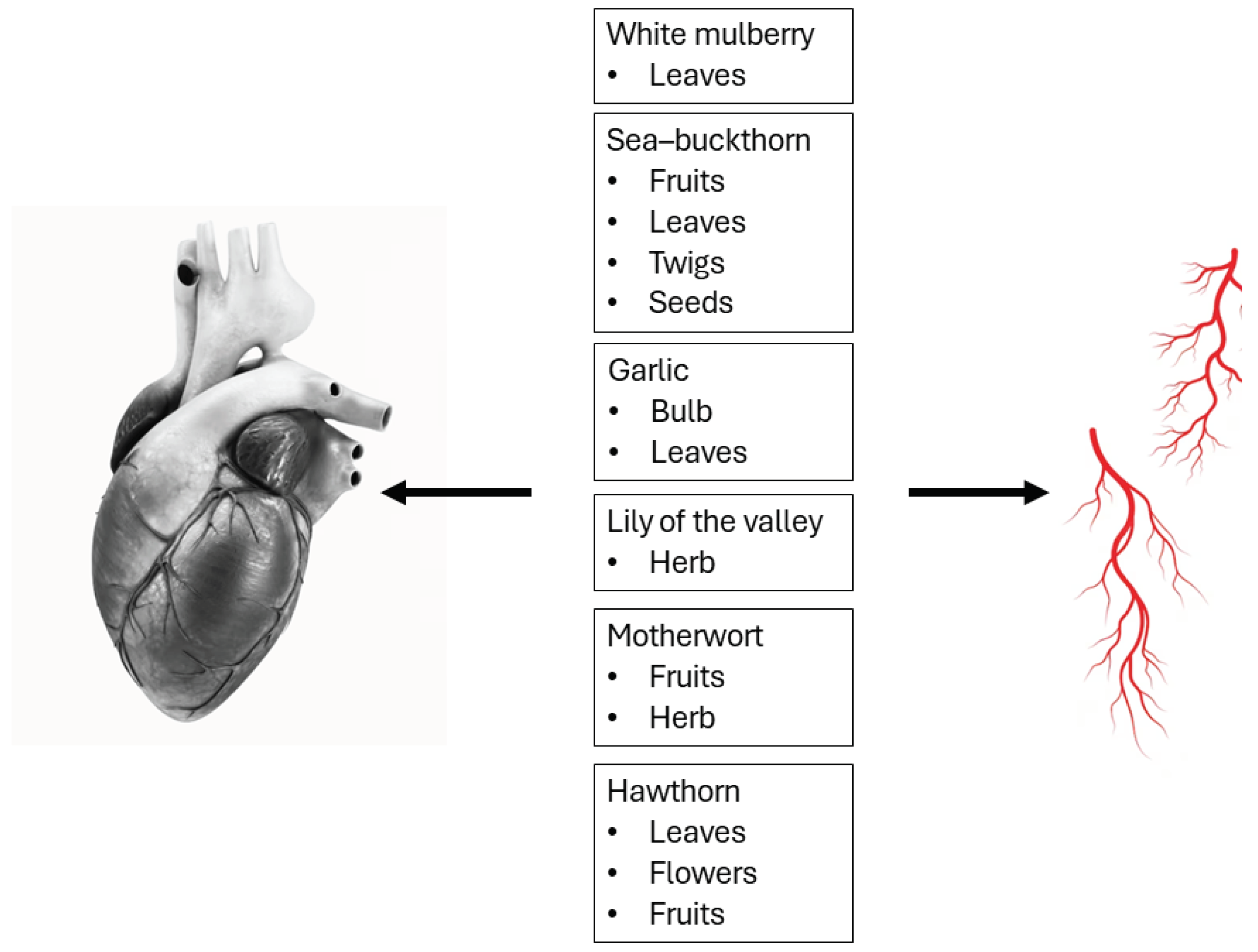

Table 1.
Cardiovascular effects of Morus alba L. (white mulberry) Moraceae family.
| Herbal Raw Material |
Active Compounds | Functions | Mechanism of Action | Model | Dose | References |
|---|---|---|---|---|---|---|
| Leaves | Phenolic groups, naringenin and quercetin | Cardioprotective Antioxidant |
↑ scavenging of the free radicals (OH) ↓ peroxidation of lipids ↓ GSH, Gpx, SOD, CAT, TBARS |
male albino Wistar rats | 500 mg/kg b.w. for 15 days p.o. | [9] |
| Flavonoids (isoflavones, flavanone, flavonols, morusin, cyclomorusin, and neocyclomorusin) novel prenylated flavonoids (isoquercetin, quercetin, astragalin, and scopoline) glycosides (namelyroseoside III and benzyl D–glucopyranoside) |
Anti–hypertensive Antioxidant Cardioprotective |
↑ cellular antioxidants (↑ GSH, SOD, and CAT, ↓ LPO) ↓ heart rate, ↓ heart weight ↓ pressure–rate index ↓ levels of cardiac damage marker enzymes |
male Wistar rats | 25, 50, 100 mg/kg b.w. for three weeks p.o. | [10] | |
| Anti–hypertensive Hypolipidemic Vascular improvement Lipid regulation agent |
↓ of cell adhesion molecules (E–selectin, VCAM–1, ICAM–1) expression in the aorta |
male rats | 100, 200 mg/kg b.w. for 14 weeks | [13] | ||
| Rutin, chlorogenic acid, astragalin | Vascular effect | ↑ NO, eNOS ↑ phosphorylation of PERK and HSP90 at threonine |
Mice | 100, 200, or 400 mg/kg b.w. | [11] | |
| Rutin and isoquercetin | Antiplatelet Antithrombotic Prevention or treatment of myocardial infarction |
↓MAPK–integrin α IIb β 3 ↓ PLA2/TXA2 routes ↓TXB2 formation ↓ serotonin secretion ↓ aggregation ↓ thrombus formation |
male Sprague Dawley rats | 100, 200, 400 mg/kg b.w. for 3 days | [12] | |
| Mulberroside A, chlorogenic acid, cryptochlorogenic acid, astragaloside, resveratrol, scopoletin, 1–deoxynojirimycin | Glucose improvement Lipid metabolism improvement |
↓TGF–β–Smad2/3 | male C57BL/6 mice | 1,000 mg/kg b.w. for 12 weeks p.o. | [14] | |
| Neochlorogenic acid | Anti–atherosclerotic | ↓ FAK/small GTPase proteins ↓ PI3K/Akt ↓ Ras–related signaling ↓ vascular smooth muscle cell (VSMC) migration and proliferation |
Ex vivo A7r5 cells (VSMCs) |
3.0 mg/mL for 24 h | [15] |
Table 2.
Cardiovascular effects of Elaeagnus rhamnoides (L.) A. Nelson (sea–buckthorn) Elaeagnaceae family.
Table 2.
Cardiovascular effects of Elaeagnus rhamnoides (L.) A. Nelson (sea–buckthorn) Elaeagnaceae family.
| Herbal raw material |
Active compounds | Functions | Mechanism of action | Model | Dose | References |
|---|---|---|---|---|---|---|
| Fruits | polyphenols (proanthocyanidins) flavonoids (flavonol glycosides isorhamnetin 3–O–hexoside–deoxyhexoside and isorhamnetin 3–O–hexoside) phenolic acids vitamins (vitamin C) fatty acids and phytosterols triterpenes and triterpene derivates (derived from isorhamnetin) compounds derived from quercetin, and kaempferol |
Anti–platelet Anticoagulant |
↓ surface exposition GPIIb/IIIa and P–selectin | In vitro | 50 μg/mL; 10 min |
[16] [18] |
| Leaves | ellagitannins (casuarinin, hippophaenin B, casuarictin, stachyurin, strictinin or their isomers), ellagic acid and its glycosides flavonoids (glycosides of isorhamnetin, kaempferol and quercetin) |
Anti–platelet | ↓ surface exposition of P–selectin ↓ surface exposition of GPIIb/IIIa active complex |
In vitro | 5 and 50 μg/mL; 30 min |
[17] |
| Twigs | Proanthocyanidins Catechin |
Anticoagulant Anti–platelet |
↓ surface exposition of P–selectin ↓ surface exposition of GPIIb/IIIa active complex |
In vitro | 5 and 50 μg/mL; 30 min |
[17] |
| Seeds | Flavonoids (glycosides of isorhamnetin, kaempferol, and quercetin), proanthocyanidins and catechin, triterpenoid saponins, and several unidentified polar and hydrophobic compounds |
Antioxidant Anticoagulant |
↓ lipid peroxidation and protein carbonylation ↓ oxidation of thiol groups |
In vitro | 0.5, 5.0, 50 µg/mL | [19] |
Table 3.
Cardiovascular effects of Allium sativum L. (garlic) Amaryllidaceae family.
| Herbal raw material |
Active compounds | Functions | Mechanism of action | Model | Dose | References |
|---|---|---|---|---|---|---|
| Bulb | Organosulfur compounds: alliin, allicin, E–ajoene, Z–ajoene, 2–vinyl–4H–1,3–dithiin, diallyl sulfide (DAS), diallyl disulfide (DADS), diallyl trisulfide (DATS) and allyl methyl sulfide (AMS) |
Hypoglycemic Hypolipidemic |
↓ total triglycerides and free fatty acids ↑ HDL–cholesterol |
In vivo mice |
0.1 mg/mL for 8 weeks Alliin (S–allyl cysteine sulfoxide) p.o. |
[34] |
| Antioxidant | ↓lipid peroxidation ↓ free radicals |
In vivo male albino Wistar rats |
2 mL S–allyl cysteine sulfoxide (SACS) p.o. |
[30] [31] [36] |
||
| Antioxidant Vascular |
↑ NO and eNOS | In vitro endothelial progenitor cells |
100 µg/mL 200 µg/mL 400 µg/mL allicin |
[39] | ||
| Antioxidant | ↑ glutathione levels in vascular endothelial cells ↓triglycerides |
In vivo male SHR |
80 mg/kg/day allicin p.o. |
[40] | ||
| Antioxidant | ↓ ROS ↓ cell growth and migration |
In vitro vascular smooth muscle cells (VSMCs) |
10 and 100 μg/L 2–vinyl–4H–1,3–Dithiin |
[41] | ||
| Antioxidant | ↓ cardiac hypertrophy markers | In vivo male Sprague Dawley |
250 mg/kg garlic | [42] | ||
| 25, 50, 100, 200 mg/kg/day Allylmethylsulfide (AMS) p.o. | [43] | |||||
| Cardioprotective Anti–apoptotic |
↑ IGF1R survival signaling pathway protein expression ↑ PI3K/Akt pathway ↓ ROS |
In vitro and in vivo cardiomyocyte cell line H9c2 and hearts from rats with streptozotocin–induced diabetes mellitus |
10 μM diallyl trisulfide (DAT) |
[44] | ||
| Antioxidant Vascular |
ND1 | In vivo obese diabetic rats |
5 mg/kg/day diallyl trisulfide (DAT) |
[45] | ||
| Anti–aggregation | ↓platelet aggregation ↓ cAMP and cGMP stimulation ↓fibrinogen binding to the GPIIb/IIIa receptor |
In vitro human platelet aggregation |
6.25% aged garlic extract |
[47] | ||
| Lipid regulation | ↓ lipogenesis and cholesterogenesis | In vivo male albino diabetic rats |
1 mL garlic aqueous leaf extract |
[35] |
1ND – not determined.
Table 4.
Cardiovascular effects of Convallaria majalis L. (lily of the valley) Asparagaceae family.
| Herbal raw material |
Active compounds | Functions | Mechanism of action | Model | Dose | References |
|---|---|---|---|---|---|---|
| Herb | cardiac glycosides: convallotoxin, convallarin, convalloside, convallasaponin, cholestane glycoside, strophanthidin, cannogenol, sarmentogenin, dipindogenin, hydroxysarmentogenin saponins |
Heart failure | ↓ Na+/K+–ATPase inhibition ↑ Na+ ions ↑ Ca2+ ions positive inotropic effect ↓ viability of HUVECs ↑ TF mRNA and protein expression |
In vitro Serum Human umbilical vein endothelial cells (HUVECs) |
50 and 100 nM for 4 h |
[52] [55] [56] |
| Anti–inflammatory | ↓ lipoxygenase inhibition | In vitro linoleic acid |
5 g of the roots | [62] | ||
| Convallatoxin/Convallaria keiskei | Cardiovascular effect | ↑ atrial stroke volume and pulse pressure, positive inotropic effect vasoconstrictor, and vasodilator effects ↑ cAMP efflux ↑ K+ concentration |
Ex vivo beating rabbit atria, male New Zealand white rabbits weighing about 2 kg |
1 x 10⁻⁵ M Convallatoxin 3 × 10⁻⁶ g/mL Convallaria keiskei |
[54] [57] [58] |
|
| convallamaroside | Anticancer | ↓ angiogenesis | Ex vivo mice on tumor angiogenesis reaction induced by tumor cells |
10,20,50,100 µg/day 1.5 h incubation |
[59] [60] [61] |
Table 5.
Cardiovascular effects of Leonurus cardiaca L. (motherwort) Lamiaceae family.
| Herbal raw material |
Active compounds | Functions | Mechanism of action | Model | Dose | References |
|---|---|---|---|---|---|---|
| Fruits | Stachydrine | Vaso–protective | ↑ expression of PTCH1 and DHFR | In vivo and ex vivo isolated rat thoracic aortas, mesenteric arteries, and renal arteries from male Sprague–Dawley rats. Bovine aorta endothelial cells (BAECs) |
10 μM for 12 hours; 0–10 μM for 24 hours |
[76] |
| ND1 | Stachydrine | Cardioprotective Antioxidant Anti–inflammatory |
↓NF–κB and JAK/STAT | In vivo male Sprague–Dawley rats |
10, 40 mg/kg b.w. for 21 days p.o. | [77] |
| Herb | Leonurine | Antiapoptotic Cardioprotective |
↑ PI3K/AKT/GSK3β | In vivo male Sprague–Dawley rats |
15 mg/ kg b.w. for 28 days p.o. |
[78] |
| labdane diterpenes: leosibirin, leosibiricin, 19–acetoxypregaleopsin flavonoid glycosides based on querectin and apigenin, phenylpropanoids: eugenol, lavandulifolioside, alkaloids: stachydrine, betonicine, leonurine, iridoids: ajugol, ajugoside, harpagide |
Antihypertensive Psycho–neurological |
ND1 | In vivo male and female patients |
1200 mg per day for 28 days |
[79] | |
| Chlorogenic acid, orientin, quercetin, hyperoside, and rutin | Cardioprotective | ↓ mitochondrial ROS generation | Ex vivo rat heart mitochondria |
6.8 μg/mL 18.2 μg/mL |
[80] |
1ND – not determined.
Table 6.
Cardiovascular effects of Crataegus spp. (hawthorn) Rosaceae family.
| Herbal raw material |
Active compounds | Functions | Mechanism of action | Model | Dose | References |
|---|---|---|---|---|---|---|
| Leaves | Flavonoids (quercetin, vitexin–2″–O–α–L–rhamnoside, hispertin, pinnatifinosides I, cratenacin), oligomeric proanthocyanidins (proanthocyanidin A2) |
Antioxidant Cardioprotective Antihypertensive Vasodilator |
↑myocardial perfusion ↓ of phosphodiesterase, ↑ in cGMP in smooth muscle cells |
In vivo diabetic and non–diabetic rats |
5 or 10 mg/kg | [109] [110] |
| Flowers | Flavonoids (rutin, hyperoside, vitexin–2″–O–rhamnoside), phenols (chlorogenic acid) oligomeric proanthocyanidins (proanthocyanidin A2) |
Antioxidant Cardioprotective Anti–inflammatory Anti–atherosclerotic Anticoagulant |
↑ improving the blood flow in blood vessels | Sprague Dawley rats | 100, 200, and 500 mg/kg for 3 weeks p.o. | [109] [111] [112] |
| Fruits | Polyphenols, flavonoids (hyperoside, hesperidin, rutoside, crataequinone A–B), vitamin C proanthocyanidins (procyanidin B2), phenols (chlorogenic acid) |
Antioxidant Cardioprotective Hypolipidemic Anti–aggregation |
ND1 | In vivo 12–week–old rats 8–week–old SHR |
1.08 g/kg body weight per day intragastrically 30 mg/kg Procyanidin |
[109] [111] [113] [114] [115] |
1ND – not determined.
Disclaimer/Publisher’s Note: The statements, opinions and data contained in all publications are solely those of the individual author(s) and contributor(s) and not of MDPI and/or the editor(s). MDPI and/or the editor(s) disclaim responsibility for any injury to people or property resulting from any ideas, methods, instructions or products referred to in the content. |
© 2024 by the authors. Licensee MDPI, Basel, Switzerland. This article is an open access article distributed under the terms and conditions of the Creative Commons Attribution (CC BY) license (http://creativecommons.org/licenses/by/4.0/).
Copyright: This open access article is published under a Creative Commons CC BY 4.0 license, which permit the free download, distribution, and reuse, provided that the author and preprint are cited in any reuse.


