
Submitted:
05 March 2024
Posted:
06 March 2024
You are already at the latest version
Abstract
Glycosphingolipids (GSLs), a subtype of glycolipids containing sphingosine, are critical components of vertebrate plasma membranes, playing a pivotal role in cellular signaling and interactions. In human articular cartilage in osteoarthritis (OA), GSL expression is known notably to decrease. This review focuses on the roles of gangliosides, a specific type of GSL, in cartilage degeneration and regeneration, emphasizing their regulatory function in signal transduction. The expression of gangliosides, whether endogenous or augmented exogenously, is regulated at the enzymatic level, targeting specific glycosyltransferases. This regulation has significant implications for the composition of cell surface gangliosides and their impact on signal transduction in chondrocytes and progenitor cells. Different levels of ganglioside expression can influence signaling pathways in various ways, potentially affecting cell properties, including malignancy. Moreover, gene manipulations against gangliosides have been shown to regulate cartilage metabolism and chondrocyte differentiation in vivo and in vitro. This review highlights the potential of targeting gangliosides in the development of therapeutic strategies for osteoarthritis and cartilage injury and addresses promising directions for future research and treatment.
Keywords:
glycosphingolipids (GSLs)
; osteoarthritis
; articular cartilage
; gangliosides
; chondrocyte differentiation
; cartilage regeneration
1. Introduction
Articular cartilage is a specialized connective tissue that resides at the interface between bones and joint space [1]. This highly specialized tissue comprises chondrocytes and a specific extracellular matrix (ECM) containing types II, IX, and XI collagen and proteoglycans, but notably lacks type I collagen. Referred to as hyaline cartilage, it is characterized by its elasticity, which plays a crucial role in absorbing and distributing loads during weight-bearing. Another function is to provide a smooth, lubricated surface facilitating a range of motion and enabling load transfer with minimal friction. Articular cartilage is avascular, aneural, lymphatic, and hypocellular, limiting its capacity for standard tissue repair mechanisms [2]. Most important, articular cartilage has a limited capacity for intrinsic healing and repair.
Osteoarthritis (OA), the most common joint disease, affects over 300 million people worldwide and contributes to an economic burden on both patients and society [3,4]. The disease costs the United States economy more than $80 billion per year [5]. OA is characterized by progressive degradation of articular cartilage and ECM, while its pathogenesis remains largely unknown despite extensive gene- and protein-based research [6]. Articular cartilage does not possess access to the nutrients or circulating chondrogenic progenitor cells and cartilage lacks the natural potential to overcome a sufficient healing response by possessing a nearly acellular nature [7]. Consequently, articular cartilage has limited healing potential; therefore, it can lead to cartilage degeneration and ultimately result in OA. In particular, the relationship between OA and glycolipids began to receive attention after the finding that the composition of glycosphingolipids (GSLs) is markedly altered in the articular cartilage of OA patients [8,9,10]. Following this report, the possibility that biomembrane glycolipids, the subject of post-genomic studies, are involved in the pathogenesis of OA has come to be considered.
GSLs are key components of cell membranes, comprising a hydrophobic ceramide and a hydrophilic oligosaccharide residue. Ceramides are embedded in the outer leaflet of the plasma membrane, while oligosaccharides project into the extracellular space [11,12]. GSLs cluster on the cell membrane surface, modulating transmembrane signaling and mediating intercellular and cell-matrix interactions [11,12,13,14]. An enzyme called glucosylceramide synthase encoded by the UDP-glucose ceramide glucosyltransferase (Ugcg) gene is responsible for directing the first committed step in GSL synthesis [15,16,17,18]. Glucosylceramide is formed when a glucose moiety is transferred from UDP-glucose to ceramide, which is the precursor of most cellular GSLs. Mice with a global disruption in UGCG are embryonic lethal (E7.5), suggesting that GSLs are essential for embryonic development and differentiation [15,16,17,19]. It is now well established that some sphingolipids can regulate key biological functions, and these include cell growth and survival, cell differentiation, angiogenesis, autophagy, cell migration, or organogenesis [20]. Furthermore, specific bioactive sphingolipids have been linked to various pathologies, including inflammation-related diseases like atherosclerosis, rheumatoid arthritis, type II diabetes, obesity, cancer, and osteoarthritis.
Here, we mainly discuss the usefulness of GSLs expressed on cell membranes as biomarkers for quality control in cartilage regenerative medicine and as therapeutic target molecules for OA.
2. Impact of GSLs on the Cartilage Homeostasis
After it was shown that a major component of glycolipids (ceramide) stimulates the mRNA expression of collagenase-1/MMP-1 and stromelysin-1/MMP-3 in human fibroblasts through the activation of three different mitogen-activated protein kinases (MAPKs), ERK1/2, SAPK/JNK and p38 in cartilage, ceramide was also found to be involved in cartilage degeneration and apoptosis [21,22]. The ceramide pathway activator suppressed the production of inflammatory cytokines and activation of the MAPK pathways [23]. As mentioned above, systemic knockout mice of the Ugcg gene are embryonic lethal because UGCG is the first committed step in the synthesis of the majority of GSLs [15,16,17,18]. GSLs form clusters on the plasma membrane and play diverse roles in regulating membrane-mediated signal transduction and mediating cell-cell and cell-extracellular matrix interactions [24,25,26,27]. Therefore, an attempt was made to sort out even the most characteristic downstream glycolipid molecules by sequential knockout of upstream glycosyltransferase genes involved in the impairment of cartilage homeostasis in chondrocytes. The contents of such research studies are summarized in Figure 1 and Table 1.
A decrease in all major gangliosides, contrasting with a marked increase in the GM3, has been demonstrated in osteoarthritic fibrillated cartilage. Some previous studies have shown that gangliosides have tissue-protective effects against oxidative stress or apoptosis in neuronal, cardiac, and hepatic cells [28,29,30,31,32]. The results of the series of studies indicate that loss of gangliosides results in greater cartilage vulnerability to interleukin (IL)-1 stimulation in the cartilage degradation process by increasing MMP-13 secretion and chondrocyte apoptosis. On the other hand, There have been indications that replenishing cells with the missing gangliosides can restore normal activation [33,34]. Based on the previous studies, it is reasonable to consider a treatment that supplements the o-, a-, and b-series gangliosides below GalNAcT. Future therapeutic trials are pending.
Glycosidase inhibitors are also considered to be an important target for cartilage regeneration. They are directly linked to osteoarthritis because N-acetyl-beta-hexosaminidase is the predominant glycosidase released by chondrocytes to degrade glycosaminoglycan [35,36]. Stimulation of chondrocytes with IL-1β selectively increases extracellular hexosaminidase activity among many enzymes, suggesting that hexosaminidase is the cartilage matrix-degrading enzyme activated by inflammatory stimuli. The inhibitor of this hexosaminidase was shown to modulate intracellular levels of glycolipids, including GM2 and GA2 (o- and a-series gangliosides).

Table 1.
Genetic defects in mouse glycan formation and physiologic consequence.
| Glycosyltransferase | Lost glycolipids | Consequences of depletion of its glycolipid | References |
| UGCG (Glucosylceramide synthase) |
GSLs |
Embryonic death. Reduced insulative capacity of the myelin sheath. Col2-Ugcg-/- mice enhance the development of OA | [16,17,19,37,38,39] |
| ST3GalⅣ (GM3S) |
Gangliosides other than the o-series | GM3 plays an immunologic role. Heightened sensitivity to insulin. Severely reduced CD4+ T cell proliferative response and cytokine production. Promote OA and RA but cartilage regeneration | [34,40,41,42,43] |
| ST8SiaⅠ (GD3S) |
b-series ganglioside | Tumor-associated carbohydrate antigens (TACA) in neuro-ectoderm-derived cancers. Suppression of age-related bone loss. Deteriorates OA with aging | [33,44,45,46,47] |
| GalNAcT (GM2/GD2S) |
Almost all gangliosides except GM3, GD3, and GT3 | Age-dependent neurodegeneration, movement disorders associated with it. Defects in spermatogenesis and learning. Exacerbating OA progression | [33,48,49,50] |
3. Role of GSLs in Cartilage Repair and Differentiation Processes
GSLs play a crucial role in the repair and differentiation processes of articular cartilage. These processes are essential for addressing cartilage defects, commonly observed in osteoarthritis. The unique properties of GSLs facilitate the regeneration of cartilage tissue, which is key to restoring joint function. Articular cartilage repair models have demonstrated the potential of specific cell types and biological factors in facilitating cartilage repair. These models highlight the importance of understanding the underlying mechanisms of cartilage regeneration, particularly focusing on the role of GSLs in this process.
In this chapter, various aspects of cartilage repair will be explored, with a focus on the role of glycolipids in promoting the regeneration and repair process through chondrogenic differentiation.
3.1. Endogenous Potential to Heal in Articular Cartilage
To enhance endogenous cell recruitment to the injury site, the biological process within a living organism after articular cartilage injury needs to be clarified. Considering species differences, appropriate animal models to replicate human articular cartilage repair processes are reported in mice [51,52,53], rats [54], rabbits [55,56,57], horses [58], and canines [59], thus molecular reaction after articular cartilage injuries is continuously disclosed [60,61,62,63,64]. Even though the species are different, the major ganglioside in articular cartilage appears to be GM3 [65,66]. GSLs consist of several types of glycolipids and are classified into several groups depending on their structural features, which include neo-lacto-series, globo-series, isoglobo-series and ganglio-series (gangliosides) [24,40]. GM3 serves as a precursor molecule for most of the more complex ganglioside species [41] and ganglioside expression pattern in cells during differentiation changes in response to cytokine and growth factor stimulation [67,68,69]. During the articular cartilage healing process, GM3 was first expressed in injured bone marrow on day 1 and gradually decreased at 2 weeks after articular cartilage injury GM3 next transiently expressed cartilage in the vicinity of remnant cartilage which co-expressed with type X collagen with a peak 6 weeks postoperative [42]. Notably, the depletion of gangliosides in mice suppressed hypertrophic differentiation in the vicinity of remnant cartilage, resulting in enhanced articular cartilage regeneration. These results suggested that gangliosides have dual roles in the recruitment of chondrogenic precursor cells to the injury site and induction of hypertrophic differentiation in chondrocytes. Manipulation of ganglioside expression may future direction in articular cartilage regeneration, however, the site- and time-specific intervention to manipulate glycosphingolipids in articular cartilage is needed.
3.2. Changes in the Glycan Structure during Chondrogenic Differentiation
Chondrogenic differentiation is the well-organized process by which cartilage is formed from condensed mesenchyme tissue, which differentiates into chondrocytes and begins secreting the molecules that form the extracellular matrix [70]. Extracellular enzymes, which include the matrix metallopeptidases, lead to the activation of cell signaling pathways and gene expression in a temporal-spatial-specific manner during the development process. The recruited mesenchymal stem cells may attempt to differentiate into chondrocytes after articular cartilage injury [71,72]. However, the response after injury is not a fully recapitulated process of development, resulting in regenerated cartilage-like tissue that does not possess typical biomolecules of hyaline cartilage such as type II collagen and aggrecan, and the proportion of the chemical constitutes of them differ from those in original cartilage [73,74]. Molecules promoting the selective differentiation of multipotent mesenchymal stem cells into chondrocytes have been reported to stimulate the repair of injured articular cartilage [75]. Therefore, the regulation system for chondrogenic differentiation is attracting attention.
Glycosylation is one of the posttranslational modifications in cell surface proteins and extracellular matrix proteins, which regulate a variety of biological functions, including enhancement of protein stability, controlling cell-to-cell communication, and adhesion [76]. In addition, this process is known to contribute to the pathogenesis of various kinds of diseases [77,78]. We previously performed quantitative and qualitative analyses of N-linked glycans and glycoproteins during chondrogenic differentiation using the glycoblotting method [79] and subsequent glycoform-focused reverse proteomics and genomics using mouse pre-chondrogenic cell line, ATDC5 cell line [80]. The levels of high-mannose type N-glycans increase during chondrogenic differentiation, suggesting that N-glycans may have key roles in differentiation and/or homeostatic maintenance of chondrocytes [81]. As for hypertrophic differentiation in chondrocytes, Yan et al. reported that resting chondrocytes exposed to concanavalin A, which binds specifically to high mannose-type structures, selectively differentiated to the hypertrophic stage [82,83]. We previously performed comprehensive glycomics, including N-glycans, O-glycans, free oligosaccharides (fOSs), glycosaminoglycans (GAGs), and GSLs and showed dynamic alterations in all classes of glycoconjugates following the differentiation process [84]. Hierarchical clustering analysis based on quantitative glycomic profiles showed that the levels of various N-glycans dramatically increased with hypertrophic progression, in contrast, those of GSL-glycans and fOSs significantly decreased. Changes in the levels of O-glycans and GAGs were transient. These results suggested that glycan markers can be used as differentiation biomarkers for chondrogenic differentiation and may help to evaluate the regenerative product after articular cartilage injury.
4. Cell Sources
As we explained in the Introduction section, articular cartilage is a type of hyaline cartilage that enables smooth movements between bones in articulating joints, which requires both weight-bearing and low-friction capability [85]. Therefore, the regenerated cartilage requires these high qualities of properties. The ultimate goal for ideal cartilage regeneration is to restore these key properties of the original hyaline cartilage in terms of histological structure and biomechanical functions, which seems to be only achieved by replacing it with healthy cartilage tissue [86]. As for now, several types of cell-based approaches have been introduced [87]. Representative strategy includes autologous cartilage implantation, mesenchymal stem cells, and induced pluripotent stem cells. Table 2 summarizes three representative cell types and reports on cartilage regenerative medicine. Here, mainly based on glycobiology, we overview the recent cell-based regenerative medicine in articular cartilage.
4.1. Autologous Cartilage/Chondrocyte Implantation
In 1993, R Langer and J P Vacanti advocated the concept of tissue engineering, which applied the principles of biology and engineering to the development of functional substitutes for injured tissue by manipulating cells, scaffolds, and stimuli such as cytokine [123]. The first target organ by tissue engineering was thought to be skin or cartilage due to the less variation of cell types, and avascular and two-dimensional nature. However, in terms of articular cartilage, it was proved to be incorrect as far as known. Articular cartilage does not possess access to the nutrients or circulating chondrogenic progenitor cells and cartilage lacks the natural potential to overcome a sufficient healing response by possessing a nearly acellular nature [124]. Consequently, articular cartilage has limited healing potential; therefore, it can lead to cartilage degeneration and ultimately result in OA.
In 1994, Brittberg et al. first introduced a cell-based therapy consisting of two staged procedures for full-thickness defects of articular cartilage in the knee [125]. This procedure requires the first harvest of chondrocytes from a non-weight-bearing area of articular cartilage. After a culture of 4 to 6 weeks, a second-stage procedure is undertaken to implant amplified chondrocytes into the defect. Considering the limited healing potential for articular cartilage, these procedures seem to be ideal. Since the first clinical report was published, several authors have demonstrated successful clinical outcomes of this procedure for cartilaginous lesions [126,127,128]. However, there remain concerns about the dedifferentiation of chondrocytes during the culture period due to the limited proliferative capacity [129,130,131]. The cartilage extracellular matrix possesses various glycosylated proteins, which contribute to the maintenance of its specific functions [132]. Sialic acids are negatively charged sugars expressed at the terminal positions of N- and O-linked oligosaccharides, which are attached to cell surfaces or secreted glycoproteins. As a result of their non-reducing terminal position, sialic acids are involved in highly specific recognition phenomena [133,134]. Primary human chondrocytes predominantly express α-2-6-specific sialyltransferases and α-2-6-linked sialic acid residues in glycoprotein N-glycans [135]. Interestingly, inflammation stimuli induced a shift from α-2-6-linked towards α-2-3-linked sialic acid, suggesting that α-2-6-linked sialic acid can be used as a biomarker for quality control in amplified human cartilage.
4.2. Mesenchymal Stem Cells (MSCs)
Mesenchymal stem cells are multipotent stem cells and can be obtained from various organs including bone marrow, synovium, periosteum, adipose tissue, and skeletal muscle [136]. MSCs are attractive cell sources in regenerative medicine based on their abilities to self-renew and differentiate into mesenchymal tissue lineage [137]. During the last few years, the use of MSCs and their cell-free derivatives has seen an increasing number of applications in disparate medical fields, including chronic musculoskeletal conditions [138,139,140,141,142]. All of these approaches would require cell harvesting and transplantation. One of the concerns is that MSCs are heterogeneous populations, whose capability to differentiate varies depending on the tissues harvested and donor age. Although MSCs are grouped together by the common characteristics of CD44, 73, 90, 105 positivity and CD31, 45 negativity, these do not necessarily define "stem cells." These cells are then expanded in vitro until a required cell number is reached, therefore, isolation methods, culture conditions, and passages should be given consideration.
Glycosylation features associated with bone marrow-derived MSCs included high-mannose type N-glycans, linear poly-N-acetyllactosamine chains, and α2-3-sialylation [143]. Their cellular differentiation stage can be determined using these glycomics. Tateno et al. carried out glycome analysis on different passages of adipose-derived human MSCs (hMSCs) using high-density lectin microarray to identify glycan markers that distinguish MSCs to have enough capability to differentiate [144]. This report indicated that α2-6-linked sialic acid-specific lectins showed stronger binding to early passage of adipose-derived hMSCs with differentiation ability to adipocytes and osteoblasts than did late passage cells without the ability. They also reported quantitative glycome analysis targeting both N- and O-glycans from early and late passages of adipose tissue-derived hMSCs and showed the expression of α2-6-sialylated N-glycans varies depending on the differentiation potential of stem cells but not O-glycans, suggesting that α2–6-sialylated N-glycans can be used biomarker for quality control of hMSCs [145]. The presence of α2-6-linked sialic acid structure is a characteristic of pluripotent stem cells that possess higher differentiation potential. This may serve as an indicator of their differentiation potential. Ryu et al. showed that the gangliosides GM3 and GD3, which contain α2-3- and α2-8-linked sialic acids, were expressed after the chondrogenic differentiation of synovium-derived hMSC aggregates [146]. In the same way, GM3 expression increased temporarily following the chondrogenic differentiation of hMSCs derived from bone marrow [147]. Considering OA cartilage is characterized by a decrease in most gangliosides, these gangliosides may be useful in developing therapeutic agents for MSC-based articular cartilage regeneration in articular cartilage disease.
4.3. Induced Pluripotent Stem Cells (iPSCs)
In 2006, Takahashi and Yamanaka reported pluripotential stem cells from mouse embryonic or adult fibroblasts by introducing four factors Oct3/4, Sox2, c-Myc, and Klf4 [148]. The method to establish human iPSCs dramatically evolved and simplified [149]. This progress provides us with opportunities to understand the disease mechanisms and promote regenerative medicine [150,151]. To avoid the rejection of differentiated cells originating from iPSC transplantation, autogenous transplantation with iPSCs originating from donors is ideal. However, these procedures require time to establish iPSCs with high enough quality for transplantation as well as the cost. In contrast, iPSCs induced from individuals with a homozygous human leukocyte antigen haplotype (HLA-homo) is a significant candidate for allogenic transplantation on the basis that HLA-homo iPSCs might not be rejected by HLA haplotype-matched patients [152,153,154]. iPSC banking recruited from healthy, consenting HLA-type homozygous donors and is made with peripheral blood-derived mononuclear cells or umbilical cord blood [155]. Many research groups have been trying to apply iPSCs-based therapy to patients, and some of them are already being administered in clinical trials [156].
As for cartilage metabolisms, the main techniques to confirm chondrogenic differentiation from iPSCs are based on the detection of upregulated chondrogenic genes or histological analysis of the extracellular matrix. We previously performed a quantitative GSL-glycan analysis to compare human iPSCs, iPSC-derived MSC-like cells, iPS-MSC-derived chondrocytes (iPS-MSC-CDs), and bone marrow-derived MSCs [147,157]. GSL-glycan profiles differed among cell types, and the GSL-glycome underwent a characteristic alteration during the process of chondrogenic differentiation. Undifferentiated human iPSCs mainly expressed globo- and lacto-series GSLs, which shifted to ganglio-series GSLs, such as GM3 when iPSCs differentiated into iPS-MSCs. After chondrogenic differentiation of both bone marrow-derived MSCs and iPS-MSC-like cells, the expression of GM3 increased temporarily. Furthermore, the GSL-glycome of normal human cartilage was closely similar to that of iPS-MSC-CDs.
In terms of tumorigenicity for iPSCs themselves, while clinical applications are going forward, the concerns that transplantation of differentiated iPSC might lead to teratoma formation in the recipient should be clarified [158,159]. Matsumoto et al. reported that R-17F antibody detects undifferentiated iPSCs harboring the Lacto-N-fucopentaose I (LNFP I) of GSLs, and as a result, exerts cytotoxic activity [160]. Recently, we developed the aminolysis-sialic acid linkage-specific alkylamidation (SALSA) method [161], which enables discrimination of sialic acid linkage isomers on GSL-glycans by mass spectrometric analysis and analyzed the GSL-glycome of co-cultured undifferentiated iPSCs and chondrocytes by glycoblotting-SALSA method. Most GSL-glycans from ~100 cells of iPSCs could also be detected. Utilizing this technique, we showed that R-17 antibody detects undifferentiated iPSCs harboring the Lacto-N-fucopentaose I (LNFP I) of GSLs have selective removal of residual iPSCs even by chondrocytes co-cultured with iPSCs [162]. Furthermore, the experiment was successfully performed to determine whether R-17 antibodies could prevent teratoma formation induced by residual iPSCs [163]. The GSLs-Glycome analysis is useful to determine the optimal condition for the removal of undifferentiated iPSCs to a level safe for transplantation.
5. Conclusions
Glycomics of mesenchymal stem/progenitor cells can be utilized to evaluate their stage of cellular differentiation. Furthermore, this review highlighted the potential that supplementing missing GSLs could play significant roles in tissue regeneration and disease modification. To guide the regeneration of degenerated or injured cartilage into articular cartilage, a multifactorial methodology that incorporates GSLs, which are closely related to cartilage homeostasis, should be developed in the future.
Author Contributions
Conceptualization, K.H. and T.O.; investigation, K.H., T.O., M.M; writing—original draft preparation, K.H. and T.O.; writing—review and editing, K.H., T.O., M.M.; supervision, N.I.; funding acquisition, K.H., T.O., N.I. All authors have read and agreed to the published version of the manuscript.
Funding
This work was supported by funds for the Japan Society for the Promotion of Science KAKENHI (Grant-in-Aid for Challenging Exploratory Research: funding numbers: 19K22672 [to N.I.]; Grant-in-Aid for Research (B): funding numbers: 22H03917 [to T.O.]; and Grant-in-Aid for Early-Career Scientists: funding number: 23K16633 [to K.H.]) and AMED (Grant Number JP22zf01270004h0002 [to N.I., T.O., K.H.]).
Acknowledgments
The sponsor of the study had no role in the report preparation.
Conflicts of Interest
The authors declare no conflict of interest.
References
- Wang, M.; Wu, Y.; Li, G.; Lin, Q.; Zhang, W.; Liu, H.; Su, J. Articular Cartilage Repair Biomaterials: Strategies and Applications. Mater. Today Bio 2024, 24, 100948. [CrossRef]
- Muthu, S.; Korpershoek, J.V.; Novais, E.J.; Tawy, G.F.; Hollander, A.P.; Martin, I. Failure of Cartilage Regeneration: Emerging Hypotheses and Related Therapeutic Strategies. Nat. Rev. Rheumatol. 2023, 19, 403–416. [CrossRef]
- Peat, G.; Thomas, M.J. Osteoarthritis Year in Review 2020: Epidemiology & Therapy. Osteoarthritis Cartilage 2021, 29, 180–189. [CrossRef]
- Chua, J.R.; Jamal, S.; Riad, M.; Castrejon, I.; Malfait, A.-M.; Block, J.A.; Pincus, T. Disease Burden in Osteoarthritis Is Similar to That of Rheumatoid Arthritis at Initial Rheumatology Visit and Significantly Greater Six Months Later. Arthritis Rheumatol. Hoboken NJ 2019, 71, 1276–1284. [CrossRef]
- Dieleman, J.L.; Cao, J.; Chapin, A.; Chen, C.; Li, Z.; Liu, A.; Horst, C.; Kaldjian, A.; Matyasz, T.; Scott, K.W.; et al. US Health Care Spending by Payer and Health Condition, 1996-2016. JAMA 2020, 323, 863–884. [CrossRef]
- Rice, S.J.; Beier, F.; Young, D.A.; Loughlin, J. Interplay between Genetics and Epigenetics in Osteoarthritis. Nat. Rev. Rheumatol. 2020, 16, 268–281. [CrossRef]
- Gobbi, A.; Lane, J.G.; Longo, U.G.; Dallo, I. Joint Function Preservation: A Focus on the Osteochondral Unit; Springer Nature, 2021; ISBN 978-3-030-82958-2.
- David, M.J.; Portoukalian, J.; Rebbaa, A.; Vignon, E.; Carret, J.P.; Richard, M. Characterization of Gangliosides from Normal and Osteoarthritic Human Articular Cartilage. Arthritis Rheum. 1993, 36, 938–942.
- David, M.J.; Hellio, M.P.; Portoukalian, J.; Richard, M.; Caton, J.; Vignon, E. Gangliosides from Normal and Osteoarthritic Joints. J. Rheumatol. Suppl. 1995, 43, 133–135.
- Bonner, W.M.; Jonsson, H.; Malanos, C.; Bryant, M. Changes in the Lipids of Human Articular Cartilage with Age. Arthritis Rheum. 1975, 18, 461–473. [CrossRef]
- Ichikawa, S.; Hirabayashi, Y. Glucosylceramide Synthase and Glycosphingolipid Synthesis. Trends Cell Biol. 1998, 8, 198–202. [CrossRef]
- Varki, A. Biological Roles of Glycans. Glycobiology 2017, 27, 3–49. [CrossRef]
- Gault, C.R.; Obeid, L.M.; Hannun, Y.A. An Overview of Sphingolipid Metabolism: From Synthesis to Breakdown. Adv. Exp. Med. Biol. 2010, 688, 1–23. [CrossRef]
- Degroote, S.; Wolthoorn, J.; van Meer, G. The Cell Biology of Glycosphingolipids. Semin. Cell Dev. Biol. 2004, 15, 375–387. [CrossRef]
- Yamashita, T.; Allende, M.L.; Kalkofen, D.N.; Werth, N.; Sandhoff, K.; Proia, R.L. Conditional LoxP-Flanked Glucosylceramide Synthase Allele Controlling Glycosphingolipid Synthesis. Genes. N. Y. N 2000 2005, 43, 175–180. [CrossRef]
- Yamashita, T.; Wada, R.; Proia, R.L. Early Developmental Expression of the Gene Encoding Glucosylceramide Synthase, the Enzyme Controlling the First Committed Step of Glycosphingolipid Synthesis. Biochim. Biophys. Acta 2002, 1573, 236–240.
- Yamashita, T.; Wada, R.; Sasaki, T.; Deng, C.; Bierfreund, U.; Sandhoff, K.; Proia, R.L. A Vital Role for Glycosphingolipid Synthesis during Development and Differentiation. Proc. Natl. Acad. Sci. U. S. A. 1999, 96, 9142–9147.
- Ichikawa, S.; Hirabayashi, Y. Glucosylceramide Synthase and Glycosphingolipid Synthesis. Trends Cell Biol. 1998, 8, 198–202. [CrossRef]
- Seito, N.; Yamashita, T.; Tsukuda, Y.; Matsui, Y.; Urita, A.; Onodera, T.; Mizutani, T.; Haga, H.; Fujitani, N.; Shinohara, Y.; et al. Interruption of Glycosphingolipid Synthesis Enhances Osteoarthritis Development in Mice. Arthritis Rheum. 2012, 64, 2579–2588. [CrossRef]
- Khavandgar, Z.; Murshed, M. Sphingolipid Metabolism and Its Role in the Skeletal Tissues. Cell. Mol. Life Sci. CMLS 2015, 72, 959–969. [CrossRef]
- Reunanen, N.; Westermarck, J.; Häkkinen, L.; Holmström, T.H.; Elo, I.; Eriksson, J.E.; Kähäri, V.M. Enhancement of Fibroblast Collagenase (Matrix Metalloproteinase-1) Gene Expression by Ceramide Is Mediated by Extracellular Signal-Regulated and Stress-Activated Protein Kinase Pathways. J. Biol. Chem. 1998, 273, 5137–5145. [CrossRef]
- Sabatini, M.; Rolland, G.; Léonce, S.; Thomas, M.; Lesur, C.; Pérez, V.; de Nanteuil, G.; Bonnet, J. Effects of Ceramide on Apoptosis, Proteoglycan Degradation, and Matrix Metalloproteinase Expression in Rabbit Articular Cartilage. Biochem. Biophys. Res. Commun. 2000, 267, 438–444. [CrossRef]
- Yang, M.; Gu, J.; Xu, F.; Wang, Y.; Wang, H.; Zhang, B. The Protective Role of Glucocerebrosidase/Ceramide in Rheumatoid Arthritis. Connect. Tissue Res. 2022, 63, 625–633. [CrossRef]
- Hakomori, S. Structure and Function of Glycosphingolipids and Sphingolipids: Recollections and Future Trends. Biochim. Biophys. Acta 2008, 1780, 325–346. [CrossRef]
- Iwabuchi, K.; Yamamura, S.; Prinetti, A.; Handa, K.; Hakomori, S. GM3-Enriched Microdomain Involved in Cell Adhesion and Signal Transduction through Carbohydrate-Carbohydrate Interaction in Mouse Melanoma B16 Cells. J. Biol. Chem. 1998, 273, 9130–9138.
- Kojima, N.; Shiota, M.; Sadahira, Y.; Handa, K.; Hakomori, S. Cell Adhesion in a Dynamic Flow System as Compared to Static System. Glycosphingolipid-Glycosphingolipid Interaction in the Dynamic System Predominates over Lectin- or Integrin-Based Mechanisms in Adhesion of B16 Melanoma Cells to Non-Activated Endothelial Cells. J. Biol. Chem. 1992, 267, 17264–17270.
- Kojima, N.; Hakomori, S. Specific Interaction between Gangliotriaosylceramide (Gg3) and Sialosyllactosylceramide (GM3) as a Basis for Specific Cellular Recognition between Lymphoma and Melanoma Cells. J. Biol. Chem. 1989, 264, 20159–20162.
- Ferrari, G.; Anderson, B.L.; Stephens, R.M.; Kaplan, D.R.; Greene, L.A. Prevention of Apoptotic Neuronal Death by GM1 Ganglioside. Involvement of Trk Neurotrophin Receptors. J. Biol. Chem. 1995, 270, 3074–3080. [CrossRef]
- Fighera, M.R.; Royes, L.F.F.; Furian, A.F.; Oliveira, M.S.; Fiorenza, N.G.; Frussa-Filho, R.; Petry, J.C.; Coelho, R.C.; Mello, C.F. GM1 Ganglioside Prevents Seizures, Na+,K+-ATPase Activity Inhibition and Oxidative Stress Induced by Glutaric Acid and Pentylenetetrazole. Neurobiol. Dis. 2006, 22, 611–623. [CrossRef]
- Sokolova, T.V.; Zakharova, I.O.; Furaev, V.V.; Rychkova, M.P.; Avrova, N.F. Neuroprotective Effect of Ganglioside GM1 on the Cytotoxic Action of Hydrogen Peroxide and Amyloid Beta-Peptide in PC12 Cells. Neurochem. Res. 2007, 32, 1302–1313. [CrossRef]
- Cavallini, L.; Venerando, R.; Miotto, G.; Alexandre, A. Ganglioside GM1 Protection from Apoptosis of Rat Heart Fibroblasts. Arch. Biochem. Biophys. 1999, 370, 156–162. [CrossRef]
- Sergent, O.; Pereira, M.; Belhomme, C.; Chevanne, M.; Huc, L.; Lagadic-Gossmann, D. Role for Membrane Fluidity in Ethanol-Induced Oxidative Stress of Primary Rat Hepatocytes. J. Pharmacol. Exp. Ther. 2005, 313, 104–111. [CrossRef]
- Momma, D.; Onodera, T.; Homan, K.; Matsubara, S.; Sasazawa, F.; Furukawa, J.; Matsuoka, M.; Yamashita, T.; Iwasaki, N. Coordinated Existence of Multiple Gangliosides Is Required for Cartilage Metabolism. Osteoarthritis Cartilage 2019, 27, 314–325. [CrossRef]
- Nagafuku, M.; Okuyama, K.; Onimaru, Y.; Suzuki, A.; Odagiri, Y.; Yamashita, T.; Iwasaki, K.; Fujiwara, M.; Takayanagi, M.; Ohno, I.; et al. CD4 and CD8 T Cells Require Different Membrane Gangliosides for Activation. Proc. Natl. Acad. Sci. U. S. A. 2012, 109, E336-342. [CrossRef]
- Shikhman, A.R.; Brinson, D.C.; Lotz, M. Profile of Glycosaminoglycan-Degrading Glycosidases and Glycoside Sulfatases Secreted by Human Articular Chondrocytes in Homeostasis and Inflammation. Arthritis Rheum. 2000, 43, 1307–1314. [CrossRef]
- Ho, C.-W.; Popat, S.D.; Liu, T.-W.; Tsai, K.-C.; Ho, M.-J.; Chen, W.-H.; Yang, A.-S.; Lin, C.-H. Development of GlcNAc-Inspired Iminocyclitiols as Potent and Selective N-Acetyl-Beta-Hexosaminidase Inhibitors. ACS Chem. Biol. 2010, 5, 489–497. [CrossRef]
- Matsubara, S.; Onodera, T.; Maeda, E.; Momma, D.; Matsuoka, M.; Homan, K.; Ohashi, T.; Iwasaki, N. Depletion of Glycosphingolipids Induces Excessive Response of Chondrocytes under Mechanical Stress. J. Biomech. 2019, 94, 22–30. [CrossRef]
- Kolesnick, R.; Golde, D.W. The Sphingomyelin Pathway in Tumor Necrosis Factor and Interleukin-1 Signaling. Cell 1994, 77, 325–328. [CrossRef]
- Hannun, Y.A. The Sphingomyelin Cycle and the Second Messenger Function of Ceramide. J. Biol. Chem. 1994, 269, 3125–3128.
- Sasazawa, F.; Onodera, T.; Yamashita, T.; Seito, N.; Tsukuda, Y.; Fujitani, N.; Shinohara, Y.; Iwasaki, N. Depletion of Gangliosides Enhances Cartilage Degradation in Mice. Osteoarthritis Cartilage 2014, 22, 313–322. [CrossRef]
- Yamashita, T.; Hashiramoto, A.; Haluzik, M.; Mizukami, H.; Beck, S.; Norton, A.; Kono, M.; Tsuji, S.; Daniotti, J.L.; Werth, N.; et al. Enhanced Insulin Sensitivity in Mice Lacking Ganglioside GM3. Proc. Natl. Acad. Sci. U. S. A. 2003, 100, 3445–3449. [CrossRef]
- Matsuoka, M.; Onodera, T.; Homan, K.; Sasazawa, F.; Furukawa, J.-I.; Momma, D.; Baba, R.; Hontani, K.; Joutoku, Z.; Matsubara, S.; et al. Depletion of Gangliosides Enhances Articular Cartilage Repair in Mice. Sci. Rep. 2017, 7, 43729. [CrossRef]
- Tsukuda, Y.; Iwasaki, N.; Seito, N.; Kanayama, M.; Fujitani, N.; Shinohara, Y.; Kasahara, Y.; Onodera, T.; Suzuki, K.; Asano, T.; et al. Ganglioside GM3 Has an Essential Role in the Pathogenesis and Progression of Rheumatoid Arthritis. PloS One 2012, 7, e40136. [CrossRef]
- Kasprowicz, A.; Sophie, G.-D.; Lagadec, C.; Delannoy, P. Role of GD3 Synthase ST8Sia I in Cancers. Cancers 2022, 14, 1299. [CrossRef]
- Liu, J.; Zheng, X.; Pang, X.; Li, L.; Wang, J.; Yang, C.; Du, G. Ganglioside GD3 Synthase (GD3S), a Novel Cancer Drug Target. Acta Pharm. Sin. B 2018, 8, 713–720. [CrossRef]
- Cao, S.; Hu, X.; Ren, S.; Wang, Y.; Shao, Y.; Wu, K.; Yang, Z.; Yang, W.; He, G.; Li, X. The Biological Role and Immunotherapy of Gangliosides and GD3 Synthase in Cancers. Front. Cell Dev. Biol. 2023, 11, 1076862. [CrossRef]
- Yo, S.; Hamamura, K.; Mishima, Y.; Hamajima, K.; Mori, H.; Furukawa, K.; Kondo, H.; Tanaka, K.; Sato, T.; Miyazawa, K.; et al. Deficiency of GD3 Synthase in Mice Resulting in the Attenuation of Bone Loss with Aging. Int. J. Mol. Sci. 2019, 20, 2825. [CrossRef]
- McGonigal, R.; Barrie, J.A.; Yao, D.; Black, L.E.; McLaughlin, M.; Willison, H.J. Neuronally Expressed A-Series Gangliosides Are Sufficient to Prevent the Lethal Age-Dependent Phenotype in GM3-Only Expressing Mice. J. Neurochem. 2021, 158, 217–232. [CrossRef]
- Sheikh, K.A.; Sun, J.; Liu, Y.; Kawai, H.; Crawford, T.O.; Proia, R.L.; Griffin, J.W.; Schnaar, R.L. Mice Lacking Complex Gangliosides Develop Wallerian Degeneration and Myelination Defects. Proc. Natl. Acad. Sci. U. S. A. 1999, 96, 7532–7537. [CrossRef]
- Chiavegatto, S.; Sun, J.; Nelson, R.J.; Schnaar, R.L. A Functional Role for Complex Gangliosides: Motor Deficits in GM2/GD2 Synthase Knockout Mice. Exp. Neurol. 2000, 166, 227–234. [CrossRef]
- Matsuoka, M.; Onodera, T.; Sasazawa, F.; Momma, D.; Baba, R.; Hontani, K.; Iwasaki, N. An Articular Cartilage Repair Model in Common C57Bl/6 Mice. Tissue Eng. Part C Methods 2015, 21, 767–772. [CrossRef]
- Fan, A.; Wu, G.; Wang, J.; Lu, L.; Wang, J.; Wei, H.; Sun, Y.; Xu, Y.; Mo, C.; Zhang, X.; et al. Inhibition of Fibroblast Activation Protein Ameliorates Cartilage Matrix Degradation and Osteoarthritis Progression. Bone Res. 2023, 11, 3. [CrossRef]
- Molin, A.N.; Contentin, R.; Angelozzi, M.; Karvande, A.; Kc, R.; Haseeb, A.; Voskamp, C.; de Charleroy, C.; Lefebvre, V. Skeletal Growth Is Enhanced by a Shared Role for SOX8 and SOX9 in Promoting Reserve Chondrocyte Commitment to Columnar Proliferation. Proc. Natl. Acad. Sci. U. S. A. 2024, 121, e2316969121. [CrossRef]
- Zhu, S.; Chen, P.; Chen, Y.; Li, M.; Chen, C.; Lu, H. 3D-Printed Extracellular Matrix/Polyethylene Glycol Diacrylate Hydrogel Incorporating the Anti-Inflammatory Phytomolecule Honokiol for Regeneration of Osteochondral Defects. Am. J. Sports Med. 2020, 48, 2808–2818. [CrossRef]
- Xu, L.; Urita, A.; Onodera, T.; Hishimura, R.; Nonoyama, T.; Hamasaki, M.; Liang, D.; Homan, K.; Gong, J.P.; Iwasaki, N. Ultrapurified Alginate Gel Containing Bone Marrow Aspirate Concentrate Enhances Cartilage and Bone Regeneration on Osteochondral Defects in a Rabbit Model. Am. J. Sports Med. 2021, 49, 2199–2210. [CrossRef]
- Zhang, L.; Dai, W.; Gao, C.; Wei, W.; Huang, R.; Zhang, X.; Yu, Y.; Yang, X.; Cai, Q. Multileveled Hierarchical Hydrogel with Continuous Biophysical and Biochemical Gradients for Enhanced Repair of Full-Thickness Osteochondral Defect. Adv. Mater. Deerfield Beach Fla 2023, 35, e2209565. [CrossRef]
- Zhao, C.; Li, X.; Han, X.; Li, Z.; Bian, S.; Zeng, W.; Ding, M.; Liang, J.; Jiang, Q.; Zhou, Z.; et al. Molecular Co-Assembled Strategy Tuning Protein Conformation for Cartilage Regeneration. Nat. Commun. 2024, 15, 1488. [CrossRef]
- González Vázquez, A.G.; Blokpoel Ferreras, L.A.; Bennett, K.E.; Casey, S.M.; Brama, P.A.; O’Brien, F.J. Systematic Comparison of Biomaterials-Based Strategies for Osteochondral and Chondral Repair in Large Animal Models. Adv. Healthc. Mater. 2021, 10, e2100878. [CrossRef]
- Yang, Z.; Wang, B.; Liu, W.; Li, X.; Liang, K.; Fan, Z.; Li, J.J.; Niu, Y.; He, Z.; Li, H.; et al. In Situ Self-Assembled Organoid for Osteochondral Tissue Regeneration with Dual Functional Units. Bioact. Mater. 2023, 27, 200–215. [CrossRef]
- Ibaraki, K.; Hayashi, S.; Kanzaki, N.; Hashimoto, S.; Kihara, S.; Haneda, M.; Takeuchi, K.; Niikura, T.; Kuroda, R. Deletion of P21 Expression Accelerates Cartilage Tissue Repair via Chondrocyte Proliferation. Mol. Med. Rep. 2020, 21, 2236–2242. [CrossRef]
- Wei, J.; Wang, K.; Hettinghouse, A.; Liu, C. Atsttrin Promotes Cartilage Repair Primarily Through TNFR2-Akt Pathway. Front. Cell Dev. Biol. 2020, 8, 577572. [CrossRef]
- Zhou, L.; Xu, J.; Schwab, A.; Tong, W.; Xu, J.; Zheng, L.; Li, Y.; Li, Z.; Xu, S.; Chen, Z.; et al. Engineered Biochemical Cues of Regenerative Biomaterials to Enhance Endogenous Stem/Progenitor Cells (ESPCs)-Mediated Articular Cartilage Repair. Bioact. Mater. 2023, 26, 490–512. [CrossRef]
- Massengale, M.; Massengale, J.L.; Benson, C.R.; Baryawno, N.; Oki, T.; Steinhauser, M.L.; Wang, A.; Balani, D.; Oh, L.S.; Randolph, M.A.; et al. Adult Prg4+ Progenitors Repair Long-Term Articular Cartilage Wounds in Vivo. JCI Insight 2023, 8, e167858. [CrossRef]
- Trengove, A.; Duchi, S.; Onofrillo, C.; Sooriyaaratchi, D.; Di Bella, C.; O’Connor, A.J. Bridging Bench to Body: Ex Vivo Models to Understand Articular Cartilage Repair. Curr. Opin. Biotechnol. 2024, 86, 103065. [CrossRef]
- Fukaya, N.; Ito, M.; Iwata, H.; Yamagata, T. Characterization of the Glycosphingolipids of Pig Cortical Bone and Cartilage. Biochim. Biophys. Acta 1989, 1004, 108–116. [CrossRef]
- Homan, K.; Onodera, T.; Hanamatsu, H.; Furukawa, J.; Momma, D.; Matsuoka, M.; Iwasaki, N. Articular Cartilage Corefucosylation Regulates Tissue Resilience in Osteoarthritis. eLife 2023, 12. [CrossRef]
- Chung, T.-W.; Kim, S.-J.; Choi, H.-J.; Kim, K.-J.; Kim, M.-J.; Kim, S.-H.; Lee, H.-J.; Ko, J.-H.; Lee, Y.-C.; Suzuki, A.; et al. Ganglioside GM3 Inhibits VEGF/VEGFR-2-Mediated Angiogenesis: Direct Interaction of GM3 with VEGFR-2. Glycobiology 2009, 19, 229–239. [CrossRef]
- Kabayama, K.; Sato, T.; Saito, K.; Loberto, N.; Prinetti, A.; Sonnino, S.; Kinjo, M.; Igarashi, Y.; Inokuchi, J. Dissociation of the Insulin Receptor and Caveolin-1 Complex by Ganglioside GM3 in the State of Insulin Resistance. Proc. Natl. Acad. Sci. U. S. A. 2007, 104, 13678–13683. [CrossRef]
- Kawashima, N.; Yoon, S.-J.; Itoh, K.; Nakayama, K. Tyrosine Kinase Activity of Epidermal Growth Factor Receptor Is Regulated by GM3 Binding through Carbohydrate to Carbohydrate Interactions. J. Biol. Chem. 2009, 284, 6147–6155. [CrossRef]
- DeLise, A.M.; Fischer, L.; Tuan, R.S. Cellular Interactions and Signaling in Cartilage Development. Osteoarthritis Cartilage 2000, 8, 309–334. [CrossRef]
- Vainieri, M.L.; Lolli, A.; Kops, N.; D’Atri, D.; Eglin, D.; Yayon, A.; Alini, M.; Grad, S.; Sivasubramaniyan, K.; van Osch, G.J.V.M. Evaluation of Biomimetic Hyaluronic-Based Hydrogels with Enhanced Endogenous Cell Recruitment and Cartilage Matrix Formation. Acta Biomater. 2020, 101, 293–303. [CrossRef]
- Martin, A.R.; Patel, J.M.; Locke, R.C.; Eby, M.R.; Saleh, K.S.; Davidson, M.D.; Sennett, M.L.; Zlotnick, H.M.; Chang, A.H.; Carey, J.L.; et al. Nanofibrous Hyaluronic Acid Scaffolds Delivering TGF-Β3 and SDF-1α for Articular Cartilage Repair in a Large Animal Model. Acta Biomater. 2021, 126, 170–182. [CrossRef]
- Xiang, S.; Lin, Z.; Makarcyzk, M.J.; Riewruja, K.; Zhang, Y.; Zhang, X.; Li, Z.; Clark, K.L.; Li, E.; Liu, S.; et al. Differences in the Intrinsic Chondrogenic Potential of Human Mesenchymal Stromal Cells and iPSC-Derived Multipotent Cells. Clin. Transl. Med. 2022, 12, e1112. [CrossRef]
- Hu, X.; Jin, M.; Sun, K.; Zhang, Z.; Wu, Z.; Shi, J.; Liu, P.; Yao, H.; Wang, D.-A. Type II Collagen Scaffolds Repair Critical-Sized Osteochondral Defects under Induced Conditions of Osteoarthritis in Rat Knee Joints via Inhibiting TGF-β-Smad1/5/8 Signaling Pathway. Bioact. Mater. 2024, 35, 416–428. [CrossRef]
- Pan, H.; Wei, Y.; Zeng, C.; Yang, G.; Dong, C.; Wan, W.; Chen, S. Hierarchically Assembled Nanofiber Scaffold Guides Long Bone Regeneration by Promoting Osteogenic/Chondrogenic Differentiation of Endogenous Mesenchymal Stem Cells. Small Weinh. Bergstr. Ger. 2024, e2309868. [CrossRef]
- Wang, J.; Wang, Y.; Jiang, X.; Xu, M.; Wang, M.; Wang, R.; Zheng, B.; Chen, M.; Ke, Q.; Long, J. Unleashing the Power of Immune Checkpoints: Post-Translational Modification of Novel Molecules and Clinical Applications. Cancer Lett. 2024, 216758. [CrossRef]
- Alghazali, R.; Nugud, A.; El-Serafi, A. Glycan Modifications as Regulators of Stem Cell Fate. Biology 2024, 13, 76. [CrossRef]
- Liu, B.; Zou, X.; Zhang, Y.; Yang, Y.; Xu, H.; Tang, F.; Yu, H.; Xia, F.; Liu, Z.; Zhao, J.; et al. Site- and Stereoselective Glycomodification of Biomolecules through Carbohydrate-Promoted Pictet-Spengler Reaction. Angew. Chem. Int. Ed Engl. 2024, e202401394. [CrossRef]
- Furukawa, J.; Shinohara, Y.; Kuramoto, H.; Miura, Y.; Shimaoka, H.; Kurogochi, M.; Nakano, M.; Nishimura, S.-I. Comprehensive Approach to Structural and Functional Glycomics Based on Chemoselective Glycoblotting and Sequential Tag Conversion. Anal. Chem. 2008, 80, 1094–1101. [CrossRef]
- Atsumi, T.; Miwa, Y.; Kimata, K.; Ikawa, Y. A Chondrogenic Cell Line Derived from a Differentiating Culture of AT805 Teratocarcinoma Cells. Cell Differ. Dev. Off. J. Int. Soc. Dev. Biol. 1990, 30, 109–116.
- Ishihara, T.; Kakiya, K.; Takahashi, K.; Miwa, H.; Rokushima, M.; Yoshinaga, T.; Tanaka, Y.; Ito, T.; Togame, H.; Takemoto, H.; et al. Discovery of Novel Differentiation Markers in the Early Stage of Chondrogenesis by Glycoform-Focused Reverse Proteomics and Genomics. Biochim. Biophys. Acta 2014, 1840, 645–655. [CrossRef]
- Yan, W.Q.; Nakashima, K.; Iwamoto, M.; Kato, Y. Stimulation by Concanavalin A of Cartilage-Matrix Proteoglycan Synthesis in Chondrocyte Cultures. J. Biol. Chem. 1990, 265, 10125–10131.
- Yan, W.; Pan, H.; Ishida, H.; Nakashima, K.; Suzuki, F.; Nishimura, M.; Jikko, A.; Oda, R.; Kato, Y. Effects of Concanavalin A on Chondrocyte Hypertrophy and Matrix Calcification. J. Biol. Chem. 1997, 272, 7833–7840.
- Homan, K.; Hanamatsu, H.; Furukawa, J.-I.; Okada, K.; Yokota, I.; Onodera, T.; Iwasaki, N. Alteration of the Total Cellular Glycome during Late Differentiation of Chondrocytes. Int. J. Mol. Sci. 2019, 20, E3546. [CrossRef]
- Lin, P.; Fu, D.; Zhang, T.; Ma, S.; Zhou, F. Microgel-Modified Bilayered Hydrogels Dramatically Boosting Load-Bearing and Lubrication. ACS Macro Lett. 2023, 12, 1450–1456. [CrossRef]
- Alkaya, D.; Gurcan, C.; Kilic, P.; Yilmazer, A.; Gurman, G. Where Is Human-Based Cellular Pharmaceutical R&D Taking Us in Cartilage Regeneration? 3 Biotech 2020, 10, 161. [CrossRef]
- Hu, H.; Liu, W.; Sun, C.; Wang, Q.; Yang, W.; Zhang, Z.; Xia, Z.; Shao, Z.; Wang, B. Endogenous Repair and Regeneration of Injured Articular Cartilage: A Challenging but Promising Therapeutic Strategy. Aging Dis. 2021, 12, 886–901. [CrossRef]
- Migliorini, F.; Maffulli, N.; Baroncini, A.; Bell, A.; Hildebrand, F.; Schenker, H. Autologous Matrix-Induced Chondrogenesis Is Effective for Focal Chondral Defects of the Knee. Sci. Rep. 2022, 12, 9328. [CrossRef]
- Migliorini, F.; Maffulli, N.; Eschweiler, J.; Götze, C.; Hildebrand, F.; Betsch, M. Prognostic Factors for the Management of Chondral Defects of the Knee and Ankle Joint: A Systematic Review. Eur. J. Trauma Emerg. Surg. Off. Publ. Eur. Trauma Soc. 2023, 49, 723–745. [CrossRef]
- Dhillon, J.; Kraeutler, M.J.; Fasulo, S.M.; Belk, J.W.; Mulcahey, M.K.; Scillia, A.J.; McCulloch, P.C. Cartilage Repair of the Tibiofemoral Joint With Versus Without Concomitant Osteotomy: A Systematic Review of Clinical Outcomes. Orthop. J. Sports Med. 2023, 11, 23259671231151707. [CrossRef]
- Ren, Z.; Liu, Y.; Ma, Y.; Huang, L.; Wang, X.; Lin, Q.; Xing, Y.; Yang, W.; Duan, W.; Wei, X. Treatment of Articular Cartilage Defects: A Descriptive Analysis of Clinical Characteristics and Global Trends Reported from 2001 to 2020. Cartilage 2023, 19476035231205695. [CrossRef]
- Dhillon, J.; Orozco, E.; Keeter, C.; Scillia, A.J.; Harris, J.D.; Kraeutler, M.J. Microfracture of Acetabular Chondral Lesions Is Not Superior to Other Cartilage Repair Techniques in Patients With Femoroacetabular Impingement Syndrome: A Systematic Review. Arthrosc. J. Arthrosc. Relat. Surg. Off. Publ. Arthrosc. Assoc. N. Am. Int. Arthrosc. Assoc. 2024, 40, 602–611. [CrossRef]
- Götze, C.; Nieder, C.; Felder, H.; Migliorini, F. AMIC for Focal Osteochondral Defect of the Talar Shoulder. Life Basel Switz. 2020, 10, 328. [CrossRef]
- Waltenspül, M.; Suter, C.; Ackermann, J.; Kühne, N.; Fucentese, S.F. Autologous Matrix-Induced Chondrogenesis (AMIC) for Isolated Retropatellar Cartilage Lesions: Outcome after a Follow-Up of Minimum 2 Years. Cartilage 2021, 13, 1280S-1290S. [CrossRef]
- Tradati, D.; De Luca, P.; Maione, A.; Uboldi, F.M.; Volpi, P.; de Girolamo, L.; Berruto, M. AMIC-Autologous Matrix-Induced Chondrogenesis Technique in Patellar Cartilage Defects Treatment: A Retrospective Study with a Mid-Term Follow-Up. J. Clin. Med. 2020, 9, 1184. [CrossRef]
- Thorey, F.; Malahias, M.-A.; Giotis, D. Sustained Benefit of Autologous Matrix-Induced Chondrogenesis for Hip Cartilage Repair in a Recreational Athletic Population. Knee Surg. Sports Traumatol. Arthrosc. Off. J. ESSKA 2020, 28, 2309–2315. [CrossRef]
- Salonius, E.; Meller, A.; Paatela, T.; Vasara, A.; Puhakka, J.; Hannula, M.; Haaparanta, A.-M.; Kiviranta, I.; Muhonen, V. Cartilage Repair Capacity within a Single Full-Thickness Chondral Defect in a Porcine Autologous Matrix-Induced Chondrogenesis Model Is Affected by the Location within the Defect. Cartilage 2021, 13, 744S-754S. [CrossRef]
- Bąkowski, P.; Grzywacz, K.; Prusińska, A.; Ciemniewska-Gorzela, K.; Gille, J.; Piontek, T. Autologous Matrix-Induced Chondrogenesis (AMIC) for Focal Chondral Lesions of the Knee: A 2-Year Follow-Up of Clinical, Proprioceptive, and Isokinetic Evaluation. J. Funct. Biomater. 2022, 13, 277. [CrossRef]
- De Lucas Villarrubia, J.C.; Méndez Alonso, M.Á.; Sanz Pérez, M.I.; Trell Lesmes, F.; Panadero Tapia, A. Acellular Matrix-Induced Chondrogenesis Technique Improves the Results of Chondral Lesions Associated With Femoroacetabular Impingement. Arthrosc. J. Arthrosc. Relat. Surg. Off. Publ. Arthrosc. Assoc. N. Am. Int. Arthrosc. Assoc. 2022, 38, 1166–1178. [CrossRef]
- Niemeyer, P.; Laute, V.; Zinser, W.; John, T.; Becher, C.; Diehl, P.; Kolombe, T.; Fay, J.; Siebold, R.; Fickert, S. Safety and Efficacy of Matrix-Associated Autologous Chondrocyte Implantation with Spheroid Technology Is Independent of Spheroid Dose after 4 Years. Knee Surg. Sports Traumatol. Arthrosc. Off. J. ESSKA 2020, 28, 1130–1143. [CrossRef]
- Eichinger, M.; Henninger, B.; Petry, B.; Schuster, P.; Herbst, E.; Wagner, M.; Rosenberger, R.; Mayr, R. Treatment of Cartilage Defects in the Patellofemoral Joint with Matrix-Associated Autologous Chondrocyte Implantation Effectively Improves Pain, Function, and Radiological Outcomes after 5-7 Years. Arch. Orthop. Trauma Surg. 2024. [CrossRef]
- Yoon, K.-H.; Lee, J.; Park, J.-Y. Costal Chondrocyte-Derived Pellet-Type Autologous Chondrocyte Implantation Versus Microfracture for the Treatment of Articular Cartilage Defects: A 5-Year Follow-up of a Prospective Randomized Trial. Am. J. Sports Med. 2024, 3635465231222797. [CrossRef]
- Snow, M.; Middleton, L.; Mehta, S.; Roberts, A.; Gray, R.; Richardson, J.; Kuiper, J.H.; ACTIVE Consortium; Smith, A.; White, S.; et al. A Randomized Trial of Autologous Chondrocyte Implantation Versus Alternative Forms of Surgical Cartilage Management in Patients With a Failed Primary Treatment for Chondral or Osteochondral Defects in the Knee. Am. J. Sports Med. 2023, 51, 367–378. [CrossRef]
- Kumagai, K.; Yamada, S.; Nejima, S.; Sotozawa, M.; Inaba, Y. Minimum 5-Year Outcomes of Osteochondral Autograft Transplantation with a Concomitant High Tibial Osteotomy for Spontaneous Osteonecrosis of the Knee with a Large Lesion. Cartilage 2022, 13, 19476035221126341. [CrossRef]
- Tsoukas, D.; Muntean, I.; Simos, C.; Sabido-Vera, R. Prospective Observational Study of a Non-Arthroscopic Autologous Cartilage Micrografting Technology for Knee Osteoarthritis. Bioeng. Basel Switz. 2023, 10, 1294. [CrossRef]
- Trofa, D.P.; Hong, I.S.; Lopez, C.D.; Rao, A.J.; Yu, Z.; Odum, S.M.; Moorman, C.T.; Piasecki, D.P.; Fleischli, J.E.; Saltzman, B.M. Isolated Osteochondral Autograft Versus Allograft Transplantation for the Treatment of Symptomatic Cartilage Lesions of the Knee: A Systematic Review and Meta-Analysis. Am. J. Sports Med. 2023, 51, 812–824. [CrossRef]
- Fong, S.; Lee, M.S.; Pettinelli, N.; Norman, M.; Park, N.; Gillinov, S.M.; Zhu, J.; Gagné, J.; Lee, A.Y.; Mahatme, R.J.; et al. Osteochondral Allograft or Autograft Transplantation of the Femoral Head Leads to Improvement in Outcomes But Variable Survivorship: A Systematic Review. Arthrosc. J. Arthrosc. Relat. Surg. Off. Publ. Arthrosc. Assoc. N. Am. Int. Arthrosc. Assoc. 2024, S0749-8063(24)00128-2. [CrossRef]
- Takei, Y.; Morioka, M.; Yamashita, A.; Kobayashi, T.; Shima, N.; Tsumaki, N. Quality Assessment Tests for Tumorigenicity of Human iPS Cell-Derived Cartilage. Sci. Rep. 2020, 10, 12794. [CrossRef]
- Tam, W.L.; Freitas Mendes, L.; Chen, X.; Lesage, R.; Van Hoven, I.; Leysen, E.; Kerckhofs, G.; Bosmans, K.; Chai, Y.C.; Yamashita, A.; et al. Human Pluripotent Stem Cell-Derived Cartilaginous Organoids Promote Scaffold-Free Healing of Critical Size Long Bone Defects. Stem Cell Res. Ther. 2021, 12, 513. [CrossRef]
- Hamahashi, K.; Toyoda, E.; Ishihara, M.; Mitani, G.; Takagaki, T.; Kaneshiro, N.; Maehara, M.; Takahashi, T.; Okada, E.; Watanabe, A.; et al. Polydactyly-Derived Allogeneic Chondrocyte Cell-Sheet Transplantation with High Tibial Osteotomy as Regenerative Therapy for Knee Osteoarthritis. NPJ Regen. Med. 2022, 7, 71. [CrossRef]
- Abe, K.; Yamashita, A.; Morioka, M.; Horike, N.; Takei, Y.; Koyamatsu, S.; Okita, K.; Matsuda, S.; Tsumaki, N. Engraftment of Allogeneic iPS Cell-Derived Cartilage Organoid in a Primate Model of Articular Cartilage Defect. Nat. Commun. 2023, 14, 804. [CrossRef]
- Eremeev, A.; Pikina, A.; Ruchko, Y.; Bogomazova, A. Clinical Potential of Cellular Material Sources in the Generation of iPSC-Based Products for the Regeneration of Articular Cartilage. Int. J. Mol. Sci. 2023, 24, 14408. [CrossRef]
- Mologne, T.S.; Bugbee, W.D.; Kaushal, S.; Locke, C.S.; Goulet, R.W.; Casden, M.; Grant, J.A. Osteochondral Allografts for Large Oval Defects of the Medial Femoral Condyle: A Comparison of Single Lateral Versus Medial Femoral Condyle Oval Grafts Versus 2 Overlapping Circular Grafts. Am. J. Sports Med. 2023, 51, 379–388. [CrossRef]
- Wang, X.; Ren, Z.; Liu, Y.; Ma, Y.; Huang, L.; Song, W.; Lin, Q.; Zhang, Z.; Li, P.; Wei, X.; et al. Characteristics and Clinical Outcomes After Osteochondral Allograft Transplantation for Treating Articular Cartilage Defects: Systematic Review and Single-Arm Meta-Analysis of Studies From 2001 to 2020. Orthop. J. Sports Med. 2023, 11, 23259671231199418. [CrossRef]
- Nuelle, C.W.; Gelber, P.E.; Waterman, B.R. Osteochondral Allograft Transplantation in the Knee. Arthrosc. J. Arthrosc. Relat. Surg. Off. Publ. Arthrosc. Assoc. N. Am. Int. Arthrosc. Assoc. 2024, 40, 663–665. [CrossRef]
- Garza, J.R.; Campbell, R.E.; Tjoumakaris, F.P.; Freedman, K.B.; Miller, L.S.; Santa Maria, D.; Tucker, B.S. Clinical Efficacy of Intra-Articular Mesenchymal Stromal Cells for the Treatment of Knee Osteoarthritis: A Double-Blinded Prospective Randomized Controlled Clinical Trial. Am. J. Sports Med. 2020, 48, 588–598. [CrossRef]
- Aldrich, E.D.; Cui, X.; Murphy, C.A.; Lim, K.S.; Hooper, G.J.; McIlwraith, C.W.; Woodfield, T.B.F. Allogeneic Mesenchymal Stromal Cells for Cartilage Regeneration: A Review of in Vitro Evaluation, Clinical Experience, and Translational Opportunities. Stem Cells Transl. Med. 2021, 10, 1500–1515. [CrossRef]
- Chen, C.-F.; Hu, C.-C.; Wu, C.-T.; Wu, H.-T.H.; Chang, C.-S.; Hung, Y.-P.; Tsai, C.-C.; Chang, Y. Treatment of Knee Osteoarthritis with Intra-Articular Injection of Allogeneic Adipose-Derived Stem Cells (ADSCs) ELIXCYTE®: A Phase I/II, Randomized, Active-Control, Single-Blind, Multiple-Center Clinical Trial. Stem Cell Res. Ther. 2021, 12, 562. [CrossRef]
- Saris, T.F.F.; de Windt, T.S.; Kester, E.C.; Vonk, L.A.; Custers, R.J.H.; Saris, D.B.F. Five-Year Outcome of 1-Stage Cell-Based Cartilage Repair Using Recycled Autologous Chondrons and Allogenic Mesenchymal Stromal Cells: A First-in-Human Clinical Trial. Am. J. Sports Med. 2021, 49, 941–947. [CrossRef]
- Sekiya, I.; Katano, H.; Mizuno, M.; Koga, H.; Masumoto, J.; Tomita, M.; Ozeki, N. Alterations in Cartilage Quantification before and after Injections of Mesenchymal Stem Cells into Osteoarthritic Knees. Sci. Rep. 2021, 11, 13832. [CrossRef]
- Carneiro, D. de C.; Araújo, L.T. de; Santos, G.C.; Damasceno, P.K.F.; Vieira, J.L.; Santos, R.R.D.; Barbosa, J.D.V.; Soares, M.B.P. Clinical Trials with Mesenchymal Stem Cell Therapies for Osteoarthritis: Challenges in the Regeneration of Articular Cartilage. Int. J. Mol. Sci. 2023, 24, 9939. [CrossRef]
- Razak, H.R.B.A.; Corona, K.; Totlis, T.; Chan, L.Y.T.; Salreta, J.F.; Sleiman, O.; Vasso, M.; Baums, M.H. Mesenchymal Stem Cell Implantation Provides Short-Term Clinical Improvement and Satisfactory Cartilage Restoration in Patients with Knee Osteoarthritis but the Evidence Is Limited: A Systematic Review Performed by the Early-Osteoarthritis Group of ESSKA-European Knee Associates Section. Knee Surg. Sports Traumatol. Arthrosc. Off. J. ESSKA 2023, 31, 5306–5318. [CrossRef]
- Langer, R.; Vacanti, J.P. Tissue Engineering. Science 1993, 260, 920–926.
- Huey, D.J.; Hu, J.C.; Athanasiou, K.A. Unlike Bone, Cartilage Regeneration Remains Elusive. Science 2012, 338, 917–921. [CrossRef]
- Brittberg, M.; Lindahl, A.; Nilsson, A.; Ohlsson, C.; Isaksson, O.; Peterson, L. Treatment of Deep Cartilage Defects in the Knee with Autologous Chondrocyte Transplantation. N. Engl. J. Med. 1994, 331, 889–895. [CrossRef]
- Brittberg, M.; Peterson, L.; Sjögren-Jansson, E.; Tallheden, T.; Lindahl, A. Articular Cartilage Engineering with Autologous Chondrocyte Transplantation. A Review of Recent Developments. J. Bone Joint Surg. Am. 2003, 85-A Suppl 3, 109–115. [CrossRef]
- Steinwachs, M.; Kreuz, P.C. Autologous Chondrocyte Implantation in Chondral Defects of the Knee with a Type I/III Collagen Membrane: A Prospective Study with a 3-Year Follow-Up. Arthrosc. J. Arthrosc. Relat. Surg. Off. Publ. Arthrosc. Assoc. N. Am. Int. Arthrosc. Assoc. 2007, 23, 381–387. [CrossRef]
- Tohyama, H.; Yasuda, K.; Minami, A.; Majima, T.; Iwasaki, N.; Muneta, T.; Sekiya, I.; Yagishita, K.; Takahashi, S.; Kurokouchi, K.; et al. Atelocollagen-Associated Autologous Chondrocyte Implantation for the Repair of Chondral Defects of the Knee: A Prospective Multicenter Clinical Trial in Japan. J. Orthop. Sci. Off. J. Jpn. Orthop. Assoc. 2009, 14, 579–588. [CrossRef]
- Roberts, S.; Menage, J.; Sandell, L.J.; Evans, E.H.; Richardson, J.B. Immunohistochemical Study of Collagen Types I and II and Procollagen IIA in Human Cartilage Repair Tissue Following Autologous Chondrocyte Implantation. The Knee 2009, 16, 398–404. [CrossRef]
- Benya, P.D.; Padilla, S.R.; Nimni, M.E. Independent Regulation of Collagen Types by Chondrocytes during the Loss of Differentiated Function in Culture. Cell 1978, 15, 1313–1321. [CrossRef]
- Tan Timur, U.; Caron, M.; van den Akker, G.; van der Windt, A.; Visser, J.; van Rhijn, L.; Weinans, H.; Welting, T.; Emans, P.; Jahr, H. Increased TGF-β and BMP Levels and Improved Chondrocyte-Specific Marker Expression In Vitro under Cartilage-Specific Physiological Osmolarity. Int. J. Mol. Sci. 2019, 20, 795. [CrossRef]
- Knudson, C.B.; Knudson, W. Cartilage Proteoglycans. Semin. Cell Dev. Biol. 2001, 12, 69–78. [CrossRef]
- Varki, A. Glycan-Based Interactions Involving Vertebrate Sialic-Acid-Recognizing Proteins. Nature 2007, 446, 1023–1029. [CrossRef]
- Malagolini, N.; Chiricolo, M.; Marini, M.; Dall’Olio, F. Exposure of Alpha2,6-Sialylated Lactosaminic Chains Marks Apoptotic and Necrotic Death in Different Cell Types. Glycobiology 2009, 19, 172–181. [CrossRef]
- Toegel, S.; Pabst, M.; Wu, S.Q.; Grass, J.; Goldring, M.B.; Chiari, C.; Kolb, A.; Altmann, F.; Viernstein, H.; Unger, F.M. Phenotype-Related Differential Alpha-2,6- or Alpha-2,3-Sialylation of Glycoprotein N-Glycans in Human Chondrocytes. Osteoarthr. Cartil. OARS Osteoarthr. Res. Soc. 2010, 18, 240–248. [CrossRef]
- Kahrizi, M.S.; Mousavi, E.; Khosravi, A.; Rahnama, S.; Salehi, A.; Nasrabadi, N.; Ebrahimzadeh, F.; Jamali, S. Recent Advances in Pre-Conditioned Mesenchymal Stem/Stromal Cell (MSCs) Therapy in Organ Failure; a Comprehensive Review of Preclinical Studies. Stem Cell Res. Ther. 2023, 14, 155. [CrossRef]
- Wong, J.K.U.; Mehta, A.; Vũ, T.T.; Yeo, G.C. Cellular Modifications and Biomaterial Design to Improve Mesenchymal Stem Cell Transplantation. Biomater. Sci. 2023, 11, 4752–4773. [CrossRef]
- Giannasi, C.; Della Morte, E.; Cadelano, F.; Valenza, A.; Casati, S.; Dei Cas, M.; Niada, S.; Brini, A.T. Boosting the Therapeutic Potential of Cell Secretome against Osteoarthritis: Comparison of Cytokine-Based Priming Strategies. Biomed. Pharmacother. Biomedecine Pharmacother. 2023, 170, 115970. [CrossRef]
- Ohnishi, T.; Homan, K.; Fukushima, A.; Ukeba, D.; Iwasaki, N.; Sudo, H. A Review: Methodologies to Promote the Differentiation of Mesenchymal Stem Cells for the Regeneration of Intervertebral Disc Cells Following Intervertebral Disc Degeneration. Cells 2023, 12, 2161. [CrossRef]
- Ma, C.-Y.; Zhai, Y.; Li, C.T.; Liu, J.; Xu, X.; Chen, H.; Tse, H.-F.; Lian, Q. Translating Mesenchymal Stem Cell and Their Exosome Research into GMP Compliant Advanced Therapy Products: Promises, Problems and Prospects. Med. Res. Rev. 2023. [CrossRef]
- Fernández-Garza, L.E.; Barrera-Barrera, S.A.; Barrera-Saldaña, H.A. Mesenchymal Stem Cell Therapies Approved by Regulatory Agencies around the World. Pharm. Basel Switz. 2023, 16, 1334. [CrossRef]
- Luo, D.; Zhu, H.; Li, S.; Wang, Z.; Xiao, J. Mesenchymal Stem Cell-Derived Exosomes as a Promising Cell-Free Therapy for Knee Osteoarthritis. Front. Bioeng. Biotechnol. 2024, 12, 1309946. [CrossRef]
- Heiskanen, A.; Hirvonen, T.; Salo, H.; Impola, U.; Olonen, A.; Laitinen, A.; Tiitinen, S.; Natunen, S.; Aitio, O.; Miller-Podraza, H.; et al. Glycomics of Bone Marrow-Derived Mesenchymal Stem Cells Can Be Used to Evaluate Their Cellular Differentiation Stage. Glycoconj. J. 2009, 26, 367–384. [CrossRef]
- Tateno, H.; Saito, S.; Hiemori, K.; Kiyoi, K.; Hasehira, K.; Toyoda, M.; Onuma, Y.; Ito, Y.; Akutsu, H.; Hirabayashi, J. A2-6 Sialylation Is a Marker of the Differentiation Potential of Human Mesenchymal Stem Cells. Glycobiology 2016, 26, 1328–1337. [CrossRef]
- Hasehira, K.; Hirabayashi, J.; Tateno, H. Structural and Quantitative Evidence of A2-6-Sialylated N-Glycans as Markers of the Differentiation Potential of Human Mesenchymal Stem Cells. Glycoconj. J. 2017, 34, 797–806. [CrossRef]
- Ryu, J.-S.; Seo, S.Y.; Jeong, E.-J.; Kim, J.-Y.; Koh, Y.-G.; Kim, Y.I.; Choo, Y.-K. Ganglioside GM3 Up-Regulate Chondrogenic Differentiation by Transform Growth Factor Receptors. Int. J. Mol. Sci. 2020, 21. [CrossRef]
- Xu, L.; Hanamatsu, H.; Homan, K.; Onodera, T.; Miyazaki, T.; Furukawa, J.-I.; Hontani, K.; Tian, Y.; Baba, R.; Iwasaki, N. Alterations of Glycosphingolipid Glycans and Chondrogenic Markers during Differentiation of Human Induced Pluripotent Stem Cells into Chondrocytes. Biomolecules 2020, 10, E1622. [CrossRef]
- Takahashi, K.; Yamanaka, S. Induction of Pluripotent Stem Cells from Mouse Embryonic and Adult Fibroblast Cultures by Defined Factors. Cell 2006, 126, 663–676. [CrossRef]
- Okita, K.; Matsumura, Y.; Sato, Y.; Okada, A.; Morizane, A.; Okamoto, S.; Hong, H.; Nakagawa, M.; Tanabe, K.; Tezuka, K.; et al. A More Efficient Method to Generate Integration-Free Human iPS Cells. Nat. Methods 2011, 8, 409–412. [CrossRef]
- Nikolouli, E.; Reichstein, J.; Hansen, G.; Lachmann, N. In Vitro Systems to Study Inborn Errors of Immunity Using Human Induced Pluripotent Stem Cells. Front. Immunol. 2022, 13, 1024935. [CrossRef]
- Şen, B.; Balcı-Peynircioğlu, B. Cellular Models in Autoinflammatory Disease Research. Clin. Transl. Immunol. 2024, 13, e1481. [CrossRef]
- de Rham, C.; Villard, J. Potential and Limitation of HLA-Based Banking of Human Pluripotent Stem Cells for Cell Therapy. J. Immunol. Res. 2014, 2014, 518135. [CrossRef]
- Zimmermann, A.; Preynat-Seauve, O.; Tiercy, J.-M.; Krause, K.-H.; Villard, J. Haplotype-Based Banking of Human Pluripotent Stem Cells for Transplantation: Potential and Limitations. Stem Cells Dev. 2012, 21, 2364–2373. [CrossRef]
- Morishima, Y.; Azuma, F.; Kashiwase, K.; Matsumoto, K.; Orihara, T.; Yabe, H.; Kato, S.; Kato, K.; Kai, S.; Mori, T.; et al. Risk of HLA Homozygous Cord Blood Transplantation: Implications for Induced Pluripotent Stem Cell Banking and Transplantation. Stem Cells Transl. Med. 2018, 7, 173–179. [CrossRef]
- Umekage, M.; Sato, Y.; Takasu, N. Overview: An iPS Cell Stock at CiRA. Inflamm. Regen. 2019, 39, 17. [CrossRef]
- Yamanaka, S. Pluripotent Stem Cell-Based Cell Therapy-Promise and Challenges. Cell Stem Cell 2020, 27, 523–531. [CrossRef]
- Hontani, K.; Onodera, T.; Terashima, M.; Momma, D.; Matsuoka, M.; Baba, R.; Joutoku, Z.; Matsubara, S.; Homan, K.; Hishimura, R.; et al. Chondrogenic Differentiation of Mouse Induced Pluripotent Stem Cells Using the Three-Dimensional Culture with Ultra-Purified Alginate Gel. J. Biomed. Mater. Res. A 2019, 107, 1086–1093. [CrossRef]
- Gropp, M.; Shilo, V.; Vainer, G.; Gov, M.; Gil, Y.; Khaner, H.; Matzrafi, L.; Idelson, M.; Kopolovic, J.; Zak, N.B.; et al. Standardization of the Teratoma Assay for Analysis of Pluripotency of Human ES Cells and Biosafety of Their Differentiated Progeny. PloS One 2012, 7, e45532. [CrossRef]
- Kawamata, S.; Kanemura, H.; Sakai, N.; Takahashi, M.; Go, M.J. Design of a Tumorigenicity Test for Induced Pluripotent Stem Cell (iPSC)-Derived Cell Products. J. Clin. Med. 2015, 4, 159–171. [CrossRef]
- Matsumoto, S.; Nakao, H.; Kawabe, K.; Nonaka, M.; Toyoda, H.; Takishima, Y.; Kawabata, K.; Yamaguchi, T.; Furue, M.K.; Taki, T.; et al. A Cytotoxic Antibody Recognizing Lacto-N-Fucopentaose I (LNFP I) on Human Induced Pluripotent Stem (hiPS) Cells. J. Biol. Chem. 2015, 290, 20071–20085. [CrossRef]
- Hanamatsu, H.; Nishikaze, T.; Miura, N.; Piao, J.; Okada, K.; Sekiya, S.; Iwamoto, S.; Sakamoto, N.; Tanaka, K.; Furukawa, J.-I. Sialic Acid Linkage Specific Derivatization of Glycosphingolipid Glycans by Ring-Opening Aminolysis of Lactones. Anal. Chem. 2018, 90, 13193–13199. [CrossRef]
- Miyazaki, T.; Hanamatsu, H.; Xu, L.; Onodera, T.; Furukawa, J.-I.; Homan, K.; Baba, R.; Kawasaki, T.; Iwasaki, N. Evaluation of Residual Human-Induced Pluripotent Stem Cells in Human Chondrocytes by Cell Type-Specific Glycosphingolipid Glycome Analysis Based on the Aminolysis-SALSA Technique. Int. J. Mol. Sci. 2019, 21, E231. [CrossRef]
- Miyazaki, T.; Hanamatsu, H.; Onodera, T.; Furukawa, J.-I.; Xu, L.; Homan, K.; Baba, R.; Kawasaki, T.; Iwasaki, N. Establishment of the Removal Method of Undifferentiated Induced Pluripotent Stem Cells Coexisting with Chondrocytes Using R-17F Antibody. Regen. Med. 2022. [CrossRef]
Figure 1.
Schematic of the biosynthetic pathway for gangliosides. Glucosylceramide (GlcCer) synthase, encoded by the Ugcg gene, synthesizes GlcCer from ceramide. Gangliosides are classified as o-, a-, and b-series according to the number of sialic acids attached to galactose. GM3 synthase (GM3S) is required for GSL synthesis downstream of LacCer, including the a-series and b-series. b-series gangliosides are synthesized from the common precursor molecule GD3, which is the product of GD3 synthase (GD3S, encoded by the Gd3s gene). β1, 4-N-acetylgalactosaminyltransferase (GalNAcT) activity is required for the elaboration of the o-, a-, and b-series precursors LacCer, GM3, and GD3, respectively. Cer, ceramide; GSLs, glycosphingolipids; GlcCer, glucosylceramide; LacCer, lactosylceramide.
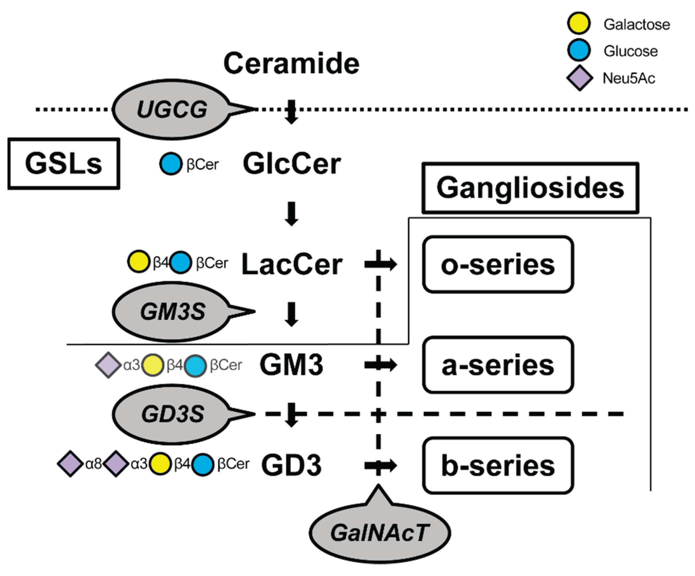
Table 2.
Cell sources and cartilage regenerative medicine.
| Clinical practice | Cell source | Lesion size (cm2) / OA grade | Performances | References |
| Microfracture | Mesenchymal stem cell (MSC) | 2.0-4.0 | Microfracture is most likely to be successful for small femoral condylar defects | [88,89,90,91,92] |
| Autologous matrix-induced chondrogenesis (AMIC) | MSC | 1.3-5.3 | Effective procedure for the treatment of mid-sized cartilage defects. Low failure rate with satisfactory clinical outcome | [88,89,93,94,95,96,97,98,99] |
| Autologous chondrocyte implantation | Chondrocyte | 2.0-10.0 | Superior structural integration with native cartilage tissue compared to microfracture and AMIC, but a two-stage treatment burden exists | [89,100,101,102,103] |
| Osteochondral autograft transplantation | Chondrocyte | 0.1-20.0 / OA grade Ⅰ-Ⅲ | Osteochondral autograft transfer system and mosaicplasty appear to be an alternative for the treatment of medium-sized focal chondral and osteochondral defects of the weight-bearing surfaces of the knee. Chondrocyte sheet and auricular cartilage micrograft for treatment of early-stage OA has been tried | [104,105,106,107] |
| Allogenic transplantation | Chondrocyte, iPSC | 2.2-4.4 / OA gradeⅡ-Ⅳ | Osteoarticular allograft transplantation was used to treat high-grade cartilage defects or arthritis. iPSC-derived cartilages are used in preclinical studies that are in the middle to late stages when clinical trials are within range | [108,109,110,111,112,113,114,115] |
| Intra-articular injection with stem cell | adipose-derived stem cell, MSC | OA grade Ⅱ-Ⅳ | Lower degenerative grades improve outcomes but are less effective for end-stage OA. The results of intra-articular administration of stem cells are better with BMSC. | [116,117,118,119,120,121,122] |
Disclaimer/Publisher’s Note: The statements, opinions and data contained in all publications are solely those of the individual author(s) and contributor(s) and not of MDPI and/or the editor(s). MDPI and/or the editor(s) disclaim responsibility for any injury to people or property resulting from any ideas, methods, instructions or products referred to in the content. |
© 2024 by the authors. Licensee MDPI, Basel, Switzerland. This article is an open access article distributed under the terms and conditions of the Creative Commons Attribution (CC BY) license (http://creativecommons.org/licenses/by/4.0/).
Copyright: This open access article is published under a Creative Commons CC BY 4.0 license, which permit the free download, distribution, and reuse, provided that the author and preprint are cited in any reuse.


