
Submitted:
26 February 2024
Posted:
27 February 2024
You are already at the latest version
Abstract
Helicobacter pylori is a gastric oncopathogen that infects over half of the world’s human population. It is a Gram-negative, microaerophilic, helix-shaped bacterium that is equipped with flagella, which provide high motility. Colonization of the stomach is asymptomatic in up to 90% of people but is a recognized risk factor for developing various gastric disorders such as gastric ulcers, gastric cancer and gastritis. Invasion of the human stomach occurs via numerous virulence factors such as CagA and VacA. Similarly, outer membrane proteins (OMPs) play an important role in H. pylori pathogenicity as a means to adapt to the epithelial environment and thereby facilitate infection. While some OMPs are porins, others are adhesins. The epithelial cell receptors SabA, BabA, AlpA, OipA, HopQ and HopZ have been extensively researched to evaluate their epidemiology, structure, role and genes. Moreover, numerous studies have been performed to seek to understand the complex relationship between these factors and gastric diseases. Associations exist between different H. pylori virulence factors, the co-expression of which appears to boost pathogenicity of the bacterium. Improved knowledge of OMPs is a major step towards combatting this global disease. Here, we provide a current overview of different H. pylori OMPs and discuss their pathogenicity, epidemiology and correlation with various gastric diseases.
Keywords:
Helicobacter pylori
; outer membrane protein
; virulence factor
; gastric cancer
; gastric disease
1. Introduction
Helicobacter pylori is considered an ancient microorganism the existence of which can be traced back to before the voyages of Christopher Columbus [1]. Yet, it took to the early 1980s for the bacterium to be identified by the Australian physicians Barry Marshall and Robin Warren. For discovering H. pylori as the principal cause of gastritis and peptic ulcer disease and mucosa-associated lymphoid-tissue (MALT) lymphoma [2,3], they were awarded the Nobel Prize in Physiology or Medicine in 2005. Chronic H. pylori infection is a predisposing factor for a range of other health conditions including ischemic stroke, Alzheimer’s disease, multiple sclerosis, autoimmune neutropenia, vitamin B12 deficiency, diabetes mellitus, cholelithiasis, idiopathic thrombocytopenic purpura, iron-deficiency anemia, cardiovascular diseases, hepatobiliary diseases, and biofilm-related infections, although further research is needed to verify each proposed link [4,5,6,7,8,9,10,11,12,13,14,15]. It is estimated that more than half of the world’s population is infected with this microorganism, its prevalence in developing countries reaching 70-90%, compared to developed nations where it is between 20-30% [16,17]. Typically, a person becomes infected with H. pylori during childhood through oral-fecal or oral-oral transmission [18]. This Gram-negative, helical bacterium is a major source of global gastric cancer mortality, so is considered as an oncogenic pathogen (oncopathogen) and hence is classified as a class I carcinogen by the World Health Organization [19]. It is equipped with different virulence factors including flagella, lipopolysaccharide (LPS), urease, and outer membrane proteins (OMPs), which are encoded by many paralogous gene families. It owes its characteristically high motility to its between 4-6 co-located flagella, which facilitate its movement and colonization of the stomach mucosa layer. Urease production provides ammonia for bacterial protein synthesis and neutralizes gastric acid, thereby making the stomach a preferred environment for colonization. This factor can damage host tissue via several mechanisms, which, together with the inflammatory immune response that this triggers, causes ulceration. Similarly, the unique structure of LPS promotes bacterial pathogenicity by facilitating attachment to gastric mucosa, thus supporting persistence of infection [20,21,22,23].
It is estimated that only about 20% of H. pylori carriers develop symptoms of disease. Chronic gastritis is a condition ascribed for H. pylori carriers without any clinical symptoms. At the same time, this pathogen is a risk factor for progression to gastric problems like a peptic ulcer [24,25,26]. Chronic gastritis follows colonization of the stomach by H. pylori, which resists clearance and causes mucosal inflammation and atrophy. Peptic ulcer formation, a consequence of damaged mucosa through stomach acid activity, is accelerated by the chronically acidic environment [27]. These sores can develop either into a lesion inside the stomach, known as a gastric ulcer, or inside the adjoining duodenum within the small intestine, termed a duodenal ulcer [28]. Importantly, having chronic gastritis increases a person’s risk of acquiring severe gastric conditions, notably gastric cancer that most often manifests as stomach adenocarcinoma [29].
An array of contributing factors, such as genetic susceptibility, diet, environmental variables, smoking and physical activity, are involved in progression to severe stomach conditions [30]. Studies showed that H. pylori is the leading cause of 63.4% of all stomach cancer and 75% of non-cardia gastric cancer (that affects the first part of the stomach) [31]. While there is now a decreasing trend in the rate of gastric cancer worldwide, it is still the second highest cause of cancer mortality [32]. In order to eradicate H. pylori, antibiotic therapy is suggested for gastric disorders. Currently, antibiotics like clarithromycin, amoxicillin or metronidazole are used in combination with proton pump inhibitors as a standard treatment [33]. It should also be noted that eradicating this microorganism may provoke some extra-gastric diseases, in particular iron deficiency, idiopathic thrombocytopenic purpura, chronic idiopathic urticaria and anemia. Further studies are required to confirm this correlation [34].
The first step for H. pylori to induce inflammation and cause infection is to colonize and attach to gastric mucosa. Usually, this happens through OMPs which play a pivotal role in adherence and pathogenicity. To date, there are about 64 members of this family which are recognized [35,36,37]. Also, five paralogous genes of OMPs have been identified. Through analyzing strains of H. pylori, 26695 and J99, many OMPs were identified. In one study, five family members of OMP, each with its own subfamily, were recognized. These families include the major OMPs Hop and Hor, Hof, Hom, iron-regulated OMPs and FecA/FrpB-like proteins, and efflux pump OMPs (Table 1) [37].
2. Helicobacter pylori Virulence Factors
2.1. Cag A and Vac A
Definition, diversity, classification and their significance: Vacuolating cytotoxin, or VacA, and Cag (cytotoxin-associated genes) pathogenicity island (PAI), encoding a bacterial type IV secretory apparatus (T4SSs), are two main factors involved in H. pylori pathogenicity (Figure 1). CagA is a protein of 116-140 kDa molecular weight that is expressed by almost 70% of strains and which produces a specific cytotoxin [38]. The significant role of this protein in H. pylori-infected patients has led to the isolates being defined as belonging to one of two groups, either CagA-positive (type I) or CagA-negative (type II). Epithelial cells and cells of the immune system are considered as two main targets for VacA, in which is expressed by all H. pylori strains [39,40]. VacA protein has cytotoxic activity that is due to its ability to drive intracellular vacuolization [41]. It has been demonstrated that various cell types are vulnerable to this toxin [40]. It has escape mechanisms to avoid the highly acidic environment of the stomach [42]. Different receptors are recognized for VacA, yet their roles and importance are not clear [43]. Similar to CagA, this virulence factor is expressed only in H. pylori type I [44]. Notably, there are three types of VacA genotype predicated on their signal sequence, namely s1a, s1b and s2, as well as m1 and m2, which is based on middle-region alleles of the vacA gene [45]. Regarding vacA allelic diversity, regions including s-region (signal), m-region (middle), i-region (intermediate), d-region (deletion) and c-regions are elucidated. Based on the deletion at the 3' end of the vacA gene, different types are investigated. The i region exists in three types (i1, i2 and i3), while all other regions are classified into two types (s1, s2, m1, m2, c1, c2, d1 and d2). More variants within these regions are proposed, of which K, E and Q-types are conspicuous [46].
The cag PAI is a 40 kb DNA sequence as that encodes type IV secretion system (T4SS) and CagA protein. This generates a pilus via which the bacterium can inject CagA protein into a host cell [47,48]. There are twelve recognized components of T4SSs in Gram-negative bacteria, including VirD4 and VirB1-11. It is organized into three parts: outer membrane core complex, inner membrane complex, and extracellular pilus [49,50]. Upon delivery of CagA into the cell and phosphorylation of a C-terminal EPIYA motif, the signaling pathway is activated via binding of CagA to the SH2 domain. Host cell changes occur after components interact with both phosphorylated and non-phosphorylated CagA. Of note are changes in cell junction, elongation, polarity, proliferation and proinflammatory response [51,52]. Various bacterial proteins such as CagM, CagX, CagY, CagT and Cag3 that form a part of CagPIA are encoded by a 41 nm long core structure. Among these, CagX and CagY are associated with the T4SS channel [53]. An interaction between CagL on the T4SS and α5β1 integrin leads to CagA transposition and pilus formation. Consequently, cells become more irregular as a result of phosphorylation at the 3' end of CagA gene (EPIYA), which is located in the PAI [44,54]. CagA is a highly immunogenic protein that comprises two types, CagAI and CagAII on the right or left segment, respectively [38,44]. This H. pylori type I virulence factor is linked to gastroduodenal disease and its gene may be acquired horizontally [44,55]. Upon bacterial attachment and infection, CagA will activate signal factors such as interleukin (IL)-8, which depend on the Cag PAI activity [44,56]. IL-8 and NFκB will pave the way for inflammation and carcinogenesis [54]. Another gene called cagE, located in the cagI and in proximity to cagA, has similarity with ptlC in Bordetella pertussis [44]. This gene is considered a better marker of pathogenicity, although further verification is needed [57]. In addition, there is a correlation among these virulence factors and other OMPs including IceA, BabA, HopQ, OipA, SabA and HopZ [58].
Geographical Variances and Clinical Associations: Several studies have investigated associations between these two antigens and different gastric conditions, yet neither is considered as an indicator of gastric cancer [59,60]. A high frequency of CagA-positive isolates in patients with gastric cancer was reported [61]. Different results obtained vary by geographical region. In one study performed in various countries, s1c-m1, s1b-m1 and s1a-m1 of vacA were the predominant genotypes in Japan and Korea, US, and Colombia, respectively. Although cagA genotype was predominant in all nations, no relationships with clinical outcomes were identified [59]. In Egypt, however, 68.7% of patients with a gastric ulcer, 50% of gastric carcinoma patients and 33.3% of gastritis cases were positive for cagA gene expression [62]. On the other hand, in an Australian cohort of H. pylori-infected individuals, 78% and 85% of cases of duodenal ulcer and gastric cancer, respectively, were positive for the cagA gene [63]. Another study showed an association between vacA s1a, cagE and cagA with gastric cancer and duodenal ulceration [64]. Additionally, a correlation between d-region and gastric atrophy and neutrophil infiltration was reported. There is a close relationship between geographical region and distribution of VacA subtypes. It is apparent that s1/m1 and i1 are predominant genotypes in northeast Asia. Also, a close relationship between VacA subtypes and gastric disorders is demonstrable. Furthermore, an association between s1a, s1c and m1 with gastric cancer, peptic ulcer and intestinal metaplasia was reported [3,65,66,67].
2.2. H. pylori Outer Membrane Proteins
Hop is the largest family of H. pylori OMPs, with 32 known members, yet they are collectively encoded by only 4% of the bacterial genome [36,37]. Hop A-E act as porin proteins as well as a channel through which antimicrobial agents permeate into the cell. Hence, many of them are potential candidates for development vaccine [68,69]. This group contains two divisions, Hop and Hor proteins. Interestingly, members of the latter lack a hop motif but still have an N-terminal motif, as do Hop proteins, and which is greatly variable in size. The former is divided into two groups based on the C-terminus [37].
Adhesion to host epithelial cells is the very first step for H. pylori colonization and persistence, which is mostly mediated by OMPs and T4SS [70]. There are three distinct steps of infection: colonization; attack of the gastric mucosa; and escape from the immune system. Attachment to mucins depends on several variables including type of mucin, anatomical site, pH, H. pylori strain and gastritis status. Also, interaction between H. pylori and host Lewis antigens, Lea,b,x,y, attributed to Hop proteins such as SabA and BabA, is vital to this process [71,72].
Protected by a mucus layer and composed mostly of MUC5AC and MUC6, the gastric epithelium is responsible for a glycosylation pattern that varies between gastric disorders. MUC2 is a type of mucin that does not exist in normal mucosa but instead is found mostly in intestinal metaplasia in which goblet cells are predominant. Understanding more about mucin expression patterns is important as H. pylori adhesion is mediated through interaction between these antigens and virulence factors [73,74,75].
2.2.1. Hop B and Hop C
HopB and HopC, also known as AlpA and AlpB, are encoded by the alp A/B locus (OMP 20 and 21, respectively). Homology of AlpA/B among various H. pylori strains is reported as more than 90%. While the role of these proteins remains to be substantiated, they are assumed to be involved in adhesion [37,76,77], for which laminin serves as a receptor. Any interruption to Hop B/C leads to diminished binding of H. pylori to laminin [92]. In addition, these proteins are responsible for producing cytokines such as IL-6, IL-8 and for activating signal transduction [76,78,79]. Gastric damage and modulation of cell signaling are consequent to AlpA/B adhesion [80]. Both play a key role in H. pylori colonization, although HopB appears to be more important [81]. New insights into the molecular mechanism of HopC indicate a function in biofilm formation. As described later, H. pylori can construct biofilm in human gastric cells, HopC being one of the OMPs with the capability to contribute to this in outer membrane vesicles (OMVs) [82].
Regarding the pathogenicity of HopB/C, there is insufficient information correlating their presence with clinical outcomes. Analysis of 200 H. pylori isolates revealed that all express these proteins, which suggests their important roles [79]. Interestingly, in another study severe gastric symptoms were associated with some H. pylori virulence factors such as HopB and VacA, with a high prevalence of HopB in cases of gastric cancer and peptic ulcers (> 80%), implying the importance of this OMP to predictions of infection outcome [67].
2.2.2. Hop H, a Phase-Variable Protein
HopH, originally identified as outer inflammatory protein or Oip A (Hpo638), is a phase-variable protein the alleles of which are present in almost all H. pylori strains. A high rate of diversity within CT dinucleotide repeats occurs in the oipA gene. Similar to other OMPs, it is assumed that HopH is involved in epithelial cell adhesion, although there are discrepancies arising from diversity between strains. This protein can also induce IL-8 production, cell-signaling and toxic events, as well as apoptosis [83,84,85]. These properties are independent of Cag PIA activity. This means that those strains which contain both virulence factors are capable of producing, for instance, higher levels of IL-8 [86]. Both functional and non-functional types of OipA are known [86,87]. Interestingly, an association between this protein and other virulence factors such as CagA, VacAs1 and BabA has been demonstrated [58,88,89].
Hop H association with clinical outcomes: A correlation between the presence of HopH and gastric disorders such as gastric cancer and peptic ulceration has been established. A study in which several virulence factors were examined together showed that gene expression could be a useful predictor of progression to gastric cancer in patients with precancerous gastric lesions, although paradoxical findings have raised doubts [90,91,92,93]. An investigation of hopH gene polymorphism led to two proposals for its pathogenicity, enhanced bacterial adhesion and correlation with the presence of other virulence factors [94]. In another study performed on gastritis and peptic ulcers, a high prevalence of the oipA gene was reported, which could imply a relationship between this gene and disease progression [95]. Similarly, a study performed on patients with gastritis, gastric carcinoma or duodenal ulcers showed an association with virulence factors such as CagA, VacA, IceA, BabA and OipA. However, only OipA was recognized as a distinctive factor for clinical outcomes. Nonetheless, as this factor is common among patients, it should be applied as a predictor only in combination with other virulence factors [87]. Several trials reported a connection between CagA and OipA expression in which slipped strand mispairing of complementary bases during DNA replication enhances bacterial adaptability. Conversely, OipA was reported as a non-significant marker in one study which used PCR to detect and differentiate H. pylori virulence factors and to predict clinical outcomes [67].
2.2.3. Hop P
This protein is also known as sialic acid-binding adhesin or SabA. for which the human Lewis (Le) histo-blood group antigens Lex and Lea are the main receptors. Sialyl-dimeric-Lewis x glycosphingolipid, defined as H. pylori receptor, is overexpressed in the stomach of infected people as sLex and sLea gene expression is upregulated during inflammation. In contrast, in the gastric mucosa of healthy people sialylated glycoconjugates are not abundant [96,97,98,99,100]. Other receptors for SabA have been identified. It can bind to α2-3-linked sialic acids and other sialic acid receptors [101], while laminin in the extracellular matrix also serves as a receptor [102]. H. pylori can bind specifically to glycosylated mucins, located in the proximity of epithelial cells, which helps it to maintain long-term infection [103]. Additionally, the polymorphic nature of H. pylori is attributed to SabA binding to sialylated carbohydrates. This is a unique strategy of adaptation for H. pylori [104], which tends to colonize those stomach areas with low acidity and high levels of HopP receptors [105].
SabA is classified as a protein that is regulated by phase variation. This means that H. pylori can switch expression of the gene on or off depending on circumstances [106]. Interestingly, sabA also undergoes gene conversion, which plays a key function in regulating SabA levels. Adhesion is affected by emerging subpopulations of H. pylori with variable expression of the protein, which is a consequence of having recombination amongst sab A, sab B and omp27 genes [107]. SabA also contributes significantly to spasmolytic polypeptide-expressing metaplasia (SPEM), which succeeds chronic atrophy and is a strategy for the stomach to reform its normal structural units following injury. It is thought that H. pylori can help SPEM progression, in which SabA adhesion to sLex plays a pivotal role [108].
Hop P and gastric disorders: Numerous studies have investigated an association between SabA and clinical outcomes. It appears that SabA is responsible for inflammation and its presence is correlated with clinical outcomes [109,110]. Also, a close relationship between this protein and gastric cancer has been found. In one study, 66% of H. pylori strains in patients with gastritis were SabA-positive, 44% were positive in individuals with duodenal ulcers and 70% in cases of gastric cancer [111]. Other studies that examined the frequency of SabA reported 93%, 86%, 80% and 23% detection in H. pylori strains in the Netherlands, France, Taiwan and Iran, respectively [58,112,113,114]. Recently, a Brazilian report revealed that SabA can accelerate gastric cancer in infected people [115].
2.2.4. Hop Q
Otherwise known as Omp27, HopQ is classified into two families, HopQI and HopQII [36,116]. Both 3' and 5' ends of hopQ alleles are highly conserved in H. pylori, but divergence occurs in the 1.1 kb mid-region, with a 75-80% similarity of nucleotide sequence. However, they are different in terms of geographical distribution, HopQI being isolated mostly in East Asia and HopQII commonly present in western countries [117,118]. Similar to other OMPs, these proteins mediate adherence to the gastric mucosa. It seems that there is a correlation between HopQ and other virulence factors like CagA and VacA [119]. Prevalence of this protein is common in those H. pylori strains with cag PAI, which is responsible for encoding CagA and a type IV secretion system [47].
A family of receptors defined as carcinoembryonic antigen-related cell adhesion molecules (CEACAMs) is recognized for HopQ and HopQ. CEACAM activation interferes with immune functions of T and NK cells [120,121]. Moreover, CEACAMs mediates various cell functions such as adhesion, proliferation, immune response and motility. CEACAM1, 5 and 6 are expressed by gastric epithelial cells. CEACAM1, 3 and 4 have both cytoplasmic and transmembrane domains, while CEACAM5, 6, 7 and 8 have glycosylphosphatidylinositol linkage to the host cell membrane. A strong connection between HopQ and CEACAM1, 3, 5 and 6 N-terminal domains facilitates H. pylori adhesion to gastric epithelial cells. Interestingly, CEACAM1, 5 and 6 are found in multiple organs. Binding between HopQ and CEACAMs plays a crucial role in CagA delivery into host cells [121,122,123].
The relationship between HopQ and CagA modulation is a focus of research interest [124]. It has been shown that inflammatory reactions follow T4SS activation and transfer of CagA oncoprotein via HopQ-CEACAMs interaction. The inflammatory response ultimately leads to gastric cancer, which supports the idea of therapeutic approaches targeting HopQ-CEACAMs [125,126]. This interaction affects human CEACAMs, responsible for CagA activation and phosphorylation in polymorphonuclear neutrophils (PMNs) but not dendritic cells and macrophages. In PMNs it lessens CagA translocation and alters expression of CEACAM receptors. Also, the presence of human CEACAMs on PMNs increases bacterial survival within phagosome, thus resisting phagocytosis [127].
Hop Q and clinical disorders: The correlation between both types of HopQ and gastric cancer is established [128,129]. Also, a high incidence of gastric cancer has been reported in patients with hopQI and vacA s1m1, or with hopQII and vacA S2 genotypes [130]. In two studies, in specific geographical regions in Iran, the rate of hopQII was higher than that of hopQI and a correlation between these OMPs and clinical outcomes was observed. However, another study showed the inverse result by which HopQI prevalence was higher with no association with gastrointestinal disorders [131,132]. Although its correlation with gastric diseases was demonstrated in several investigations, paradoxically HopQ could even be used therapeutically, as trials have shown good efficacies against melanoma metastasis [133].
2.2.5. Hop S, Hop T and Hop U
HopS, HopT and HopU were first identified as blood group antigen-binding adhesin A (BabA) or OMP 28 (~80 kDa), BabB or OMP 19, and BabC or OMP 9, respectively. They each mediate attachment of H. pylori to histo-blood group antigens on gastric epithelial cells except for BabC, the function of which is not yet clear. Notably, there is extensive homology at the 3' and 5' segments of babA and babB [134]. There are two types of babA, namely babA1 and babA2, with the latter divided into two subtypes with high and low protein production (Bab A-H and Bab A-L) [135,136]. An evaluation of glycosphingolipids as a receptor reported that H. pylori varies in its attachment to different blood groups including A Rh+/- and O Rh–. Moreover, H. pylori could not adhere to glycosphingolipids in people with blood group O but could bind extremely well in A Rh+/- individuals. In this study, Leb hexaosyceramide, pentaosylceramide, heptaosylceramide, lactosylceramide, lactotetraosylceramide, neolactohexaosylceramide and pentaosylceramide were reported as BabA receptors [137]. In addition to Leb, fucosylated blood group A, B and O antigens are noteworthy receptors [138]. Depending on the mid region and ability to bind ABO antigens, there are two classifications of BabA, specialist and generalist. The former refers to those H. pylori strains that can attach to ALeb (A-Lewis a), whereas the latter refers to those that bind to ALeb, BLeb (B-Lewis b) and Leb [139]. Also, analysis of variation of babA and babB revealed that there are five and three groups of alleles, including AD1-5 and BD1-3, for BabA and BabB, respectively [136].
Helicobacter pylori is able to achieve compatibility with the variable gastric acidic environment through recombination and mutation in babA genes. This enables mediated attachment via this protein, which is responsible for this phenomenon, thereby increasing the risk of progression to gastric cancer [140]. BabA is an antigen that is commonly expressed by H. pylori and which is related to specific clinical outcomes including peptic ulcers and gastric cancer. Also, colonization occurs predominantly in the lowermost antrum of the stomach [141,142]. Based on recent studies, recombination between the three bab genes frequently happens [143]. BabA undergoes genetic regulation through phase variation, which modulates its role in adherence. Also, it can be affected by recombination between babA and babB genes [144]. This genetic regulation is beneficial for H. pylori adaptation to its gastric environment in which the bacterium is exposed to a high level of physiological stress [145].
Correlation with gastric disorders: Several studies have investigated a correlation between babA gene expression and gastric disorders such as peptic ulcers and gastric cancer. Reportedly, inflammation induced by BabA adhesion results in gastric conditions such as precancerous transformations and intestinal metaplasia [146,147,148]. Also, a correlation between Leb and low binding activity and risk of duodenal ulcers was found [149]. Notably, undertaken the correlation between this genotype and gastric cancer was demonstrated separately in Germany, Portugal, Japan, Taiwan, China, USA and Brazil [90,150,151,152,153,154]. Similarly, babA2 gene was recently found at high frequency in patients with gastric cancer or peptic ulcers, although discrepancies arise regarding whether or not development to the severe gastric condition is associated with this genotype. A possible reason for this could be a lack of expression of BabA protein despite the presence of the gene [67]. This agrees with a meta-analysis of twenty studies that indicated a strong association between BabA2 and increased risk of gastric cancer in Asian populations compared to South American ones, suggesting a significant role of this virulence factor in pathogenicity [155].
2.2.6. Hop Z
This protein, also known as HP9, has a role in adherence to gastric epithelial cells, although its receptor is not yet recognized [72]. The hopZ gene undergoes slipped-strand mispairing and is regulated by a phase-variable CT repeat, which means whether it is switched on or off depends on the prevailing in vivo situation. There are two types, HopZI and HopZII. This differentiation dates to the era in prehistory before migration of humans from Africa [69,156]. Its relationship with infection is suggested by some findings [157,158]. In one investigation, an association between this protein and gastric cancer was reported, but a correlation between HopZ and chronic atrophic gastritis has yet to be found [72,156].
2.2.7. Hop V, Hop W and Other OMPs
These porin members belong to the Hop A/E family. This is due to homologous N-terminal and C-terminus regions. In terms of their pore size, HopV and HopW are similar to E. coli OmpF porin. Among H. pylori isolates, their expression is relatively less. Hop X/Y have been identified as porins similar to Hop A-D [37,159,160,161]. Colonization attributed to OMPs is mediated by H. pylori OMP 18 [162].
In the Helicobacter outer membrane (Hom) family, four members (HomA, B, C and D) are recognized, of which HomB is the most studied. Hom A/B exhibit variation in regard to genes copies and genomic localization in different geographical areas. The rate of homology between homA/B genomes is estimated at 90%, with only a 300 bp difference. Similar to other OMPs, recombination and phase variation are involved in gene duplication [37]. HomA/B are known for their significant roles in adherence, antibiotic resistance, biofilm formation and gastric malignancies [163]. Two important functions ascribed to HomB are IL-8 secretion and adherence [164]. While no specific association with clinical outcomes has been found for either of these proteins, they are likely to be involved in H. pylori persistence [165].
3. H. pylori Survival Strategies
3.1. Genetic Characteristics of H. pylori OMPs Contribute to Its Ability to Survive
A feature of the genome of H. pylori is its appreciable plasticity. This is due to genetic recombination which results in a high level of mutation, notably reported for babA2 gene expression [167,168]. This pathogen uses various micro- and macro-diverse tools to survive in the gastric mucosal environment [169,170]. Genetic incongruity is especially pronounced among omp genes [37]. Most studies have been performed on two H. pylori well-researched strains, 26695 and J99, which are thought to be representative of clinically significant isolates [116].
There are three categories of H. pylori genes: phase-variable; structure-variable; and strain-specific [26]. Some phase-variable genes use a specific method to escape from immune surveillance whereby not only does the expression of antigens change, but also the bacterium becomes more heterogenous. To date, six genes, including sabA/B, babB/C, oipA and hopZ, have been identified that are regulated by this mechanism [26,171].
One interesting finding is that H. pylori can upregulate expression of Leb and Lex, yielding BabA and SabA receptors, respectively. This function is performed by deposition of these antigens, which facilitates increased colonization [172].
There is broad similarity between H. pylori strains in terms of ribosome-binding sites (nucleotide number). However, the shorter spacing that is observed in some H. pylori genes may cause a change in the gene expression reported for seven orthologous pairs of omp genes. Examples can be seen in babA genes. Slipped-strand repair plays a pivotal role in altering expression of these proteins, thereby providing a mechanism by which H. pylori can evade the host immune system. The Com-B system in bab A/B/C is integral to this mechanism. While the central region of these genes is diverse, the 5' and 3' ends are similar. Slipped-strand repair has been reported in several genes and is thought to underpin antigenic variation and genetic diversity that is observed among H. pylori strains. Five hop orthologs undergo this regulation to change signal sequence, while the final product of expression remains the same [36,37,136,168,173]. In addition, gene duplication, in which there are two copies of an allele, is described for babA, hopJ, hopK, hopQ, hopM and hopN genes. This event differs between various hop genes depending on the H. pylori strain [37,134].
As well as OMPs, based on recent studies there are two other genes that affect H. pylori pathogenicity. ice A1 and ice A2 are a pair of novel genes that are considered as risk factors for various gastric conditions. Transcription of either ice A gene can be induced by H. pylori attachment to gastric epithelial cells. Their distribution among different geographical areas and gastric diseases is variable [174,175]. There is a relationship between this gene and other virulence factors such as CagA and VacA. The findings of one study suggest that ice A and cagA may be used as potential markers for clinical outcomes [62]. However, as the findings are paradoxical, elucidation through further research is required [59,176]. The duodenal ulcer-promoting gene dupA, which is located in the plasticity region of cag PAI, is thought to provide an increased risk for duodenal ulcers. Expression of this gene induces IL-8 and neutrophil activity [177]. On the other hand, in patients with gastric cancer its prevalence is much lower. dup A may provide a good candidate to predict clinical outcomes such as duodenal ulcers [26,177,178,179].
3.2. H. pylori Biofilm, an Alternative Strategy for Survival
Another medical challenge that is presented by H. pylori is its capacity to form biofilm. Under the protection of the impervious matrix of extracellular polymeric substances (EPS), bacteria are refractory to antibiotic penetration, thus greatly reducing the efficacy of standard treatment approaches [180]. OMVs are an integral component of H. pylori biofilm EPS matrix. Produced during bacterial growth, OMVs are implicated in pathogenesis through biofilm formation. A recent study demonstrated that an α-class carbonic anhydrase (CA) is found in OMVs, synthesized by both biofilm-producing and planktonic H. pylori strains [181]. On the other hand, genes responsible for encoding CAs in H. pylori are distributed to cytoplasmic β-CA (hpβCA) and α-CA (hpαCA). Also, expression of these two genes is accelerated at low pH and their joint activity with urease helps H. pylori to withstand the acidic gastric conditions. Therefore, hpCA has been considered as a new therapeutic candidate [181,182,183,184,185].
4. H. pylori and Host Responses
The protective host immune response to H. pylori helps to lessen the threat that colonization poses. However, this noted pathogen has evolved a unique strategy to overcome host defenses. Long-term infection is a consequence of remodeling of the host-pathogen interface as well as immune evasion due to multiple virulence factors expressed by H. pylori [186].
It is known that cag PAI is a potent driver of IL-8 and NfκB secretion. A cascade of intracellular activities is involved in H. pylori-dependent signal transduction. Nucleotide-binding oligomerization domain (NOD)1 is an intracellular pattern recognition receptor that recognizes bacterial peptidoglycan, among other danger signals, and thus plays a fundamental role in innate and adaptive defenses and control of inflammation1 [187]. This protein also has an important function in cancer development. Following interaction between CagA and PAR1b, BRCA1 disturbs phosphorylation, which can lead to the promotion of DNA double-strand breaks and BRCAness. This phenomenon is expanded via p53 inactivation, enabling DNA-damaged cells to escape from apoptosis and proliferate. This propels a “hit-and-run mechanism”, which is a significant cause of gastric carcinogenesis [188].
Similarly, VacA activity affects the immune system in multiple ways. For example, paracellular permeability during carcinogenesis, TGF-β1 production and heightened inflammation. At the same time, through vacuolization, antigen proteolysis decreases, which subsequently reduces peptide presentation and thereby inhibits T cell stimulation. This leads to down-modulation of CD4+ and CD8+ responses, thus facilitating the persistence of H. pylori infection [189].
Composed of lipid A and polysaccharide, LPS is an H. pylori endotoxin within the outer cell membrane. It is intrinsically involved in septic shock and sepsis through triggering proinflammatory mediators such as TNF-α and IL-1 [190]. LPS can promote gastric cancer through inhibiting inflammatory immune responses as well as preventing invasion of gastric cancer cells by immune mediators. Specifically, LPS derived from H. pylori can weaken cytotoxicity of mononuclear cells towards gastric cancer cells, as well as cytotoxic activity of gastric epithelia and NK cells. In addition, H. pylori LPS selectively elevates production of IL-18 and IL-12 and activates signal transduction patterns related to TLR4- and toll/IL-1 receptor [190,191,192]. Multiple studies have shown the significant role of TLR4-LPS in initiation and escalation of gastric cancer. The synthesis of two important factors in promoting cancer, TNF-α and IL-8, is accelerated after TLR4-LPS binding [193,194].
A series of mechanisms is utilized by this pathogen to enable it to evade the host immune system. H. pylori is a motile bacterium that is equipped with at least four flagella, the coordinated actions of which propel it through the gastric mucus layer. Each flagellum comprises several components including hook, basal body, filament and sheath. Interestingly, reports show that they contribute to biofilm formation [189,195]. As alluded to above, urease production is another means to combat the immune system. As it helps to alleviate the acidic environment, this implies a role in chronicity of infection and bacterial persistence. Also, ammonium produced by urease may cause damage to host cells [196,197]. Additional recognized roles for urease include a chemotactic effect on immune cells as well as angiogenesis, which may promote development of infection to gastric cancer [198,199].
A “founder colony” is a newly proposed model of escape and persistence in which H. pylori penetrates deep within the microenvironment of gastric glands to initiate colonization. These small colonies then expand to form persistent clonal population islands. They are distinct from planktonic bacteria in the superficial layer and gaining the space for new bacterial growth presents a challenge [200].
5. Advancements and Challenges in Therapeutic Strategies Targeting H. pylori
In order to achieve a protective immune response some therapeutic strategies such as T cell activation and targeting inhibitory receptors are of note. In this regard, promising results were obtained using MDX-1106 (anti-PD-1), lambrolizumab, and rapamycin to control the mTOR/p70 S6 kinase pathway. However, further clinical research is required to acquire a deep knowledge of immunity to H. pylori. A better understanding of these mechanisms is critical to design an appropriate vaccine [201]. Several prototype vaccines are in development and currently undergoing trials are in progress (Table 2). Regarding other therapeutic approaches, there are several targets suggested for H. pylori treatment. These include shikimate pathways (involved in ubiquinone and aromatic acid synthesis), flavodoxin (electron carrier protein), coenzyme A, succinylase pathway, and urease inhibitory compounds [202]. As discussed below, there are several innovative therapeutic approaches against H. pylori, including novel treatments targeting key virulence factors and host-microbe interactions.
Regarding therapies that target Cag A, progress has been made using ATPase Cagα inhibitors to target the Cag type IV secretion system. CHIR-1, a kinase-targeting compound, and difluoromethylornithine, have both shown promising results. However, technical limitations make it difficult to achieve full inhibition. It is necessary to preincubate bacteria with CHIR-1 to reach the strongest level of inhibition [203,204,205,206]. In summary, a major role of VacA and CagA in H. pylori pathogenicity and disease progression is evident. Irrespective of the geographical differences, CagA is a good indicator of patient outcome and targeting this protein could provide a potentially effective means of treatment [207].
Targeting OMPs is a promising innovative therapeutic approach against H. pylori, given their importance and roles in gastric conditions (Table 3). Hop B and Hop C are considered potential targets for vaccine therapy as they are involved during the early colonization stages of infection with H. pylori. In immunization studies in mice, when HopB, either on its own or in combination with other antigens (BabB, urease, catalase), was conjugated to the DC-Chol mucosal adjuvant, enhanced cellular and humoral protective responses were observed [208]. In a further promising evaluation of HopB immunogenicity in recombinant plasmids, HopB recombinant protein was introduced as a novel means of infection prophylaxis and eradication [209].
It has shown that OipA is a promising candidate for an oral vaccine. In murine studies, inoculating IgA raised against OipA significantly ameliorated H. pylori infection [210]. Similarly, a Salmonella typhimurium bacterial ghost-based DNA vaccine that delivers the oipA gene is proposed as a novel immunogen. This oral vaccine was capable of boosting immune responses, observed as heightened antibody and cytokine levels, and minimized bacterial colonization [211]. In another study, the efficiency of a recombinant OipA vaccine in mice was indicated by an elevated interferon-γ response [212]. Hence, a series of investigations shows a connection between this protein and gastric disorders, but using a combination of factors is suggested as a more accurate predictor of clinical outcomes.
Porcine milk has shown promising therapeutic potential by interfering with SabA adhesion. Apparently, this product has an inhibitory effect on H. pylori adhesion by expressing Lewis B glycans, as well as sialyl Lewis X [213]. Multiple trials demonstrated the efficacy of SabA as a potential recombinant vaccine candidate. One study evaluated a novel immunogenic cocktail, including VacA, BabA, SabA, FecA and Omp16, using a reverse vaccinology approach [214,215]. Similarly, a multiepitope oral vaccine composed of BabA, SabA, OipA, VacA, CagA, cholera toxin subunit B (CTB) and other components, serves as another promising vaccine candidate [216]. The significant role of SabA in anchoring H. pylori and its ability to adapt to the gastric environment reinforces the idea of using this protein as the basis for vaccine design. Furthermore, there is promise in evaluating glycosphingolipids as therapeutic targets to develop new treatments for pathogenic host-microbe interactions in the human stomach [217,218].
Research has been performed to evaluate anti-adhesive agents on hopQ genotypes. In one study, HopQ1 was more sensitive than HopQII to different dietary ingredients [219]. It is suggested that engineered CEACAMs conjugated to antimicrobial agents, with higher specificity and affinity for HopQ, can improve antibacterial efficiencies [220]. Further research is needed regarding prevalence of this protein, correlation with clinical outcomes and potential targets.
When the efficacy of various drugs to interfere with the interaction between BabA and gastric mucosa was evaluated, rhamnogalacturonans showed potential as inhibitors of this protein [221]. In terms of treatment, some progress has been made using mucolytic agents. Findings show that N-acetylcysteine has the ability to disrupts BabA adhesion to the gastric mucosa. Also, this conserved disulfide has a synergic effect with antibiotic therapy that boosts the efficiency of each [222]. Pectin and rhamnogalacturonans show promise as BabA adhesion blockers, indicating that BabA could potentially serve as a target for designing receptor-mediated adhesion drugs [221].

Table 2.
Improvement of vaccine design and progression of development pathway.
| Vaccine | Type | Status | Reference | Time |
|---|---|---|---|---|
| Imevax/IMX101 | Multicomponent | I | [223] | Ongoing |
| VacA, CagA, NAP (NCT00736476) | Recombinant | I/II | [224] | 2018 |
| HpaA expression by Vibrio cholera | Recombinant | Preclinical | [225] | 2017 |
| Cholera toxin B and H. pylori Lpp20 | Epitope | Preclinical | [226] | 2016 |
| H. pylori vaccine | Oral recombinant | III | [227] | 2015 |
| CTB-UreI-UreB (BIB) | Recombinant multi-epitope | Preclinical | [228] | 2014 |
| HelicoVax | Multi-epitope | Preclinical | [229] | 2011 |
Table 3.
Different H. pylori OMPs, their given names, receptors, and roles in gastric disorders.
| OMP | Also known as | Receptor | PU | GC | GA | DU | MALT | Reference(s) |
|---|---|---|---|---|---|---|---|---|
| Cag A | Cytotoxicity in associated gene | Epithelial cell | ✓ | ✓ | EPIYAD/C | - | ✓ | [54] |
| Cag L | ||||||||
| Vac A | Vacuolating cytotoxin | RPTP-α | VacAs1m1VacAs2m2 | VacAs1m1 | VacAs1m1 | - | - | [43,54] |
| RPTP-β | ||||||||
| Lipids | ||||||||
| Heparin sulphate | ||||||||
| Sphingomyelin | ||||||||
| Fibronectin | ||||||||
| Β2- integrin | ||||||||
| EGFR | ||||||||
| Hop B/C | Alp A/B | Laminin | - | - | - | - | - | [230,231] |
| Collagen IV | ||||||||
| Hop H | Oip A | Not known | ✓ | ✓ | - | ✓ | [54,87,90] | |
| Hop P | Sab A | sLex | - | - | - | - | ✓ | [58] |
| sLea | ||||||||
| Hop S | Bab A | sLeb | Bab A2 | ✓ | ✓ | - | - | [54] |
| A, B, O blood group | ||||||||
| Hop Q | - | CEACAMs | - | ✓ | - | - | - | [121] |
Abbreviations: PU, peptic ulcer; GC, gastric cancer; GA, gastric adenocarcinoma; DU, duodenal ulcer; MALT, mucosa-associated lymphoid tissue; sLe, sialyl-Lewis; EGFR, epidermal growth factor receptor; ✓, genopositive.
6. Conclusions
Helicobacter pylori is the principal cause of gastric conditions including peptic ulcers, gastric carcinoma, mucosa-associated lymphoid tissue lymphoma and gastritis. Its pathogenicity is due to a combination of virulence factors including – but not limited to – urease, flagella, and OMPs [22,231,232,233,234,235]. The progression of H. pylori infection to gastric cancer happens through a series of events. Primary inflammation may develop into acute gastritis and chronic gastritis. At this stage, multiple factors such as stomach pH, genetic diversity and environmental factors can gradually alter the gastric condition to cancer. During early stages, most patients are unaware of their condition and so treatment is not started until symptoms are more advanced. Hence, developing earlier and more accurate screening methods to enable prevention and eradication of H. pylori at the community level, as well as better treatment strategies to combat existing infection in patients, are warranted [236]. This will require an in-depth knowledge of different features of this bacterium.
Various indices are involved in determining clinical outcome. Notable among these are host genetics, particularly relating to the functioning of an individual’s immune system, as well as H. pylori virulence factors These belong to one of three groups that relate to colonization, development, and disease [89,237]. OMPs exist in all H. pylori strains as a tool for initial attachment, so are therefore considered as potential targets for candidate vaccine design. Recombinant vaccines incorporating CagA, VacA, urease, BabA, SabA, OipA and porin proteins show promise in ongoing trials [215].
Adherence to and colonization of epithelial cells play integral, initial roles in the pathogenicity of H. pylori. These interactions are mediated via OMPs and other virulence factors. Through adhesion to gastric mucosa and harnessing its type IV secretion system, the pathogen transfers toxins and effector molecules into the host cell. OMPs also facilitate inflammation, metaplasia and the ultimate pathological outcome of gastric cancer [238]. Each OMP has a distinct receptor, so gaining a clear understanding of them all aids diagnosis of infection and benefits clinical outcomes. In this context, combined evaluation of different OMPs can be both more rapid and accurate than any single identification. In one study, by considering various OMPs including OipA, BabA and SabA, the accuracy of gastric cancer prediction reached 77% [239]. In addition, an association between the production of some OMPs has been identified. While there is an inverse relationship between OipA and CagPAI, the presence of this OMP is a prerequisite for CagA translocation [85]. Similarly, at an 83% rate of H. pylori infection, a close relationship between VacA and chronic gastritis is apparent. Also, a correlation between VacA, BabA2 and OipA with increased risk of gastric cancer has been revealed [103]. When considering these relationships as potential prognostic markers a number of challenges such as limited time of survival and geographical regionality of occurrence should be considered.
Although several H. pylori-related virulence factors are involved in promoting gastric disorders, the causal relationships that underlie severe gastric conditions are still to be elucidated. The importance of these factors is crucial from both treatment and management perspectives [71]. It is also necessary to gain a precise evaluation of the epidemiology of each OMP, as its prevalence at a population level is different based on geographical region, even within the same country. Other criteria such as patient age gender, and bacterial genotype are also important [240,241,242]. To date, despite considerable research efforts there is no vaccine candidate that is sufficiently far advanced to be of interest to a pharmaceutical company to take through commercial development. Therefore, further research and greater investment are warranted in order to improve vaccine design and efficacy in terms of both prevention and lessening medical burden.
Author Contributions
Conceptualization, Z.S.; article collation, Z.S. and A.W.T-R.; writing—original draft preparation, Z.S.; writing—review and editing, A.W.T-R. Both authors have read and agreed to the published version of the manuscript.
Funding
This work received no external funds.
Conflicts of Interest
The authors declare no conflict of interest.
References
- Yamaoka, Y.; Orito, E.; Mizokami, M.; Gutierrez, O.; Saitou, N.; Kodama, T.; Osato, M.S.; Kim, J.G.; Ramirez, F.C.; Mahachai, V.; et al. Helicobacter pylori in North and South America before Columbus. FEBS Lett. 2002, 517, 180–184. [Google Scholar] [CrossRef]
- Watts, G. Nobel prize is awarded to doctors who discovered H. pylori. BMJ 2005, 331, 795–795. [Google Scholar] [CrossRef] [PubMed]
- Hocker, M.; Hohenberger, P. Helicobacter pylori virulence factors — one part of a big picture. Lancet 2003, 362, 1231–1233. [Google Scholar] [CrossRef]
- Zhou, D.; Zhang, Y.; Gong, W.; Mohamed, S.O.; Ogbomo, H.; Wang, X.; Liu, Y.; Quan, Z. Are Helicobacter pylori and other Helicobacter species infection associated with human biliary lithiasis? A meta-analysis. PloS ONE 2011, 6, e27390. [Google Scholar] [CrossRef]
- Yonezawa, H.; Osaki, T.; Woo, T.; Kurata, S.; Zaman, C.; Hojo, F.; Hanawa, T.; Kato, S.; Kamiya, S. Analysis of outer membrane vesicle protein involved in biofilm formation of Helicobacter pylori. Anaerobe 2011, 17, 388–390. [Google Scholar] [CrossRef]
- Gravina, A.G.; Zagari, R.M.; De Musis, C.; Romano, L.; Loguercio, C.; Romano, M. Helicobacter pylori and extragastric diseases: A review. World J. Gastroenterol. 2018, 24, 3204–3221. [Google Scholar] [CrossRef]
- Capurso, G.; Lahner, E.; Marcheggiano, A.; Caruana, P.; Carnuccio, A.; Bordi, C.; Delle Fave, G.; Annibale, B. Involvement of the corporal mucosa and related changes in gastric acid secretion characterize patients with iron deficiency anaemia associated with Helicobacter pylori infection. Aliment. Pharmacol. Ther. 2001, 15, 1753–1761. [Google Scholar] [CrossRef] [PubMed]
- Pellicano, R.; Franceschi, F.; Saracco, G.; Fagoonee, S.; Roccarina, D.; Gasbarrini, A. Helicobacters and extragastric diseases. Helicobacter 2009, 14 Suppl 1, 58–68. [Google Scholar] [CrossRef]
- Arnold, D.M.; Bernotas, A.; Nazi, I.; Stasi, R.; Kuwana, M.; Liu, Y.; Kelton, J.G.; Crowther, M.A. Platelet count response to H. pylori treatment in patients with immune thrombocytopenic purpura with and without H. pylori infection: a systematic review. Haematologica 2009, 94, 850–856. [Google Scholar] [CrossRef] [PubMed]
- Pirouz, T.; Zounubi, L.; Keivani, H.; Rakhshani, N.; Hormazdi, M. Detection of Helicobacter pylori in paraffin-embedded specimens from patients with chronic liver diseases, using the amplification method. Dig. Dis. Sci. 2009, 54, 1456–1459. [Google Scholar] [CrossRef]
- Zhou, X.; Zhang, C.; Wu, J.; Zhang, G. Association between Helicobacter pylori infection and diabetes mellitus: a meta-analysis of observational studies. Diabetes Res. Clin. Pract. 2013, 99, 200–208. [Google Scholar] [CrossRef]
- Shin, D.W.; Kwon, H.T.; Kang, J.M.; Park, J.H.; Choi, H.C.; Park, M.S.; Park, S.M.; Son, K.Y.; Cho, B. Association between metabolic syndrome and Helicobacter pylori infection diagnosed by histologic status and serological status. J. Clin. Gastroenterol. 2012, 46, 840–845. [Google Scholar] [CrossRef] [PubMed]
- Vijayvergiya, R.; Vadivelu, R. Role of Helicobacter pylori infection in pathogenesis of atherosclerosis. World J. Cardiol. 2015, 7, 134–143. [Google Scholar] [CrossRef] [PubMed]
- Osawa, H.; Kawakami, M.; Fujii, M.; Kubo, N.; Iwanaka, H.; Yamamoto, W.; Saitoh, M.; Yaginuma, T.; Sugano, K. Helicobacter pylori infection and coronary heart disease in Japanese patients. Cardiology 2001, 95, 14–19. [Google Scholar] [CrossRef] [PubMed]
- Wu, X.Z.; Chen, D. Helicobacter pylori and hepatocellular carcinoma: Correlated or uncorrelated? J. Gastroenterol. Hepatol. 2006, 21, 345–347. [Google Scholar] [CrossRef]
- Hooi, J.K.Y.; Lai, W.Y.; Ng, W.K.; Suen, M.M.Y.; Underwood, F.E.; Tanyingoh, D.; Malfertheiner, P.; Graham, D.Y.; Wong, V.W.S.; Wu, J.C.Y.; et al. Global prevalence of Helicobacter pylori infection: Systematic review and meta-analysis. Gastroenterology 2017, 153, 420–429. [Google Scholar] [CrossRef]
- Dunn, B.E.; Cohen, H.; Blaser, M.J. Helicobacter pylori . Clin. Microbiol. Rev. 1997, 10, 720–741. [Google Scholar] [CrossRef]
- Goh, K.-L.; Chan, W.-K.; Shiota, S.; Yamaoka, Y. Epidemiology of Helicobacter pylori infection and public health implications. Helicobacter 2011, 16 Suppl 1, 1–9. [Google Scholar] [CrossRef]
- Parkin, D.M.; Bray, F.; Ferlay, J.; Pisani, P. Global cancer statistics, 2002. CA: A Cancer J. Clin. 2005, 55, 74–108. [Google Scholar] [CrossRef]
- Shao, S.-H.; Wang, H.; Chai, S.-G.; Liu, L.-M. Research progress on Helicobacter pylori outer membrane protein. World J. Gastroenterol. 2005, 11, 3011–3013. [Google Scholar] [CrossRef] [PubMed]
- Moran, A.P. The role of lipopolysaccharide in Helicobacter pylori pathogenesis. Aliment. Pharmacol. Ther. 1996, 10 Suppl 1, 39–50. [Google Scholar] [CrossRef]
- Gu, H. Role of flagella in the pathogenesis of Helicobacter pylori. Curr. Microbiol. 2017, 74, 863–869. [Google Scholar] [CrossRef]
- Mobley, H.L. The role of Helicobacter pylori urease in the pathogenesis of gastritis and peptic ulceration. Aliment. Pharmacol. Ther. 1996, 10 Suppl 1, 57–64. [Google Scholar] [CrossRef]
- Hentschel, E.; Brandstatter, G.; Dragosics, B.; Hirschl, A.M.; Nemec, H.; Schutze, K.; Taufer, M.; Wurzer, H. Effect of ranitidine and amoxicillin plus metronidazole on the eradication of Helicobacter pylori and the recurrence of duodenal ulcer. New Engl. J. Med. 1993, 328, 308–312. [Google Scholar] [CrossRef]
- Sibony, M.; Jones, N.L. Recent advances in Helicobacter pylori pathogenesis. Curr. Opin. Gastroenterol. 2012, 28, 30–35. [Google Scholar] [CrossRef]
- Yamaoka, Y. Roles of the plasticity regions of Helicobacter pylori in gastroduodenal pathogenesis. J. Med. Microbiol. 2008, 57, 545–553. [Google Scholar] [CrossRef]
- Kobayashi, M.; Lee, H.; Nakayama, J.; Fukuda, M. Roles of gastric mucin-type O-glycans in the pathogenesis of Helicobacter pylori infection. Glycobiology 2009, 19, 453–461. [Google Scholar] [CrossRef] [PubMed]
- Kuna, L.; Jakab, J.; Smolic, R.; Raguz-Lucic, N.; Vcev, A.; Smolic, M. Peptic ulcer disease: a brief review of conventional therapy and herbal treatment options. J. Clin. Med. 2019, 8, 179. [Google Scholar] [CrossRef] [PubMed]
- Lauren, P. The two histological main types of gastric carcinona: diffuse and so-called intestinal type carcinoma: an attempt at a histo-clinical classification. Acta Pathol. Et Microbiol. Scand. 1965, 64, 31–49. [Google Scholar] [CrossRef] [PubMed]
- Forman, D.; Burley, V.J. Gastric cancer: global pattern of the disease and an overview of environmental risk factors. Best Pract. Research. Clin. Gastroenterol. 2006, 20, 633–649. [Google Scholar] [CrossRef]
- Parkin, D.M. The global health burden of infection-associated cancers in the year 2002. Int. J. Cancer 2006, 118, 3030–3044. [Google Scholar] [CrossRef]
- Ferlay, J.; Shin, H.R.; Bray, F.; Forman, D.; Mathers, C.; Parkin, D.M. Estimates of worldwide burden of cancer in 2008: GLOBOCAN 2008. Int. J. Cancer 2010, 127, 2893–2917. [Google Scholar] [CrossRef]
- Papastergiou, V.; Georgopoulos, S.D.; Karatapanis, S. Treatment of Helicobacter pylori infection: meeting the challenge of antimicrobial resistance. World J. Gastroenterol. 2014, 20, 9898–9911. [Google Scholar] [CrossRef]
- Goodman, K.J.; Joyce, S.L.; Ismond, K.P. Extragastric diseases associated with Helicobacter pylori infection. Curr. Gastroenterol. Rep. 2006, 8, 458–464. [Google Scholar] [CrossRef]
- Guruge, J.L.; Falk, P.G.; Lorenz, R.G.; Dans, M.; Wirth, H.P.; Blaser, M.J.; Berg, D.E.; Gordon, J.I. Epithelial attachment alters the outcome of Helicobacter pylori infection. Proc. Natl. Acad. Sci. USA 1998, 95, 3925–3930. [Google Scholar] [CrossRef]
- Tomb, J.F.; White, O.; Kerlavage, A.R.; Clayton, R.A.; Sutton, G.G.; Fleischmann, R.D.; Ketchum, K.A.; Klenk, H.P.; Gill, S.; Dougherty, B.A.; et al. The complete genome sequence of the gastric pathogen Helicobacter pylori. Nature 1997, 388, 539–547. [Google Scholar] [CrossRef] [PubMed]
- Alm, R.A.; Bina, J.; Andrews, B.M.; Doig, P.; Hancock, R.E.W.; Trust, T.J. Comparative genomics of Helicobacter pylori: Analysis of the outer membrane protein families. Infect. Immun. 2000, 68, 4155–4168. [Google Scholar] [CrossRef]
- Covacci, A.; Censini, S.; Bugnoli, M.; Petracca, R.; Burroni, D.; Macchia, G.; Massone, A.; Papini, E.; Xiang, Z.; Figura, N. Molecular characterization of the 128-kDa immunodominant antigen of Helicobacter pylori associated with cytotoxicity and duodenal ulcer. Proc. Natl. Acad. Sci. USA 1993, 90, 5791–5795. [Google Scholar] [CrossRef] [PubMed]
- Cover, T.L.; Blanke, S.R. Helicobacter pylori VacA, a paradigm for toxin multifunctionality. Nat. Rev. Microbiol. 2005, 3, 320–332. [Google Scholar] [CrossRef] [PubMed]
- Rieder, G.; Fischer, W.; Haas, R. Interaction of Helicobacter pylori with host cells: function of secreted and translocated molecules. Curr. Opin. Microbiol. 2005, 8, 67–73. [Google Scholar] [CrossRef] [PubMed]
- Leunk, R.D.; Johnson, P.T.; David, B.C.; Kraft, W.G.; Morgan, D.R. Cytotoxic activity in broth-culture filtrates of Campylobacter pylori. J. Med. Microbiol. 1988, 26, 93–99. [Google Scholar] [CrossRef]
- de Bernard, M.; Papini, E.; de Filippis, V.; Gottardi, E.; Telford, J.; Manetti, R.; Fontana, A.; Rappuoli, R.; Montecucco, C. Low pH activates the vacuolating toxin of Helicobacter pylori, which becomes acid and pepsin resistant. J. Biol. Chem. 1995, 270, 23937–23940. [Google Scholar] [CrossRef]
- Backert, S.; Tegtmeyer, N. the versatility of the Helicobacter pylori vacuolating cytotoxin vacA in signal transduction and molecular crosstalk. Toxins 2010, 2, 69–92. [Google Scholar] [CrossRef]
- Censini, S.; Lange, C.; Xiang, Z.; Crabtree, J.E.; Ghiara, P.; Borodovsky, M.; Rappuoli, R.; Covacci, A. cag, a pathogenicity island of Helicobacter pylori, encodes type I-specific and disease-associated virulence factors. Proc. Natl. Acad. Sci. USA 1996, 93, 14648–14653. [Google Scholar] [CrossRef]
- Atherton, J.C.; Cao, P.; Peek, R.M., Jr.; Tummuru, M.K.; Blaser, M.J.; Cover, T.L. Mosaicism in vacuolating cytotoxin alleles of Helicobacter pylori. Association of specific vacA types with cytotoxin production and peptic ulceration. J. Biol. Chem. 1995, 270, 17771–17777. [Google Scholar] [CrossRef]
- Soyfoo, D.M.; Doomah, Y.H.; Xu, D.; Zhang, C.; Sang, H.-M.; Liu, Y.-Y.; Zhang, G.-X.; Jiang, J.-X.; Xu, S.-F. New genotypes of Helicobacter pylori VacA d-region identified from global strains. BMC Mol. Cell Biol. 2021, 22, 4–4. [Google Scholar] [CrossRef] [PubMed]
- Backert, S.; Selbach, M. Role of type IV secretion in Helicobacter pylori. Cell. Microbiol. 2008, 10, 1573–1581. [Google Scholar] [CrossRef]
- Backert, S.; Meyer, T.F. Type IV secretion systems and their effectors in bacterial pathogenesis. Curr. Opin. Microbiol. 2006, 9, 207–217. [Google Scholar] [CrossRef] [PubMed]
- Waksman, G. From conjugation to T4S systems in Gram-negative bacteria: a mechanistic biology perspective. EMBO Rep. 2019, 20. [Google Scholar] [CrossRef] [PubMed]
- Grohmann, E.; Christie, P.J.; Waksman, G.; Backert, S. Type IV secretion in Gram-negative and Gram-positive bacteria. Mol. Microbiol. 2018, 107, 455–471. [Google Scholar] [CrossRef]
- Takahashi-Kanemitsu, A.; Knight, C.T.; Hatakeyama, M. Molecular anatomy and pathogenic actions of Helicobacter pylori CagA that underpin gastric carcinogenesis. Cell. Mol. Immunol. 2020, 17, 50–63. [Google Scholar] [CrossRef]
- Mimuro, H.; Suzuki, T.; Tanaka, J.; Asahi, M.; Haas, R.; Sasakawa, C. Grb2 is a key mediator of Helicobacter pylori CagA protein activities. Mol. Cell 2002, 10, 745–755. [Google Scholar] [CrossRef]
- Frick-Cheng, A.E.; Pyburn, T.M.; Voss, B.J.; McDonald, W.H.; Ohi, M.D.; Cover, T.L. Molecular and structural analysis of the Helicobacter pylori cag type IV secretionsystem core complex. mBio 2016, 7, e02001–e02015. [Google Scholar] [CrossRef] [PubMed]
- Chang, W.L.; Yeh, Y.C.; Sheu, B.S. The impacts of H. pylori virulence factors on the development of gastroduodenal diseases. J. Biomed. Sci. 2018, 25, 68. [Google Scholar] [CrossRef]
- Xiang, Z.; Censini, S.; Bayeli, P.F.; Telford, J.L.; Figura, N.; Rappuoli, R.; Covacci, A. Analysis of expression of CagA and VacA virulence factors in 43 strains of Helicobacter pylori reveals that clinical isolates can be divided into two major types and that CagA is not necessary for expression of the vacuolating cytotoxin. Infect. Immun. 1995, 63, 94–98. [Google Scholar] [CrossRef]
- Odenbreit, S.; Puls, J.; Sedlmaier, B.; Gerland, E.; Fischer, W.; Haas, R. Translocation of Helicobacter pylori CagA into gastric epithelial cells by type IV secretion. Science 2000, 287, 1497–1500. [Google Scholar] [CrossRef]
- Ikenoue, T.; Maeda, S.; Ogura, K.; Akanuma, M.; Mitsuno, Y.; Imai, Y.; Yoshida, H.; Shiratori, Y.; Omata, M. Determination of Helicobacter pylori virulence by simple gene analysis of the cag pathogenicity island. Clin. Diagn. Lab. Immunol. 2001, 8, 181–186. [Google Scholar] [CrossRef]
- Lehours, P.; Menard, A.; Dupouy, S.; Bergey, B.; Richy, F.; Zerbib, F.; Ruskone-Fourmestraux, A.; Delchier, J.C.; Megraud, F. Evaluation of the association of nine Helicobacter pylori virulence factors with strains involved in low-grade gastric mucosa-associated lymphoid tissue lymphoma. Infect. Immun. 2004, 72, 880–888. [Google Scholar] [CrossRef] [PubMed]
- Yamaoka, Y.; Kodama, T.; Gutierrez, O.; Kim, J.G.; Kashima, K.; Graham, D.Y. Relationship between Helicobacter pylori iceA, cagA, and vacA status and clinical outcome: studies in four different countries. J. Clin. Microbiol. 1999, 37, 2274–2279. [Google Scholar] [CrossRef] [PubMed]
- Qiao, W.; Hu, J.-L.; Xiao, B.; Wu, K.-C.; Peng, D.-R.; Atherton, J.C.; Xue, H. cagA and vacA genotype of Helicobacter pylori associated with gastric diseases in Xi'an area. World J. Gastroenterol. 2003, 9, 1762–1766. [Google Scholar] [CrossRef]
- Parsonnet, J.; Friedman, G.; Orentreich, N.; Vogelman, H. Risk for gastric cancer in people with CagA positive or CagA negative Helicobacter pylori infection. Gut 1997, 40, 297–301. [Google Scholar] [CrossRef]
- Abu-Taleb, A.M.F.; Abdelattef, R.S.; Abdel-Hady, A.A.; Omran, F.H.; El-Korashi, L.A.; Abdel-Aziz El-Hady, H.; El-Gebaly, A.M. Prevalence of Helicobacter pylori cagA and iceA genes and their association with gastrointestinal diseases. Int. J. Microbiol. 2018, 2018, 4809093. [Google Scholar] [CrossRef]
- Kamogawa-Schifter, Y.; Yamaoka, Y.; Uchida, T.; Beer, A.; Tribl, B.; Schöniger-Hekele, M.; Trauner, M.; Dolak, W. Prevalence of Helicobacter pylori and its CagA subtypes in gastric cancer and duodenal ulcer at an Austrian tertiary referral center over 25 years. PloS One 2018, 13, e0197695. [Google Scholar] [CrossRef]
- Erzin, Y.; Koksal, V.; Altun, S.; Dobrucali, A.; Aslan, M.; Erdamar, S.; Dirican, A.; Kocazeybek, B. Prevalence of Helicobacter pylori vacA, cagA, cagE, iceA, babA2 genotypes and correlation with clinical outcome in Turkish patients with dyspepsia. Helicobacter 2006, 11, 574–580. [Google Scholar] [CrossRef]
- Choe, Y.H.; Kim, P.S.; Lee, D.H.; Kim, H.K.; Kim, Y.S.; Shin, Y.W.; Hwang, T.S.; Kim, H.J.; Song, S.U.; Choi, M.S. Diverse vacA allelic types of Helicobacter pylori in Korea and clinical correlation. Yonsei Med. J. 2002, 43, 351–356. [Google Scholar] [CrossRef] [PubMed]
- Aydin, F.; Kaklikkaya, N.; Ozgur, O.; Cubukcu, K.; Kilic, A.O.; Tosun, I.; Erturk, M. Distribution of vacA alleles and cagA status of Helicobacter pylori in peptic ulcer disease and non-ulcer dyspepsia. Clin. Microbiol. Infect. 2004, 10, 1102–1104. [Google Scholar] [CrossRef]
- Lee, D.H.; Ha, J.H.; Shin, J.I.; Kim, K.M.; Choi, J.G.; Park, S.; Park, J.S.; Seo, J.H.; Park, J.S.; Shin, M.K.; et al. Increased risk of severe gastric symptoms by virulence factors vacAs1c, alpA, babA2, and hopZ in Helicobacter pylori infection. J. Microbiol. Biotechnol. 2021, 31, 368–379. [Google Scholar] [CrossRef] [PubMed]
- Nikaido, H.; Vaara, M. Molecular basis of bacterial outer membrane permeability. Microbiol. Rev. 1985, 49, 1–32. [Google Scholar] [CrossRef]
- Peck, B.; Ortkamp, M.; Diehl, K.D.; Hundt, E.; Knapp, B. Conservation, localization and expression of HopZ, a protein involved in adhesion of Helicobacter pylori. Nucleic Acids Res. 1999, 27, 3325–3333. [Google Scholar] [CrossRef]
- Bonsor, D.A.; Sundberg, E.J. Roles of adhesion to epithelial cells in gastric colonization by Helicobacter pylori. Adv. Exp. Med. Biol. 2019, 1149, 57–75. [Google Scholar] [CrossRef] [PubMed]
- Lindén, S.K.; Wickström, C.; Lindell, G.; Gilshenan, K.; Carlstedt, I. Four modes of adhesion are used during Helicobacter pylori binding to human mucins in the oral and gastric niches. Helicobacter 2008, 13, 81–93. [Google Scholar] [CrossRef] [PubMed]
- Sheu, B.S.; Yang, H.B.; Yeh, Y.C.; Wu, J.J. Helicobacter pylori colonization of the human gastric epithelium: a bug's first step is a novel target for us. J. Gastroenterol. Hepatol. 2010, 25, 26–32. [Google Scholar] [CrossRef] [PubMed]
- Reis, C.A.; David, L.; Nielsen, P.A.; Clausen, H.; Mirgorodskaya, K.; Roepstorff, P.; Sobrinho-Simões, M. Immunohistochemical study of MUC5AC expression in human gastric carcinomas using a novel monoclonal antibody. Int. J. Cancer 1997, 74, 112–121. [Google Scholar] [CrossRef]
- Reis, C.A.; David, L.; Carvalho, F.; Mandel, U.; de Bolós, C.; Mirgorodskaya, E.; Clausen, H.; Sobrinho-Simões, M. Immunohistochemical study of the expression of MUC6 mucin and co-expression of other secreted mucins (MUC5AC and MUC2) in human gastric carcinomas. J. Histochem. Cytochem. 2000, 48, 377–388. [Google Scholar] [CrossRef] [PubMed]
- de Bolos, C.; Real, F.X.; Lopez-Ferrer, A. Regulation of mucin and glycoconjugate expression: from normal epithelium to gastric tumors. Front. Biosci. 2001, 6, D1256–D1263. [Google Scholar] [CrossRef]
- Odenbreit, S.; Faller, G.; Haas, R. Role of the alpAB proteins and lipopolysaccharide in adhesion of Helicobacter pylori to human gastric tissue. Int. J. Med. Microbiol. : IJMM 2002, 292, 247–256. [Google Scholar] [CrossRef] [PubMed]
- Odenbreit, S.; Till, M.; Haas, R. Optimized BlaM-transposon shuttle mutagenesis of Helicobacter pylori allows the identification of novel genetic loci involved in bacterial virulence. Mol. Microbiol. 1996, 20, 361–373. [Google Scholar] [CrossRef] [PubMed]
- Loke, M.F.; Lui, S.Y.; Ng, B.L.; Gong, M.; Ho, B. Antiadhesive property of microalgal polysaccharide extract on the binding of Helicobacter pylori to gastric mucin. FEMS Immunol. Med. Microbiol. 2007, 50, 231–238. [Google Scholar] [CrossRef]
- Odenbreit, S.; Swoboda, K.; Barwig, I.; Ruhl, S.; Borén, T.; Koletzko, S.; Haas, R. Outer membrane protein expression profile in Helicobacter pylori clinical isolates. Infect. Immun. 2009, 77, 3782–3790. [Google Scholar] [CrossRef]
- Lu, H.; Wu, J.Y.; Beswick, E.J.; Ohno, T.; Odenbreit, S.; Haas, R.; Reyes, V.E.; Kita, M.; Graham, D.Y.; Yamaoka, Y. Functional and intracellular signaling differences associated with the Helicobacter pylori AlpAB adhesin from Western and East Asian strains. J. Biol. Chem. 2007, 282, 6242–6254. [Google Scholar] [CrossRef]
- de Jonge, R.; Durrani, Z.; Rijpkema, S.G.; Kuipers, E.J.; van Vliet, A.H.; Kusters, J.G. Role of the Helicobacter pylori outer-membrane proteins AlpA and AlpB in colonization of the guinea pig stomach. J. Med. Microbiol. 2004, 53, 375–379. [Google Scholar] [CrossRef]
- Yonezawa, H.; Osaki, T.; Fukutomi, T.; Hanawa, T.; Kurata, S.; Zaman, C.; Hojo, F.; Kamiya, S. Diversification of the AlpB outer membrane protein of Helicobacter pylori affects biofilm formation and cellular adhesion. J. Bacteriol. 2017, 199, e00729–e00716. [Google Scholar] [CrossRef]
- Teymournejad, O.; Mobarez, A.M.; Hassan, Z.M.; Talebi Bezmin Abadi, A. Binding of the Helicobacter pylori OipA causes apoptosis of host cells via modulation of Bax/Bcl-2 levels. Sci. Rep. 2017, 7, 8036. [Google Scholar] [CrossRef]
- Farzi, N.; Yadegar, A.; Aghdaei, H.A.; Yamaoka, Y.; Zali, M.R. Genetic diversity and functional analysis of oipA gene in association with other virulence factors among Helicobacter pylori isolates from Iranian patients with different gastric diseases. Infect. Genet. Evol. 2018, 60, 26–34. [Google Scholar] [CrossRef]
- Horridge, D.N.; Begley, A.A.; Kim, J.; Aravindan, N.; Fan, K.; Forsyth, M.H. Outer inflammatory protein a (OipA) of Helicobacter pylori is regulated by host cell contact and mediates CagA translocation and interleukin-8 response only in the presence of a functional cag pathogenicity island type IV secretion system. Pathog. Dis. 2017, 75. [Google Scholar] [CrossRef]
- Yamaoka, Y.; Kwon, D.H.; Graham, D.Y. A M(r) 34,000 proinflammatory outer membrane protein (oipA) of Helicobacter pylori. Proc. Natl. Acad. Sci. USA 2000, 97, 7533–7538. [Google Scholar] [CrossRef]
- Yamaoka, Y.; Kikuchi, S.; el-Zimaity, H.M.; Gutierrez, O.; Osato, M.S.; Graham, D.Y. Importance of Helicobacter pylori oipA in clinical presentation, gastric inflammation, and mucosal interleukin 8 production. Gastroenterology 2002, 123, 414–424. [Google Scholar] [CrossRef] [PubMed]
- Ando, T.; Peek, R.M.; Pride, D.; Levine, S.M.; Takata, T.; Lee, Y.C.; Kusugami, K.; van der Ende, A.; Kuipers, E.J.; Kusters, J.G.; et al. Polymorphisms of Helicobacter pylori HP0638 reflect geographic origin and correlate with cagA status. J. Clin. Microbiol. 2002, 40, 239–246. [Google Scholar] [CrossRef] [PubMed]
- Peek, R.M., Jr.; Blaser, M.J. Helicobacter pylori and gastrointestinal tract adenocarcinomas. Nat. Rev. Cancer 2002, 2, 28–37. [Google Scholar] [CrossRef]
- Gerhard, M.; Lehn, N.; Neumayer, N.; Boren, T.; Rad, R.; Schepp, W.; Miehlke, S.; Classen, M.; Prinz, C. Clinical relevance of the Helicobacter pylori gene for blood-group antigen-binding adhesin. Proc. Natl. Acad. Sci. USA 1999, 96, 12778–12783. [Google Scholar] [CrossRef] [PubMed]
- Quiroga, A.J.; Cittelly, D.M.; Bravo, M.M. BabA2, oipA and cagE Helicobacter pylori genotypes in Colombian patients with gastroduodenal diseases. Biomedica 2005, 25, 325–334. [Google Scholar] [CrossRef]
- Bartpho, T.S.; Wattanawongdon, W.; Tongtawee, T.; Paoin, C.; Kangwantas, K.; Dechsukhum, C. Precancerous gastric lesions with Helicobacter pylori acA+/babA2+/oipA+genotype increase the risk of gastric cancer. BioMed Res. Int. 2020, 2020, 7243029–7243029. [Google Scholar] [CrossRef]
- Dabiri, H.; Jafari, F.; Baghaei, K.; Shokrzadeh, L.; Abdi, S.; Pourhoseingholi, M.A.; Mohammadzadeh, A. Prevalence of Helicobacter pylori vacA, cagA, cagE, oipA, iceA, babA2 and babB genotypes in Iranian dyspeptic patients. Microb. Pathog. 2017, 105, 226–230. [Google Scholar] [CrossRef]
- Dossumbekova, A.; Prinz, C.; Mages, J.; Lang, R.; Kusters, J.G.; Van Vliet, A.H.; Reindl, W.; Backert, S.; Saur, D.; Schmid, R.M.; et al. Helicobacter pylori HopH (OipA) and bacterial pathogenicity: genetic and functional genomic analysis of hopH gene polymorphisms. J. Infect. Dis. 2006, 194, 1346–1355. [Google Scholar] [CrossRef]
- Salih, B.A.; Abasiyanik, M.F.; Ahmed, N. A preliminary study on the genetic profile of cag pathogenicity-island and other virulent gene loci of Helicobacter pylori strains from Turkey. Infect. Genet. Evol. 2007, 7, 509–512. [Google Scholar] [CrossRef] [PubMed]
- Aspinall, G.O.; Monteiro, M.A.; Pang, H.; Walsh, E.J.; Moran, A.P. Lipopolysaccharide of the Helicobacter pylori type strain NCTC 11637 (ATCC 43504): structure of the O antigen chain and core oligosaccharide regions. Biochemistry 1996, 35, 2489–2497. [Google Scholar] [CrossRef] [PubMed]
- Sherburne, R.; Taylor, D.E. Helicobacter pylori expresses a complex surface carbohydrate, Lewis X. Infect. Immun. 1995, 63, 4564–4568. [Google Scholar] [CrossRef] [PubMed]
- Mahdavi, J.; Sonden, B.; Hurtig, M.; Olfat, F.O.; Forsberg, L.; Roche, N.; Angstrom, J.; Larsson, T.; Teneberg, S.; Karlsson, K.A.; et al. Helicobacter pylori SabA adhesin in persistent infection and chronic inflammation. Science 2002, 297, 573–578. [Google Scholar] [CrossRef]
- Alper, J. Searching for medicine's sweet spot. Science 2001, 291, 2338–2343. [Google Scholar] [CrossRef]
- Madrid, J.F.; Ballesta, J.; Castells, M.T.; Hernández, F. Glycoconjugate distribution in the human fundic mucosa revealed by lectin- and glycoprotein-gold cytochemistry. Histochemistry 1990, 95, 179–187. [Google Scholar] [CrossRef]
- Walz, A.; Odenbreit, S.; Mahdavi, J.; Boren, T.; Ruhl, S. Identification and characterization of binding properties of Helicobacter pylori by glycoconjugate arrays. Glycobiology 2005, 15, 700–708. [Google Scholar] [CrossRef]
- Roche, N.; Angstrom, J.; Hurtig, M.; Larsson, T.; Boren, T.; Teneberg, S. Helicobacter pylori and complex gangliosides. Infect. Immun. 2004, 72, 1519–1529. [Google Scholar] [CrossRef]
- Schreiber, S.; Konradt, M.; Groll, C.; Scheid, P.; Hanauer, G.; Werling, H.-O.; Josenhans, C.; Suerbaum, S. The spatial orientation of Helicobacter pylori in the gastric mucus. Proc. Natl. Acad. Sci. USA 2004, 101, 5024–5029. [Google Scholar] [CrossRef]
- Aspholm, M.; Olfat, F.O.; Norden, J.; Sonden, B.; Lundberg, C.; Sjostrom, R.; Altraja, S.; Odenbreit, S.; Haas, R.; Wadstrom, T.; et al. SabA is the H. pylori hemagglutinin and is polymorphic in binding to sialylated glycans. PLoS Pathog. 2006, 2, e110. [Google Scholar] [CrossRef]
- Bjorkholm, B.; Salama, N.R. Genomics of Helicobacter 2003. Helicobacter 2003, 8 Suppl 1, 1–7. [Google Scholar] [CrossRef]
- Saunders, N.J.; Peden, J.F.; Hood, D.W.; Moxon, E.R. Simple sequence repeats in the Helicobacter pylori genome. Mol. Microbiol. 1998, 27, 1091–1098. [Google Scholar] [CrossRef]
- Talarico, S.; Whitefield, S.E.; Fero, J.; Haas, R.; Salama, N.R. Regulation of Helicobacter pylori adherence by gene conversion. Mol. Microbiol. 2012, 84, 1050–1061. [Google Scholar] [CrossRef] [PubMed]
- Sáenz, J.B.; Vargas, N.; Mills, J.C. Tropism for spasmolytic polypeptide-expressing metaplasia allows Helicobacter pylori to expand its intragastric niche. Gastroenterology 2019, 156, 160–174. [Google Scholar] [CrossRef] [PubMed]
- Kusters, J.G.; van Vliet, A.H.M.; Kuipers, E.J. Pathogenesis of Helicobacter pylori infection. Clin. Microbiol. Rev. 2006, 19, 449–490. [Google Scholar] [CrossRef]
- Yamaoka, Y. Increasing evidence of the role of Helicobacter pylori SabA in the pathogenesis of gastroduodenal disease. J. Infect. Dev. Ctries. 2008, 2, 174–181. [Google Scholar] [CrossRef] [PubMed]
- Yamaoka, Y.; Ojo, O.; Fujimoto, S.; Odenbreit, S.; Haas, R.; Gutierrez, O.; El-Zimaity, H.M.; Reddy, R.; Arnqvist, A.; Graham, D.Y. Helicobacter pylori outer membrane proteins and gastroduodenal disease. Gut 2006, 55, 775–781. [Google Scholar] [CrossRef]
- Sheu, B.S.; Odenbreit, S.; Hung, K.H.; Liu, C.P.; Sheu, S.M.; Yang, H.B.; Wu, J.J. Interaction between host gastric Sialyl-Lewis X and H. pylori SabA enhances H. pylori Density Patients Lacking Gastric Lewis B Antigen. Am. J. Gastroenterol. 2006, 101, 36–44. [Google Scholar] [CrossRef]
- Jonge, R.; Pot, R.; Loffeld, R.; van Vliet, A.; Kuipers, E.; Kusters, J. The functional status of the Helicobacter pylori sabB adhesin gene as a putative marker for disease outcome. Helicobacter 2004, 9, 158–164. [Google Scholar] [CrossRef]
- Hossein, G.; Hanieh, R.; Mitra, R.; Afsoon, T. Determination of the status of Helicobacter pylori sabA gene in relation to clinical findings. J. Med. Bacteriol. 2012, 1, 3–8. [Google Scholar]
- Pereira, J.; Orcini, W.; Peruquetti, R.; Cardoso Smith, M.; Payão, S.; Rasmussen, L. Prevalence of Helicobacter pylori cag A and sab A genotypes in patients with gastric disease. Adv. Microbiol. 2019, 09, 239–247. [Google Scholar] [CrossRef]
- Alm, R.A.; Ling, L.S.; Moir, D.T.; King, B.L.; Brown, E.D.; Doig, P.C.; Smith, D.R.; Noonan, B.; Guild, B.C.; deJonge, B.L.; et al. Genomic-sequence comparison of two unrelated isolates of the human gastric pathogen Helicobacter pylori. Nature 1999, 397, 176–180. [Google Scholar] [CrossRef] [PubMed]
- Cao, P.; Lee, K.J.; Blaser, M.J.; Cover, T.L. Analysis of hopQ alleles in East Asian and Western strains of Helicobacter pylori. FEMS Microbiol. Lett. 2005, 251, 37–43. [Google Scholar] [CrossRef]
- Cao, P.; Cover, T.L. Two different families of hopQ alleles in Helicobacter pylori. J. Clin. Microbiol. 2002, 40, 4504–4511. [Google Scholar] [CrossRef] [PubMed]
- Sicinschi, L.A.; Correa, P.; Bravo, L.E.; Peek, R.M., Jr.; Wilson, K.T.; Loh, J.T.; Yepez, M.C.; Gold, B.D.; Thompson, D.T.; Cover, T.L.; et al. Non-invasive genotyping of Helicobacter pylori cagA, vacA, and hopQ from asymptomatic children. Helicobacter 2012, 17, 96–106. [Google Scholar] [CrossRef] [PubMed]
- Gur, C.; Maalouf, N.; Gerhard, M.; Singer, B.B.; Emgård, J.; Temper, V.; Neuman, T.; Mandelboim, O.; Bachrach, G. The Helicobacter pylori HopQ outer membrane protein inhibits immune cell activities. Oncoimmunology 2019, 8, e1553487. [Google Scholar] [CrossRef] [PubMed]
- Königer, V.; Holsten, L.; Harrison, U.; Busch, B.; Loell, E.; Zhao, Q.; Bonsor, D.A.; Roth, A.; Kengmo-Tchoupa, A.; Smith, S.I.; et al. Helicobacter pylori exploits human CEACAMs via HopQ for adherence and translocation of CagA. Nat. Microbiol. 2016, 2, 16188. [Google Scholar] [CrossRef]
- Kuespert, K.; Pils, S.; Hauck, C.R. CEACAMs: their role in physiology and pathophysiology. Curr. Opin. Cell Biol. 2006, 18, 565–571. [Google Scholar] [CrossRef] [PubMed]
- Javaheri, A.; Kruse, T.; Moonens, K.; Mejías-Luque, R.; Debraekeleer, A.; Asche, C.I.; Tegtmeyer, N.; Kalali, B.; Bach, N.C.; Sieber, S.A.; et al. Helicobacter pylori adhesin HopQ engages in a virulence-enhancing interaction with human CEACAMs. Nat. Microbiol. 2016, 2, 16189. [Google Scholar] [CrossRef] [PubMed]
- Loh, J.T.; Torres, V.J.; Algood, H.M.; McClain, M.S.; Cover, T.L. Helicobacter pylori HopQ outer membrane protein attenuates bacterial adherence to gastric epithelial cells. FEMS Microbiol. Lett. 2008, 289, 53–58. [Google Scholar] [CrossRef] [PubMed]
- Xia, R.; Zhang, B.; Wang, X.; Jia, Q. Pathogenic interactions between Helicobacter pylori adhesion protein HopQ and human cell surface adhesion molecules CEACAMs in gastric epithelial cells. Iran. J. Basic Med. Sci. 2019, 22, 710–715. [Google Scholar]
- Hamway, Y.; Taxauer, K.; Moonens, K.; Neumeyer, V.; Fischer, W.; Schmitt, V.; Singer, B.B.; Remaut, H.; Gerhard, M.; Mejías-Luque, R. Cysteine residues in Helicobacter pylori adhesin HopQ are required for CEACAM-HopQ interaction and subsequent CagA translocation. Microorganisms 2020, 8. [Google Scholar] [CrossRef]
- Behrens, I.K.; Busch, B.; Ishikawa-Ankerhold, H.; Palamides, P.; Shively, J.E.; Stanners, C.; Chan, C.; Leung, N.; Gray-Owen, S.; Haas, R. The HopQ-CEACAM interaction controls CagA translocation, phosphorylation, and phagocytosis of Helicobacter pylori in neutrophils. mBio 2020, 11. [Google Scholar] [CrossRef]
- Talebi Bezmin Abadi, A.; Mohabbati Mobarez, A. High prevalence of Helicobacter pylori hopQ II genotype isolated from Iranian patients with gastroduodenal disorders. J. Pathog. 2014, 2014, 842469–842469. [Google Scholar] [CrossRef]
- Yakoob, J.; Abbas, Z.; Khan, R.; Salim, S.A.; Awan, S.; Abrar, A.; Jafri, W. Helicobacter pylori outer membrane protein Q allele distribution is associated with distinct pathologies in Pakistan. Infect. Genet. Evol. 2016, 37, 57–62. [Google Scholar] [CrossRef]
- Ohno, T.; Sugimoto, M.; Nagashima, A.; Ogiwara, H.; Vilaichone, R.K.; Mahachai, V.; Graham, D.Y.; Yamaoka, Y. Relationship between Helicobacter pylori hopQ genotype and clinical outcome in Asian and Western populations. J. Gastroenterol. Hepatol. 2009, 24, 462–468. [Google Scholar] [CrossRef]
- Dara, M.; Khashei, R.; Dehghani, B. High frequency of hopQ genotypes among Iranian Helicobacter pylori clinical isolates. Le Infez. Med. 2017, 25, 123–126. [Google Scholar]
- Sedarat, Z.; Khashei, R.; Shirzad, H.; Bagheri, N.; Sadeghiani, M.; Shahi, H.; Zamanzad, B. Frequency of Helicobacter pylori hopQI, hopQII and sabA genes among Iranian patients with gastroduodenal diseases. Jundishapur J. Microbiol. 2018, 11, e56017. [Google Scholar] [CrossRef]
- Park, S.H.; Yoon, S.J.; Choi, S.; Kim, J.S.; Lee, M.S.; Lee, S.J.; Lee, S.H.; Min, J.K.; Son, M.Y.; Ryu, C.M.; et al. Bacterial type III effector protein HopQ inhibits melanoma motility through autophagic degradation of vimentin. Cell Death Dis. 2020, 11, 231. [Google Scholar] [CrossRef]
- Ilver, D.; Arnqvist, A.; Ogren, J.; Frick, I.M.; Kersulyte, D.; Incecik, E.T.; Berg, D.E.; Covacci, A.; Engstrand, L.; Boren, T. Helicobacter pylori adhesin binding fucosylated histo-blood group antigens revealed by retagging. Science 1998, 279, 373–377. [Google Scholar] [CrossRef]
- Fujimoto, S.; Olaniyi Ojo, O.; Arnqvist, A.; Wu, J.Y.; Odenbreit, S.; Haas, R.; Graham, D.Y.; Yamaoka, Y. Helicobacter pylori BabA expression, gastric mucosal injury, and clinical outcome. Clin. Gastroenterol. Hepatol. 2007, 5, 49–58. [Google Scholar] [CrossRef]
- Pride, D.T.; Meinersmann, R.J.; Blaser, M.J. Allelic Variation within Helicobacter pylori babA and babB. Infect. Immun. 2001, 69, 1160–1171. [Google Scholar] [CrossRef] [PubMed]
- Jin, C.; Barone, A.; Boren, T.; Teneberg, S. Helicobacter pylori-binding nonacid glycosphingolipids in the human stomach. J. Biol. Chem. 2018, 293, 17248–17266. [Google Scholar] [CrossRef] [PubMed]
- Boren, T.; Falk, P.; Roth, K.A.; Larson, G.; Normark, S. Attachment of Helicobacter pylori to human gastric epithelium mediated by blood group antigens. Science 1993, 262, 1892–1895. [Google Scholar] [CrossRef]
- Aspholm-Hurtig, M.; Dailide, G.; Lahmann, M.; Kalia, A.; Ilver, D.; Roche, N.; Vikstrom, S.; Sjostrom, R.; Linden, S.; Backstrom, A.; et al. Functional adaptation of BabA, the H. pylori ABO blood group antigen binding adhesin. Science 2004, 305, 519–522. [Google Scholar] [CrossRef] [PubMed]
- Bugaytsova, J.A.; Björnham, O.; Chernov, Y.A.; Gideonsson, P.; Henriksson, S.; Mendez, M.; Sjöström, R.; Mahdavi, J.; Shevtsova, A.; Ilver, D.; et al. Helicobacter pylori adapts to chronic infection and gastric disease via pH-responsive BabA-mediated adherence. Cell Host Microbe 2017, 21, 376–389. [Google Scholar] [CrossRef]
- Hansen, L.M.; Gideonsson, P.; Canfield, D.R.; Borén, T.; Solnick, J.V. Dynamic expression of the BabA adhesin and its BabB paralog during Helicobacter pylori infection in rhesus macaques. Infect. Immun. 2017, 85, e00094–00017. [Google Scholar] [CrossRef] [PubMed]
- Sheu, S.M.; Sheu, B.S.; Chiang, W.C.; Kao, C.Y.; Wu, H.M.; Yang, H.B.; Wu, J.J. H. H. pylori clinical isolates have diverse babAB genotype distributions over different topographic sites of stomach with correlation to clinical disease outcomes. BMC Microbiol. 2012, 12, 89. [Google Scholar] [CrossRef] [PubMed]
- Colbeck, J.C.; Hansen, L.M.; Fong, J.M.; Solnick, J.V. Genotypic profile of the outer membrane proteins BabA and BabB in clinical isolates of Helicobacter pylori. Infect. Immun. 2006, 74, 4375–4378. [Google Scholar] [CrossRef] [PubMed]
- Solnick, J.V.; Hansen, L.M.; Salama, N.R.; Boonjakuakul, J.K.; Syvanen, M. Modification of Helicobacter pylori outer membrane protein expression during experimental infection of rhesus macaques. Proc. Natl. Acad. Sci. USA 2004, 101, 2106–2111. [Google Scholar] [CrossRef]
- Ansari, S.; Yamaoka, Y. Helicobacter pylori BabA in adaptation for gastric colonization. World J. Gastroenterol. 2017, 23, 4158–4169. [Google Scholar] [CrossRef]
- Ishijima, N.; Suzuki, M.; Ashida, H.; Ichikawa, Y.; Kanegae, Y.; Saito, I.; Borén, T.; Haas, R.; Sasakawa, C.; Mimuro, H. BabA-mediated adherence is a potentiator of the Helicobacter pylori type IV secretion system activity. J. Biol. Chem. 2011, 286, 25256–25264. [Google Scholar] [CrossRef]
- Kable, M.E.; Hansen, L.M.; Styer, C.M.; Deck, S.L.; Rakhimova, O.; Shevtsova, A.; Eaton, K.A.; Martin, M.E.; Gideonsson, P.; Borén, T.; et al. Host determinants of expression of the Helicobacter pylori BabA adhesin. Sci. Rep. 2017, 7, 46499. [Google Scholar] [CrossRef]
- Asl, S.F.; Pourvahedi, M.; Mojtahedi, A.; Shenagari, M. Analysis of babA, cagE and cagA genes in Helicobacter pylori from upper gastric patients in the north of Iran. Infect. Disordorders Drug Targets 2019, 19, 274–278. [Google Scholar] [CrossRef]
- Saberi, S.; Schmidt, A.; Eybpoosh, S.; Esmaili, M.; Talebkhan, Y.; Mohajerani, N.; Oghalaie, A.; Eshagh Hosseini, M.; Mohagheghi, M.A.; Bugaytova, J.; et al. Helicobacter pylori strains from duodenal ulcer patients exhibit mixed babA/B genotypes with low levels of BabA adhesin and Lewis b binding. Dig. Dis. Sci. 2016, 61, 2868–2877. [Google Scholar] [CrossRef]
- Thoreson, A.C.; Hamlet, A.; Celik, J.; Bystrom, M.; Nystrom, S.; Olbe, L.; Svennerholm, A.M. Differences in surface-exposed antigen expression between Helicobacter pylori strains isolated from duodenal ulcer patients and from asymptomatic subjects. J. Clin. Microbiol. 2000, 38, 3436–3441. [Google Scholar] [CrossRef]
- Bai, Y.; Zhang, Y.-L.; Chen, Y.; Jin, J.-F.; Zhang, Z.-S.; Zhou, D.-Y. Cloning and expression and immunogenicity of Helicobacter pylori BabA2 gene. World J. Gastroenterol. 2004, 10, 2560–2562. [Google Scholar] [CrossRef]
- Yu, J.; Leung, W.K.; Go, M.Y.Y.; Chan, M.C.W.; To, K.F.; Ng, E.K.W.; Chan, F.K.L.; Ling, T.K.W.; Chung, S.C.S.; Sung, J.J.Y. Relationship between Helicobacter pylori babA2 status with gastric epithelial cell turnover and premalignant gastric lesions. Gut 2002, 51, 480–484. [Google Scholar] [CrossRef] [PubMed]
- Chomvarin, C.; Namwat, W.; Chaicumpar, K.; Mairiang, P.; Sangchan, A.; Sripa, B.; Tor-Udom, S.; Vilaichone, R.K. Prevalence of Helicobacter pylori vacA, cagA, cagE, iceA and babA2 genotypes in Thai dyspeptic patients. Int. J. Infect. Dis. 2008, 12, 30–36. [Google Scholar] [CrossRef] [PubMed]
- Oliveira, A.G.; Santos, A.; Guerra, J.B.; Rocha, G.A.; Rocha, A.M.C.; Oliveira, C.A.; Cabral, M.M.D.A.; Nogueira, A.M.M.F.; Queiroz, D.M.M. babA2- and cagA-positive Helicobacter pylori strains are associated with duodenal ulcer and gastric carcinoma in Brazil. J. Clin. Microbiol. 2003, 41, 3964–3966. [Google Scholar] [CrossRef] [PubMed]
- Kpoghomou, M.-A.; Wang, J.; Wang, T.; Jin, G. Association of Helicobacter pylori babA2 gene and gastric cancer risk: a meta-analysis. BMC Cancer 2020, 20, 465–465. [Google Scholar] [CrossRef] [PubMed]
- Kennemann, L.; Brenneke, B.; Andres, S.; Engstrand, L.; Meyer, T.F.; Aebischer, T.; Josenhans, C.; Suerbaum, S. In vivo sequence variation in HopZ, a phase-variable outer membrane protein of Helicobacter pylori. Infect. Immun. 2012, 80, 4364–4373. [Google Scholar] [CrossRef] [PubMed]
- Kennemann, L.; Didelot, X.; Aebischer, T.; Kuhn, S.; Drescher, B.; Droege, M.; Reinhardt, R.; Correa, P.; Meyer, T.F.; Josenhans, C.; et al. Helicobacter pylori genome evolution during human infection. Proc. Natl. Acad. Sci. USA 2011, 108, 5033–5038. [Google Scholar] [CrossRef] [PubMed]
- Giannakis, M.; Backhed, H.K.; Chen, S.L.; Faith, J.J.; Wu, M.; Guruge, J.L.; Engstrand, L.; Gordon, J.I. Response of gastric epithelial progenitors to Helicobacter pylori isolates obtained from Swedish patients with chronic atrophic gastritis. J. Biol. Chem. 2009, 284, 30383–30394. [Google Scholar] [CrossRef]
- Lienlaf, M.; Morales, J.P.; Díaz, M.I.; Díaz, R.; Bruce, E.; Siegel, F.; León, G.; Harris, P.R.; Venegas, A. Helicobacter pylori HopE and HopV porins present scarce expression among clinical isolates. World J. Gastroenterol. 2010, 16, 320–329. [Google Scholar] [CrossRef]
- Bina, J.; Bains, M.; Hancock, R.E. Functional expression in Escherichia coli and membrane topology of porin HopE, a member of a large family of conserved proteins in Helicobacter pylori. J. Bacteriol. 2000, 182, 2370–2375. [Google Scholar] [CrossRef]
- Peck, B.; Ortkamp, M.; Nau, U.; Niederweis, M.; Hundt, E.; Knapp, B. Characterization of four members of a multigene family encoding outer membrane proteins of Helicobacter pylori and their potential for vaccination. Microbes Infect. 2001, 3, 171–179. [Google Scholar] [CrossRef] [PubMed]
- Shan, Y.; Lu, X.; Han, Y.; Li, X.; Wang, X.; Shao, C.; Wang, L.; Liu, Z.; Tang, W.; Sun, Y.; et al. Helicobacter pylori outer membrane protein 18 (Hp1125) is involved in persistent colonization by evading interferon-gamma signaling. BioMed Res. Int. 2015, 2015, 571280. [Google Scholar] [CrossRef]
- Tamrakar, A.; Singh, R.; Kumar, A.; Makde, R.D.; Ashish; Kodgire, P. Biophysical characterization of the homodimers of HomA and HomB, outer membrane proteins of Helicobacter pylori. Sci. Rep. 2021, 11, 24471–24471. [Google Scholar] [CrossRef] [PubMed]
- Oleastro, M.; Cordeiro, R.; Ferrand, J.; Nunes, B.; Lehours, P.; Carvalho-Oliveira, I.; Mendes, A.I.; Penque, D.; Monteiro, L.; Mégraud, F.; et al. Evaluation of the clinical significance of homB, a novel candidate marker of Helicobacter pylori strains associated with peptic ulcer disease. J. Infect. Dis. 2008, 198, 1379–1387. [Google Scholar] [CrossRef]
- Oleastro, M.; Cordeiro, R.; Ménard, A.; Yamaoka, Y.; Queiroz, D.; Mégraud, F.; Monteiro, L. Allelic diversity and phylogeny of homB, a novel co-virulence marker of Helicobacter pylori. BMC Microbiol. 2009, 9, 248–248. [Google Scholar] [CrossRef]
- Cheng, L.; Mirko, R.; Sara, L.; Medea, P.; Caroline, B.; Eva, B.; Myrthe, J.; Bram, F.; Wim, V.D.; Richard, D.; et al. The Helicobacter heilmannii hofE and hofF genes are essential for colonization of the gastric mucosa and play a role in IL-1beta-induced gastric MUC13 expression. Helicobacter 2016, 21, 504–522. [Google Scholar] [CrossRef] [PubMed]
- Suerbaum, S.; Smith, J.M.; Bapumia, K.; Morelli, G.; Smith, N.H.; Kunstmann, E.; Dyrek, I.; Achtman, M. Free recombination within Helicobacter pylori. Proc. Natl. Acad. Sci. USA 1998, 95, 12619–12624. [Google Scholar] [CrossRef] [PubMed]
- Vallström, A.; Lundberg, C.; Kersulyte, D.; Berg, D.; Borén, T.; Arnqvist, A. Metastability of Helicobacter pylori bab adhesin genes and dynamics in Lewis b antigen binding. Proc. Natl. Acad. Sci. USA 2004, 101, 16923–16928. [Google Scholar] [CrossRef]
- Odenbreit, S.; Haas, R. Helicobacter pylori: impact of gene transfer and the role of the cag pathogenicity island for host adaptation and virulence. Curr. Top. Microbiol. Immunol. 2002, 264, 1–22. [Google Scholar]
- Suerbaum, S.; Josenhans, C. Helicobacter pylori evolution and phenotypic diversification in a changing host. Nat. Rev. Microbiol. 2007, 5, 441–452. [Google Scholar] [CrossRef]
- Bergman, M.P.; Prete, G.; Kooyk, Y.; Appelmelk, B. Helicobacter pylori phase variation, immune modulation and gastric autoimmunity. Nat. Rev. Microbiol. 2006, 4, 151–159. [Google Scholar] [CrossRef]
- Gonciarz, W.; Walencka, M.; Moran, A.P.; Hinc, K.; Obuchowski, M.; Chmiela, M. Upregulation of MUC5AC production and deposition of Lewis determinants by Helicobacter pylori facilitate gastric tissue colonization and the maintenance of infection. J. Biomed. Sci. 2019, 26, 23–23. [Google Scholar] [CrossRef] [PubMed]
- Yamaoka, Y.; Kita, M.; Kodama, T.; Imamura, S.; Ohno, T.; Sawai, N.; Ishimaru, A.; Imanishi, J.; Graham, D.Y. Helicobacter pylori infection in mice: role of outer membrane proteins in colonization and inflammation. Gastroenterology 2002, 123, 1992–2004. [Google Scholar] [CrossRef]
- Peek, R.M., Jr.; Thompson, S.A.; Donahue, J.P.; Tham, K.T.; Atherton, J.C.; Blaser, M.J.; Miller, G.G. Adherence to gastric epithelial cells induces expression of a Helicobacter pylori gene, iceA, that is associated with clinical outcome. Proc. Assoc. Am. Physicians 1998, 110, 531–544. [Google Scholar] [PubMed]
- Ashour, A.A.; Collares, G.B.; Mendes, E.N.; de Gusmão, V.R.; Queiroz, D.M.; Magalhães, P.P.; de Carvalho, A.S.; de Oliveira, C.A.; Nogueira, A.M.; Rocha, G.A.; et al. iceA genotypes of Helicobacter pylori strains isolated from Brazilian children and adults. J. Clin. Microbiol. 2001, 39, 1746–1750. [Google Scholar] [CrossRef] [PubMed]
- Zhang, S.H.; Xie, Y.; Li, B.M.; Liu, D.S.; Wan, S.H.; Luo, L.J.; Xiao, Z.J.; Li, H.; Yi, L.J.; Zhou, J.; et al. Prevalence of Helicobacter pylori cagA, vacA, and iceA genotypes in children with gastroduodenal diseases]. Chin. J. Contemp. Pediatr. 2016, 18, 618–624. [Google Scholar] [CrossRef]
- Lu, H.; Hsu, P.-I.; Graham, D.Y.; Yamaoka, Y. Duodenal ulcer promoting gene of Helicobacter pylori. Gastroenterology 2005, 128, 833–848. [Google Scholar] [CrossRef]
- Gomes, L.I.; Rocha, G.A.; Rocha, A.M.; Soares, T.F.; Oliveira, C.A.; Bittencourt, P.F.; Queiroz, D.M. Lack of association between Helicobacter pylori infection with dupA-positive strains and gastroduodenal diseases in Brazilian patients. Int. J. Med. Microbiol. IJMM 2008, 298, 223–230. [Google Scholar] [CrossRef]
- Souod, N.; Sarshar, M.; Dabiri, H.; Momtaz, H.; Kargar, M.; Mohammadzadeh, A.; Abdi, S. The study of the oipA and dupA genes in Helicobacter pylori strains and their relationship with different gastroduodenal diseases. Gastroenterol. Hepatol. Bed Bench 2015, 8, S47–S53. [Google Scholar]
- Cammarota, G.; Sanguinetti, M.; Gallo, A.; Posteraro, B. Biofilm formation by Helicobacter pylori as a target for eradication of resistant infection. Aliment. Pharmacol. Ther. 2012, 36, 222–230. [Google Scholar] [CrossRef]
- Ronci, M.; Del Prete, S.; Puca, V.; Carradori, S.; Carginale, V.; Muraro, R.; Mincione, G.; Aceto, A.; Sisto, F.; Supuran, C.T.; et al. Identification and characterization of the α-CA in the outer membrane vesicles produced by Helicobacter pylori. J. Enzym. Inhib. Med. Chem. 2019, 34, 189–195. [Google Scholar] [CrossRef]
- Wen, Y.; Feng, J.; Scott, D.R.; Marcus, E.A.; Sachs, G. The HP0165-HP0166 two-component system (ArsRS) regulates acid-induced expression of HP1186 alpha-carbonic anhydrase in Helicobacter pylori by activating the pH-dependent promoter. J. Bacteriol. 2007, 189, 2426–2434. [Google Scholar] [CrossRef]
- Sachs, G.; Kraut, J.A.; Wen, Y.; Feng, J.; Scott, D.R. Urea transport in bacteria: acid acclimation by gastric Helicobacter spp. J. Membr. Biol. 2006, 212, 71–82. [Google Scholar] [CrossRef] [PubMed]
- Grande, R.; Di Giulio, M.; Bessa, L.J.; Di Campli, E.; Baffoni, M.; Guarnieri, S.; Cellini, L. Extracellular DNA in Helicobacter pylori biofilm: a backstairs rumour. J. Appl. Microbiol. 2011, 110, 490–498. [Google Scholar] [CrossRef]
- Kulp, A.; Kuehn, M.J. Biological functions and biogenesis of secreted bacterial outer membrane vesicles. Annu. Rev. Microbiol. 2010, 64, 163–184. [Google Scholar] [CrossRef] [PubMed]
- Prashar, A.; Capurro, M.I.; Jones, N.L. Under the radar: Strategies used by Helicobacter pylori to evade host responses. Annu. Rev. Physiol. 2022, 84, 485–506. [Google Scholar] [CrossRef]
- Pachathundikandi, S.K.; Tegtmeyer, N.; Backert, S. Signal transduction of Helicobacter pylori during interaction with host cell protein receptors of epithelial and immune cells. Gut Microbes 2013, 4, 454–474. [Google Scholar] [CrossRef]
- Imai, S.; Ooki, T.; Murata-Kamiya, N.; Komura, D.; Tahmina, K.; Wu, W.; Takahashi-Kanemitsu, A.; Knight, C.T.; Kunita, A.; Suzuki, N.; et al. Helicobacter pylori CagA elicits BRCAness to induce genome instability that may underlie bacterial gastric carcinogenesis. Cell Host Microbe 2021, 29, 941–958. [Google Scholar] [CrossRef]
- Cheok, Y.Y.; Lee, C.Y.Q.; Cheong, H.C.; Vadivelu, J.; Looi, C.Y.; Abdullah, S.; Wong, W.F. An overview of Helicobacter pylori survival tactics in the hostile human stomach environment. Microorganisms 2021, 9. [Google Scholar] [CrossRef]
- Ito, N.; Tsujimoto, H.; Ueno, H.; Xie, Q.; Shinomiya, N. Helicobacter pylori-mediated immunity and signaling transduction in gastric cancer. J. Clin. Med. 2020, 9, 3699. [Google Scholar] [CrossRef] [PubMed]
- Rudnicka, K.; Matusiak, A.; Miszczyk, E.; Rudnicka, W.; Tenderenda, M.; Chmiela, M. Immunophenotype of peripheral blood natural killer cells and IL-10 serum levels in relation to Helicobacter pylori status. APMIS 2013, 121, 806–813. [Google Scholar] [CrossRef]
- Rudnicka, K.; Miszczyk, E.; Matusiak, A.; Walencka, M.; Moran, A.P.; Rudnicka, W.; Chmiela, M. Helicobacter pylori-driven modulation of NK cell expansion, intracellular cytokine expression and cytotoxic activity. Innate Immun. 2015, 21, 127–139. [Google Scholar] [CrossRef]
- Zhao, Y.; Kong, X.; Li, X.; Yan, S.; Yuan, C.; Hu, W.; Yang, Q. Metadherin mediates lipopolysaccharide-induced migration and invasion of breast cancer cells. PloS ONE 2011, 6, e29363. [Google Scholar] [CrossRef] [PubMed]
- Chen, Y.; Wu, Z.; Yuan, B.; Dong, Y.; Zhang, L.; Zeng, Z. MicroRNA-146a-5p attenuates irradiation-induced and LPS-induced hepatic stellate cell activation and hepatocyte apoptosis through inhibition of TLR4 pathway. Cell Death Dis. 2018, 9, 22. [Google Scholar] [CrossRef]
- Hathroubi, S.; Zerebinski, J.; Ottemann, K.M. Helicobacter pylori biofilm involves a multigene stress-biased response, including a structural role for flagella. mBio 2018, 9, e01973–01918. [Google Scholar] [CrossRef]
- Debowski, A.W.; Walton, S.M.; Chua, E.G.; Tay, A.C.; Liao, T.; Lamichhane, B.; Himbeck, R.; Stubbs, K.A.; Marshall, B.J.; Fulurija, A.; et al. Helicobacter pylori gene silencing in vivo demonstrates urease is essential for chronic infection. PLoS Pathog. 2017, 13, e1006464. [Google Scholar] [CrossRef] [PubMed]
- Mégraud, F.; Neman-Simha, V.; Brügmann, D. Further evidence of the toxic effect of ammonia produced by Helicobacter pylori urease on human epithelial cells. Infect. Immun. 1992, 60, 1858–1863. [Google Scholar] [CrossRef]
- Olivera-Severo, D.; Uberti, A.F.; Marques, M.S.; Pinto, M.T.; Gomez-Lazaro, M.; Figueiredo, C.; Leite, M.; Carlini, C.R. A new role for Helicobacter pylori urease: contributions to angiogenesis. Front. Microbiol. 2017, 8, 1883. [Google Scholar] [CrossRef] [PubMed]
- Uberti, A.F.; Olivera-Severo, D.; Wassermann, G.E.; Scopel-Guerra, A.; Moraes, J.A.; Barcellos-de-Souza, P.; Barja-Fidalgo, C.; Carlini, C.R. Pro-inflammatory properties and neutrophil activation by Helicobacter pylori urease. Toxicon 2013, 69, 240–249. [Google Scholar] [CrossRef]
- Fung, C.; Tan, S.; Nakajima, M.; Skoog, E.C.; Camarillo-Guerrero, L.F.; Klein, J.A.; Lawley, T.D.; Solnick, J.V.; Fukami, T.; Amieva, M.R. High-resolution mapping reveals that microniches in the gastric glands control Helicobacter pylori colonization of the stomach. PLoS Biol. 2019, 17, e3000231. [Google Scholar] [CrossRef]
- Lina, T.T.; Alzahrani, S.; Gonzalez, J.; Pinchuk, I.V.; Beswick, E.J.; Reyes, V.E. Immune evasion strategies used by Helicobacter pylori. World J. Gastroenterol. 2014, 20, 12753–12766. [Google Scholar] [CrossRef]
- Debraekeleer, A.; Remaut, H. Future perspective for potential Helicobacter pylori eradication therapies. Future Microbiol. 2018, 13, 671–687. [Google Scholar] [CrossRef]
- Hilleringmann, M.; Pansegrau, W.; Doyle, M.; Kaufman, S.; MacKichan, M.L.; Gianfaldoni, C.; Ruggiero, P.; Covacci, A. Inhibitors of Helicobacter pylori ATPase Cagalpha block CagA transport and cag virulence. Microbiology 2006, 152, 2919–2930. [Google Scholar] [CrossRef]
- Barry, D.P.; Asim, M.; Leiman, D.A.; de Sablet, T.; Singh, K.; Casero, R.A., Jr.; Chaturvedi, R.; Wilson, K.T. Difluoromethylornithine is a novel inhibitor of Helicobacter pylori growth, CagA translocation, and interleukin-8 induction. PloS ONE 2011, 6, e17510. [Google Scholar] [CrossRef]
- Sayer, J.R.; Walldén, K.; Pesnot, T.; Campbell, F.; Gane, P.J.; Simone, M.; Koss, H.; Buelens, F.; Boyle, T.P.; Selwood, D.L.; et al. 2- and 3-substituted imidazo[1,2-a]pyrazines as inhibitors of bacterial type IV secretion. Bioorgic Med. Chem. 2014, 22, 6459–6470. [Google Scholar] [CrossRef]
- Doua, F.; Boa, F.Y.; Schechter, P.J.; Miézan, T.W.; Diai, D.; Sanon, S.R.; De Raadt, P.; Haegele, K.D.; Sjoerdsma, A.; Konian, K. Treatment of human late stage gambiense trypanosomiasis with alpha-difluoromethylornithine (eflornithine): efficacy and tolerance in 14 cases in Côte d'Ivoire. Am. J. Trop. Med. Hyg. 1987, 37, 525–533. [Google Scholar] [CrossRef] [PubMed]
- Broutet, N.; Marais, A.; Lamouliatte, H.; de Mascarel, A.; Samoyeau, R.; Salamon, R.; Mégraud, F. cagA status and eradication treatment outcome of anti-Helicobacter pylori triple therapies in patients with nonulcer dyspepsia. J. Clin. Microbiol. 2001, 39, 1319–1322. [Google Scholar] [CrossRef]
- Sanchez, V.; Gimenez, S.; Haensler, J.; Geoffroy, C.; Rokbi, B.; Seguin, D.; Lissolo, L.; Harris, B.; Rizvi, F.; Kleanthous, H.; et al. Formulations of single or multiple H. pylori antigens with DC Chol adjuvant induce protection by the systemic route in mice Optimal prophylactic combinations are different from therapeutic ones. FEMS Immunol. Med. Microbiol. 2001, 30, 157–165. [Google Scholar] [CrossRef] [PubMed]
- Xue, J.; Bai, Y.; Chen, Y.; Wang, J.-D.; Zhang, Z.-S.; Zhang, Y.-L.; Zhou, D.-Y. Expression of Helicobacter pylori AlpA protein and its immunogenicity. World J. Gastroenterol. 2005, 11, 2260–2263. [Google Scholar] [CrossRef] [PubMed]
- Mahboubi, M.; Falsafi, T.; Sadeghizadeh, M.; Mahjoub, F. The role of outer inflammatory protein A (OipA) in vaccination of theC57BL/6 mouse model infected by Helicobacter pylori. Turk. J. Med. Sci. 2017, 47, 326–333. [Google Scholar] [CrossRef]
- Chen, J.; Li, N.; She, F. Helicobacter pylori outer inflammatory protein DNA vaccine-loaded bacterial ghost enhances immune protective efficacy in C57BL/6 mice. Vaccine 2014, 32, 6054–6060. [Google Scholar] [CrossRef]
- Soudi, H.; Falsafi, T.; Mahboubi, M.; Gharavi, S. Evaluation of Helicobacter pylori OipA protein as a vaccine candidate and propolis as an adjuvant in C57BL/6 mice. Iran. J. Basic Med. Sci. 2021, 24, 1220–1230. [Google Scholar] [CrossRef]
- Gustafsson, A.; Hultberg, A.; Sjöström, R.; Kacskovics, I.; Breimer, M.E.; Borén, T.; Hammarström, L.; Holgersson, J. Carbohydrate-dependent inhibition of Helicobacter pylori colonization using porcine milk. Glycobiology 2006, 16, 1–10. [Google Scholar] [CrossRef] [PubMed]
- Naz, A.; Awan, F.M.; Obaid, A.; Muhammad, S.A.; Paracha, R.Z.; Ahmad, J.; Ali, A. Identification of putative vaccine candidates against Helicobacter pylori exploiting exoproteome and secretome: a reverse vaccinology based approach. Infect. Genet. Evol. 2015, 32, 280–291. [Google Scholar] [CrossRef] [PubMed]
- Keikha, M.; Eslami, M.; Yousefi, B.; Ghasemian, A.; Karbalaei, M. Potential antigen candidates for subunit vaccine development against Helicobacter pylori infection. J. Cell. Physiol. 2019, 234, 21460–21470. [Google Scholar] [CrossRef] [PubMed]
- Urrutia-Baca, V.H.; Gomez-Flores, R.; De La Garza-Ramos, M.A.; Tamez-Guerra, P.; Lucio-Sauceda, D.G.; Rodríguez-Padilla, M.C. Immunoinformatics approach to design a novel epitope-based oral vaccine against Helicobacter pylori. J. Comput. Biol. 2019, 26, 1177–1190. [Google Scholar] [CrossRef] [PubMed]
- Doohan, D.; Rezkitha, Y.A.A.; Waskito, L.A.; Yamaoka, Y.; Miftahussurur, M. Helicobacter pylori BabA-SabA key roles in the adherence phase: the synergic mechanism for successful colonization and disease development. Toxins 2021, 13, 485. [Google Scholar] [CrossRef] [PubMed]
- Benktander, J.; Barone, A.; Johansson, M.M.; Teneberg, S. Helicobacter pylori SabA binding gangliosides of human stomach. Virulence 2018, 9, 738–751. [Google Scholar] [CrossRef] [PubMed]
- Yakoob, J.; Abbas, Z.; Mehmood, M.H.; Tariq, K.; Saleem, S.A.; Awan, S.; Malik, A.; Hamid, S.; Khan, R.; Jafri, W. Helicobacter pylori outer membrane protein Q genotypes and their susceptibility to anti-adhesive phytotherapeutic agents. J. Integr. Med. 2017, 15, 398–406. [Google Scholar] [CrossRef]
- Bonsor, D.A.; Zhao, Q.; Schmidinger, B.; Weiss, E.; Wang, J.; Deredge, D.; Beadenkopf, R.; Dow, B.; Fischer, W.; Beckett, D.; et al. The Helicobacter pylori adhesin protein HopQ exploits the dimer interface of human CEACAMs to facilitate translocation of the oncoprotein CagA. EMBO J. 2018, 37, e98664. [Google Scholar] [CrossRef]
- Gottesmann, M.; Paraskevopoulou, V.; Mohammed, A.; Falcone, F.H.; Hensel, A. BabA and LPS inhibitors against Helicobacter pylori: pectins and pectin-like rhamnogalacturonans as adhesion blockers. Appl. Microbiol. Biotechnol. 2020, 104, 351–363. [Google Scholar] [CrossRef]
- Gurbuz, A.K.; Ozel, A.M.; Ozturk, R.; Yildirim, S.; Yazgan, Y.; Demirturk, L. Effect of N-acetyl cysteine on Helicobacter pylori. South. Med. J. 2005, 98, 1095–1097. [Google Scholar] [CrossRef]
- ClinicalTrials.gov. Phase 1a/b study on safety of IMX101 in H. pylori-negative and H. pylori-infected healthy volunteers. 2019. https://clinicaltrials.gov/study/NCT03270800.
- Malfertheiner, P.; Selgrad, M.; Wex, T.; Romi, B.; Borgogni, E.; Spensieri, F.; Zedda, L.; Ruggiero, P.; Pancotto, L.; Censini, S.; et al. Efficacy, immunogenicity, and safety of a parenteral vaccine against Helicobacter pylori in healthy volunteers challenged with a Cag-positive strain: a randomised, placebo-controlled phase 1/2 study. Lancet Gastroenterol. Hepatol. 2018, 3, 698–707. [Google Scholar] [CrossRef]
- Tobias, J.; Lebens, M.; Wai, S.N.; Holmgren, J.; Svennerholm, A.M. Surface expression of Helicobacter pylori HpaA adhesion antigen on Vibrio cholerae, enhanced by co-expressed enterotoxigenic Escherichia coli fimbrial antigens. Microb. Pathog. 2017, 105, 177–184. [Google Scholar] [CrossRef]
- Li, Y.; Chen, Z.; Ye, J.; Ning, L.; Luo, J.; Zhang, L.; Jiang, Y.; Xi, Y.; Ning, Y. Antibody production and Th1-biased response induced by an epitope vaccine composed of cholera toxin B unit and Helicobacter pylori Lpp20 epitopes. Helicobacter 2016, 21, 234–248. [Google Scholar] [CrossRef] [PubMed]
- Zeng, M.; Mao, X.H.; Li, J.X.; Tong, W.D.; Wang, B.; Zhang, Y.J.; Guo, G.; Zhao, Z.J.; Li, L.; Wu, D.L.; et al. Efficacy, safety, and immunogenicity of an oral recombinant Helicobacter pylori vaccine in children in China: a randomised, double-blind, placebo-controlled, phase 3 trial. Lancet 2015, 386, 1457–1464. [Google Scholar] [CrossRef]
- Wang, B.; Pan, X.; Wang, H.; Zhou, Y.; Zhu, J.; Yang, J.; Li, W. Immunological response of recombinant H. pylori multi-epitope vaccine with different vaccination strategies. Int. J. Clin. Exp. Pathol. 2014, 7, 6559–6566. [Google Scholar] [PubMed]
- Moss, S.F.; Moise, L.; Lee, D.S.; Kim, W.; Zhang, S.; Lee, J.; Rogers, A.B.; Martin, W.; De Groot, A.S. HelicoVax: epitope-based therapeutic Helicobacter pylori vaccination in a mouse model. Vaccine 2011, 29, 2085–2091. [Google Scholar] [CrossRef]
- Senkovich, O.A.; Yin, J.; Ekshyyan, V.; Conant, C.; Traylor, J.; Adegboyega, P.; McGee, D.J.; Rhoads, R.E.; Slepenkov, S.; Testerman, T.L. Helicobacter pylori AlpA and AlpB bind host laminin and influence gastric inflammation in gerbils. Infect. Immun. 2011, 79, 3106–3116. [Google Scholar] [CrossRef]
- Matsuo, Y.; Kido, Y.; Yamaoka, Y. Helicobacter pylori outer membrane protein-related pathogenesis. Toxins 2017, 9, 101. [Google Scholar] [CrossRef]
- Parsonnet, J.; Friedman, G.D.; Vandersteen, D.P.; Chang, Y.; Vogelman, J.H.; Orentreich, N.; Sibley, R.K. Helicobacter pylori infection and the risk of gastric carcinoma. New Engl. J. Med. 1991, 325, 1127–1131. [Google Scholar] [CrossRef] [PubMed]
- Talley, N.J.; Zinsmeister, A.R.; Weaver, A.; DiMagno, E.P.; Carpenter, H.A.; Perez-Perez, G.I.; Blaser, M.J. Gastric adenocarcinoma and Helicobacter pylori infection. J. Natl. Cancer Inst. 1991, 83, 1734–1739. [Google Scholar] [CrossRef] [PubMed]
- Mori, M.; Suzuki, H.; Suzuki, M.; Kai, A.; Miura, S.; Ishii, H. Catalase and superoxide dismutase secreted from Helicobacter pylori. Helicobacter 1997, 2, 100–105. [Google Scholar] [CrossRef]
- Valenzuela-Valderrama, M.; Cerda-Opazo, P.; Backert, S.; González, M.F.; Carrasco-Véliz, N.; Jorquera-Cordero, C.; Wehinger, S.; Canales, J.; Bravo, D.; Quest, A.F.G. The Helicobacter pylori urease virulence factor is required for the induction of hypoxia-induced factor-1α in gastric cells. Cancers 2019, 11, 799. [Google Scholar] [CrossRef] [PubMed]
- Correa, P. Is gastric cancer preventable? Gut 2004, 53, 1217–1219. [Google Scholar] [CrossRef]
- Rugge, M.; Busatto, G.; Cassaro, M.; Shiao, Y.H.; Russo, V.; Leandro, G.; Avellini, C.; Fabiano, A.; Sidoni, A.; Covacci, A. Patients younger than 40 years with gastric carcinoma: Helicobacter pylori genotype and associated gastritis phenotype. Cancer 1999, 85, 2506–2511. [Google Scholar] [CrossRef]
- Oleastro, M.; Ménard, A. The role of Helicobacter pylori outer membrane proteins in adherence and pathogenesis. Biology 2013, 2, 1110–1134. [Google Scholar] [CrossRef]
- Su, Y.L.; Huang, H.L.; Huang, B.S.; Chen, P.C.; Chen, C.S.; Wang, H.L.; Lin, P.H.; Chieh, M.S.; Wu, J.J.; Yang, J.C.; et al. Combination of OipA, BabA, and SabA as candidate biomarkers for predicting Helicobacter pylori-related gastric cancer. Sci. Rep. 2016, 6, 36442. [Google Scholar] [CrossRef]
- Sohrabi, M.; Khashei, R.; Alizadeh, M.; Asl, M.H.; Nejati, M.A.; Dara, M.; Bazargani, A. Low rate of babA2 genotype among Iranian Helicobacter pylori clinical isolates. J. Clin. Diagn. Res. 2017, 11, Dc32–Dc36. [Google Scholar] [CrossRef]
- Honarmand-Jahromy, S.; Siavoshi, F.; Malekzadeh, R.; Nejad Sattari, T.; Latifi-Navid, S. Reciprocal impact of host factors and Helicobacter pylori genotypes on gastric diseases. World J. Gastroenterol. 2015, 21, 9317–9327. [Google Scholar] [CrossRef] [PubMed]
- Safaralizadeh, R.; Dastmalchi, N.; Hosseinpourfeizi, M.; Latifi-Navid, S. Helicobacter pylori virulence factors in relation to gastrointestinal diseases in Iran. Microb. Pathog. 2017, 105, 211–217. [Google Scholar] [CrossRef] [PubMed]
Figure 1.
Helicobacter pylori type IV secretion system (T4SS) and Cag A pathogenicity. In the intricate interplay between H. pylori adhesins and epithelial cells, various receptors play a crucial role in mediating binding. A noteworthy homology has been observed between H. pylori outer membrane proteins and Vir proteins in Agrobacterium., The cagA pathogenicity island consists of distinct elements within the multicomponent T4SS complex. Specifically, Cag X, T, and Y contribute to the core complex, while CagE, W, and V participate in the inner membrane complex. Additionally, CagC, L, and I are instrumental in pilus formation. Subsequent to the interaction between host cells and binding proteins, the CagA substrate is delivered through assembled pili. Integrin receptors play a pivotal role in facilitating this interaction with CagA, Y, L, and I. In the lower section of the diagram, CagE, Z, α, and β are implicated in generating energy through dephosphorylation of nucleoside triphosphates (NTP), ultimately leading to translocation of CagA. Notably, Cagγ, situated in the peptidoglycan layer, assumes responsibility for hydrolyzing peptidoglycan.
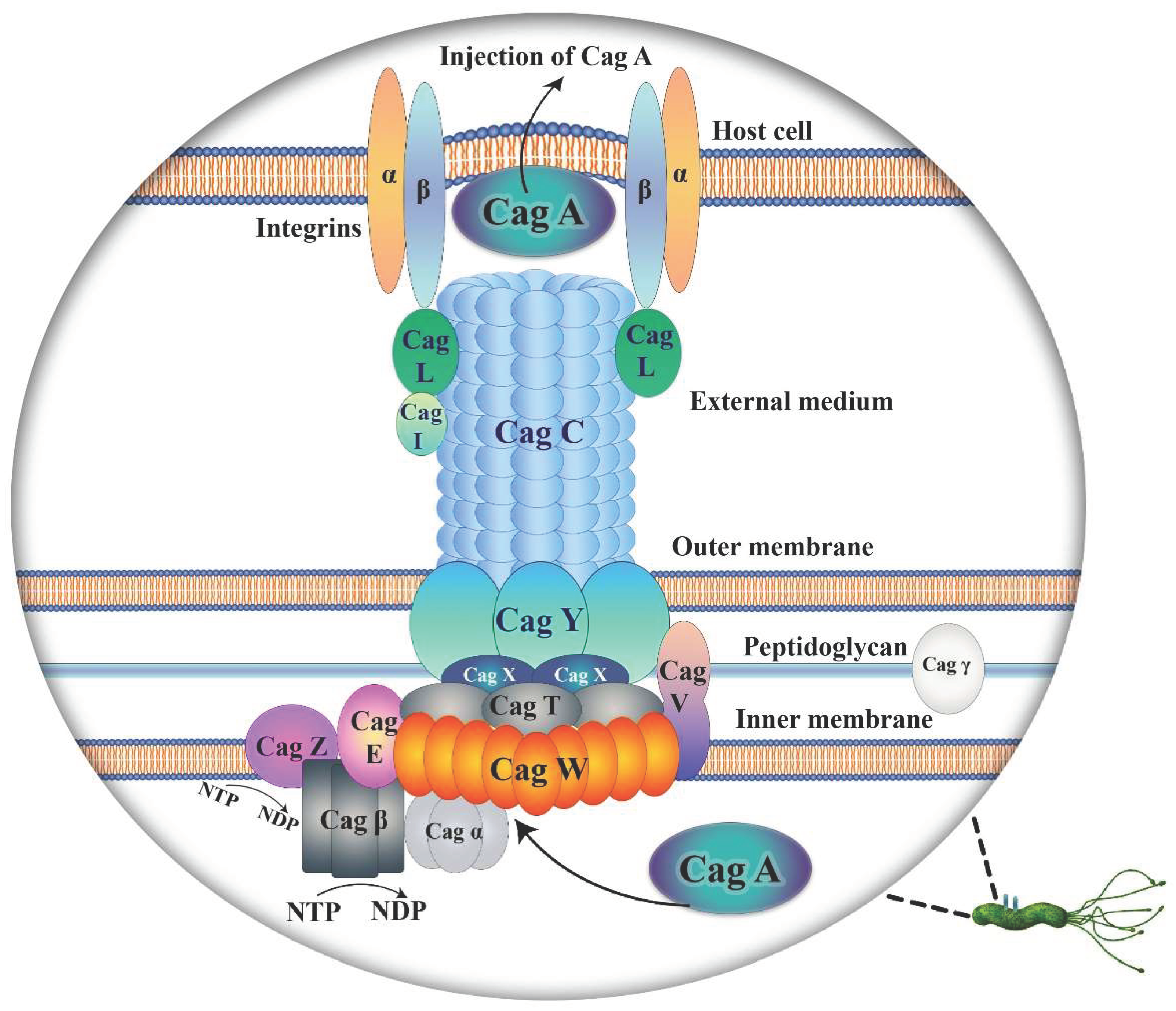
Table 1.
Classification of Helicobacter pylori outer membrane proteins.
| Protein family | Number of sub-family | Sub-family genes |
|---|---|---|
| Hop | 22 |
hopZ, hopD, hopM, hopA, hopF, hopG, hopJ, hopH, hopE, hopO, hopP, hopC, hopB, hopK, hopI, hopL, hopQ, hopQ, hopN, hopU babA, babB |
| Hor | 12 | horA, horB, horC, horD, horE, horF, horG, horH, horI, horJ, horK, horL |
| Hof | 8 | hofA, hofB, hofC, hofD, hofE, hofF, hofG, hofH |
| Hom | 4 | homA, homB, homC, homD |
| FecA-like | 3 | fecA-1, fecA-2, fecA-3 |
| FrpB-like | 3 | frpB-1, frpB-2, frpB-3 |
| Efflux pump | 6 |
hefA, hefD, hefG flgH palA lpp20 |
Disclaimer/Publisher’s Note: The statements, opinions and data contained in all publications are solely those of the individual author(s) and contributor(s) and not of MDPI and/or the editor(s). MDPI and/or the editor(s) disclaim responsibility for any injury to people or property resulting from any ideas, methods, instructions or products referred to in the content. |
© 2024 by the authors. Licensee MDPI, Basel, Switzerland. This article is an open access article distributed under the terms and conditions of the Creative Commons Attribution (CC BY) license (http://creativecommons.org/licenses/by/4.0/).
Copyright: This open access article is published under a Creative Commons CC BY 4.0 license, which permit the free download, distribution, and reuse, provided that the author and preprint are cited in any reuse.


