Submitted:
04 February 2024
Posted:
05 February 2024
Read the latest preprint version here
Abstract
A rare genetic disorder caused by col18.a1 mutations, inherited mostly in autosomal recessive and rarely in autosomal dominant pattern, giving rise to ocular abnormalities and other associated disorders, the condition is referred to as Knobloch syndrome. This paper overviews the anterior and posterior ocular symptoms in Knobloch patients, thus demonstrating the major findings in Knobloch cases including the mutational analysis which discusses the proteins involved and exon number on which mutations are detected. Animal modeling done by knocking out col18.a1 has also been discussed to determine the functions of this gene which have been identified based on anomalies found in knock-out mice.
Keywords:
Keywords: Mutation
; Col18A1
; Knobloch syndrome
; mouse modeling
; treatments
Introduction
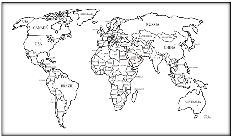
Knobloch syndrome is an autosomal recessive disorder characterized by abnormalities most commonly in ophthalmic regions. The most common symptoms of Knobloch syndrome include retinal detachment, occipital encephaloceles, and high myopia (1). Apart from ocular and occipital skull abnormalities, Knobloch syndrome can give a range of phenotypic discoveries like lung hypoplasia, hyperextensible joints, duplication of the renal collecting system, epilepsy, neuronal migration abnormalities, and dysmorphic findings such as midface hypoplasia, high-arched palate, micrognathia, flat nasal bridge, and dental abnormalities (4). It is a genetic disorder that is caused by mutations in the gene Col18A1 and is inherited in an autosomal recessive pattern. This condition is acquired in an autosomal latent example, which implies that mutations have occurred in both duplicates and copies of every cell's gene. One copy of a mutated gene will be present in both parents of the person with an autosomal recessive condition means the parents are a carrier, however, they commonly don't give indications and manifestations of their condition. This syndrome is named after one of the two authors named W.H Knobloch who first reported this disease along with his companion in a family where five out of ten siblings were affected. Their parents had a non-consanguineous marriage. To date, three types of Knobloch syndromes have been reported. Type 1 is because of mutations in the col18A1 gene however the causative genes for type 2 and 3 have still not been identified. Influenced people may likewise have irregularities in the focal space of the retina, called the macula. The macula is accountable for sharp focal vision, which is required for point-by-point errands like perusing, driving, and perceiving faces. Because of irregularities in the retina, vitreous, and macula, individuals with Knobloch syndrome can raise visual deficiency or blindness in one or both eyes. Knobloch syndrome occurs due to biallelic mutations in Col18A1 (2). Col18A1 is associated with Knobloch syndrome. Collagen XVIII is a non-fibril-framing. It is more like a short isoform communicated in epithelial and endothelial base layers all through the body however especially in the eye, where it plays a significant formative part (3). Knobloch disorder is an uncommon condition. Although, the specific prevalence of the condition is obscure. The countries from where Knobloch cases were reported; are shown by red dots in the map given below.
SYMPTOMS TABLE DESCRIPTION
Supplementary table.1 shows the data of case reports of Knobloch patients reported to date. The symptoms are divided based on an anterior and posterior division of the eye and other clinical observations and associations found in those patients such as skeletal abnormalities, facial dysmorphisms, neurological disorders, and other interlinked malformations. Each row individually represents the records of a single research publication and includes all the cases discussed in that paper.
MUTATIONS TABLE DESCRIPTION
Supplementary table.2 discusses the type of mutations identified after genome sequencing of Knobloch patients. A total of 65 mutations have been reported in Col18.a1 and PAK2 gene, being a cause of Knobloch syndrome. Out of which, 28 were deletions, 6 were insertions, 5 were duplications and 26 were substitutions. Within the insertion and deletions, 28 were frameshift.
Causes
Mutations in COL18.A1 are the novel cause of this syndrome. This gene plays its role in the building of collagen XVIII which is an important structural protein found in the body’s connective tissues. Collagen produces endostatin, endostatin is a signaling molecule and it retards the proliferation and migration of endothelial cells. (5) Presence of collagen in appropriate amounts is very significant for normal eye development. 21q22.3 was the very first locus identified for Knobloch syndrome in 1996 in 11 affected people in a Brazilian family by sertie et al. (6) Recently mutations in another gene have been identified in Knobloch type 2 patients which is PAK 2 in two affected siblings with unaffected parents. They confirmed the mutant variant in affected children with the help of Sanger sequencing. PAK2 gene plays its role in cell growth, survival, and migration (7). Homology modeling of the PAK 2 gene showed that Glu435, Ser371, and Trp409 are present in the catalytic active site of the gene. These residues are significant for the correct conformation and stability of helical structures. A point mutation in (Glu435Lys) which is present at the active region domain of the PAK2 gene seems important for the kinase activity of the gene because this mutation has led to a decrease in phosphorylation at the catalytic site (8). Nonfunctional or haploinsufficiency of the PAK2 gene has resulted in patients with autism-related behaviors, cytoskeleton impairment, dysplasia+, bifid thumb growth delay, and related neurological disorders (9)(10)(11). All these clinical findings are also seen in patients with Knobloch syndrome having mutational COL18.A1 gene which provides clear evidence of a strong association between the two genes. Antonarakis et al hypothesized that some functional correlation might exist between COL18.A1 and PAK2 because both work in association with integrins and are activated by RAC1 which is involved in the regulation of many cell processes in the body (8).
Methodology
A review of the literature was conducted from 1992 to 2021 and all the papers accomplishing the inclusion criteria and published by the International scholars were considered. The search strategy adopted was an article title/keyword/abstract-based search using the following key terms: ‘Knobloch/recessive and dominant’, ‘Knobloch/congenital’, ‘Knobloch /congenital ophthalmological disorders’. KNO1 reported under the study title of birth defects, ophthalmological disorder, and congenital ocular deformities were included. PubMed, Science Direct, and Google Scholar were the search engines employed for literature search. The appropriate information including authors, institute, study site, time, sample size, age group, objectives, and management strategy, was extracted. Data were maintained in excel sheet.
Clinical Techniques
Radiological examinations, CT scan, Optical Coherence Tomography (OCT), Anomaloscope plate test-5, histological examinations, ophthalmic evaluation, magnetic resonance imaging MRI, chest radiography, Echocardiogram, intelligence tests, abdominal ultrasounds, renal and cranial ultrasounds, ELISA to measure plasma concentrations, urinary tract ultrasound scan, vertebral column radiography, slit-lamp biomicroscopy, fundus photography, B scan ultrasonography, Goldmann kinetic perimetry dark adaptometry, Ganzfeld ERGs, LKC technologies, skull radiography, ocular examinations, fluorescein angiography, ultra-sonographic investigations during pregnancy, pathological analysis, psychometric evaluations, retinopathy, teeth examinations, fundus finding after pars plana vitrectomy, DNA extraction, electroencephalography, retinoscopy, vitreoretinopathy, goniotomy, trabeculotomy, auditory screening, gonioscopy were used to determine the clinical symptoms in Knobloch patients.
Genetic Molecular Techniques
RNA purification by Guanidium isothiocyanate method, PCR , silver staining for PCR production visualization, genotype analysis using polymorphic markers, dbEST database searching, bidirectional sequencing through PCR, SSCP analysis of chromosomes, DNA sequencing through Dye Deoxy Terminator Cycle Sequencing, RFLP analysis, Pyrosequencing, flanking primers through primer 3 for mutational analysis, standard chromosome testing, fish analysis, CD68 staining, direct sequencing of COL18.A1 exons, surface plasma resonance assays, immunohistochemistry, RT PCR analysis, BLAST (NCBI) for sequence retrieval, PFAM, GenBank, knockout analysis to determine if knocking out of the gene COL18.A1 has any outcome on expression, autozygosity mapping along with exome sequencing, SNP genotyping, RNA in situ hybridization, Illumina sequencing, whole-genome genotyping, confirmatory sanger sequencing, genotyping and linkage analysis, mitochondrial DNA sequencing, segregation analysis, western blotting procedures were used for the mutational analysis of patients’ genome.
Mouse Modelling
In recent years, mouse modeling has been performed for the functional analysis of COL18.A1 which is a causative gene for Knobloch syndrome, confirmed through genomic molecular analysis of several Knobloch patients. Col18.a1 directs the conformation of the protein to make a protein named collagen. Absence of collagen and its derivative endostatin results in the underdevelopment of retina and retinal vessels as well as blood vessels in the ophthalmic vitreous region. Dissociation of hyaloid vessels from the surface of the retina, leading to retinal vasculature (13)(15), and absence of VHP capillaries was evident in col18.a1 null mice which were homozygous mutant. Similarly, collagen 18 null mice also showed signs of structural anomalies along inner eye membranes and delayed expression of vascular endothelial growth factors in the retina (12). Ocular examination of Col18.a1 null mice at the age of 22 months showed varied phenotypes and irregularities when compared with wild-type species. Abnormalities included a detachment of layers of IPE cells from the iris, abnormal developmental pattern, and irregular bending of retinal vessels. Mutant mice also showed an unusual increase in thickness of BM zone present in the anterior region of iris (13)(14)(15). Ophthalmic studies along with fluorescence angiography in Col18.a1null mice also showed pigmented regions formed by the migration of cells from iris stroma leading to cluster formation and giving the appearance of iris clumps, thus blocking light from retinal vessels (13)(15). Utrianen et al in 2004 experimentally showed that Col18.a1 null mice also exhibit abnormal skull enlargements as well as irregularities associated with renal structure and functions (14). The latest evidence also shows the signs of iris atrophy and ciliary body abnormalities in collagen 18 null mice which are evident symptoms in Knobloch patients (15). Col18a1 mice and Hspg2 mutant mice were taken under observation out of which col18a1 mice have raised plasma triglycerides comparatively to that of wild type. Hypertriglyceridemia is encountered in mice due to deficiency of col18. Hypertriglyceridemia was developed due to amended extrahepatic clearance triglyceride-rich lipoproteins. In col18a1 mice, the level of lipoprotein lipase (lpl) is notably low in comparison to wild-type mice. Since the Col18a1 mice had hypertriglyceridemia, and a decrease in plasma and endothelium-bound Lpl levels in their blood; this evoked to be tested on human patients who had Col18-deficiency (16). Several anomalies were observed in collagen 18 null mouse eyes. The excessive protrusion was visible from the anterior side of the lens capsule. An examination through a Transmission electron microscope indicated an uncontrolled fibrillar stuff accumulation in an aqueous medium besides the ciliary body of transgenic mice also some inflammation was seen in the fibrillar space on top of the lens capsule. Some retinal and vitreous anomalies were suspected and encountered too. Shortage of collagen 18 resultantly gives led to enlarged eyes, lens subluxation, and lower intraocular pressure. Knobloch sufferers generate truncated protein as a result of missense mutations; they might be deleterious and harmful, this is all disclosed due to the transgenic mice’s study in which overexpression of the Tsp-1 domain or the C-terminal endostatin domain was seen (17).
Treatment
Moreover, some common abnormalities in patients with Knobloch syndrome-like cataracts, encephaloceles, blurred vision, etc. can be treated accordingly. Cataract surgery was performed on a Caucasian boy aged 12 years reported with Knobloch syndrome which improved its visual acuity in the right eye to 20/200 (18). Similarly high myopic conditions can be treated with eye surgeries to enhance vision but require proper evaluation from patient to patient (19). Likewise, Ebrahimiadib et al reported a case of an Afghani girl aged 7 years who was suffering from Knobloch syndrome confirmed through genetic testing having severe retinal detachment, underwent medical treatment including scleral buckling surgery and cryotherapy in her left eye. She showed signs of effective retinal detachment repair and improved visual impairment a few months post-surgery (20). Alsulaiman et al in 2019 published a paper that discusses the surgical treatments given to patients having retinal detachment within the timeframe of 7 years (January 2012 till December 2018). Different treatment procedures included silicone oil tamponade with scleral buckling surgery or devoid of it, pars plana vitrectomy (PPV), endolaser photocoagulation, retinectomy, triamcinolone-assisted posterior hyaloid peeling, and vitrectomy. 7 out of 9 eyes underwent operative treatments, which couldn’t be performed on the rest of two because one had a retinal detachment that was restricted to the atrophic lesion and in the other case, the parent’s consent was not available. Post-surgical procedures, 5 out of 9 eyes showed re-attachment of the retina, and two eyes showed re-detachment (21).
Discussion
The relationship of occipital encephaloceles and vitreoretinal degeneration with high nearsightedness and retinal separation was portrayed first by Knobloch and Layer in 1971. As they reported 5 siblings out of 10 in a family showed the above-mentioned conditions i.e. occipital encephaloceles and vitreoretinal degeneration with high myopia. The vitreoretinal degeneration with high nearsightedness and inevitable retinal separation or say detachment are all universally inclusive in this syndrome. The study inferred that innate occipital scalp defects in preference to genuine encephaloceles may, as is valid at times of Meckel condition, go with Knobloch syndrome (22). Clinical flexibility is available in the appearance of this syndrome’s condition (23). COL18A1 is situated on the long arm of chromosome 21 (chr21q22.3) and is made out of 43 exons. It encodes the collagen XVIII protein, which has been disclosed to be a significant part of base films (24). COL18A1 has no less than three particular isoforms of various lengths. Regardless of the fact that COL18A1 is pervasively communicated or expressed, its isoforms have distinctive tissue and formative circulation. In expansion to these three isoforms, COL18A1 can create endostatin through proteolytic cleavage. Endostatin is a gesturing molecule known to hinder the relocation and expansion of endothelial cells and is fit for stifling angiogenesis (25)(26). Through genetic investigations it has been noticed that most common mutations occur at exons 30 through 42 of COL18A1; in Knobloch condition patients. In any case, there stays a lot of heterogeneity in mutational site conveyance (27). With advancements in research recently it has also been observed that germline compound heterozygous mutations were likewise portrayed in patients with Knobloch condition (28). Some accessory anomalies in certain patients have been noticed by Caglayan A. O et al 2014; anomalies included atrial septal imperfections, seizures, and minor dysmorphic discoveries/findings. These findings foreground the huge phenotypic range that the Knobloch condition can include and can further represent the significance of type XVIII collagen in the typical improvement of various organ frameworks (29). In this study, we have thoroughly considered case reports of Knobloch syndrome patients till now. We deliberated the reports being mentioned in research papers from 1982 to 2021 and from those cases the symptoms were extracted out as shown in the table. This is collective data that is representing the most common and least commonly occurring symptoms in people being affected by Knobloch syndrome. Not only symptoms, but our analysis also highlights the major causes of Knobloch syndrome and techniques that have been used to track out this syndrome. By performing throughout analysis of all cases; the basic understanding of this syndrome is described in our study. Knobloch syndrome is a condition that is comprised of various ocular defects as well as several clinical abnormalities.
Results
A total of 83 articles have been published on Knobloch syndrome since 1971 out of which we reviewed the cases reported in patients to date. To the best of our knowledge, 49 case reports have been published in literature which reports a total of approximately 240 patients of this ophthalmic disorder up till now. Molecular techniques that were involved in determining the clinical findings in the patients of Knobloch syndrome included clinical molecular techniques and genetic molecular techniques.
Future Recommendations
As it is evident that Knobloch syndrome involves ocular abnormalities that vary among patients but high myopia, encephaloceles, and occipital defects are the most common symptoms of this condition (30). Because a lot of mutations are a cause of this particular disorder, there is no specific treatment introduced to tackle these kinds of mutations, however, genome sequencing can tell us about the faulty gene and precautions can be taken in this regard. The incidence of inherited genetic disorders among children from a consanguineous marriage is double the rate of children from parents that are not related to each other (31). Consanguinity can be eluded to avoid such genetic disorders.
References
- Wilson, C., Aftimos, S., Pereira, A., & McKay, R. (1998). Report of two sibs with Knobloch syndrome (encephalocoele and viteroretinal degeneration) and other anomalies. American journal of medical genetics, 78(3), 286–290. [CrossRef]
- Aldahmesh, M. A., Khan, A. O., Mohamed, J. Y., Levin, A. V., Wuthisiri, W., Lynch, S., McCreery, K., & Alkuraya, F. S. (2013). No evidence for locus heterogeneity in Knobloch syndrome. Journal of medical genetics, 50(8), 565–566. [CrossRef]
- Seppinen, L., & Pihlajaniemi, T. (2011). The multiple functions of collagen XVIII in development and disease. Matrix Biology, 30(2), 83-92. [CrossRef]
- Keren B, Suzuki OT, Gerard-Blanluet M, et al. CNS malformations in Knobloch syndrome with splice mutation in COL18A1 gene. Am J Med Genet. 2007; 143A:1514-1518. [CrossRef]
- Schuch, G., Heymach, J. V., Nomi, M., Machluf, M., Force, J., Atala, A., ... & Soker, S. (2003). Endostatin inhibits the vascular endothelial growth factor-induced mobilization of endothelial progenitor cells. Cancer research, 63(23), 8345-8350.
- Sertié, A. L., Quimby, M., Moreira, E. S., Murray, J., Zatz, M., Antonarakis, S. E., & Passos-Bueno, M. R. (1996). A gene which causes severe ocular alterations and occipital encephalocele (Knobloch syndrome) is mapped to 21q22.3. Human molecular genetics, 5(6), 843–847. [CrossRef]
- Ran, M., Weng, B., Cao, R., Li, Z., Peng, F., Luo, H., ... & Chen, B. (2018). miR-26a inhibits proliferation and promotes apoptosis in porcine immature Sertoli cells by targeting the PAK2 gene. Reproduction in Domestic Animals, 53(6), 1375-1385. [CrossRef]
- Antonarakis, S. E., Holoubek, A., Rapti, M., Rademaker, J., Meylan, J., Iwaszkiewicz, J., Zoete, V., Wilson, C., Taylor, J., Ansar, M., Borel, C., Menzel, O., Kuželová, K., & Santoni, F. A. (2021). Dominant monoallelic variant in the PAK2 gene causes Knobloch syndrome type 2. Human molecular genetics, ddab026. Advance online publication. [CrossRef]
- Wang, Y., Zeng, C., Li, J., Zhou, Z., Ju, X., Xia, S., ... & Sun, Z. S. (2018). PAK2 haploinsufficeincy results in synaptic cytoskeleton impairment and autism-related behavior. Cell reports, 24(8), 2029-2041.
- Pollazzon, M., Grosso, S., Papa, F. T., Katzaki, E., Marozza, A., Mencarelli, M. A., ... & Renieri, A. (2009). A 9.3 Mb microdeletion of 3q27. 3q29 associated with psychomotor and growth delay, tricuspid valve dysplasia and bifid thumb. European journal of medical genetics, 52(2-3), 131-133. [CrossRef]
- Zhang, K., Wang, Y., Fan, T., Zeng, C., & Sun, Z. S. (2020). The p21-activated kinases in neural cytoskeletal remodeling and related neurological disorders. Protein & Cell, 1-20. [CrossRef]
- Fukai, N., Eklund, L., Marneros, A. G., Oh, S. P., Keene, D. R., Tamarkin, L., Niemelä, M., Ilves, M., Li, E., Pihlajaniemi, T., & Olsen, B. R. (2002). Lack of collagen XVIII/endostatin results in eye abnormalities. The EMBO journal, 21(7), 1535–1544. [CrossRef]
- Marneros, A. G., & Olsen, B. R. (2003). Age-dependent iris abnormalities in collagen XVIII/endostatin deficient mice with similarities to human pigment dispersion syndrome. Investigative ophthalmology & visual science, 44(6), 2367–2372. [CrossRef]
- Utriainen, A., Sormunen, R., Kettunen, M., Carvalhaes, L. S., Sajanti, E., Eklund, L., Kauppinen, R., Kitten, G. T., & Pihlajaniemi, T. (2004). Structurally altered basement membranes and hydrocephalus in a type XVIII collagen deficient mouse line. Human molecular genetics, 13(18), 2089–2099. [CrossRef]
- Marneros, A. G., & Olsen, B. R. (2005). Physiological role of collagen XVIII and endostatin. FASEB journal : official publication of the Federation of American Societies for Experimental Biology, 19(7), 716–728. [CrossRef]
- Bishop, J. R., Passos-Bueno, M. R., Fong, L., Stanford, K. I., Gonzales, J. C., Yeh, E., Young, S. G., Bensadoun, A., Witztum, J. L., Esko, J. D., & Moulton, K. S. (2010). Deletion of the basement membrane heparan sulfate proteoglycan type XVIII collagen causes hypertriglyceridemia in mice and humans. PloS one, 5(11), e13919. [CrossRef]
- Aikio, M., Hurskainen, M., Brideau, G., Hägg, P., Sormunen, R., Heljasvaara, R., Gould, D. B., & Pihlajaniemi, T. (2013). Collagen XVIII short isoform is critical for retinal vascularization, and overexpression of the Tsp-1 domain affects eye growth and cataract formation. Investigative ophthalmology & visual science, 54(12), 7450–7462. [CrossRef]
- Bongiovanni, C. S., Ferreira, C. C. S., Rodrigues, A. P. S., Fortes Filho, J. B., & Tartarella, M. B. (2011). Cataract surgery in Knobloch syndrome: a case report. Clinical Ophthalmology (Auckland, NZ), 5, 735.
- https://stanfordhealthcare.org/medical-conditions/eyes-and-vision/high-myopia/treatments.html.
- Ebrahimiadib, N., Modjtahedi, B. S., Ferenchak, K., Papakostas, T. D., Mantagos, J. S., & Vavvas, D. G. (2017). Optical coherence tomography findings and successful repair of retinal detachment in Knobloch syndrome. Digital journal of ophthalmology: DJO, 23(1), 29–32. [CrossRef]
- Alsulaiman, S. M., Al-Abdullah, A. A., Alakeely, A., Aldhibi, H., Engelbrecht, L., Ghazi, N. G., & Mura, M. (2020). Macular Hole-Related Retinal Detachment in Children with Knobloch Syndrome. Ophthalmology. Retina, 4(5), 498–503. [CrossRef]
- Seaver, L. H., Joffe, L., Spark, R. P., Smith, B. L., & Hoyme, H. E. (1993). Congenital scalp defects and vitreoretinal degeneration: redefining the Knobloch syndrome. American journal of medical genetics, 46(2), 203-208. [CrossRef]
- Sniderman, L. C., Koenekoop, R. K., O'Gorman, A. M., Usher, R. H., Sufrategui, M. R., Moroz, B., ... & Der Kaloustian, V. M. (2000). Knobloch syndrome involving midline scalp defect of the frontal region. American journal of medical genetics, 90(2), 146-149. [CrossRef]
- Halfter, W., Dong, S., Schurer, B., & Cole, G. J. (1998). Collagen XVIII is a basement membrane heparan sulfate proteoglycan. Journal of Biological Chemistry, 273(39), 25404-25412.
- Suzuki, O. T., Sertie, A. L., Der Kaloustian, V. M., Kok, F., Carpenter, M., Murray, J., ... & Passos-Bueno, M. R. (2002). Molecular analysis of collagen XVIII reveals novel mutations, presence of a third isoform, and possible genetic heterogeneity in Knobloch syndrome. The American Journal of Human Genetics, 71(6), 1320-1329.
- Schuch, G., Heymach, J. V., Nomi, M., Machluf, M., Force, J., Atala, A., ... & Soker, S. (2003). Endostatin inhibits the vascular endothelial growth factor-induced mobilization of endothelial progenitor cells. Cancer research, 63(23), 8345-8350.
- Suzuki, O., Kague, E., Bagatini, K., Tu, H., Heljasvaara, R., Carvalhaes, L., ... & Passos-Bueno, M. R. (2009). Novel pathogenic mutations and skin biopsy analysis in Knobloch syndrome. Molecular vision, 15, 801.
- Joyce, S., Tee, L., Abid, A., Khaliq, S., Mehdi, S. Q., & Maher, E. R. (2010). Locus heterogeneity and Knobloch syndrome. American journal of medical genetics. Part A, 152(11), 2880-2881.
- Caglayan, A. O., Baranoski, J. F., Aktar, F., Han, W., Tuysuz, B., Guzel, A., ... & Gunel, M. (2014). Brain malformations associated with Knobloch syndrome—review of literature, expanding clinical spectrum, and identification of novel mutations. Pediatric neurology, 51(6), 806-813. [CrossRef]
- White, R. J., Wang, Y., Tang, P., & Montezuma, S. R. (2017). Knobloch syndrome associated with Polymicrogyria and early onset of retinal detachment: two case reports. BMC ophthalmology, 17(1), 214. [CrossRef]
- Merten, M. (2019). Keeping it in the family: consanguineous marriage and genetic disorders, from Islamabad to Bradford. BMJ: British Medical Journal (Online), 365. [CrossRef]
Disclaimer/Publisher’s Note: The statements, opinions and data contained in all publications are solely those of the individual author(s) and contributor(s) and not of MDPI and/or the editor(s). MDPI and/or the editor(s) disclaim responsibility for any injury to people or property resulting from any ideas, methods, instructions or products referred to in the content. |
© 2024 by the authors. Licensee MDPI, Basel, Switzerland. This article is an open access article distributed under the terms and conditions of the Creative Commons Attribution (CC BY) license (http://creativecommons.org/licenses/by/4.0/).
Copyright: This open access article is published under a Creative Commons CC BY 4.0 license, which permit the free download, distribution, and reuse, provided that the author and preprint are cited in any reuse.


