
Submitted:
26 December 2023
Posted:
27 December 2023
You are already at the latest version
Abstract
Tongue mobility is an obstructive sleep apnoea (OSA) marker and myofunctional therapy (MFT) target. For this reason, all patients with sleep-disordered breathing should require a combined functional assessment from an ear, nose, and throat (ENT) specialist and a phonoaudiologist to confirm or rule out the presence of tongue-tie. To our knowledge, this is the first case of a 13-year-old girl diagnosed with severe OSA and a significant decrease of 94% in her apnoea index (AI), requiring frenotomy with immediate postoperative change in the tongue position. A drug-induced sleep en-doscopy (DISE) was performed before and immediately post-frenotomy, and the anatomical changes provoked by this surgery during sleep were confirmed for the first time.
Keywords:
Ankyloglossia
; Drug induced sleep endoscopy
; frenulectomy
; sleep apnoea
1. Introduction
Most obstructive sleep apnoea (OSA) patients share a common physiopathology: the narrowing of the upper airway, mainly at the retropalatal and retroglossal levels. This can be due to increased soft tissue from fatty deposits and tissue hypertrophy owing to the chronic inflammatory process, a volumetric reduction of the facial skeletal framework caused by craniofacial characteristics related to genetic or environmental factors, alterations in neuromuscular tone, or a combination of these factors. OSA has a multifactorial aetiology, characterized by the abnormal (total or partial) collapse of the upper airway during sleep that produces disturbances in ventilation and sleep architecture [1]. It occurs in 3–10% of children with two prevalence peaks, one in the 2–8 years of age range and another in adolescence, related to weight gain.
The outcomes of multiple investigations in the paediatric population reveal a pattern: early dysfunction involving abnormal nasal breathing, sucking, and chewing leading to progressive dysmorphia that results in increased upper airway collapsibility during sleep [2].
Several risk factors for developing paediatric OSA have been described, including obesity, adenoid and tonsillar hypertrophy, allergic rhinitis, and septal deviation. Short lingual frenulum has also been described as a risk factor. According to the guidelines of the American Academy of Otolaryngology–Head and Neck Surgery (AAO-HNS), ankyloglossia is defined as a condition of limited tongue mobility caused by a restrictive lingual frenulum that has an incidence of 2.8–10.7% [3]. Tongue-tie is a congenital anomaly with autosomal dominant X-linked inheritance, being more common in men (with a sex ratio of 2:1) [2]. It is also related to patients with Beckwith-Wiedemann syndrome, orofacial digital syndrome, cleft palate, and Optiz syndrome [2].
Ankyloglossia does not allow the tongue to rest on the palate, meaning that during sleep, the tongue falls towards the pharynx and obstructs the airway, which explains the appearance of OSA. The balance between the tongue and the buccinator muscles is responsible for the normal development of the maxillary and mandibular arch. The stimuli of abnormal bone growth, the absence of nasal breathing, and hypotonia with the consequent development of oral breathing associated with ankyloglossia are responsible for the abnormal orofacial development, thus increasing the risk of collapse in the upper airway.
An ear, nose, and throat (ENT) specialist should consider a functional assessment for patients with OSA, including examining the lingual frenulum [3]. However, our group demonstrated [4] that the evaluation of a short lingual frenulum was not included in any otolaryngology clinical guidelines. Therefore, the diagnosis of sleep breathing disorder (SBD) in patients with ankyloglossia is delayed, unlike in children with tonsillar hypertrophy [2]. All of these patients must be evaluated for the craniofacial region, breathing, elongated face shape, the coexistence of retrognathia, dental crowding, high and narrow palate, tongue posture, and the length and mobility of the lingual frenulum [3].
Pediatric Drug induced sleep endoscopy (DISE) [5] is considered in children with persistent, postadenotonsillectomy obstructive sleep apnea who refuse or fail positive pressure therapy. However, many questions regarding timing of DISE, patient selection, ideal scoring method and threshold for operative intervention remain unanswered as no controlled studies of DISE-assisted surgery in children have been conducted. Up to the moment, there is no case reported in a pedatric OSA patient showing anatomic changes after a therapeutical intervention. Since our group considered mandatory use DISE to explain anatomical findings in patients without a reasonable explanation of their OSA [6] and also to improve adherence to myofunctional therapy [7,8]. We introduce the first OSA pediatric case where tongue tie surgery combined with MT resolves the disease demonstrated by DISE performed pre and postoperatively.
2. CASE DESCRIPTION
A 13-year-old female patient with sleep problems witnessed apnoeas and slight snoring. She had a level III sleep study (nocturnal respiratory polygraphy was performed using an Embletta portable diagnostic system (ResMed, Sydney, Australia) according to the technical specifications of the American Academy of Sleep Medicine [9]. Measurements were obtained using a snoring sensor, nasal thermistor, and nasal pressure cannula to register airflow; thoracic and abdominal belts to assess ribcage and abdominal movements; electrocardiography; actigraphy to detect body position; oxygen saturation; and heart rate. in February 2022) with the result of apnoea–hypopnoea index (AHI) 17.3/hour, 14 obstructive apnoeas, 15 central apnoeas, 10 mixed apnoeas, and 99 hypopneas. Oxygen desaturation index (ODI) was 2.4; T90 was 0.1%, mean oximetry 95%, and nadir 83%. Epworth sleepiness questionnaire (ESS) scored 18. On physical evaluation, she presented BMI 21 Kg/m2, moderate hypertrophy of the turbinates, non-adenotonsillar hypertrophy, normal palate, Angle 1 classification, teeth malposition, and the unique significant finding was the tongue-tie. This was evaluated following the Marchesani protocol [10]. The measurement was taken during the maximum opening of the lower left incisor to the upper left incisor, and then a measurement was taken with the mouth wide open with the tip of the tongue touching the incisor papilla. This measure was considered normal if the difference between the two was <50%. Our protocol functional assessment [7] included measurements with Iowa Oral Performance Instrument (IOPI) were: maximum tongue strength 36 kilopascal (Kps) (Normal 65 Kps) and maximum buccinator strength 24Kps (Normal 35 Kps).
Many otolaryngologists have examined her without any objective finding to explain the disease. The patient was referred to speech therapy and; in the assessment, a frenotomy was considered to manage the short-lingual frenulum (Figure 1).
Before surgical time, a drug-induced sleep endoscopy (DISE) was performed using an inhaled anesthetic agent allowing for intravenous access to be obtained. The inhalation agent is then discontinued and a propofol infusion dosed appropriately for the child’s age and weight is used for the remainder of the procedure. We visualized anteroposterior collapse due to tongue base with secondary lateral epiglottic collapse and twisted epiglottis. Vote classification [11] was V1APO1LT2APE2L. Then, the patient underwent turbinoplasty combined with lingual frenotomy under sedation and a new DISE was performed immediately after the operation, showing the adequate position of the tongue with improvement in retroglossal collapse and complete decrease in epiglottic collapse, with an adequate response to Chin lift and Esmarch manoeuvres Vote classification improved to V1APO1T0PE0. (See video 1) The postoperative period was uneventful, and the patient and since the first day postoperatively her family reported improved sleep patterns and decreased daytime sleepiness. (See video 2)
Patient started to perform myofunctional therapy and a new sleep study was requested two months after the procedure. Postoperative control in August 2023 lacked OSA with AHI 4.2/hour (1 obstructive apnoea – 3,2 hypopneas)., 0 central apnoeas, 0 mixed apnoeas, and 30 hypopneas. ODI and T90 were 0%, mean oximetry 95%, and nadir 91%.ESS scored 24, her IOPI tongue measurement was 52 Kps and her buccinator evaluation was 30 Kps.
Up to date (24/12/23) there is no recurrence of symptoms with similar results obtained in a new sleep study. See Table 1
3. Discusion
This is the first paediatric DISE where anatomical changes after a frenuloplasty are objectively documented by performing another immediate postoperative DISE.
The tongue is a dynamic organ that affects breath, language, early weaning, breastfeeding, sucking, and swallowing, with a critical role in facial development and the oropharynx's patency [3]. Lingual frenulum is a vestigial embryological element of mostly fibrous consistency, a product of the adhesion between the tongue and floor of the mouth that keeps the hard and soft tissue balanced to promote adequate growth. During tongue formation, cells of the lingual frenulum undergo gene-mediated apoptosis and migrate distally to the medial dorsum of the tongue; incomplete or absent migration results in the development of ankyloglossia [2,3].
Ankyloglossia, commonly described as "tongue-tie", is a short or altered insertion of the lingual frenulum that restricts the tongue's mobility. It also hurts orofacial development (dentofacial anomalies, abnormal growth of the maxilla with a narrow palatine arch, and imbalanced mandible growth). It modifies the respiratory function because of the reduced calibre of the upper airway with the risk of collapsibility during sleep [2,4]. Tongue-tie generates the use of additional facial muscles and modifies the cervical muscles, and is associated with incorrect head posture, nasal obstruction, and oral breathing [2].
The untreated short lingual frenulum is associated with OSA. In ankyloglossia, the tongue rests on the mouth's floor, contrary to its normal position, resting against the upper incisors and hard palate [3]. Oral breathing, one of the nocturnal symptoms of children with SBD and particularly with OSA, was more prevalent in patients with reduced mobility of the tongue, and compared with healthy children, they were also more prone to developing hypotonia as measured by IOPI [4]. This confirms that the position of the tongue has special importance in the development of OSA.
Huang et al. concluded that assessment of the frenulum length should be performed in all children with OSA, as well as OSA screening and surgical management in all children with ankyloglossia as soon as possible [12]. Guilleminault et al. [1]. reported a relationship between OSA and ankyloglossia in 150 children with SBD, and found short lingual frenulum in 64 patients associated with oral breathing. Besides, Man Yuen [13]. collected data from 82 paediatric patients and found a high prevalence of OSA in patients with short lingual frenulum. Brozek et al. [14] reported that children with positive questionnaires for high risk of OSA had a short tongue-tie and, more likely, a high-arched palate, which is also a risk factor for OSA. These findings have as well been described by Guilleminault, who reported a strong association between high-arched palate and children with ankyloglossia compared with the control with statistically significant results 80% vs. 8.75%, respectively [1].
Removal of tongue-tie in a patient with adequate muscular strength in the tongue allows it to be able to spread the oral cavity and avoid collapse. Under normal conditions, the tongue contacts the hard palate and does not invade the retroglossal space [1,3]. The tongue's mobility is affected by the length of the lingual frenulum and the elasticity of the floor of the mouth, and patients with reduced length of the frenulum could have normal mobility of the tongue and enough force to ensure the normal development of the maxilla [8]. In patients with a lack of strength in the tongue muscles, an upward vector cannot be established with subsequent difficulty for stimulation of the intermaxillary synchondrosis, affecting bone growth and favoring the development of a V-shaped maxilla, creating a narrower nasal floor, affecting the oropharyngeal space, and increasing the risk of nasal obstruction, which once again explains the relationship between lingual frenulum and OSA.
Frenotomy in children is a safe and well-tolerated procedure, although there is no consensus on the ideal or suitable specific surgical technique. The surgical approach to the tongue-tie is referred to intermittently in the literature. Still, the term “frenotomy” or “frenulotomy” is routinely used for procedures performed in children in which a simple incision of the tongue-tie is made. Frenuloplasty is an incision of the lingual frenulum with a reorganization of the tissue, and frenectomy is used to remove the lingual frenulum. In 2023, our group suggested that lingual frenulum surgery (frenotomy) was an option for patients with OSA that can improve the results of MFT because ankyloglossia limits the success and adherence to this therapy [4].
MFT improves muscle tone and position of the tongue and decreases respiratory events. A reduction in AHI of 62% has been observed in the paediatric population [15]. For this reason, our protocol recommends that all patients who will be managed with frenotomy, including children, should be evaluated by speech therapists and undergo MFT before and after the surgical procedure to achieve better results [4]. On the other hand, IOPI measurements allow the patients to have an objective evaluation of their tongue strength and allow the therapist to select the proper patient to perform MFT [16].
The frenotomy technique used for us consists of a three-step procedure. First, we do a DISE, followed by a frenotomy under sedation using local anesthesia to carry out a cold dissection and bipolar coagulation. Finally, a new DISE is essential to assess postoperative changes in the upper airway [8]. Complications of lingual frenotomy include haemorrhage, airway obstruction, injury to salivary structures, and abnormal scarring [8]. Up to date we have not found any significant complications, so we recommend this procedure due to its safety and feasibility [17].
Despite the results of multiple studies, AAO guidelines suggest insufficient evidence to associate ankyloglossia with OSA [3]. In fact, it is considered that, in some cases, tongue-tie can prevent subsequent collapse, and frenotomy could worsen OSA [8]. This point is made based on two case reports with insufficient evidence. On the other hand, this group has described that the release of the tongue with correct muscle tone can prevent collapse and does not lead to glossoptosis. Furthermore, the tongue can rest on its normal position on the palate, as evidenced in this case.
In the lecture given by Professor Dr. Eric Kezirian in 2020, available on his Youtube® Channel [18], he refered that tongue tie surgery should not be considered as a treatment for OSA. We demonstrated that in selected cases like this, it can be a potential tool to treat OSA. We are aware about the risk of universalizing this treatment due to its feasibility and low morbidity between other therapists not focused in OSA. It is important to notice, that frenotomy should be used after excluding other anatomical potential causes of OSA by an ENT specialist.
We believe this technique of Dise-Frenotomy-Dise is an useful procedure to assure the benefits of the surgery allowing an evaluation of further complementary treatments if required. Furthermore, this technique increase adherence to MFT giving the patient a visual feedback about the anatomical reasons of his disease [6]. We are working with the predictive factors about success with this procedure as we are aware of not all the patients with tongue tie are candidates for this procedure. We believe that the tongue must have space to come to its optimal position in the oral cavity and always postoperative rehabilitation with a speech therapist will be necessary [8].
5. Conclusions
Functional ankyloglossia examination should always be performed in all SBD patients. Tongue-tie should always be suspected when there are no other anatomical possible causes of OSA. DISE performed pre and post frenuloplasty is useful for evaluating anatomical changes and future therapeutic measures.
Supplementary Materials
The following supporting information can be downloaded at the website of this paper posted on Preprints.org.
Author Contributions
Conceptualization, J.X.V. C.O.R. and P.B.; methodology, G.P.; writing—original draft preparation, J.X.V.; writing—review and editing, L.R.A. and M.G.I.;. All authors have read and agreed to the published version of the manuscript.
Funding
No funding.
Institutional Review Board Statement
Not applicable.
Informed Consent Statement
Informed consent was obtained from all subjects involved in the study.
Data Availability Statement
Suggested Data Availability Statements are available in section “MDPI Research Data Policies” at https://www.mdpi.com/ethics.
Conflicts of Interest
No conflict of interest.
References
- Guilleminault C, Huseni S, Lo L. A frequent phenotype for paediatric sleep apnoea: Short lingual frenulum. ERJ Open Res 2016;2(3): pii.00043-2016. [CrossRef]
- Villa MP, Evangelisti M, Barreto M, Cecili M, Kaditis A. Short lingual frenulum as a risk factor for sleep-disordered breathing in school-age children. Sleep Med. 2020;66:119-122. [CrossRef]
- Correa, E. J., et al. (2023). What are we missing in adult obstructive sleep apnea clinical evaluation? Review of official guidelines. International Journal of Orofacial Myology and Myofunctional Therapy, 49(2), 1-10. [CrossRef]
- O’Connor-Reina C, Garcia JMI, Alcala LR, Ruiz ER, Iriarte MTG, Morente JCC, et al. Improving adherence to myofunctional therapy in the treatment of sleep-disordered breathing. J Clin Med. 2021;10(24):5772. [CrossRef]
- Kirkham EM. Pediatric Drug-Induced Sleep Endoscopy. Otolaryngol Clin North Am. 2022 Dec;55(6):1165-1180. [CrossRef]
- O'Connor Reina C, Plaza Mayor G, Ignacio-Garcia JM, Baptista Jardin P, Garcia-Iriarte MT, Casado-Morente JC. Floppy Closing Door Epiglottis Treated Successfully with an Mhealth Application Based on Myofunctional Therapy: A Case Report. Case Rep Otolaryngol. 2019 Jul 1;2019:4157898. [CrossRef]
- Rodriguez-Alcalá L, Ignacio-García J, Serrano Angulo MS et al. Tongue+ protocol for the diagnosis of obstructive sleep apnoea in Quirónsalud Marbella hospital [version 1; peer review: 2 approved with reservations]. F1000Research 2022, 11:322. [CrossRef]
- Correa EJ, O’Connor-Reina C, Rodríguez-Alcalá L, Benjumea F, Casado-Morente JC, Baptista PM, et al. Does frenotomy modify upper airway collapse in OSA adult patients? Case report and systematic review. J Clin Med. 2023;12(1): 201. [CrossRef]
- Berry RB, Gamaldo CE, Harding SM, Brooks R, Lloyd RM, Vaughn BV, et al. AASM Scoring Manual Version 2.2 Updates: New Chapters for Scoring Infant Sleep Staging and Home Sleep Apnea Testing. J Clin Sleep Med 2015 Nov 15;11(11):1253-1254. 2015. [CrossRef]
- Marchesan IQ. Lingual frenulum protocol. Int J Orofacial Myology. 2012 Nov;38:89-103. [CrossRef]
- Kezirian EJ, Hohenhorst W, de Vries N. Drug-induced sleep endoscopy: the VOTE classification. Eur Arch Otorhinolaryngol. 2011 Aug;268(8):1233-1236. Epub 2011 May 26. [CrossRef]
- Huang YS, Guilleminault C. Pediatric Obstructive Sleep Apnea: Where Do We Stand? Adv Otorhinolaryngol. 2017;80:136-144. Epub 2017 Jul 17. [CrossRef]
- Brożek-Mądry E, Burska Z, Steć Z, Burghard M, Krzeski A. Short lingual frenulum and head-forward posture in children with the risk of obstructive sleep apnea. Int J Pediatr Otorhinolaryngol. 2021 May;144:110699. Epub 2021 Mar 29. [CrossRef]
- Yuen HM, Au CT, Chu WCW, Li AM, Chan KC. Reduced tongue mobility: an unrecognized risk factor of childhood obstructive sleep apnea. Sleep. 2022 Jan 11;45(1):zsab217. [CrossRef]
- Carrasco-Llatas M, O'Connor-Reina C, Calvo-Henríquez C. The Role of Myofunctional Therapy in Treating Sleep-Disordered Breathing: A State-of-the-Art Review. Int J Environ Res Public Health. 2021 Jul 8;18(14):7291. [CrossRef]
- O'Connor-Reina C, Rodriguez-Alcala L, Ignacio JM, Baptista P, Garcia-Iriarte MT, Plaza G. Assessment of Muscular Weakness in Severe Sleep Apnea Patients: A Prospective Study. Otolaryngol Head Neck Surg. 2023 Sep;169(3):725-733. Epub 2023 Feb 7. [CrossRef]
- Zaghi S, Valcu-Pinkerton S, Jabara M, Norouz-Knutsen L, Govardhan C, Moeller J, Sinkus V, Thorsen RS, Downing V, Camacho M, Yoon A, Hang WM, Hockel B, Guilleminault C, Liu SY. Lingual frenuloplasty with myofunctional therapy: Exploring safety and efficacy in 348 cases. Laryngoscope Investig Otolaryngol. 2019 Aug 26;4(5):489-496. [CrossRef]
- Kezirian E. Myofunctional Therapy and Frenuloplasty Are Not Appropriate Treatments for Obstructive Sleep Apnea. Available online: https://www.youtube.com/watch?v=29fOg5eDe2c (accessed on 14 December 2022).
Figure 1.
Tongue tie associated with teeth malposition.
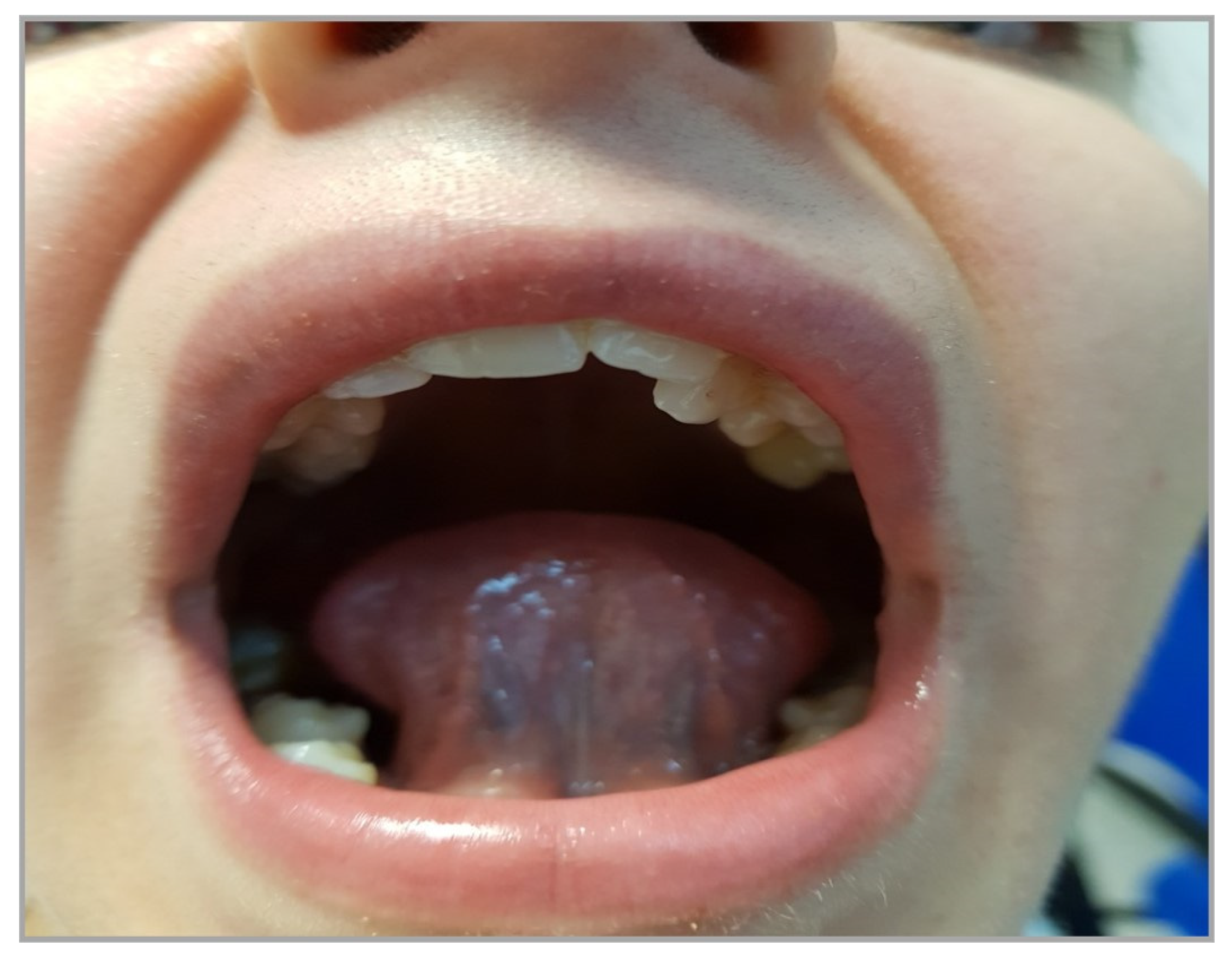

Table 1.
Pre and postoperative 3 and 6 months polygraphy results.
| Before surgery | 3 months after surgery | 6 months after surgery | |
|---|---|---|---|
| AHI | 17,3 | 4,1 | 2,2 |
| CA | 15 | 0 | 0 |
| AI | 4,9 | 0,1 | 0 |
| HI | 12,4 | 4,1 | 2,2 |
| SI | 74,7 | 36,7 | 21,3 |
| Min Sat O2 | 83% | 91 | 93 |
| ODI | 2,4 | 0 | 0 |
| Max PR | 90 | 54 | 60 |
AHÍ: Apnea hypopnea index, CA: Central Apneas, MA; Mixed apneas, AI: Apnea Index, HI. Hypopnea Index, SI: Snore Index; Min Sat O2: Minimal Saturation O2, Max PR: Maximal heart rate.
Disclaimer/Publisher’s Note: The statements, opinions and data contained in all publications are solely those of the individual author(s) and contributor(s) and not of MDPI and/or the editor(s). MDPI and/or the editor(s) disclaim responsibility for any injury to people or property resulting from any ideas, methods, instructions or products referred to in the content. |
© 2023 by the authors. Licensee MDPI, Basel, Switzerland. This article is an open access article distributed under the terms and conditions of the Creative Commons Attribution (CC BY) license (http://creativecommons.org/licenses/by/4.0/).
Copyright: This open access article is published under a Creative Commons CC BY 4.0 license, which permit the free download, distribution, and reuse, provided that the author and preprint are cited in any reuse.


