
Submitted:
28 November 2023
Posted:
29 November 2023
You are already at the latest version
Preprints on COVID-19 and SARS-CoV-2
Abstract
Myocarditis has been recognized as a possible rare complication of COVID-19 mRNA vaccination. It concerns between one and five vaccinated people per 100,000 in the general population, with increased incidence in adolescent and young adult men. Most often, cases of myocarditis have been reported in the days following the second dose of vaccine mainly in younger male patients. This rare complication of vaccination usually resolves within days or weeks. However, the pathophysiological events responsible for the increase in frequency of myocarditis after COVID-19 vaccination remain unclear. Several recent reports have highlighted that free spike proteins circulating in the blood of patients at high levels appear to play a major role in myocarditis. Here, we review the most relevant data that partly lift the veil on the molecular mechanisms of the induction of myocarditis following mRNA-based COVID-19 vaccination. We hypothesize that a mechanism of molecular mimicry of the viral spike triggers transient dysregulation of angiotensin-converting enzyme 2, leading to increased soluble angiotensin II binding to the transmembrane receptor angiotensin II type I receptor, similar to what is observed during SARS-CoV-2 infection. We suggest to standardize management of suspected cases of mRNA-based COVID-19 vaccine-induced myocarditis, including angiotensin II and spike antigenemia monitoring.
Keywords:
COVID-19 vaccines
; mRNA vaccine
; SARS-CoV-2 spike
; myocarditis
; ACE2
; renin-angiotensin system
1. Introduction
The coronavirus disease 2019 (COVID-19) pandemic is caused by a Sarbecovirus named severe acute respiratory syndrome coronavirus 2 (SARS-CoV-2). Most infected individuals have mild symptoms before they recover, but SARS-CoV-2 infection has sometimes resulted, mostly in the first two years of the pandemic, in a severe acute respiratory illness requiring mechanical ventilation with about1% mortality [1,2,3,4]. A detailed analysis of temporal variations of excess cardiovascular mortality (observed deaths versus expected deaths predicted by the negative binomial log-linear regression model) during the COVID-19 pandemic found an excess cardiovascular death percentage (5.7% and 4.0% in men and women, respectively) in the COVID-19 era [5]. According to the World Health Organization (WHO), as of September 21, 2023, there was 770,778,396 confirmed cases of SARS-CoV-2 infection and 6,958,499 deaths reported worldwide (WHO Coronavirus COVID-19 dashboard; accessed on September 27, 2023). Faced with an infectious pathogen that was spreading rapidly across the planet to the point of being declared a pandemic by the WHO, creating an effective vaccine was the method of choice to try to protect the population.
Acute myocarditis is commonly associated with viral infections [6,7] including SARS-CoV-2. The focal/diffuse degrees of myocardial inflammation determine the severity of symptoms in patients with myocarditis and the fulminant myocarditis cases predominantly progress with elevated serum cardiac troponins [8,9,10,11,12]. Cardiovascular complications have been described in many COVID-19 patients and myocarditis (from subclinical myocardial injury to fulminant lethal myocarditis) has been proposed to account for a small fraction of cardiac injury among patients infected with SARS-CoV-2 [13,14,15,16,17,18,19,20,21]. (Figure 1).
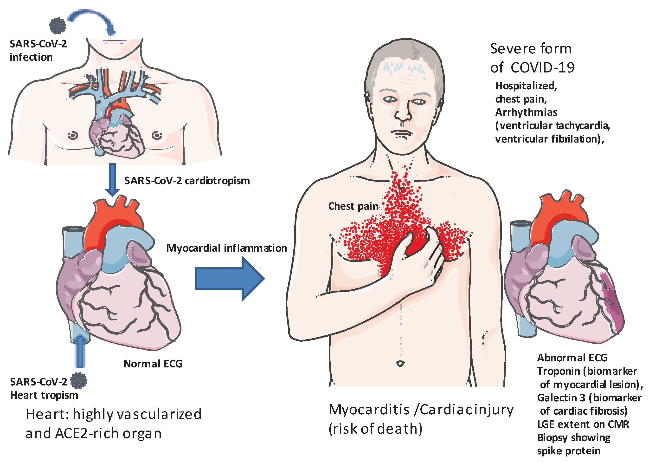
Figure 1.
Schematic representation of myocarditis in patients with COVID-19. Clinical presentation: Most frequently associated with raised troponin blood level, abnormal electrocardiogram (ECG), abnormal echocardiography, late gadolinium enhancement (LGE) involving subepicardial regions, more than 1 abnormality in cardiac magnetic resonance (CMR). Since 2009, a CMR-based diagnosis of myocarditis has been supported by the Lake Louise criteria (LLC), targeting three aspects of myocardial inflammation: oedema, hyperaemia and necrosis and/or fibrosis. Most of the cases of COVID-19-related myocarditis were diagnosed based on CMR findings, with endomyocardial biopsy performed in very few cases because it requires a specific expertise and it is not performed in stable patients. However, endomyocardial biopsy (EMB) remains the gold standard for the diagnosis of myocarditis with identification of SARS-CoV-2 in the myocardium. Beside the identification of myocardial damage, a fundamental step in the diagnostic of myocarditis is the exclusion of obstructive coronary artery disease, especially when the clinical presentation resembles to an acute coronary syndrome. Myocarditis remain uncommon in COVID-19 patients. Since failed human hearts have a higher percentage of ACE2-expressing cardiomyocytes, patients with heart failure are likely to be more susceptible to myocarditis.
Recently, it has been reported that SARS-CoV-2 infects coronary vessels, inducing pro-atherogenic inflammatory responses that could trigger acute cardiovascular complications and increase the long-term cardiovascular risk [22]. With the rapid rollout of COVID-19 vaccinations, numerous associated and suspected adverse events have been reported nationally and worldwide and their occurrence remains unpredictable. Literature reporting confirmed cases of pericarditis and myocarditis following SARS-CoV-2 mRNA vaccinations [21,23,24,25,26,27,28,29,30,31]. For example, from the 27.3 million Pfizer-BioNTech (BNT162b2) doses administered in Australia to 2 January 2022, there have been 415 reports of likely myocarditis and 735 reports of likely pericarditis and that from the 1.8 million Moderna (mRNA-1273) doses administered in Australia to 2 January 2022, there have been 40 reports of likely myocarditis and 52 reports of likely pericarditis (COVID-19 vaccine weekly safety report - 06-01-2022 | Therapeutic Goods Administration (TGA); accessed on 07.01.2022). However, it is unclear how many of these cases are a direct consequence of the vaccine versus coincidental. An elegant single-centre retrospective analysis of all patients presenting to St Vincent's Hospital, Sydney, Australia with suspected COVID-19 vaccine-related myocarditis (9 suspected cases) and pericarditis (97 suspected cases) was recently published [32]. These authors used the Brighton Collaboration Case Definition of Myocarditis and Pericarditis [12] to categorize patients into groups based on diagnostic certainty and cardiac magnetic resonance imaging findings were reviewed against updated Lake Louise Criteria (LLC, which target three aspects of myocardial inflammation: edema, hyperemia, and necrosis and/or fibrosis) [8,10] for diagnosing patients with suspected myocarditis (Table 1). They confirmed 10 cases of possible or probable myocarditis and pericarditis of which 80% had electrocardiogram abnormalities and one patient had multisystem inflammatory syndrome following vaccination with severely impaired left ventricular ejection fraction.

Table 1.
Diagnostic criteria for myocarditis.
| Diagnostic criteria for myocarditis | CDC* criteria |
Brighton Collaboration criteria |
|---|---|---|
|
Level 1 (confirmed) Symptoms consistent with myocarditis and at least one of: Abnormal histopathology OR Elevated troponin AND abnormal CMR |
Level 1 (definitive) Symptoms consistent with myocarditis and at least one of: Abnormal histopathology OR Elevated troponin AND abnormal CMR OR Elevated troponin AND abnormal TTE |
|
| Level 2 (probable) | Level 2 (probable) | |
| Symptoms consistent with myocarditis and at least one of: Elevated troponin OR Abnormal ECG OR Abnormal TTE OR Abnormal CMR |
Symptoms consistent with myocarditis and at least one of: Elevated troponin OR CKMB OR Abnormal ECG OR Abnormal TTE |
|
| Level 3 (possible case) | ||
| Symptoms consistent with myocarditis AND Enlarged heart on CXR OR non-specific ECG abnormalities |
||
|
CMR diagnostic criteria for myocarditis |
Diagnostic target Myocardial edema Myocardial injury Hyperemia Myocardial necrosis |
Lake Louise criteria (LLC) T2-weighted imaging, increased Bright signal intensity Increased global early gadolinium enhancement ratio between myocardium and skeletal muscle. At least one focal lesion with non-ischemic regional distribution on late gadolinium enhancement Pericardial effusion; Systolic left ventricular wall motion abnormality |
* Abbreviations: .CDC: Center for Disease Control, CMR: Cardiac magnetic resonance imaging; TTE: transthoracic echocardiogram; ECG: electrocardiogram; CKMB: creatine kinase myocardial band; CXR: chest X-ray; LLC: Lake Louise criteria. If two Lake Louise criteria are positive, CMR is considered indicative of active myocardial inflammation. Parametric mapping with CMR permits the routine spatial visualization and quantification of changes in myocardial composition based on changes in T1, T2, and T2*(star) relaxation times and extracellular volume (ECV). The clinical recommendations for CMR mapping can be found in the publication by Messroghli and colleagues [33].
Another study in Canada, reported that among 19740741 doses of COVID-19 mRNA administered, there were 297 reports of myocarditis or pericarditis [34]. CDC advocates myocarditis and pericarditis screening for patients who develop acute chest pain, shortness of breath, or palpitations, particularly in adolescents and young adults palpitations within 7 days of receiving the COVID-19 mRNA vaccine. Younger children who have myocarditis or pericarditis may have non-specific symptoms such as irritability, vomiting, poor feeding, tachypnea (fast breathing), or lethargy (CDC last reviewed October 10, 2023; https://www.cdc.gov/vaccines/covid-19/clinical-considerations/myocarditis.html; accessed on November 12, 2023).
Despite the potential adverse effects of vaccination, it has been globally recognized that its benefits far outweigh the risks in the management of COVID-19. Most often, the prognosis of the rare cases of post-COVID-19 vaccine myocarditis appears to be favorable, with full recovery in both males and females. Even today, the pathophysiological events which could lead to the increase in frequency of myocarditis after COVID-19 vaccination remain unclear and their occurrence remains unpredictable. However, several recent papers reported evidence suggesting that free spike proteins circulating in the blood of patients at high levels could be associated with the induction of post-vaccinal myocarditis. This opens new perspectives to try to explain the molecular crosstalk underlying this pathological process and to understand why it is a rare undesirable effect.
2.1. Toward the generation of COVID-19 mRNA vaccines
Vaccine manufacturers use a large panel of routine strategies to design their vaccines, including the traditional live attenuated virus (mutant virus), the inactivated virus (inactivated by chemical or physical treatments), with or without adjuvant such as aluminum salts, the use of viral vector vaccines expressing a recombinant envelope protein of the target infectious pathogen, and the production of recombinant envelope proteins or epitope-based synthetic polypeptides delivered by polymers, or liposome nanoparticles [35,36].
Alongside the classic vaccine approaches, another path using next-generation mRNA technology vaccines [36], the "mRNA-based vaccine strategy", was recently made possible and rapidly gained attention, because it theoretically present several advantageous: (1) it is easy to design; (2) it involves short development and production cycles; (3) it presents great flexibility in term of manipulating the coding sequence; (4) it makes it possible to design multiple mRNAs in a single vaccine dose; and, (5) it can be produced at a low cost. The objective of such vaccines was to safely induce immunity limiting the pandemic and reducing the frequency of the severe forms of COVID-19. These apparent advantages of next-generation mRNA technology vaccines lead to an unprecedented rapid development and approval of mRNA-based COVID-19 vaccines resulting in the mass production of full-length SARS-CoV-2 spike protein in the vaccinated subjects [37,38,39]. BNT162b2 by Pfizer-BioNTech and mRNA-1273 by Moderna were planned for use in mass-immunization programs to curb the pandemic.
Since their approval by the FDA in December 2020, several billion doses of this type of vaccine have been injected to humans in the world over the last three years which saw successive and concomitant outbreaks of SARS-CoV-2 variants. According to the WHO, up to 13,505,089,801 vaccine doses had been administered as of September 19, 2023 (WH0 Coronavirus COVID-19 dashboard; accessed on 27 September 2023), a large majority of which were mRNA-based COVID-19 vaccines. This strategy led to Professors Katalin Kariko and Drew Weissman, who developed the technology behind mRNA COVID-19 vaccine, to recently be awarded the 2023 Nobel Prize in Physiology or Medicine. However, several studies have provided insight into rare but clearly documented myocarditis cases, seen with Pfizer/BioNTech’s BNT162b2 and Moderna’s mRNA-1273 vaccines [40,41].
2.2. The Pfizer/BioNTech’s BNT162b2 and Moderna’s mRNA-1273 vaccines and post-vaccine myocarditis
Following mass vaccination using mRNA-based COVID-19 vaccines [38,42], caregivers and epidemiologists observed a small but significant increase in the frequency of acute viral myocarditis and acute coronary syndrome associated with rare but sometimes fatal cardiovascular complications [43,44,45]. In response to the surveillance alerts regarding the COVID-19 vaccines, the WHO vaccine safety committee noted in the mid-2021 that myocarditis and pericarditis following vaccination with mRNA-based COVID-19 vaccines required further investigation of cases.
Numerous studies worldwide reported similar observations regarding the adverse effects of mRNA-based COVID-19 vaccines. These detrimental effects of the vaccine are characterized by the inflammation of the myocardium resulting in myocyte death and tissue necrosis [23,24]. A study of an Israeli cohort of patients who suffer from myocarditis symptoms following administration of the Pfizer/BioNTech BNT162b2 vaccine (n=304 patients; symptoms were mild in 95% of vaccine recipients), found an increased risk of myocarditis in young adult males after injection of the second dose of vaccine [25]. A large scale retrospective Danish study [26] reviewed data from five million residents over the age of twelve, four million of whom had received mRNA-based COVID-19 vaccines (either the Pfizer/BioNTech BNT162b2 or the Moderna mRNA-1273 vaccines), and reported that the incidence of myocarditis after mRNA-based COVID-19 vaccines was of 1.4/100,000 vaccinated individuals. With the Moderna mRNA-1273 vaccine, myocarditis or myopericarditis was highlighted with an overall incidence of 4.2/100,000 (76% male subjects; 86% had received a second dose of vaccine). The risk was substantially higher after the second dose of vaccine. A British study [27] analyzed the incidence of deaths from myocarditis, pericarditis and cardiac arrhythmias following administration of mRNA-based COVID-19 vaccines (Pfizer/BioNTech BNT162b2, n = 16,993,389; Moderna mRNA-1273, n = 1,006,191) and reported an increased risk of myocarditis associated with the first dose of the Pfizer/BioNTech BNT162b2 vaccine and the first and second doses of the Moderna mRNA-1273 vaccine over the 1–28 day post-vaccination period. The incidence of myocarditis was estimated as an extra 0.1/100,000 event per person vaccinated with the Pfizer/BioNTech BNT162b2 and 0.6/100,000 event per person vaccinated with the Moderna mRNA-1273. An increased risk of cardiac arrhythmias was also evidenced after a second dose of the Moderna mRNA-1273 vaccine. A study of a cohort in Hong Kong [46] analyzed 160 patients with myocarditis and 1,533 without, linking health care records to vaccination records. The BNT162b2 mRNA vaccine was reported to be associated with 20 cases of myocarditis, leading the authors to conclude that patients who were vaccinated with the Pfizer/BioNTech BNT162b2 vaccine had a three-fold increased risk of developing myocarditis compared to unvaccinated subjects. This risk was seen predominantly among young males after the second dose of vaccine. The highest incidence of myocarditis after vaccination with mRNA vaccines has occurred within three to four days after the second vaccination in young males.
Most benefit/risk studies regarding COVID-19 vaccine concluded on the clear benefits of COVID-19 mRNA vaccination with respect to myocarditis. A paper published in Nature Reviews Cardiology [28] stated : ""The risk of acute myocarditis associated with COVID-19 mRNA vaccination has garnered intense (social) media attention. However, myocarditis after COVID-19 mRNA vaccination is rare and usually resolves within days or weeks. Moreover, the risks of hospitalization and death associated with COVID-19 are greater than the risk associated with COVID-19 vaccination. Therefore, COVID-19 vaccination should be recommended in adolescents and adults". This paper highlights that in mRNA-based COVID-19 vaccine-associated myocarditis, up to 90% of patients will functionally recover, usually after a chest pain syndrome [28,29].
Currently, the overall incidence of myocarditis after injection of mRNA-based COVID-19 vaccines is estimated to be between 0.3 and five cases per 100,000 people but with significant differences according to age and gender. Nonetheless, despite a low absolute risk, these detrimental effects of mRNA-based COVID-19 vaccines cannot be ignored, particularly as it concerns young populations who have a low risk of death from SARS-CoV-2 infection.
2.3. Shedding light on the manufacture of mRNA-based COVID-19 vaccines
The Pfizer/BioNTech BNT162b2 mRNA vaccine is a lipid nanoparticle (LNP)-encapsulated, nucleoside-modified RNA vaccine (codon-optimized modified spike mRNA), that encodes a SARS-CoV-2 full-length spike protein stabilized in prefusion conformation [42,47].
The production process of the mRNA-based COVID-19 vaccines is quite easy to set up, with a simpler manufacturing process compared to other "more traditional" vaccines, and it theoretically offers several advantages including rapid response and rapid adaptability, enhanced physical stability, and safety. However, this production process involves a number of steps requiring extreme rigor to ensure the presence of intact RNA in the batches [48,49] and an acceptable purity of samples intended for human vaccination.
The manufacturing process of the mRNA-based COVID-19 vaccines includes (Figure 2):
- Isolation of SARS-CoV-2 and the extraction of its RNA genome. Synthesis by reverse transcription (RT) of a double stranded (ds) DNA template of the gene coding for the spike protein. A synthetic DNA sequence encoding the viral spike protein is inserted into a bacterial plasmid (7,824 base pairs for the Pfizer BNT162b2 mRNA vaccine and 6,777 base pairs for the Moderna mRNA 1273 vaccine) that contains a bacterial origin (ori) of replication and a kanamycin resistant (aminoglycoside phosphotransferase Neo/Kan) gene [50]. Notably, the wild type spike sequence (NCBI accession: NC_045512) is found to be 45.3% identical to the BNT162b2 vaccine and the GC content is 37.3% for the wild type and 56.9% for the BNT162b2 vaccine. Eight small ORFs were found to overlap the Pfizer BNT162b2 mRNA vaccines compared to eleven overlapping ORFs in the wild type [51].
- The recombinant bacterial plasmid containing a double stranded DNA copy of the gene coding for the spike protein as well as a DNA-dependent RNA-polymerase promoter and a kanamycin-resistance selection gene are stored at -150°C until use. The, plasmid is then transfected into an Escherichia coli (E. coli) bacteria that has been made competent for DNA uptake (the construct includes missense codons leading to two major changes in the S2 spike protein sequence with K986P and V987P substitution aimed at stabilizing the protein, the proline is a very rigid amino acid forming a bend aimed at improving the stability of the spike protein by preventing the conformational change of the pre-fusion into the post-fusion structure) [52,53].
- E.coli colonies are grown at 37°C for 24 hours on Petri dishes filled with solid medium. During this process the plasmid is transmitted to daughter bacteria of the E.coli colonies when the bacteria divide (bacteria multiply every 20 minutes). To avoid event of plasmid loss its maintenance is enforced by selection with a kanamycin antibiotic added to the growth medium. Bacteria are then grown into flasks filled with medium and then moved into a large fermenter that contains up to 300 liters of a nutrient broth where they are grown for four days. After amplification in bacteria serving as a master cell bank [54], bacteria are chemically broken down and the plasmid DNA is purified from bacterial debris. The products were tested for purity and gene sequence control. Each one liter batch of plasmid DNA is intended to finally produce about one million doses of the vaccine.
- The ring-shaped plasmid is linearized through the action of a restriction enzyme releasing the sequence encoding the synthetic spike. The cell-free in vitro transcription of DNA into RNA is achieved using a T7 RNA polymerase to generate a synthetic mRNA with a 5' cap. The sequence of mRNA is 4100-4300 nucleotides long with a 5'cap [55]. At this stage, the synthetic nucleoside N-methyl-pseudouridine (mψU) is incorporated into the artificial RNA instead of the natural uridine nucleoside to further increase RNA stability, to enhance translation efficiency in host cells, and to remove alternative start codons - avoid overlapping ORFs - and internal ribosome entry sites, thus preventing non-specific recognition by ribosomal complexes [56]. The addition of a 5′ cap structure is a critical part of this production step that has been improved by new technology suitable for large-scale production [57,58]. A poly(A) tail is needed for efficient translation of mRNA vaccines and is also a critical part during manufacture [59].
- In vitro transcription is followed by several steps of mRNA purification, including the removal of DNA and dsRNA, which could lead to an excessive innate immune response by dsRNA sensing [60] and mRNA is filtered and frozen. Analysis of mRNA requires diverse techniques such as RT-qPCR, capillary and gel electrophoresis, high-pressure liquid chromatography (HPLC) and immunoblotting. The Food and Drug Administration (FDA, USA), recommends manufacturers to limit amount of residual DNA in the final product to be below 10 ng/dose and the size of DNA to be below the size of a functional gene [61].
- The thawed mRNA is mixed with water. In a separate process, the oily lipids are mixed with ethanol, and mRNA and lipids (including phospholipids, cholesterol, cationic lipids and polyethylene glycol lipids that are mixed together) are mixed to create lipid nanoparticles [62]. When the lipids come into contact with the mRNA, electric charge pulls them together in a nanosecond. The mRNA is enveloped in several layers of clinically translatable lipid nanoparticles (multilayer liposomes), forming an oily protective vaccine particle. Liposomes or lipid nanoparticles (LNPs) which facilitate the mRNA cytosolic transport, are known to function as adjuvants can also modulate the immune response [63,64]. The newly made vaccine is filtered to remove the ethanol, concentrated and filtered again to remove any impurities, and finally sterilized. Machines inject 0.45 ml of a concentrated vaccine solution into vials, enough for six doses after dilution. The vials are sealed with foil and capped with purple lids and stored at -70°C. After further quality testing, the vials from the same batch are ready to ship.
- Packaged vaccine doses (preserved at low temperature), are shipped and processed for the market. The mRNA-based COVID-19 vaccine is administered by injection into the deltoid muscle leading to capture of mRNA by muscle cells. The lipid nanoparticles protect RNA (a fragile molecule) from RNAse-dependent degradation and facilitate cellular uptake by lipid fusion with lipids of cell membrane. Spike coding RNA is released into target cell cytoplasm.
Figure 2.
Schematic representation of the main steps in the production process of mRNA-based COVID-19 vaccine mRNAs: the Pfizer BNT162b2 mRNA vaccine is a lipid nanoparticle-encapsulated, nucleoside-modified RNA vaccine that encodes a SARS-CoV-2 full-length spike protein stabilized in prefusion conformation (see the main text for details).
This vaccine strategy is expected to limit vaccine diffusion near the site of injection and local lymph nodes and is considered safe. However controversial results about the safety of the mRNA-based COVID-19 vaccine have increasingly been reported by different teams [65,66]. There is a concern that the Pfizer BNT162b2 mRNA vaccines are not totally pure and contain traces of plasmid DNA. Furthermore, it was claimed that the robustness of the DNAse I digestion step does not destroy all DNA compounds and that between 10% and 35% of the nucleic acid contained in the vaccines is actually DNA [67]. More recently, Speicher and colleagues [50] reported that all mRNA-based COVID-19 vaccine "exceed the guidelines for residual DNA set by FDA and WHO of 10 ng/dose by 188-509-fold". It was also assumed that the recombinant DNA found in mRNA vaccines can be introduced into the cells at the time of vaccine administration, favored by lipid nanoparticles, just as with the mRNA itself, leading to possible integration [68], an hypothesis that remains the subject of debate [69]. The mRNA vaccines are analyzed using a range of time-consuming and costly methods. Recently Gunter and colleagues [70] described a streamlined method to analyze mRNA vaccines using long read nanopore sequencing (VAX-seq). According to these authors, VAX-seq can comprehensively measure sequence, length, integrity, nucleoside modifications, and purity of mRNA vaccines. Currently, there is also a concern that the vaccine activates the endogenous LINE-1 reverse transcriptase, leading to possible reverse transcription of the mRNA vaccine into DNA with a risk of integration into the host genome [71]. Recently, deep sequencing experiments have provided evidence of heterogeneity in the purity of the mRNA-based COVID-19 vaccine batches and significant traces of DNA [50].
The European Medicines Agency (EMA) guidelines on the quality of clinical aspects associated with DNA vaccines stipulates that DNA should be no more than 0.033% of the total nucleic acids in vaccine doses (Rolling Review Critical Report: https://factreview.gr/wp-content/uploads/2023/07/Rolling-Review-Report-Quality-COVID-19-mRNA-Vaccine-BioNTech.pdf, page 74).
2.4. Biodistribution and persistence of COVID-19 mRNA vaccines
The biodistribution and duration of persistence of the COVID-19 mRNA vaccines may be important for understanding of the adverse side-effects of the vaccine. After injection into the deltoid muscle, the vaccine spike mRNA nanoparticles are expected to be captured and endocytosed by muscle cells. The cellular endocytosis of the mRNA is expected to take about one minute and the translation into spike protein is expected to take about two minutes (as calculated considering a translation speed of 10 amino acids per second and a protein made up of 1273 amino acids) [72,73]. This newly synthesized vaccine spike protein is expected to be delivered at the cell surface in a prefusion conformation, as previously reported by Wrapp and colleagues [47], and to activate an anti-spike immune response including the induction of neutralizing antibodies [74,75,76]. The spike mRNA nanoparticles can also be picked up by the immune cells in the proximal lymph node. Using a cellular model, Fertig and colleagues [77] observed that the endolysosomal compartments of most vaccine-treated cells were enriched with electron dense, multilayered lipid structures as compared to controls, suggesting the successful endocytosis of LNPs, followed by the endosomal-mediated disintegration of LNPs, translation into spike protein and synthesis of spike-like structures clustering on isolated protrusions of the plasma membrane of some cells after 12 hours incubation with the Pfizer/BioNTech BNT162b2 vaccine (10 µg mRNA per 1 × 10⁶ cells). The biodistribution and persistence of mRNA vaccines after intramuscular injection, has been studied in rodents and non-human primates.
These studies indicated that intramuscular injection leads to an initial accumulation of the vaccine spike protein at the injection site within hours of the vaccine injection [77,78]. LNPs are then rapidly transported to proximal lymph nodes by passive draining as well as actively carried by antigen-presenting cells and neutrophils, while the remaining unprocessed LNPs reach systemic circulation [77,79,80]. A study by Krauson and colleagues [81] found that COVID-19 mRNA vaccines elicit antigen-specific germinal center B cell responses only in draining lymph nodes but in mediastinal lymph nodes Using a specific RT-qPCR based assays to detect COVID-19 mRNA vaccine these authors found the presence of spike mRNA in the axillary lymph nodes in the majority of patients who died within 30 days of vaccination. Vaccine was also detected in the myocardium in a subset of patients within 30 days of death. It was reported that a single intramuscular immunization with the Pfizer/BioNTech BNT162b2 vaccine (containing the branched-tail ionizable lipid ALC-0315 that includes a tertiary amine, branched tails and ester linkers biodegradable structure) can activate dendritic cells, monocytes and macrophages in the draining lymph nodes to produce interferons [82]. Another ionizable lipid SM102 (sharing structural features with ALC-0315) used in the Moderna mRNA-1273 vaccine LNPs was found to activate IL-1 Ra cytokine production [83]. It has also been found that exosomes with a spike protein on their surface are induced by the Pfizer/BioNTech BNT162b2 vaccine and circulate prior to the development of specific antibodies. Circulating exosomes expressing spike proteins were found on day 14 after vaccination, followed by anti-spike antibodies being detected 14 days after the second dose [84,85].The beneficial effect of vaccination is usually determined by monitoring the specific anti-spike antibody responses [86], although this represents only part of the immune response against the virus. In a murine animal model, the intramuscular administration of LNPs containing ionizable lipid and mRNA encoding the Spike of SARS-CoV-2, the titres of antibodies against SARS-CoV-2 was increased tenfold with respect to the vaccine encoding for the unadjuvanted antigen [87].
The mRNA-based COVID-19 vaccines, consist of nonreplicating mRNA and are expected to be naturally degraded after translation through intracellular RNAse activities within the cytosol. They should therefore be rapidly eliminated from the injection site. The half-life of this foreign mRNA in the vaccinated subject is unknown, theoretically from a few hours to ten days based on general data for cellular mRNA [88,89], but vaccine-associated synthetic mRNA has been reported to persist in systemic circulation for at least two weeks [77] and full-length or fragments of SARS-CoV-2 spike mRNA vaccine sequences have been found in blood up to four weeks after COVID-19 vaccination [90]. In another study, both the mRNA vaccine and spike protein were found in germinal center in a lymph node up to eight weeks post-injection in some cases [91]. Thus, in contrast to disrupted germinal centers in lymph nodes observed during SARS-CoV-2 infection, the Pfizer/BioNTech BNT162b2 vaccine stimulates robust germinal centers containing vaccine mRNA and vaccine spike antigen.
In a study by Ogata and colleagues [92], 11 of 13 participants exhibited spike antigen in their plasma after a first injection of the Moderna mRNA 1273 COVID-19 vaccine, whereas nucleocapsid concentrations were insignificant in all participants, indicating that the detected spike originated from the vaccination and not from a natural infection. This study suggested that the spike protein leaves the site of the COVID-19 vaccine injection and enters the bloodstream, accumulating in other parts of the body (Figure 3).
Figure 3.
Hypothetical model accounting for the possible route taken by vaccine-derived compounds (mRNA coding for the viral spike and/or spike protein) which can be found in the blood circulation and the heart after injection of mRNA-based COVID-19 vaccine in the upper arm (deltoid muscle). Men aged between 18 and 24 are at higher risk of developing myocarditis following mRNA-based COVID-19 vaccination. If undetected, there is a risk of sudden death. When detected, exercises must be prohibited.
This was confirmed by Brogna and colleagues [93] using mass spectrometry examination of biological samples. In vaccinated subjects (n = 20), these authors evidenced the presence of the spike protein translated from the mRNA vaccine (distinguishable from the wild-type protein due to specific amino acid variations introduced to maintain the protein in a prefusion state) in 50% of vaccinated individuals while this was not the case in unvaccinated individual (n = 20).
2.5. Previous hypothesis proposed in an attempt to explain the increased risk of post mRNA-based COVID-19 vaccines myocarditis
It has been hypothesized that the rare adverse side effects reported in young subjects after injection of mRNA-based COVID-19 vaccines (e.g., myocarditis and multisystem inflammatory syndrome), could be the unintended consequence of an inappropriate use of the vaccine such as at the time when the antiviral immune response is already highly stressed during a low-noise infection in the process of healing, leading to exacerbated cytotoxic T cell response killing cells and triggering tissue damage [94,95,96,97,98].The potential implication of lipid nanoparticles present in the vaccine and epigenetic modifications which occur with inoculations with vaccines, have also been suggested as being possibly involved in the pathogenesis of myocarditis associated with mRNA-based COVID-19 vaccines [63,64,99,100,101]. Myocarditis was also suggested to be associated with a specific immune genetic background such as genetic variants in genes encoding HLA, desmosomal, cyctoskeletal or sarcomeric proteins [102]. Stress response, hypertensive response, and/or alcohol consumption known to affect the expression of angiotensin-converting enzyme 2 (ACE2) have also been suggested as being responsible for the adverse effects of mRNA-based COVID-19 vaccines [103].
A French retrospective analysis reported that among 21,909 subjects who had received at least one dose of the Pfizer/BioNTech BNT162b2 COVID-19 vaccine, 8,121 people (37.1%) exhibited high blood pressure after the first injection [104]. Another report investigating post-injection adverse effects of mRNA-based COVID-19 vaccines among a population of 1870 subjects who had received one or two doses of COVID-19 vaccine indicated that 153 subjects (8%) showed an increase in blood pressure values after vaccination, with higher frequency of blood pressure alterations at the second or booster dose [105]. Similarly, the prevalence of abnormalities or increases in blood pressure in a meta-analysis including 357,387 subjects was 3.2% and that of stage III hypertension was 0.6%, mainly in elderly and frail people [106].
Gundry [107] who used the PULS Cardiac Test (Predictive Health Diagnostics Co., Irvine, CA) a clinically utilized measurement of multiple protein biomarkers including IL-16 (proinflammatory cytokine), soluble FAS (inducer of apoptosis) and HGF (T cells chemotaxis) to monitor the increased in endothelial tissues inflammation among a cohort of 566 patients who received mRNA-based COVID-19 vaccine. Dramatic changes in the PULS score (from 11% to 25%) were observed with patients immunized using the Pfizer/BioNTech BNT162b2 and Moderna mRNA 1273 COVID-19 vaccines, and these changes persisted for at least two-and-a-half months after the second dose of vaccine. The author used the results to calculate a five-year risk score (percentage of risk) for new acute coronary syndrome and concluded that the administration of mRNA-based COVID-19 vaccines was associated with an increase in endothelial inflammatory processes, thrombosis and acute coronary syndrome risk. Attention has also been drawn to cases of multisystem inflammatory syndrome that have been reported after administration to children of doses of mRNA-based COVID-19 vaccines [40,108]. This pathophysiological process was linked to an abnormal activation of the immune response. The clinical symptoms affecting these children were found to be associated with elevated anti-AT1R, anti-endothelin receptor, anti-α1 adrenergic receptor, anti-β1 adrenergic receptor, anti-β2 adrenergic receptor and anti-muscarinic cholinergic receptor-2/3/4 autoantibodies [109]. Recently, myocarditis following mRNA-based COVID-19 vaccination has been linked to autoantibodies against endogenous interleukin-1 receptor antagonist (IL-1RA) the function of which is to inhibit interleukin-1 signaling and inflammation [110]. Since development of ACE2 autoantibodies after SARS-CoV-2 infection was described [111,112], it can be speculated that the synthetic spike could also induce this type of autoantibodies. However, a recent study found that ACE2 IgG levels in COVID-19 patients are too low to impair the regulatory activity of ACE2 [113]. It remains to be verified that the vaccination does not induce more ACE2 autoantibodies than SARS-CoV-2 infection.
2.6. Possible role of circulating spike protein in post-mRNA-based COVID-19 vaccines associated myocarditis
Soon after the discovery of the first cases of COVID-19 and the characterization of the SARS-CoV-2, it was demonstrated that the virus receptor was the ACE2 molecule [114,115,116]. A critical step in the SARS-CoV-2 infection cycle is the binding of the homotrimeric viral spike protein to the peptidase domain of ACE2 [117,118,119]. Once bound to ACE2, SARS-CoV-2 down-regulates the cellular expression of the ACE2 gene and ACE2 protein and the unopposed action of Ang II was deemed responsible for worsening the outcome of COVID-19 [120]. In vitro, SARS-CoV-2 decreases ACE2 methylation and ACE2 lysine 31 hypermethylation decreases binding to SARS-CoV-2 spike protein [121,122].
From the start of the pandemic, we and others warned against considering ACE2 as a simple receptor, knowing the major role this molecule plays in the RAS pathway [20,123,124,125]. More precisely, we hypothesized that by attaching to ACE2 the virus was likely to influence the balance regulating the production of angiotensin II, thus modulating blood pressure, coagulation and inflammatory processes [20,126,127,128]. We were subsequently able to demonstrate that the presence of the virus in patients can induce an overproduction of angiotensin II [129]. Similar results regarding increased angiotensin II were reported by others [130,131]. On the molecular level, we postulate that the pathophysiological dysfunction observed in COVID-19 mainly involves the Angiotensin II/ transmembrane receptor angiotensin II type I receptor (AT1R)/Hypoxia-inducible factor-1 (HIF-1) axis [132]. As this physiopathological process initiated by viral infection is dependent on the interaction between the spike of the virus and ACE2, it was very likely that the spike expressed as part of vaccine could induce the same adverse effects as the virus itself. We have recently reviewed this issue [133]. The fact that symptomatic hypertension (malaise, headache, tingling in the mouth, diaphoresis and increased blood pressure) subsequent to vaccination with the Pfizer/BioNTech BNT162b2 was reported [134,135] is not surprising and confirmed our hypothesis.
Using a mouse model, Rhea and colleagues [136] had previously shown that intravenous injection of the radioiodinated S1 (I-S1) spike protein can cross the blood–brain barrier in male mice, and was also taken up by several organs. Thus when a huge quantity of mRNA encapsulated in LNPs is injected, LNPs, exosomes expressing the spike and free spike, can pass into the lymphatic capillaries then axillary/supraclavicular lymph nodes and it is possible that these compounds can enter the venous bloodstream. Thus, these components are likely to diffuse into different organs and therefore to cross the heart many times. Besides, LNPs are likely to fuse with cardiomyocytes where spike mRNA can be translated into spike proteins.
Of course, it appeared quite logical to think that the interaction between the circulating vaccine spike and ACE2 expressed on the endothelium could be at the origin of vaccine-induced myocarditis. However, previously there was a lack of experimental evidence to support this hypothesis. According to a work recently published by Yonker and colleagues [137], all post-vaccine myocarditis subjects (n = 16) and age-matched vaccinated controls (n=45) showed similar rise in anti-spike antibodies and anti-spike T-cell response after vaccination (with either the Pfizer/BioNTech BNT162b2 or Moderna mRNA 1273 COVID-19 vaccine). However, one major significant difference between both groups was the high level of circulating full-length spike protein (33.9±22.4 pg/mL), in the plasma of myocarditis patients in association with slight elevations in cardiac troponin T, C-reactive protein and cytokine production [137,138,139]. This is the first evidence of a correlation between myocarditis and the presence of circulating spikes that remains detectable for up to three weeks after vaccination, suggesting that the spike protein translated from the mRNA vaccine may be the causal agent of myocarditis. This is consistent with the work by Krauson and colleagues [81] who detected spike mRNA vaccine in the myocardium of vaccinated patients who died within the 4 weeks after vaccination. The cardiac ventricles of the deceased people in which vaccine was detected had healing myocardial injury and had more myocardial macrophages than controls in which vaccine was not detected.
3. Conclusion
The elegant study by Yonker and colleagues [137] bridges the gap in the model that we proposed of a direct effect of the vaccine spike on ACE2 inducing a process of microthrombosis and inflammation that can lead to myocarditis. When a large amount of free spike protein circulates in the bloodstream, it may damage the cardiac pericytes or endothelium by acting on ACE2, reducing nitric oxide production and activating the production of inflammatory molecules in some ways similar to that which has been observed with SARS-CoV-2 infection which has been shown to trigger acute myocardial infarction [140,141,142]. It can be emphasized that a Danish study of 5119 patients diagnosed with COVID-19 estimated that the incidence rate of acute myocardial infarction was five times higher during the 14 days after COVID-19 diagnosis [143]. Alternatively, it could act by potential molecular mimicry between spike protein and endogenous antigens that elicit cardiac targeted autoantibodies [144,145]. For the model to be credible, it must be admitted that the circulating rate of mRNA nanoparticles and/or spike will vary depending on the individual who receive the vaccine (to the extent that only a small proportion of individuals who receive the vaccine experience these adverse effects) which then suggests that the dose of vaccine will contain variable quantities of intact mRNA, depending on the batch available to clinical staff.
The mRNA integrity directly impacts the effectiveness of the mRNA-based vaccines. The mRNA transcription can be abortive (smaller fragments), mRNA can possibly be fragmented by RNAses or hydrolyzed, spike species can derive from cryptic mRNA transcription start sites. It could be considered that the quality of the vaccine could vary depending on the more or less perfect method of storage of vaccine vials (e.g. if the number of -70°C freezers and storage spaces were limited) in the facility managing the vaccination process. Obviously, if this is the case, then the abnormal dose delivery of mRNA is simply a technical problem that might be solved by a better production control by the manufacturer. The main source of concern is the prolonged existence of mRNA that evades destruction, the mechanism of which is debated [70,138]. As above mentioned, foreign mRNA in the vaccinated subject was predicted to persist in an active form during a period ranging from a few hours to ten days [88,89], but vaccine-associated synthetic mRNA was reported to persist in systemic circulation for at least two to four weeks [77,81] and full-length SARS-CoV-2 spike mRNA vaccine sequences were found in the blood up to four to eight weeks post-injection in some cases [81,90,91]. It was recently reported that both the Pfizer/BioNTech BNT162b2 and Moderna mRNA 1273 COVID-19 vaccines induce specific dysfunction of cardiomyocytes after 48h of spike expression, and that both the cardiac ryanodine receptor (RyR2) impairment and sustained protein kinase A (PKA) activation may likely increase the risk of acute cardiac events [146]. It has also been claimed that COVID-19 mRNA vaccines contain excessive amounts of residual bacterial DNA, including spike encoding DNA sequences, packaged in the LNPs and thus resistant to DNase I and that billions to hundreds of billions of DNA molecules can be present per dose of vaccine [50]. DNA contamination may be prothrombotic, particularly for fragments with high GC contents [147,148]. It has also been suggested that getting the plasmids out of the E. coli, may result in residual bacterial endotoxin, in the vaccines. The debate has not yet been settled on the purity of samples during the manufacturing process of mRNA COVID-19 vaccines.
Another question recently addressed by Bozkurt [138] is "why circulating spike protein levels remained elevated despite adequate levels and functionality of anti-spike antibodies ?". This author suggested hypothetical explanations such as vaccine overdose, the possible role of anti-idiotype, and prolonged existence of mRNA. Recently, Japanese radiologists published a study [149] on myocardial 18Fluorine-fluorodeoxyglucose (18F-FDG) uptake on images in 700 asymptomatic vaccinated subjects (The majority of the vaccinated individuals,77.6%, received the Pfizer/BioNTech BNT162b2 vaccine followed by 21% who received the Moderna mRNA-1273 mRNA vaccine. The 700 subjects underwent positron emission tomography (PET)/computed tomography (CT) within a period of 1–180 days after their second vaccination and increased 18F-FDG uptake (a marker of myocardial inflammation of diverse origin including viral myocarditis) was found compared with the unvaccinated group (n = 303). However, this increase was not seen in subjects who underwent imaging more than 180 days after vaccination. These results are corroborated by the data reported in the Krauson's studies [81]. Krauson and colleagues detected spike mRNA vaccine in the axillary lymph nodes and myocardium from recently vaccinated deceased patients. Vaccine was detected in the majority of patients dying within 4 weeks of vaccination, but not in patients dying more than 4 weeks from vaccination.(Figure 4)

Figure 4.
Schematic mechanism of action of the COVID-19 mRNA vaccine associated myocarditis. The spike mRNA vaccine was detected in the myocardium from recently vaccinated deceased patients [81]. The LNPs packaging of the COVID-19 mRNA vaccine allows nucleoside-modified RNA vaccine to be stably delivered and enter cardiomyocytes after LNPs had entered the general bloodstream. Once inside a cardiomyocyte, this mRNA forms a complex with initiation factors and the small subunit of the ribosome, where elongation of the spike polypeptide chain starts. The S proteins first assemble to form homotrimers into the cytoplasm and then migrate to the cell surface to protrude with a native-like conformation. Cardiomyocytes expressing the spike protein can possibly be targets for a specific cytotoxic T cells anti-spike immune response and destroyed.
Whatever the conclusions of this important debate, we now know with certainty that the vaccine spike circulates longer than initially assumed, and that it is directly associated with the increased frequency of myocarditis in young men. Recently Barmada and colleagues [150] investigated a cohort of patients who developed myocarditis and/or pericarditis with elevated troponin and C-reactive protein levels. The patients showed cardiac imaging abnormalities shortly after COVID-19 mRNA vaccination, late gadolinium enhancement, and elevations in circulating interleukins (IL-1 , IL-1RA, and IL-15), chemokines (CCL4, CXCL1, and CXCL10) and matrix metalloproteases (MMP1, MMP8, MMP9 and TIMP1). Their immune responses analysis indicated expansion of activated cytotoxic T cells, NK cells in the heart and inflammatory and profibrotic monocytes. The precise molecular mechanisms by which the COVID-19 mRNA vaccines lead to myocardial injury and myocarditis remain to be characterized but we can reasonably hypothesize that it is a process which passes through the soluble angiotensin II binding to the transmembrane receptor angiotensin II type I receptor (AT1R; expressed in the arterioles and several organs including heart), and the intracellular factor hypoxia-inducible factor 1 alpha (HIF-1α). (Figure 5)
Figure 5.
Potential interaction of COVID-19 mRNA vaccine/spike with the renin-angiotensin system (RAS). A free floating spike can be released from cells expressing the COVID-19 mRNA vaccine, leading to a massive interaction with the cell surface ACE2 receptor. As a result, an increased Ang II level is likely to lead to an overactivation of AT1R signaling which initiates activation of PKC and c-Src that is required for superoxide production by NADPH oxidases (NOX1 and NOX2). NOX2 also stimulates the production of reactive oxygen species (ROS) by mitochondria. The consequence is an inhibition of the hypoxia inducible factor 1 α (HIF-1α) hydroxylation by the prolyl hydroxylase (PHD) pathway and of its polyubiquitinylation by the ubiquitin ligase usually leading to HIF-1 proteosomal degradation (a mechanism contributing to cell homeostasis). After ROS production, HIF-1α translocates to the cell nucleus where it forms heterodimers with the HIF- subunit and binds to the HRE element in the promoter of hypoxia-inducible genes and recruits histone acetyltransferases CREB Binding Protein (CBP)/p300. Thus, it up-regulates ACE1 which contributes to Ang II production and abnormal functioning of the RAS pathway. HIF-1α nuclear translocation also activates the transient receptor potential ankyrin 1 (TRPA1) gene expression which leads to increase in intracellular Ca2+ and cell injury as well as expression of several genes involved in the control of cell viability and proliferation among others [132]. In parallel, Ang II triggers an increase in cytoplasmic Ca2+ that induces NOX5 to generate H2O2. HIF also contributes to the down regulation of the ACE2 gene and activation of ADAM17 which leads to cleavage of the ACE2 protein and the release of soluble ACE2 (sACE2). AT1R signaling activates a number of signaling pathways, such as G protein–mediated (Gq and Gi), Janus kinase/signal transducers and activators of transcription, extracellular signal regulated kinase (ERK), NF-κB, NLRP3 procaspase 1 pathways leading to induction of pro-inflammatory cytokines.
The prevalence of post-vaccinal myocarditis in young men could be associated with the level of expression of ACE2 which varies according to sex and age [151]. A negative association was reported between age and soluble ACE2 plasma concentrations in people above the age of 55 [152]. It is also known that polymorphisms in ACE2 gene 5′ upstream regions might influence ACE2 expression. Differences of more than 1% in minor allele frequency in the 10Kb region upstream to ACE2 analyzed using data from the 1,000 Genomes project, found 57 polymorphisms [20,153] which could explain why not all young men are affected by vaccination in the same way. In addition, there is a known polymorphism of ACE2 and dozen of human ACE2 variants have been characterized, which could impact the affinity for the viral spike or could modify the ACE2 protein stability [154,155,156,157]. These individual variations may be highly important in the outcome of vaccination, since the expression of ACE2 in the heart is known to be higher than in the lungs. ACE2 is found in the endothelial cells of coronary arteries, arterioles, venules, and capillaries as well as vascular smooth muscle cells, and is strongly expressed by cardiac fibroblasts, cardiomyocytes, and cardiac pericytes [158,159].
Myocarditis has been recognized as a complication of COVID-19 mRNA vaccination and remains as issues of concern. The medical management of COVID-19 mRNA vaccine-associated myocarditis mainly relies on corticosteroids to challenge the progression of non-specific immune system activation [6,160]. Beta-blockers are often employed in treatment of acute myocarditis, even in uncomplicated disease, presumably by virtue of the perceived protection they provide against arrhythmic events [7]. In a recent study by Yu and colleagues [161], a subset of adolescent patients diagnosed with COVID-19 mRNA vaccine– associated myocarditis were found to show impairment of left ventricular and right ventricular myocardial deformation and persistence of late gadolinium enhancement with up to 1 year of follow-up while global systolic ventricular function appeared to be preserved. As we move forward into the next long-term phase of the pandemic with booster vaccinations [162], routine monitoring of angiotensin II and spike antigenemia following mRNA-based COVID-19 vaccines and/or patients with myocarditis should be performed. If spike antigenemia is detected, administration of recombinant soluble ACE2 (hrsACE2) or anti-spike antibodies [130,163,164,165,166,167] could potentially be required to prevent or reverse post-vaccinal myocarditis.
Author Contributions
All authors contributed to the design of the review. CAD wrote the manuscript and all authors agreed with the manuscript submitted for publication.
Funding
This research was funded by the French Government under the " Investissements d’avenir " (Investments for the Future) programme managed by the Agence Nationale de la Recherche (French ANR: National Agency for Research), (reference: Méditerranée Infection 10-IAHU-03), and annual funding from Aix-Marseille university and IRD to the MEPHI research unit.
Institutional Review Board Statement
The study did not require ethical approval.
Informed Consent Statement
Not applicable.
Data Availability Statement
Data supporting reported results can be found in the list of references of this paper.
Acknowledgments
Figures were designed using the Servier Medical Art supply of images available under a Creative Commons CC BY 3.0 license.
Conflicts of Interest
CAD declares a link of interest with the Sanofi and Merck pharmaceutical companies. The other authors declare that the research was conducted in the absence of any commercial or financial relationships that could be construed as a potential conflict of interest.
References
- Zhou P, Yang XL, Wang XG, Hu B, Zhang L, Zhang W, Si HR, Zhu Y, Li B, Huang C, et al. A Pneumonia Outbreak Associated With a New Coronavirus of Probable Bat Origin. Nature (2020) 579: 270–273. [CrossRef]
- Zhu N, Zhang D, Wang W, Li X, Yang B, Song J, Zhao X, Huang B, Shi W, Lu R, et al. A Novel Coronavirus From Patients With Pneumonia in China, 2019. N. Engl. J. Med. (2020) 382:727–733. [CrossRef]
- Huang C, Wang Y, Li X, Ren L, Zhao J, Hu Y, Zhang L, Fan G, Xu J, Gu X, et al. Clinical Features of Patients Infected With 2019 Novel Coronavirus in Wuhan, China. Lancet (2020) 395(10223): 497–506. [CrossRef]
- Devaux CA, Lagier JC. Unraveling the Underlying Molecular Mechanism of ‘Silent Hypoxia’ in COVID-19 Patients Suggests a Central Role for Angiotensin II Modulation of the AT1R-Hypoxia-Inducible Factor Signaling Pathway. J. Clin. Med. (2023) 12: 2445. [CrossRef]
- Han L, Zhao S, Li S, Gu S, Deng X, Yang L, Ran J. Excess cardiovascular mortality across multiple COVID-19 waves in the United States from March 2020 to March 2022. Nature Cardiovasc. Res. (2023) 2: 322-333. [CrossRef]
- Pollack A, Kontorovich AR, Fuster V, Dec GW. Viral myocarditis—diagnosis, treatment options, and current controversies. Nature Rev. Cardiol. (2015)12: 670–680. [CrossRef]
- Sozzi FB, Gherbesi E, Faggiano A, Gnan E, Maruccio A, Schiavonne M, Lacuzio L, Carugo S. Viral Myocarditis: Classification, Diagnosis, and Clinical Implications. Front. Cardiovasc. Med. (2022) 9: 908663. [CrossRef]
- Friedrich MG, Sechtem U, Schulz-Menger J, Holmvang G, Alakija P, Cooper LT, White JA, Abdel-Aty H, Gutberlet M, Prasad S, et al. Cardiovascular magnetic resonance in myocarditis: a JACC White paper. J. Am. Coll. Cardiol. (2009) 53(17): 1475-1487. [CrossRef]
- Caforio AL, Pankuweit S, Arbustini E, Basso C, Gimeno-Blanes J, Felix SB, Fu M, Helio T, Heymans S, Jahns R. et al. Current state of knowledge on aetiology, diagnosis, management, and therapy of myocarditis: a position statement of the European Society of Cardiology Working Group on myocardial and pericardial diseases. Eur. Heart J. (2013) 34(33): 2636-2648. [CrossRef]
- Luetkens JA, Faron A, Isaak A, Dabir D, Kuetting D, Feisst A, Schmeel FC, Sprinkart AMThomas D. Comparison of Original and 2018 Lake Louise Criteria for diagnosis of acute myocarditis: results of a validation cohort. Radiol. Cardiothor. Imag. (2019) 1(3): e190010. [CrossRef]
- Kociol RD, Cooper LT, Fang JC, Moslehi JJ, Pang PS, Sabe MA, Shah RV, Sims DB, Thiene G, Vardeny O. Recognition and initial management of fulminant myocarditis: a scientific statement from the American heart association. Circulation (2020) 141(6): e69-92. [CrossRef]
- Marshall TR, Schrader S, Voss L, Buttery JP, Crawford NW, Cheng DR. A comparison of post-COVID vaccine myocarditis classification using the Brighton Collaboration criteria versus Centre for Dieasise Control criteria. Australian Gov. Dept Health and Aged Care, Com. Dis. Intell. (2023) 47: electronic publication 19/01/2023. [CrossRef]
- Hamadeh A, Aldujeli A, Briedis K, Tecson KM, Sanz-Sanchez J, Al Dujeili M, Al-Obeidi A, Diez JL, Zaliunas R, Stoler R, McCullough PA. Characteristics and Outcomes in Patients Presenting With COVID-19 and ST-Segment Elevation Myocardial Infarction. Am. J. Cardiol. (2020) 131: 1-6. [CrossRef]
- Mele D, Flamigni F, Rapezzi C, Ferrari R. Myocarditis in COVID-19 patients: current problems. Intern. Emerg. Med. (2021) 16: 1123–1129. [CrossRef]
- Xie Y, Xu E, Bowe B, Al-Aly Z. Long-term cardiovascular outcomes of COVID-19. Nature Med. (2022) 28: 583-590. [CrossRef]
- Castiello T, Georgiopoulos G, Finocchiaro G, Claudia M, Gianatti A, Delialis D, Aimo A, Prasad S. COVID-19 and myocarditis: a systematic review and overview of current challenges. Heart Failure Rev. (2022) 27: 251–261. [CrossRef]
- Kornowski R, Witberg G. Acute myocarditis caused by COVID-19 disease and following COVID-19 vaccination. Openheart (2022) 9: e001957. [CrossRef]
- Pillay J, Gaudet L, Wingert A, Bialy L, Mackie AS, Paterson DI, Hartling L. Incidence, risk factors, natural history, and hypothesised mechanisms of myocarditis and pericarditis following covid-19 vaccination: living evidence syntheses and review. Brit. Med. J. (2022) 378: e069445. [CrossRef]
- Heidecker B, Dagan N, Balicer R, Eriksson U, Rosano G, Coats A, Tschöpe C, Kelle S, Poland GA, Frustaci A, et al. Myocarditis following COVID-19 vaccine: incidence, presentation, diagnosis, pathophysiology, therapy, and outcomes put into perspective. A clinical consensus document supported by the Heart Failure Association of the European Society of Cardiology (ESC) and the ESC Working Group on Myocardial and Pericardial Diseases. Eur. J. Heart Fail. (2022) 24(11): 10.1002/ejhf.2669. [CrossRef]
- Devaux CA, Camoin-Jau L An update on angiotensin-converting enzyme 2 structure/functions, polymorphism, and duplicitous nature in the pathophysiology of coronavirus disease 2019: Implications for vascular and coagulation disease associated with severe acute respiratory syndrome coronavirus infection. Front. Microbiol. (2022) 13: 1042200. [CrossRef]
- Katoto PDMC, Byamungu LN, Brand AS, Tamuzi JL, Kakubu MAM, Wiysonge CS, Gray G. Systematic review and meta-analysis of myocarditis and pericarditis in adolescents following COVID-19 BNT162b2 vaccination. npj Vaccines (2023) 8:89. [CrossRef]
- Eberhardt N, Noval MG, Kaur R, Amadori L, Gildea M, Sajja S, Amadori L, Das D, Cihoroz B, Stewart OJ, et al. SARS-CoV-2 infection triggers pro-atherogenic inflammatory responses in human coronary vessels. Nature Cardiovasc. Res. (2023) published online 28 September 2023. [CrossRef]
- Oleszak F, Maryniak A., Botti E, Abrahim C, Salifu MO,Youssef M, Henglein VL, McFariane SJ. Myocarditis associated with COVID-19. Am. J. Med. Case Rep. (2020) 8: 498–502. [CrossRef]
- Wu S, Zou G, Lin K, Zhang D. Effects of COVID-19 on the cardiovascular system and implications for management. J. Xiangya Med. (2021) 6: 7. [CrossRef]
- Mevorach D, Anis E, Cedar N, Bromberg M, Haas EJ, Nadir E, Olsha-Castell S, Arad D, Hasin T, Levi N, et al. Myocarditis after BNT162b2 mRNA Vaccine against COVID-19 in Israel. N. Engl. J. Med. (2021) 385: 2140–2149. [CrossRef]
- Husby A, Vinslov Hansen J, Fosbol E, Myrup Thiesson E, Madsen M, Thomsen RW, Sorensen HT, Andersen M, Wohlfahrt J, Gislason G, et al. SARS-CoV-2 vaccination and myocarditis or myopericarditis: Population based cohort study. Brit. Med. J. (2021) 375: e068665. [CrossRef]
- Patone, M. Patone M., Mei XW, Handunnetthi L, Dixon S, Zaccardi F, Shankar-Hari M, Watkinson P, Khunti K, Harnden A, Coupland CAC, et al. Risks of myocarditis, pericarditis, and cardiac arrhythmias associated with COVID-19 vaccination or SARS-CoV-2 infection. Nature Med. (2022) 28(2), 410–422. [CrossRef]
- Heymans S, Cooper LT. Myocarditis after COVID-19 mRNA vaccination: clinical observations and potential mechanisms. Nature Rev. Cardiol. (2022) 19: 75-77. [CrossRef]
- Heymans S, Cooper LT. Author Correction: Myocarditis after COVID-19 mRNA vaccination: clinical observations and potential mechanisms. Nature Rev. Cardiol. (2023) 20: 575. [CrossRef]
- Pastor Pueyo P, Gambo-Ruberte E, Gayan Ordas J, Blanco LM, Figal DP, Larranaga Moreira JM, Gomez Barrado JJ, Gonzalez Clla D, Almenar Bonet L, Corbi Pascual MJ, et al. Vaccine–carditis study: Spanish multicenter registry of inflammatory heart disease after COVID-19 vaccination. Clin. Res. Cardiol. (2023) Jun 27. [CrossRef]
- Jiang J, Chan L, Kauffman J, Narula J, Charney AW, Oh W, Nadkami G, N3C Consortium. Impact of vaccination on major adverse cardiovascular events in patients with COVID-19 infection. J. Am. Coll. Cardiol. (2023) 81(9): 928-930. [CrossRef]
- Wassif M, Lo P, Satouris P, Swan L, Tardo D, Kovacic JC, Muller D, Muthiah K, Kotlyar E, Bart NK. Acute Myocarditis and Pericarditis after mRNA COVID-19 vaccinations - A single-centre retrospective analysis. Heart Lung Circul. (2023) 32: 467-479. [CrossRef]
- Messroghli DR, Moon JC, Ferreira VM, Grosse-Wortmann L, He T, Kellman P, Mascherbauer J, Nezafat R, Salerno M, Schelbert EB, et al. Clinical recommendations for cardiovascular magnetic resonance mapping of T1, T2, T2* and extracellular volume: A consensus statement by the Society for Cardiovascular Magnetic Resonance (SCMR) endorsed by the European Association for Cardiovascular Imaging (EACVI). J. Cardiovasc. Magn. Reson. (2017) 19: 75. [CrossRef]
- Buchan SA, Seo CY, Johnson C, Alley S, Kwong JC, Nasrren S, Calzavara A, Lu D, Harris TM, Yu K, Wilson SE. Epidemiology of Myocarditis and Pericarditis Following mRNA Vaccination by Vaccine Product, Schedule, and Interdose Interval Among Adolescents and Adults in Ontario, Canada. JAMA Network Open. (2022) 5(6):e2218505. [CrossRef]
- Bramwell VW, Perrie Y. The rational design of vaccines. Drug Discov. Today (2005) 10: 1527–1534. [CrossRef]
- Schijns V, Majhen D, van der Ley P, Thakur A, Summerfield A, Berisio R, Nativi C, Fernandez-Tejada A, Alvarez-Dominguez C, Gizurarson S, Zamyatina A, et al. Rational vaccine design in times of emerging diseases: the critical choices of immunological correlates of protection, vaccine antigen and immunomodulation. Pharmaceutics (2021) 13(4): 501. [CrossRef]
- Walsh EE, Frenck RW Jr., Falsey AR, Kitchin N, Absalon J, Gurtman A, Lockhart S, Neuzil K, Mulligan MJ, Bailey R, et al. Safety and immunogenicity of two RNA-based Covid-19 vaccine candidates. N. Engl. J. Med. (2020) 383(25): 2439-2450 . [CrossRef]
- Baden LR, El Sahly HM, Essink B, Kotloff K, Frey S, Novak R, Diemert D, Spector SA, Rouphael N, Creech CB, et al. COVE Study Group. Efficacy and safety of the mRNA-1273 Sars-Cov-2 vaccine. N. Engl. J. Med. (2021) 384(5): 403-416. [CrossRef]
- Teo, SP. Teo SP. Review of COVID-19 mRNA vaccines: BNT162b2 and mRNA-1273. J. Pharm. Pract. (2021) 35(6):947-951. [CrossRef]
- Barda N, Dagan N, Ben-Shlomo Y, Kepten E, Waxman J, Ohana R, Hernán MA, Lipsitch M, Kohane I, Netzer D, et al. Safety of the BNT162b2 mRNA COVID-19 vaccine in a nationwide setting. N Engl J Med. (2021) 385: 1078– 1090. [CrossRef]
- Bozkurt B, Kamat I, Hotez PJ. Myocarditis with COVID-19 mRNA vaccines. Circulation (2021) 144: 471–484. [CrossRef]
- Polack FP, Thomas SJ, Kitchin N, Absalon J, Gurtman A, Lockhart S, Perez JL, Pérez Marc G, Moreira ED, Zerbini C, et al. Safety and Efficacy of the BNT162b2 mRNA Covid-19 Vaccine. N. Engl. J. Med. (2020) 383(27): 2603-2615. [CrossRef]
- Lodigiani C, Iapichino G, Carenzo L, Cecconi M, Ferrazzi P, Sebastian T, Kucher N, Studt JD, Sacco C, Bertuzzi A, et al. Venous and arterial thromboembolic complications in COVID-19 patients admitted to an academic hospital in Milan. Italy. Thromb. Res.( 2020) 191: 9–14. [CrossRef]
- Salah HM, Mehta JL. COVID-19 vaccine and myocarditis. Am. J. Cardiol. (2021) 157: 146–148. [CrossRef]
- Shiravi AA, Ardekani A, Sheikhbahaei E, Heshmat-Ghahdarijani K. Cardiovascular complications of SARS-CoV-2 vaccines: An overview. Cardiol. Ther. (2022) 11: 13–21. [CrossRef]
- Lai FTT, Li X, Peng K, Huang L, Ip P, Tong X, Ling Chui CS, Fai Wan EY, Ho Wong CK, Yin Chan EW, et al. Carditis After COVID-19 Vaccination with a Messenger RNA Vaccine and an Inactivated Virus Vaccine: A Case–Control Study. Ann. Intern. Med.( 2022) 175: 362–370. [CrossRef]
- Wrapp D, Wang N, Corbett KS, Goldsmith JA, Hsieh CL, Abiona O, Graham BS, McLellan JS. Cryo-EM structure of the 2019-nCoV spike in the prefusion conformation. Science (2020), 367(6483): 1260-1263. [CrossRef]
- Beasley, DW. Beasley DW. New international guidance on quality, safety and efficacy of DNA vaccines. npj Vaccines (2020) 5: 53. [CrossRef]
- Tinari, S. Tinari S. The EMA covid-19 data leak, and what it tells us about mRNA instability. Brit. Med. J. (2021) 372: n627. [CrossRef]
- Speicher DJ, Rose J, Gutschi LM, Wiseman D, McKernan K. DNA fragments detected in monovalent and bivalent Pfizer/BioNTech and Moderna modRNA COVID-19 vaccines from Ontario, Canada: Exploratory dose response relationship with serious adverse events. Open Sci. Framework (2023). Preprint not peer reviewed. https://osf.io/xv3nz.
- Beaudoin CA, Bartas M, Volna´ A, Pecinka P, Blundell TL Are there hidden genes in DNA/RNA vaccines? Front. Immunol. (2022) 13: 801915. [CrossRef]
- Nance KD, Meier JL. Modifications in an emergency: the role of N1-Methylpseudouridine in COVID-19 vaccines. ACS Cent Sci. (2021) 7:748–756. [CrossRef]
- Schoenmaker L, Witzigmann D, Kulkarni JA, Verbeke R, Kersten G, Jiskoot W, Crommelin DJA. mRNA-lipid nanoparticle COVID-19 vaccines: structure and stability. Int. J. Pharm. (2021) 601: 120586. [CrossRef]
- Cott E, deBruyn E, Corum J April 28, 2021 (The New York Times). How Pfizer Make its Covid-19 Vaccine. https://www.nytimes.com/interactive/2021/health/pfizer-coronavirus-vaccine.html (Accessed on 20 june 2023).
- Abu Abed, O.S. Abu Abed, O.S. Gene therapy avenues and COVID-19 vaccines. Genes Immun. (2021) 22: 120–124. [CrossRef]
- Xia, X. Xia X. Detailed dissection and critical evaluation of the Pfizer/BioNTech and Moderna mRNA vaccines. Vaccines (2021) 9(7): 734. [CrossRef]
- Kyriakopoulos AM, Mc Cullough PA. Synthetic mRNAs; Their Analogue Caps and Contribution to Disease. Diseases (2021) 9(3): 57. [CrossRef]
- Kim SC, Sekhon SS, Shin WR, Ahn G, Cho BK, Ahn JY, Kim YH. Modifications of mRNA vaccine structural elements for improving mRNA stability and translation efficiency. Mol. Cell. Toxicol. (2022) 18(1): 1-8. [CrossRef]
- Jackson NAC, Kester KE, Casimiro D, Gurunathan S, DeRosa F. The promise of mRNA vaccines: a biotech and industrial perspective. npj Vaccines (2020) 5: 11. [CrossRef]
- Nelson J, Sorensen EW, Mintri S, Rabideau AE, Zheng W, Besin G, Khatwani N, Su SV, Miracco EJ, Issa WJ, et al. Impact of mRNA chemistry and manufacturing process on innate immune activation. Sci. Adv. (2020) 6(26): eaaz6893. [CrossRef]
- Shin J, Wood D, Robertson J, Minor P, Penden K, WHO Informal Consultation Group. WHO informal consultation on the application of molecular methods to assure the quality, safety and efficacy of vaccines, Geneva, Switzerland, 7-8 April 2005. Biologicals (2007) 35(1): 63-71. [CrossRef]
- Han X, Zhang H, Butowska K, Swingle KL, Alameh MG, Weissman D, Mitchell MJ. An ionizable lipid toolbox for RNA delivery. Nature Com. (2021) 12: 7233. [CrossRef]
- Pulendran B, Arunachalam PS, O'Hagan DT. Emerging concepts in the science of vaccine adjuvants. Nature Rev. Drug Discov. (2021) 20: 454-475. [CrossRef]
- Kobiyama K, Ishii KJ. Making innate sense of mRNA vaccine adjuvanticity. Nature Immunol. (2022) 23: 472-482. [CrossRef]
- Yan B, Chakravorty S, Mirabelli C, Wang L, Trujillo-Ochoa JL, Chauss D, Kumar D, Lionakis MS, Olson MR, Wobus CE, et al. Host-virus chimeric events in SARS-CoV-2-infected cells are infrequent and artifactual. J. Virol. (2021) 95: e00294-21. [CrossRef]
- Grigoriev A, Kelley JJ, Guan L. Sequences of SARS-CoV-2 "hybrids" with the human genome: signs of non-coding RNA ?. J. Virol. (2022) 96(2): e01462-21. [CrossRef]
- McKernan K, Helbert Y, Kane LT, Mc Laughlin S. Sequencing of bivalent Moderna and Pfizer mRNA vaccines reveals nanogram to microgram quantities of expression vector dsDNA per dose. Scienceopen (2023) preprint not peer reviewed. [CrossRef]
- Zhang L, Richards A, Barrasa MI, Hughes SH, Young RA, Jaenisch RA. Reverse-transcribed SARS-CoV-2 RNA can integrate into the genome of cultured human cells and can be expressed in patient-derived tissues. Proc. Natl. Acad. Sci., USA (2021) 118(21): e2105968118. [CrossRef]
- Smits N, Rasmussen J, Bodea GO, Amarilla AA, Gerdes P, Sanchez-Luque FJ, Ajjikuttira P, Modhiran N, Liang B, Faivre J, et al. No evidence of human genome integration of SARS-CoV-2 found by long-read DNA sequencing. Cell Rep. (2021) 36(7): 109530. [CrossRef]
- Gunter HM, Idrisoglu S, Singh S, Han DJ, Ariens E, Peters JR, Wong T, Cheetham SW, Xu J, Rai SK, et al. mRNA vaccine quality analysis using RNA sequencing. Nature Com. (2023) 14: 5663. [CrossRef]
- Aldén M, Olofsson Falla, F, Yang D, Barghouth M, Luan C, Rasmussen M, De Marinis Y. Intracellular Reverse Transcription of Pfizer BioNTech COVID-19 mRNA Vaccine BNT162b2 In Vitro in Human Liver Cell Line. Curr. Issues Mol. Biol. (2022) 44: 1115–1126. [CrossRef]
- Alberts B, Johnson A, Lewis J, Raff M, Roberts K, Walter P. From RNA to Protein. In Molecular Biology of the Cell, 4th ed.; Garland Science: New York, NY, USA (2002) Available online: https://www.ncbi.nlm.nih.gov/books/NBK26829/ (accessed on 6 April 2023).
- Shamir M, Baron Y, Phillips R, Milo R. SnapShot: Timescales in cell biology. Cell (2016) 164: 1302. [CrossRef]
- Amanat F, Thapa M, Lei T, Ahmed SMS, Adelsberg DC, Carreño JM, Strohmeier S, Schmitz AJ, Zafar S, Zhou JQ, et al. SARS-CoV-2 mRNA vaccination induces functionally diverse antibodies to NTD, RBD, and S2. Cell (2021) 184: 3936–3948.e10. [CrossRef]
- Angyal A, Longet S, Moore SC, Payne RP, Harding A, Tipton T, Rongkard P, Ali M, Hering LM, Meardon N, et al. T-Cell and antibody responses to first BNT162b2 vaccine dose in previously infected and SARS-CoV-2-naive UK health-care workers: A multicentre prospective cohort study. Lancet Microbe (2022) 3: e21–e31. [CrossRef]
- Cantoni D, Siracusano G, Mayora-Neto M, Pastori C, Fantoni T, Lytras S. Analysis of Antibody Neutralisation Activity against SARS-CoV-2 Variants and Seasonal Human Coronaviruses NL63, HKU1, and 229E Induced by Three Different COVID-19 Vaccine Platforms. Vaccines (2023) 11: 58. [CrossRef]
- Fertig TE, Chitoiu L, Marta DS, Ionescu VS, Cismasiu VB, Radu E, Angheluta G, Dobre M, Serbanescu A, Hinescu ME, Gherghiceanu M. Vaccine mRNA Can Be Detected in Blood at 15 Days Post-Vaccination. Biomedicines (2022) 10: 1538. [CrossRef]
- Cognetti JS, Miller BL Monitoring serum spike protein with disposable photonic biosensors following SARS-CoV-2 vaccination. Sensors (2021). 21(17): 17. [CrossRef]
- Liang F, Lindgren G, Lin A, Thompson EA, Ols S, Röhss J, John S, Hassett K, Yuzhakov O, Bahl K, et al. Efficient Targeting and Activation of Antigen-Presenting Cells In Vivo after Modified MRNA Vaccine Administration in Rhesus Macaques. Mol. Ther. (2017) 25: 2635–2647. [CrossRef]
- Chaudhary N, Weissman D, Whitehead KA. MRNA Vaccines for Infectious Diseases: Principles, Delivery and Clinical Translation. Nat. Rev. Drug Discov.( 2021) 20: 817–838. [CrossRef]
- Krauson AJ, Casimero FVC, Siddiquee Z, Stone JR. Duration of SARS-CoV-2 mRNA vaccine persistence and factors associated with cardiac involvement in recently vaccinated patients. npj Vaccines (2023) 8: 141. [CrossRef]
- Li C, Lee A, Grigoryan L, Arunachalam PS, Scott MKD, Trisal M, Wimmers F, Sanyal M, Weidenbacher PA, Feng Y, et al. Mechanisms of innate and adaptive immunity to the Pfizer-BioNTech BNT162b2 vaccine. Nature Immunol. (2022) 23: 543–555. [CrossRef]
- Tahtinen S, Tong AJ, Himmels P, Oh J, Paler-Martinez A, Kim L, Wichner S, Oei Y, McCarron MJ, Freund EC, et al. IL-1 and IL-1ra are key regulators of the inflammatory response to RNA vaccines. Nature Immunol. (2022) 23: 532–542. [CrossRef]
- Bansal S, Perincheri S, Fleming T, Poulson C, Tiffany B, Bremner RM, Mohanakumar T. Cutting Edge: Circulating exosomes with COVID spike protein are induced by BNT162b2 (Pfizer-BioNTech) vaccination prior to development of antibodies: A novel mechanism for immune activation by mRNA vaccines. J. Immunol. (2021) 207: 2405-2410. [CrossRef]
- Seneff S, Nigh G, Kyriakopoulos, Mc Cullough PA. Innate immune suppression by SARS-CoV-2 mRNA vaccinations: The role of G-quadruplexes, exosomes, and MicroRNAs. Food Chem. Toxicol. (2022) 164: 113008. [CrossRef]
- Meo SA, Bukhari AI, Akram J, Meo AS, Klonoff DC. COVID-19 vaccines: comparison of biological, pharmacological characteristics and adverse effects of Pfizer/BioNTech and Moderna Vaccines. Eur. Rev. Med. Pharmacol. Sci. (2021) 25: 1663-1669. [CrossRef]
- Li B, Jiang AY, Raji I, Atyeo C, Raimondo TM, Gordon AGR, Rhym LH, Samad T, MacIsaac C, Witten J., et al. Enhancing the immunogenicity of lipid-nanoparticle mRNA vaccines by adjuvanting the ionizable lipid and the mRNA. Nature Biomed. Eng. (2023) Sep 7. Online ahead of print. [CrossRef]
- Pardi N, Tuyishime S, Muramatsu H, Kariko K, Mui BL, Tam YK, Madden TD, Hope MJ, Weissman D. Expression kinetics of nucleosidemodified mRNA delivered in lipid nanoparticles to mice by various routes. J. Control Release. (2015) 217: 345–351. [CrossRef]
- Wayment-Steele HK, Kim DS, Choe CA, Nicol JJ, Wellington-Oguri R, Watkins AM, Parra Sperberg RA, Huang PS, Participants E, Das R. Theoretical basis for stabilizing messenger RNA through secondary structure design. Nucleic Acids Res. (2021) 49 (18): 10604–10617. [CrossRef]
- Castruita JAS, Schneider UV, Mollerup S, Leineweber TD, Weis N, Bukh,J, Pedersen MS, Westh H. SARS-CoV-2 spike mRNA vaccine sequences circulate in blood up to 28 days after COVID-19 vaccination. Apmis (2023) 131(3): 128–132. [CrossRef]
- Röltgen K, Nielsen SC, Silva O, Younes SF, Zaslavsky M, Costales C, Yang F, Wirz OF, Solis D, Hoh RA, Wang A, et al. Immune imprinting, breadth of variant recognition, and germinal center response in human SARS-CoV-2 infection and vaccination. Cell (2022) 185, 1025–1040. [CrossRef]
- Ogata AF, Cheng CA, Desjardins M, Senussi Y, Sherman AC, Powell M, Novack L, Von S, Li X, Baden LR, Walt DR. Circulating Severe Acute Respiratory Syndrome Coronavirus 2 (SARS-CoV-2) vaccine antigen detected in the plasma of mRNA-1273 vaccine recipients. Clin. Infect. Dis. (2022) 74(4): 715–718. [CrossRef]
- Brogna C, Cristoni S, ,Marino G, Montano L, Viduto V, Fabrowski M, Lettieri G, Piscopo M. Detection of recombinant Spike protein in the blood of individuals vaccinated against SARS-CoV-2: Possible molecular mechanisms. Proteomics Clin Applications (2023): 2300048. [CrossRef]
- King WW, Petersen MR, Matar RM, Budweg JB, Cuervo Pardo L, Petersen JW. Myocarditis following mRNA vaccination against SARS-CoV-2, a case series. Am. Heart. J. Plus Cardiol. Res. Pract. (2021) 8: 100042. [CrossRef]
- Williams CB, Choi JI, Hosseini F, Roberts J, Ramanathan K, Ong K. Acute myocarditis following mRNA-1273 SARS-CoV-2 vaccination. CJC Open (2021) 3: 1410–1412. [CrossRef]
- Torjsesen, I. Torjsesen I. COVID-19: Pfizer-BioNTech vaccine is “likely” responsible for deaths of some elderly patients, Norwegian review finds. Brit. Med. J. (2021) 373: n1372. [CrossRef]
- Sun CLF, Jaffre E, Levi R. Increased emergency cardiovascular events among under-40 population in Israel during vaccine rollout and third COVID-19 wave. Nat. Sci. Rep. (2022) 12: 6978. [CrossRef]
- Husby A, Lovdal Gulseth H, Hovi P, Vinslov Hansen J, Pihlström N, Gunnes N, Härkänen T, Dahl J, Karistad O, Heliö T, et al. Clinical outcomes of myocarditis after SARS-CoV-2 mRNAvaccination in four Nordic countries: Population based cohort study. Brit. Med. J. Med. (2023) 2: e000373. [CrossRef]
- Tsilingiris D, Vallianou NG, Karampela I, Liu J, Dalamaga M. Potential implications of lipid nanoparticles in the pathogenesis of myocarditis associated with the use of mRNA vaccines against SARS-CoV-2. Metab. Open (2022) 13: 100159. [CrossRef]
- Tang H, Tanaka G, Unterman T, Bursztajn H. Detoxifying the fear of epigenetic changes due to COVID vaccination. Am. J. Med. (2022) 135(6): 665-666. [CrossRef]
- Yamaguchi Y, Kato Y, Edahiro R, Sondergaard JN, Murakami T, Amiya S, Nameki S, Yoshimine Y, Morita T, Takesshima Y, et al. Consecutive BNT162b2 mRNA vaccination induces short-term epigenetic memory in innate immune cells. JCI Insight (2022) 7(22): e163347. [CrossRef]
- Heymans S, Eriksson U, Lehtonen J, Cooper LT Jr. The quest for new approaches in myocarditis and inflammatory cardiomyopathy. J. Am. Coll. Cardiol. (2016) 68: 2348–2364. [CrossRef]
- Khani E, Entezari-Maleki T. Hypertensive crisis following COVID-19 vaccination. J. Clin. Pharmacol. (2022) 62: 1047–1048. [CrossRef]
- Bouhanick B, Brusq C, Bongard V, Tessier S, Montastruc JL, Senard JM, Montastruc F, Herin F. Blood pressure measurements after mRNA-SARS-CoV-2 tozinameran vaccination: A retrospective analysis in a university hospital in France. J. Hum. Hypertens. (2022) 36: 580–581. [CrossRef]
- Simonini M, Scarale MG, Tunesi F, Manunta P, Lanzani C. COVID-19 vaccines effect on blood pressure. Eur. J. Intern. Med. (2022) 105: 109–110. [CrossRef]
- Angeli F, Reboldi G, Trapasso M, Santilli G, Zappa M, Verdecchia P. Blood pressure increase following COVID-19 vaccination: A systematic overview and meta- analysis. J. Cardiovasc. Dev. Dis. (2022) 9: 150. [CrossRef]
- Gundry, SF. Gundry SF. mRNA COVID Vaccines Dramatically Increase Endothelial Inflammatory Markers and ACS Risk as Measured by PULS Cardiac Test: A Warning. Circulation (2021) 144, A10712. correction to: Abstract 10712. [CrossRef]
- Salzman MB, Huang CW, O’Brien CM, Castillo RD. Multisystem Inflammatory Syndrome after SARS-CoV-2 Infection and COVID-19 Vaccination. Emerg. Infect. Dis. (2021) 27: 1944–1948. [CrossRef]
- Buchhorn R, Meyer C, Schulze-Forster K, Junker J, Heidecke H. Autoantibody Release in Children after Corona Virus mRNA Vaccination: A risk factor of Multisystem Inflammatory Syndrome? Vaccines (2021) 9: 1353. [CrossRef]
- Thurner L, Kessel C, Fadle N, Seidel F, Kindermann I, Tschöpe C, Kheiroddin P, Kiblboeck D, Hoffmann MC, Bette B, et al. IL-1RA antibodies in myocarditis after SARS-CoV-2 vaccination. N. Engl. J. Med. (2022) 387(16): 1524-1527. [CrossRef]
- Arthur JM, Forrest JC, Boehme KW, Kennedy JL, Owens S, Herzog C, Liu J, Harville TO. Development of ACE2 autoantibodies after SARS-CoV-2 infection. PLos One (2021) 16(9): e0257016. [CrossRef]
- Hallmann E, Sikora D, Poniedzialek B, Szymanski K, Kondratiuk K, Zurawski J, Brydak L, Rzymski P. IgG autoantibodies against ACE2 in SARS-CoV-2 infected patients. J. Med. Virol. (2023) 95(1): e28273. [CrossRef]
- Lebedin M, Vazquez Garcia C, Spatt L, Ratswohl C, Thibeault C, Ostendorf L, Alexander T, Paul F, Sander LE, Kurth F, de la Rosa K. Discriminating promiscuous from target-specific autoantibodies in COVID-19. Eur J Immunol.(2023) 53(5):e2250210. [CrossRef]
- Qiu Y, Zhao YB, Wang Q, Li JY, Zhou ZJ, Liao CH, Ge XY. Predicting the Angiotensin Converting Enzyme 2 (ACE2) Utilizing Capability as the Receptor of SARS-Cov-2. Microbes Infect. (2020) 22(4-5): 221–225. [CrossRef]
- Ou X, Liu Y, Lei X, Li P, Mi D, Ren L, Guo L, Guo R, Chen T, Hu J, et al. Characterization of spike glycoprotein of SARS-CoV-2 on virus entry and its immune cross-reactivity with SARS-CoV. Nature Com. (2020) 11: 1620. [CrossRef]
- Hoffmann M, Kleine-Weber H, Schroeder S, Krüger N, Herrler T, Erichsen S, Schiergens TS, Herrler G, Wu NH, Nitsche A, et al. SARS-CoV-2 Cell Entry Depends on ACE2 and TMPRSS2 and Is Blocked by a Clinically Proven Protease Inhibitor. Cell (2020) 181: 271–280.e8 : . [CrossRef]
- Lan J, Ge J, Yu J, Shan S, Zhou H, Fan S, Zhang Q, Shi X, Wang Q, Zhang L, Wang X. Structure of the SARSCoV-2 spike receptor-binding domain bound to the ACE2 receptor. Nature (2020). 581: 215–220. [CrossRef]
- Shang J, Ye G, Shi K, Wan Y, Luo C, Aihara H, Geng Q, Auerbach A, Li F. Structural basis of receptor recognition by SARS-CoV-2. Nature (2020) 581: 221–224. [CrossRef]
- Yan R, Zhang Y, Li Y, Xia L, Guo Y, Zhou Q. Structural basis for the recognition of the SARS-CoV-2 by full-length human ACE2. Science (2020) 367: 1444–1448. [CrossRef]
- Hendren NS, Drazner MH, Bozkurt B, Cooper LT. Description and proposed management of the acute COVID-19 cardiovascular syndrome. Circulation (2020). 141: 1903–1914. [CrossRef]
- Tu WJ, McCuaing RD, Melino M, Rawle DJ, Le TT, Yan K, Suhrbier A, Johnston RL, Koufariotis LT, Waddell N, et al. Targeting novel LSD1-dependent ACE2 demethylation domains inhibits SARS-CoV-2 replication. Cell Discov. (2021) 7: 37. [CrossRef]
- Tu WJ, Melino M, Dunn J, McCuaig RD, Bielefeldt-Ohmann H, Tsimbalyuk S, Forwood JK, Ahuja T, Vandermeide J, Tan X, et al. In vivo inhibition of nuclear ACE2 translocation protects against SARS-CoV-2 replication and lung damage through epigenetic imprinting. Nature Com. (2023) 14: 3680. [CrossRef]
- Devaux CA, Rolain JM, Raoult D. ACE2 Receptor Polymorphism: Susceptibility to SARS-Cov-2, Hypertension, Multi-Organ Failure, and COVID-19 Disease Outcome. J. Microbiol. Immunol. Infect. (2020) 53(3): 425–435. [CrossRef]
- Zheng YY, Ma YT, Zhang JY, Xie X. COVID-19 and the cardiovascular system. Nature Rev. Cardiol. (2020) 17: 259-260. [CrossRef]
- Silhol F, Sarlon G, Deharo JC, Vaïsse B. Downregulation of ACE2 induces overstimulation of the renin–angiotensin system in COVID-19: should we block the renin–angiotensin system? Hypertens. Res. (2020) 43: 854–856. [CrossRef]
- Miesbach, W. Miesbach W. Pathological role of angiotensin II in severe COVID-19. TH Open (2020) 4(2): e138-e144. [CrossRef]
- Jahani, M. Jahani, M., Dokaneheifard, S., Mansouri, K. Hypoxia: A key feature of COVID-19 launching activation of HIF-1 and cytokine storm. J. Inflamm. (2020). 17: 33. [CrossRef]
- Costela-Ruiz VJ, Illescas6Montes R, Puerta-Puerta JM, Ruiz C, Melguizo-Rodriguez L. SARS-CoV-2 infection: The role of cytokines in COVID-19 disease. Cytokine Growth Factor Rev .(2020) 54: 62-75. [CrossRef]
- Osman IO, Melenotte C, Brouqui P, Million M, Lagier JC, Parola P, Stein A, La Scola B, Meddeb L, Mege JL, Raoult D, Devaux CA. Expression of ACE2, Soluble ACE2, Angiotensin I, Angiotensin II and Angiotensin-(1-7) Is Modulated in COVID-19 Patients. Front. Immunol. (2021) ,12: 625732. [CrossRef]
- Zoufaly A, Poglitsch M, Aberle JH, Hoepler W, Seitz T, Traugott M, Grieb A, Pawelka E, Laferl H, Wenisch C, et al. Human Recombinant Soluble ACE2 in Severe COVID-19. Lancet Resp. Med. (2020) 8: 115–158. [CrossRef]
- Reindl-Schwaighofer R, Hödlmoser S, Domenig O, Krenn K, Eskandary F, Krenn S, Schörgenhofer C, Rumpf B, Karolyi M, Traugott MT, et al. The systemic renin-angiotensin system in COVID-19. Sci. Rep. (2022). 12: 20117. [CrossRef]
- Devaux CA, Raoult D The impact of COVID-19 on populations living at high altitude: Role of hypoxiainducible factors (HIFs) signaling pathway in SARS-CoV-2 infection and replication. Front. Physiol. (2022) 13: 960308. [CrossRef]
- Devaux CA, Camoin-Jau L. Molecular Mimicry of the Viral Spike in the SARS-CoV-2 Vaccine Possibly Triggers Transient Dysregulation of ACE2, Leading to Vascular and Coagulation Dysfunction Similar to SARS-CoV-2 Infection. Viruses (2023) 15: 1045. [CrossRef]
- Meylan S, Livio F, Foerster M, Genoud PJ, Marguet F, Wuerzner G. CHUV COVID Vaccination Center. Stage III hypertension in patients after mRNA-based SARS-CoV-2 vaccination. Hypertension (2021) 77: e56–e57. [CrossRef]
- Zappa M, Verdecchia P, Spanevello A, Visca D, Angeli F. Blood pressure increase after Pfizer/BioNTech SARS-CoV-2 vaccine. Eur. J. Intern. Med. (2021) 90: 111–113. [CrossRef]
- Rhea EM, Logsdon AF, Hansen KM, Williams LM, Reed MJ, Baumann KK, Holden SJ, Raber J, Banks WA, Erickson MA. The S1 protein of SARS-CoV-2 crosses the blood–brain barrier in mice. Nature Neurosci. (2021) 24: 368-378. [CrossRef]
- Yonker, L. Yonker L.M, Swank Z, Bartsch YC, Burns MD, Kane A, Boribong BP, Davis JP, Loiselle M, Novak T, Senussi Y, et al. Circulating spike protein detected in post–COVID-19 mRNA vaccine myocarditis. Circulation (2023) 147: 867-876. [CrossRef]
- Bozkurt B, Shedding Light on Mechanisms of Myocarditis With COVID-19 mRNA Vaccines. Circulation (2023) 147: 877–880. [CrossRef]
- Forte, E. Forte E. Circulating spike protein may contribute to myocarditis after COVID-19 vaccination. Nat Cardiovasc. Res. (2023). 2: 100 . [CrossRef]
- Bilaloglu S, Aphinyanaphongs Y, Jones S, Iturrate E, Hochman J, Berger JS. Thrombosis in hospitalized patients with COVID-19 in a New York City health system. JAMA (2020) 324: 799-801 . [CrossRef]
- Bangalore S, Sharma A, Slotwiner A, Yatskar L, Harari R, Shah B, Ibrahim H, Friedman GH, Thompson C, Alviar CL, et al. ST-segment elevation in patients with covid-19 — a case series. N. Engl. J. Med. (2020) 382: 2478-2480. [CrossRef]
- Toidokoro D, Hiroi Y. Cardiovascular implications of the COVID-19 pandemic. J. Cardiol. (2022) 79(4): 460-467. [CrossRef]
- Modin D, Claggett B, Sindet-Pedersen C, Lassen MCH, Skaarup KG,Jensen JUS, Fralick M, Schou M, Lamberts M, Gerds T, et al. Acute COVID-19 and the incidence of ischemic stroke and acute myocardial infarction. Circulation (2020) 142: 2080-2082. [CrossRef]
- Mohkhedkar M, Krishna Venigalia SS, Janakiraman V. Untangling COVID-19 and autoimmunity: Identification of plausible targets suggests multi organ involvement. Mol. Immunol. (2021) 137: 105–113. [CrossRef]
- Moody R, Sonda S, Johnston FH, Smith KJ, Stephens N, McPherson M, Flanagan KL, Poebanski M. Antibodies against Spike protein correlate with broad autoantigen recognition 8 months post SARS-CoV-2 exposure, and anti-calprotectin autoantibodies associated with better clinical outcomes. Front. Immunol. (2022) 13: 945021. [CrossRef]
- Schreckenberg R, Woitasky N, Itani N, Czech L, Ferdinandy P, Schulz R. Cardiac side effects of RNA-based SARS-CoV-2 vaccines: Hidden cardiotoxic effects of mRNA-1273 and BNT162b2 on ventricular myocyte function and structure. Brit. J. Pharmacol. (2023) Oct 12. Online ahead of print. [CrossRef]
- Gaitzsch E, Czermak T, Ribeiro A, Heun Y, Bohmer M, Merkle M, Merkle M, Mannell H, Schulz C, Wörnle M, Pircher J. Double-stranded DNA induces a prothrombotic phenotype in the vascular endothelium. Sci. Rep. (2017) 7(1):1112. [CrossRef]
- Ma X, Xin D, She R, Liu D, Ge J, Mei Z. Novel insight into cGAS-STING pathway in ischemic stroke: from pre- to post-disease. Front Immunol. (2023) 14: e1275408. [CrossRef]
- Nakahara T, Iwabuchi Y, Miyazawa R, Tonda K, Shiga T, Strauss HW, Antoniades C, Narula J, Jinzaki M. Assessment of Myocardial 18F-18F-FDG Uptake at PET/CT in Asymptomatic SARS-CoV-2-vaccinated and Nonvaccinated individuals. Radiology (2023) 308 (3), Published Online Sep 19 2023. https://pubs.rsna.org/doi/10.1148/radiol.23074.
- Barmada A, Klien J, Ramaswamy A, Brodsky NN, Jaycox JR, Sheilkha H, Jones KM, Habet V, Campbell M, Sumida TS, et al. Cytokinopathy with aberrant cytotoxic lymphocytes and profibrotic myeloid response in SARS-CoV-2 mRNA vaccine–associated myocarditis. Sci. Immunol. (2023) 8: eadh3455. [CrossRef]
- Gebhard C, Regitz-Zagrosek V, Neuhauser HK, Morgan R, Klein SL. Impact of Sex and Gender on COVID-19 Outcomes in Europe. Biol Sex Differ (2020) 11: 29. [CrossRef]
- AlGhatrif M, Tanaka T, Moore AZ, Bandinelli S, Lakatta EG, Ferrucci L. Age-associated difference in circulating ACE2, the gateway for SARS-COV-2, in humans: results from the InCHIANTI study. GeroScience (2021) 43: 619–627. [CrossRef]
- Lanjanian H, Moazzam-Jazi M, Hedayati M, Akbarzadeh M, Guity K, Sedaghati-Khayat B, Azizi F, Daneshpour MS. SARS-CoV-2 infection susceptibility infuenced by ACE2 genetic polymorphisms: insights from Tehran cardio-metabolic genetic study. Sci. Rep. (2021) 11: 1529. [CrossRef]
- Cao Y, Li L, Feng Z, Wan S, Huang P, Sun X, Wen F, Huang X, Ning G, Wang W. Comparative genetic analysis of the novel coronavirus (2019-nCoV/SARS-CoV-2) receptor ACE2 in different populations. Cell Discov. (2020) 6: 4–7. [CrossRef]
- Benetti E, Tita R, Spiga O, Ciolfi A, Birolo G, Bruselles A, Doddato G, Giliberti A, Marconi C, Musacchia F, et al. ACE2 gene variants may underlie interindividual variability and susceptibility to COVID-19 in the Italian population. Eur. J. Hum. Genet. (2020) 28: 1602–1614. [CrossRef]
- Othman H, Bouslama Z, Brandenburg JT, da Rocha J, Hamdi Y, Ghedira K, Srairi-Abid N, Hazelhurst S. Interaction of the spike protein RBD from SARS-CoV-2 with ACE2: similarity with SARS-CoV, hot-spot analysis and effect of the receptor polymorphism. Biochem. Biophys. Res. Commun. (2020) 527: 702–708. [CrossRef]
- Suryamohan K, Diwanji D, Stawiski EW, Gupta R, Miersch S, Liu J, Chen C, Jiang YP, Fellouse FA, Sathirapongsasuti JF, et al. Human ACE2 receptor polymorphisms and altered susceptibility to SARSCoV-2. Comm. Biol. (2021). 4: 475. [CrossRef]
- Chen L, Li X, Chen M, Feng Y, Xiong C. The ACE2 expression in human heart indicates new potential mechanism of heart injury among patients infected with SARS-CoV-2. Cardiovasc. Res. (2020) 116: 1097–1100. [CrossRef]
- Hikmet F, Méar L, Edvinsson A, Micke P, Uhlén M, Lindskog, C. The protein expression profile of ACE2 in human tissues. Mol. Syst. Biol. (2020) 16: e9610. [CrossRef]
- Nassar M, Nso N, Gonzalez C, Lakhdar S, Alshamam M, Elshafey M, Abdalazeem Y, Nyein A, Punzalan B, Durrance RJ, et al. COVID-19 vaccine-induced myocarditis: Case report with literature review. Diab. Metabolism Synd. : Clin. Res. Rev (2021) 15: 102205. [CrossRef]
- Yu CKM, Tsao S, Ng CWK, Chua GT, Chan KL, Shi J, Chan YYT, lp P, Kwan MYW, Cheung YF. Cardiovascular assessment up to one year after COVID-19 vaccine-associated myocarditis. Circulation (2023) 148: 436-439. [CrossRef]
- Stowe J, Miller E, Andrews N, Whitaker HJ Risk of myocarditis and pericarditis after a COVID-19 mRNA vaccine booster and after COVID-19 in those with and without prior SARSCoV-2 infection: A self-controlled case series analysis in England. PLoS Med (2023) 20(6): e1004245. [CrossRef]
- Abd El-Aziz TM, Al-Sabi A, Stockand JD. Human recombinant soluble ACE2 (hrsACE2) shows promise for treating severe COVID19. Sign. Transduct. Target Ther. (2020) 5: 258 . [CrossRef]
- Higuchi Y, Suzuki T, Arimori T, Ikemura N, Mihara E, Kirita Y, Ohgitani E, Mazda O, Motooka D, Nakamura S, et al. Engineered ACE2 receptor therapy overcomes mutational escape of SARS-CoV-2. Nature Com. (2021) 12: 3802 . [CrossRef]
- Krishnamurthy S, Lockey RF, Kolliputi N. Soluble ACE2 as a potential therapy for COVID-19. Am. J. Physiol. Cell Physiol. (2021) 320: C279–C281. [CrossRef]
- Focosi D, McConnell S, Casadevall A, Cappello E, Valdiserra G, Tuccori M. Monoclonal antibody therapies against SARS-CoV-2. Lancet Inf. Dis. (2022) 22(11): E311-E326. [CrossRef]
- Onodera Y, Liang J, Li Y, Griffin B, Thanabalasingam T, Lu C, Zhu JY, Liu M, Moraes T, Zheng W, et al. Inhalation of ACE2 as a therapeutic target on sex-bias differences in SARS-CoV-2 infection and variant of concern. iScience (2023) 26: 107470. [CrossRef]
Disclaimer/Publisher’s Note: The statements, opinions and data contained in all publications are solely those of the individual author(s) and contributor(s) and not of MDPI and/or the editor(s). MDPI and/or the editor(s) disclaim responsibility for any injury to people or property resulting from any ideas, methods, instructions or products referred to in the content. |
© 2023 by the authors. Licensee MDPI, Basel, Switzerland. This article is an open access article distributed under the terms and conditions of the Creative Commons Attribution (CC BY) license (http://creativecommons.org/licenses/by/4.0/).
Copyright: This open access article is published under a Creative Commons CC BY 4.0 license, which permit the free download, distribution, and reuse, provided that the author and preprint are cited in any reuse.


