
Submitted:
20 October 2023
Posted:
25 October 2023
You are already at the latest version
Abstract
Due to its distinctive chemical and biological properties, pectin has recently drawn much attention in biomedical and tissue engineering applications. Polymers like pectin with cell-instructive properties are attractive natural biomaterials for tissue repair and regeneration. Besides, bioactive pectin and pectin-based composites exhibit improved characteristics to deliver active molecules. Pectin and pectin-based composites serve as interactive matrices or scaffolds by stimulating cell adhesion and cell proliferation and enhancing tissue remodeling by forming an extracellular matrix in vivo. Several bioactive properties, such as immunoregulatory, antibacterial, anti-inflammatory, anti-tumor, and antioxidant activities, contribute to the pectin's and pectin-based composite's enhanced applications in tissue engineering and drug delivery systems.This paper reviews the promising characteristics of pectin or pectic polysaccharides and highlights its potential applications in drug delivery and tissue engineering. Tissue engineering scaffolds containing pectin and pectin-based conjugates or composites demonstrate essential features such as non-toxicity, tunable mechanical properties, biodegradability, and suitable surface properties. The design and fabrication of pectic composites are versatile for tissue engineering and drug delivery applications.
Keywords:
Pectin
; Tissue Engineering
; Composites
; Biomedical Applications
1. Introduction
Recent studies have focused on the natural polymer pectin due to its inexpensive cost and biological properties that enable it to be used in various pharmacological and biomedical applications (Minzanova et al., 2018). Pectin is an essential part of the cell wall that is needed for the development of plants. Structurally, pectin is classified in a multifunctional family of covalently linked D-galacturonic acid-rich polysaccharides found in terrestrial plants' primary cell walls (Coimbra et al., 2011). The covalently linked 1-4-alpha-D-galacturonic units are interchangeable with 1-2 attached alpha-L-rhamnopyranosyl remnants that carry saccharide polymers (Neufeld & Bianco-Peled, 2017). The galacturonic remnants found in pectin are typically present as salts or methyl esters. The precise chemical structure of pectin is difficult to deduce. It depends on the source and conditions they extract in location and other surrounding factors, making their chemical arrangement different. Commercially, pectin is extracted from plant materials such as citrus peel, apple pomace, and sugar beets. The polysaccharide helps to provide intercellular adhesion, rigidity, and mechanical resistance for the cell walls of plants. This support is needed to survive plants living in harmful environments related to temperature, pollutants, and other environmental stressors. The multifunctional component of pectin has allowed it to provide numerous target sites for chemical modifications (Pereira et al., 2018). The properties of pectin, such as its non-toxicity, emulsion behavior, diverse chemical composition, biocompatibility, and high stability, enable it to be a commonly used polymer (Lara-Espiniza et al., 2018). Industrially, pectin is used for various applications such as food manufacturing and biomedical engineering. Biomedical applications include drug delivery, tissue engineering, and wound healing (Figure 1).
The structure of pectin differs and depends on the type of plants and cell types that it develops in. Based on the source that the pectin emerges from, the polymer will vary in size, acetylation type, the degree to which it is esterified, and other variables that are controlled by the differences of the galacturonic acid that is leading the homogalacturonan chain and the side chain type of the rhamnogalacturonan-1 (Khotimchenko et al., 2012). Rhamnogalacturonan-1 generally forms the branched regions of the pectin polysaccharide, which are the leading carbohydrate chains. Interestingly, homogalacturonan forms the linear fragment of the polysaccharide, and sometimes, the chain forms the component that rhamnogalacturonan-1 generally makes up (Figure 2). The pectin polysaccharide varies based on the source from which it is groomed and the conditions from which it is extracted (Perez, Mazeau, & Penhoat, 2000). Regardless of the diversity found within the pectin polysaccharide, the structure is classified as canonical. Though pectin was discovered over two hundred years ago, the design of its composition has yet to be ultimately interpreted.
The pectin polymer is a core structure alternating alpha-1, 4- linked D-galacturonic acid and alpha-1, 2- L- rhamnose units. The system of pectin regulates the influence that the polysaccharide has on cytokine production; this proves that the elemental characteristics that are found in the polysaccharide are related to its ability to impact cellular environmental conditions. The diversity in the structure of pectin polymers from different plant origins enables it to be used in multiple applications. Pectin extracted from various sources of plants generally has similar structural characteristics, but the structures ultimately differ based on the species and the plant’s physiological stage. With the effect of structural features, the chemical composition of pectin, such as the galacturonic acid proportion, methyl group content, and grade of acetylation, determines the polymer’s function (Minzanova et al., 2018).
Tissue engineering is the strategy of regenerating damaged tissue with the use of biomaterials (Figure 3). The goal of this application is to use materials such as polymers that can mimic the natural cell formation and aid in the attachment, proliferation, and differentiation of cells (Cuijpers, Walboomers, & Jansen, 2011). In the process of tissue engineering, damaged tissues are recovered when the body's cells and the highly porous tissue scaffold are integrated as a template for the formation of the tissue's new growth (O'Brien, 2011). In tissue engineering, pectin generally acts as a matrix material (Kumar et al., 2012). The development of synthetic tissue engineering scaffolds that emerge from endogenous or transplanted parent cells gains the capacity to function based on environments that properly integrate signals that restore proper cellular processes. Immune reactivity is a factor that influences the use of pectin within applications. In applications for pathological conditions, immunomodulators are essential to regulate the body’s distinctive immune response to foreign materials and antigens from foreign or transplanted cells. The purpose of utilizing immunomodulators such as pectin is to not eliminate the immune response but to regulate the reactivity and further the efficiency of the applications that require the modulation of the immune system. Past studies have noted that pectin can weaken inflammatory reactivity by stimulating anti-inflammatory cytokines and decreasing the assembly of pro-inflammatory cytokines (Salman et al., 2008).
2. Properties of Pectic Polysaccharides
Pectin's versatile properties allow it to be prospectively used in other applications, like medicine, as a carrier vehicle for drug delivery and a scaffold in tissue engineering or regenerative medicine.
2.1. Immunoregulatory Activity
The structural features of pectin provide a polysaccharide with biological activities such as immunomodulation. Immunomodulation is classified as a group of therapeutic interventions to regulate the immune system. Immunomodulators respond to the immune system by two different mechanisms: immunostimulation and immunosuppression. Immunosuppressive activity occurs on the backbone of pectin polysaccharides (Popov & Ovodov, 2013). The structural changes in the galacturonic chain of the pectin control the macromolecule’s capacity to reduce immune reactivity (Boehler et al., 2011). The presence of a high quantity of galacturonic acid residues displays an increased immunosuppression activity. The amount of galacturonic acid residue fragments found on pectin determines the immunomodulatory. The injection of a glucan, zymosan, enables the pectin that contains more than 80% of the content of galacturonic acid residues to lower the production of macrophages. The polysaccharides of pectin that have 75% galacturonic acid residues or less do not reduce the gathering of macrophages stimulated by the injection of zymosan. Certain plants that produce pectin contain a significant percentage of galacturonic acid residues, while others do not. Plants that have a high quantity of galacturonic acid residues, such as Potamogeton natans L., which are pond weeds that produce the pectin called Potamogeton Anand; and Vaccinium oxycoccos L., a cranberry plant that produces the pectin called oxycoccusan. Plants that give rise to the pectins that have lower than 75% of galacturonic acid are pectins derived from Butomus, which is derived from Butomaceae; and Lemna emerged from Araceae. Table 1 shows pectin’s immunoregulatory activities.
2.2. Anti-Inflammatory Activity
Different degrees of methyl esterification affect the inflammatory properties of pectin. The various degrees of methyl esterification of pectin plays a role in determining the polysaccharide’s capacity to prevent the functional activity of white blood cells and leukocytes. In observing the influence of methyl esterification on pectin macromolecules, it is essential to analyze the make-up and characteristics of the pectin’s progenitor’s raw materials and the methods used to isolate the pectin. Table 2 summarizes the anti-inflammatory properties of pectin.
2.3. Antibacterial Activity
Biomedical applications of antimicrobial natural systems have gained much attention in recent years. Biodegradable natural products based on pectin, pectin-linoleate, pectin-oleate, and pectin palmitate were reported to inhibit the microbial effect on several bacterial strains, including E. coli and S. aureus. Table 3 shows the reported data on the antibacterial properties of pectin. Table 3 exhibits the antibacterial properties of various pectin.
2.4. Anticancer Activity of Pectin and Pectin-Based Composites
Effective cancer treatment, a significant global disease, is highly challenging. Even though there is a substantial advancement in surgery, gene therapy, immunotherapy, chemo, and radiotherapy, the mortality rate due to metastatic cancer is still alarming. Drug resistance of cancer tumor cells and adverse side effects of chemotherapies have been considered the critical drawbacks of cancer treatment. Several in vitro and in vivo studies reported the anti-tumor activity of pectin that showed the decrease of tumor cell adhesion and proliferation and stimulation of cell apoptosis (Chandel et al. 2022). Table 4 displays the anticancer activity of pectin and other pectin-based composites.
3. Pectin for Drug Delivery Applications
Using pectin within the drug delivery system has broadly been explored because pectin can release drugs. Generally, researchers in the drug delivery system want the drug to be safely and efficiently immobilized or covalently attached to a biomaterial vehicle such as pectin (Munarin, Tanzi, & Petni, 2012). Industrially, when integrated into a drug component, pectin is broadly utilized to treat radioactive isotopes and heavy metal poisoning (Zaitseva et al., 2020). Concerning heavy metals, pectin can act as a chelating agent by removing or preventing the interactions of toxic heavy metals within the human body, such as iron, copper, and mercury. When pectin polysaccharides interact with metal ions, esterification, and chelation occur according to the number of non-methyl esterified galacturonosyl residues. Pectin molecules not esterified can form gels when surrounded by bivalent cations.
For this reason, when ionic crosslinks between galacturonan chains containing six or more adjacent residues increase, the metal binding of pectin molecules expands, and the degree of methyl esterification decreases (Zaitseva et al., 2020). The degree of esterification describes the percentage of galacturonic acids that are reacted with methanol and converted into an ester. There are generally two categories of pectin: high-methoxyl pectin and methoxyl pectin. When calcium surrounds the low-methoxyl pectin, the pectin gains the capacity to form gel because of the ionic crosslinking between the homogalacturonan chains. The egg box mechanism is classified as one of the few gelation mechanisms that have been discovered (Khotimchenko et al., 2012). In the egg-box process, six or more contiguous and non-esterified galacturonic residues are contained between each formed homogalacturonan chain within the calcium crosslinked junction zones (Khotimchenko, et al., 2012). As a result of this interaction, an absorbent polymer network is formed (O'Neill et al., 2004). The degree of methyl esterification of the galacturonosyl residues plays a crucial role in leading the gelation of the pectin polysaccharides and in determining the physical properties of the pectin. The pectin polysaccharides' ability to form gels is one of the main reasons why they are used within developed applications from various areas of the profession, such as physics, chemistry, biochemistry, biotechnology, cryobiology, and medicine (Zaitseva, et al., 2020).
Pectin has been used as a nourishing dietary component, and the polysaccharide has also been used in drugs to treat diseases that are developed from within the digestive system. Unmodified pectin is not digestible (Zhao et al., 2008). Within the human body's digestive system, pectin activates the movement and peristalsis of the digestive system. Peristalsis is known as the wave-like movement that occurs for muscle contraction. Pectin can also cleanse the small intestine's villi, and its gel properties enable it to improve the absorption of food intake and biologically stimulated materials. Pectin has been used as a drug delivery vehicle within the deliveries of colon-specific drugs and hydrogel-based drug delivery systems. Hydrogels generally consist of crosslinked, hydrophilic polymer chains that form three-dimensional networks (Eswaramma, Reddy, & Rao, 2017). In the hydrogel-based drug delivery system, pectin as a drug delivery vehicle can release the desired medication at a specific rate and area in the body (Neufeld & Bianco-Reed, 2017). In colon-specific delivery, the polymer can prevent specific drugs from traveling into the upper intestines, and instead, the drug is delivered into the colon. The polymer also can control the release of drugs at specific rates that are desired. Pectin’s magnitude of interaction with other diverse bio-polymers leads to the production of new composite materials used in applications such as tissue engineering.
4. Pectin for Tissue Engineering Applications
Pectin has several advantages in tissue engineering, such as biodegradability, biocompatibility, low toxicity levels, antibacterial characteristics, and the polymer’s ability to promote controlled drug release. Hundreds of research articles have been documented in the past seven years on pectin’s applications in tissue engineering. Pectin is very attractive in tissue engineering because when it is fabricated into a tissue scaffold, it can control the release of drugs from the scaffold, which accelerates the local regenerative activity (Rambhia & Ma, 2015). Pectin can also be modified in numerous ways with compounds and other biopolymers. When adjusting the pectin biomaterial for applications such as tissue engineering, it is essential to use biopolymers and compounds in combination with it that have good interactions together and promote the overall efficiency of the composite. There are two main ways that interactions can occur between polysaccharides and compounds: (1) repulsion by steric exclusion and (2) attraction between the molecules (Lara-Espinoza et al., 2018). When pectin is modified with oligopeptide-arginine-glycine-aspartic, the fabricated composite can improve the generation of preosteoblasts via the action of cell adhesion and differentiation better than pectin alone (Munarin et al., 2011). Recently, pectin constructed with chitosan has been explored as a scaffold to regenerate damaged tissue such as bone and skin in tissue engineering. Combined, chitosan and pectin create a polyelectrolyte complex that results in a scaffold that has improved mechanical resistance, porous microstructures, swelling capacity, stabilized crosslinking, and biocompatibility (Bombaldi de Souza et al., 2019). Porous pectin-based tissue scaffolds are generally fabricated using standardized techniques such as freeze-drying.
Modified pectin has even been explored in regenerating tissue parts such as the ear and nose. A 3D anatomical-shaped scaffold of the ear and nose was developed in a study using the composite of pectin and (3-glycidyloxypropyl) trimethoxysilone (pectin-GpTMS). The pectin-GpTMS was a great benefit as a biomaterial in mimicking different tissues for patient-specific scaffolds (Lapromarda et al., 2020). The diverse abilities that pectin has are why it is widely used globally in various biomedical applications. Table 5 demonstrates the application of pectin systems in tissue engineering strategy.
5. Conclusion and Future Prospects
This review summarizes the important biological properties and recent development of pectin and pectin-based composites in tissue engineering applications. Pectin and pectin-based composites are attractive for tissue engineering applications due to their non-toxicity, biocompatibility, and biodegradability and their anti-tumor, antibacterial, and anticancer characteristics. These materials also have excellent chemical reactivity and emulsification properties, which make them widely used and an advanced candidate for drug delivery and tissue engineering applications. Developing functionalized scaffolds for bone tissue, skin, biological valves, injectable scaffolds, etc., is being explored, promising natural macromolecular materials for medical applications. However, further investigations are needed to explore the insights into the roles of bioactivity of pectins in vivo. Lack of available research on the mechanisms of pectin's anticancer protection mechanisms, as well as clinical trials, have restricted pectin's application in medicine and drug development. A combination of in vitro and in vivo degradation kinetics, information on digested products, and the mechanisms of actions could further illuminate how pectin can be further technologically explored to expand its applications in the clinical setting of the tissue engineering field.
Acknowledgment
The author acknowledges the support from the Texas Undergraduate Medical Academy, Prairie View A&M University, USA.
References
- An, H.; Yang, Y.; Zhou, Z.; Bo, Y.; Wang, Y.; He, Y.; Wang, D.; Qin, J. Pectin-based injectable and biodegradable self-healing hydrogels for enhanced synergistic anticancer therapy. Acta Biomater. 2021, 131, 149–161. [Google Scholar] [CrossRef] [PubMed]
- Bai, F., J. Diao, Y. Wang, S. Sun, H. Zhang, Y. Liu, Y. Wang and J. Cao (2017). "A New Water-Soluble Nanomicelle Formed through Self-Assembly of Pectin–Curcumin Conjugates: Preparation, Characterization, and Anticancer Activity Evaluation." Journal of Agricultural and Food Chemistry 65(32): 6840-6847. [CrossRef]
- Boehler, R.M.; Graham, J.G.; Shea, L.D. Tissue engineering tools for modulation of the immune response. BioTechniques 2011, 51, 239–254. [Google Scholar] [CrossRef] [PubMed]
- de Souza, F.C.B.; de Souza, R.F.B.; Drouin, B.; Mantovani, D.; Moraes. M. Comparative study on complexes formed by chitosan and different polyanions: Potential of chitosan-pectin biomaterials as scaffolds in tissue engineering. Int. J. Biol. Macromol. 2019, 132, 178–189. [Google Scholar] [CrossRef] [PubMed]
- Coimbra, P.; Ferreira, P.; de Sousa, H.; Batista, P.; Rodrigues, M.; Correia, I.; Gil, M. Preparation and chemical and biological characterization of a pectin/chitosan polyelectrolyte complex scaffold for possible bone tissue engineering applications. Int. J. Biol. Macromol. 2011, 48, 112–118. [Google Scholar] [CrossRef] [PubMed]
- Chandel, V., D. Biswas, S. Roy, D. Vaidya, A. Verma and A. Gupta (2022). "Current Advancements in Pectin: Extraction, Properties and Multifunctional Applications." Foods 11(17): 2683. [CrossRef]
- Chen, J.; Mei, M.-S.; Xu, Y.; Shi, S.; Wang, S.; Wang, H. Versatile functionalization of pectic conjugate: From design to biomedical applications. Carbohydr. Polym. 2023, 306, 120605. [Google Scholar] [CrossRef] [PubMed]
- Chen, W.; Gou, Y.; Li, W.; Zhang, P.; Chen, J.; Wu, H.; Hu, F.; Cheng, W. Activation of Intrinsic Apoptotic Signaling Pathway in A549 Cell by a Pectin Polysaccharide Isolated from Codonopsis pilosula and Its Selenized Derivative. J. Carbohydr. Chem. 2015, 34, 475–489. [Google Scholar] [CrossRef]
- Cheng, H.; Zhang, Z.; Leng, J.; Liu, D.; Hao, M.; Gao, X.; Tai, G.; Zhou, Y. The inhibitory effects and mechanisms of rhamnogalacturonan I pectin from potato on HT-29 colon cancer cell proliferation and cell cycle progression. Int. J. Food Sci. Nutr. 2012, 64, 36–43. [Google Scholar] [CrossRef]
- Coimbra, P.; Ferreira, P.; de Sousa, H.; Batista, P.; Rodrigues, M.; Correia, I.; Gil, M. Preparation and chemical and biological characterization of a pectin/chitosan polyelectrolyte complex scaffold for possible bone tissue engineering applications. Int. J. Biol. Macromol. 2011, 48, 112–118. [Google Scholar] [CrossRef]
- Cuijpers, V.M.; Walboomers, X.F.; Jansen, J.A. Scanning Electron Microscopy Stereoimaging for Three-Dimensional Visualization and Analysis of Cells in Tissue-Engineered Constructs: Technical Note. Tissue Eng. Part C: Methods 2011, 17, 663–668. [Google Scholar] [CrossRef]
- Daguet, D.; Pinheiro, I.; Verhelst, A.; Possemiers, S.; Marzorati, M. Arabinogalactan and fructooligosaccharides improve the gut barrier function in distinct areas of the colon in the Simulator of the Human Intestinal Microbial Ecosystem. J. Funct. Foods 2016, 20, 369–379. [Google Scholar] [CrossRef]
- Delphi, L.; Sepehri, H. Apple pectin: A natural source for cancer suppression in 4T1 breast cancer cells in vitro and express p53 in mouse bearing 4T1 cancer tumors, in vivo. Biomed. Pharmacother. 2016, 84, 637–644. [Google Scholar] [CrossRef]
- Nascimento, G.E.D.; Winnischofer, S.M.B.; Ramirez, M.I.; Iacomini, M.; Cordeiro, L.M.C. The influence of sweet pepper pectin structural characteristics on cytokine secretion by THP-1 macrophages. Food Res. Int. 2017, 102, 588–594. [Google Scholar] [CrossRef]
- Donadio, J.L.S.; Prado, S.B.R.D.; Rogero, M.M.; Fabi, J.P. Effects of pectins on colorectal cancer: targeting hallmarks as a support for future clinical trials. Food Funct. 2022, 13, 11438–11454. [Google Scholar] [CrossRef]
- Dziadek, M.; Dziadek, K.; Salagierski, S.; Drozdowska, M.; Serafim, A.; Stancu, I.-C.; Szatkowski, P.; Kopec, A.; Rajzer, I.; Douglas, T.E.; et al. Newly crosslinked chitosan- and chitosan-pectin-based hydrogels with high antioxidant and potential anticancer activity. Carbohydr. Polym. 2022, 290, 119486. [Google Scholar] [CrossRef] [PubMed]
- El-Batal, A.I.; Mosalam, F.M.; Ghorab, M.; Hanora, A.; Elbarbary, A.M. Antimicrobial, antioxidant and anticancer activities of zinc nanoparticles prepared by natural polysaccharides and gamma radiation. Int. J. Biol. Macromol. 2018, 107, 2298–2311. [Google Scholar] [CrossRef] [PubMed]
- Eswaramma, S.; Reddy, N.S.; Rao, K.S.V.K. Phosphate crosslinked pectin based dual responsive hydrogel networks and nanocomposites: Development, swelling dynamics and drug release characteristics. Int. J. Biol. Macromol. 2017, 103, 1162–1172. [Google Scholar] [CrossRef] [PubMed]
- Gaikwad, D.; Shewale, R.; Patil, V.; Mali, D.; Gaikwad, U.; Jadhav, N. Enhancement in in vitro anti-angiogenesis activity and cytotoxicity in lung cancer cell by pectin-PVP based curcumin particulates. Int. J. Biol. Macromol. 2017, 104, 656–664. [Google Scholar] [CrossRef]
- Guerra-Rosas, M.; Morales-Castro, J.; Cubero-Márquez, M.; Salvia-Trujillo, L.; Martín-Belloso, O. Antimicrobial activity of nanoemulsions containing essential oils and high methoxyl pectin during long-term storage. Food Control. 2017, 77, 131–138. [Google Scholar] [CrossRef]
- Gupta, V.K.; Pathania, D.; Asif, M.; Sharma, G. Liquid phase synthesis of pectin–cadmium sulfide nanocomposite and its photocatalytic and antibacterial activity. J. Mol. Liq. 2014, 196, 107–112. [Google Scholar] [CrossRef]
- Guzmán-Chávez, M.L.; Claudio-Rizo, J.A.; Caldera-Villalobos, M.; Cabrera-Munguía, D.A.; Becerra-Rodríguez, J.J.; Rodríguez-Fuentes, N. Novel bioactive collagen-polyurethane-pectin scaffolds for potential application in bone regenerative medicine. Appl. Surf. Sci. Adv. 2022, 11. [Google Scholar] [CrossRef]
- Hassan, E.A.; Elseoud, W.S.A.; Abo-Elfadl, M.T.; Hassan, M.L. New pectin derivatives with antimicrobial and emulsification properties via complexation with metal-terpyridines. Carbohydr. Polym. 2021, 268, 118230. [Google Scholar] [CrossRef]
- Ho, G.T.T.; Zou, Y.-F.; Aslaksen, T.H.; Wangensteen, H.; Barsett, H. Structural characterization of bioactive pectic polysaccharides from elderflowers ( Sambuci flos ). Carbohydr. Polym. 2016, 135, 128–137. [Google Scholar] [CrossRef]
- Hu, Z.; Cheng, J.; Xu, S.; Cheng, X.; Zhao, J.; Low, Z.W.K.; Chee, P.L.; Lu, Z.; Zheng, L.; Kai, D. PVA/pectin composite hydrogels inducing osteogenesis for bone regeneration. Mater. Today Bio 2022, 16, 100431. [Google Scholar] [CrossRef]
- Kapoor, S.; Dharmesh, S.M. Pectic Oligosaccharide from tomato exhibiting anticancer potential on a gastric cancer cell line: Structure-function relationship. Carbohydr. Polym. 2017, 160, 52–61. [Google Scholar] [CrossRef]
- Khotimchenko, Y.; Khozhaenko, E.; Kovalev, V.; Khotimchenko, M. Cerium Binding Activity of Pectins Isolated from the Seagrasses Zostera marina and Phyllospadix iwatensis. Mar. Drugs 2012, 10, 834–848. [Google Scholar] [CrossRef] [PubMed]
- Kumar, P.S.; Ramya, C.; Jayakumar, R.; Nair, S.K.V.; Lakshmanan, V.-K. Drug delivery and tissue engineering applications of biocompatible pectin–chitin/nano CaCO3 composite scaffolds. Colloids Surfaces B: Biointerfaces 2013, 106, 109–116. [Google Scholar] [CrossRef] [PubMed]
- Lapomarda, A.; De Acutis, A.; Chiesa, I.; Fortunato, G.M.; Montemurro, F.; De Maria, C.; Belmonte, M.M.; Gottardi, R.; Vozzi, G. Pectin-GPTMS-Based Biomaterial: toward a Sustainable Bioprinting of 3D scaffolds for Tissue Engineering Application. Biomacromolecules 2020, 21, 319–327. [Google Scholar] [CrossRef] [PubMed]
- Leclere, L.; Fransolet, M.; Cote, F.; Cambier, P.; Arnould, T.; Van Cutsem, P.; Michiels, C. Heat-Modified Citrus Pectin Induces Apoptosis-Like Cell Death and Autophagy in HepG2 and A549 Cancer Cells. PLOS ONE 2015, 10, e0115831–e0115831. [Google Scholar] [CrossRef] [PubMed]
- Leivas, C.L.; Nascimento, L.F.; Barros, W.M.; Santos, A.R.; Iacomini, M.; Cordeiro, L.M. Substituted galacturonan from starfruit: Chemical structure and antinociceptive and anti-inflammatory effects. Int. J. Biol. Macromol. 2015, 84, 295–300. [Google Scholar] [CrossRef] [PubMed]
- Liu, Z.; Dang, J.; Wang, Q.; Yu, M.; Jiang, L.; Mei, L.; Shao, Y.; Tao, Y. Optimization of polysaccharides from Lycium ruthenicum fruit using RSM and its anti-oxidant activity. Int. J. Biol. Macromol. 2013, 61, 127–134. [Google Scholar] [CrossRef]
- Luan, F.; Peng, L.; Lei, Z.; Jia, X.; Zou, J.; Yang, Y.; He, X.; Zeng, N. Traditional Uses, Phytochemical Constituents and Pharmacological Properties of Averrhoa carambola L.: A Review. Front. Pharmacol. 2021, 12. [Google Scholar] [CrossRef]
- Maxwell, E.G.; Colquhoun, I.J.; Chau, H.K.; Hotchkiss, A.T.; Waldron, K.W.; Morris, V.J.; Belshaw, N.J. Modified sugar beet pectin induces apoptosis of colon cancer cells via an interaction with the neutral sugar side-chains. Carbohydr. Polym. 2016, 136, 923–929. [Google Scholar] [CrossRef]
- Minzanova, S.T.; Mironov, V.F.; Arkhipova, D.M.; Khabibullina, A.V.; Mironova, L.G.; Zakirova, Y.M.; Milyukov, V.A. Biological Activity and Pharmacological Application of Pectic Polysaccharides: A Review. Polymers 2018, 10, 1407. [Google Scholar] [CrossRef]
- Mubarok, W.; Elvitigala, K.C.M.L.; Kotani, T.; Sakai, S. Visible light photocrosslinking of sugar beet pectin for 3D bioprinting applications. Carbohydr. Polym. 2023, 316, 121026. [Google Scholar] [CrossRef]
- Munarin, F.; Guerreiro, S.G.; Grellier, M.A.; Tanzi, M.C.; Barbosa, M.A.; Petrini, P.; Granja, P.L. Pectin-Based Injectable Biomaterials for Bone Tissue Engineering. Biomacromolecules 2011, 12, 568–577. [Google Scholar] [CrossRef] [PubMed]
- Munarin, F.; Tanzi, M.C.; Petrini, P. Advances in biomedical applications of pectin gels. Int. J. Biol. Macromol. 2012, 51, 681–689. [Google Scholar] [CrossRef]
- Mzoughi, Z., A. Abdelhamid, C. Rihouey, D. Le Cerf, A. Bouraoui and H. Majdoub (2018). "Optimized extraction of pectin-like polysaccharide from Suaeda fruticosa leaves Characterization, antioxidant, anti-inflammatory and analgesic activities." Carbohydr Polym 185: 127-137. [CrossRef]
- Neufeld, L.; Bianco-Peled, H. Pectin–chitosan physical hydrogels as potential drug delivery vehicles. Int. J. Biol. Macromol. 2017, 101, 852–861. [Google Scholar] [CrossRef] [PubMed]
- Nisar, T.; Yang, X.; Alim, A.; Iqbal, M.; Wang, Z.-C.; Guo, Y. Physicochemical responses and microbiological changes of bream (Megalobrama ambycephala) to pectin based coatings enriched with clove essential oil during refrigeration. Int. J. Biol. Macromol. 2018, 124, 1156–1166. [Google Scholar] [CrossRef]
- O'Brien, F.J. Biomaterials & scaffolds for tissue engineering. Mater. Today 2011, 14, 88–95. [Google Scholar] [CrossRef]
- Ochoa-Villarreal, M.; Aispuro-Hernández, E.; Vargas-Arispuro, I.; Ngel, M. Plant Cell Wall Polymers: Function, Structure and Biological Activity of Their Derivatives. In Polymerization; InTech: 2012; pp. 63–86.
- Ogbonna, C. and D. Kavaz (2022). "Development of novel silver-apple pectin nanocomposite beads for antioxidant, antimicrobial and anticancer studies." Biologia 77(3): 879-891.
- Ogutu, F.O.; Mu, T.-H.; Sun, H.; Zhang, M. Ultrasonic Modified Sweet Potato Pectin Induces Apoptosis like Cell Death in Colon Cancer (HT-29) Cell Line. Nutr. Cancer 2018, 70, 136–145. [Google Scholar] [CrossRef]
- O'Neill, M.A.; Ishii, T.; Albersheim, P.; Darvill, A.G. RHAMNOGALACTURONAN II: Structure and Function of a Borate Cross-Linked Cell Wall Pectic Polysaccharide. Annu. Rev. Plant Biol. 2004, 55, 109–139. [Google Scholar] [CrossRef]
- Oueslati, S.; Ksouri, R.; Falleh, H.; Pichette, A.; Abdelly, C.; Legault, J. Phenolic content, antioxidant, anti-inflammatory and anticancer activities of the edible halophyte Suaeda fruticosa Forssk. Food Chem. 2012, 132, 943–947. [Google Scholar] [CrossRef]
- Pathania, D., G. Sharma and R. Thakur (2015). "Pectin @ zirconium (IV) silicophosphate nanocomposite ion exchanger: Photo catalysis, heavy metal separation and antibacterial activity." Chemical Engineering Journal 267: 235-244. [CrossRef]
- Pedrosa, L. F., A. Raz and J. P. Fabi (2022). "The Complex Biological Effects of Pectin: Galectin-3 Targeting as Potential Human Health Improvement?" Biomolecules 12(2). [CrossRef]
- Peng, Q.; Xu, Q.; Yin, H.; Huang, L.; Du, Y. Characterization of an immunologically active pectin from the fruits of Lycium ruthenicum. Int. J. Biol. Macromol. 2013, 64, 69–75. [Google Scholar] [CrossRef]
- Pereira, R.F.; Barrias, C.C.; Bártolo, P.J.; Granja, P.L. Cell-instructive pectin hydrogels crosslinked via thiol-norbornene photo-click chemistry for skin tissue engineering. Acta Biomater. 2018, 66, 282–293. [Google Scholar] [CrossRef]
- Pérez, S.; Mazeau, K.; du Penhoat, C.H. The three-dimensional structures of the pectic polysaccharides. Plant Physiol. Biochem. 2000, 38, 37–55. [Google Scholar] [CrossRef]
- Perez, S.; Rodríguez-Carvajal, M.; Doco, T. A complex plant cell wall polysaccharide: rhamnogalacturonan II. A structure in quest of a function. Biochimie 2003, 85, 109–121. [Google Scholar] [CrossRef]
- Popov, S.V.; Markov, P.A.; Popova, G.Y.; Nikitina, I.R.; Efimova, L.; Ovodov, Y.S. Anti-inflammatory activity of low and high methoxylated citrus pectins. Biomed. Prev. Nutr. 2013, 3, 59–63. [Google Scholar] [CrossRef]
- Popov, S.V.; Ovodov, Y.S. Polypotency of the immunomodulatory effect of pectins. Biochem. (Moscow) 2013, 78, 823–835. [Google Scholar] [CrossRef] [PubMed]
- Prado, S.B.R.D.; Ferreira, G.F.; Harazono, Y.; Shiga, T.M.; Raz, A.; Carpita, N.C.; Fabi, J.P. Ripening-induced chemical modifications of papaya pectin inhibit cancer cell proliferation. Sci. Rep. 2017, 7, 1–17. [Google Scholar] [CrossRef]
- Rambhia, K.J.; Ma, P.X. Controlled drug release for tissue engineering. J. Control. Release 2015, 219, 119–128. [Google Scholar] [CrossRef] [PubMed]
- Salman, H.; Bergman, M.; Djaldetti, M.; Orlin, J.; Bessler, H. Citrus pectin affects cytokine production by human peripheral blood mononuclear cells. Biomed. Pharmacother. 2008, 62, 579–582. [Google Scholar] [CrossRef]
- Sherry, C. L., S. S. Kim, R. N. Dilger, L. L. Bauer, M. L. Moon, R. I. Tapping, G. C. Fahey, K. A. Tappenden and G. G. Freund (2010). "Sickness behavior induced by endotoxin can be mitigated by the dietary soluble fiber, pectin, through up-regulation of IL-4 and Th2 polarization." Brain, Behavior, and Immunity 24(4): 631-640. [CrossRef]
- Sriamornsak, P.; Sungthongjeen, S.; Puttipipatkhachorn, S. Use of pectin as a carrier for intragastric floating drug delivery: Carbonate salt contained beads. Carbohydr. Polym. 2007, 67, 436–445. [Google Scholar] [CrossRef]
- Suganya, K.U.; Govindaraju, K.; Kumar, V.G.; Karthick, V.; Parthasarathy, K. Pectin mediated gold nanoparticles induces apoptosis in mammary adenocarcinoma cell lines. Int. J. Biol. Macromol. 2016, 93, 1030–1040. [Google Scholar] [CrossRef]
- Suliman, S.; Mieszkowska, A.; Folkert, J.; Rana, N.; Mohamed-Ahmed, S.; Fuoco, T.; Finne-Wistrand, A.; Dirscherl, K.; Jørgensen, B.; Mustafa, K.; et al. Immune-instructive copolymer scaffolds using plant-derived nanoparticles to promote bone regeneration. Inflamm. Regen. 2022, 42, 1–17. [Google Scholar] [CrossRef]
- Supreetha, R., S. Bindya, P. Deepika, H. M. Vinusha and B. P. Hema (2021). "Characterization and biological activities of synthesized citrus pectin-MgO nanocomposite." Results in Chemistry 3. [CrossRef]
- Vogt, L.M.; Sahasrabudhe, N.M.; Ramasamy, U.; Meyer, D.; Pullens, G.; Faas, M.M.; Venema, K.; Schols, H.A.; de Vos, P. The impact of lemon pectin characteristics on TLR activation and T84 intestinal epithelial cell barrier function. J. Funct. Foods 2016, 22, 398–407. [Google Scholar] [CrossRef]
- Wu, L. , Wang, H., Zhu X. H., Hou Y. C., Liu W. W., Yang, G. M., Jiang, A. (2015) Pectin-chitosan complex: Preparation and application in colon-specific capsule. Int J Agric & Biol Eng, 8(4), 151-160. [CrossRef]
- Zaitseva, O.; Khudyakov, A.; Sergushkina, M.; Solomina, O.; Polezhaeva, T. Pectins as a universal medicine. Fitoterapia 2020, 146, 104676. [Google Scholar] [CrossRef] [PubMed]
- Zhang, T.; Zhou, P.; Zhan, Y.; Shi, X.; Lin, J.; Du, Y.; Li, X.; Deng, H. Pectin/lysozyme bilayers layer-by-layer deposited cellulose nanofibrous mats for antibacterial application. Carbohydr. Polym. 2015, 117, 687–693. [Google Scholar] [CrossRef] [PubMed]
- Zhang, W.; Zhao, X.J.; Jiang, Y.; Zhou, Z. Citrus pectin derived silver nanoparticles and their antibacterial activity. Inorg. Nano-Metal Chem. 2017, 47, 15–20. [Google Scholar] [CrossRef]
- Zhao, Z.Y.; Liang, L.; Fan, X.; Yu, Z.; Hotchkiss, A.; Wilk, B.J.; Eliaz, I. The role of modified citrus pectin as an effective chelator of lead in children hospitalized with toxic lead levels. . 2008, 14, 34–8. [Google Scholar] [PubMed]
Figure 1.
Several biomedical applications of pectin-based composites.
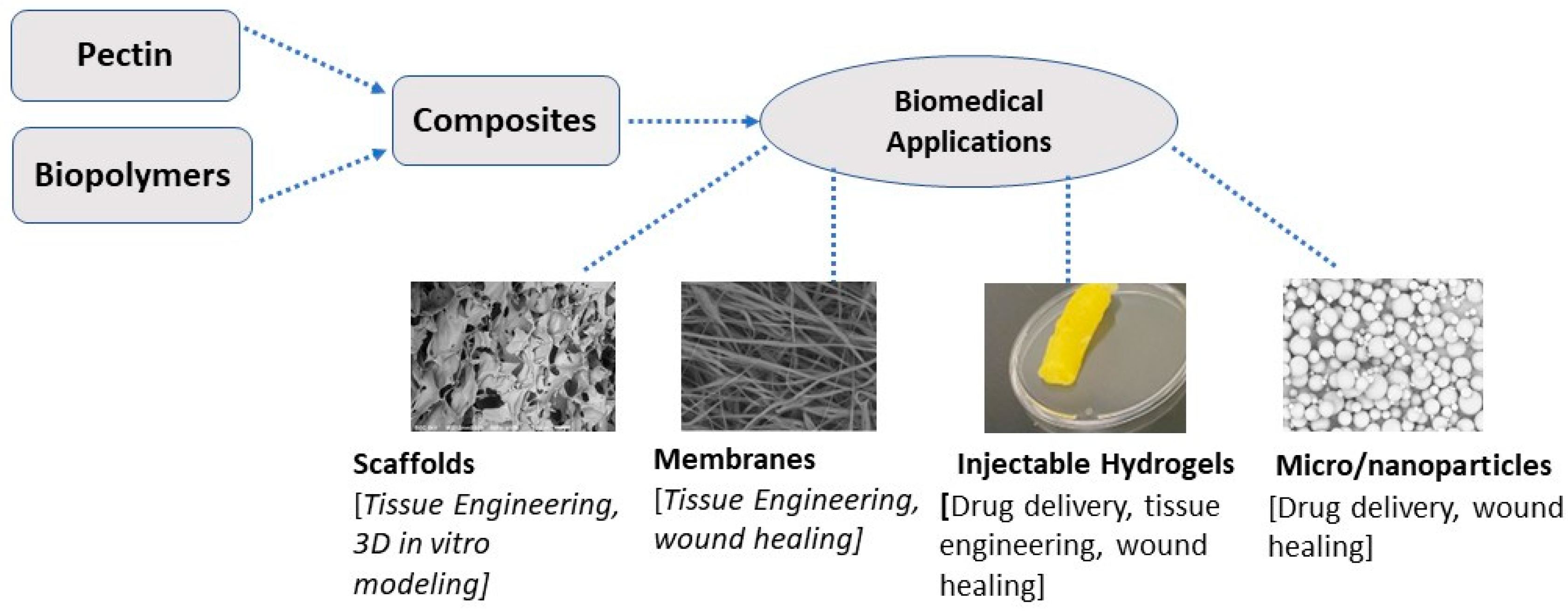
Figure 2.
Homogalacturonan structure of pectin polysaccharides. Homogalacturonan is a linear polymer of α -(1,4)-D galacturonic acid with methyl-esterified at C-6 and acetyl-esterified at positions O-2 and O-3 (Ochoa-Villareal et al. 2012).
Figure 2.
Homogalacturonan structure of pectin polysaccharides. Homogalacturonan is a linear polymer of α -(1,4)-D galacturonic acid with methyl-esterified at C-6 and acetyl-esterified at positions O-2 and O-3 (Ochoa-Villareal et al. 2012).
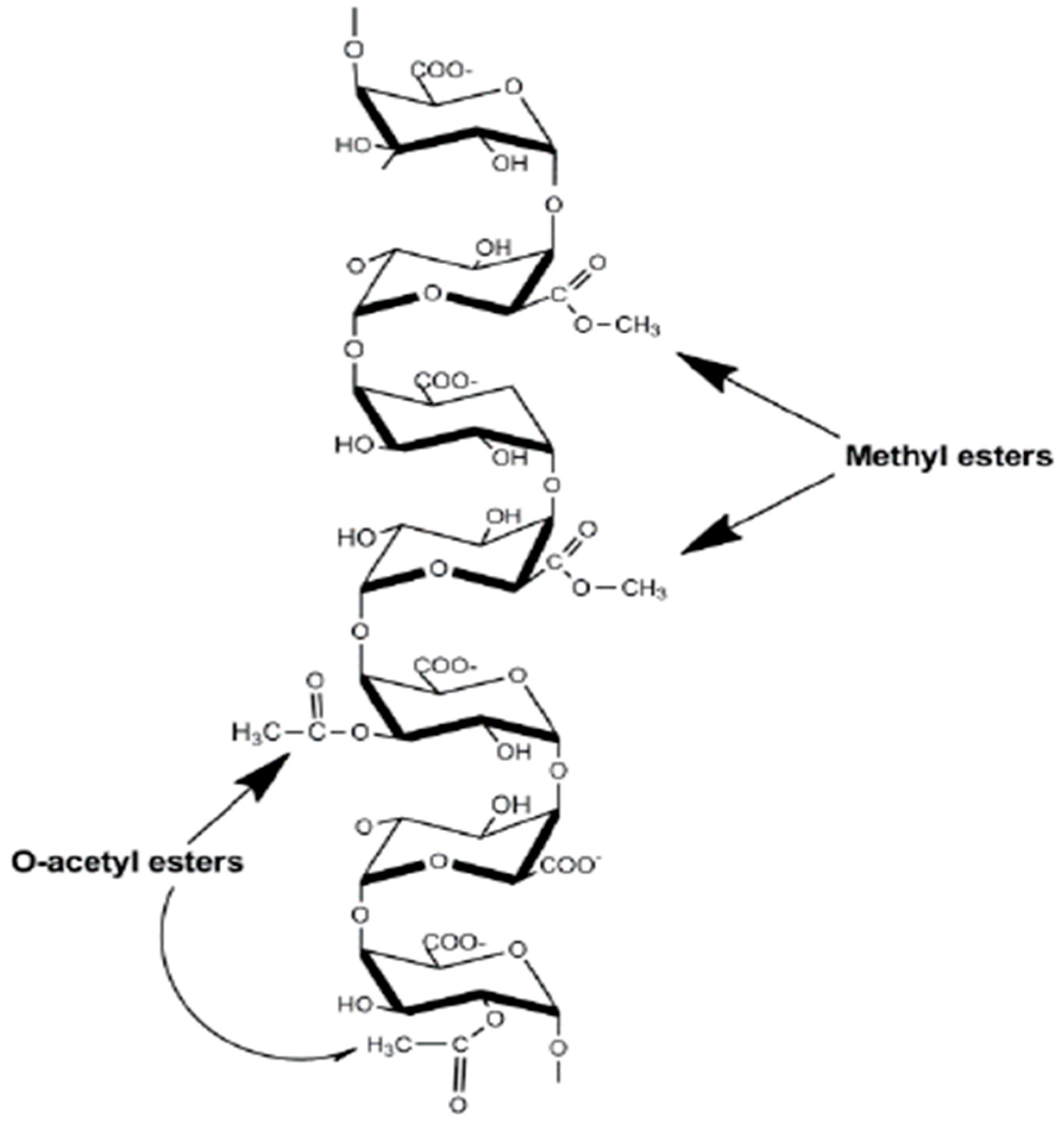
Figure 3.
Fundamental tissue engineering strategy.
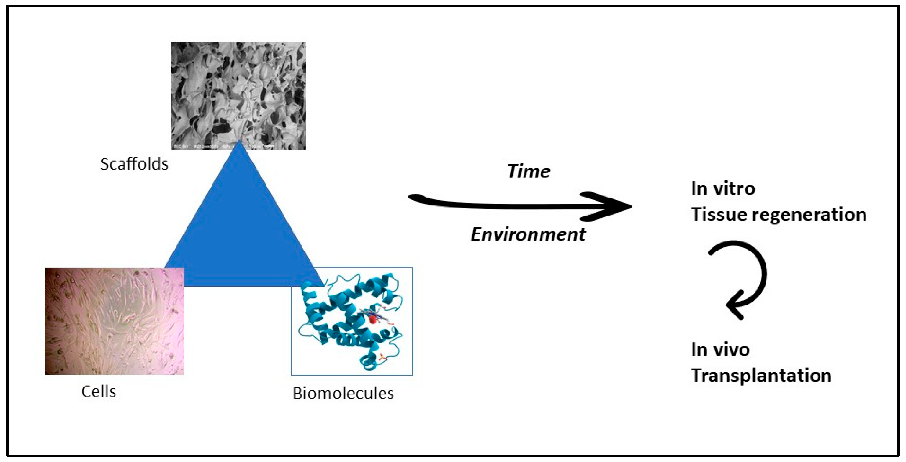

Table 1.
Pectin’s immunoregulatory activities: source and mechanism of action.
| Pectin source | Uses and mechanism of action | Reference |
|---|---|---|
| i. Lemon Pectin | The physical-chemical characteristics of lemon pectin, for example, the degree of methyl esterification and the extent of polymerization, influence the immunostimulatory properties and are significantly essential to utilize pectins to improve immune response. | (Daguet et al. 2016, Vogt et al. 2016) |
| ii. Sumbuci floss or elderflower | Used to heal various diseases linked with the immune system, for example, influenza, chill, or pyrexia. Extracts from S. nigra flowers have stimulation effects on macrophages. In vitro studies reported that the biological activity of rhamnogalacturonan I (RG-1) comprising polysaccharides of elderflowers contributes to higher immunomodulation activity and enhanced macrophage-stimulating effects. | (Ho et al. 2016, Minzanova et al. 2018) |
| iii. Tomato Pectin | Pectic oligosaccharides in sour raw tomatoes demonstrated potential as an anticancer on a gastric cancer cell line in vitro. | (Kapoor and Dharmesh 2017) |
| iv. Lycium ruthenium | Polysaccharides in L. ruthenium suppressed the levels of pro-inflammatory cytokines in lipopolysaccharide-stimulated macrophages and exhibited antifatigue, antioxidation, and hypoglycemic activity. | (Liu et al. 2013, Peng et al. 2014) |
Table 2.
Anti-inflammatory properties of pectin.
| Pectin source | Mechanism of action | Reference |
|---|---|---|
| i. Star fruit (Averrhoa carambola L.) | In vivo, the study reported that the polysaccharides from starfruit exhibited antinociceptive and anti-inflammatory properties and were beneficial for controlling inflammatory pain. | (Leivas et al. 2016, Luan et al. 2021) |
| ii. Suaeda fruiticosa (L.) Forssk | Polysaccharides, phenolic compounds, and bioactive flavonoids from S. fruticose, comprising free radical scavenging and lipid peroxidation, function as an anti-inflammatory agent and analgesic or antioxidant. | (Oueslati et al. 2012, Mzoughi et al. 2018) |
| iii. Citrus pectin | An in vivo study demonstrated that low methyl-esterified pectin from citrus fruits inhibited systemic and local inflammation, whereas a high degree of esterification inhibited intestinal inflammation. | (Sherry et al. 2010, Popov et al. 2013) |
| iv. Sweet Pepper fruits | Both native and modified pectin possessed the inherent activity to control THP-1 macrophages. Due to the availability of lipopolysaccharides, anti-inflammatory properties occur by inhibiting pro-inflammatory and promoting anti-inflammatory cytokines. | (do Nascimento et al. 2017, Pedrosa et al. 2022) |
Table 3.
Antibacterial properties of pectin-based materials.
| Pectin-based system | Mechanism of action | Reference |
|---|---|---|
| i. Citrus Pectin-coated Ag nanoparticles (NPs) | Citrus pectin-coated Ag NPs exhibited great antibacterial activities toward Gram-negative E. coli and Gram-positive S. Aureus. | (Zhang et al. 2017) |
| ii. Pectin-cadmium sulfide nanocomposite (Pc/CSNC); Pectin-zirconium (IV) silicophosphate nanocomposite (Pc/ZSPNC) | Pc/CSNC exhibited a significant effect of antibacterial activity against E. coli. PC/ZSPNC showed substantial antibacterial activity towards E. coli and S. aureus. | (Gupta et al. 2014, Pathania et al. 2015, Hassan et al. 2021) |
| iii. Citrus pectin-MgO Nanocomposites | Pectin-MgO showed significant antibacterial activity against clinical pathogens lactobacillus and Bacillus subtills. | (Supreetha et al. 2021) |
| iv. Pectin/lysozymes layer by layer nanofibrous mats | Pectin/lysosome nanofibrous mats exhibited significant antibacterial effects against E. coli and S. aureus. | (Zhang et al. 2015) |
| v. Essential oils (EOs)/Pectin nanoemulsion | EOs/Pectin nanoemulsion exhibited antibacterial activity towards E. coli and L. innocua populations. | (Guerra-Rosas et al. 2017, Nisar et al. 2019) |
Table 4.
Anticancer activity of pectin and pectin-based composites.
| Pectin source or pectin-based system | Target cancer cell line | Mechanism of action | Reference |
|---|---|---|---|
| Pectin from potato | Human colon cancer HT-29 cells | Rhamnogalacturonan (RG)-I domain-rich potato pectin showed the inhibitory effect of HT-29 cell proliferation in vitro. | (Cheng et al. 2013, Donadio et al. 2022) |
| Pectin from sugar beet | Human colon cancer cell lines (HT-29 and DLD-1) | An in vitro study reported that the pectin from sugar beet exhibited anti-proliferative activity toward colon cancer cells—alkali-treated sugar beet pectin extract induced apoptosis. | (Maxwell et al. 2016) |
| Pectin from sweet potato | Human colon cancer HT-29 cells | Sweet potato pectin modified by ultrasonication inhibited HT-29 cell proliferation and induced apoptosis in vitro. | (Ogutu et al. 2018) |
| Pectin from apple | Breast cancer cells 4T1 | Pectic acid from apple pectin inhibited 4T1 breast cancer cell growth, reduced cell attachment, and induced apoptosis in vitro. In vivo, results exhibited that pectic acid inhibited tumor progression and increased apoptosis cell number. | (Delphi and Sepehri 2016) |
| Citrus pectin | Liver hepatocellular carcinoma cells HepG2 and Adenocarcinoma human alveolar basal epithelial cells A549 | Citrus pectin (heat-modified) induced classical apoptosis and indicated the activation of autophagy in both HepG2 and A549 cancer cell lines. | (Leclere et al. 2015) |
| Pectin from papaya | Colon cancer cell, prostate cancer cell | Papaya pectin extracted from intermediate ripening phases significantly decreased cell viability and induced necroptosis in cancer cell lines in vitro. | (Prado et al. 2017) |
| Pectin-Curcumin | Breast and hepatic cervical cancer cells | The pectin-curcumin complex had better inhibitory activity against cancer cells than only curcumin due to the increased stability and solubility of the composites. | (Bai et al. 2017, Chen et al. 2023) |
| Pectic polysaccharide/Selenium | Adenocarcinomas human alveolar basal epithelial cells | Pectic polysaccharide/Selenium showed a higher inhibiting capacity for cell migration and initiated cell apoptosis than the original pectin polysaccharides. | (Chen et al. 2015) |
| Pectin-polyvinyl pyrrolidone-curcumin | Lung cancer cells A549 | Pectin-polyvinyl pyrrolidone-curcumin particulates showed increased anti-tumor effects than curcumin alone. | (Gaikwad et al. 2017) |
| Pectin/silver (Ag) nanocomposites | Epithelial human breast cancer cell line MDA-MB-231 | Pectin/Ag nanocomposites showed a significantly high inhibitory effect on breast cancer cell proliferation. | (Ogbonna and Kavaz 2022) |
| Pectin/gold nanoparticles | Mammary adenocarcinoma | Pectin/gold nanoparticles induced apoptosis and decreased the viability of the cancer cells. | (Suganya et al. 2016) |
| Citrus pectin/Znnanoparticles | Ehrlich Ascites Carcinoma and human colon adenocarcinoma | The citrus pectin/Zn nanoparticles showed anticancer properties by influencing cancer cell cytotoxicity. | (El-Batal et al. 2018) |
| Pectin/Chitosan | Human colon cancer HT-29 cells | Pectin/chitosan composites exhibited anti-proliferative effects on cancer cells but no cytotoxic effects on normal cells. | (Dziadek et al. 2022) |
| Pectin aldehyde/poly(N-isopropyl acrylamide-stat-acyl hydrazide) P(NIPAM-stat-AH) | Colon carcinoma cells CT26 | In vivo, the study revealed that the self-healing and injectable composites had the potential for anticancer therapy. | (An et al. 2021) |
Table 5.
A summary of several pectin systems in a tissue engineering approach.
| Pectin systems | Method | Application | References |
|---|---|---|---|
| Low methoxyl citrus pectin | UV photocrosslinking with peptide crosslinkers (cell-degradable) and adhesive ligands (integrin-specific); Lyophilization | Skin tissue engineering | (Pereira et al. 2018) |
| Sugar beet pectin (SBP) crosslinked by visible light | Applying 405 nm visible light in the presence of tris(bipyridine)ruthenium (II) chloride hexahydrate and sodium persulfate, rapid hydrogenation of SBP was obtained. 3D hydrogel constructs were obtained using 3D bioprinting. | Promising for liver and other soft tissue engineering | (Mubarok et al. 2023) |
| Citrus peel's pectin crosslinked with (3glycidyloxypropyl)trimethoxysilane (GPTMS) | Freeze drying or 3D bioprinting | Various tissue regeneration | (Lapomarda et al. 2020) |
| Pectin/ chitin/nano CaCO3 | Lyophilization | Bone regeneration | (Kumar et al. 2013) |
| Pectin/chitosan | Freeze drying | Bone tissue engineering | (Coimbra et al. 2011) |
| Pectin/Strontium/Hydroxyapatite | Solution-based chemical technique | Bone regeneration | (Akshata et al. 2023) |
| Collagen/polyurethane/pectin | Semi-interpenetration process | Bone regeneration | (Guzmán-Chávez et al. 2022) |
| Pectin/PVA | Freezing-thawing | Bone regeneration | (Hu et al. 2022) |
| Poly(L-lactide-co-ɛ-caprolactone) (PLCA) /pectin | Scaffolds functionalized with pectin. | In vitro and in vivo bone regeneration | (Suliman et al. 2022) |
Disclaimer/Publisher’s Note: The statements, opinions and data contained in all publications are solely those of the individual author(s) and contributor(s) and not of MDPI and/or the editor(s). MDPI and/or the editor(s) disclaim responsibility for any injury to people or property resulting from any ideas, methods, instructions or products referred to in the content. |
© 2023 by the authors. Licensee MDPI, Basel, Switzerland. This article is an open access article distributed under the terms and conditions of the Creative Commons Attribution (CC BY) license (http://creativecommons.org/licenses/by/4.0/).
Copyright: This open access article is published under a Creative Commons CC BY 4.0 license, which permit the free download, distribution, and reuse, provided that the author and preprint are cited in any reuse.


