
Submitted:
16 October 2023
Posted:
18 October 2023
You are already at the latest version
Abstract
Currently, synthetic diamonds are finding new applications. Quantum devices can be created based on diamond plates with the desired concentration of defects in the structure of the crystal lattice. NV color centers can be used in quantum applications by irradiating plates with high-energy particles, followed by annealing at temperatures above 800°C to develop the spin-optical properties of the centers Diamond plates are subjected to electron irradiation and much less often to proton irradiation. As a result, synthetic diamond plates with NV centers ob-tained by proton irradiation are less studied, although proton irradiation makes it possible to obtain a layered structure of NV centers. In this work, single crystal diamond plates irradiated with protons were studied using polarization microscopy, IR spectroscopy and ODMR spectros-copy. It is shown that when creating NV centers, the distribution and concentration of C – defects in the sample, as well as mechanical stresses before and after irradiation (with subsequent an-nealing), can differ significantly from each other.
Keywords:
NV centers
; IR spectroscopy
; anisotropy of mechanical tension
; defects
; ODMR
; spatial distribution
1. Introduction
It is known that due to its cubic crystal structure with strong covalent bonds of carbon atoms, as well as record-high atomic density, diamond is the most promising wide-band semiconductor [1,2]. The unique properties and prospects of the high-tech application of diamonds are determined by the presence and concentration of various types of defects in the crystal lattice [3,4]. A necessary part of the process of creating plates with unique properties is irradiation with electron or proton beams [5,6]. Studies of diamond plates during the creation of vacancies in the diamond structure mainly occur during their electron irradiation. Electron irradiation of diamond plates is the simplest in comparison with other methods and allows the creating of a high concentration of vacancies, and in subsequent annealing, the concentration of NV centers [7,8]. Of course, vacancies can be created not only by electron irradiation but also by other methods, one of which is proton irradiation. Proton irradiation does not create such high concentrations of vacancies as electron irradiation, but these vacancies can be created in layers, which is promising and deserves more in-depth study. For wide application, it is necessary to develop methods for irradiation and characterization of diamond elements. [9,10], including quantum technologies.
Of great interest is a NV complex. This is an atom of the most common impurity—nitrogen, replacing a carbon atom in the crystal lattice of diamond, while the neighboring lattice site remains vacant. This defect can be in different charge states: neutral (NV0) and negatively charged (NV−). The uniqueness of this defect lies in the fact that its electronic spins are easily manipulated by light, magnetic, electric, and microwave fields, which makes it possible to record quantum information (qubits) on the back of the core of the center. Such manipulation is possible even at room temperature [11,12]. The main interest in quantum technologies is represented by the so-called NV centers, which have an additional electron located at the vacancy site and forming a pair with spin S = 1 with one of the vacancy electrons.
Defects in diamonds can be studied well by IR spectroscopy [13,14]. The study of the defects themselves by IR spectroscopy alone will not give a complete picture of the material itself and its properties. Moreover, during the growth of diamond plates and their processing, internal mechanical stresses may appear. It is well known that NV centers are optically active, which means that diamond plates can be studied by optically detectable magnetic resonance (ODMR). NV centers can also be investigated by X-ray diffraction analysis using ultrashort pulses [15].
In this work, studies of diamonds with NV centers were carried out, the vacancies of which were obtained by irradiating the diamond with protons. During the study, three main characteristics of the diamond were obtained:
- Distribution of nitrogen-containing defects before and after obtaining NV centers (IR spectroscopy method),
- Distribution of mechanical tensions before and after obtaining NV tensions,
- Qualitative assessment of the concentration of NV centers (ODMR method).
2. Experimental. Samples and Proton Irradiation
Synthetic diamond plates S#1 and S#2 grown by HTHP technology using the temperature gradient method on a tandem accelerator with vacuum insulation at the Institute of Nuclear Physics SB RAS were tested for proton irradiation. The accelerator provides a stationary monoenergetic proton beam with an energy varying from 0.3 to 2.3 MeV, a current varying from 0.5 to 10 mA, and a transverse size from 1 cm to 3 cm [16,17]. The size of the samples: S#1 is a square 4 by 4 mm and thickness is 1.5 mm. Trapezoidal plate S#2, where the length is 3.3 mm and the width is 1.3 mm approximately and thickness is 0.5 mm. Samples before irradiation are shown in Figure 1:
Sample S#1 was irradiated with protons with an energy of 1 MeV at a dose of 1015 p/cm2. Sample S#2 was irradiated with protons at a dose of 2 × 10¹⁶ p/cm2, (the first time with an energy of 1 MeV at a dose of 1016 p/cm2, and then the second time with protons with an energy of 2 MeV at a dose of 1016 p/cm2).
The proton penetration depth was calculated using the SRIM—2013 (The Stopping and Range of Ions in Matter) program [18] for sample S#1, the penetration depth is 8 microns at an energy of 1 MeV. For sample S#2, the proton penetration depth is 8 microns at an energy of 1 MeV and 24.4 microns at an energy of 2 MeV.
After irradiation, the samples were annealed in an oven at a temperature of 900 °C for two hours in a vacuum under a pressure of 2.5 mBar created by argon buffer gas.
The color of the diamonds has not changed visually, which indicates a fairly low concentration of the formed NV centers, but does not refute their presence. A diamond containing NV centers of high concentration should have a pink color in its color.
3. Absorption IR Spectroscopy Method
IR spectroscopy methods in the single-phonon region of the diamond spectrum in the middle IR range register signals from the main nitrogen centers (C, C+, A, B1). In the studied synthetic diamonds from this set, only C-centers (single substituting nitrogen atoms) are present, and after irradiation, the same centers in the ionized state (C+ center) are also present. In this work, the FT-801 IR-Fourier spectrometer with a MICRAN-3 IR microscope manufactured by Simex (Novosibirsk) was used. IR absorption spectra were obtained at points located along the grid in increments of 41.3 µm with a light spot size of 100 × 100 µm. The radiation was recorded by a CMT (cadmium-mercury-tellurium) receiver cooled with liquid nitrogen. The spectral resolution was 8 cm−1, 4 scans were performed at each point. The reference spectrum was recorded in the transmission mode without a sample, that is, the reference spectrum of air was recorded. Next, the scanning location was chosen in such a way as to cover an area characterized by axial symmetry with an axis directed along the crystallographic direction <111> [19]. A scanned map of sample S#1 with a square of approximately is 2064 μm2 and scanned map of sample S#2 with the area is about 1900 by 900 microns. Each spectrum of the diamond plate obtained by the IR method is normalized by the value of 12.8 ± 0.3 cm−1 of the intrinsic absorption of the lattice at values of 2170 and 2030 cm−1 [20]. Further, the absorption spectrum was decomposed into components related to nitrogen centers C characteristic of the HPHT of synthetic diamonds. The concentration of defects C in this diamond is calculated by the formula [21]:
The absolute value of the concentration is determined by this formula before annealing and irradiation. During irradiation, part of the C defects, due to the loss of the fifth valence electron, turns into C+ defects. In this case, the signal from the C centers is weakened and a signal from C+ appears in the single-phonon region with the main line at 1332 cm−1. The content of centers C can now be judged by subtracting from the initial concentration of defects C the concentration of defects C+, which is determined using the relation N(С+) (ppm) = (5.5 ± 1) × µ1332. But when it is necessary to take into account that some of the C centers are also involved in the formation of defects responsible for the electron-vibrational system with a zero-phonon line (ZPL) 523 nm in the visible range and, possibly, defects associated with the line 1530 cm−1 in mid infrared [21].
4. Optical Anisotropy Method
The effect of double refraction in diamonds was used to study mechanical tensions. To detect double refraction, it is necessary to apply the method of polarization microscopy. When polarized light passes through a birefringent sample, there is a phase difference between an ordinary and an extraordinary beam. By fixing the intensity of the light that has passed through the sample, this phase difference can be found. The registration of optical anisotropy makes it possible to estimate the internal stresses in the diamond. By article [22], we have developed a stand representing a microscope with a motorized table mounted on it and a rotating linear polarizer. A CMOS camera is used as a photodetector, with the help of which it is possible to form a map of internal voltages.
5. Optically Detectable Magnetic Resonance (ODMR) Method
The principle of optically detectable magnetic resonance (ODMR) is to detect the fluorescence that occurs in a diamond when it is pumped with a laser. NV centers have bright, stable fluorescence at 532 nm excitation. The ground and excited state are triplets consisting of spin sublevels. For the side sublevels of the excited state, a transition to the ground state through a system of auxiliary levels is possible, which leads to optical polarization of the spin. To perform magnetometry, the dependence of the frequencies of microwave transitions between the spin sublevels of the ground state on the magnetic field due to the Zeeman effect is used. To register the signal of an optically detectable magnetic resonance signal (ODMR), we developed and used a stand, the schematic diagram of which is presented in [23]. The ODMR signal from the studied plates was recorded in continuous mode in a zero magnetic field. Optical excitation was carried out by a semiconductor laser Cobolt Samba with a wavelength of 532 nm, then through the lens MY5X-802—5X laser beam fell on a diamond plate, forming a spot with a diameter of about 100 microns. The luminescence excited by this laser was focused on a photodetector of the APDC12703 series. Microwave radiation was supplied by a planar antenna with a diameter of 1 mm in the form of a printed circuit board located directly under the sample [24].
6. IR Spectroscopy Distribution Maps of Sample S#1
Below are the defect distribution maps. The vertical scale is the concentration of defect C in ppm (C defect or donor nitrogen is one nitrogen atom, which isomorphically replaces the carbon atom in the diamond lattice).
Figure 2 shows the distribution of the C-defect with an area of 2064 μm2 of sample S#1 before irradiation and after irradiation with concomitant annealing at 900 °C for two hours in vacuum.
A noticeable change in concentration indicates the transformation of the C defect into an NV defect.
In the central zone, the concentration of C is 59, while at the periphery, between the rays, it is 189 ppm. After irradiation and annealing, the concentration at the corresponding points is about 13 and 154 ppm.
The concentration difference shown in Figure 3 obtained by subtracting the concentration C of the defect after irradiation to the concentration before irradiation, normalized to the maximum concentration value. The difference is represented in hundredths units.
According to this distribution, it can be seen that concentration decreased to 40%.
7. IR Spectroscopy Distribution Maps of Sample S#2
Figure 4 shows the distribution of the C-defect of plate S#2 before irradiation and after irradiation with concomitant annealing at 900 °C for two hours in a vacuum. Defect C or donor nitrogen is a single nitrogen atom that isomorphically replaces the carbon atom in the diamond lattice. The scanning area was covered almost entirely, the entire plate. The size of the area is about 1900 by 900 microns.
The dominant concentration is ~150 ppm, locally up to ~200 ppm. After proton irradiation and subsequent annealing, the dominant concentration of defect C remains approximately the same, but local spots with increased concentration increase.
The concentration difference shown in Figure 5 obtained by subtracting the concentration C of the defect after irradiation to the concentration before irradiation, normalized to the maximum concentration value. The difference is represented in hundredths units.
According to this distribution, the concentration of the defect decreased to 10% in the whole area of the plate. This can be seen by the negative values of the difference in the concentration of defect C before and after irradiation, which indicates the conversion of nitrogen atoms into other related complexes, in particular into NV centers.
8. Comparison of Results before and after Irradiation. Internal Tensions. Anisotropy Coefficient
Pronounced changes are observed on plate S#1. In the center of this plate, there is a cross, visually transparent.
Figure 6 shows the distribution of internal stresses. The scale is dimensionless, characterizes the anisotropy of internal tensions |sinδ|.
Comparing these results, it can be seen that the plate has become more homogeneous in internal stresses. This is obviously due to the post-processing (annealing) of the plates. The comparison of these two plates The difference of internal tensions shown in Figure 7 obtained by subtracting the values of tensions after irradiation to the values of tensions before irradiation, normalized to the maximum values of tensions value. The difference is represented in hundredths units.
According to this distribution, the voltage has not changed in general, but at the boundaries of heterogeneity, in the sacral region, the stresses have decreased to 30%.
No changes in the internal stresses of the S#2 plate were observed. The removal of strong stresses occurred, obviously due to the complex effects of proton irradiation and annealing.
9. ODMR Spectra of Diamond Plates after Proton Irradiation
To study the magnetic properties of diamond plates directly, measurements were made directly on the stand of optically detectable magnetic resonances. The ODMR signal from the samples under consideration is shown in Figure 8. It can be seen that the ODMR signal at a resonant frequency of 2871 MHz from diamond S#2 irradiated with a dose of 2 × 10¹⁶ p/cm2 is more pronounced and differs by an order of magnitude from the ODMR signal received at the same frequency from diamond S#1 irradiated with a dose of 1015 p/cm2.
The signal at the resonant frequency of diamond S3 differs from the signal received at the same frequency of diamond S#1 by 40 times with the same tuning. This indirectly suggests that the concentration of NV in the diamond plate S#2 is significantly higher.
10. Conclusions
The distribution of inclusions in the IR range has been studied for two proton-irradiated diamond plates. After irradiation and annealing in vacuum, the concentration of C defects decreased in samples S#1 and S#2, which indicates the transition from the defect to the NV center.
The anisotropy coefficient decreased after irradiation and annealing in vacuum, and more homogeneous regions appeared. At the boundaries of the inhomogeneity, the stresses decreased. In other areas, the voltage has not changed.
The ODMR method revealed that the signal at the resonant frequency of diamond S#2, irradiated with a higher dose, is visible and differs from the signal received at the same frequency of diamond S#1 by an order of magnitude. This indirectly suggests that the concentration of NV- in the diamond plate S#2 is much higher.
We emphasize that in the works [5,6], samples with uniform proton irradiation over the sample area were studied.
A conditional boundary has been found at which luminescence can be detected by the ODMR method in continuous operation. The minimum radiation dose of the diamond plate should be greater than 1015 p/cm2.
Author Contributions
Conceptualization, methodology, М.E. and D.M.; methodology ODMR, R.B.; experiments using method ODMR, polarization microscopy, I.P., A.L. and A.Kh.; experiments using method IR-spectroscopy, A.K.; experiments with proton beams, S.T., D.K., I.K., S.S. and I.S.; post-growth engineering, V.V. and A.E.; validation, M.E. and D.M.; formal analysis, M.E., K.M. and D.M.; writing—original draft preparation, M.E. and A.K.; writing—review and editing, A.K. and M.E.; project administration, M.E. All authors have read and agreed to the published version of the manuscript.
Funding
The research was supported by of the Russian Science Foundation No. 23-12-20014, https://rscf.ru/project/23-12-20014/ and of the state assignment the Russian Federation No. FSRU 2021-0008.
References
- Chen, G.; Wang, W.; Lin, F.; Zhang, M.; Wei, Q.; Yu, C.; Wang, H.-X. Electrical Characteristics of Diamond MOSFET with 2DHG on a Heteroepitaxial Diamond Substrate. Materials 2022, 15, 2557. [Google Scholar] [CrossRef]
- Crnjac, A.; Skukan, N.; Provatas, G.; Rodriguez-Ramos, M.; Pomorski, M. Electronic Properties of a Synthetic Single-Crystal Diamond Exposed to High Temperature and High Radiation. Materials 2020, 13, 2473. [Google Scholar] [CrossRef]
- Pratas, S.; Silva, E.L.; Neto, M.A.; Fernandes, C.M.; Fernandes, A.J.S.; Figueiredo, D.; Silva, R.F. Boron Doped Diamond for Real-Time Wireless Cutting Temperature Monitoring of Diamond Coated Carbide Tools. Materials 2021, 14, 7334. [Google Scholar] [CrossRef]
- Bougas, L.; Wilzewski, A.; Dumeige, Y.; Antypas, D.; Wu, T.; Wickenbrock, A.; Bourgeois, E.; Nesladek, M.; Clevenson, H.; Braje, D.; Englund, D.; Budker, D. On the Possibility of Miniature Diamond-Based Magnetometers Using Waveguide Geometries. Micromachines 2018, 9, 276. [Google Scholar] [CrossRef]
- Schabikowskia, M.; Wojciechowskib, A.M.; Mitura-Nowaka, M.; Mrózekb, M.; Krukb, A.; Rajchela, B.; Gawlikb, W.; Marszałeka, M. Optical Characterization of Nitrogen-Vacancy Centers Created by Proton Implantation in Diamond. Materials 2021, 14, 833. [Google Scholar] [CrossRef]
- Mrózek, M.; Schabikowski, M.; Mitura-Nowak, M.; Lekki, J.; Marszałek, M.; Wojciechowski, A.M.; Gawlik, W. Nitrogen-Vacancy Color Centers Created by Proton Implantation in a Diamond. Materials 2021, 14, 833. [Google Scholar] [CrossRef]
- Meshkov, I.N.; Eseev, M.K.; Kuziv, I.V.; et al. Vacancy Determination in Single-Crystal Diamond Plates Using Positron Annihilation Spectroscopy. Phys. Part. Nuclei Lett. 2023, 20, 757–762. [Google Scholar] [CrossRef]
- Eseev, M.; Kuziv, I.; Kostin, A.; Meshkov, I.; Sidorin, A.; Orlov, O. Investigation of Nitrogen and Vacancy Defects in Synthetic Diamond Plates by Positron Annihilation Spectroscopy. Materials 2023, 16, 203. [Google Scholar] [CrossRef]
- Liu, G.-Q.; Pan, X.-Y. Quantum information processing with nitrogen–vacancy centers in diamond. Chin. Phys. B. 2018, 27, 020304. [Google Scholar] [CrossRef]
- Zhang, H.; et al. Spin-torque oscillation in a magnetic insulator probed by a single-spin sensor. Physical Review B. 2020, 102, 2. [Google Scholar]
- Balasubramanian, G.; Chan, I.Y.; Kolesov, R.; Al-Hmoud, M.; Tisler, J.; Shin, C.; Kim, C.; Wojcik, A.; Hemmer, P.R.; Krueger, A.; et al. Nanoscale imaging magnetometry with diamond spins under ambient conditions. Nature 2008, 455, 648–651. [Google Scholar] [CrossRef]
- Doherty, M.W.; Manson, N.B.; Delaney, P.; Jelezko, F. The nitrogen- vacancy col-ourcentre in diamond. Physics Reports 2013, 528, 1–45. [Google Scholar] [CrossRef]
- Kudryashov, S.; Danilov, P.; Kuzmin, E.; Smirnov, N.; Gorevoy, A.; Vins, V.; Pomazkin, D.; Paholchuk, P.; Muratov, A.; Kirichenko, A.; et al. Productivity of Concentration Dependent Conversion of Substitutional Nitrogen Atoms into Ni-trogen-Vacancy Quantum Emitters in Synthetic-Diamond by Ultrashort Laser Pulses. Micromachines 2023, 14, 1397. [Google Scholar] [CrossRef]
- Khomich, A.A.; Khmelnitskii, R.; Kozlova, M.; Khomich, A.V.; Ralchenko, V. IR Spectroscopy of Vacancy Clusters (Am-ber Centers) in CVD Diamonds Nanostructured by Fast Neutron Irradiation. C—Journal of Carbon Research 2023, 9, 55. [Google Scholar] [CrossRef]
- Eseev, M.; Makarova, K.; Makarov, D. Scattering of Ultrashort X-ray Pulses on Diamonds with NV Centers. Crystals 2022, 12, 1417. [Google Scholar] [CrossRef]
- Taskaev, S. Accelerator Based Epithermal Neutron Source. Physics of Particles and Nuclei 2015, 46, 956–990. [Google Scholar] [CrossRef]
- Taskaev, S.; Berendeev, E.; Bikchurina, M.; Bykov, T.; Kasatov, D.; Kolesnikov, I.; Koshkarev, A.; Makarov, A.; Ostreinov, G.; Porosev, V.; Savinov, S.; Shchudlo, I.; Sokolova, E.; Sorokin, I.; Sycheva, T.; Verkhovod, G. Neutron Source Based on Vacuum Insulated Tandem Accelerator and Lithium Target. Biology 2021, 10, 350. [Google Scholar] [CrossRef]
- http://www.srim.org/.
- Tucker, O.D.; Newton, M.E.; Baker, J.M. EPR and 14N electron-nuclear double-resonance measurements on the ionized nearest-neighbor dinitrogen center in diamond. Phys. Rev. B. 1994, 50, 15586–15596. [Google Scholar] [CrossRef]
- Yelisseyev, A.; Vins, V.; Lobanov, S.; Afonin, D.; Maksimov, A.; Blinkov, A. Aggregation of Donor Nitrogen in Irradiated Ni-Containing Synthetic Diamonds. Journal of Crystal Grows 2011, 318, 539–544. [Google Scholar] [CrossRef]
- Lawson, S.C.; Fisher, D.; Hunt, D.C.; Newton, M.E. On the existence of positively charged single-substitutional nitrogen in diamond. Journal of Physics: Condensed Matter 1998, 10, 6171. [Google Scholar] [CrossRef]
- Glazer, A.M.; Lewis, J.G.; Kaminsky, W. An automatic optical imaging system for birefringent media. Department of Physics: Oxford OX1 3PU, UK, 2015. [Google Scholar]
- Babunts, R.A.; Muzafarova, M.V.; Anisimov, A.N; et al. Diagnostics of NV defect structure orientation in diamond using optically detected magnetic resonance with a modulated magnetic field. Tech. Phys. Lett. 2015, 41, 583–586. [Google Scholar] [CrossRef]
- Sasaki, K.; et al. Broadband, large-area microwave antenna for optically detected magnetic resonance of nitrogen-vacancy centers in diamond. Rev. Sci. Instrum. 2016, 87, 053904. [Google Scholar] [CrossRef]
Figure 1.
Image of samples before proton irradiation: left—S#1 left and right—S#2.
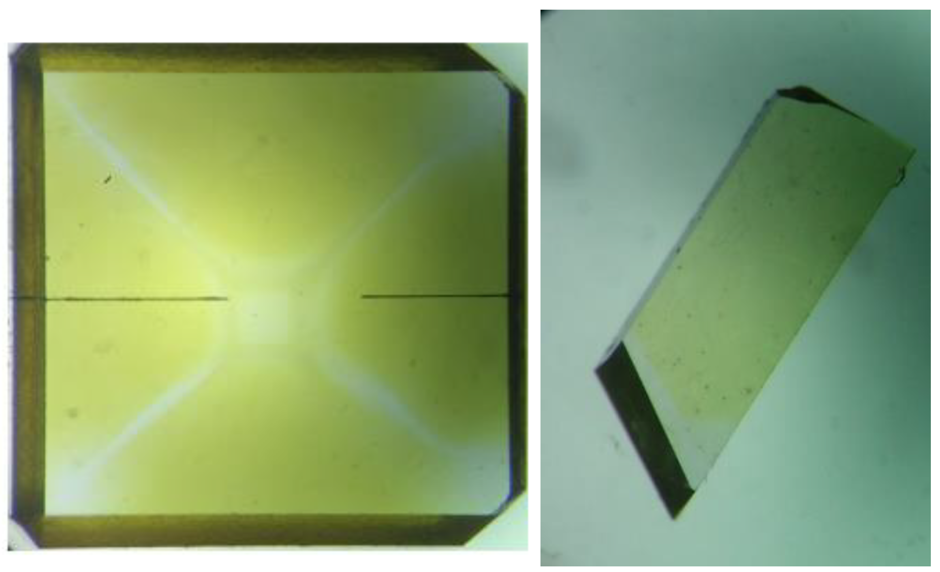
Figure 2.
IR spectrum of the distribution of C-defects in sample S#1. (on the left—before irradiation, on the right—after irradiation in combination with subsequent annealing at 900 °C for two hours in a vacuum).
Figure 2.
IR spectrum of the distribution of C-defects in sample S#1. (on the left—before irradiation, on the right—after irradiation in combination with subsequent annealing at 900 °C for two hours in a vacuum).
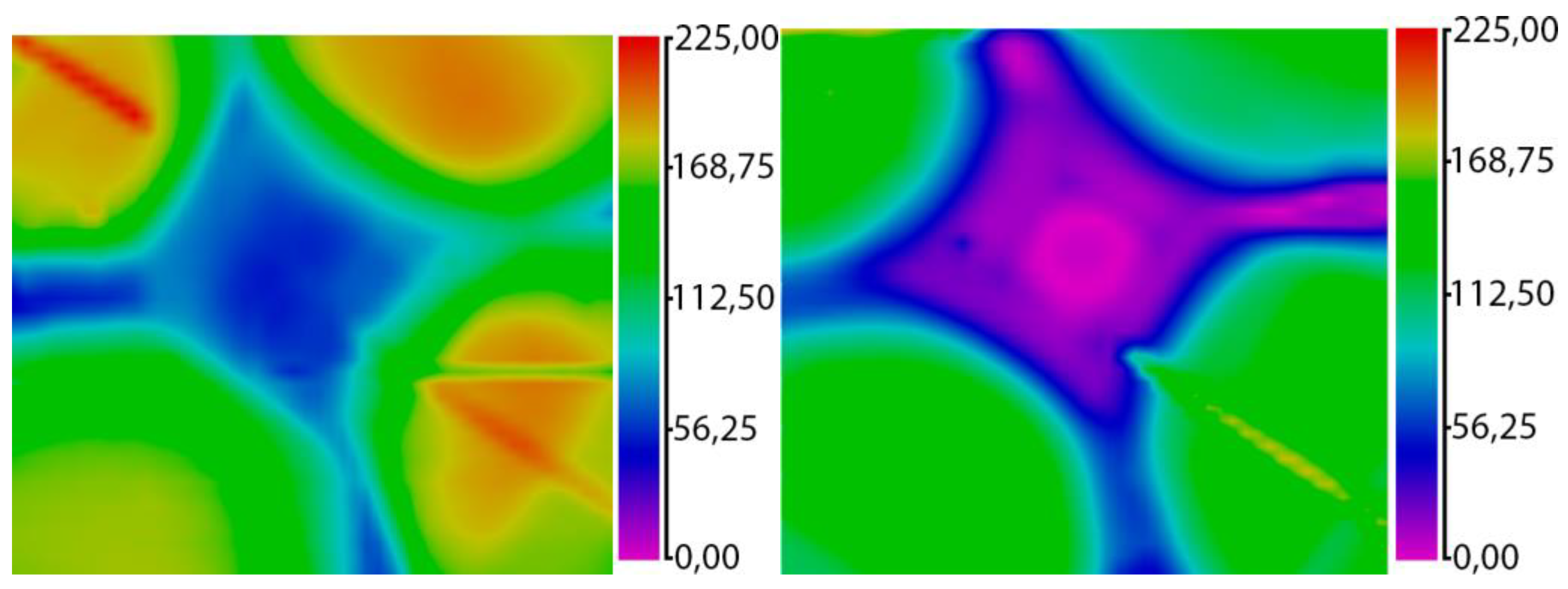
Figure 3.
The difference in the concentration of the C defect in sample S#1 in fractions before irradiation and after irradiation in combination with subsequent annealing at 900 °C for two hours in a vacuum.
Figure 3.
The difference in the concentration of the C defect in sample S#1 in fractions before irradiation and after irradiation in combination with subsequent annealing at 900 °C for two hours in a vacuum.
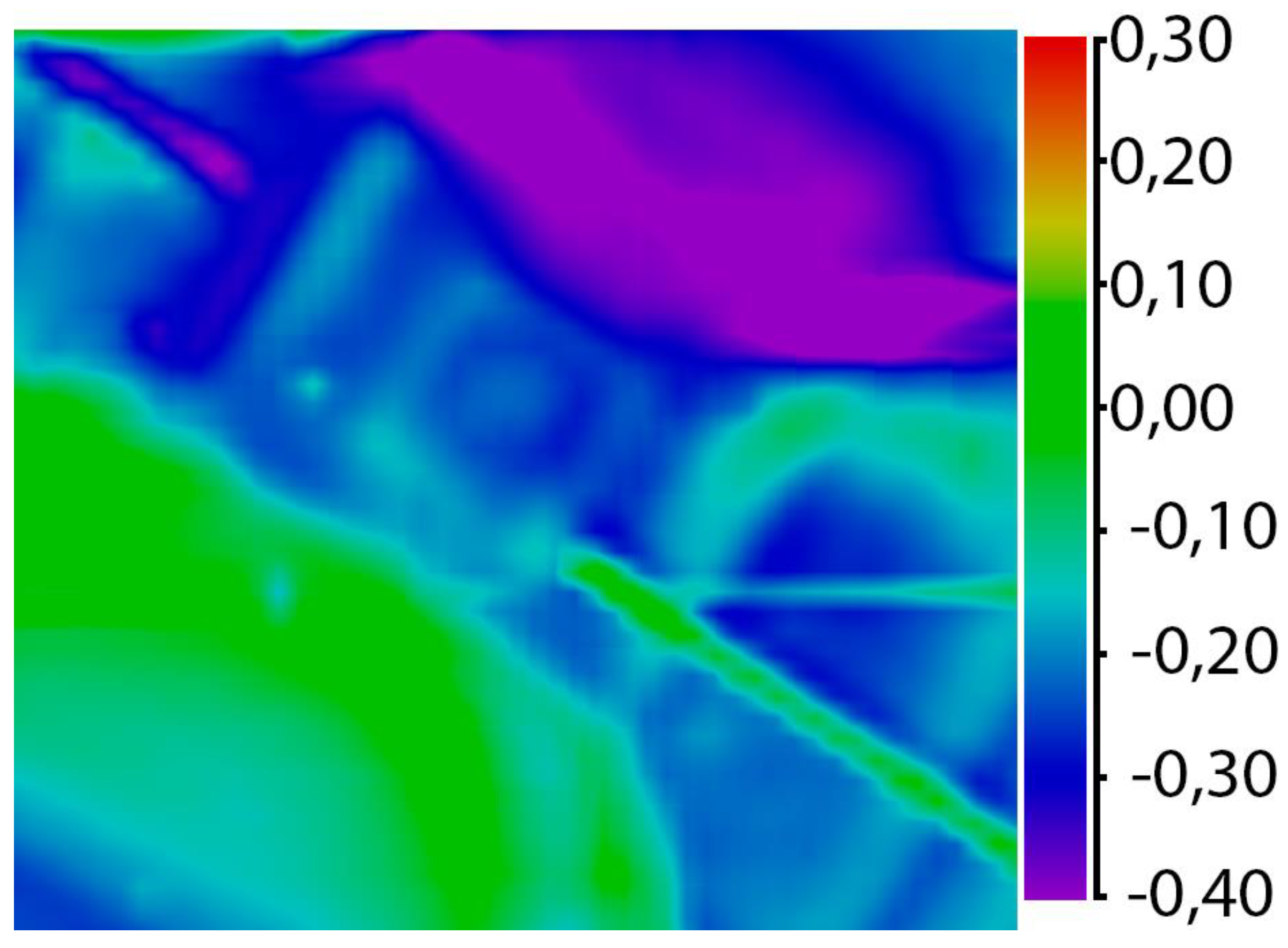
Figure 4.
The IR spectrum of the distribution of C-defects in sample S#2. (on the left—before irradiation, on the right—after irradiation in combination with subsequent annealing at 900 °C for two hours in a vacuum).
Figure 4.
The IR spectrum of the distribution of C-defects in sample S#2. (on the left—before irradiation, on the right—after irradiation in combination with subsequent annealing at 900 °C for two hours in a vacuum).
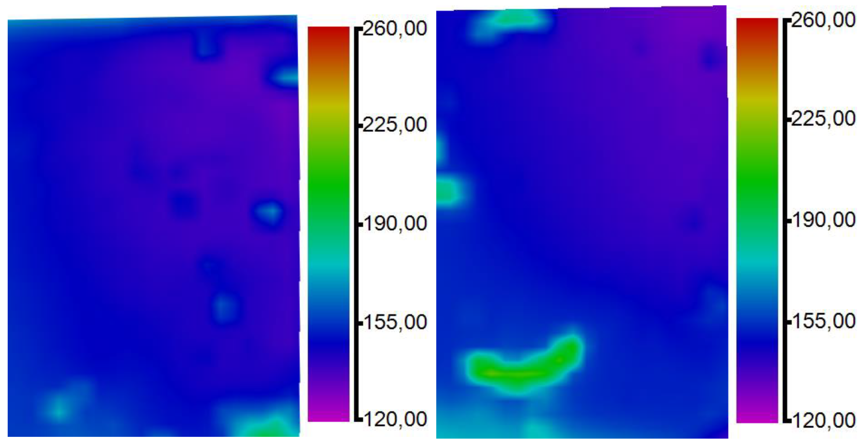
Figure 5.
The difference in the concentration of the C defect in sample S#2 in fractions before irradiation and after irradiation in combination with subsequent annealing at 900 °C for two hours in a vacuum.
Figure 5.
The difference in the concentration of the C defect in sample S#2 in fractions before irradiation and after irradiation in combination with subsequent annealing at 900 °C for two hours in a vacuum.
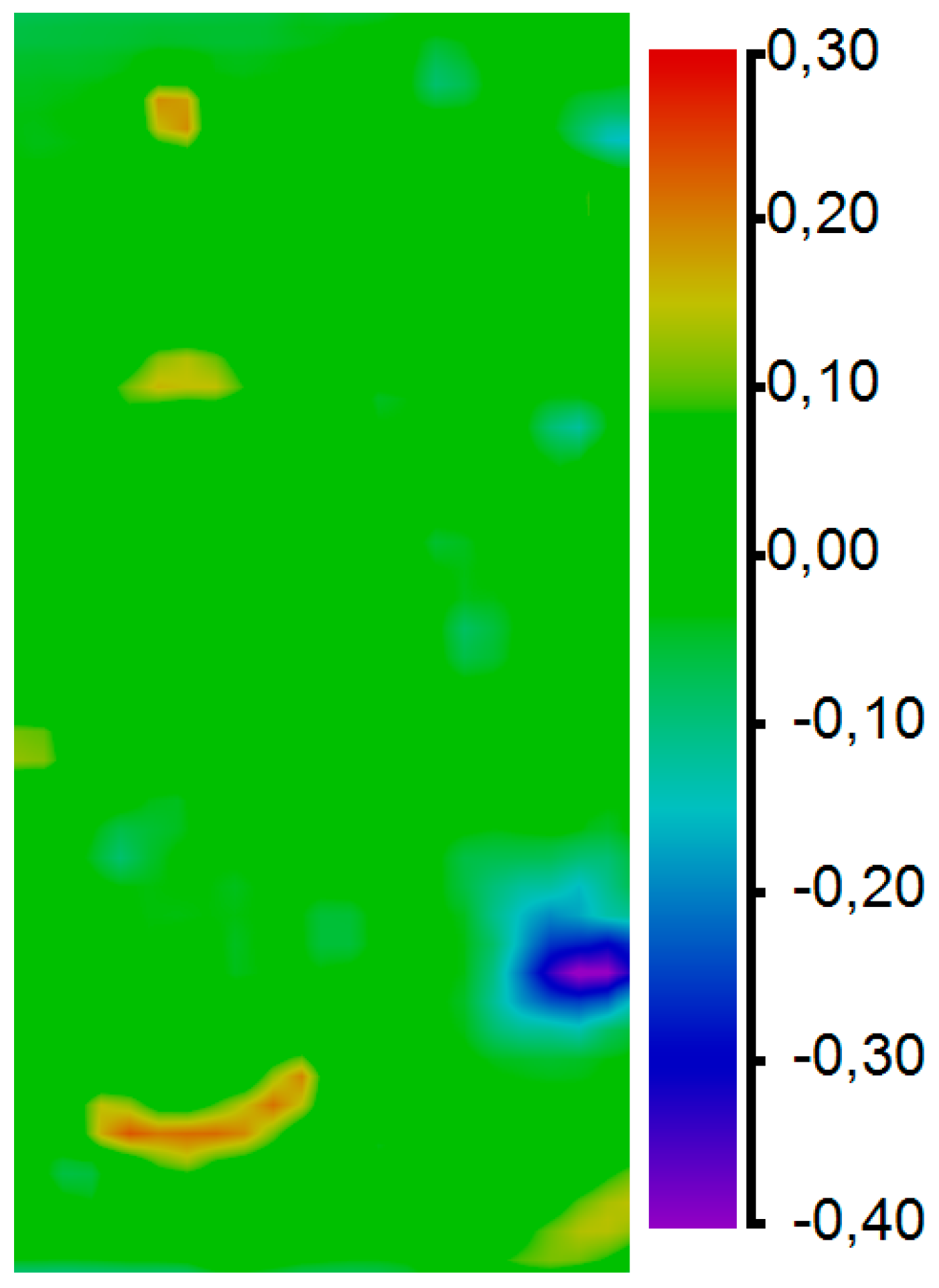
Figure 6.
Distribution of internal tensions |sinδ| of sample S#1: on the left—before irradiation (|sinδ|= 0.04 ÷ 0.32) and on the right—after irradiation in combination with subsequent annealing at 900 °C for two hours in vacuum (|sin δ| = 0.02 ÷ 0.25).
Figure 6.
Distribution of internal tensions |sinδ| of sample S#1: on the left—before irradiation (|sinδ|= 0.04 ÷ 0.32) and on the right—after irradiation in combination with subsequent annealing at 900 °C for two hours in vacuum (|sin δ| = 0.02 ÷ 0.25).
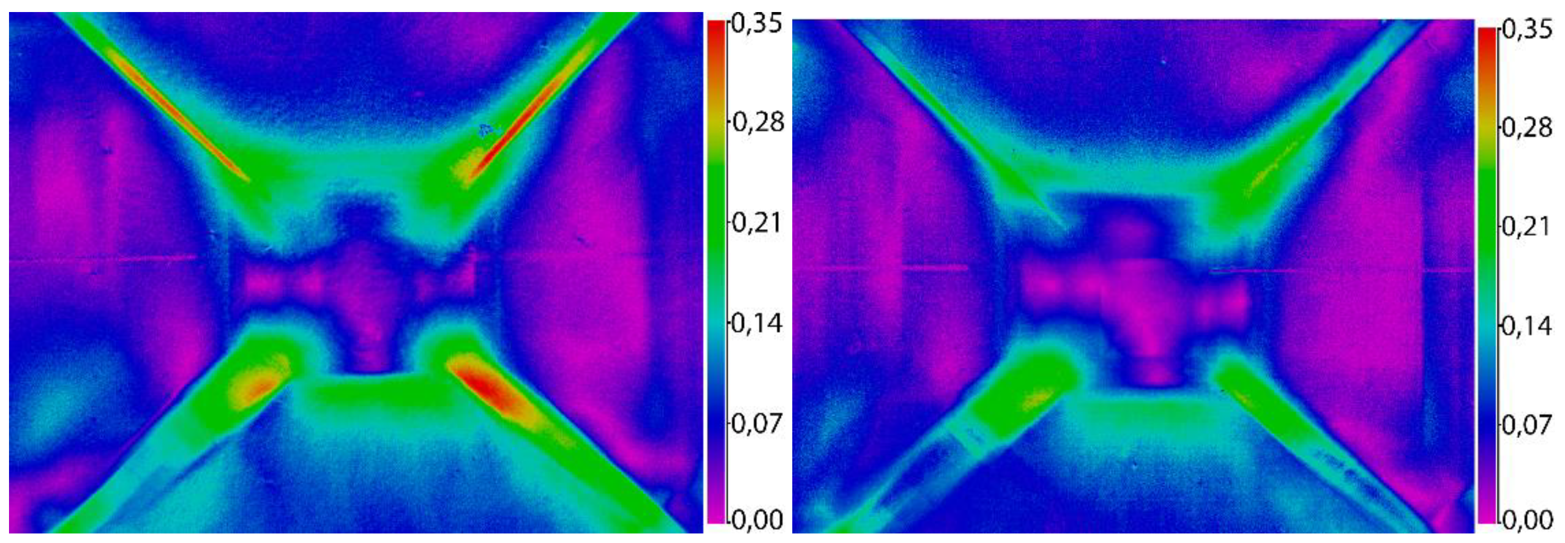
Figure 7.
The difference of internal tensions |sin δ| in fractions before irradiation and after irradiation in combination with subsequent annealing at 900 °C for two hours in the vacuum of plate S#1.
Figure 7.
The difference of internal tensions |sin δ| in fractions before irradiation and after irradiation in combination with subsequent annealing at 900 °C for two hours in the vacuum of plate S#1.
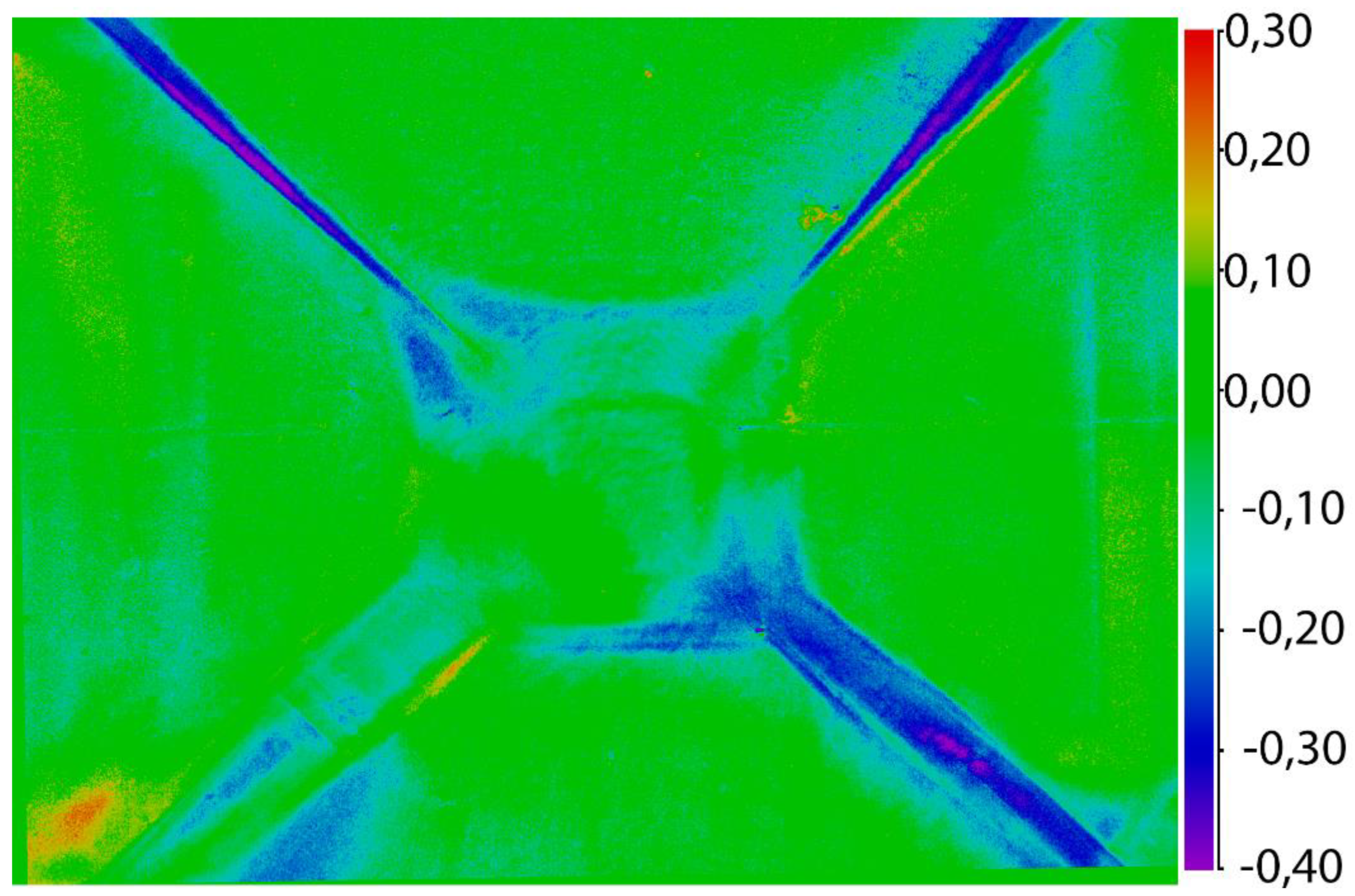
Figure 8.
ODMR spectra of plates, S#1, S#2 after irradiation in combination with subsequent annealing at 900 °C for two hours in a vacuum.
Figure 8.
ODMR spectra of plates, S#1, S#2 after irradiation in combination with subsequent annealing at 900 °C for two hours in a vacuum.

Disclaimer/Publisher’s Note: The statements, opinions and data contained in all publications are solely those of the individual author(s) and contributor(s) and not of MDPI and/or the editor(s). MDPI and/or the editor(s) disclaim responsibility for any injury to people or property resulting from any ideas, methods, instructions or products referred to in the content. |
© 2023 by the authors. Licensee MDPI, Basel, Switzerland. This article is an open access article distributed under the terms and conditions of the Creative Commons Attribution (CC BY) license (http://creativecommons.org/licenses/by/4.0/).
Copyright: This open access article is published under a Creative Commons CC BY 4.0 license, which permit the free download, distribution, and reuse, provided that the author and preprint are cited in any reuse.


