
Submitted:
21 September 2023
Posted:
22 September 2023
Read the latest preprint version here
Abstract
Background: Antibiotic-containing carrier systems are one option that offers the advantage of releasing active ingredients over a longer period of time. In vitro sustained drug release from a carrier system consisting of microporous β-TCP ceramic and alginate has been reported in previous works. Alginate dialdehyde (ADA) gelatin gel showed both better mechanical properties when loaded into a β-TCP ceramic and higher biodegradability than pure alginate. Methods: Dual release of daptomycin and BMP-2 was measured on days 1, 2, 3, 6, 9, 14, 21, and 28 by HPLC and ELISA. After release, the microbial efficacy of the daptomycin was verified and the biocompatibility of the composite was tested in cell culture. Results: Daptomycin and the model compound FITC protein A (n=30) were released from the composite over 28 days. A Daptomycin release above the minimum inhibitory concentration (MIC) by day 9 and a burst release of 71.7 ± 5.9 % were observed in the loaded ceramics. Low concentrations of BMP-2 were released from the loaded ceramics over 28 days.
Keywords:
dual release
; daptomycin
; BMP-2
; β-TCP scaffold
; ADA-gelatin gel
; bone infection
; bone regeneration
1. Introduction
Osteomyelitis is an inflammation of the bone that usually affects the bone (osteitis), the bone marrow (osteomyelitis), and the periosteum (periostitis) [1]. The surrounding soft tissue may also be affected. It is caused by infection with various microorganisms and ultimately leads to destruction of the bone [2]. Etiologically, a distinction is made between hematogenous, locally transmitted (per continuitatem), exogenous, and specific osteomyelitis [3]. In pediatrics, hematogenous osteomyelitis mainly affects the long bones; in adults, it manifests mainly as spondylitis [4]. The source of infection can be, for example, a banal skin infection or contamination during intravenous drug intake, which is flushed hematogenously into the bone, causing osteomyelitis [5]. Staphylococcus aureus is the most common causative agent of primary hematogenous and locally transmitted osteomyelitis [5]. In the case of implant-associated infection, the small colony variants (SCV) of Staphylococcus and coagulase-negative Staphylococcus should be mentioned [6]. Depending on the duration of infection or the histological form of inflammation, osteomyelitis can be initially divided into acute and chronic forms [7]. Osteomyelitis is considered chronic when the causative agent persists for more than 6 weeks, although the boundary may be blurred [3]. In contrast, the appearance of sequesters on CT or MRI is a definite criterion for chronic infection [4]. Sequesters are necrotic pieces of bone that are rejected by healthy tissue and thus prevent the infection from healing [8]. The usual therapy consists of debridement, i.e. surgical removal of the infected tissue, and systemic antibiotic therapy [9] tailored to the specific pathogen. Local antibiotic therapy is much more effective [10] and is sometimes already applied with the use of gentamicin-loaded PMMA chains (Septopal®)[11]. The disadvantage, however, is that they have to be removed again in a second surgical procedure and, in addition, a not inconsiderable proportion of the antibiotics remains in the PMMA [12] because it is biodegradable. Delayed in vitro drug release from a carrier system [13] consisting of microporous β-TCP ceramic and alginate has been previously reported in the literature [14,15,16,17]. ADA gelatin gel showed both better mechanical properties in a β-TCP ceramic and higher biodegradability than pure alginate [18]. Since it has already been tested in terms of drug release [17,19], it is a promising hydrogel for dedicated directed flow loading. The possibility of dual drug release has not yet been investigated in this system, which could improve patient outcomes by combining anti-infective treatment, bone growth promoting therapy, and a better stability in bone through the β-TCP ceramic.
2. Materials and Methods
2.1. Preparing the microporous β-TCP ceramics
The ceramics were produced as previously described [20]. 80 g of a-tricaclium phosphate and 20 g of tricalcium phosphate (Art. No. 102143, Merck; mixture of an apatite and some calcium hydrogenphosphate) were mixed with 60.0 ± 0.2 g of a solution containing 0.2M Na2HPO4 and 1% polyacrylic acid (Art. No. 81132, Fluka; Mw = 5.1 kDa). It was stirred and the paste was poured into plastic syringes with their tips removed (Ø = 23 mm). The paste hardened for 45 min, was then covered with 10 ml of PBS 7.4 solution (Art. No. P5368, Sigma) and incubated for 3 days at 60 °C. The samples (Ø = 23 mm; L = 70 mm) were dried at the same temperature and sintered at 1250 °C for 4 h. Heating and cooling took place at 1 °C/min. The cylinders were trimmed to a length of 25mm and a diameter of 7mm. Finally, the samples were washed in ethanol to remove residual particles and calcined at 900 °C to burn off all organic residues. Before use, the ceramics were shortened to cylinders with a length of 6mm and washed again. The samples were sterilized at 200 °C for 4 h.
2.2. Characterization of the microporous β-TCP ceramics
The characterization was analogous to previously published work [16,21]. The structure and pore sizes were analyzed by ESEM (FEI Quanta 250 FEG, Hilsboro, USA), µCT (Scanco Micro-CT 50, Brüttisellen, Switzerland) and porosimetry (Porotec Pascal 140/440, Hofheim, Germany). An accelerating voltage of 10 kV was used for the ESEM, and the parameters of the µCT were 90 kV, 4W, 44 µA at a resolution of 2 µm and an integration time of 5000 ms. The pore sizes were measured with the Pascal 140 porosimeter in the range from 1000 µm to 1.4 µm with a pressure increase to 0.1 kPa and with the Pascal 440 porosimeter in the range from 1.4 µm to 1.8 nm with a pressure increase to 400 MPa. The composition of the ceramics was determined using EDX (Oxford Instruments, Abingdon, UK) and XRD (Bruker D8 Advance, Billerica, USA). EDX was performed with an accelerating voltage of 12 kV and a measurement time of 100 s (live time-corrected). Measurement conditions for XRD were Bragg-Brentano geometry equipped with Cu anode and secondary graphite monochromator, scintillation counter, 40 kV/40 mA, 1°2-theta/ min, step size 0.02°2theta. Parameters of Scanco Micro-CT 50 were 90 kV, 4 W, 44 µA at a resolution of 2 µm and an integration time of 5000 ms.
2.3. Preparation and Characterization of the ADA-gelatin hydrogels
Gelatin and alginate were sterilized by using low temperature hydrogen peroxide gas plasma sterilization. The fabrication of alginate di-aldehyde (ADA) was carried out as already described by us elsewhere [21]. Daptomycin hydrochloride (Cubicin 500mg Daptomycin i.v.; MSD Sharp&Dohme; PZN 06708869) was dissolved together with gelatin in a final concentration after mixing ADA and gelatin 1:1 together of 50 mg/ml. 1.5 g ADA from plasma-sterilized alginate were dissolved in 600 µl BMP stock solution (equivalent to 300 µg BMP-2) and 29.4 ml PBS.
2.3.1. Gel permeation chromatography (GPC)
GPC analysis was carried out to determine the molar mass distribution and the mean molar masses of the used alginate, ADA and gelatin. 20 mg of the sample were dissolved in 10 ml of the eluent over two days at room temperature. Before measurement, the solutions were filtered through a PTFE filter membrane with a porosity of 1 µm. For calibration, several pullulan standards were used in the separation area of the column combination.
2.3.2. Rheology
The Malvern Kinexus lab+ KNX2110 rheometer (Malvern, UK) was used for the rheological investigations. The cone plate used (CP1/40 SR3033 SS) had a diameter of 40 mm and an angle of 1°. The distance to the fixed plate (PLS40 S2345 SS) was 23 µm. The measurements with frequency ramp were performed with a shear strain of 1% and a temperature of 25 °C in the range of 0.02 Hz–16 Hz. The measurements were performed for ADA and gelatin gels separately, as well as for ADA-gelatin (with/without daptomycin and BMP-2).
2.4. Loading the microporous β-TCP ceramics
The loading principle developed by Seidenstuecker [14] using a flow chamber was used and 6 chambers were connected in parallel to the vacuum pump (KNF Neuberger SC920, Freiburg, Germany) so that several ceramics could be loaded at the same time. After the loading process, the ceramics were placed in CaCl2 solution for crosslinking. The CaCl2 solutions also contained the same concentration of daptomycin and/or BMP-2 as in the gel.
2.5. Release experiments
The loaded ceramics were weighed again and transferred to 5 ml vials in the same way as the unloaded negative controls. Covered with 3 ml of distilled water, they were placed in the warming cabinet, protected from light, at 37 °C for 28 days. At day 1, 2, 3, 6, 9, 14, 21 and 28 after the experiment, the ceramics were transferred to a 5 ml vial with fresh distilled water and the old vial was frozen at −20 °C.
2.6. Determination of the release kinetics of daptomycin and BMP-2
All samples were thawed and sterile filtered with 0.2 µm disposable filters (Chromafil Xtra H-PTFE-20/25, Art. No. 729245, Macherey-Nagel, Düren, Germany). The antibiotic content of the samples was determined by HPLC, the BMP2 content by ELISA.
2.6.1. HPLC
The determination of Daptomycin with HPLC (Shimadzu CBM-20A, CTO-20AC, DGU-20A5R, LC-20ADXR, Reservoir Tray, RF-20A, SIL-30AC, SPD-M20A IVDD, Kyōto, Japan; Macherey-Nagel precolumn EC 4/3 Nucleodur 300-5 C4ec, column EC 250/3 Nucleodur 300-5 C4ec, Düren, Germany) was carried out at a temperature of 25 °C, 15 min running time, 1 ml/min flow rate and λ = 221 nm. ACN and 25.08 mM Na2HPO4 (pH 5.5) in 30:70 ratio was used as mobile phase.
2.6.2. ELISA
ELISA was performed using Sino Biological's Human BMP-2 ELISA Kit (Art. No. KIT10426, Beijing, China) according to the manufacturer's instructions. A 96-well plate coated with capture antibody was already included in the kit. After rinsing three times with 300 µl wash buffer, 100 µl of each release sample was pipetted into the wells. In addition, a BMP-2 standard (0–2500 pg/ml) was prepared and treated like the samples. The samples and the standard were completely pipetted off within 15 minutes and incubated for 2 hours at room temperature. The wells were then rinsed again three times, 100 μl of the detection antibody were added and incubated at room temperature for 1 hour. The wells were rinsed three times and 200 µl of the substrate solution was pipetted into the wells. After 20 minutes of incubation at room temperature in the dark, the color reaction was stopped with 50 µl stop solution
2.7. Biocompatibility
All cell culture experiments were performed with MG-63 cells (ATCC-CRL 1429). In addition, the ceramics used for the cell culture experiments were sawn to a thickness of 2mm and filled with ADA-gelatin as described before. Live-dead assay (PromoCell Live/Dead Cell Staining Kit II, Art. No. PK-CA70730002, Heidelberg, Germany), WST assay (Roche Cell Proliferation Reagent WST-1, Art. No. 11644807001, Basel, Switzerland) and LDH assay (Roche Cytotoxicity Detection Kit (LDH), Art. No. 11644793001, Basel, Switzerland) were performed according to the manufacturer’s instructions. For live-dead assay 20k cells per sample, for WST and LDH assay 50k cells per sample were used.
2.8. Anti-microbial activity
To demonstrate that the antibiotics used were still microbially effective after 28 days, their minimum inhibitory concentration was determined. For this purpose, ISO Standard 20776-1 and EUCAST [22] were followed. Samples from release days 1, 2, 3, 6, 9, 14, 21, and 28 of daptomycin BMP-2 release were tested. The exact procedure is already described elsewhere [16,21]. Defined dilutions of each concentration (original concentration as well as 4 above and 4 below the MIC) were prepared which were then tested for MIC in a microtiter experiment with S. aureus (ATCC 29213).
2.9. Statistics
Data was presented as average ± standard deviation and analyzed using one-way analysis of variance (ANOVA). The comparison of averages was done according to Fisher LSD. The statistical significance level was set at p < 0.05. Calculations were performed using OriginPro 2022 SR1 (OriginLabs, Northampton, MA, USA).
3. Results
3.1. Characterization of the microporous β-TCP
The mean pore size of the ceramics was determined to be 4.9 µm and a total porosity of 45% using scanning electron microscopy and porosimetry. The EDX shows a Ca/P ratio of 1.48, which identified the sample as β-TCP. The XRD measurement confirmed the result with the subsequent Rietveld refinement analysis (Profex 4.3., www.profex.org, Freeware). 99.5% β-TCP and traces of CPP from the manufacturing process could be detected. We came to similar conclusions in other studies of β-TCP [16,18,21] (pls. see Figure 1).
3.2. Characterization of the ADA-gelatin gel
Both alginate and gelatin show no changes in molecular weight before and after plasma sterilization. Unsterile ADA as well as ADA prepared from sterile alginate also show similar values Mn 52–55 kDa; Mw 298–320 kDa; Mz 1150–1300 kDa. The greatest differences can be observed in ADA that has been plasma sterilized after preparation. The values for Mn decrease from the range 50 kDa (before) to 7–13 kDa (after). PDI (Mw/Mn) halves from 5–6 to 2–3. The Table 1 below shows an overview of the plasma-sterilized gels. It becomes clear that the influence of the sterilization process on the gelatin and alginate is not as great as on ADA.
The complex viscosity of the ADA was found to be 0.2 ± 0.02 Pa∙s independent of the frequency. The shear viscosity of gelatin, on the other hand, was very strongly dependent on the frequency (3 Pa∙s at 1 Hz, 50 Pa∙s at 0.1 Hz and 0.3 Pa∙s at 10 Hz).
3.3. Release kinetics of dual release of daptomycin and BMP-2
The release of daptomycin and BMP-2 out of the composite showed a burst release of 71.7 ± 5.9% for daptomycin, but only 4.8 ± 7.1% for BMP-2, based on the amount of active agent released over the entire study period (see Table 2). According to Diederen et al. [23] this concentration lies within the MIC (0.125–1 mg/l) against S. aureus. Based on the initial weight during production, 105.13 ± 9.14% daptomycin was released, i.e. the entire initial weight, but only 0.72 ± 0.16% BMP-2.
3.4. Biocompatibility
In live/dead staining, living cells predominated with 65–78% on days 3, 7 and 10. The empty ceramic as control showed 89–96%, whereas ceramics with ADA/gelatin (control 2) showed 82–97% live cells. An overview of the results is shown in Table 3. We introduced the intermediate section because there were cells that were stained both green and red and were clearly not dead in the microscopy.
Cell proliferation of MG-63 cells on the composites of ADA-gelatin and daptomycin and BMP-2 was decreased compared to controls (empty TCP) and TCP with ADA/gelatin. In addition, proliferation was approximately constant over the study period, whereas it increased in both the empty ceramics and the ADA/gelatin-filled ceramics (see Figure 2a).
In terms of cytotoxicity, both controls (empty TCP, gel-filled TCP) showed similar values to the negative control (cells only) in the range of 0% (negative values also correspond to 0%) over the 3-day study period. The composite with daptomycin and BMP-2 showed almost constant values of 19–22% (see Figure 2b).
3.5. Antimicrobial activity
The MIC for daptomycin BMP-2 release ranged from 0.475 to 1 mg/l (see Table 4). For release days 14 to 28, no antimicrobial activity was detected. The inoculum control (GC) provided a bacterial concentration of 7 x 10⁵ ml-1.
4. Discussion
Both EDX and XRD showed that the RMS ceramic consisted of β-TCP. Rietveld refinement analysis also confirmed this. In ESEM, µCT and porosimetry the pore structure could be verified (requirement ca 5µm pore diameter). These findings were in agreement with existing work on the ceramic. Rietveld refinement analysis also confirmed this [16,18,24].
GPC measured a molar mass of 198 kDa for alginate and 52.4 kDa for ADA. Sarker et al. [25] determined the molar masses of alginate and ADA via their viscosities. This resulted in 422.3 ± 5.3 kDa for alginate and 185.5 ± 2.8 kDa for ADA, significantly lower molar masses than those determined via GPC in this work. This again showed the wide variation in the batches of alginate used, although the same alginate with the same order number from Sigma was used in both papers. In loading experiments by Seidenstücker et al. [16] performed with similar procedures, alginate with a molar mass Mn of 93 ± 18 kDa determined in GPC was used; before plasma sterilization, this value was 343 ± 45 kDa. Again, the molar mass of the alginate was higher than that of the alginate used in this work. It was noteworthy that the molar mass of the alginate in the work of Seidenstuecker et al. [16] was significantly reduced by plasma sterilization, whereas in the present work, plasma sterilization hardly changed the molar mass of the alginate. Low temperature hydrogen peroxide gas/plasma sterilization thus proved to be a gentler process.
In the literature, in a similar loading procedure, a 2.5% alginate gel required a time of 10 ± 3.1 min for complete loading of the ceramics [14]. Rheology for this showed a complex viscosity of 0.35 Pa∙s at a frequency of 10 rad/s (= 1.6 Hz). At the same frequency, the complex viscosity of the ADA gelatin gel in the present work increased from 0.09 Pa∙s to 0.16 Pa∙s after 10 min and 0.5 Pa∙s after 30 min during an exemplary crosslinking (measurement 1). Despite the slightly different measurement parameters, this resulted in a similar expected loading time for the ADA gelatin gel.
In comparison with another work [26], where both the gel preparation and the rheological measurement parameters corresponded, a significantly longer crosslinking time was shown. In Sarker et al. [26] this was 8.2 min, in the present work at least 43 min.
The burst release of daptomycin at 71% is significantly greater than comparable results for vancomycin from alginate in a previous work [16]. There, the burst release was only 35.2 ± 1.5%. In contrast to the present work, however, alginate and not ADA gelatin was used. The BMP-2 release was significantly lower than in previous studies and was in the single-digit percentage range, whereas in Kissling’s [15] case 45.4% released within the first 48h.
The percentage of live cells on daptomycin and BMP-2-containing hydrogel was between 10–20% lower than on the blank ceramic. In comparison to this, the ADA-gelatin loaded ceramic showed a lower percentage of live cells of 5–20%. Nevertheless, the cytotoxicity measurements showed little difference from other work with CDHA [27]. The differences are due to the effects of BMP-2 concentration, as also seen in cell proliferation [15]. However, there is a habituation effect after 1 week with increasing numbers of living cells.
The concentrations of daptomycin released were effective against S. aureus for 9 days. Unfortunately, the concentration after 14 days, which was still in the range described by Diederen et al. [23], was no longer effective. EUCAST even demand a significantly higher classification of the MIC with 1 mg/ml [22].
Author Contributions
Conceptualization, M.S.; methodology, M.S.; software, M.S., H.S.; validation, L.R., P.S. and M.S.; formal analysis, L.R.; investigation, L.R. and P.S.; resources, H.S.; data curation, L.R. and M.S..; writing—original draft preparation, M.S. and L.R..; writing—review and editing, M.S. and L.R..; visualization, L.R.; supervision, M.S.; project administration, M.S:; funding acquisition, M.S. All authors have read and agreed to the published version of the manuscript
Funding
This research was funded by German Research Foundation (DFG) grant number 388988890. The article processing charge was funded by the Baden-Württemberg Ministry of Science, Research and Art and the University of Freiburg in the funding program Open Access Publishing.
Institutional Review Board Statement
Not applicable.
Informed Consent Statement
Not applicable.
Data Availability Statement
The data presented in this article are available on request from the corresponding author.
Acknowledgments
The authors would like to thank Isabelle Caseley for proof-reading.
Conflicts of Interest
The authors declare no conflict of interest.
References
- Lee, Y.J.; Sadigh, S.; Mankad, K.; Kapse, N.; Rajeswaran, G.J.Q.I.i.M.; Surgery. The imaging of osteomyelitis. Quantitative Imaging in Medicine and Surgery 2016, 6, 184–198. [Google Scholar] [CrossRef] [PubMed]
- Oliveira, T.C.; Gomes, M.S.; Gomes, A.C. The crossroads between infection and bone loss. Microorganisms 2020, 8, 1765. [Google Scholar] [CrossRef] [PubMed]
- Zimmerli, W. Clinical presentation and treatment of orthopaedic implant-associated infection. Journal of Internal Medicine 2014, 276, 111–119. [Google Scholar] [CrossRef]
- Hatzenbuehler, J.; Pulling, T.J. Diagnosis and management of osteomyelitis. Am. Fam. Physician 2011, 84, 1027–1033. [Google Scholar]
- Sia, I.G.; Berbari, E.F. Infection and musculoskeletal conditions: Osteomyelitis. Best practice & research. Clinical rheumatology 2006, 20, 1065–1081. [Google Scholar] [CrossRef]
- Kapadia, B.H.; Berg, R.A.; Daley, J.A.; Fritz, J.; Bhave, A.; Mont, M.A. Periprosthetic joint infection. Lancet 2016, 387, 386–394. [Google Scholar] [CrossRef]
- Lew, D.P.; Waldvogel, F.A. Osteomyelitis. Lancet 2004, 364, 369–379. [Google Scholar] [CrossRef] [PubMed]
- Schmitt, S.K. Osteomyelitis. Infectious disease clinics of North America 2017, 31, 325–338. [Google Scholar] [CrossRef]
- Fraimow, H.S. Systemic antimicrobial therapy in osteomyelitis. Seminars in Plastic Surgery 2009, 23, 90–99. [Google Scholar] [CrossRef]
- Gogia, J.S.; Meehan, J.P.; Di Cesare, P.E.; Jamali, A.A. Local antibiotic therapy in osteomyelitis. Semin Plast Surg 2009, 23, 100–107. [Google Scholar] [CrossRef]
- Blaha, J.D.; Calhoun, J.H.; Nelson, C.L.; Henry, S.L.; Seligson, D.; Esterhai, J.L.J.; Heppenstall, R.B.; Mader, J.; Evans, R.P.; Wilkins, J.; et al. Comparison of the clinical efficacy and tolerance of gentamicin pmma beads on surgical wire versus combined and systemic therapy for osteomyelitis. Clinical Orthopaedics and Related Research® 1993, 295. [Google Scholar] [CrossRef]
- Díez-Peña, E.; Frutos, G.; Frutos, P.; Barrales-Rienda, J.M. Gentamicin sulphate release from a modified commercial acrylic surgical radiopaque bone cement. I. Influence of the gentamicin concentration on the release process mechanism. Chem. Pharm. Bull. (Tokyo) 2002, 50, 1201–1208. [Google Scholar] [CrossRef] [PubMed]
- Kumar Giri, T.; Thakur, D.; Alexander, A.; Ajazuddin; Badwaik, H.; Krishna Tripathi, D. Alginate based hydrogel as a potential biopolymeric carrier for drug delivery and cell delivery systems: Present status and applications. Curr. Drug Del. 2012, 9, 539–555. [Google Scholar] [CrossRef] [PubMed]
- Seidenstuecker, M.; Kissling, S.; Ruehe, J.; Suedkamp, N.; Mayr, H.; Bernstein, A. Novel method for loading microporous ceramics bone grafts by using a directional flow. J Funct Biomater 2015, 6, 1085. [Google Scholar] [CrossRef]
- Kissling, S.; Seidenstuecker, M.; Pilz, I.H.; Suedkamp, N.P.; Mayr, H.O.; Bernstein, A. Sustained release of rhbmp-2 from microporous tricalciumphosphate using hydrogels as a carrier. BMC Biotechnol. 2016, 16, 44. [Google Scholar] [CrossRef]
- Seidenstuecker, M.; Ruehe, J.; Suedkamp, N.P.; Serr, A.; Wittmer, A.; Bohner, M.; Bernstein, A.; Mayr, H.O. Composite material consisting of microporous β-tcp ceramic and alginate for delayed release of antibiotics. Acta Biomater. 2017, 433–446. [Google Scholar] [CrossRef]
- Schrade, S.; Ritschl, L.; Süss, R.; Schilling, P.; Seidenstuecker, M. Gelatin nanoparticles for targeted dual drug release out of alginate-di-aldehyde-gelatin gels. Gels 2022, 8, 365. [Google Scholar] [CrossRef]
- Seidenstuecker, M.; Schmeichel, T.; Ritschl, L.; Vinke, J.; Schilling, P.; Schmal, H.; Bernstein, A. Mechanical properties of the composite material consisting of β-tcp and alginate-di-aldehyde-gelatin hydrogel and its degradation behavior. Materials 2021, 14, 1303. [Google Scholar] [CrossRef]
- Balakrishnan, B.; Jayakrishnan, A.; Kumar, S.S.P.; Nandkumar, A.M. Anti-bacterial properties of an in situ forming hydrogel based on oxidized alginate and gelatin loaded with gentamycin. Trends Biomater Artificial Organs 2012, 26, 139–145. [Google Scholar]
- Mayr, H.O.; Klehm, J.; Schwan, S.; Hube, R.; Sudkamp, N.P.; Niemeyer, P.; Salzmann, G.; von Eisenhardt-Rothe, R.; Heilmann, A.; Bohner, M. , et al. Microporous calcium phosphate ceramics as tissue engineering scaffolds for the repair of osteochondral defects: Biomechanical results. Acta Biomater. 2013, 9, 4845–4855. [Google Scholar] [CrossRef]
- Ritschl, L.; Schilling, P.; Wittmer, A.; Bohner, M.; Bernstein, A.; Schmal, H.; Seidenstuecker, M. Composite material consisting of microporous beta-tcp ceramic and alginate-dialdehyde-gelatin for controlled dual release of clindamycin and bone morphogenetic protein 2. J. Mater. Sci. Mater. Med. 2023, 34, 39. [Google Scholar] [CrossRef] [PubMed]
- EUCAST. Breakpoint tables for interpretation of mics and zone diameters. The European Committee on Antimicrobial Susceptibility Testing - EUCAST: Växjö, 2023. V: The European Committee on Antimicrobial Susceptibility Testing - EUCAST, 2023. [Google Scholar]
- Diederen, B.M.; van Duijn, I.; Willemse, P.; Kluytmans, J.A. In vitro activity of daptomycin against methicillin-resistant staphylococcus aureus, including heterogeneously glycopeptide-resistant strains. Antimicrob. Agents Chemother. 2006, 50, 3189–3191. [Google Scholar] [CrossRef] [PubMed]
- Kuehling, T.; Schilling, P.; Bernstein, A.; Mayr, H.O.; Serr, A.; Wittmer, A.; Bohner, M.; Seidenstuecker, M. A human bone infection organ model for biomaterial research. Acta Biomater. 2022, 230–241. [Google Scholar] [CrossRef] [PubMed]
- Sarker, B.; Rompf, J.; Silva, R.; Lang, N.; Detsch, R.; Kaschta, J.; Fabry, B.; Boccaccini, A.R. Alginate-based hydrogels with improved adhesive properties for cell encapsulation. International Journal of Biological Macromolecules 2015, 78, 72–78. [Google Scholar] [CrossRef]
- Sarker, B.; Papageorgiou, D.G.; Silva, R.; Zehnder, T.; Gul-E-Noor, F.; Bertmer, M.; Kaschta, J.; Chrissafis, K.; Detsch, R.; Boccaccini, A.R. Fabrication of alginate-gelatin crosslinked hydrogel microcapsules and evaluation of the microstructure and physico-chemical properties. Journal of Materials Chemistry B 2014, 2, 1470–1482. [Google Scholar] [CrossRef]
- Blankenburg, J.; Vinke, J.; Riedel, B.; Zankovic, S.; Schmal, H.; Seidenstuecker, M. Alternative geometries for 3d bioprinting of calcium phosphate cement as bone substitute. Biomedicines 2022, 10, 3242. [Google Scholar] [CrossRef]
Figure 1.
Overview of β-TCP characteristics: (a): ESEM Image HFW 46.6 µm; HV: 10 kV; LFD detector @ 100 Pa; (b): µCT of porosity of the β-TCP taken by Scanco Micro-CT 50 at 90 kV, 4 W, 44 µA at a resolution of 2 µm and an integration time of 5000 ms ; (c): Pore size distribution; measurements of the Pascal-140 porosimeter (1000 µm–1.4 µm; pressure increase to 0.1 kPa) in blue and the Pascal-440 porosimeter (1.4 µm–1.8 nm; pressure increase to 400 MPa) in purple (Porotec Pascal 140/440, Hofheim, Germany); (d): XRD pattern of the TCP (Bruker D8 Advance, Billerica, USA; Bragg-Brentano geometry; Cu anode; secondary graphite monochromator; scintillation counter; 40 kV/40 mA; 1°2-theta/ min; step size 0.02°2theta) and Rietveld Refinement with Profex 4.3.
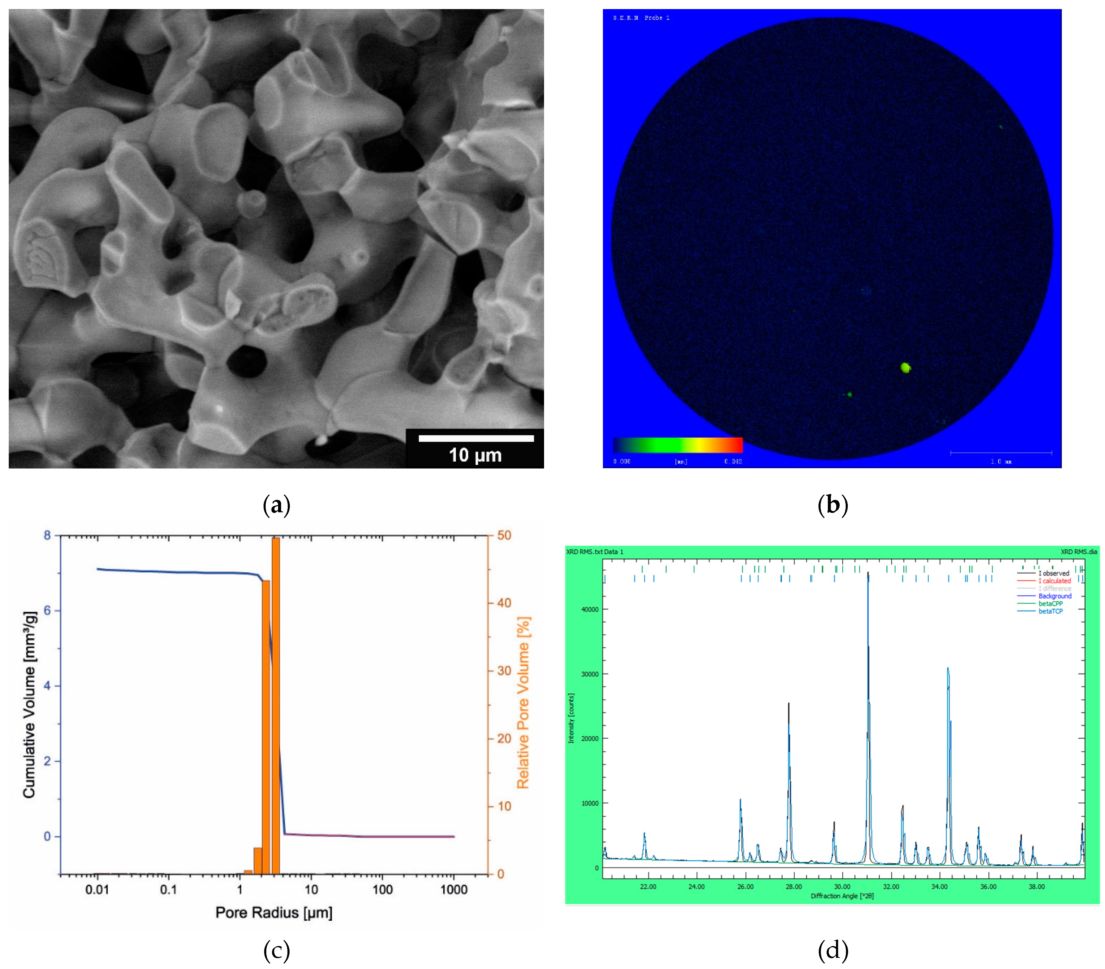
Figure 2.
Overview cell viability (a) and cytotoxicity (b).
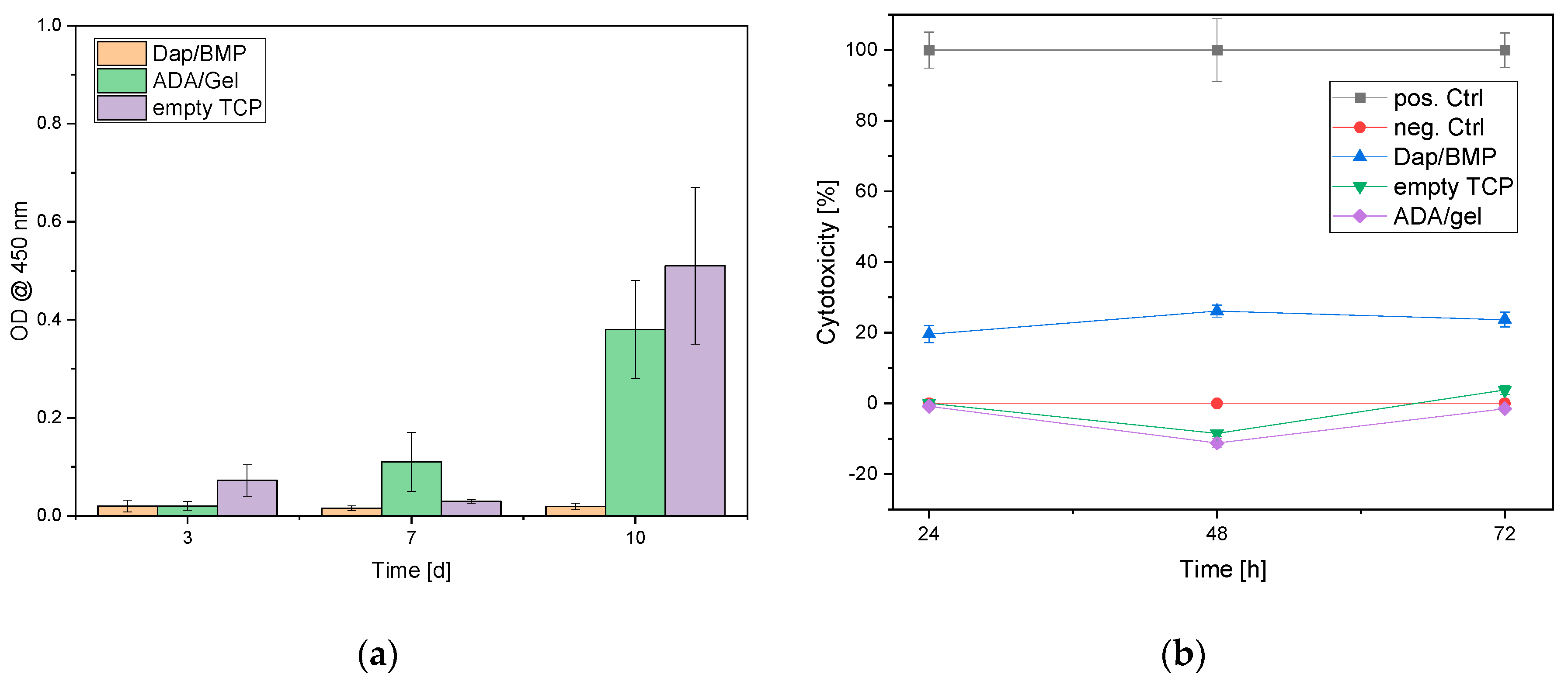

Table 1.
Molecular weights of alginate, ADA and gelatin at different times in the sterilization process.
Table 1.
Molecular weights of alginate, ADA and gelatin at different times in the sterilization process.
| Sample | Mn [kDa] | Mw [kDa] | Mz [kDa] | PDI (= Mw/Mn) |
|---|---|---|---|---|
| Alginate before plasma sterilization | 198 | 729 | 1440 | 3.68 |
| Alginate after plasma sterilization | 201 | 767 | 1530 | 3.81 |
| ADA unsterile | 52.4 | 316 | 1280 | 6.03 |
| ADA out of plasma-sterile alginate | 55.3 | 298 | 1160 | 5.38 |
| ADA out of unsterile alginate, plasma sterilized after manufacturing | 6.91 | 17.8 | 39.8 | 2.57 |
| ADA out of plasma-sterile alginate and plasma sterilized again after manufacturing | 13.1 | 41 | 116 | 3.13 |
| Gelatin before plasma sterilization | 16.3 | 114 | 254 | 6.99 |
| Gelatine after plasma sterilization | 15.9 | 105 | 231 | 6.61 |
Table 2.
Overview of released daptomycin and BMP-2 concentrations.
| Release period [d] | Daptomycin-Release [µg/ml] | BMP-2 Release [ng/ml] |
|---|---|---|
| 1 | 1026.05 ± 84.28 | 59.80 ± 90.0 |
| 2 | 318.01 ± 34.85 | 101.86 ± 92.04 |
| 3 | 72.43 ± 48.64 | 669.00 ± 148.40 |
| 6 | 8.62 ± 2.65 | 208.96 ± 43.20 |
| 9 | 2.76 ± 0.18 | 71.54 ± 47.63 |
| 14 | 1.45 ± 1.30 | 148.31 ± 100.86 |
| 21 | 0.37 ± 0.98 | 1.75 ± 6.14 |
| 28 | 0.18 ± 0.67 | 2.63 ± 9.15 |
| Recovery [%]: | 105.13 ± 9.14 | 0.72 ± 0.16 |
Table 3.
Relative cell counts MG-63 cells on composite vs empty ceremics.
| Sample | Relative cell count [%] | ||
| Alive | Intermediate | Dead | |
| Day3 | |||
| Dap/BMP-2 | 74.85 ± 48.16 | 5.56 ± 2.82 | 19.59 ± 9.08 |
| Control (empty ceramics) | 95.93 ± 14.21 | 0 ± 0 | 4.07 ± 1.20 |
| Control 2 (ADA/Gel no drugs) | 96.48 ± 31,22 | 0 ± 0 | 3.52 ± 1.93 |
| Day 7 | |||
| Dap/BMP-2 | 65.83 ± 27.14 | 0 ± 0 | 34.17 ± 23.41 |
| Control (empty ceramics) | 93.53 ± 11.33 | 0 ± 0 | 6.47 ± 1.48 |
| Control 2 (ADA/Gel no drugs) | 82.95 ± 31.48 | 0 ± 0 | 17.05 ± 4.80 |
| Day 10 | |||
| Dap/BMP-2 | 77.41 ± 63.13 | 0 ± 0 | 22.59 ± 22.97 |
| Control (empty ceramics) | 89.07 ± 53.61 | 0 ± 0 | 10.93 ± 3.84 |
| Control 2 (ADA/Gel no drugs) | 82.79 ± 27.79 | 0 ± 0 | 17.21 ± 10.67 |
intermediate = cells that were stained both green and red and were clearly not dead in the microscopy.
Table 4.
Overview of the antimicrobial activity of the released Daptomycin exemplary on one sample.
| Sample | Dilution levels of the original concentration [mg/l] | ||||||||||||
| 16 | 8 | 4 | 2 | 1 | 0.5 | 0.25 | 0.125 | 0.063 | 0.031 | GC | EW | MIC | |
| Control | - | - | - | - | - | - | +++ | +++ | +++ | +++ | +++ | - | 0.5 |
| 1291 | 8 | 4 | 2 | 1 | 0.5 | 0.25 | 0.125 | 0.063 | 0.031 | GC | EW | MIC | |
| D1P1 | - | - | - | - | - | - | +++ | +++ | +++ | +++ | +++ | - | 0.5 |
| 308 | 8 | 4 | 2 | 1 | 0.5 | 0.25 | 0.125 | 0.063 | 0.031 | GC | EW | MIC | |
| D2P1 | - | - | - | - | - | - | +++ | +++ | +++ | +++ | +++ | - | 0.5 |
| 110.5 | 8 | 4 | 2 | 1 | 0.5 | 0.25 | 0.125 | 0.063 | 0.031 | GC | EW | MIC | |
| DT3P1 | - | - | - | - | - | +++ | +++ | +++ | +++ | +++ | +++ | - | 1 |
| 18.59 | 8 | 4 | 2 | 1 | 0.5 | 0.25 | 0.125 | 0.063 | 0.031 | GC | EW | MIC | |
| D6P1 | - | - | - | - | - | - | +++ | +++ | +++ | +++ | +++ | - | 0.5 |
| - | 0.95 | 0.475 | 0.238 | 0.119 | 0.059 | 0.03 | 0.015 | 0.007 | 0.004 | GC | EW | MIC | |
| D9P1 | / | - | - | +++ | +++ | +++ | +++ | +++ | +++ | +++ | +++ | - | 0.475 |
| - | 0 | 0 | 0 | 0 | 0 | 0 | 0 | 0 | 0 | GC | EW | MIC | |
| D14P1 | / | ++ | +++ | +++ | +++ | +++ | +++ | +++ | +++ | +++ | +++ | - | > 0 |
| - | 0 | 0 | 0 | 0 | 0 | 0 | 0 | 0 | 0 | GC | EW | MIC | |
| D21P1 | / | ++ | +++ | +++ | +++ | +++ | +++ | +++ | +++ | +++ | +++ | - | > 0 |
| - | 0 | 0 | 0 | 0 | 0 | 0 | 0 | 0 | 0 | GC | EW | MIC | |
| D28P1 | / | ++ | +++ | +++ | +++ | +++ | +++ | +++ | +++ | +++ | +++ | - | > 0 |
D1…D28 = day 1 …28; GC = growth control, only bacteria; EW = empty well; (-) no growth; (+) growth; (++) strong growth; (+++) very strong growth.
Disclaimer/Publisher’s Note: The statements, opinions and data contained in all publications are solely those of the individual author(s) and contributor(s) and not of MDPI and/or the editor(s). MDPI and/or the editor(s) disclaim responsibility for any injury to people or property resulting from any ideas, methods, instructions or products referred to in the content. |
© 2023 by the authors. Licensee MDPI, Basel, Switzerland. This article is an open access article distributed under the terms and conditions of the Creative Commons Attribution (CC BY) license (https://creativecommons.org/licenses/by/4.0/).
Copyright: This open access article is published under a Creative Commons CC BY 4.0 license, which permit the free download, distribution, and reuse, provided that the author and preprint are cited in any reuse.


