
Submitted:
05 September 2023
Posted:
07 September 2023
You are already at the latest version
Abstract
Imaging-based biomarkers have developed as an effective tool in neurology, providing vital understandings of the structural, functional, and molecular changes associated with neurological disorders. Imaging techniques such as magnetic resonance imaging (MRI), positron emission tomography (PET), single-photon emission computed tomography (SPECT), and computed tomography (CT) have been widely employed to record disease-related alterations in the brain. These techniques provide a wide range of biomarkers, such as functional connectivity patterns, volumetric measurements, molecular imaging agents, and perfusion parameters, enabling the correct identification of neurological disorders. These biomarkers have proven useful in early diagnosis, disease progression tracking, therapy response prediction, and surgical planning. This review emphasizes the various obstacles and limitations that are associated with imaging-based biomarkers. Technical constraints, standardization obstacles, ethical concerns, regulatory challenges, and cost-effectiveness concerns all offer substantial barriers to wider use. It is vital to overcome these challenges if imaging biomarkers are to be successfully integrated into routine clinical practice. Imaging technology advancements like high-resolution imaging, multimodal imaging, and artificial intelligence-based analysis hold immense promise for imaging-based biomarkers in the future. While more study and standardization are needed, their ongoing development and integration into clinical practice have the potential to revolutionize the diagnosis, treatment, and management of neurological disorders, resulting in better patient care and outcomes.
Keywords:
PET
; MRI
; SPECT
; CT
; Depression
; Epilepsy
; Atrophy
1. Introduction
Neurological disorders present significant challenges in terms of accurate diagnosis and efficient treatment due to their complexity and heterogeneity. Traditional diagnostic approaches frequently rely on subjective assessments and clinical evaluations, which can lead to limitations in accuracy and reliability. However, the introduction of imaging-based biomarkers has transformed the area of neurology [1]. MRI, PET, SPECT, and CT are the imaging techniques that have distinct benefits in visualising and quantifying structural, functional, and molecular changes in the brain [2,3,4]. These imaging methods provide useful information regarding tissue integrity, neurochemical changes, and cerebral blood flow, among other things. Researchers and physicians can develop objective and quantitative biomarkers that aid in the diagnosis, prognosis, and monitoring of neurological illnesses by exploiting these imaging properties [5,6].
Imaging-based biomarkers (IBB) are important because they can give a non-invasive and thorough assessment of neurological disorders. These biomarkers allow for early detection, precise diagnosis, and distinction of disease subtypes. They also offer insights into disease progression, treatment response, and personalized therapeutic interventions [7,8]. Moreover, IBB facilitate the evaluation of novel therapies in clinical trials and contribute to the development of precision medicine approaches in neurology. The integration of IBB in clinical practice has the potential to transform patient care by improving diagnostic accuracy, enabling personalized treatment strategies, and facilitating disease management [9]. Clinicians can use IBB to make informed judgements about the best treatment options for specific patients, ultimately optimising results. Furthermore, IBB can help us better understand the underlying mechanisms and pathophysiology of neurological illnesses. These biomarkers provide crucial insights into disease processes by visualising and quantifying brain alterations, assisting in the identification of novel treatment targets and the development of tailored interventions [10].
2. Biomarkers in Neurological Diseases
2.1. Biomarkers for diagnosis
IBB utilize various imaging techniques to visualize and quantify structural, functional, or molecular changes in the brain [11,12]. Structural imaging techniques, MRI, provides precise images of the brain's architecture and can identify anomalies linked with neurological disorders [13,14]. Biomarkers obtained from structural imaging include the measurement of brain volume loss or atrophy, which is frequently employed as a biomarker for neurodegenerative conditions such as Alzheimer's disease (AD) or frontotemporal dementia [15]. White matter hyper-intensities seen on MRI scans are indicative of small vessel disease and can assist in diagnosing conditions like vascular dementia [16]. Functional imaging techniques capture brain activity and can help to identify patterns associated with the neurological diseases. Functional MRI (fMRI) measures changes in blood flow and oxygenation, can identify aberrant activation patterns in regions of interest, aiding in the diagnosis of conditions such as epilepsy or psychiatric disorders [17,18]. Positron Emission Tomography (PET) imaging with specific radiotracers can detect abnormal protein accumulation, such as amyloid plaques in Alzheimer's disease or dopaminergic dysfunction in Parkinson's disease [19,20,21]. Molecular Imaging Biomarkers techniques utilize radiotracers that bind to specific molecules or receptors in the brain. These biomarkers help diagnose neurological diseases by detecting molecular alterations associated with the condition [22,23,24]. The use of radiotracers like 18F-florbetapir or 11C-Pittsburgh compound B (PiB) in PET imaging allows for the visualization and quantification of amyloid plaques in Alzheimer's disease [20,25]. PET or SPECT imaging with radiotracers like 123I-FP-CIT or 18F-FDOPA can evaluate dopamine transporter function, aiding in the diagnosis of movement disorders like Parkinson's disease [26,27,28,29]. These imaging-based biomarkers provide objective and quantitative measures that assist in the diagnosis of neurological diseases. They complement clinical evaluations, improve diagnostic accuracy, and aid in early detection, allowing for timely intervention and appropriate management of these conditions.
2.2. Biomarkers for disease progression
IBB have proven valuable for assessing disease progression in neurological diseases. Structural imaging biomarkers are frequently employed as a biomarker for neurodegenerative conditions [30,31]. Longitudinal brain volume evaluation can reveal the rate of atrophy, which is valuable for tracking disease development such as AD, Multiple sclerosis (MS), or Huntington's disease (HD) [32,33,34,35,36]. Progressive ventricular enlargement can be seen in neurodegenerative disorders and can be used as a biomarker for disease progression, such as PD or AD [37,38,39,40,41]. Functional Imaging Biomarkers, such as fMRI or PET, can offer information regarding changes in brain function that occur as a result of disease improvement. These biomarkers can assist in determining the dynamic nature of neurological conditions. Changes in functional connectivity networks, as measured by fMRI, can signal disease progression in diseases such as AD, PD and MS [42,43,44,45,46,47,48,49]. PET imaging using radiotracers such as fluorodeoxyglucose (FDG) can assess glucose metabolism in the brain, which can indicate disease progression in AD [50,51,52]. PET and SPECT are molecular imaging modalities that can track the molecular changes associated with disease progression. These biomarkers enable the detection and quantification of specific molecular targets [53,54]. Tau protein build up, as measured by tau PET imaging, is a biomarker for disease progression in AD and other tauopathies [55,56]. Dopamine transporter availability changes, as measured by PET or SPECT imaging, can indicate disease progression in PD [57,58]. These imaging-based disease progression biomarkers provide important insights into the dynamic changes that occur in neurological disorders throughout time.
2.3. Biomarkers for treatment response
IBB provide objective and quantitative measures of changes in brain structure, function, or molecular profiles following therapeutic interventions. The evaluation of lesion volume on MRI images in conditions such as MS or stroke can show the efficacy of disease-modifying medicines or interventions targeted at minimizing the extent of brain damage [59,60,61]. Monitoring changes in hippocampus volume during conditions such as AD can act as a biomarker for treatment response and disease progression [62,63]. fMRI and PET scans provide information about changes in brain function and connectivity after treatment, indicating treatment response in disorders such as major depressive disorder (MDD) or schizophrenia [64,65,66]. Furthermore, these approaches can detect changes in brain activation patterns during specific tasks or cognitive problems, providing information regarding treatment response in disorders such as ADHD or traumatic brain injury [67,68,69,70,71].
These imaging-based biomarkers for treatment response provide valuable insights into the effects of therapeutic interventions on brain structure, function, or molecular profiles. They aid in assessing treatment efficacy, guiding treatment decisions, and optimizing patient management.
2.4. Predictive biomarkers
Predictive biomarkers derived from imaging techniques in neurological diseases are valuable tools for identifying patients who are more likely to respond to specific treatments or have a higher risk of disease progression. Combining imaging data with genetic information can help identify predictive biomarkers [72,73]. Certain genetic variants may influence treatment response or disease progression. For example, in multiple sclerosis, specific genetic markers, when combined with imaging biomarkers like lesion load or brain volume, can help predict the likelihood of disease progression or response to disease-modifying therapies [19,74]. Identifying specific imaging patterns or signatures that are associated with treatment response or disease progression can serve as predictive biomarkers. By identifying predictive biomarkers, clinicians can better stratify patients, optimize treatment selection, and personalize therapeutic interventions.
2.5. Prognostic biomarkers
Prognostic biomarkers provide insights into the likely disease course, progression, or outcomes for individual patients. MRI, provide valuable biomarkers for assessing disease prognosis. Measurement of brain volume loss or atrophy over time can serve as a prognostic biomarker in AD, PD or MS [75,76,77,78,79]. Greater rates of brain atrophy often indicate a more severe disease course or worse outcomes. In conditions like MS, the volume and distribution of brain lesions assessed through MRI can provide prognostic information. Higher lesion load often correlates with more aggressive disease progression or disability accumulation. Functional imaging techniques, such as fMRI or PET, offer insights into brain function and connectivity that can serve as prognostic biomarkers [80,81,82,83,84,85]. Alterations in functional connectivity networks assessed by fMRI can provide prognostic information in AD, stroke, or traumatic brain injury [86,87]. PET imaging can assess metabolic activity in the brain, providing prognostic information in conditions like brain tumors or epilepsy. Higher metabolic activity in certain regions may indicate more aggressive tumor behavior or seizure recurrence [88,89,90]. Molecular imaging techniques, including PET or SPECT, can offer prognostic biomarkers by assessing molecular targets associated with disease progression or treatment response.
3. Application of Imaging Biomarkers (IB) in Specific Neurological Diseases
IB play a crucial role in the evaluation and management of various neurological disorders. They provide valuable insights into the structural, functional, and molecular changes that occur in the brain, aiding in early diagnosis, differential diagnosis, disease staging, and monitoring of treatment response (Table 1).
3.1. AD and other dementias
The use of IB into the clinical practise has changed the way AD and other dementias are assessed and managed. Amyloid PET imaging with radiotracers like 18F-florbetapir (FTP) or PiB allows for the visualization and quantification of amyloid plaques in the brain. These biomarkers help in the early and accurate diagnosis of AD, as the presence of amyloid plaques is a hallmark of the disease [91,92,93,94]. Amyloid PET imaging can differentiate AD from other forms of dementia and aid in patient stratification for clinical trials [95,96,97]. Tau PET imaging, using FTP, allows for the detection and quantification of tau pathology in the brain. Tau pathology, including neurofibrillary tangles, is closely associated with disease progression in AD and other tauopathies [95,98,99,100]. Structural MRI is widely used in AD and other dementias to evaluate brain atrophy which differentiate between normal aging and pathological changes associated with neurodegenerative diseases [101,102]. Structural MRI biomarkers, such as hippocampal volume or whole-brain volume, can aid in the early detection and tracking of disease progression [103,104,105]. In AD and other dementias, fMRI can reveal alterations in functional connectivity patterns, such as disruptions in the default mode network [106,107]. These biomarkers aid in the understanding of disease mechanisms, assessing disease severity, and predicting cognitive decline.
3.2. Parkinson's disease
Imaging techniques such as PET or SPECT with radiotracers targeting dopamine transporters, such as 123I-FP-CIT or 18F-FDOPA, can assess the integrity and availability of dopaminergic neurons in the brain [108]. Dopamine transporter imaging also helps in monitoring disease progression and evaluating the response to dopaminergic therapies. fMRI or PET, provide insights into changes in brain function and connectivity in PD include resting-sate functional connectivity, task-based activation, structural imaging etc [109,110,111,112,113]. Altered functional connectivity patterns, such as disruptions in the default mode network or corticostriatal networks, have been observed in PD. fMRI or PET during specific motor tasks can assess changes in brain activation patterns. They provide information about the effects of PD on motor circuitry and help evaluate the response to therapeutic interventions, such as deep brain stimulation [114,115,116]. High-resolution MRI or specific imaging sequences, such as susceptibility-weighted imaging (SWI), can visualize and measure the substantia nigra. These imaging biomarkers aid in the detection of substantia nigra degeneration, a characteristic feature of PD [117,118,119]. MRI-based volumetric analysis can evaluate changes in specific brain regions, such as the basal ganglia or cortical areas. These biomarkers can help in disease staging and monitoring disease progression [120]. PET or SPECT, can provide insights into molecular changes associated with PD [121]. Radiotracers targeting alpha-synuclein aggregates, a pathological hallmark of PD, are under development [122,123]. These biomarkers may help in the early diagnosis and monitoring of disease progression. An illustartion with respect to immaging biomarkers used in AD and PD are depcetd in Figure 1.
3.3. Neuropsychiatric disorders
Imaging biomarkers have important applications in the evaluation and management of neuropsychiatric disorders. These biomarkers enhance our understanding of the underlying neural mechanisms and help guide personalized treatment approaches.
3.3.1. Major Depressive Disorder (MDD):
Resting-state functional connectivity assessed by fMRI can reveal alterations in functional networks, such as the default mode network or the limbic system, in individuals with MDD. These biomarkers help in understanding the neurobiology of depression and predicting treatment response. Structural MRI biomarkers, such as hippocampal volume, have been associated with MDD. Reduced hippocampal volume may indicate increased vulnerability to depression or treatment resistance [126,127,128,129].
3.3.2. Schizophrenia:
Structural imaging techniques can detect alterations in brain structure, such as decreased gray matter volume in specific regions like the prefrontal cortex or hippocampus. These biomarkers aid in the diagnosis and staging of schizophrenia [130,131]. Resting-state fMRI can reveal disrupted functional connectivity networks, such as the default mode network or the salience network, in individuals with schizophrenia [132,133]. These biomarkers help in understanding the underlying neural circuitry abnormalities and predicting clinical outcomes.
3.3.3. Bipolar Disorder:
Diffusion Tensor Imaging (DTI) can assess white matter integrity and identify alterations in fiber tracts in individuals with bipolar disorder. White matter connection disruptions may be used as indicators for disease diagnosis and development [134,135,136]. In patients with bipolar disorder, task-based fMRI can reveal aberrant activation patterns during cognitive activities, emotional processing, or reward processing [137,138,139]. These biomarkers help researchers understand the brain underpinnings of symptoms and predict treatment response.
3.3.4. Obsessive-Compulsive Disorder (OCD):
Individuals with OCD exhibit altered functional connection patterns, such as enhanced connectivity between the orbitofrontal cortex and the basal ganglia. These biomarkers aid in the knowledge of the brain circuits involved in the pathophysiology of OCD [140,141,142]. MRI-based measurements of cortical thickness can identify regional abnormalities in individuals with OCD, particularly in regions associated with cortico-striato-thalamo-cortical circuits [143,144,145].
3.4. Epilepsy
MRI, a structural imaging technique play a crucial role in the evaluation of epilepsy, helps in identifying the underlying structural abnormalities that can cause seizures. High-resolution structural MRI allows for the detection of focal cortical dysplasia, hippocampal sclerosis, brain tumors, vascular malformations, and other structural lesions associated with epilepsy. These biomarkers aid in the localization and characterization of the epileptogenic zone [146,147,148]. Quantitative analysis of brain regions, such as the hippocampus or amygdala, can help identify abnormalities related to mesial temporal lobe epilepsy (MTLE) [149,150]. Functional imaging techniques provide insights into brain function and connectivity in epilepsy. They help in localizing the epileptogenic zone and understanding the network abnormalities associated with seizures [151,152]. Simultaneous electroencephalography (EEG) and fMRI recordings allow for the identification of blood oxygen level-dependent (BOLD) signal changes associated with epileptic activity. These biomarkers aid in localizing the epileptogenic zone and mapping the functional connectivity network associated with seizures [153,154,155]. Resting-state fMRI can reveal changes in functional connectivity networks such as the default mode network or the salience network. These indicators can help with surgical planning by providing insight into the functional abnormalities associated with epilepsy [156,157]. PET, can provide biomarkers related to specific molecular targets in epilepsy. PET imaging with FDG can assess regional glucose metabolism in the brain. Hypometabolism in specific regions, such as the temporal lobe, may indicate the epileptogenic focus or the extent of the epileptic network [152,158]. PET imaging with radiotracers targeting specific neurotransmitter receptors, such as the GABA-A receptor or the serotonin transporter, can provide insights into neurotransmitter abnormalities in epilepsy [159].
3.5. Multiple sclerosis
Structural imaging techniques, such as MRI, play a crucial role in the diagnosis, monitoring, and prognosis of multiple sclerosis. The quantification of T2 hyperintense lesions on MRI scans provides a biomarker of disease burden and dissemination in space, aiding in the diagnosis and monitoring of MS progression. The detection and quantification of contrast-enhancing lesions on post-contrast MRI scans indicate acute inflammation and blood-brain barrier disruption. These biomarkers help in identifying disease activity and monitoring treatment response [160,161,162,163]. Longitudinal assessment of brain volume loss or atrophy provides a biomarker of neurodegeneration and disease progression in MS. It correlates with physical disability and cognitive impairment [164]. Diffusion Tensor Imaging (DTI) measures the diffusion of water molecules in the brain's white matter, providing insights into the integrity of fiber tracts [165]. DTI-based biomarkers in multiple sclerosis includes Fractional Anisotropy (FA) and Mean Diffusivity (MD). Reduced FA values indicate axonal damage and demyelination in white matter tracts. Decreased FA in specific regions, such as the corpus callosum or corticospinal tracts, correlates with physical disability and disease progression [166,167]. Increased MD values reflect tissue damage and inflammation. Elevated MD is associated with active lesions and predicts disability progression in MS [168]. Functional imaging techniques, such as fMRI, provide insights into brain activity and functional connectivity in multiple sclerosis, include Resting-State Functional Connectivity for altered functional connectivity patterns, such as disruptions in the default mode network or sensorimotor networks have been observed in MS [169].
PET imaging techniques offer molecular imaging biomarkers in multiple sclerosis. PET imaging with radiotracers targeting microglial activation, such as PK11195 or TSPO, can visualize neuroinflammation in MS. Increased uptake of these radiotracers is associated with disease activity and severity. Emerging PET radiotracers targeting myelin, such as 11C-PIB or 18F-GE180, hold promise for assessing myelin integrity and repair in MS [170,171].
3.6. Stroke
Computed Tomography (CT) Imaging is widely used for the initial assessment of stroke patients due to its availability and speed. Non-contrast CT scans can rapidly detect acute ischemic changes and differentiate between ischemic and hemorrhagic strokes. They provide information about the location and extent of early ischemic changes [172,173,174]. CT Angiography (CTA) is a technique for visualising blood arteries in the brain and determining occlusions or stenosis. It aids in the identification of the underlying aetiology of a stroke, such as atherosclerosis, arterial dissection, or embolism. MRI imaging techniques provide precise information on the structure of the brain and can distinguish between different stroke subtypes [175,176]. Diffusion-Weighted Imaging (DWI) can detect acute ischemic lesions within minutes of stroke onset. It provides valuable information about the affected brain tissue and helps determine the viability of the tissue at risk (Okorie et al., 2015). Perfusion-Weighted Imaging (PWI) assesses cerebral blood flow and can help to identify areas of hypoperfusion or ischemia. It aids in estimating the extent of the penumbra, which is potentially salvageable tissue, and guides decisions regarding reperfusion therapies [177,178]. Magnetic Resonance Angiography (MRA) provides detailed images of blood vessels and helps in visualizing the site and extent of vessel occlusion or stenosis. It aids in determining the appropriate treatment approach, such as endovascular intervention or anticoagulation [179,180,181]. Perfusion imaging techniques, including CT or MRI-based perfusion imaging, provide quantitative measures of cerebral blood flow and help assess tissue viability. They aid in determining the extent of the ischemic penumbra, which guides decisions regarding reperfusion therapies [182,183,184,185]. These imaging biomarkers by providing objective measures of brain damage and vascular abnormalities, imaging biomarkers enhance clinical decision-making, optimize patient care, and improve long-term outcomes in stroke patients (Table 2).
4. Challenges and Limitations
4.1. Technical limitations
While IBB have shown great promise in the treatment of neurological disorders, they also face certain technical limitations and challenges. Spatial resolution affects accurate detection of small-scale changes or lesions, with fMRI having lower resolution than histopathology. Temporal resolution can be slow, making it difficult to capture quick changes in brain activity. Signal-to-Noise Ratio (SNR) impacts data quality and reliability, with low SNR reducing sensitivity. Image artifacts, patient movement, and limited contrast agents contribute to lower SNR. Expertise is required for interpreting imaging biomarkers. Developing reliable algorithms for data analysis and extracting meaningful biomarkers is complex. Validation against gold-standard measures or clinical outcomes is necessary to establish reliability and clinical utility.
4.2. Standardization and reproducibility
Standardization and reproducibility pose challenges IBB due to protocol variations, acquisition parameters, data analysis methods, and software tools. Consensus on standardized imaging protocols is lacking, resulting in variability in image quality and biomarker measurements. Diverse analysis methods and software tools contribute to inconsistencies in measurements. Multicentre studies face additional challenges from equipment variations, requiring calibration and harmonization efforts. Longitudinal studies rely on consistent protocols and reliable follow-up imaging. Maintaining consistent acquisition parameters and minimizing platform or software changes are crucial for longitudinal biomarker comparability and reproducibility.
5. Future Directions and Potential Impact
Advancements in imaging technology, including multimodal imaging, molecular imaging, AI integration, and real-time imaging, will provide comprehensive insights into neurological disorders. This will enhance diagnosis, treatment decision-making, and targeted therapies. Furthermore, integrating imaging biomarkers into clinical practice holds promise for neurological disorder treatment. They enhance diagnostic accuracy, guide personalized treatment, monitor disease progression, aid surgical planning, and enable prognostic predictions. Integration facilitates early and accurate diagnosis, tailored interventions, and timely treatment adjustments. Imaging technology in telemedicine expands access to specialized care.
6. Conclusions
IBB have shown promise in neurological disorder treatment, aiding in diagnosis, treatment selection, monitoring, and prognostication. Technical limits, standardisation concerns, ethics, legislation, and cost-effectiveness are among the challenges. Imaging technology advancements such as high-resolution, multimodal, molecular imaging, AI, and real-time imaging will improve accuracy and sensitivity. Integration with personalized medicine and precision imaging will enhance outcomes. Standardizing protocols, analysis, and addressing ethical/regulatory aspects will facilitate clinical integration. The future of IBB in neurological disorder treatment is promising but requires concerted efforts for widespread implementation.
Author Contributions
Conceptualization, S.K., M.K.B. and N.L.W.; methodology, M.B.K., N.L.W., A.B.U., M.J.U., S.R.K.; software, M.B.K. and S.K.; validation, M.B.K., N.L.W., S.R.K., and S.K.; formal analysis, B.G.T., A.B.U., S.R.K., and M.B.K.; investigation, M.B.K., N.L.W., and S.K.; resources, S.R.K., M.B.K., and S.K.; data curation, M.B.K., N.L.W., S.R.K., B.G.T. and A.B.U.; writing—original draft preparation, M.B.K., N.L.B., S.K., S.R.K.; writing—review and editing, M.B.K., S.R.K., and S.K.; visualization, M.B.K. and SA.K.; supervision, S.K.; project administration, M.B.K., S.R.K. and S.K.; funding acquisition, S.K. and S.R.K. All authors have read and agreed to the published version of the manuscript.
Funding
This research received no external funding.
Institutional Review Board Statement
Not applicable
Informed Consent Statement
Not applicable
Data Availability Statement
Not applicable
Acknowledgments
This work was supported by Sejong University and Konkuk University, South Korea.
Conflicts of Interest
The authors declare no conflict of interest.
References
- Korolev, I.O.; Symonds, L.L.; Bozoki, A.C. Predicting Progression from Mild Cognitive Impairment to Alzheimer’s Dementia Using Clinical, MRI, and Plasma Biomarkers via Probabilistic Pattern Classification. PLoS ONE 2016, 11, e0138866. [CrossRef]
- Vrenken, H.; Jenkinson, M.; Horsfield, M.A.; Battaglini, M.; van Schijndel, R.A.; Rostrup, E.; Geurts, J.J.G.; Fisher, E.; Zijdenbos, A.; Ashburner, J.; et al. Recommendations to Improve Imaging and Analysis of Brain Lesion Load and Atrophy in Longitudinal Studies of Multiple Sclerosis. J Neurol 2013, 260, 2458–2471. [CrossRef]
- Enzinger, C.; Barkhof, F.; Ciccarelli, O.; Filippi, M.; Kappos, L.; Rocca, M.A.; Ropele, S.; Rovira, À.; Schneider, T.; de Stefano, N.; et al. Nonconventional MRI and Microstructural Cerebral Changes in Multiple Sclerosis. Nat Rev Neurol 2015, 11, 676–686. [CrossRef]
- Amiri, H.; de Sitter, A.; Bendfeldt, K.; Battaglini, M.; Gandini Wheeler-Kingshott, C.A.M.; Calabrese, M.; Geurts, J.J.G.; Rocca, M.A.; Sastre-Garriga, J.; Enzinger, C.; et al. Urgent Challenges in Quantification and Interpretation of Brain Grey Matter Atrophy in Individual MS Patients Using MRI. Neuroimage Clin 2018, 19, 466–475. [CrossRef]
- Jo, T.; Nho, K.; Saykin, A.J. Deep Learning in Alzheimer’s Disease: Diagnostic Classification and Prognostic Prediction Using Neuroimaging Data. Front Aging Neurosci 2019, 11. [CrossRef]
- Janssen, R.J.; Mourão-Miranda, J.; Schnack, H.G. Making Individual Prognoses in Psychiatry Using Neuroimaging and Machine Learning. Biol Psychiatry Cogn Neurosci Neuroimaging 2018, 3, 798–808. [CrossRef]
- Kim, B.; Kim, H.; Kim, S.; Hwang, Y. A Brief Review of Non-Invasive Brain Imaging Technologies and the near-Infrared Optical Bioimaging. Appl Microsc 2021, 51, 9. [CrossRef]
- Meijboom, R.; Wiseman, S.J.; York, E.N.; Bastin, M.E.; Valdés Hernández, M. del C.; Thrippleton, M.J.; Mollison, D.; White, N.; Kampaite, A.; Ng Kee Kwong, K.; et al. Rationale and Design of the Brain Magnetic Resonance Imaging Protocol for FutureMS: A Longitudinal Multi-Centre Study of Newly Diagnosed Patients with Relapsing-Remitting Multiple Sclerosis in Scotland. Wellcome Open Res 2022, 7, 94. [CrossRef]
- Chiu, F.-Y.; Yen, Y. Imaging Biomarkers for Clinical Applications in Neuro-Oncology: Current Status and Future Perspectives. Biomark Res 2023, 11, 35. [CrossRef]
- Smith, E.T.S. Clinical Applications of Imaging Biomarkers. Part 1. The Neuroradiologist’s Perspective. Br J Radiol 2011, 84, S196–S204. [CrossRef]
- Varghese, T.; Sheelakumari, R.; James, J.S.; Mathuranath, P. A Review of Neuroimaging Biomarkers of Alzheimer’s Disease. Neurol Asia 2013, 18, 239–248, doi:25431627.
- Bachli, M.B.; Sedeño, L.; Ochab, J.K.; Piguet, O.; Kumfor, F.; Reyes, P.; Torralva, T.; Roca, M.; Cardona, J.F.; Campo, C.G.; et al. Evaluating the Reliability of Neurocognitive Biomarkers of Neurodegenerative Diseases across Countries: A Machine Learning Approach. Neuroimage 2020, 208, 116456. [CrossRef]
- Du, F.; Cooper, A.J.; Thida, T.; Shinn, A.K.; Cohen, B.M.; Öngür, D. Myelin and Axon Abnormalities in Schizophrenia Measured with Magnetic Resonance Imaging Techniques. Biol Psychiatry 2013, 74, 451–457. [CrossRef]
- Du, F.; Öngür, D. Probing Myelin and Axon Abnormalities Separately in Psychiatric Disorders Using MRI Techniques. Front Integr Neurosci 2013, 7. [CrossRef]
- Frenzel, S.; Wittfeld, K.; Habes, M.; Klinger-König, J.; Bülow, R.; Völzke, H.; Grabe, H.J. A Biomarker for Alzheimer’s Disease Based on Patterns of Regional Brain Atrophy. Front Psychiatry 2020, 10. [CrossRef]
- Kynast, J.; Lampe, L.; Luck, T.; Frisch, S.; Arelin, K.; Hoffmann, K.-T.; Loeffler, M.; Riedel-Heller, S.G.; Villringer, A.; Schroeter, M.L. White Matter Hyperintensities Associated with Small Vessel Disease Impair Social Cognition beside Attention and Memory. Journal of Cerebral Blood Flow & Metabolism 2018, 38, 996–1009. [CrossRef]
- Glover, G.H. Overview of Functional Magnetic Resonance Imaging. Neurosurg Clin N Am 2011, 22, 133–139. [CrossRef]
- Perez, D.L.; Nicholson, T.R.; Asadi-Pooya, A.A.; Bègue, I.; Butler, M.; Carson, A.J.; David, A.S.; Deeley, Q.; Diez, I.; Edwards, M.J.; et al. Neuroimaging in Functional Neurological Disorder: State of the Field and Research Agenda. Neuroimage Clin 2021, 30, 102623. [CrossRef]
- Ayubcha, C.; Revheim, M.-E.; Newberg, A.; Moghbel, M.; Rojulpote, C.; Werner, T.J.; Alavi, A. A Critical Review of Radiotracers in the Positron Emission Tomography Imaging of Traumatic Brain Injury: FDG, Tau, and Amyloid Imaging in Mild Traumatic Brain Injury and Chronic Traumatic Encephalopathy. Eur J Nucl Med Mol Imaging 2021, 48, 623–641. [CrossRef]
- Uzuegbunam, B.C.; Librizzi, D.; Hooshyar Yousefi, B. PET Radiopharmaceuticals for Alzheimer’s Disease and Parkinson’s Disease Diagnosis, the Current and Future Landscape. Molecules 2020, 25, 977. [CrossRef]
- Maschio, C.; Ni, R. Amyloid and Tau Positron Emission Tomography Imaging in Alzheimer’s Disease and Other Tauopathies. Front Aging Neurosci 2022, 14. [CrossRef]
- Beaurain, M.; Salabert, A.-S.; Ribeiro, M.J.; Arlicot, N.; Damier, P.; Le Jeune, F.; Demonet, J.-F.; Payoux, P. Innovative Molecular Imaging for Clinical Research, Therapeutic Stratification, and Nosography in Neuroscience. Front Med (Lausanne) 2019, 6. [CrossRef]
- Villa, C.; Lavitrano, M.; Salvatore, E.; Combi, R. Molecular and Imaging Biomarkers in Alzheimer’s Disease: A Focus on Recent Insights. J Pers Med 2020, 10, 61. [CrossRef]
- Lu, F.-M.; Yuan, Z. PET/SPECT Molecular Imaging in Clinical Neuroscience: Recent Advances in the Investigation of CNS Diseases. Quant Imaging Med Surg 2015, 5, 433–447. [CrossRef]
- Bao, W.; Xie, F.; Zuo, C.; Guan, Y.; Huang, Y.H. PET Neuroimaging of Alzheimer’s Disease: Radiotracers and Their Utility in Clinical Research. Front Aging Neurosci 2021, 13. [CrossRef]
- Wallert, E.; Letort, E.; van der Zant, F.; Winogrodzka, A.; Berendse, H.; Beudel, M.; de Bie, R.; Booij, J.; Raijmakers, P.; van de Giessen, E. Comparison of [18F]-FDOPA PET and [123I]-FP-CIT SPECT Acquired in Clinical Practice for Assessing Nigrostriatal Degeneration in Patients with a Clinically Uncertain Parkinsonian Syndrome. EJNMMI Res 2022, 12, 68. [CrossRef]
- Booth, T.C.; Nathan, M.; Waldman, A.D.; Quigley, A.-M.; Schapira, A.H.; Buscombe, J. The Role of Functional Dopamine-Transporter SPECT Imaging in Parkinsonian Syndromes, Part 1. American Journal of Neuroradiology 2015, 36, 229–235. [CrossRef]
- Marner, L.; Korsholm, K.; Anderberg, L.; Lonsdale, M.N.; Jensen, M.R.; Brødsgaard, E.; Denholt, C.L.; Gillings, N.; Law, I.; Friberg, L. [18F]FE-PE2I PET Is a Feasible Alternative to [123I]FP-CIT SPECT for Dopamine Transporter Imaging in Clinically Uncertain Parkinsonism. EJNMMI Res 2022, 12, 56. [CrossRef]
- Palermo, G.; Giannoni, S.; Bellini, G.; Siciliano, G.; Ceravolo, R. Dopamine Transporter Imaging, Current Status of a Potential Biomarker: A Comprehensive Review. Int J Mol Sci 2021, 22, 11234. [CrossRef]
- Mazón, M.; Vázquez Costa, J.F.; Ten-Esteve, A.; Martí-Bonmatí, L. Imaging Biomarkers for the Diagnosis and Prognosis of Neurodegenerative Diseases. The Example of Amyotrophic Lateral Sclerosis. Front Neurosci 2018, 12. [CrossRef]
- Jovicich, J.; Barkhof, F.; Babiloni, C.; Herholz, K.; Mulert, C.; Berckel, B.N.M.; Frisoni, G.B. Harmonization of Neuroimaging Biomarkers for Neurodegenerative Diseases: A Survey in the Imaging Community of Perceived Barriers and Suggested Actions. Alzheimer’s & Dementia: Diagnosis, Assessment & Disease Monitoring 2019, 11, 69–73. [CrossRef]
- Wang, J.; Napoli, E.; Kim, K.; McLennan, Y.; Hagerman, R.; Giulivi, C. Brain Atrophy and White Matter Damage Linked to Peripheral Bioenergetic Deficits in the Neurodegenerative Disease FXTAS. Int J Mol Sci 2021, 22, 9171. [CrossRef]
- Marino, S.; Bonanno, L.; Lo Buono, V.; Ciurleo, R.; Corallo, F.; Morabito, R.; Chirico, G.; Marra, A.; Bramanti, P. Longitudinal Analysis of Brain Atrophy in Alzheimer’s Disease and Frontotemporal Dementia. Journal of International Medical Research 2019, 47, 5019–5027. [CrossRef]
- McWhinney, S.R.; Abé, C.; Alda, M.; Benedetti, F.; Bøen, E.; del Mar Bonnin, C.; Borgers, T.; Brosch, K.; Canales-Rodríguez, E.J.; Cannon, D.M.; et al. Association between Body Mass Index and Subcortical Brain Volumes in Bipolar Disorders–ENIGMA Study in 2735 Individuals. Mol Psychiatry 2021, 26, 6806–6819. [CrossRef]
- Kinnunen, K.M.; Mullin, A.P.; Pustina, D.; Turner, E.C.; Burton, J.; Gordon, M.F.; Scahill, R.I.; Gantman, E.C.; Noble, S.; Romero, K.; et al. Recommendations to Optimize the Use of Volumetric MRI in Huntington’s Disease Clinical Trials. Front Neurol 2021, 12. [CrossRef]
- Cole, J.H.; Poudel, R.P.K.; Tsagkrasoulis, D.; Caan, M.W.A.; Steves, C.; Spector, T.D.; Montana, G. Predicting Brain Age with Deep Learning from Raw Imaging Data Results in a Reliable and Heritable Biomarker. Neuroimage 2017, 163, 115–124. [CrossRef]
- Chen, C.-C. V.; Tung, Y.-Y.; Chang, C. A Lifespan MRI Evaluation of Ventricular Enlargement in Normal Aging Mice. Neurobiol Aging 2011, 32, 2299–2307. [CrossRef]
- Apostolova, L.G.; Green, A.E.; Babakchanian, S.; Hwang, K.S.; Chou, Y.-Y.; Toga, A.W.; Thompson, P.M. Hippocampal Atrophy and Ventricular Enlargement in Normal Aging, Mild Cognitive Impairment (MCI), and Alzheimer Disease. Alzheimer Dis Assoc Disord 2012, 26, 17–27. [CrossRef]
- Geriatric Neurology; Nair, A.K., Sabbagh, M.N., Eds.; John Wiley & Sons, Ltd: Chichester, UK, 2014; ISBN 9781118730676.
- Mak, E.; Su, L.; Williams, G.B.; Firbank, M.J.; Lawson, R.A.; Yarnall, A.J.; Duncan, G.W.; Mollenhauer, B.; Owen, A.M.; Khoo, T.K.; et al. Longitudinal Whole-Brain Atrophy and Ventricular Enlargement in Nondemented Parkinson’s Disease. Neurobiol Aging 2017, 55, 78–90. [CrossRef]
- Tanaka, M.; Vécsei, L. Editorial of Special Issue ‘Dissecting Neurological and Neuropsychiatric Diseases: Neurodegeneration and Neuroprotection.’ Int J Mol Sci 2022, 23, 6991. [CrossRef]
- Du, Y.; Fu, Z.; Calhoun, V.D. Classification and Prediction of Brain Disorders Using Functional Connectivity: Promising but Challenging. Front Neurosci 2018, 12. [CrossRef]
- Filippi, M.; Spinelli, E.G.; Cividini, C.; Agosta, F. Resting State Dynamic Functional Connectivity in Neurodegenerative Conditions: A Review of Magnetic Resonance Imaging Findings. Front Neurosci 2019, 13. [CrossRef]
- Cai, R.; Shen, G.; Wang, H.; Guan, Y. Brain Functional Connectivity Network Studies of Acupuncture: A Systematic Review on Resting-State FMRI. J Integr Med 2018, 16, 26–33. [CrossRef]
- Jonckers, E.; Van Audekerke, J.; De Visscher, G.; Van der Linden, A.; Verhoye, M. Functional Connectivity FMRI of the Rodent Brain: Comparison of Functional Connectivity Networks in Rat and Mouse. PLoS ONE 2011, 6, e18876. [CrossRef]
- Jonckers, E.; Van Audekerke, J.; De Visscher, G.; Van der Linden, A.; Verhoye, M. Functional Connectivity FMRI of the Rodent Brain: Comparison of Functional Connectivity Networks in Rat and Mouse. PLoS ONE 2011, 6, e18876. [CrossRef]
- ZHANG, H.; WANG, S.; XING, J.; LIU, B.; MA, Z.; YANG, M.; ZHANG, Z.; TENG, G. Detection of PCC Functional Connectivity Characteristics in Resting-State FMRI in Mild Alzheimer’s Disease. Behavioural Brain Research 2009, 197, 103–108. [CrossRef]
- Lin, H.; Cai, X.; Zhang, D.; Liu, J.; Na, P.; Li, W. Functional Connectivity Markers of Depression in Advanced Parkinson’s Disease. Neuroimage Clin 2020, 25, 102130. [CrossRef]
- Luo, C.; Song, W.; Chen, Q.; Zheng, Z.; Chen, K.; Cao, B.; Yang, J.; Li, J.; Huang, X.; Gong, Q.; et al. Reduced Functional Connectivity in Early-Stage Drug-Naive Parkinson’s Disease: A Resting-State FMRI Study. Neurobiol Aging 2014, 35, 431–441. [CrossRef]
- Toussaint, P.-J.; Perlbarg, V.; Bellec, P.; Desarnaud, S.; Lacomblez, L.; Doyon, J.; Habert, M.-O.; Benali, H. Resting State FDG-PET Functional Connectivity as an Early Biomarker of Alzheimer’s Disease Using Conjoint Univariate and Independent Component Analyses. Neuroimage 2012, 63, 936–946. [CrossRef]
- Yan, S.; Zheng, C.; Cui, B.; Qi, Z.; Zhao, Z.; An, Y.; Qiao, L.; Han, Y.; Zhou, Y.; Lu, J. Multiparametric Imaging Hippocampal Neurodegeneration and Functional Connectivity with Simultaneous PET/MRI in Alzheimer’s Disease. Eur J Nucl Med Mol Imaging 2020, 47, 2440–2452. [CrossRef]
- Zhang, M.; Guan, Z.; Zhang, Y.; Sun, W.; Li, W.; Hu, J.; Li, B.; Ye, G.; Meng, H.; Huang, X.; et al. Disrupted Coupling between Salience Network Segregation and Glucose Metabolism Is Associated with Cognitive Decline in Alzheimer’s Disease – A Simultaneous Resting-State FDG-PET/FMRI Study. Neuroimage Clin 2022, 34, 102977. [CrossRef]
- Pysz, M.A.; Gambhir, S.S.; Willmann, J.K. Molecular Imaging: Current Status and Emerging Strategies. Clin Radiol 2010, 65, 500–516. [CrossRef]
- Lu, F.-M.; Yuan, Z. PET/SPECT Molecular Imaging in Clinical Neuroscience: Recent Advances in the Investigation of CNS Diseases. Quant Imaging Med Surg 2015, 5, 433–447. [CrossRef]
- Okamura, N.; Harada, R.; Furumoto, S.; Arai, H.; Yanai, K.; Kudo, Y. Tau PET Imaging in Alzheimer’s Disease. Curr Neurol Neurosci Rep 2014, 14, 500. [CrossRef]
- Leuzy, A.; Cicognola, C.; Chiotis, K.; Saint-Aubert, L.; Lemoine, L.; Andreasen, N.; Zetterberg, H.; Ye, K.; Blennow, K.; Höglund, K.; et al. Longitudinal Tau and Metabolic PET Imaging in Relation to Novel CSF Tau Measures in Alzheimer’s Disease. Eur J Nucl Med Mol Imaging 2019, 46, 1152–1163. [CrossRef]
- Valli, M.; Mihaescu, A.; Strafella, A.P. Imaging Behavioural Complications of Parkinson’s Disease. Brain Imaging Behav 2019, 13, 323–332. [CrossRef]
- Xian, W.; Shi, X.; Luo, G.; Yi, C.; Zhang, X.; Pei, Z. Co-Registration Analysis of Fluorodopa and Fluorodeoxyglucose Positron Emission Tomography for Differentiating Multiple System Atrophy Parkinsonism Type From Parkinson’s Disease. Front Aging Neurosci 2021, 13. [CrossRef]
- Hemond, C.C.; Glanz, B.I.; Bakshi, R.; Chitnis, T.; Healy, B.C. The Neutrophil-to-Lymphocyte and Monocyte-to-Lymphocyte Ratios Are Independently Associated with Neurological Disability and Brain Atrophy in Multiple Sclerosis. BMC Neurol 2019, 19, 23. [CrossRef]
- Zhu, T.; Wang, L.; Tian, F.; Zhao, X.; Pu, X.-P.; Sun, G.-B.; Sun, X.-B. Anti-Ischemia/Reperfusion Injury Effects of Notoginsenoside R1 on Small Molecule Metabolism in Rat Brain after Ischemic Stroke as Visualized by MALDI–MS Imaging. Biomedicine & Pharmacotherapy 2020, 129, 110470. [CrossRef]
- Thomalla, G.; Rossbach, P.; Rosenkranz, M.; Siemonsen, S.; Krützelmann, A.; Fiehler, J.; Gerloff, C. Negative Fluid-Attenuated Inversion Recovery Imaging Identifies Acute Ischemic Stroke at 3 Hours or Less. Ann Neurol 2009, 65, 724–732. [CrossRef]
- Cummings, J. The National Institute on Aging—Alzheimer’s Association Framework on Alzheimer’s Disease: Application to Clinical Trials. Alzheimer’s & Dementia 2019, 15, 172–178. [CrossRef]
- Murphy, M.C.; Huston, J.; Jack, C.R.; Glaser, K.J.; Manduca, A.; Felmlee, J.P.; Ehman, R.L. Decreased Brain Stiffness in Alzheimer’s Disease Determined by Magnetic Resonance Elastography. Journal of Magnetic Resonance Imaging 2011, 34, 494–498. [CrossRef]
- Ding, Y.-D.; Yang, R.; Yan, C.-G.; Chen, X.; Bai, T.-J.; Bo, Q.-J.; Chen, G.-M.; Chen, N.-X.; Chen, T.-L.; Chen, W.; et al. Disrupted Hemispheric Connectivity Specialization in Patients with Major Depressive Disorder: Evidence from the REST-Meta-MDD Project. J Affect Disord 2021, 284, 217–228. [CrossRef]
- Köhler-Forsberg, K.; Jorgensen, A.; Dam, V.H.; Stenbæk, D.S.; Fisher, P.M.; Ip, C.-T.; Ganz, M.; Poulsen, H.E.; Giraldi, A.; Ozenne, B.; et al. Predicting Treatment Outcome in Major Depressive Disorder Using Serotonin 4 Receptor PET Brain Imaging, Functional MRI, Cognitive-, EEG-Based, and Peripheral Biomarkers: A NeuroPharm Open Label Clinical Trial Protocol. Front Psychiatry 2020, 11. [CrossRef]
- Fitzgerald, P.B.; Srithiran, A.; Benitez, J.; Daskalakis, Z.Z.; Oxley, T.J.; Kulkarni, J.; Egan, G.F. An FMRI Study of Prefrontal Brain Activation during Multiple Tasks in Patients with Major Depressive Disorder. Hum Brain Mapp 2008, 29, 490–501. [CrossRef]
- Paloyelis, Y.; Mehta, M.A.; Kuntsi, J.; Asherson, P. Functional MRI in ADHD: A Systematic Literature Review. Expert Rev Neurother 2007, 7, 1337–1356. [CrossRef]
- Zhu, C.-Z.; Zang, Y.-F.; Cao, Q.-J.; Yan, C.-G.; He, Y.; Jiang, T.-Z.; Sui, M.-Q.; Wang, Y.-F. Fisher Discriminative Analysis of Resting-State Brain Function for Attention-Deficit/Hyperactivity Disorder. Neuroimage 2008, 40, 110–120. [CrossRef]
- Amen, D.G.; Henderson, T.A.; Newberg, A. SPECT Functional Neuroimaging Distinguishes Adult Attention Deficit Hyperactivity Disorder From Healthy Controls in Big Data Imaging Cohorts. Front Psychiatry 2021, 12. [CrossRef]
- Stojanovski, S.; Felsky, D.; Viviano, J.D.; Shahab, S.; Bangali, R.; Burton, C.L.; Devenyi, G.A.; O’Donnell, L.J.; Szatmari, P.; Chakravarty, M.M.; et al. Polygenic Risk and Neural Substrates of Attention-Deficit/Hyperactivity Disorder Symptoms in Youths With a History of Mild Traumatic Brain Injury. Biol Psychiatry 2019, 85, 408–416. [CrossRef]
- Tran, K.; Wu, J. Case Report: Neuroimaging Analysis of Pediatric ADHD-Related Symptoms Secondary to Hypoxic Brain Injury. Brain Inj 2019, 33, 1402–1407. [CrossRef]
- Rowe, C.C.; Bourgeat, P.; Ellis, K.A.; Brown, B.; Lim, Y.Y.; Mulligan, R.; Jones, G.; Maruff, P.; Woodward, M.; Price, R.; et al. Predicting Alzheimer Disease with Β-amyloid Imaging: Results from the Australian Imaging, Biomarkers, and Lifestyle Study of Ageing. Ann Neurol 2013, 74, 905–913. [CrossRef]
- Yan, J.; Huang, H.; Risacher, S.L.; Kim, S.; Inlow, M.; Moore, J.H.; Saykin, A.J.; Shen, L. Network-Guided Sparse Learning for Predicting Cognitive Outcomes from MRI Measures. In; 2013; pp. 202–210. [CrossRef]
- Fleischer, V.; Ciolac, D.; Gonzalez-Escamilla, G.; Grothe, M.; Strauss, S.; Molina Galindo, L.S.; Radetz, A.; Salmen, A.; Lukas, C.; Klotz, L.; et al. Subcortical Volumes as Early Predictors of Fatigue in Multiple Sclerosis. Ann Neurol 2022, 91, 192–202. [CrossRef]
- Young, P.N.E.; Estarellas, M.; Coomans, E.; Srikrishna, M.; Beaumont, H.; Maass, A.; Venkataraman, A. V.; Lissaman, R.; Jiménez, D.; Betts, M.J.; et al. Imaging Biomarkers in Neurodegeneration: Current and Future Practices. Alzheimers Res Ther 2020, 12, 49. [CrossRef]
- Bottani, M.; Banfi, G.; Lombardi, G. The Clinical Potential of Circulating MiRNAs as Biomarkers: Present and Future Applications for Diagnosis and Prognosis of Age-Associated Bone Diseases. Biomolecules 2020, 10, 589. [CrossRef]
- Kamagata, K.; Andica, C.; Kato, A.; Saito, Y.; Uchida, W.; Hatano, T.; Lukies, M.; Ogawa, T.; Takeshige-Amano, H.; Akashi, T.; et al. Diffusion Magnetic Resonance Imaging-Based Biomarkers for Neurodegenerative Diseases. Int J Mol Sci 2021, 22, 5216. [CrossRef]
- Burns, D.K.; Alexander, R.C.; Welsh-Bohmer, K.A.; Culp, M.; Chiang, C.; O’Neil, J.; Evans, R.M.; Harrigan, P.; Plassman, B.L.; Burke, J.R.; et al. Safety and Efficacy of Pioglitazone for the Delay of Cognitive Impairment in People at Risk of Alzheimer’s Disease (TOMMORROW): A Prognostic Biomarker Study and a Phase 3, Randomised, Double-Blind, Placebo-Controlled Trial. Lancet Neurol 2021, 20, 537–547. [CrossRef]
- Ferrando, R.; Damian, A. Brain SPECT as a Biomarker of Neurodegeneration in Dementia in the Era of Molecular Imaging: Still a Valid Option? Front Neurol 2021, 12. [CrossRef]
- Sasabayashi, D.; Koike, S.; Nakajima, S.; Hirano, Y. Editorial: Prognostic Imaging Biomarkers in Psychotic Disorders. Front Psychiatry 2022, 13. [CrossRef]
- Sutphen, C.L.; Fagan, A.M.; Holtzman, D.M. Progress Update: Fluid and Imaging Biomarkers in Alzheimer’s Disease. Biol Psychiatry 2014, 75, 520–526. [CrossRef]
- Chew, S.; Atassi, N. Positron Emission Tomography Molecular Imaging Biomarkers for Amyotrophic Lateral Sclerosis. Front Neurol 2019, 10. [CrossRef]
- Cortese, R.; Giorgio, A.; Severa, G.; De Stefano, N. MRI Prognostic Factors in Multiple Sclerosis, Neuromyelitis Optica Spectrum Disorder, and Myelin Oligodendrocyte Antibody Disease. Front Neurol 2021, 12. [CrossRef]
- Melhem, E.R. MR Imaging Biomarkers in Amyotrophic Lateral Sclerosis. Acad Radiol 2017, 24, 1185–1186. [CrossRef]
- Kotian, R.P.; Koteshwar, P. FA Characteristics as Imaging Biomarkers Among the Indian Population in Early Parkinson’s Disease. In Diffusion Tensor Imaging and Fractional Anisotropy; Springer Nature Singapore: Singapore, 2022; pp. 131–153. [CrossRef]
- Konstantinou, N.; Pettemeridou, E.; Stamatakis, E.A.; Seimenis, I.; Constantinidou, F. Altered Resting Functional Connectivity Is Related to Cognitive Outcome in Males With Moderate-Severe Traumatic Brain Injury. Front Neurol 2019, 9. [CrossRef]
- Hoehn, M.; Aswendt, M. Structure–Function Relationship of Cerebral Networks in Experimental Neuroscience: Contribution of Magnetic Resonance Imaging. Exp Neurol 2013, 242, 65–73. [CrossRef]
- Clément, A.; Zaragori, T.; Filosa, R.; Ovdiichuk, O.; Beaumont, M.; Collet, C.; Roeder, E.; Martin, B.; Maskali, F.; Barberi-Heyob, M.; et al. Multi-Tracer and Multiparametric PET Imaging to Detect the IDH Mutation in Glioma: A Preclinical Translational in Vitro, in Vivo, and Ex Vivo Study. Cancer Imaging 2022, 22, 16. [CrossRef]
- Stafstrom, C.E.; Carmant, L. Seizures and Epilepsy: An Overview for Neuroscientists. Cold Spring Harb Perspect Med 2015, 5. [CrossRef]
- Huang, Z.; Liu, H.; Wu, Y.; Li, W.; Liu, J.; Wu, R.; Yuan, J.; He, Q.; Wang, Z.; Zhang, K.; et al. Automatic Brain Structure Segmentation for 18F-Fluorodeoxyglucose Positron Emission Tomography/Magnetic Resonance Images via Deep Learning. Quant Imaging Med Surg 2023, 13, 4447–4462. [CrossRef]
- Flores, S.; Chen, C.D.; Su, Y.; Dincer, A.; Keefe, S.J.; Perez-Carrillo, G.G.; Hornbeck, R.C.; Goyal, M.S.; Vlassenko, A.G.; Schwarz, S.; et al. Characteristics and Quantitative Impact of Off-target Skull Binding in Tau PET Studies of Alzheimer Disease. Alzheimer’s & Dementia 2022, 18. [CrossRef]
- Veitch, D.P.; Weiner, M.W.; Aisen, P.S.; Beckett, L.A.; DeCarli, C.; Green, R.C.; Harvey, D.; Jack, C.R.; Jagust, W.; Landau, S.M.; et al. Using the Alzheimer’s Disease Neuroimaging Initiative to Improve Early Detection, Diagnosis, and Treatment of Alzheimer’s Disease. Alzheimer’s & Dementia 2022, 18, 824–857. [CrossRef]
- Anand, K.; Sabbagh, M. Amyloid Imaging: Poised for Integration into Medical Practice. Neurotherapeutics 2017, 14, 54–61. [CrossRef]
- O’Brien, J.T.; Herholz, K. Amyloid Imaging for Dementia in Clinical Practice. BMC Med 2015, 13, 163. [CrossRef]
- Lois, C.; Gonzalez, I.; Johnson, K.A.; Price, J.C. PET Imaging of Tau Protein Targets: A Methodology Perspective. Brain Imaging Behav 2019, 13, 333–344. [CrossRef]
- Hellwig, S.; Frings, L.; Bormann, T.; Vach, W.; Buchert, R.; Meyer, P.T. Amyloid Imaging for Differential Diagnosis of Dementia: Incremental Value Compared to Clinical Diagnosis and [18F]FDG PET. Eur J Nucl Med Mol Imaging 2019, 46, 312–323. [CrossRef]
- Vandenberghe, R.; Nelissen, N.; Salmon, E.; Ivanoiu, A.; Hasselbalch, S.; Andersen, A.; Korner, A.; Minthon, L.; Brooks, D.J.; Van Laere, K.; et al. Binary Classification of 18F-Flutemetamol PET Using Machine Learning: Comparison with Visual Reads and Structural MRI. Neuroimage 2013, 64, 517–525. [CrossRef]
- Li, C.-H.; Chen, T.-F.; Chiu, M.-J.; Yen, R.-F.; Shih, M.-C.; Lin, C.-H. Integrated 18F-T807 Tau PET, Structural MRI, and Plasma Tau in Tauopathy Neurodegenerative Disorders. Front Aging Neurosci 2021, 13. [CrossRef]
- Ricci, M.; Cimini, A.; Camedda, R.; Chiaravalloti, A.; Schillaci, O. Tau Biomarkers in Dementia: Positron Emission Tomography Radiopharmaceuticals in Tauopathy Assessment and Future Perspective. Int J Mol Sci 2021, 22, 13002. [CrossRef]
- Hall, B.; Mak, E.; Cervenka, S.; Aigbirhio, F.I.; Rowe, J.B.; O’Brien, J.T. In Vivo Tau PET Imaging in Dementia: Pathophysiology, Radiotracer Quantification, and a Systematic Review of Clinical Findings. Ageing Res Rev 2017, 36, 50–63. [CrossRef]
- Hojjati, S.H.; Ebrahimzadeh, A.; Babajani-Feremi, A. Identification of the Early Stage of Alzheimer’s Disease Using Structural MRI and Resting-State FMRI. Front Neurol 2019, 10. [CrossRef]
- Farina, F.R.; Emek-Savaş, D.D.; Rueda-Delgado, L.; Boyle, R.; Kiiski, H.; Yener, G.; Whelan, R. A Comparison of Resting State EEG and Structural MRI for Classifying Alzheimer’s Disease and Mild Cognitive Impairment. Neuroimage 2020, 215, 116795. [CrossRef]
- Liu, M.; Li, F.; Yan, H.; Wang, K.; Ma, Y.; Shen, L.; Xu, M. A Multi-Model Deep Convolutional Neural Network for Automatic Hippocampus Segmentation and Classification in Alzheimer’s Disease. Neuroimage 2020, 208, 116459. [CrossRef]
- Sørensen, L.; Igel, C.; Liv Hansen, N.; Osler, M.; Lauritzen, M.; Rostrup, E.; Nielsen, M. Early Detection of Alzheimer’s Disease Using M <scp>RI</Scp> Hippocampal Texture. Hum Brain Mapp 2016, 37, 1148–1161. [CrossRef]
- Frisoni, G.B.; Fox, N.C.; Jack, C.R.; Scheltens, P.; Thompson, P.M. The Clinical Use of Structural MRI in Alzheimer Disease. Nat Rev Neurol 2010, 6, 67–77. [CrossRef]
- Wu, X.; Li, R.; Fleisher, A.S.; Reiman, E.M.; Guan, X.; Zhang, Y.; Chen, K.; Yao, L. Altered Default Mode Network Connectivity in Alzheimer’s Disease-A Resting Functional MRI and Bayesian Network Study. Hum Brain Mapp 2011, 32, 1868–1881. [CrossRef]
- Eyler, L.T.; Elman, J.A.; Hatton, S.N.; Gough, S.; Mischel, A.K.; Hagler, D.J.; Franz, C.E.; Docherty, A.; Fennema-Notestine, C.; Gillespie, N.; et al. Resting State Abnormalities of the Default Mode Network in Mild Cognitive Impairment: A Systematic Review and Meta-Analysis. Journal of Alzheimer’s Disease 2019, 70, 107–120. [CrossRef]
- Wallert, E.; Letort, E.; van der Zant, F.; Winogrodzka, A.; Berendse, H.; Beudel, M.; de Bie, R.; Booij, J.; Raijmakers, P.; van de Giessen, E. Comparison of [18F]-FDOPA PET and [123I]-FP-CIT SPECT Acquired in Clinical Practice for Assessing Nigrostriatal Degeneration in Patients with a Clinically Uncertain Parkinsonian Syndrome. EJNMMI Res 2022, 12, 68. [CrossRef]
- Li, W.; Lao-Kaim, N.P.; Roussakis, A.-A.; Martín-Bastida, A.; Valle-Guzman, N.; Paul, G.; Soreq, E.; Daws, R.E.; Foltynie, T.; Barker, R.A.; et al. Longitudinal Functional Connectivity Changes Related to Dopaminergic Decline in Parkinson’s Disease. Neuroimage Clin 2020, 28, 102409. [CrossRef]
- Wu, T.; Wang, L.; Chen, Y.; Zhao, C.; Li, K.; Chan, P. Changes of Functional Connectivity of the Motor Network in the Resting State in Parkinson’s Disease. Neurosci Lett 2009, 460, 6–10. [CrossRef]
- Shine, J.M.; Muller, A.J.; O’Callaghan, C.; Hornberger, M.; Halliday, G.M.; Lewis, S.J. Abnormal Connectivity between the Default Mode and the Visual System Underlies the Manifestation of Visual Hallucinations in Parkinson’s Disease: A Task-Based FMRI Study. NPJ Parkinsons Dis 2015, 1, 15003. [CrossRef]
- Wang, H.-Y.; Ren, L.; Li, T.; Pu, L.; Huang, X.; Wang, S.; Song, C.; Liang, Z. The Impact of Anxiety on the Cognitive Function of Informal Parkinson’s Disease Caregiver: Evidence from Task-Based and Resting-State FNIRS. Front Psychiatry 2022, 13. [CrossRef]
- Berman, S.B.; Miller-Patterson, C. PD and DLB: Brain Imaging in Parkinson’s Disease and Dementia with Lewy Bodies. In; 2019; pp. 167–185. [CrossRef]
- Cerasa, A.; Pugliese, P.; Messina, D.; Morelli, M.; Cecilia Gioia, M.; Salsone, M.; Novellino, F.; Nicoletti, G.; Arabia, G.; Quattrone, A. Prefrontal Alterations in Parkinson’s Disease with Levodopa-Induced Dyskinesia during FMRI Motor Task. Movement Disorders 2012, 27, 364–371. [CrossRef]
- Troisi Lopez, E.; Minino, R.; Liparoti, M.; Polverino, A.; Romano, A.; De Micco, R.; Lucidi, F.; Tessitore, A.; Amico, E.; Sorrentino, G.; et al. Fading of Brain Network Fingerprint in Parkinson’s Disease Predicts Motor Clinical Impairment. Hum Brain Mapp 2023, 44, 1239–1250. [CrossRef]
- Herz, D.M.; Eickhoff, S.B.; Løkkegaard, A.; Siebner, H.R. Functional Neuroimaging of Motor Control in Parkinson’s Disease: A Meta-Analysis. Hum Brain Mapp 2014, 35, 3227–3237. [CrossRef]
- Haller, S.; Badoud, S.; Nguyen, D.; Barnaure, I.; Montandon, M.-L.; Lovblad, K.-O.; Burkhard, P. Differentiation between Parkinson Disease and Other Forms of Parkinsonism Using Support Vector Machine Analysis of Susceptibility-Weighted Imaging (SWI): Initial Results. Eur Radiol 2013, 23, 12–19. [CrossRef]
- Reiter, E.; Mueller, C.; Pinter, B.; Krismer, F.; Scherfler, C.; Esterhammer, R.; Kremser, C.; Schocke, M.; Wenning, G.K.; Poewe, W.; et al. Dorsolateral Nigral Hyperintensity on 3.0T Susceptibility-Weighted Imaging in Neurodegenerative Parkinsonism. Movement Disorders 2015, 30, 1068–1076. [CrossRef]
- Bae, Y.J.; Song, Y.S.; Choi, B.S.; Kim, J.-M.; Nam, Y.; Kim, J.H. Comparison of Susceptibility-Weighted Imaging and Susceptibility Map-Weighted Imaging for the Diagnosis of Parkinsonism with Nigral Hyperintensity. Eur J Radiol 2021, 134, 109398. [CrossRef]
- Tuite, P. Brain Magnetic Resonance Imaging (MRI) as a Potential Biomarker for Parkinson’s Disease (PD). Brain Sci 2017, 7, 68. [CrossRef]
- Fang, Y.-H.D.; Chiu, S.-C.; Lu, C.-S.; Yen, T.-C.; Weng, Y.-H. Fully Automated Quantification of the Striatal Uptake Ratio of [ 99m Tc]-TRODAT with SPECT Imaging: Evaluation of the Diagnostic Performance in Parkinson’s Disease and the Temporal Regression of Striatal Tracer Uptake. Biomed Res Int 2015, 2015, 1–11. [CrossRef]
- Capotosti, F.; Vokali, E.; Molette, J.; Ravache, M.; Delgado, C.; Kocher, J.; Pittet, L.; Dimitrakopoulos, I.K.; Di-Bonaventura, I.; Touilloux, T.; et al. The Development of [ 18 F]ACI-12589, a High Affinity and Selective Alpha-synuclein Radiotracer, as a Biomarker for Parkinson’s Disease and Other Synucleinopathies. Alzheimer’s & Dementia 2021, 17. [CrossRef]
- Kotzbauer, P.T.; Tu, Z.; Mach, R.H. Current Status of the Development of PET Radiotracers for Imaging Alpha Synuclein Aggregates in Lewy Bodies and Lewy Neurites. Clin Transl Imaging 2017, 5, 3–14. [CrossRef]
- Sachin, K.; Sourabh, S. Alzheimer MRI Preprocessed Dataset. Data set. Kaggle 2022. [CrossRef]
- Mak, E.; Su, L.; Williams, G.B.; Firbank, M.J.; Lawson, R.A.; Yarnall, A.J.; Duncan, G.W.; Mollenhauer, B.; Owen, A.M.; Khoo, T.K.; et al. Longitudinal Whole-Brain Atrophy and Ventricular Enlargement in Nondemented Parkinson’s Disease. Neurobiol Aging 2017, 55, 78–90. [CrossRef]
- Javaheripour, N.; Li, M.; Chand, T.; Krug, A.; Kircher, T.; Dannlowski, U.; Nenadić, I.; Hamilton, J.P.; Sacchet, M.D.; Gotlib, I.H.; et al. Altered Resting-State Functional Connectome in Major Depressive Disorder: A Mega-Analysis from the PsyMRI Consortium. Transl Psychiatry 2021, 11, 511. [CrossRef]
- Zhang, S.; She, S.; Qiu, Y.; Li, Z.; Wu, X.; Hu, H.; Zheng, W.; Huang, R.; Wu, H. Multi-Modal MRI Measures Reveal Sensory Abnormalities in Major Depressive Disorder Patients: A Surface-Based Study. Neuroimage Clin 2023, 39, 103468. [CrossRef]
- Liu, W.; Wu, Z.; Sun, M.; Zhang, S.; Yuan, J.; Zhu, D.; Yan, G.; Hou, K. Association between Fasting Blood Glucose and Thyroid Stimulating Hormones and Suicidal Tendency and Disease Severity in Patients with Major Depressive Disorder. Bosn J Basic Med Sci 2022, 22, 635–642. [CrossRef]
- Wang, Z.; Zhang, D.; Guan, M.; Ren, X.; Li, D.; Yin, K.; Zhou, P.; Li, B.; Wang, H. Increased Thalamic Gray Matter Volume Induced by Repetitive Transcranial Magnetic Stimulation Treatment in Patients with Major Depressive Disorder. Front Psychiatry 2023, 14. [CrossRef]
- Karlsgodt, K.H.; Sun, D.; Cannon, T.D. Structural and Functional Brain Abnormalities in Schizophrenia. Curr Dir Psychol Sci 2010, 19, 226–231. [CrossRef]
- Cetin-Karayumak, S.; Di Biase, M.A.; Chunga, N.; Reid, B.; Somes, N.; Lyall, A.E.; Kelly, S.; Solgun, B.; Pasternak, O.; Vangel, M.; et al. White Matter Abnormalities across the Lifespan of Schizophrenia: A Harmonized Multi-Site Diffusion MRI Study. Mol Psychiatry 2020, 25, 3208–3219. [CrossRef]
- Huang, J.; Ke, P.; Chen, X.; Li, S.; Zhou, J.; Xiong, D.; Huang, Y.; Li, H.; Ning, Y.; Duan, X.; et al. Multimodal Magnetic Resonance Imaging Reveals Aberrant Brain Age Trajectory During Youth in Schizophrenia Patients. Front Aging Neurosci 2022, 14. [CrossRef]
- Li, S.; Hu, N.; Zhang, W.; Tao, B.; Dai, J.; Gong, Y.; Tan, Y.; Cai, D.; Lui, S. Dysconnectivity of Multiple Brain Networks in Schizophrenia: A Meta-Analysis of Resting-State Functional Connectivity. Front Psychiatry 2019, 10. [CrossRef]
- Podwalski, P.; Szczygieł, K.; Tyburski, E.; Sagan, L.; Misiak, B.; Samochowiec, J. Magnetic Resonance Diffusion Tensor Imaging in Psychiatry: A Narrative Review of Its Potential Role in Diagnosis. Pharmacological Reports 2021, 73, 43–56. [CrossRef]
- Chitty, K.M.; Lagopoulos, J.; Lee, R.S.C.; Hickie, I.B.; Hermens, D.F. A Systematic Review and Meta-Analysis of Proton Magnetic Resonance Spectroscopy and Mismatch Negativity in Bipolar Disorder. European Neuropsychopharmacology 2013, 23, 1348–1363. [CrossRef]
- Lagopoulos, J.; Hermens, D.F.; Hatton, S.N.; Tobias-Webb, J.; Griffiths, K.; Naismith, S.L.; Scott, E.M.; Hickie, I.B. Microstructural White Matter Changes in the Corpus Callosum of Young People with Bipolar Disorder: A Diffusion Tensor Imaging Study. PLoS ONE 2013, 8, e59108. [CrossRef]
- Shaffer, J.J.; Johnson, C.P.; Fiedorowicz, J.G.; Christensen, G.E.; Wemmie, J.A.; Magnotta, V.A. Impaired Sensory Processing Measured by Functional MRI in Bipolar Disorder Manic and Depressed Mood States. Brain Imaging Behav 2018, 12, 837–847. [CrossRef]
- Shi, X.-F.; Forrest, L.N.; Kuykendall, M.D.; Prescot, A.P.; Sung, Y.-H.; Huber, R.S.; Hellem, T.L.; Jeong, E.-K.; Renshaw, P.F.; Kondo, D.G. Anterior Cingulate Cortex Choline Levels in Female Adolescents with Unipolar versus Bipolar Depression: A Potential New Tool for Diagnosis. J Affect Disord 2014, 167, 25–29. [CrossRef]
- Magnotta, V.A.; Xu, J.; Fiedorowicz, J.G.; Williams, A.; Shaffer, J.; Christensen, G.; Long, J.D.; Taylor, E.; Sathyaputri, L.; Richards, J.G.; et al. Metabolic Abnormalities in the Basal Ganglia and Cerebellum in Bipolar Disorder: A Multi-Modal MR Study. J Affect Disord 2022, 301, 390–399. [CrossRef]
- Liu, W.; Hua, M.; Qin, J.; Tang, Q.; Han, Y.; Tian, H.; Lian, D.; Zhang, Z.; Wang, W.; Wang, C.; et al. Disrupted Pathways from Frontal-Parietal Cortex to Basal Ganglia and Cerebellum in Patients with Unmedicated Obsessive Compulsive Disorder as Observed by Whole-Brain Resting-State Effective Connectivity Analysis – a Small Sample Pilot Study. Brain Imaging Behav 2021, 15, 1344–1354. [CrossRef]
- Parmar, A.; Sarkar, S. Neuroimaging Studies in Obsessive Compulsive Disorder: A Narrative Review. Indian J Psychol Med 2016, 38, 386–394. [CrossRef]
- Picó-Pérez, M.; Moreira, P.S.; de Melo Ferreira, V.; Radua, J.; Mataix-Cols, D.; Sousa, N.; Soriano-Mas, C.; Morgado, P. Modality-Specific Overlaps in Brain Structure and Function in Obsessive-Compulsive Disorder: Multimodal Meta-Analysis of Case-Control MRI Studies. Neurosci Biobehav Rev 2020, 112, 83–94. [CrossRef]
- Boedhoe, P.S.W.; Heymans, M.W.; Schmaal, L.; Abe, Y.; Alonso, P.; Ameis, S.H.; Anticevic, A.; Arnold, P.D.; Batistuzzo, M.C.; Benedetti, F.; et al. An Empirical Comparison of Meta- and Mega-Analysis With Data From the ENIGMA Obsessive-Compulsive Disorder Working Group. Front Neuroinform 2019, 12. [CrossRef]
- Shaw, P.; Sharp, W.; Sudre, G.; Wharton, A.; Greenstein, D.; Raznahan, A.; Evans, A.; Chakravarty, M.M.; Lerch, J.P.; Rapoport, J. Subcortical and Cortical Morphological Anomalies as an Endophenotype in Obsessive-Compulsive Disorder. Mol Psychiatry 2015, 20, 224–231. [CrossRef]
- Cui, H.; Zhang, Y.; Zhao, Y.; Zhao, Y.; Ding, Q.; Chen, R.; Manssuer, L.; Zhang, C.; Liu, W.; Li, D.; et al. Mechanisms Underlying Capsulotomy for Refractory Obsessive-Compulsive Disorder: Neural Correlates of Negative Affect Processing Overlap with Deep Brain Stimulation Targets. Mol Psychiatry 2023. [CrossRef]
- Cendes, F.; Theodore, W.H.; Brinkmann, B.H.; Sulc, V.; Cascino, G.D. Neuroimaging of Epilepsy. In; 2016; pp. 985–1014.
- Álvarez-Linera Prado, J. Structural Magnetic Resonance Imaging in Epilepsy. Radiología (English Edition) 2012, 54, 9–20. [CrossRef]
- Sone, D.; Beheshti, I.; Maikusa, N.; Ota, M.; Kimura, Y.; Sato, N.; Koepp, M.; Matsuda, H. Neuroimaging-Based Brain-Age Prediction in Diverse Forms of Epilepsy: A Signature of Psychosis and Beyond. Mol Psychiatry 2021, 26, 825–834. [CrossRef]
- Memarian, N.; Thompson, P.M.; Engel, J.; Staba, R.J. Quantitative Analysis of Structural Neuroimaging of Mesial Temporal Lobe Epilepsy. Imaging Med 2013, 5. [CrossRef]
- Moghaddam, H.S.; Aarabi, M.H.; Mehvari-Habibabadi, J.; Sharifpour, R.; Mohajer, B.; Mohammadi-Mobarakeh, N.; Hashemi-Fesharaki, S.S.; Elisevich, K.; Nazem-Zadeh, M.-R. Distinct Patterns of Hippocampal Subfield Volume Loss in Left and Right Mesial Temporal Lobe Epilepsy. Neurological Sciences 2021, 42, 1411–1421. [CrossRef]
- van Graan, L.A.; Lemieux, L.; Chaudhary, U.J. Methods and Utility of EEG-FMRI in Epilepsy. Quant Imaging Med Surg 2015, 5, 300–312. [CrossRef]
- Centeno, M.; Carmichael, D.W. Network Connectivity in Epilepsy: Resting State FMRI and EEG–fMRI Contributions. Front Neurol 2014, 5. [CrossRef]
- Ebrahimzadeh, E.; Shams, M.; Rahimpour Jounghani, A.; Fayaz, F.; Mirbagheri, M.; Hakimi, N.; Rajabion, L.; Soltanian-Zadeh, H. Localizing Confined Epileptic Foci in Patients with an Unclear Focus or Presumed Multifocality Using a Component-Based EEG-FMRI Method. Cogn Neurodyn 2021, 15, 207–222. [CrossRef]
- Sadjadi, S.M.; Ebrahimzadeh, E.; Shams, M.; Seraji, M.; Soltanian-Zadeh, H. Localization of Epileptic Foci Based on Simultaneous EEG–FMRI Data. Front Neurol 2021, 12. [CrossRef]
- Ebrahimzadeh, E.; Saharkhiz, S.; Rajabion, L.; Oskouei, H.B.; Seraji, M.; Fayaz, F.; Saliminia, S.; Sadjadi, S.M.; Soltanian-Zadeh, H. Simultaneous Electroencephalography-Functional Magnetic Resonance Imaging for Assessment of Human Brain Function. Front Syst Neurosci 2022, 16. [CrossRef]
- Scheid, B.H.; Bernabei, J.M.; Khambhati, A.N.; Mouchtaris, S.; Jeschke, J.; Bassett, D.S.; Becker, D.; Davis, K.A.; Lucas, T.; Doyle, W.; et al. Intracranial Electroencephalographic Biomarker Predicts Effective Responsive Neurostimulation for Epilepsy Prior to Treatment. Epilepsia 2022, 63, 652–662. [CrossRef]
- Sparacia, G.; Parla, G.; Lo Re, V.; Cannella, R.; Mamone, G.; Carollo, V.; Midiri, M.; Grasso, G. Resting-State Functional Connectome in Patients with Brain Tumors Before and After Surgical Resection. World Neurosurg 2020, 141, e182–e194. [CrossRef]
- Li, R.; Wang, L.; Chen, H.; Guo, X.; Liao, W.; Tang, Y.-L.; Chen, H. Abnormal Dynamics of Functional Connectivity Density in Children with Benign Epilepsy with Centrotemporal Spikes. Brain Imaging Behav 2019, 13, 985–994. [CrossRef]
- Juhász, C.; Mittal, S. Molecular Imaging of Brain Tumor-Associated Epilepsy. Diagnostics 2020, 10, 1049. [CrossRef]
- Marcille, M.; Hurtado Rúa, S.; Tyshkov, C.; Jaywant, A.; Comunale, J.; Kaunzner, U.W.; Nealon, N.; Perumal, J.S.; Zexter, L.; Zinger, N.; et al. Disease Correlates of Rim Lesions on Quantitative Susceptibility Mapping in Multiple Sclerosis. Sci Rep 2022, 12, 4411. [CrossRef]
- Kaunzner, U.W.; Kang, Y.; Monohan, E.; Kothari, P.J.; Nealon, N.; Perumal, J.; Vartanian, T.; Kuceyeski, A.; Vallabhajosula, S.; Mozley, P.D.; et al. Reduction of PK11195 Uptake Observed in Multiple Sclerosis Lesions after Natalizumab Initiation. Mult Scler Relat Disord 2017, 15, 27–33. [CrossRef]
- Hemond, C.C.; Bakshi, R. Magnetic Resonance Imaging in Multiple Sclerosis. Cold Spring Harb Perspect Med 2018, 8. [CrossRef]
- Bruschi, N.; Boffa, G.; Inglese, M. Ultra-High-Field 7-T MRI in Multiple Sclerosis and Other Demyelinating Diseases: From Pathology to Clinical Practice. Eur Radiol Exp 2020, 4, 59. [CrossRef]
- Lorefice, L.; Fenu, G.; Pitzalis, R.; Scalas, G.; Frau, J.; Coghe, G.; Musu, L.; Sechi, V.; Barracciu, M.A.; Marrosu, M.G.; et al. Autoimmune Comorbidities in Multiple Sclerosis: What Is the Influence on Brain Volumes? A Case–Control MRI Study. J Neurol 2018, 265, 1096–1101. [CrossRef]
- Bao, J.; Tu, H.; Li, Y.; Sun, J.; Hu, Z.; Zhang, F.; Li, J. Diffusion Tensor Imaging Revealed Microstructural Changes in Normal-Appearing White Matter Regions in Relapsing–Remitting Multiple Sclerosis. Front Neurosci 2022, 16. [CrossRef]
- Mustafi, S.M.; Harezlak, J.; Kodiweera, C.; Randolph, J.S.; Ford, J.C.; Wishart, H.A.; Wu, Y.-C. Detecting White Matter Alterations in Multiple Sclerosis Using Advanced Diffusion Magnetic Resonance Imaging. Neural Regen Res 2019, 14, 114–123. [CrossRef]
- Fujimori, J.; Uryu, K.; Fujihara, K.; Wattjes, M.P.; Suzuki, C.; Nakashima, I. Measurements of the Corpus Callosum Index and Fractional Anisotropy of the Corpus Callosum and Their Cutoff Values Are Useful to Assess Global Brain Volume Loss in Multiple Sclerosis. Mult Scler Relat Disord 2020, 45, 102388. [CrossRef]
- Naismith, R.T.; Xu, J.; Tutlam, N.T.; Scully, P.T.; Trinkaus, K.; Snyder, A.Z.; Song, S.K.; Cross, A.H. Increased Diffusivity in Acute Multiple Sclerosis Lesions Predicts Risk of Black Hole. Neurology 2010, 74, 1694–1701. [CrossRef]
- Storelli, L.; Azzimonti, M.; Gueye, M.; Vizzino, C.; Preziosa, P.; Tedeschi, G.; De Stefano, N.; Pantano, P.; Filippi, M.; Rocca, M.A. A Deep Learning Approach to Predicting Disease Progression in Multiple Sclerosis Using Magnetic Resonance Imaging. Invest Radiol 2022, 57, 423–432. [CrossRef]
- Airas, L.; Rissanen, E.; Rinne, J.O. Imaging Neuroinflammation in Multiple Sclerosis Using TSPO-PET. Clin Transl Imaging 2015, 3, 461–473. [CrossRef]
- Morbelli, S.; Bauckneht, M.; Capitanio, S.; Pardini, M.; Roccatagliata, L.; Nobili, F. A New Frontier for Amyloid PET Imaging: Multiple Sclerosis. Eur J Nucl Med Mol Imaging 2019, 46, 276–279. [CrossRef]
- Birenbaum, D.; Bancroft, L.W.; Felsberg, G.J. Imaging in Acute Stroke. West J Emerg Med 2011, 12, 67–76, doi:Birenbaum et al., 2011.
- Potter, C.A.; Vagal, A.S.; Goyal, M.; Nunez, D.B.; Leslie-Mazwi, T.M.; Lev, M.H. CT for Treatment Selection in Acute Ischemic Stroke: A Code Stroke Primer. RadioGraphics 2019, 39, 1717–1738. [CrossRef]
- Muir, K.W. Imaging of Acute Stroke and Transient Ischaemic Attack. J Neurol Neurosurg Psychiatry 2005, 76, iii19–iii28. [CrossRef]
- Saba, L.; Anzidei, M.; Marincola, B.C.; Piga, M.; Raz, E.; Bassareo, P.P.; Napoli, A.; Mannelli, L.; Catalano, C.; Wintermark, M. Imaging of the Carotid Artery Vulnerable Plaque. Cardiovasc Intervent Radiol 2014, 37, 572–585. [CrossRef]
- Lim, S.; Yoon, H.Y.; Jang, H.J.; Song, S.; Kim, W.; Park, J.; Lee, K.E.; Jeon, S.; Lee, S.; Lim, D.-K.; et al. Dual-Modal Imaging-Guided Precise Tracking of Bioorthogonally Labeled Mesenchymal Stem Cells in Mouse Brain Stroke. ACS Nano 2019, 13, 10991–11007. [CrossRef]
- Sodaei, F.; Shahmaei, V. Identification of Penumbra in Acute Ischemic Stroke Using Multimodal MR Imaging Analysis: A Case Report Study. Radiol Case Rep 2020, 15, 2041–2046. [CrossRef]
- Kim, I.; Kang, H.; Yoon, H.J.; Chung, B.M.; Shin, N.-Y. Deep Learning–Based Image Reconstruction for Brain CT: Improved Image Quality Compared with Adaptive Statistical Iterative Reconstruction-Veo (ASIR-V). Neuroradiology 2021, 63, 905–912. [CrossRef]
- Degnan, A.J.; Gallagher, G.; Teng, Z.; Lu, J.; Liu, Q.; Gillard, J.H. MR Angiography and Imaging for the Evaluation of Middle Cerebral Artery Atherosclerotic Disease. American Journal of Neuroradiology 2012, 33, 1427–1435. [CrossRef]
- Campbell, B.C. V; Ma, H.; Ringleb, P.A.; Parsons, M.W.; Churilov, L.; Bendszus, M.; Levi, C.R.; Hsu, C.; Kleinig, T.J.; Fatar, M.; et al. Extending Thrombolysis to 4·5–9 h and Wake-up Stroke Using Perfusion Imaging: A Systematic Review and Meta-Analysis of Individual Patient Data. The Lancet 2019, 394, 139–147. [CrossRef]
- Aeschbacher, S.; Blum, S.; Meyre, P.B.; Coslovsky, M.; Vischer, A.S.; Sinnecker, T.; Rodondi, N.; Beer, J.H.; Moschovitis, G.; Moutzouri, E.; et al. Blood Pressure and Brain Lesions in Patients With Atrial Fibrillation. Hypertension 2021, 77, 662–671. [CrossRef]
- Demeestere, J.; Wouters, A.; Christensen, S.; Lemmens, R.; Lansberg, M.G. Review of Perfusion Imaging in Acute Ischemic Stroke. Stroke 2020, 51, 1017–1024. [CrossRef]
- Shah, M.K.; Shin, W.; Parikh, V.S.; Ragin, A.; Mouannes, J.; Bernstein, R.A.; Walker, M.T.; Bhatt, H.; Carroll, T.J. Quantitative Cerebral MR Perfusion Imaging: Preliminary Results in Stroke. Journal of Magnetic Resonance Imaging 2010, 32, 796–802. [CrossRef]
- Tarpley, J.; Franc, D.; Tansy, A.P.; Liebeskind, D.S. Use of Perfusion Imaging and Other Imaging Techniques to Assess Risks/Benefits of Acute Stroke Interventions. Curr Atheroscler Rep 2013, 15, 336. [CrossRef]
- Scalzo, F.; Nour, M.; Liebeskind, D.S. Data Science of Stroke Imaging and Enlightenment of the Penumbra. Front Neurol 2015, 6. [CrossRef]
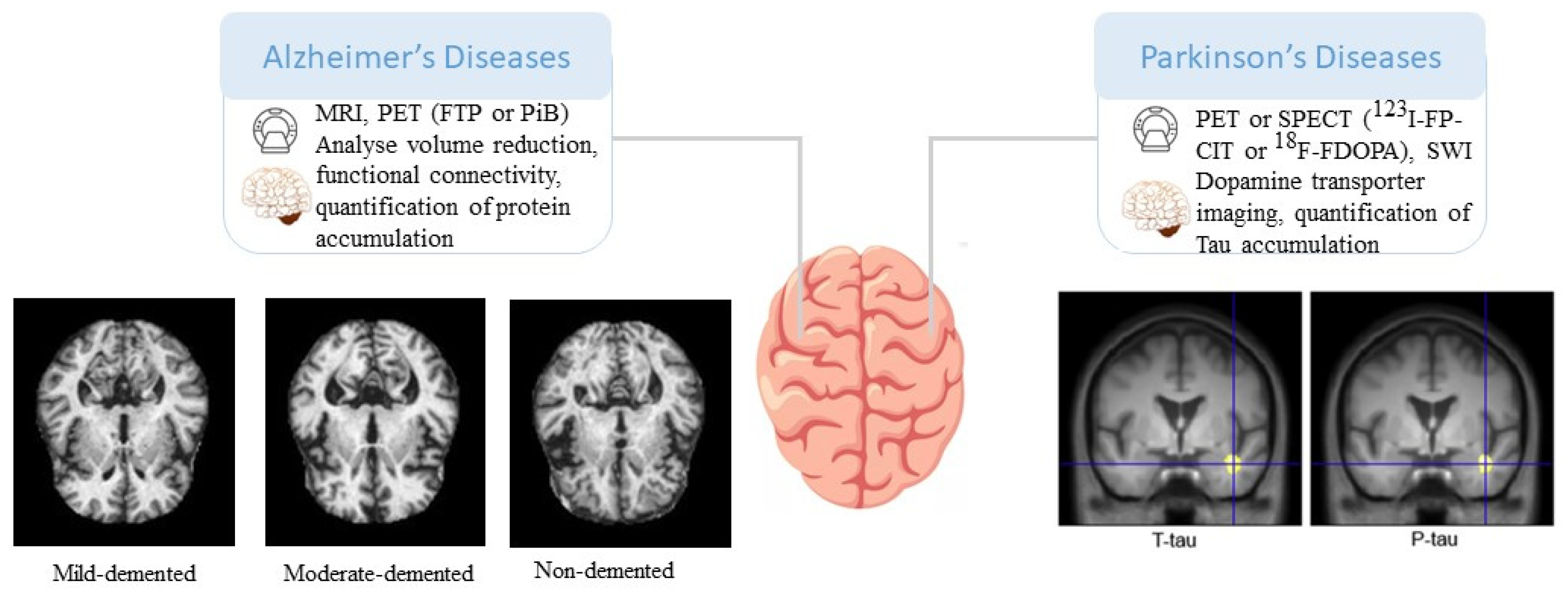

Table 1.
Outlining imaging-based biomarkers in specific neurological disorders.
| Neurological Disorders | Imaging-based Biomarkers |
|---|---|
| Alzheimer's disease | Amyloid PET imaging, Tau PET imaging, Hippocampal volume, Cortical thickness, Functional connectivity disruption, FDG-PET hypometabolism |
| Parkinson's disease | DaTscan SPECT imaging, Dopamine transporter imaging, Substantia nigra hyperechogenicity, Diffusion tensor imaging (DTI) alterations, Functional connectivity changes |
| Depression | Prefrontal cortex alterations, Hippocampal volume reduction, Amygdala hyperactivity, Default mode network dysfunction, Serotonin transporter imaging |
| Epilepsy | Hippocampal sclerosis on MRI, Cortical dysplasia on MRI, Epileptic network characterization using functional connectivity, PET/SPECT imaging for seizure focus localization |
| Multiple Sclerosis | Corticospinal tract degeneration on DTI, Whole-brain atrophy, Motor cortex hyperexcitability on fMRI, Hypometabolism on FDG-PET, Functional connectivity alterations |
| Stroke | Infarct volume and location on MRI, Perfusion imaging for assessment of ischemic penumbra, Collateral circulation evaluation, Functional connectivity changes, Vascular imaging (CTA/MRA) for stenosis/occlusion detection |
Table 2.
summarizing the imaging techniques commonly used in the assessment of stroke and their respective roles.
Table 2.
summarizing the imaging techniques commonly used in the assessment of stroke and their respective roles.
| Imaging Technique | Role |
|---|---|
| Non-contrast CT | Rapidly detects acute ischemic changes and differentiates between ischemic and hemorrhagic strokes. Provides information about the location and extent of early ischemic changes. |
| CT Angiography (CTA) | Visualizes blood vessels in the brain and identifies occlusions or stenosis. Helps determine the underlying cause of stroke. |
| MRI | Provides detailed information about brain structure and differentiates between stroke subtypes |
| Diffusion-Weighted Imaging | Detects acute ischemic lesions within minutes of stroke onset. Provides information about affected brain tissue and helps determine tissue viability. |
| Perfusion-Weighted Imaging | Assesses cerebral blood flow and identifies areas of hypoperfusion or ischemia. Aids in estimating the extent of the penumbra |
| Magnetic Resonance Angiography (MRA) | Provides detailed images of blood vessels. Helps visualize vessel occlusion or stenosis and determine treatment approach. |
| Perfusion Imaging (CT or MRI) | Provides quantitative measures of cerebral blood flow. Assists in assessing tissue viability and determining the extent of the ischemic penumbra. |
Disclaimer/Publisher’s Note: The statements, opinions and data contained in all publications are solely those of the individual author(s) and contributor(s) and not of MDPI and/or the editor(s). MDPI and/or the editor(s) disclaim responsibility for any injury to people or property resulting from any ideas, methods, instructions or products referred to in the content. |
© 2023 by the authors. Licensee MDPI, Basel, Switzerland. This article is an open access article distributed under the terms and conditions of the Creative Commons Attribution (CC BY) license (http://creativecommons.org/licenses/by/4.0/).
Copyright: This open access article is published under a Creative Commons CC BY 4.0 license, which permit the free download, distribution, and reuse, provided that the author and preprint are cited in any reuse.


