
Submitted:
16 August 2023
Posted:
17 August 2023
You are already at the latest version
Abstract
The thyroid-heart relationship has a long and articulated history of its own, a history that en-compasses physiological and pathophysiological knowledge, the latter particularly in the context of cardiac diseases such as heart failure, arrhythmias and ischaemic heart disease. In recent years, molecular biology studies, in an experimental context, have highlighted the extraordinary dialogue that exists among the two systems in the field of cardioprotection, being an extremely important area for the treatment of cardiac diseases in both acute and chronic phases. In addi-tion, in the last few years, several studies have been carried out on the prognostic impact of al-terations in thyroid function, including subclinical ones, in heart disease, in particular in heart failure and acute myocardial infarction, with evidence of a negative prognostic impact of these and therefore with the suggestion to treat these alterations in order to prevent cardiac events, such as death. This review provides a comprehensive summary of the heart-thyroid relation-ship.
Keywords:
thyroid
; heart
; thyroid abnormalities
; cardioprotection
; heart failure
; acute myocardial infarction
History of the relationship between heart and thyroid
The thyroid-heart relationship has a very long and articulated history, encompassing physiological and pathophysiological knowledge, especially in cases of heart disease such as heart failure (HF), arrhythmias, and ischemic heart disease. The hypothesis of a relationship between heart and thyroid was, in fact, born well over two hundred years ago, in 1813, when Caleb Hillier Parry, an expert on heart disease in England, first hypothesized a link between cardiac events and thyroid disease. His writings include a detailed description of eight cases of thyroid gland enlargement combined with the presence of heart palpitation [1].
About a century later, in the 19th century, the co-presence of cardiac disorders and thyroid disorders was described by many other authors. In particular in 1802 in Italy, Giuseppe Flajani, described the case of a 22-year-old patient with thyroid symptoms and 'extraordinary palpitations in the region of the heart' and in 1828 the term 'Kropfherz' or 'cardiac goiter' was coined [2,3]. The descriptions of other similar cases seem to spread like wildfire, in fact even in Dublin, during a lecture Robert James Graves described three patients (all women) with heart palpitations and goiter. These symptoms were also joined by the characteristic eye prominence found by William Stokes in another patient. In 1835 Graves then described these four cases as a cardiac syndrome [4] while Carl Adolph von Basedow simultaneously described the association between goiter, exophthalmos, and palpitations, focusing more on the eyes and giving rise to the Merseburg triad [5,6]. By the end of the 19th century and the beginning of the 20th century, the connection between these symptoms was recognized and made official; thus, more modern studies began. In 1896, Mobius contributed to highlighting a relationship between the thyroid gland and the heart by pointing out that patients died mainly of heart-related causes in thyrotoxicosis syndrome [7]. In 1898, Friedrich Kraus introduced the concept of 'toxic cardiac goiter’ [8]. Then, at the beginning of the 20th century, surgical hypothyroidism was proposed as an extracardiac approach for the treatment of ischaemic heart disease [9]. This approach, however, was abandoned due to poor results.
In 1918, Symmers noted that the sympathology present in thyrotoxic heart disease was virtually identical to that of clinical cases described as 'idiopathic dilatation and hypertrophy of the heart' [10]. In the same year, White and Aub studied the effect of thyroid disease on the heart by combining physiological parameters measured with the electrocardiogram [11]. At rest, hypertension, tachycardia, and tremors were frequently observed. There were also T-wave variations, tachycardia, paroxysms of auricular fibrillation, pronounced auricular fibrillation, and signs of left ventricular preponderance. Krumbhaar observed the effects of 'prolonged thyrotropic intoxication' on the hearts of 51 patients and demonstrated that these effects are in fact progressive [12]. His studies reported the presence of tachycardia, T-wave alterations, high pulse pressure, hypertrophy, arrhythmias and heart block. However, some of these alterations disappeared as the hyperthyroidism improved after medical or surgical treatment. Although several cases with the presence of auricular fibrillation had already been described, the first to describe this symptomatology in presence of a toxic thyroid adenoma was, in 1922, Schoonmaker and Webb until Wishart, in 1929, reported auricular fibrillation as the most frequently encountered rhythm disturbance in thyrotoxicosis, pointing out that this could, in fact, be the first detectable sign of thyrotoxicosis [13,14].
In 1922, Hamilton classified Thyrotoxicosis of the heart into two classes: the first without cardiac damage with tachycardia as the main symptom, the second with cardiac damage and no evidence of rheumatic or other heart disease [15]. The following year, Kerr and Hensel described the cardiovascular characteristics of 181 thyrotoxic patients at the University of California Hospital, showing that tachycardia was the earliest symptom [16]. In 1924, Hamilton first described cases of adenomatous or exophthalmic goiter complicated by congestive heart failure, finding that these patients could be cured after thyroid removal [17]. In the following years, some research was published on this subject [18,19]. In the 1940s, it became possible to inhibit thyroid gland with the use of chemical compounds. Raab and Sharpey-Schafer successfully treated patients with angina pectoris and severe congestive HF using thiouracil, which reduced heart's sensitivity to epinephrine [20,21]. Canary et al. in the 1957 instead used antiadrenergic agents in the treatment of hyperthyroidism [22]. In the 1970s, beta-adrenergic blocking drugs, represented by D,L-propranolol, were finally incorporated into treatment, improving the adrenergic signs and symptoms associated with hyperthyroidism [23]. Also in the 1970s, an interventional study called the Coronary Drug Project (CDP) demonstrated negative outcomes on the pro-arrhythmic effects of D-T4 (inactive form of thyroxine, while L-T4 represents the active form) [24]. Indeed, the clinical outcomes of administering 6 mg/day of D-T4 were unclear (more than doubling endogenous T4 production) [25]. Only later was it discovered that the dose of D-T4 was actually contaminated with a high level of active L-T4, so the total dose administered was much higher than that expected to correct manifest hypothyroidism [26]. Thus, D-T4 was indeed shown to have a toxic effect in patients with coronary artery disease, as was already hypothesized before the study began. It is precisely for this reason, therefore, that CDP was unable to provide relevant information on TH supplementation in the treatment of coronary patients. Despite numerous studies, the question of a connection between hyperthyroidism and HF was a source of debate for a long time, partly due to the clinic-pathological diversity of patients with these diseases [27,28]. To better understand this connection, animal studies were also conducted, particularly in rabbits treated with thyroxine [29]. While in the mid-20th century, cardiac damage in hyperthyroidism was thought to be due to an interaction between direct toxic effects on cardiac muscles and indirect effects due to increased metabolism and oxygen consumption, it was not until Tata et al. that the TH effect on cardiac physiology and function was actually understood, demonstrating a TH effect on 44 protein synthesis at the transcription level [30,31]. Subsequent studies showed a correlation between changes in myosin ATPase activity and the contractile properties of the heart and in 1972, it was discovered through the rat study that the basic unit of TH action is the triiodothyronine nuclear receptor complex [32,33]. TH has also been shown to regulate the expression of ventricular myosin isoenzymes by stimulating α-MHC mRNA synthesis and inhibiting β-MHC mRNA expression [34]. In the late 1980s, it was finally demonstrated that hyperthyroidism was able to induce a 61% decrease in phospholamban mRNA level and that cardiac genes regulated by TH include sodium-potassium ATPase ion channels, voltage-gated potassium channels, sodium-calcium exchanger and β1-adrenergic receptor genes [35-37].
Thyroid and heart: a tight physiological relation
THs strongly influence the growth, development, and metabolism of cells, organs, and systems. Among these, THs play a pivotal role in cardiovascular (CV) homeostasis maintenance, both directly influencing all components of the system, and indirectly via regulation of autonomous nervous system, renin-angiotensin-aldosterone system, and systemic vasculature, with involvement in cardiac contractility, electrophysiological functions, and cardiac morphology and structure [38].
These effects are mainly mediated by different signaling pathways clustered into genomic and non-genomic actions. THs influence cardiac function with genomic pathways through binding to nuclear receptors that contribute to the regulation of target gene expression. In more details, these actions, inducing the enhancement of contractile function and diastolic relaxation, are essentially due to gene expression through specific nuclear a and bTH receptors (TRs) [39]. T3 regulates different myocyte genes related to cardiac contractile function, such as sarco-plasmic reticulum (SR)Ca2þ ATPase (SERCA2), phospholamban (PLB), and the myosin heavy chains (MHC), α and β, that encode the protein isoforms of the thick filament. In this regard, many plasma-membrane ion transporters involved in synchronising electrochemical functions in the myocardium, and different voltage-gated potassium channels are regulated by T3 [40]. In the cardiac myocite and on the systemic vasculature, THs has also non-genomic effects, not involving TH response elements (TRE)-mediated transcriptional events via interactions with cytoplasmic and membrane-associated TRs. These properties are related to changes in various membrane ion channels for sodium, potassium, and calcium, both in the heart and vascular smooth muscle cells (VSM). THs exert their effects directly on myocardial structure, modifying the interstitial collagen content, promoting the development of angiogenesis, and consequently regulate cardiac function trough chronotropic, inotropic, and dromotropic effects. Also, THs, via direct action on mitochondria influence inflammation, neuroendocrine systems, and oxidative stress pathways [41]. Usually, the non-genomic and genomic actions of T3 work in concert to regulate cardiac function and CV hemodynamics. As depicted in Figure 1, TH actions on the heart and peripheral vasculature are realised in a decreased systemic vascular resistance (SVR) and an increased heart rate at rest, left ventricular contractility, and blood volume. THs are involved also in the control of cardiac pacemaker activity influencing the action potential duration and repolarization currents in myocytes and regulating the transcription of the pacemaker-related genes [42,43]. Furthermore, although not all studies show a correlation, evidences obtained on studies of community-based population indicate that thyroid function is involved into control of blood pressure [44]. THs enhance basal metabolic rate in mostly organs and tissues with subsequent raised metabolic demands that lead to modifications in cardiac output, SVR, and blood pressure [44].
Hyperthyroidism, Hyporthyroidism and Subclinical conditions
It is now recognised that clinical characterization of abnormal thyroid metabolism is linked to the TH effects on the heart and CV system [45] (see Figure 1). Both pathological conditions, hyperthyroidism and hypothyroidism cause alterations in cardiac patterns, such as contractility, myocardial oxygen consumption, cardiac output, blood pressure, and SVR [46].
Thyroid diseases are quite common, with a prevalence in female population between 9% and 15%, while in male population the incidence is lower, probably due to autoimmune mechanisms for the most widespread forms, including both Graves’ and Hashimoto’s disease [47].
Hyperthyroidism is a condition affected by stimulation of the TSH receptors by autoantibodies (Graves’ disease) or as a consequence of the autonomous production of THs by thyroid nodules [48]. In patients with hyperthyroidism, many of the clinical consequences are related to the heart and CV system, including palpitations, exercise intolerance, dyspnea on exertion, widened pulse pressure, and occasionally atrial fibrillation (see Figure 1). In addition, peripheral vascular resistance is reduced, while cardiac contractility and heart rate at rest are improved, resulting in an increase by 50% to 300% of the normal of cardiac output [49]. The reduction of SVR determines low renal perfusion, which in turn sets the renin-angiotensin-aldosterone system (RAAS) into action, leading to a growth of total blood volume. Cardiac arrhythmias or electrocardiogram alterations, sinus tachycardia, atrial fibrillation, and reduced PR and QT intervals, are documented in patients with hyperthyroidism, and, in rare instances, also atrio-ventricular blockage [50,51]. Hyperthyroidism has been shown to result in haemodynamic changes with a lowered ejection fraction (EF) and cardiac output due to a decrease in myocardial contractile reserve [52]. Diametrically opposed CV signs and symptoms, as bradycardia, diastolic hypertension, cold intolerance, and fatigue are typical for hypothyroidism. Hypothyroidism occurs in 3% of the adult female population and is linked to an increase of SVR, a reduction in cardiac contractility and cardiac output, and an enhanced atherosclerosis [53]. In contrast to hyperthyroidism in which atrial arrhythmias are well documented, hypothyroidism is characterized by a prolonged of QT interval that, in turn, affect to ventricular irritability [54].
Subclinical hyperthyroidism is a condition characterized by low serum TSH levels and normal levels of serum T3 and T4. Usually, its prevalence increases in advancing age with concomitant risk for CV mortality and atrial fibrillation [55,56]. Clinical evidences have documented a relationship between subclinical hyperthyroidism and incident CV death, HF, and atrial fibrillation [57-59]. Subclinical hypothyroidism is instead the condition associated with serum TSH level higher than normal range, but normal TH levels. Although subclinical disease is frequently “asymptomatic,” cardiac characterization of patients with subclinical hypothyroidism displays alterations such as an impaired ventricular filling, systolic functional changes, an increased C-Reactive protein, as risk factors [60-62]. Atherosclerosis, coronary heart disease, and myocardial infarction risk in female population (aged 60 and older) were also documented [63].
Thyroid and cardioprotection
Cardioprotection has been defined as including any intervention (and as such both physiological adaptive and compensatory mechanisms as well as therapeutic strategies) that contribute to heart preservation by reducing or even preventing myocardial damage [64]. TH exert recognized cardioprotective effects through modulation of different key cellular pathways, including preservation of mitochondrial activity and morphology, antifibrotic and proangiogenic actions, and promotion of cell regeneration and growth after the postischemic injury [65]. Specifically, the large amount of available experimental data suggest multiple TH effects such as regulation of prosurvival pathways (e.g. activation of PI3K)/Akt and PKC, and inhibition of p38MAPK signaling), remodeling of myocardial interstitium (e.g. TGF-β1 signaling), proangiogenic effects on coronary circulation (e.g. mediated by integrin αVβ3 and HIF-1a expression), anti-inflammatory and anti-oxidative properties (e.g. cytokine reduction, direct/indirect antioxidant action as iodide, can act scavenger of free radicals, or enhancement or inhibition of the activity of antioxidant enzymes and free radical scavengers), effects on the neuroendocrine system (e.g. deactivation of the neuroendocrine system, characterized by a reduction in plasma levels of noradrenaline, NTproBNP, and aldosterone), epigenetic effects and chromatin changes (e.g. regulation of α and β-MHC genes), post-transcriptional regulation of miRNA (e.g. miR-214, miR-208) [41].
In particular, 3,5,3′-triiodothyronine (T3) and its precursor thyroxine (T4), are considered key regulators of mitochondrial function [66]. Accordingly, reduced levels of these hormones induce unbalance of cardiac mitochondrial activity, and subsequently leads to adverse consequences at cardiac muscle level within the myocardium [67]. Thus, the low T3 syndrome (LT3S), a status characterized by decreased total serum T3 and free T3 (fT3) with normal levels of thyroxine (T4) and thyrotropin (TSH), observed in acute post-ischemic and chronic heart pathologies and generally considered an adaptive response, can actually retains negative effects, favouring the progressive worsening of cardiac function and myocardial remodelling and representing a strong predictor of mortality and adverse CV events in these patients [68].
In view of the beneficial effects of TH on CV function, several attempts have been made utilizing TH replacement therapy in short-term and long-term administration, T3 or T4 at different doses and modality of administration [68]. However, there are great differences in the population cohorts in which these treatments are given, including HF patients with a normal or abnormal TH profile, stable or unstable HF, or acute myocardial infarction (AMI) patients [68]. Moreover, attention must be paid to avoid important adverse effects due to possible pharmacological-induced hyperthyroidism. Consequently, a shared consensus on the use of the TH replacement therapy has not been achieved yet by the scientific community. Nonetheless, the American Association of Clinical Endocrinologists, in conjunction with the American Thyroid Association, guidelines propose TH treatment in patients with HF and TSH levels exceeding the high normality reference limit up to 10 μIU/mL, for restoration and maintenance of a normal TH status, monitoring status in order to prevent over-treatment [69].
Recently, a number of additive underlying mechanisms related to TH the cardioprotective effects of TH are furtherly emerging, detailing more and more the possible key cellular pathways involved.
One recent advancement concerns the opening of the ATP-sensitive mitochondrial potassium channel (mitoK-ATP), with an important antioxidant and protective role against post-ischemia/reperfusion mitochondrial dysfunction and cell loss [70]. In particular, data obtained in experimental (rats) and in vitro (rat cardiomyocytes) models suggested that T3 administration (given to restore the physiological concentration) was able to modulate the expression level of both the channel subunits (mitoK and mitoSur), downregulated under the stress conditions [71]. These data identify the post-ischemic LT3S as a permissive condition for the inhibition of mitoK and mitoSur; translated to clinical setting, it may suggest that restoring T3 plasma and myocardial levels to euthyroidism could represent a beneficial tool to limit the evolution of post-ischemic left ventricular dysfunction, because done in a window where the activation of dangerous mitochondrial regulatory events can be still prevented or mitigated, at least in part, by derepression of the mitoK-ATP channel.
Another additive key underlying mechanism involves the nuclear factor erythroid 2-related factor (Nrf2) pathway, one of the main endogenous antioxidant responses, effector of the cardioprotective-related TH effects [72]. Nrf2 is a crucial factor in TH-related cardioprotection, which affects different pathways related to oxidative stress, inflammation, cell growth, and energy supply [72]. Interestingly, different natural antioxidants (e.g. vitamin E, curcumin and quercetin) may modulate Nrf2 signaling improving ltered TH level conditions [72]. This fact opens the possibility of targeting the Nrf2 pathway for thyroid disease prevention or improvement. However, there are difficulties related to the involvement of this factor in a number of diseases and the possibility of eliciting unwanted collateral adverse effects, equally harmful for other pathophysiological conditions.
An exciting development in this field is instead the issue regarding the role of vitamin D, and the possibility to use 25-Hydroxyvitamin D (25(OH)D) administration to modulate cardioprotection, also through effects linked to TH modulation. Vitamin D has a number of beneficial effects on the cardiometabolic disease (e.g., through modulation of endothelial and smooth muscle cell activity, renin-angiotensin-aldosterone system, nitric oxide, oxidative stress, and inflammatory response) [73,74]. Moreover, the administration of vitamin D3 in diabetic rats increases deiodinase (DIO) 2 expression and, consequently, leads to an increase in fT3 and a decrease in fT4 levels [75]. In vitro studies evidenced that 1,25(OH)2 administration may increase D2 activity, suggesting a cross-talk between TH and vitamin D [76,77]. Other in vitro data suggested a role for of vitamin D through central and peripheral activities in the modulation of TSH and THs, as 1,25-(OH)2D3 inhibit TSH-stimulated adenylyl cyclase activity and iodide uptake in rat thyroid cells, whereas increases TRH-induced TSH release in the normal pituitary cells [78,79]. Moreover, vitamin D administration may increase the serum levels of fT3, fT4 in offspring of female rats administered 100 times the normal dose of iodide (100 HI; 750 μg/d) during pregnancy and lactation, as well as the protein expression levels of TRα1 and TRβ1 in their thyroid cells; in addition, 1,25(OH)2D3 supplementation, reverse the imbalance in pro-inflammatory and anti-inflammatory cytokines (IL-17A, IFN-γ, and IL-10) [80]. At clinical levels, there is evidence of a relationship between vitamin D [25(OH)D] deficiency and autoimmune thyroid diseases or cancer, as well as between 25(OH)D levels and titres of antibodies and thyroid autoimmunity replacement, although some studies reported a lack of association [81,82]. Many factors may contribute to this high variability between the available results, including the use of different methods to assess circulating 25(OH)D levels, cut-off used to define normality, and the confounding effects of sex, age, obesity, and seasonal blood withdrawal. We previously observed a relationship between LT3S and reduced 25(OH)D levels in acute myocardial infarction (AMI) [83]. These findings suggest that 25(OH)D might serve as an additional tool to counteract LT3S in acute settings, and, as the association was more evident in those with severe 25(OH)D hypovitaminosis, this particular subgroup of patients may benefit more from vitamin D administration [78].
In the majority of studies evaluating the relationship between vitamin D supplementation and anti-thyroid antibodies (generally in patients with autoimmune thyroid diseases) a decrease of TPOAb and TgAb levels has been observed; available findings about TSH and TH were less clear, also reporting null association [82]. Nonetheless, one study enrolled a general population of more than 11.000 subjects receiving vitamin D supplementation (in order to achieve blood 25(OH)D levels over 100 nmol/L at 12 month follow-up) in a health and wellness program; hypothyroidism was found in 2% (23% including subclinical hypothyroidism) of subjects at baseline decreasing to 0.4% (or 6% with subclinical) at follow-up. Moreover, 25(OH)D levels ≥125 nmol/L were associated with a 30% reduced risk of hypothyroidism and a 32% reduced risk of elevated anti-thyroid antibodies [84].
Actually, the strategy of T3 replacement, conducted in order to replace fT3 blood levels without eliciting major systemic adverse effects, represents the best tool to be utilized in clinical practice. New insights may identify additional determinants to better understand key mechanisms related to replacement of T3 (e.g. the mitoK-ATP channel). At the same time, other possibilities, such as those emerging in the oxidative stress field (e.g., modulation of the Nrf2 pathway) may represent alternative/additive therapeutic targets, although this knowledge is still in its infancy. Instead, for it concerns vitamin D, if it is true that clinical data are controversial and far from be definitive, it would be wrong to consider irrelevant an hypothesis highly plausible in view of many experimental findings supporting a relationship between vitamin D and TH. Ultimately, vitamin D supplementation is safe as very rarely toxic even at high doses, unlike thyroid hormones which can cause hyperthyroidism with all the consequences that may arise from this condition; thus its use alone, but especially in combination with TH treatment in the CV clinical settings, may be of interest and merits further investigation, representing an interesting issue for future research (Figure 2).
The prognostic impact of TH abnormalities in heart failure and acute myocardial infarction
There is a large amount of data showing that subclinical TH abnormalities have a negative prognostic impact in patients with heart disease. In this context, HF and AMI were the main cardiac diseases in which prognostic impact of TH mild abnormalities were assessed. Iervasi et al showed that LT3S is a strong predictor of cardiac death. In this study, fT3 was the main independent predictor of overall death. Interestingly, fT3 increased prognostic prediction of cumulative death after considering all of the other conventional variables (age/sex, cardiac risk factors, historical and clinical data) [85]. LT3S is counted among the subclinical TH disorders, although it is not the expression of a primary thyroid disorder but rather the result of an acute or chronic disease. Instead, it is considered as an adaptive TH metabolic phenomenon to minimize energy expenditure. However, experimental data, as shown above, and clinical data questioned this theory because of the negative structural, histological, cellular and functional effects experimentally documented in line with prognostic data. Therefore, the new hypothesis is that LT3S is initially adaptive, but, whether persistent, can shift into a maladaptive mechanism, favouring HF progression. This hypothesis comes up from the pathophysiological evidence that the continuous activation of other systems, such as the inflammatory and the sympathetic ones, causes the shift from adaptive to maladaptive and thus toxic effects in chronic diseases, such as HF. In addition, this hypothesis finds its reason in the pathophysiology of allostatic overload determined by the continuous activation of the stress-dedicated systems to physiological response, which mainly efficiency is the up and down activation. In this context, a reduction in peripheral production of biologically active T3, probably linked to increased proinflammatory markers (interleukin 6), has been documented in asymptomatic or mildly symptomatic patients with non-ischemic dilated cardiomyopathy [86].
Iervasi et al also showed that the primary subclinical TH abnormalities have negative prognostic implications in cardiac patients. Specifically, after adjustment for several risk factors, hazard ratios for cardiac death were significantly higher in SCH, (HR, 2.40), in SCT (HR, 2.32) and LT3 (1.63) in comparison to euthyroidism [57]. The same impact has been shown also in a setting of patients with acute cardiac disease, including mainly patients with acute HF and acute coronary syndrome [87,88]. In particular, overt hypothyroidism and low fT3 levels were independently in-hospital predictors of all-cause mortality. Actually, the major part of the studies on the effects of TH abnormalities in patient with cardiac diseases are focused on HF and AMI. The studies on TH prognostic impact in HF patients are reassumed in the Table 1. In general, in HF patients, all the three types of subclinical TH abnormalities are predictors of cardiac events, including both hard and soft ones. When we clustered patients according to left ventricular ejection fraction (LVEF), low T3 circulating levels identified patients at higher probability of overall death in presence of a LVEF value lower than 20%. In fact, survival of patients with a LVEF ≤ 20% and LT3S (total T3 level ≤ 1.2 pmol/L) was significantly lower than that in patients with similar LVEF and without LT3S (61vs 83%). Similarly, the probability of survival of patients with a LVEF >20% and without LT3s was significantly greater than that in patients with the same LVEF value and with LT3S (90 vs 73%) [111] Also, LT3S showed additive prognostic stratification power when associated to BNP value for overall and cardiac death: patients with LT3S and high BNP (>cut-off value 165 ng/L for cardiac death) had a survival probability of 46% in comparison to those normal T3 values and lower BNP (90%) [108]. A relationship between TH and inflammatory markers has been documented also in HF patients, with IL-6, TNFα and CRP correlating inversely with fT3 [113]. Interestingly, Shen et al showed that FT3 and inflammatory patterns (neutrophil-to-lymphocyte ratio) were independently associated to all-cause mortality or HF rehospitalization [114]. Cittadini et al. showed that the occurrence of multiple hormonal and metabolic deficiency syndrome, encompassing several anabolic systems, the somatotropic axis (growth hormone and its tissue effector insulin-like growth factor-1), anabolic steroids (testosterone and DHEA-S), and THs, was associated to increased overall mortality and CV hospitalization in HF [115]. More recently, the prognostic impact of altered thyroid metabolism has been also documented in patients with HF and preserved LVEF (HFpEF). In particular, a low FT3/FT4 ratio was associated with clinical and cardiac instrumental changes, and predicted higher risk of diuretic intensification, urgent HF visits, HF hospitalization, or cardiac death in HFpEF.
However, the results on the prognostic role of subclinical TH abnormalities in HF are non-consensual, and the heterogeneity can depend from different factors, including type and severity of disease, sex, age, race, definition, modality of acquisition and severity of TH dysfunction, type and number of events considered at follow up, duration of follow-up. As shown in the Table 1, the studies on HF included patients with different level of left ventricular dysfunction, different age and with stable and unstable ischemic and non-ischemic HF. The age ranged from 37 to 75 years, and the events included both hard and soft ones. Two examples may account for these discrepancies.
In the study of Mahal S et al. the conclusion was that in hospitalized patients with HF and subclinical hypothyroidism (SCH) there was no increase in mortality and major morbidity, but there were only differences in the hospital characteristics [116]. Similarly, the study of Perez showed that hypothyroidism was associated with an increased risk of the composite outcome (cardiac death or HF hospitalization and all cause death), but this association disappeared when NT-proBNP was included in the model [105]. However, in both the studies, the major part of population was old (> 80 years, around 90% in the Mahal study, mean age 73 years old in the Perez study) and this may create trouble in the diagnosis of hypothyroidism. Accordingly, TSH normal reference range increases with age, and this may justify the use of different reference interval in patients >60 years [117]. Another example is the enrolment of patients taking TH substitutive therapy, as in the study of Frey in which levothyroxine treatment was in 20% of patients with subclinical hyperthyroidism (SHY) (iatrogenic SHY), and in 27% of SCH patients [106]. However, a limitation of all the above-mentioned studies is that TH was assessed only one time, at the beginning of the enrolment, without any follow up. This limit is critical if we consider that TH disorders can develop in 27% of HF patients (LT3S 12.5%, SCH 10.4%, overt hypothyroidism 6.2%) during follow up, highlighting the need to monitor TH metabolism longitudinally [118], also considering that these abnormalities were associated to additive worse prognosis factors. Moreover, considering HF as a systemic disease, it is conceivable that TH metabolic abnormalities may further aggravate the function of other organ and systems, such as patients with renal insufficiency or those with anaemia, inducing thus the development of a vicious circle favouring HF progression [119,120].
In AMI, LT3S has been more extensively assessed in term of prognostic impact in comparison to the other TH abnormalities. This is in line with the evidence that in the acute phase of AMI, T3 circulating levels rapidly down-regulates with maximal changes 36 hours after onset of symptoms [121]. This down regulation is linked to an increase in the inactive T3 metabolite (reverse T3), which occurs rapidly within 12 hours the onset of symptoms. The reduction is strictly linked with the acute phase of the disease and its severity, as evidenced by the direct relationship among clinical status, myocardial necrosis extent and LV dysfunction as well as the intensity of the inflammatory and stress response and levels of T3 [123,124]. Also, changes in T3 levels were associated to early and late recovery of cardiac function after AMI [124]. Interestingly, the persistence of low TSH value in the acute phase of AMI and after 4 months was associated to post-ischemic LV remodelling, as well as the persistence of SCH was associated to more severe coronary artery stenosis and occurrence of cardiac events [125,126]. These data highlight the importance to maintain TH homeostasis, and this should also a key point of the TH replacement therapy. In general, LT3S or low T3/T4 ratio were associated to a worse prognosis. The association with other predictive variables, such as NT-proBNP and GRACE risk score increased the weight of prognostic stratification identifying patients at higher risk of cardiac events [128,129]. Also, the negative prognostic impact of LT3S has been shown in patients with myocardial infarction with nonobstructive coronary arteries [129].
TH replacement therapy in heart failure and acute myocardial infarction
As we stated in a previous review, TH replacement therapy has been still considered a pillar in patients with cardiac disorders. Actually, if we consider LT3S an acute physiological response to acute disease phase, it is correct to avoid any treatment. However, according to the experimental evidences showing the key role of TH in cardioprotection, we have also to hypothesize that prolonged TH abnormalities could reduce or make cardioprotective mechanisms ineffective. Moreover, as already reported in the first paragraph of this review, the Coronary Drug Project (CDP) reinforced this dogma [130]. In HF and AMI patients, thyroxine and triiodothyronine were both used. Table 2 summarizes the main characteristics and findings of these studies in HF patients. In general, the studies were done in a few numbers of patients, with different clinical characteristics, and with different targets, mainly functional and morphological ones. Furthermore, the main questions regard the type, duration, administration modalities, TH dosage. In our study, we administered T3 intravenously, for three consecutive days in stable hospitalized HF patients having a severe LV dysfunction and LT3S [135]. The drug was well tolerated without any side effects, or HR increase or arrhythmias at Holter EKG monitoring. The results showed a significant increase in LV stroke volume, LVEF, and cardiac index. The improved cardiac function paralleled neuroendocrine de-activation, with a significant reduction in the vasoconstrictor/sodium-retaining norepinephrine and aldosterone, and in the plasma levels of their counterpart NT-proBNP. In contrast, Holmager et al. administered 3-month oral T3 therapy, in stable chronic HF patients, without any clinical or functional benefit [132]. However, the differences between the two studies are consistent and could account for the contrasting results. First, patients of our study had lower LVEF and higher –NT-pro-BNP indicating a more severe clinical and cardiac status than those of Holmager’s study. Furthermore, the type of administration and T3 dosage were definitely different and this determined a higher increase in fT3 in our treated patients in respect to those of Holmager, from 1.74 pg/ml (range 1.62-1.93 pg/ml) to 3.43 (range 3.20-3.84 pg/ml) and from 1.4 (0.9-1.6 nmol/l) to 1.7 (1.3-3.4 nmol/l), respectively. Indeed, continuous intravenous administration of T3 guarantees the stability of circulating plasma T3 levels that is unlikely with oral administration of two daily T3 doses. One suggestion coming from the discrepancies of these two studies is that patient selection is a key point for the effectiveness of TH replacement therapy. Thus, we can argue that the positive effect of TH replacement therapy in HF patients occurs in those with more severe clinical and cardiac status. A proof of this is the result of the Malik’s study in which thyroxine (20 µg/h) was administered intravenously in patients with severe left ventricular dysfunction, evolving to cardiogenic shock, who were unresponsive to conventional inotropic therapy and intra-aortic balloon counterpulsation. All patients had improvements in cardiac index and hemodynamics at 24 and 36 hours after beginning thyroxine. These effects were maintained to complete surgical treatment consisting of heart transplantation or LV device [137]. Another critical point is the modality of administration. Whether intravenous continuous infusion guaranties the maintenance of a stable TH circulating level, but it is not feasible in the daily life, oral administration cannot guarantee this. One solution could be the use of slow-release T3 patches, but there are no studies on their application in HF patients. Importantly, two meta-analysis showed TH replacement therapy was well tolerated and confirmed the positive effects of TH replacement therapy in HF patients with a reduction in neuroendocrine and sympathetic activation, improvement in cardiac function, cardiac output and diastolic function [140,141].
In the context of AMI, in a phase II, randomized, treated/untreated patients’ study (THYRST Study), we showed that T3 replacement therapy was safe and improved regional systolic function in patients with STEMI and LT3 [142]. Accordingly, the discharge/follow-up decrease in wall motion score index, a semiquantitative method to assess regional systolic wall function, was significantly greater in the T3-treated group, whereas there were no significant changes in systolic global function and in reduction in necrosis extent between the treated and untreated T3 groups. T3 was administered orally 3 times daily with a maximum dose of 15 mcg/m2/day, and continued for 6 months starting after 72 hours from hospital admission [142]. The effort was to maintain T3 circulating levels within normal ranges. Also, in the ThyRepair Study, which aim was the effects of acute administration of T3 in patients with anterior AMI, with the treatment starting after coronary revascularization intravenously at a bolus injection of 0.8 μg/kg followed by a constant infusion of 0.113 μg/kg/h i.v. for 48 hours [143]. The results showed a reduced intra-hospital LV remodelling, but not at follow up (lower LV end-diastolic and end-systolic volumes in treated patients) and a tendency to reduction in myocardial necrosis extent at follow up in treated than in untreated patients. In contrast, the study of Jabbar et al showed that levothyroxine treatment in AMI patients did not improve global systolic function [144]. The initial T4 dose was 25 microg per day with the target to maintain TSH between 0.4 and 2.5 mU/L. The starting dose was within 21 days of AMI up to 52 weeks. In this study, mean TSH value, at the beginning of the treatment, was 5.7 mU/L, and LVEF was quite normal (> 50%). It is noteworthy that the enrolled patients of these three studies were clinically stable with a normal mean LVEF (>50%), and this, very likely, mitigated the effects of T3 therapy. Moreover, TH dose regimen was different, as well as the beginning and the lasting of treatment, and type of TH used. Thus, according to the results of the meta-analysis of Tharmapoopathy, there are still no tight indications to TH replacement therapy in patients with AMI [145]. Another critical point that could account for the discrepancies of the results on the efficacy of TH treatment in HF and AMI patients is that TH levels may not accurately reflect myocardial TH levels. In particular, cardiac diseases induce an increased expression of cardiac D3 deiodinase with the consequent conversion of T4 into reverse T3, that is the inactive T3 form. Probably, patients with increased activation of this enzyme can benefit from the TH replacement therapy.
Conclusion
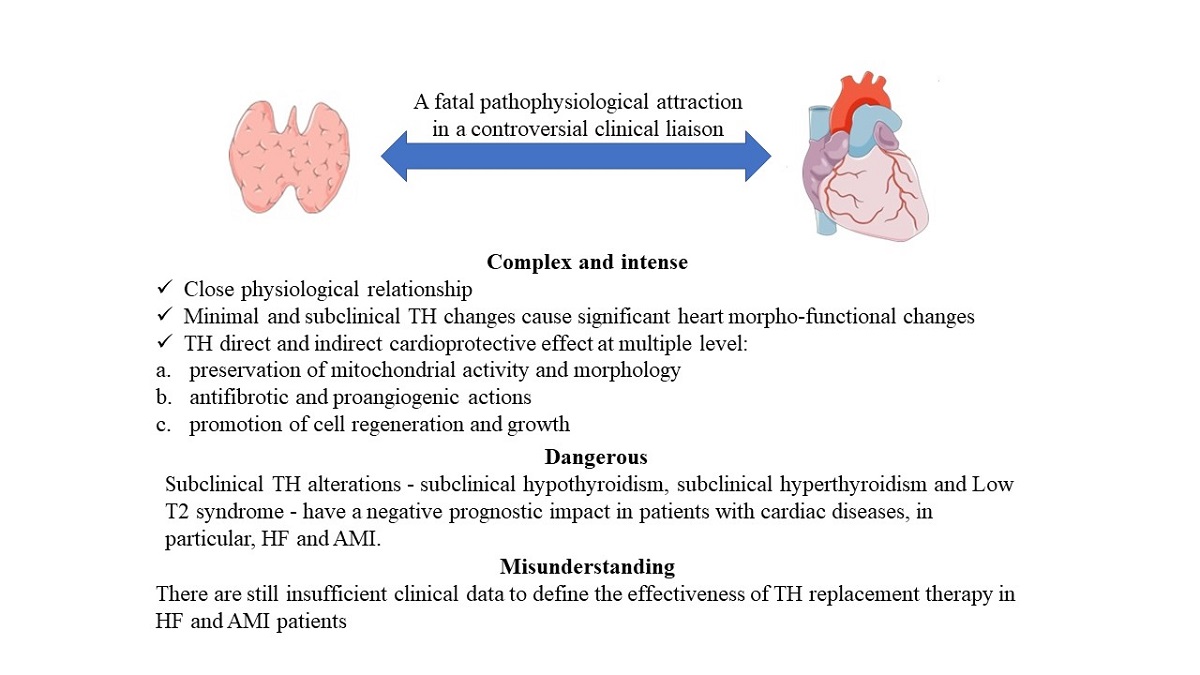
In this review, we have addressed several aspects of the complex, intense dangerous and still misunderstood liaison between heart and thyroid. We have seen that this was discovered more than a century ago; its complexity and intensity lie in the close physiological relationship and on the evidence that even minimal and subclinical TH metabolic alterations cause significant morpho-functional changes of the heart, as well as TH exerts a direct and indirect cardioprotective effect at multiple levels. The dangerousness of this relationship lies in the fact that the same small TH alterations have a negative prognostic weight in both HF and AMI patients, identifying patients at higher risk of even major cardiac events. Further, the misunderstanding lies in the fact that there are still insufficient clinical data to define the effectiveness of TH replacement therapy in HF and AMI patients. According to Cittadini, TH abnormalities are part of the multiple hormonal and metabolic deficiency syndrome that is a promising therapeutic target [115]. Finally, we like to quote the title of a recent editorial by Gerdes MA et al.: "Ignoring a basic pathophysiological mechanism of heart failure progression will not make it go away” [146].
Author Contributions
A.P, F.M, M.F.L.L, C.V.: Drafting and revising the manuscript; and final approval of the manuscript to be submitted for publication. All authors have read and agreed to the published version of the manuscript.
Funding
No funds: grants, or other support was received.
Conflicts of Interest
The authors declare no conflict of interest. The figure was created with Power Point.
References
- Wicomb W, Cooper DK, Hassoulas J, Rose AG, Barnard CN. Orthotopic transplantation of the baboon heart after 20 to 24 hours’ preservation by continuous hypothermic perfusion with an oxygen- ated hyperosmolar solution. J Thorac Cardiovasc Surg. 1982, 83, 133–140. [Google Scholar] [CrossRef]
- Cooper DK, Wicomb WN, Rose AG, Barnard CN. Orthotopic allotransplantation and autotrans- plantation of the baboon heart following twenty-four hours’ storage by a portable hypothermic perfusion system. Cryobiology. 1983, 20, 385–394. [Google Scholar] [CrossRef] [PubMed]
- Novitzky D, Cooper DKC, Barnard CN. The surgi- cal technique of heterotopic heart transplantation. Ann Thorac Surg. 1983, 36, 476–482. [Google Scholar] [CrossRef] [PubMed]
- Cooper DK, Wicomb WN, Barnard CN. Storage of the donor heart by a portable hypothermic perfu- sion system: experimental development and clinical experience. J Heart Transplant. 1983, 2, 104–110. [Google Scholar]
- Wicomb WN, Cooper DK, Novitzky D, Barnard CN. Cardiac transplantation following storage of the donor heart by a portable hypothermic perfusion sys- tem. Ann Thorac Surg. 1984, 373, 243–248. [Google Scholar]
- Wicomb WN, Cooper DKC, Lanza RP, Novitzky D, Isaacs S. The effects of brain death and 24 hours’ storage by hypothermic perfusion on donor heart function in the pig. J Thorac Cardiovasc Surg. 1986, 91, 896–909. [Google Scholar] [CrossRef]
- Novitzky D, Wicomb WN, Cooper DKC, Rose AG, Fraser RC, Barnard CN. Electrocardiographic, hae- modynamic and endocrine changes occurring during experimental brain death in the Chacma baboon. J Heart Transplant. 1984, 4, 63–69. [Google Scholar]
- Cushing, H. Some experimental and clinical observa- tions concerning states of increased intracranial ten- sion. Am J Med Sci. 1902, 124, 373–400. [Google Scholar] [CrossRef]
- Kocher, A. Ueber morbus Basedowi. Mitt Grenzgeb Med Chir. 1901, 1, 1–13. [Google Scholar]
- Cooper DKC, Novitzky D, Wicomb WN. The patho- physiological effects of brain death on potential donor organs, with particular reference to the heart. Ann R Coll Surg Engl. 1989, 71, 261–266. [Google Scholar]
- Novitzky D, Rose AG, Cooper DKC, Reichart B. Interpretation of endomyocardial biopsy after heart transplantation. Potentially confusing factors. S Afr Med J. 1986, 70, 789–792. [Google Scholar]
- Novitzky D, Wicomb WN, Cooper DKC, Rose AG, Reichart B. Prevention of myocardial injury during brain death by total cardiac sympathectomy in the Chacma baboon. Ann Thorac Surg. 1986, 41, 520–524. [Google Scholar] [CrossRef]
- Novitzky D, Cooper DKC, Rose AG, Reichart B. Prevention of myocardial injury by pretreatment with verapamil hydrochloride prior to experimental brain death: efficacy in a baboon model. Am J Emerg Med. 1987, 5, 11–18. [Google Scholar] [CrossRef] [PubMed]
- Novitzky D, Cooper DKC, Morrell D, Isaacs S. Change from aerobic to anaerobic metabolism after brain death, and reversal following triiodothyronine (T3) therapy. Transplantation. 1988, 45, 32–36. [Google Scholar] [CrossRef] [PubMed]
- Hamilton, BE. Clinical notes on hearts in hyperthy- roidism. Boston Med Surg J. 1922, 186, 216–218. [Google Scholar] [CrossRef]
- Kerr WJ, Hensel GC. Observations of the cardiovas- cular system in thyroid disease. Arch Intern Med. 1923, 31, 398–410. [Google Scholar] [CrossRef]
- Pratschke J, Wilhelm MJ, Kusaka M, et al. Brain death and its influence on donor organ quality and outcome after transplantation. Transplantation. 1999, 67, 343–348. [Google Scholar] [CrossRef]
- Margolies A, Wood ERS. The heart in thyroid disease. I. The effect of thyroidectomy on the orthodiagram. J Clin Invest. 1935, 14, 483–496. [Google Scholar]
- Gilligan DR, Berlin DD, Volk MC, Stern B, Blumgart HL. Therapeutic effect of total ablation of normal thyroid on congestive heart failure and angina pecto- ris. IX. Postoperative parathyroid function. Clinical observations and serum calcium and phosphorus studies. J Clin Invest. 1934, 13, 789–806. [Google Scholar]
- Goetsch, E. Newer methods in the diagnosis of thyroid disorders: pathological and clinical: B. Adrenaline hypersensitiveness in clinical states of hyperthyroid- ism. NY State J Med. 1918, 18, 259–267. [Google Scholar]
- McDonald CH, Shepeard WL, Green MF, DeGroat AF. Response of the hyperthyroid heart to epineph- rine. Am J Phys. 1935, 112, 227–230. [Google Scholar]
- Gaffney TE, Braunwald E, Kahler RL. Effects of gua- nethidine on triiodothyronine-induced hyperthyroid- ism in man. New Eng J Med. 1961, 265, 16–20. [Google Scholar] [CrossRef] [PubMed]
- Levey, GS. The adrenergic nervous system in hyper- thyroidism: therapeutic role of beta adrenergic block- ing drugs. Pharmacol Ther. 1976, 1, 431–443. [Google Scholar]
- No authors listed. The coronary drug project. Findings leading to further modifi cations of its protocol with respect to dextrothyroxine. The coronary drug project research group. JAMA. 1972, 220, 996–1008. [Google Scholar]
- Pilo A, Iervasi G, Vitek F, Ferdeghini M, Cazzuola F, Bianchi R. Thyroidal and peripheral production of 3,5,3 ’ - triiodothyronine in humans by multicompartmental analysis. Am J Physiol. 1990, 258, E715–26. [Google Scholar]
- Young WF Jr, Gorman CA, Jiang NS, Machacek D, Hay ID. L-thyroxine contamination of pharmaceutical D-thyroxine: probable cause of therapeutic effect. Clin Pharmacol Ther. 1984, 36, 781–787. [Google Scholar] [CrossRef]
- Cookson, H. The thyroid and the heart. Br Med J. 1959, 1, 254–259. [Google Scholar] [CrossRef]
- Summers VK, Surtees SJ. Thyrotoxicosis and heart disease. Acta Med Scand. 1961, 169, 661–671. [Google Scholar]
- Yater, WM. The tachycardia, time factor, sur- vival period and seat of action of thyroxine in the perfused hearts of thyroxinized rabbits. Am J Phys. 1931, 98, 338–343. [Google Scholar] [CrossRef]
- Papp, C. The heart in thyroid dysfunction. Postgrad Med J. 1945, 21, 45–51. [Google Scholar] [CrossRef]
- Tata JR, Ernster L, Lindberg O, Arrhenius E, Pedersen S, Hedman R. The action of thyroid hormones at the cell level. Biochem J. 1963, 86, 408–428. [Google Scholar] [CrossRef] [PubMed]
- Dillmann, WH. Hormonal influences on cardiac myo- sin ATPase activity and myosin isoenzyme distribu- tion. Mol Cell Endocrinol. 1984, 34, 169–181. [Google Scholar] [CrossRef] [PubMed]
- Oppenheimer JH, Koerner D, Schwartz HL, Surks MI. Specific nuclear triiodothyronine bind- ing sites in rat liver and kidney. J Clin Endocrinol Metab. 1972, 35, 330–333. [Google Scholar] [CrossRef]
- Lompré AM, Nadal-Ginard B, Mahdavi V. Expression of the cardiac ventricular alpha- and beta-myosin heavy chain genes is developmentally and hormonally regulated. J Biol Chem. 1984, 259, 6437–6446. [Google Scholar] [CrossRef]
- Klein I, Ojamaa K. Thyroid hormone and the car- diovascular system. N Engl J Med. 2001, 344, 501–509. [Google Scholar] [CrossRef]
- Razvi S, Jabbar A, Pingitore A, Danzi S, Biondi B, Klein I, Peeters R, Zaman A, Iervasi G. Thyroid hormones and cardiovascular function and diseases. J Am Coll Cardiol. 2018, 24, 1781–1796. [Google Scholar]
- Kahaly GJ, Dillmann WH. Thyroid hormone action in the heart. Endocr Rev. 2005, 26, 704–728. [Google Scholar] [CrossRef]
- Jabbar A, Pingitore A, Pearce SH, Zaman A, Iervasi G, Razvi S (2017) Thyroid hormones and cardiovascular disease. Nat Rev Cardiol 14(1):39–55.
- Hartong R, Wang N, Kurokawa R, Lazar MA, Glass CK, Apriletti, Dillmann WH. Delineation of three different thyroid hormone-response elements in promoter of rat sarcoplasmic reticulum Ca2_-ATPase gene. J Biol Chem. 1994, 269, 13021–13029. [Google Scholar] [CrossRef]
- Klein, I. Endocrine disorders and cardiovascular disease. In: Mann, ed.(Chapter81). Braunwald’s Heart Disease.
- Mastorci F, Sabatino L, Vassalle C, Pingitore A. Cardioprotection and Thyroid Hormones in the Clinical Setting of Heart Failure. Front Endocrinol (Lausanne). 2020, 10, 927. [Google Scholar] [CrossRef]
- Sun Z, Ojamaa K, Coetzee WA, Artman M, Klein I. Effects of thyroid hormone on action potential and repolarization currents in rat ventricular myocytes. Am J Physiol Endocrinol Metab. 2000, 278, E302–E307. [Google Scholar] [CrossRef]
- Pachucki J, Burmeister LA, Larsen PR. Thyroid hormone regulates hyperpolarization-activated cyclic nucleotide-gated channel (HCN2) mRNA in the rat heart. Circ Res. 1999, 85, 498–503. [Google Scholar] [CrossRef] [PubMed]
- Danzi S, Klein I. Thyroid hormone and blood pressure regulation. Curr Hypertens Rep. 2003, 5, 513–520. [Google Scholar] [CrossRef] [PubMed]
- Klein I, Ojamaa K. Thyroid hormone and the cardiovascular system. N Engl J Med. 2001, 344, 501–509. [Google Scholar] [CrossRef]
- Biondi B, Palmieri EA, Lombardi G, Fazio S. Effects of thyroid hormone on cardiac function: the relative importance of heart rate, loading conditions, and myocardial contractility in the regulation of cardiac performance in human hyperthyroidism. J Clin Endocrinol Metab. 2002, 87, 968–974. [Google Scholar] [CrossRef]
- Volpe, R. Immunoregulation in autoimmune thyroid disease. Thyroid. 1994, 4, 373–377. [Google Scholar] [CrossRef]
- Cooper, D. S. , and Biondi, B. ( 2012). Subclinical thyroid disease. Lancet 379, 1142–1154. [CrossRef]
- Biondi B, Palmieri EA, Lombardi G, Fazio S. Effects of thyroid hormone on cardiac function: the relative importance of heart rate, loading conditions, and myocardial contractility in the regulation of cardiac performance in human hyperthyroidism. J Clin Endocrinol Metab. 2002, 87, 968–974. [Google Scholar] [CrossRef]
- Kahaly, G. J. , and Dillmann, W. H. ( 2005). Thyroid hormone action in the heart.Endocr. Rev. 26, 704–728. [CrossRef]
- Mohr-Kahaly, S. , Kahaly, G., and Meyer, J. (1996). [Cardiovascular effects of thyroid hormones]. Z. Kardiol. 85(Suppl. 6), 219–231.
- Weltman NY, Wang D, Redetzke RA, Gerdes AM. Longstanding hyperthyroidism is associated with normal or enhanced intrinsic cardiomyocyte function despite decline in global cardiac function. PLoS One. 2012, 7, e46655. [CrossRef]
- Klein I. Endocrine disorders and cardiovascular disease. In: Zipes DP, Libby P, Bonow R, Braunwald E, eds. Braunwald’s Heart Disease: A Textbook of Cardiovascular Medicine. 7th ed. Philadelphia, Pa. W.B. Saunders; 2005:2051–2065.
- Fredlund BO, Olsson SB. Long QT interval and ventricular tachycardia of “torsade de pointe” type in hypothyroidism. Acta Med Scand. 1983, 213, 231–235.
- Sawin CT, Geller A, Wolf PA, Belanger AJ, Baker E, Bacharach P, Wilson PW, Benjamin EJ, D’Agostino RB. Low serum thyrotropin concentrations as a risk factor for atrial fibrillation in older persons. N Engl J Med. 1994, 331, 1249–1252. [Google Scholar] [CrossRef] [PubMed]
- 56. Parle JV, Maisonneuve P, Sheppard MC, Boyle P, Franklyn JA. Prediction of all-cause and cardiovascular mortality in elderly people from one low serum thyrotropin result: a 10-year cohort study. Lancet. 2001, 358, 861–865. [CrossRef] [PubMed]
- Iervasi, G., Molinaro, S., Landi, P., Taddei, M. C., Galli, E., Mariani, F., et al. (2007). Association between increased mortality and mild thyroid dysfunction in cardiac patients. Arch. Intern. Med. 167, 1526–1532. [CrossRef]
- Cappola, A. R., Fried, L. P., Arnold, A. M., Danese, M. D., Kuller, L. H., Burke, G. L., et al. (2006). Thyroid status, cardiovascular risk, and mortality in older adults. JAMA 295, 1033–1041. [CrossRef]
- Rodondi, N., Bauer, D. C., Cappola, A. R., Cornuz, J., Robbins, J., Fried, L. P., et al. (2008). Subclinical thyroid dysfunction, cardiac function, and the risk of heart failure. The Cardiovascular Health study. J. Am. Coll. Cardiol. 52, 1152–1159. [CrossRef]
- Rodondi N, Aujesky D, Vittinghoff E, Cornuz J, Bauer DC. Subclinical hypothyroidism and the risk of coronary heart disease: a meta-analysis. Am J Med. 2006, 119, 541–551. [Google Scholar] [CrossRef]
- Monzani, F. , Di Bello, V., Caraccio, N., Bertini, A., Giorgi, D., Giusti, C., et al. (2001). Effect of levothyroxine on cardiac function and structure in subclinical hypothyroidism: a double blind, placebo-controlled study. J. Clin. Endocrinol. Metab. 86, 1110–1115. [CrossRef]
- Ripoli, A. , Pingitore, A., Favilli, B., Bottoni, A., Turchi, S., Osman, N. F., et al. (2005). Does subclinical hypothyroidism affect cardiac pump performance? Evidence from a magnetic resonance imaging study. J. Am. Coll. Cardiol. 45, 439–445. [CrossRef]
- Hak AE, Pols HA, Visser TJ, Drexhage HA, Hofman A, Witteman JC. Subclinical hypothyroidism is an independent risk factor for atherosclerosis and myocardial infarction in elderly women: the Rotterdam Study. Ann Intern Med. 2000, 132, 270–278. [Google Scholar] [CrossRef]
- Kübler W, Haass M. Cardioprotection: definition, classification, and fundamental principles. Heart. 1996, 75, 330–333. [Google Scholar] [CrossRef]
- Pingitore A, Nicolini G, Kusmic C, Iervasi G, Grigolini P, Forini F. Cardioprotection and thyroid hormones. Heart Fail Rev. 2016, 21, 391–399. [Google Scholar] [CrossRef]
- 66. Forini F, Pitto L, Nicolini G. Thyroid Hormone, Mitochondrial Function and Cardioprotection (chapter 9). In: Iervasi et al. (eds.), Thyroid and Heart. Springer Nature Switzerland AG 2020 G.
- Goldenthal MJ, Ananthakrishnan R, Marín-García J. Nuclear-mitochondrial cross-talk in cardiomyocyte T3 signaling: a time-course analysis. J Mol Cell Cardiol. 2005, 39, 319–326. [Google Scholar] [CrossRef]
- Galli E, Pingitore A, Iervasi G. The role of thyroid hormone in the pathophysiology of heart failure: clinical evidence. Heart Fail Rev. 2010, 15, 155–169. [Google Scholar] [CrossRef] [PubMed]
- Garber JR, Cobin RH, Gharib H, Hennessey JV, Klein I, Mechanick JI, et al.. Clinical practice guidelines for hypothyroidism in adults: cosponsored by the American Association of Clinical Endocrinologists and the American Thyroid Association. Thyroid. (2012) 22:1200–35.
- Pan, Y. , Wang Y., Shi W., Liu Y., Cao S., Yu T. Mitochondrial proteomics alterations in rat hearts following ischemia/reperfusion and diazoxide post conditioning. Mol. Med. Rep. 2021, 23, 161. [Google Scholar] [CrossRef] [PubMed]
- Canale P, Nicolini G, Pitto L, Kusmic C, Rizzo M, Balzan S, Iervasi G, Forini F. Role of miR-133/Dio3 Axis in the T3-Dependent Modulation of Cardiac mitoK-ATP Expression. Int J Mol Sci. 2022, 23, 6549.
- Sabatino, L. Nrf2-Mediated Antioxidant Defense and Thyroid Hormone Signaling: A Focus on Cardioprotective Effects. Antioxidants (Basel). 2023, 12, 1177. [Google Scholar] [CrossRef]
- Della Nera G, Sabatino L, Gaggini M, Gorini F, Vassalle C. Vitamin D Determinants, Status, and Antioxidant/Anti-inflammatory-Related Effects in Cardiovascular Risk and Disease: Not the Last Word in the Controversy. Antioxidants (Basel). 2023, 12, 948. [Google Scholar] [CrossRef]
- Al-Oanzi ZH, Alenazy FO, Alhassan HH, Alruwaili Y, Alessa AI, Alfarm NB, Alanazi MO, Alghofaili SI. The Role of Vitamin D in Reducing the Risk of Metabolic Disturbances That Cause Cardiovascular Diseases. J Cardiovasc Dev Dis. 2023, 10, 209. [Google Scholar]
- Alrefaie Z, Awad H. Effect of vitamin D3 on thyroid function and deiodinase 2 expression in diabetic rats. Arch Physiol Biochem. 2015;121(5):206-9.
- Miura M, Tanaka K, Komatsu Y, Suda M, Yasoda A, Sakuma Y, Ozasa A, Nakao K 2002 A novel interaction between thyroid hormones and 1,25(OH)(2)D(3) in osteoclast formation. Biochem Biophys Res Commun 291: 987–994.
- Gouveia CH, Christoffolete MA, Zaitune CR, Dora JM, Harney JW, Maia AL, Bianco AC. Type 2 iodothyronine selenodeiodinase is expressed throughout the mouse skeleton and in the MC3T3-E1 mouse osteoblastic cell line during differentiation. Endocrinology. 2005, 146, 195–200. [Google Scholar] [CrossRef]
- Berg JP, Liane KM, Bjørhovde SB, Bjøro T, Torjesen PA, Haug E. Vitamin D receptor binding and biological effects of cholecalciferol analogues in rat thyroid cells. J Steroid Biochem Mol Biol. 1994, 50, 145–150. [Google Scholar] [CrossRef]
- D'Emden MC, Wark JD. 1,25-Dihydroxyvitamin D3 enhances thyrotropin releasing hormone induced thyrotropin secretion in normal pituitary cells. Endocrinology. 1987, 121, 1192–1194. [Google Scholar] [CrossRef]
- Wang Y, Liu Q, Dong H, Feng Y, Raguthu C, Liang X, Liu C, Zhang Z, Yao X. The protective effect of iodide intake adjustment and 1,25(OH)2D3 supplementation in rat offspring following excess iodide intake. Ther Adv Endocrinol Metab. 2020, 11, 2042018820958295. [Google Scholar]
- Vassalle C, Parlanti A, Pingitore A, Berti S, Iervasi G, Sabatino L. Vitamin D, Thyroid Hormones and Cardiovascular Risk: Exploring the Components of This Novel Disease Triangle. Front Physiol. 2021, 12, 722912. [Google Scholar] [CrossRef]
- Babić Leko M, Jureško I, Rozić I, Pleić N, Gunjača I, Zemunik T. Vitamin D and the Thyroid: A Critical Review of the Current Evidence. Int J Mol Sci. 2023, 24, 3586. [Google Scholar] [CrossRef]
- Pingitore A, Mastorci F, Berti S, Sabatino L, Palmieri C, Iervasi G, Vassalle C. Hypovitaminosis D and Low T3 Syndrome: A Link for Therapeutic Challenges in Patients with Acute Myocardial Infarction. J Clin Med. 2021, 10, 5267. [Google Scholar] [CrossRef] [PubMed]
- Mirhosseini N, Brunel L, Muscogiuri G, Kimball S. Physiological serum 25-hydroxyvitamin D concentrations are associated with improved thyroid function-observations from a community-based program. Endocrine. 2017, 58, 563–573. [Google Scholar] [CrossRef] [PubMed]
- Iervasi G, Pingitore A, Landi P, Raciti M, Ripoli A, Scarlattini M, L’Abbate A, Donato L. Low-T3 syndrome: a strong prognostic predictor of death in patients with heart disease. Circulation. 2003, 107, 708–713. [Google Scholar] [CrossRef] [PubMed]
- Pingitore A, Iervasi G, Barison A, Prontera C, Pratali L, Emdin M, Giannessi D, Neglia D. Early activation of an altered thyroid hormone profile in asymptomatic or mildly symptomatic idiopathic left ventricular dysfunction. J Card Fail 2006, 12, 520–526. [Google Scholar] [CrossRef] [PubMed]
- Molinaro S, Iervasi G, Lorenzoni V, Coceani M, Landi P, Srebot V, Mariani F, L'Abbate A, Pingitore A. Persistence of mortality risk in patients with acute cardiac diseases and mild thyroid dysfunction. Am J Med Sci. 2012, 343, 65–70. [Google Scholar] [CrossRef]
- De Matteis G, Covino M, Burzo ML, Della Polla DA, Petti A, Bruno C, Franceschi F, Mancini A, Gambassi G Prognostic role of hypothyroidism and low free-triiodothyronine levels in patients hospitalized with acute heart failure. Intern Emerg Med. 2021, 16, 1477–1486. [CrossRef]
- Zhou P, Huang LY, Zhai M, Huang Y, Zhuang XF, Liu HH, Zhang YH, Zhang J. [The prognostic value of free triiodothyronine/free thyroxine ratio in patients hospitalized with heart failure]. Zhonghua Yi Xue Za Zhi. 2023, 103, 1679–1684 Chinese. [CrossRef] [PubMed]
- Wang C, Han S, Li Y, Tong F, Li Z, Sun Z. Value of FT3/FT4 Ratio in Prognosis of Patients With Heart Failure: A Propensity-Matched Study. Front Cardiovasc Med. 2022, 9, 859608. [Google Scholar] [CrossRef]
- Samuel NA, Cuthbert JJ, Brown OI, Kazmi S, Cleland JGF, Rigby AS, Clark AL. Relation Between Thyroid Function and Mortality in Patients With Chronic Heart Failure. Am J Cardiol. 2021, 139, 57–63. [Google Scholar] [CrossRef] [PubMed]
- 92. Iacoviello M, Parisi G, Gioia MI, Grande D, Rizzo C, Guida P, Lisi F, Giagulli VA, Licchelli B, Di Serio F, Guastamacchia E, Triggiani V. Thyroid Disorders and Prognosis in Chronic Heart Failure: A Long-Term Follow-Up Study. Endocr Metab Immune Disord Drug Targets. 2020, 20, 437–445. [CrossRef] [PubMed]
- Li X, Yao Y, Chen Z, Fan S, Hua W, Zhang S, Fan X. Thyroid-stimulating hormone within the normal range and risk of major adverse cardiovascular events in nonischemic dilated cardiomyopathy patients with severe left ventricular dysfunction. Clin Cardiol. 2019, 42, 120–128. [Google Scholar] [CrossRef]
- Sato Y, Yoshihisa A, Kimishima Y, Kiko T, Kanno Y, Yokokawa T, Abe S, Misaka T, Sato T, Oikawa M, Kobayashi A, Yamaki T, Kunii H, Nakazato K, Takeishi Y. Low T3 Syndrome Is Associated With High Mortality in Hospitalized Patients With Heart Failure. J Card Fail 2019, 25, 195–203. [Google Scholar] [CrossRef] [PubMed]
- Kannan L, Shaw PA, Morley MP, Brandimarto J, Fang JC, Sweitzer NK, Cappola TP, Cappola AR. Thyroid dysfunction in heart failure and cardiovascular outcomes. Circ Heart Fail. 2018, 11, e005266. [Google Scholar] [CrossRef]
- Sato Y, Yoshihisa A, Kimishima Y, Kiko T, Watanabe S, Kanno Y, Abe S, Miyata M, Sato T, Suzuki S, Oikawa M, Kobayashi A, Yamaki T, Kunii H, Nakazato K, Ishida T, Takeishi Y. Subclinical hypothyroidism is associated with adverse prognosis in heart failure patients. Can J Cardiol. 2018, 34, 80–87. [Google Scholar] [CrossRef]
- Chen YY, Shu XR, Su ZZ, Lin RJ, Zhang HF, Yuan WL, Wang JF, Xie SL. A low-normal free triiodothyronine level is associated with adverse prognosis in euthyroid patients with heart failure receiving cardiac resynchronization therapy. Int Heart J. 2017, 58, 908–914. [Google Scholar] [CrossRef] [PubMed]
- Hayashi T, Hasegawa T, Kanzaki H, Funada A, Amaki M, Takahama H, Ohara T, Sugano Y, Yasuda S, Ogawa H, Anzai T. Subclinical hypothyroidism is an independent predictor of adverse cardiovascular outcomes in patients with acute decompensated heart failure. ESC Heart Fail. 2016, 3, 168–176. [Google Scholar] [CrossRef] [PubMed]
- Wang W, Guan H, Fang W, Zhang K, Gerdes AM, Iervasi G, et al. Free triiodothyronine level correlates with myocardial injury and prognosis in idiopathic dilated cardiomyopathy: evidence from cardiac MRI and SPECT/PET imaging. Sci Rep. 2016, 6, 39811. [Google Scholar] [CrossRef]
- Okayama D, Minami Y, Kataoka S, Shiga T, Hagiwara N. Thyroid function on admission and outcome in patients hospitalized for acute decompensated heart failure. J Cardiol. 2015, 66, 205–211. [Google Scholar] [CrossRef]
- Wang W, Guan H, Gerdes M, Iervasi G, Yang Y, Tang Y. Thyroid status, cardiac function and mortality in patients with idiopathic dilated cardiomyopathy. J Clin Endocrinol Metab 2015, 100, 3210–3218. [Google Scholar] [CrossRef]
- Chen S, Shauer A, Zwas DR, Lotan C, Keren A, Gotsman I. The effect of thyroid function on clinical outcome in patients with heart failure. Eur J Heart Fail 2014, 16, 217–226. [Google Scholar] [CrossRef] [PubMed]
- Chuang CP, Jong YS, Wu CY, Lo HM. Impact of triiodothyronine and N-terminal pro-B-type natriuretic peptide on the long term survival of critically ill patients with acute heart failure. Am J Cardiol 2014, 113, 845–850. [Google Scholar] [CrossRef] [PubMed]
- Li X, Yang X, Wang Y, Ding L, Wang J, Hua W. The prevalence and prognostic effects of subclinical thyroid dysfunction in dilated cardiomyopathy patients: a single-center cohort study. J Card Fail. 2014, 20, 506–512. [Google Scholar] [CrossRef] [PubMed]
- Perez AC1, Jhund PS1, Stott DJ2, Gullestad L3, Cleland JG4, van Veldhuisen DJ5, Wikstrand J6, Kjekshus J3, McMurray JJ7. Thyroid-stimulating hormone and clinical outcomes: the CORONA trial (controlled rosuvastatin multinational study in heart failure). JACC Heart Fail. 2014, 2, 35–40. [Google Scholar] [CrossRef]
- Frey A, Kroiss M, Berliner D, Seifert M, Allolio B, Güder G, Ertl G, Angermann CE, Störk S, Fassnacht M. Prognostic impact of subclinical thyroid dysfunction in heart failure. Int J Cardiol. 2013, 168, 300–305. [Google Scholar] [CrossRef]
- Mitchell JE, Hellkamp AS, Mark DB, Anderson J, Johnson GW, Poole JE, Lee KL, Bardy GH. Thyroid function in heart failure and impact on mortality. JACC Heart Fail. 2013, 1, 48–55. [Google Scholar] [CrossRef]
- Passino C, Pingitore A, Landi P, Fontana M, Zyw L, Clerico A, Emdin M, Iervasi G. Prognostic value of combined measurement of brain natriuretic peptide and triiodothyronine in heart failure. J Card Fail. 2009, 15, 35–40. [Google Scholar] [CrossRef]
- Iacoviello M, Guida P, Guastamacchia E, Triggiani V, Forleo C, Catanzaro R, Cicala M, Basile M, Sorrentino S, Favale S. Prognostic role of sub-clinical hypothyroidism in chronic heart failure outpatients. Curr Pharm Des. 2008, 14, 2686–2692. [Google Scholar] [CrossRef]
- Kozdag G, Ural D, Vural A, Agacdiken A, Kahraman G, Sahin T, Ural E, Komsuoglu B. Relation between free triiodothyronine/free thyroxine ratio, echocardiographic parameters and mortality in dilated cardiomyopathy. Eur J Heart Fail. 2005, 7, 113–118. [Google Scholar] [CrossRef]
- Pingitore A1, Landi P, Taddei MC, Ripoli A, L'Abbate A, Iervasi G. Triiodothyronine levels for risk stratification of patients with chronic heart failure. Am J Med. 2005, 118, 132136. [Google Scholar]
- Hamilton MA, Stevenson LW, Luu M, Walden JA. Altered thyroid hormone metabolism in advanced heart failure. J Am Coll Cardiol. 1990, 16, 91–95. [Google Scholar] [CrossRef]
- Lubrano V, Pingitore A, Carpi A, Iervasi G. Relationship between triiodothyronine and proinflammatory cytokines in chronic heart failure. Biomed Pharmacother. 2010, 64, 165–169. [Google Scholar] [CrossRef] [PubMed]
- Shen Y, Chen G, Su S, Zhao C, Ma H, Xiang M. Independent Association of Thyroid Dysfunction and Inflammation Predicts Adverse Events in Patients with Heart Failure via Promoting Cell Death. J Cardiovasc Dev Dis. 2022, 9, 290. [Google Scholar] [CrossRef]
- Cittadini A, Salzano A, Iacoviello M, Triggiani V, Rengo G, Cacciatore F, Maiello C, Limongelli G, Masarone D, Perticone F, Cimellaro A, Perrone Filardi P, Paolillo S, Mancini A, Volterrani M, Vriz O, Castello R, Passantino A, Campo M, Modesti PA, De Giorgi A, Monte IP, Puzzo A, Ballotta A, D'Assante R, Arcopinto M, Gargiulo P, Sciacqua A, Bruzzese D, Colao A, Napoli R, Suzuki T, Eagle KA, Ventura HO, Marra AM, Bossone E; T. O.S.CA. Investigators. Multiple hormonal and metabolic deficiency syndrome predicts outcome in heart failure: the T. O.S.CA. Registry. Eur J Prev Cardiol. 2021, 28, 1691–1700. [Google Scholar] [CrossRef] [PubMed]
- Mahal S, Datta S, Ravat V, Patel P, Saroha B, Patel RS. Does subclinical hypothyroidism affect hospitalization outcomes and mortality in congestive cardiac failure patients? Cureus. 2018, 10, e2766. [CrossRef]
- 117. Fontes R, Coeli CR, Aguiar F, Vaisman M. Reference interval of thyroid stimulating hormone and free thyroxine in a reference population over 60 years old and in very old subjects (over 80 years): comparison to young subjects. Thyroid Research 2013, 6, 13. [CrossRef]
- Silva-Tinoco R, Castillo-Martı´nez L, Orea-Tejeda A, et al. Developing thyroid disorders is associated with poor prognosis factors in patient with stable chronic heart failure. Int J Cardiol 2011, 147, e24–e25. [Google Scholar] [CrossRef]
- Merla R, Martinez JD, Martinez MA, et al. Hypothyroidism and renal function in patients with systolic heart failure. Tex Heart Inst J 2010, 37, 66–69. [Google Scholar]
- Drechsler C, Schneider A, Gutjahr-Lengsfeld L, et al. Thyroid function, cardiovascular events, and mortality in diabetic hemodialysis patients. Am J Kidney Dis 2014, 63, 988–996. [Google Scholar] [CrossRef]
- Friberg L, Werner S, Eggertsen G, Ahnve S. Rapid down-regulation of thyroid hormones in acute myocardial infarction: is it cardioprotective in patients with angina?Arch Intern Med. 2002, 162, 1388–1394. [CrossRef]
- Wang WY, Tang YD, Yang M, Cui C, Mu M, Qian J, Yang YJ. Free triiodothyronine level indicates the degree of myocardial injury in patients with acute ST-elevation myocardial infarction. Chin Med J (Engl). 2013, 126, 3926–3929. [Google Scholar]
- Ceremuzyński L, Górecki A, Czerwosz L, Chamiec T, Bartoszewicz Z, Herbaczyńska-Cedro Low serum triiodothyronine in acute myocardial infarction indicates major heart injury. K.Kardiol Pol. 2004, 60, 468–480.
- Lymvaios I, Mourouzis I, Cokkinos DV, Dimopoulos MA, Toumanidis ST, Pantos C Thyroid hormone and recovery of cardiac function in patients with acute myocardial infarction: a strong association? .Eur J Endocrinol. 2011, 165, 107–114. [CrossRef] [PubMed]
- Reindl M, Feistritzer HJ, Reinstadler SJ, Mueller L, Tiller C, Brenner C, Mayr A, Henninger B, Mair J, Klug G, Metzler B. Thyroid-stimulating hormone and adverse left ventricular remodeling following ST-segment elevation myocardial infarction. Eur Heart J Acute Cardiovasc Care. 2019, 8, 717–726. [Google Scholar] [CrossRef] [PubMed]
- Han C, Xu K, Wang L, Zhang Y, Zhang R, Wei A, Dong L, Hu Y, Xu J, Li W, Li T, Liu C, Qi W, Jin D, Zhang J, Cong H. Impact of persistent subclinical hypothyroidism on clinical outcomes in non-ST-segment elevation acute coronary syndrome undergoing percutaneous coronary intervention. Clin Endocrinol (Oxf). 2022, 96, 70–81. [Google Scholar] [CrossRef] [PubMed]
- Brozaitiene J, Mickuviene N, Podlipskyte A, Burkauskas J, Bunevicius R. Relationship and prognostic importance of thyroid hormone and N-terminal pro-B-Type natriuretic peptide for patients after acute coronary syndromes: a longitudinal observational study. Cardiovasc Disord. 2016, 16, 45. [Google Scholar] [CrossRef]
- Yu T, Tian C, Song J, He D, Wu J, Wen Z, Sun Z, Sun Z. Value of the fT3/fT4 ratio and its combination with the GRACE risk score in predicting the prognosis in euthyroid patients with acute myocardial infarction undergoing percutaneous coronary intervention: a prospective cohort study. BMC Cardiovasc Disord. 2018, 18, 181, PMID: 30200880; PMCID: PMC6131820. [Google Scholar] [CrossRef]
- Gao S, Ma W, Huang S, Lin X, Yu M. Impact of low triiodothyronine syndrome on long-term outcomes in patients with myocardial infarction with nonobstructive coronary arteries. Ann Med. 2021, 53, 741–749. [Google Scholar] [CrossRef]
- Pingitore A, Chen Y, Gerdes AM, Iervasi G. Acute myocardial infarction and thyroid function: new pathophysiological and therapeutic perspectives. Annals of Medicine. Ann Med. 2012, 44, 745–757. [Google Scholar] [CrossRef]
- Amin A, Chitsazan M, Taghavi S, Ardeshiri M. Effects of triiodothyronine replacement therapy in patietns with chronic stable heart failure and low-triidotrhyronine syndrome: a randomized, double-blind, placebo-controlled study. ESC Heart Failure 2015, 2, 5–11. [Google Scholar] [CrossRef]
- Holmager P, Schmidt U, Mark P, Andersen U, Dominguez H, Raymond I, Zerahn B, Nygaard B1, Kistorp C, Faber J. Long-term L-Triiodothyronine (T3) treatment in stable systolic heart failure patients: a randomised, double-blind, cross-over, placebo-controlled intervention study. Clin Endocrinol (Oxf). 2015, 83, 931–937. [Google Scholar] [CrossRef]
- Curotto Grasiosi J, Peresotti B, Machado RA, et al. Improvement in functional capacity after levothyroxine treatment in patients with chronic heart failure and sublinical hypothyroidism. Endocrinol Nutr 2013, 60, 427–432. [Google Scholar]
- Goldman S, McCarren M, Morkin E, et al. DITPA (3,5-Diiodothyropropionic Acid), a thyroid hormone analog to treat heart failure: phase II trial Veterans Affairs Cooperative Study. Circulation 2009, 119, 3093–3100. [Google Scholar] [CrossRef] [PubMed]
- Pingitore A, Galli E, Barison A, et al. Acute effects of triiodothyronine (T3) replacement therapy in patients with chronic heart failure and low-T3 syndrome: a randomized, placebo-controlled study. J Clin Endocrinol Metab 2008, 93, 1351–1358. [Google Scholar] [CrossRef] [PubMed]
- Iervasi G, Emdin M, Colzani RMP, et al. Beneficial effects of long-term triiodothyronine (T3) infusion in patients with advanced heart failure and low T3 syndrome. In: Kimchi A, editor. Second International Congress on Heart Disease - new trends in research, diagnosis and treatment. Medimond Medical Publications;Englewood, NJ, USA: 2001. p. 549-53.
- Malik FS, Mehra MR, Uber PA, et al. Intravenous thyroid hormone supplementation in heart failure with cardiogenic shock. J Card Fail 1999, 5, 31–37. [Google Scholar] [CrossRef] [PubMed]
- Hamilton MA, Stevenson LW, Fonarow GC, et al. Safety and hemodynamic effects of intravenous triiodothyronine in advanced congestive heart failure. Am J Cardiol 1998, 81, 443–447. [Google Scholar] [CrossRef]
- Moruzzi P, Doria E, Agostoni PG. Medium-term effectiveness of L-thyroxine treatment in idiopathic dilated cardiomyopathy. Am J Med 1996, 101, 461–467. [Google Scholar] [CrossRef]
- Shi C, Bao Y, Chen X, Tian L. The Effectiveness of Thyroid Hormone Replacement Therapy on Heart Failure and Low-Triiodothyronine Syndrome: An Updated Systematic Review and Meta-analysis of Randomized Controlled Trials. Endocr Pract. 2022, 28, 1178–1186. [Google Scholar] [CrossRef]
- Chen X, Bao Y, Shi C, Tian L. Effectiveness and Safety of Thyroid Hormone Therapy in Patients with Dilated Cardiomyopathy: A Systematic Review and Meta-analysis of RCTs. Am J Cardiovasc Drugs. 2022, 22, 647–656. [Google Scholar] [CrossRef]
- Pingitore A, Mastorci F, Piaggi P, Aquaro GD, Molinaro S, Ravani M, De Caterina A, Trianni G, Ndreu R, Berti S, Vassalle C, Iervasi G. Usefulness of Triiodothyronine Replacement Therapy in Patients With ST Elevation Myocardial Infarction and Borderline/Reduced Triiodothyronine Levels (from the THIRST Study). Am J Cardiol 2018, 00, 1–8. [Google Scholar]
- Pantos C et al Effects of Acute Triiodothyronine Treatment in Patients with Anterior Myocardial Infarction Undergoing Primary Angioplasty: Evidence from a Pilot Randomized Clinical Trial (ThyRepair Study). Thyroid. 2022, 32, 714–724. [CrossRef] [PubMed]
- Jabbar A et al Effect of Levothyroxine on Left Ventricular Ejection Fraction in Patients With Subclinical Hypothyroidism and Acute Myocardial Infarction: A Randomized Clinical Trial. JAMA. 2020, 324, 249–258. [CrossRef] [PubMed]
- Tharmapoopathy M, Thavarajah A; Kenny RPW, Pingitore A, Iervasi G, Dark J, Bano A, Razvi S. Efficacy and Safety of Triiodothyronine Treatment in Cardiac Surgery or Cardiovascular Diseases: A Systematic Review and Meta-Analysis of Randomized Controlled Trials. Thyroid. 2022, 32, 879–896. [Google Scholar] [CrossRef]
- Gerdes AM, Portman MA, Iervasi G, Pingitore A, Cooper DKC, Novitzky D. Ignoring a basic pathophysiological mechanism of heart failure progression will not make it go away. Am J Physiol Heart Circ Physiol. 2021, 320, H1919–H1922. [Google Scholar] [CrossRef] [PubMed]
Figure 1.
Interaction of the Thyroid and the Heart. SVR: Systemic Vascular Resistance; HR: Heart Rate; CV: Cardiovascular; ECG: Electrocardiogram.
Figure 1.
Interaction of the Thyroid and the Heart. SVR: Systemic Vascular Resistance; HR: Heart Rate; CV: Cardiovascular; ECG: Electrocardiogram.
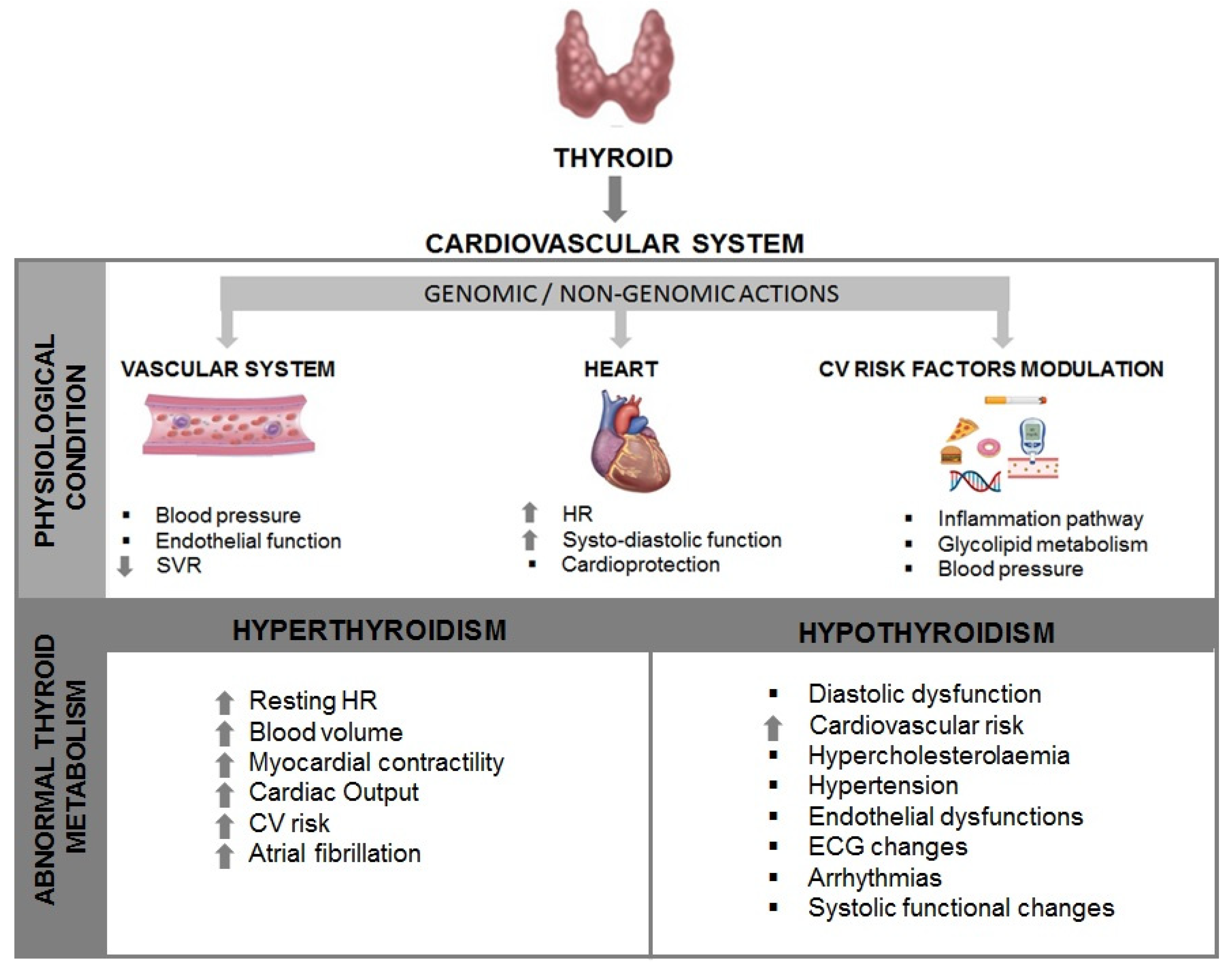
Figure 2.
Main possible vitamin D-related mechanisms in thyroid functioning. LT3S: low T3 syndrome; 25(OH)D: 25-HydroxyvitaminD.
Figure 2.
Main possible vitamin D-related mechanisms in thyroid functioning. LT3S: low T3 syndrome; 25(OH)D: 25-HydroxyvitaminD.
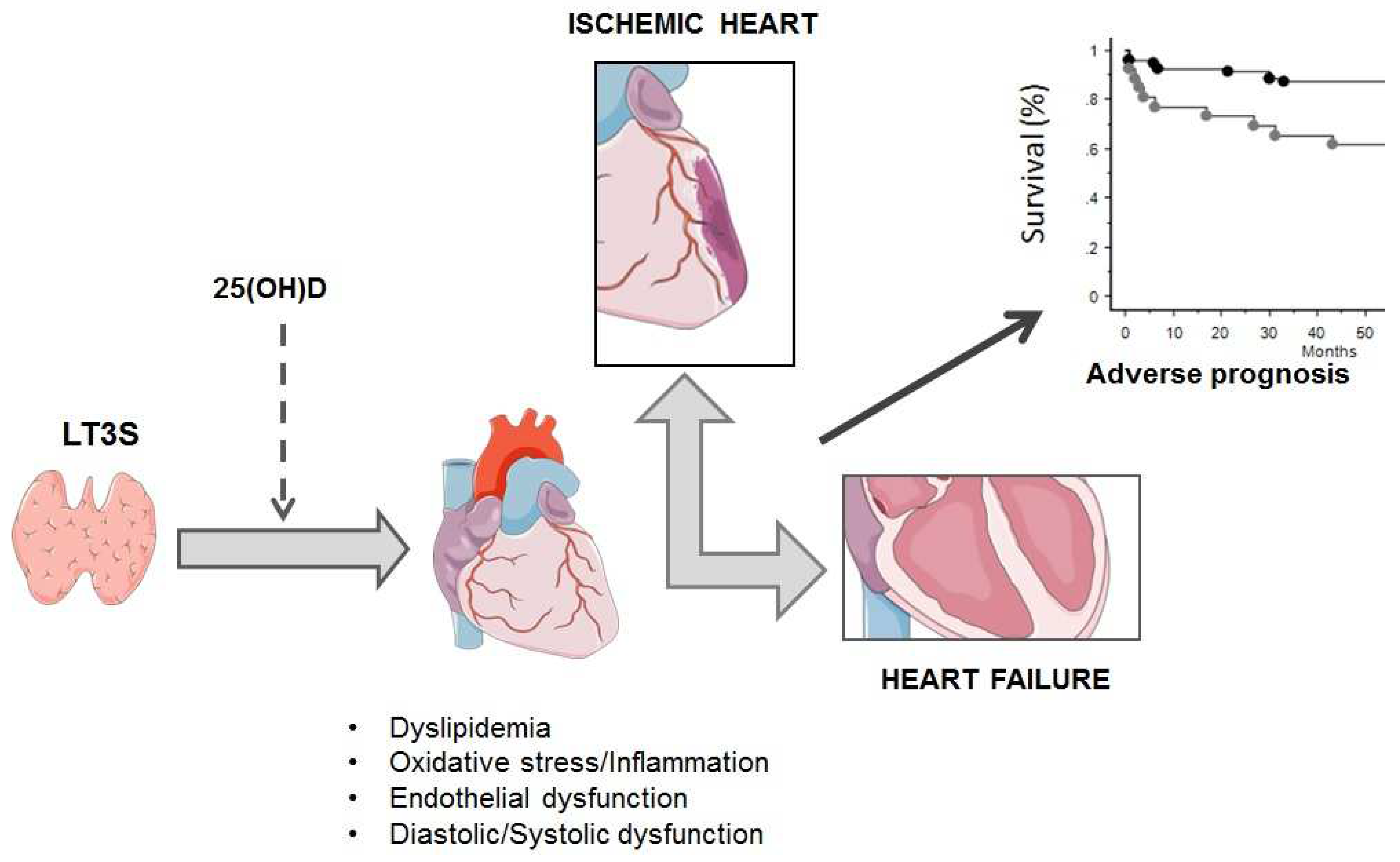

Table 1.
Prognostic impact of altered thyroid metabolism in HF patients.
| TH Dysfunction | Events (n) | N° PTS (W,%) | Age (yy) | LVEF (%) | NYHA class III-IV | Prognostic weight | Ref. |
|---|---|---|---|---|---|---|---|
| fT3/fT4 ratio <2.15 | Cardiac Death, Transplantation, LV device implantation | 3257 (18) | 57 | ND | ND | HR values of FT3/FT4 ratio predicting the risk of composite endpoint in pts with LVEF <40%, 40-49%, and≥50% were 0.91, 0.83, and 0.65, respectively | [89] |
| fT3/fT4 cutoff 0.233 | CV death (29%) , Overall Death (25%) | 8887 (46) | 69 | 50 | 85% | HR of all-cause mortality and for CV death for pts with a high FT3/FT4 ratio was 0.841 and 0.844 times less than that in pts with a low FT3/FT4 ratio | [90] |
| TSH >4.70 mIU/l, TSH <0.35 mIU/l. | Overall death | 4992 (45) | 74 | ND | 34% | Hypothyroidism (HR 1.259) and hyperthyroidism (HR 1.21) had a greater risk of death compared to euthyroidism. | [91] |
| LT3, Hypothyroidism | Overall death | 762 | ND | ND | ND | Independent association with death significant in pts with TSH >10 mIU/L. LT3 was independently associated with HF hospitalization and death | [92] |
| SCH: (TSH 4-10 microUI/mL), HYPO: (TSH >10 microUI/mL), LT3: (fT3 < 1.8 pg/ml) | in-hospital death | 1018 (55) | 81 | ND | 80% | Mortality rate was 27% among HYPO pts , 17% in SCH pts, and 11% among euthyroid pts. HYPO (HR 2.1) and fT3 levels (HR 3.4) were associated with an increased likelihood of in-hospital death. | [88] |
| TSH quartiles (≤ 1.3; 1.4-2.2; 2.3-3.5; ≥3.6 mlU/L) | Cardiac Death (28), non-Cardiac Death (30) HF impairment (40), Cardiac transplantation (11), Ventricular Arrhythmias (24) | 180 (21) | 37 | 28 | 35% | Serum TSH levels (> 2.67 nIU/L) may provide help for the stratification of the risk of ventricular arrhythmias | [93] |
| LT3:fT3≤2.03 pg/ml | CV Death (88), non-CV death (105) | 911 (41) | 68 | 60 | 3% | LT3 at discharge is associated with higher cardiac and all cause-mortalitY, accompanied by high central venous pressure, lower nutritional status and impaired exercise capacity | [94] |
| SCH:TSH >4.51 mlU/L ; LT3: total T3 < 80 ng/dl | cardiac transplantation (104), VAD replacement (31), Overall death (327) | 1,365 (35) | 57 | 34 | No data | SCH (HR 1.82) and LT3 (HR 2.12) were associated with increased risk of composite end-point | [95] |
| SCH:TSH >4 µlU/mL | Worsening HF (232), CV death (108), non-CV death (128) | 1043 (41) | 67 | 42 | 3% | SCH is an independent predictor of cardiac event (HR 1.42) and all-cause mortality (1.421) after adjustment with other confounders | [96] |
| fT3< 3.00 pmol/L | CV Death (30), non-CV death (6), Hospitalization (45) | 113 (3.5) | 61 | 31 | 64% | Patients with fT3 < 3.00 pmol/L had higher overall mortality and HF hospitalization | [97] |
| SCH: TSH of 4.5 to 19.9mIU/L; SHY: TSH <0.45 mIU/L | CV Death (27), Rehospitalization (80) | 274 (70) | 70 | 39 | 100% | Higher TSH is independently associated with composite CV events. SCH is an independent predictor (HR: 2.31) of composite CV events | [98] |
| fT3 <2.77 pg/mL | CV death (19), non-CV death (4) | 71 (34) | 54 | 26 | No data | FT3 < 2.77 pg/mL was identified as predictor of events (HR: 8.623) | [99] |
| LT3:fT3 ≤2.05 pg/ml | CV death (16), non-CV death (10) | 270 (31) | 68 | 67 | 100% | LT3 on admission is associated with higher in-hospital all-cause, cardiac, and non-cardiac death rates, and with increased 1-year death | [100] |
| LT3:fT3 <1.79 pg/mL; SCH:TSH >4.78 mlU/L normal fT3 and or fT4; SHY: TSH <0.55 mlU/L and normal fT3 and or fT4; HYPO: TSH >4.78 mlU/L and < fT3 and or Ft4 | non-CV death (ND) | 458 (29) | 51 | 32 | ND | HYPO was the strongest predictor of death (HR 4.189), followed by LT3 (HR 3.147) and SHYPO (HR 2.869) | [101] |
| TSH quartiles (≤ 1.3; 1.4-2.2; 2.3-3.5; ≥3.6 mlU/L) | CV death (ND) CV hospitalization (ND) | 5599 (51) | 75 | <50 | ND | Increased risk of death in the highest TSH group (HR 1.54).TSH as an independent predictor of the combined endpoint | [102] |
| Total T3 ≤ 52.3 ng/dl | CV Death (38), non-CV death (16) | 144 (49) | 71 | 42 | 100% | T3 as independent predictors for both all-cause and cardiac mortalities among critical ill patients with HF, and high NT-proBNP and low T3 levels predict a worse long-term outcome | [103] |
| SHY: TSH <0.35 µIU/mLSCH: TSH >5.5 µIU/mL | Overall death | 963 (26) | 52 | 32 | 72% | SHY, SCH have higher all-cause mortality rate. However, only SHY (HR 1.793), not SCH , is an independent predictor for increased risk of overall death | [104] |
| SHY: TSH <0.3 µIU/mLSCH: TSH >5.0 µIU/mL | CV Death (1104), Hospitalization (1210), non-CV death (1402) | 4750 (22) | 73 | 31 | 63% | SCH associated with an increased risk of the composite outcome of CV death or HF hospitalization (HR: 1.29), as well as all-cause death (HR: 1.36). When NT-proBNP was added to the predictive models, the association between SCH and all outcomes was eliminated | [105] |
| LT3: fT3 <2.7 pmol/l SHY: TSH <0.3 mIU/l SCH: TSH >4.0 mIU/l HYPER: TSH <0.1 mIU/l HYPO: TSH >4.0 mIU/l | CV death (153), non-CV death (111) | 758 (29) | 68 | 30 | ND | SCH, SHY; HYPO, HYPER are not relevant prognostic factors. LT3 is a significant indicator of poor prognosis | [106] |
| HYPO (>5.0 μU/ml), HYPER (<0.3 μU/ml) | non-CV death (ND) | 2,225 (48) | 59 | 24 | ND | HYPO and HYPER were associated with 58% and 85% increases in the risk for death (HR: 1.58; HR: 1.85) | [107] |
| LT3: fT3 <2.1 ng/L | CV Death (64), non-CV death (46) | 442 (25) | 65 | 33 | 37% | Pts with LT3 and higher BNP showed the highest risk of all-cause and cardiac death | [108] |
| SCH: TSH >5.5 mlU/L | Hospitalization (55), non-CV death (18), transplantation (6) | 338 (33) | 64 | 32 | ND | TSH levels even slightly above normal range are independently associated with a greater likelihood of HF progression | [109] |
| fT3/fT4 ratio ≤1.7 | CV Death (15), VF (1) | 111 (31) | 62 | 29 | ND | fT3/fT4 ratio ≤ 1.7 was associated with an increased risk of mortality, independent of other prognostic markers. Sensitivity, specificity, positive and negative predictivity of fT3/fT4 ratio ≤ 1.7 for cardiac mortality were 100%, 71%, 36% and 100% | [110] |
| free T3, 3.2 to 6.5 pmol/L (2 to 4.2 pg/mL); | CV Death (47), non-CV death (17) | 281 (17) | 68 | 28 | ND | Low T3 levels are an independent predictor of mortality adding prognostic information to conventional clinical (age) and functional cardiac parameters (LVEF) | [111] |
| fT3/reverseT3 ratio ≤4 | Cardiac Death (17), Transplantation (6) | 84 (16) | 50 | 18 | 100% | A low fT3/reverse T3 ratio was a predictor of cardiac events with a survival rate of 37%. The lowest ratio was associated with the poorest prognosis | [112] |
SCH: Subclinical Hypothyroidism; SCH: subclinical hypothyroidism; SHY: Subclinical hyperthyroidism; HYPO: Overt Hypothyroidism; SHYPER: Overt Herthyroidism; HT: VAD: ventricular assist device; CV: cardiovascular; LT3: low T3 syndrome; fT3: free Triiodothyronine; fT4: free Thyroxine; TSH: thyrotropin; W: Women; ND: No data.
Table 2.
TH replacement therapy in patients with ischemic and non-ischemic HF.
| Patients (N) | Study design | LVEF (%) | TH dose treatment | Main findings | Ref. |
|---|---|---|---|---|---|
| 39 ischemic and non-ischemic HF and LT3 | Randomized, prospective, double-blind, placebo controlled | 31± 6 | T3 0.025 mg/day, OS | Improvement in NYHA class, ↑ LVEF, ↓ LVESV, ↓ NT-proBNP, ↓ hs-C-reactive protein, ↑ 6-MWD | [131] |
| 13 ischemic and non-ischemic HF and LT3 | Randomized, double-blind, cross-over, placebo-controlled | 43 (37-52) | Oral T3 twice daily for 3 months (tablet dose 20 µg) | No clinical or functional benefit observerd | [132] |
| 163 ischemic, non-ischemic HF, SCH | Uncontrolled | N/A | T4 dose necessary to normalize TSH | ↑ Physical performance at 6 minute walking test | [133] |
| 86 ischemic and non-ischemic HF | Randomized (2:1) placebo controlled | 28 ± 6 | DTPA twice daily 90 mg increments (every 2 wks to maximum 360 mg) | ↑ CI ↓ SVR, lipoproteins and cholesterol | [134] |
| 20 ischemic and non-ischemic HF and LT3 | Randomized, placebo controlled | 25 (18-32) | T3 3 days continuously infused (initial dose 20 g/m2) | ↑ LVSV, LVEDV, ↓ NT-proBNP, Aldosterone, NA | [135] |
| 6 ischemic and non-ischemic HF | Uncontrolled | 24 ± 3 | T3 initial dose 20 mg/m2bs/d Continuous infusion (4 d) | ↓SVR ↑ CO and UO | [136] |
| 10 cardiogenic shock | Uncontrolled | N/A | T4 20 mg/h bolus + continuous infusion (36h) | ↑ CI, PCWP and MAP | [137] |
| 23 ischemic and non-ischemic HF | Uncontrolled | 22 ± 1 | T3 cumulative dose 0.15 -2.7 mg/kg bolus + continuous infusion (6-12 h) | ↓SVR ↑ CO | [138] |
| 10 non-ischemic HF | Randomized (1:1) placebo controlled | 29 ± 6 | T4 100 mg/d OS for 3 months | Improvement in cardiovascular performance at rest, exercise and dobutamine stress test | [139] |
CI: Cardiac index; CO: Cardiac output; DITPA: 3,5-Diiodothyropropionic acid; HF: Heart failure; LVEF: Left ventricular ejection fraction; LVEDV: Left ventricular end diastolic volume; LVSV: Left ventricular stroke volume; MAP: Mean arterial pressure; NT-proBNP: N-terminal pro-Brain natriuretic peptide; PCWP: Pulmonary capillary wedge pressure; T3: Triiodothyronine; T4: Thyroxine; SVR: Systemic vascular resistance; UO: Urinary output, 6-MWD: 6 minute walking distance; SCH: Subclinical Hypothyroidism; LT3: low T3 syndrome; NA: not applicable.
Disclaimer/Publisher’s Note: The statements, opinions and data contained in all publications are solely those of the individual author(s) and contributor(s) and not of MDPI and/or the editor(s). MDPI and/or the editor(s) disclaim responsibility for any injury to people or property resulting from any ideas, methods, instructions or products referred to in the content. |
© 2023 by the authors. Licensee MDPI, Basel, Switzerland. This article is an open access article distributed under the terms and conditions of the Creative Commons Attribution (CC BY) license (http://creativecommons.org/licenses/by/4.0/).
Copyright: This open access article is published under a Creative Commons CC BY 4.0 license, which permit the free download, distribution, and reuse, provided that the author and preprint are cited in any reuse.


