
Submitted:
10 July 2023
Posted:
11 July 2023
You are already at the latest version
Abstract
Titanium (Ti) and Ti-based alloys are commonly used in dental implants, and surface modifications of dental implants are important for achieving osseointegration (i.e., direct connection between the implant surface and bone). This study investigated the effect of an eco-friendly etching solution—a hydrogen peroxide–sodium bicarbonate mixture—on the surface properties and contact angles of and osteoblast adhesion and proliferation on Ti surfaces. Disk-shaped Ti specimens were prepared using different surface treatments (machining, sandblasting, and sandblasting/acid-etching), and they were immersed in the etching solution and then ultrasonically cleaned. Surface characterization was performed using scanning electron microscopy, digital microscopy, contact angle analysis, and X-ray photoelectron spectroscopy. MG-63 osteoblasts were cultured on the specimens, and their adhesion to the specimen surface and their proliferation were examined using staining and the MTT assay, respectively. Additional etching with the etching solution caused the formation of nano/micro hierarchical structures, increased the surface roughness, and enhanced the hydrophilicity. Osteoblast adhesion and proliferation were found to improve on the modified surfaces. The eco-friendly etching method has the potential to enhance the biological properties of Ti implant surfaces and to thereby improve the dental implant performance.
Keywords:
surface treatment
; titanium
; hierarchical structures
; dental implant
1. Introduction
Titanium (Ti) and Ti-based alloys are the most commonly used implant materials in the dental field owing to their excellent mechanical/physical properties and biocompatibility [1]. When dental implants are used, it is important to achieve osseointegration, which refers to the direct connection between the implant surface and living bone without any soft tissue interference [2]. It is known that the surface topography of implants plays a critical role in the interaction between their surface and adjacent bone tissue [3], and early osseointegration is generally achieved through surface modifications such as the modification of the chemical composition or surface roughness [4,5]. In particular, rough Ti surfaces have been found to elicit better osteoblast responses than smooth ones [6]. Protein/implant and cell/implant interactions are also influenced by the surface morphology of the implant [7].
Structures such as scallops, bulges, and holes that are similar in size to cells can significantly affect osseointegration. The response of cells to microscale surface features, which includes changes in their shape, location, and polarization, is known as contact induction [8]. It is widely acknowledged that different levels of surface roughness lead to different cellular responses; for example, micro-rough structures are favorable for cell attachment, while nano-rough structures promote cell differentiation, protein synthesis, and gene expression [9,10]. Moreover, nanoscale surface features have been shown to enhance antimicrobial properties [11,12], thereby reducing the risk of inflammation around the implant site. In particular, rough surfaces can significantly enhance mechanical interlocking between the implant material and bone tissue, resulting in high stability and strong fixation of the implant.
In the medical field, Ti and its alloys have lower osteoblast attachment than modified surfaces because of their machined surfaces. In a previous study, faster osteoblast attachment was observed on a modified surface compared with that on smooth, machined, or polished surfaces [13]. The surfaces of Ti and Ti alloy implants are commonly modified through sandblasting/acid-etching (SLA), a process that involves blasting the surface with coarse abrasive particles and then subjecting it to dual acid-etching by using strong acids [14]. This process produces an isotropic topography with irregularities on the macroscale and interconnected cavities on the micron and sub-micron scale. The enhanced osseointegration properties of the surfaces are believed to result from stronger mechanical interlocking with the surrounding bone, as well as increased surface area, surface energy, protein adsorption, and cell adhesion during the initial stages of wound healing [15,16,17]. Compared with machined implants, Ti surfaces with micro-roughness have been observed to cause variations in the proliferation, differentiation, and secretion patterns of osteogenic cells [18,19,20].
Wennerberg and Albrektsson found that high surface roughness accelerates bone formation [21]. According to studies conducted by Berglundh et al., an implant’s surface should be moderately rough. Hydrophilicity also plays a significant role in implant performance [22]. To enhance an implant surface’s hydrophilicity and bioactivity, methods such as physical, chemical, and biological modifications have been employed [23,24]. Sandblasted and acid-etched surfaces show good implant-cell interactions, which makes them a preferred choice for most dental implants used clinically [25]. The modification of implant surfaces not only enhances bone healing but also improves the primary stability of the implant-bone interface. However, high surface roughness can also increase plaque accumulation [26,27]. Therefore, there is a need for effective implant decontamination strategies that do not involve the alteration of the surface topography, to ensure the long-term stability of surface treated dental implants, especially in patients with compromised conditions [26,27,28].
Among the various surface modification techniques, large-grit SLA is the most successfully commercialized surface treatment for Ti-based dental implants [29,30]. The micron-sized surface structures provide strong implant-bone mechanical interlocking and a large bone-to-implant contact area for the stable fixation of the implants [31]. In this method, sandblasted Ti implants with micron and submicron topographies are realized by immersing the implants in an etching solution consisting of concentrated sulfuric acid (H2SO4) and hydrochloric acid (HCl) [3] However, in the case of Ti-based alloys subjected to SLA, poor osteoblast adhesion in the early stages of the placement of the alloys poses a major problem [29,32]. Furthermore, this technique involves the use of strong acids and heat, and hence, it requires long and complex post-etching cleaning processes [3,33].
Recently, an eco-friendly Ti implant surface modification technique was developed. In the technique, a hydrogen peroxide (H2O2)/sodium bicarbonate (NaHCO3) mixture is used as the immersing solution for Ti etching, instead of strong acids such as H2SO4 and HCl [3,33]. Simple immersion in the oxidative solution produces reproducible nano/micro structures on Ti implant surfaces, without any need for sandblasting [3]. This new technique may be applied to Ti implants subjected to SLA to further enhance the biological properties of their surfaces.
This study tested the null hypothesis that etching with the eco-friendly solution is ineffective. To validate this hypothesis, we analyzed the effect of additional etching of Ti surfaces subjected to SLA surfaces and a subsequent treatment (machining or sandblasting), with an H2O2/NaHCO3 mixture on the properties and contact angles of and osteoblast adhesion and proliferation on the surfaces.
2. Materials and Methods
2.1. Sample preparation
A total of 60 disk-shaped (diameter: 10 mm; thickness: 3 mm) grade 4 Ti specimens (MEGAGEN Implant Co. Ltd., Korea) were prepared, and they were divided into three groups on the basis of their processing: machined (M), sandblasted (SL), and sandblasted/acid-etched groups. The M group specimens were not subjected to any surface treatment, the SL group specimens were sandblasted with Al2O3 grit with a size of 0.25–0.50 m [34], and the SLA group specimens were sandblasted with Al2O3 grit with a size of 0.25–0.50 m and subjected to acid-etching and subsequent etching with HCl (10%–16%)/H2SO4 (68%–75%) at a temperature of 80–90 °C [34]. Half of the specimens (10 specimens) in each of the three groups were immersed for 2 h in a 30 wt% H2O2/5 wt% NaHCO3 aqueous mixture at room temperature [3] and classified into new groups (ModM, ModSL, ModSLA). The different groups of specimens used in this study are listed in Table 1. The etched specimens were ultrasonically cleaned for 15 min in each of three solvents, namely acetone, ethanol, and water, and then air-dried.
2.2. Surface characterization
The surface morphology of the unetched and etched Ti specimens were examined using field emission scanning electron microscopy (FE-SEM, SU8010, Hitachi, Japan). The secondary electron mode at high vacuum with an acceleration voltage of 15 kV was selected for analysis, and the morphology of specimens was imaged at magnifications of 1,000×, 5,000×, and 50,000×.
Surface roughness data and 3D images of the specimens were obtained using a digital microscope (VHX-7000, Keyence, Itasca, IL, USA), and the roughness data were analyzed with VHX-H5M software. The specimen was positioned at a tilt angle of 0° to obtain images with a magnification of 300×. A total of 20 images of size 256 μm × 256 μm were captured and aligned to obtain surface roughness data and 3D images (n = 3). Three specimens from each group were evaluated by observing ten random spots on each of them, and the average Ra (mean surface profile roughness) and surface texture scan Sa (the center plane average) values were calculated.
The hydrophilicities of the nano-, micro-, and hierarchical micro/nano-structured surfaces were evaluated using the sessile drop method with a contact angle analyzer (Phoenix-MT(A), SEO Co., Ltd., Gyeonggi-do, Korea). At room temperature, droplets of equal volume (1.0 μL) were dispensed onto the specimen, and the left and right angles were measured (n = 3). The contact angle was analyzed using an image analysis program, Surfaceware 7 software. The surface chemistry was investigated using X-ray photoelectron spectroscopy (XPS, Nexsa, Thermofisher, UK) with Al Kα radiation. The binding energies for each spectrum were calibrated on the basis of the C 1s spectra at 285.0 eV.
2.3. Cell culture and adhesion
The morphology of the cells grown on each specimen was determined by staining them. MG-63 human osteoblasts were the cells grown, and 1 × 10⁵ cells were aliquoted and cultured [35]. The cultured cells were stained with green fluorescent Alexa Fluor™ 488 phalloidin (Lot No. M18I049, Thermo Fisher Scientific, OR, USA) and washed with phosphate-buffered saline. Each specimen was stained with ProLong™ Gold Antifade Reagent with DAPI (Lot No. 2305156, Thermo Fisher Scientific, OR, USA) for microscopic observation, mounted on a microscope slide, and observed under a fluorescence microscope (BX43, Olympus, Japan) at 400× magnification [36].
2.4. Cell proliferation
Cell proliferation on the Ti surfaces was evaluated using a modified MTT assay [34]. Each specimen was placed in a well plate, and osteoblasts (4×10⁴ cells/mL) were seeded on the specimen and cultured. After one day, the cells were gently washed with phosphate-buffered saline, 5 mg/mL MTT solution was added to the cells on each specimen, and the cells were incubated for 4 h at 37 °C. The insoluble formazan produced by the viable cells was removed and dissolved in dimethyl sulfoxide. The absorbance of the MTT solution was measured using a microplate reader (ELISA analyzer Sunrise, Tecan Trading AG, Switzerland) at 570 nm.
3. Results and Discussion
Figure 1 shows scanning electron microscopy (SEM) images of all groups listed in Table 1. For the M group machined with cutting tools, typical groove pattern images and profiles were obtained, whereas the SL group had a sharp, fractured surface since the sandblasting particles were sprayed onto the surface of milled Ti. The SLA groups showed larger and deeper cavities, which resulted from sandblasting, than the M groups, and small micropores caused by acid-etching [37]. For the machined Ti surfaces that were etched, low-magnification images (1,000× and 5,000×) showed the microtopography of the surfaces, and high-magnification (50,000×) images clearly revealed the formation of nanostructures on the surfaces [3]. On the additionally etched ModM, ModSL, and ModSLA groups, in addition to cavities and microstructures similar to those found on the SLA surfaces, nanochannels with a comb-like pattern were newly formed [2].
Figure 2A is a side-by-side indication of SEM images of all Ti surfaces and 3D profile images generated from the corresponding SEM images. Figure 2B shows the surface roughness values (Ra and Sa) and the water contact angles of the all Ti specimens. The additional etching significantly increased the Ra and Sa values in all cases (p < 0.05) because of the formation of nano/micro hierarchical structures on the surfaces (Figure 2A,B), but significantly decreased the contact angles (p < 0.05) (Figure 2C). Before additional etching, the SLA group showed higher contact angles than the M group. For specimens that were not etched with the eco-friendly solution, the Ti surface was hydrophobic. However, in the case of the groups etched with the eco-friendly solution, the Ti surface had become hydrophilic. Furthermore, in these groups, the higher wettability of Ti surfaces treated with the H2O2/NaHCO3 mixture was directly associated with the unique nanotopography of interconnected, comb-like nanochannels [33]. This was because the surface wettability was highly dependent on the surface energy. High surface wettability improves the interaction between the implant surface and the biological environment and enhances cellular activity [38]. MacDonald et al. [39] and Rupp et al. [40] reported that osseointegration is easily achieved when the wettability of an implant is excellent. An implant reacts with the surrounding tissue fluids in the early stages after its placement, and adsorption of cell adhesion proteins, such as fibronectin, occurs on its surface. In particular, implants with rough surfaces and high surface energies show high protein adsorption in the initial stages.
Ti-based implants with high surface roughness and a large surface area show high bioactivity. Furthermore, the mechanical stability between bone and the implant is high after the implant’s placement [41]. In particular, a high surface energy results in a surface morphology that can effectively retain blood clots [42]. Boyan et al. [43] reported that the surface roughness influences cell behavior, with rough surfaces promoting the adhesion and proliferation of osteoblastic cells because of high collagen synthesis, and smooth surfaces being more favorable for the attachment of fibroblast and epithelial cells.
Junker et al. [44] defined surface roughness in the range of 1–10 μm as micro-roughness and reported that micro-roughness maximizes the interlocking between the implant surface and mineralized bone. Brett et al. [45] reported that nanometer roughness in the range of 1–100 nm plays an important role in protein adsorption and osteointegration involving osteoblastic cell attachment. In this study, the additionally etched groups showed micro-roughness and comb-like nano/micro-roughness. A moderately rough surface (Sa: 1.0–2.0 μm) has been reported to enhance osteoblast adhesion to Ti implants.
Storing cleaned Ti implants in water to maintain the surface free energy of the TiO2 surface layer can render the implant surfaces chemically active [46]. By contrast, air exposure can immediately reduce the wettability of a clean TiO2 layer, owing to spontaneous adsorption of hydrocarbons and carbon dioxide [46]. The contact angles of the additionally etched SLA surfaces were found to be lower than those of the etched machined surfaces.
To minimize the initial hydrophobicity of SLA surfaces caused by their microtopography and atmospheric contamination, studies have proposed the use of SLActive surfaces and normal saline as the storage medium. However, there is no strong evidence showing that SLActive is superior to SLA surfaces in immediate and/or early occlusal loading protocols [27].
Figure 3 and Figure 4 depict SEM images showing the morphology of cells cultured on sample surfaces for 1 and 24 h, respectively; the images are shown at 2000× magnification. After 1 h culturing, the cells in every group were similar and circular, and the number of cells was negligible. On the other hand, after 24 h culturing, the cells were spread more uniformly on the entire surface than those cultured for 1 h. Furthermore, the morphology of osteoblasts showed that they were better spread on the additionally etched specimens compared with the cells on the unetched specimens. In particular, the ModSL sample showed a better maintained comb-like microstructure and surface micro-roughness than the ModSLA sample. This shows that treatment with the eco-friendly solution after sandblasting resulted in a superior surface compared with SLA treatment. Previous studies have identified factors contributing to the attachment and proliferation of osteoblasts, Kilpadi et al. [47] reported that the passivation process performed with 20%–45% nitric acid according to the ASTM F86 protocol can minimize the corrosion of Ti. Furthermore, the cell attachment mechanism can be expected to improve when the surface energy is increased. Pan et al. [48] reported that 30% peroxide treatment increased the thickness of the TiO2 layer on a Ti surface. Ti implant surface reacted with Ca/P in body fluids to form a hydroxycarbonated apatite (HCA) layer that promoted mineralization.
Figure 5 shows the results of cell staining before and after etching. Cell shapes were similar in the SL and SLA groups, except for the surface of the M group, before etching. However, after etching, the surface of all groups had better cell shapes, and similar to the results of cell adhesion, the surface adhesion after etching was higher than that before etching.
Implant surface treatments have been found to impact bone formation and bone remodeling, and several studies have reported that the roughness of an implant surface has a positive effect on osteoblast activity [49]. Furthermore, through cell response experiments involving osteoblasts, it has been reported that implants with irregular rough surfaces exhibit high cell attachment [50, 51].
However, studies that have performed a direct comparison between sandblasted surfaces and sandblasted and etched surfaces are scarce [20]. Many of the studies that have found osteoblast differentiation at high surface micro-roughness appear to have investigated machined or polished Ti surfaces and to have compared those groups with surfaces subjected to surface treatments that produce different levels of micro-roughness [52]. On the other hand, studies that have directly compared the effect of etched surfaces with that of sandblasted and etched surfaces on osteoblast behavior have found higher osteoblast differentiation on etched surfaces [53].
Figure 6 shows the cell survival results, expressed by the optical density at 570 nm, for all Ti specimens. On day 1, the additional etching did not show any increased cell survival results compared with the unetched conditions (p > 0.05). These findings indicate that the additional etching, and consequently the formation of nano/micro hierarchical structures on the Ti surfaces (SLA as well as machined), definitely enhanced the human osteoblast proliferation.
These results are in agree with the results of Conserva et al. [54], who found higher differentiation after additional eco-friendly solution etching compared with SLA surfaces. Studies have also investigated the effect of implant surface properties on cell attachment and proliferation, Rosalez-Leal et al. [55] and Keller et al. [53] observed higher attachment of cells on an SLA surface after one hour. However, compared with a surface etched with an eco-friendly solution, higher proliferation was observed after 24 h. Except for the study of Keller et al., who evaluated osteoblast attachment at a single time point (1 h), our findings corroborate the results of previous studies [53].
The binding energies of Ti 2p, O 1s, and C 1s core levels are shown in Figure 7 and Figure 8. The figures show a comparison of the intensities of different elements. The O 1s peak of TiO2 was observed around 530 eV for all specimens, and the Ti 2p peak was observed around 458 eV, with a sub-peak around 464 eV. The C 1s peak, supposed to originate from a hydrocarbon (C-H), was observed around 285 eV, with a sub-peak that was attributed to a carbonyl group being observed around 288 eV. Kang et al. [56] noted that the standard binding energies of Ti implant surfaces were as follows: Ti 2p: 458.7 eV; O 1s: 530.1 eV; and C 1s: 284.8 eV. They also observed that when an additional cleaning treatment was performed, the Ti 2p peak split into Ti 2p1 and the Ti 2p3 peaks. In other words, the Ti 2p peak was separated into Ti 2p1 and Ti 2p3 peaks at 458.7 ± 0.3 eV for TiO2, 457.1 ± 0.3 eV for Ti2O3, and 455.3 ± 0.1eV for TiO. Therefore, the binding energy of the Ti 2p peak measured in the current study ranged from 458.4 to 459.2 eV, which indicates the formation of a TiO2 oxide layer.
Table 2 shows relative atomic concentrations (at%) and the binding energy of the surface residual elements in the specimens subjected to different surface treatments. The amount of O was the largest in the SL group, probably because of the absorption of O from the air during the sandblasting treatment, and it was followed by the ModSL group. The amount of residual C was the largest in the M group, in the unetched specimens, and it was smallest in the SL and Mod SL groups. The main peaks were Ti and O, while the weak peak was C and it resulted from carbon contamination, these observations were consistent with the results of XPS analysis of the surfaces of all specimens.
The residual amount of C in the ModSL group etched with the eco-friendly solution after sandblasting was lower than those in the M and SLA groups, while the residual amount of O was higher. Therefore, the production of TiO2 was higher in the ModSL group, which would have increased the attachment area and speed of osteoblast proliferation. Consequently, the rate of osseointegration was increased because of the migration and proliferation of osteoblasts, and when an implant treated with eco-friendly solution etching after sandblasting was implanted, its initial stability improved and the interfacial contact surface with bone tissue increased. This resulted in the removal torque value increasing to guarantee the long-term success rate of the implant. The combination of sandblasting treatment and eco-friendly etching treatment has the potential to replace the existing SLA treatment method involving a strong acid mixture (HCl/H2SO4).
When the Ti specimen was treated with the eco-friendly H2O2/NaHCO3 mixture, it exhibited a nanoscale surface morphology with a comb-like pattern, and the surface roughness and wettability increased. Previous studies, including that of Kim et al. [3], have suggested that the removal effects of Ti surface residues could be expected from the treatment of a Ti alloy specimen with an eco-friendly H2O2/NaHCO3 mixture. In other words, a surface cleaning effect without any change in the surface chemical composition is observed, as H2O2 is easily decomposed into H2O and O with the aid of NaHCO3. Moreover, H2O2 in the eco-friendly mixture caused the formation of a hierarchical structure in which micro-pits and comb-like nano-channels were formed. Furthermore, the formation of the hydroxyl radical (OH), a strong oxidizer, resulted in the Ti surface being oxidized, which increased the cell affinity, wettability, and hydrophilicity [33].
This study examined whether the acid-etching process, which appears to be problematic in the commonly used surface treatment process, can be replaced with an eco-friendly solution, by comparing the ModSL specimen (etched with the eco-friendly H2O2/NaHCO3 mixture after sandblasting) with the ModSLA specimen (subjected to SLA and etched with HCl/H2SO4, a commonly used strong acid mixture). It was found that the biological surface characteristics of the former were somewhat better than that of the latter. The eco-friendly H2O2/NaHCO3 mixture therefore has the potential to replace the currently used HCl/H2SO4.
4. Conclusion
The findings of this in vitro study suggest that nanoscale topographies with a comblike pattern were formed on the optimized SLA Ti surfaces through simple immersion in the H2O2/NaHCO3 mixture at room temperature. The additional etching procedure of grade 4 Ti surfaces significantly increased their surface roughness and wettability. It was found that the nano/micro hierarchical structures on the Ti surface also enhanced osteoblast adhesion and proliferation. Although the additional etching of the SLA surface is considered to be a promising approach, further research is required to assess its merits.
Author Contributions
Conceptualization, J.-S.I., T.-Y.K. and M.-H.H.; methodology, H.-S.C., H.-W.A. and T.-Y.K..; formal analysis, J.-S.I., H.-S.C., H.-W.A. and M.-H.H.; investigation, J.-S.I. and T.-Y.K.; resources, M.-H.H.; writing—original draft preparation, J.-S.I., H.-S.C. and M.-H.H.; visualization, J.-S.I. and H.-W.A.; funding acquisition, M.-H.H. All authors have read and agreed to the published version of the manuscript.
Funding
This work was supported by a National Research Foundation of Korea (NRF) grant funded by the Korea government (MSIT) (No. 2022R1F1A1066517).
Institutional Review Board Statement
Not applicable.
Informed Consent Statement
Not applicable.
Data Availability Statement
Not applicable.
Acknowledgments
The authors thank MEGAGEN Implants Co., Ltd., for providing and supporting samples for this work.
Conflicts of Interest
The authors declare no conflict of interest. The funders had no role in the design of the study; in the collection, analyses, or interpretation of data; in the writing of the manuscript; or in the decision to publish the results.
References
- Aziz-Kerrzo, M.; Conroy, K.G.; Fenelon, A.M.; Farrell, S.T.; Breslin, C.B. Electrochemical studies on the stability and corrosion resistance of titanium-based implant materials. Biomaterials 2001, 22, 1531–1539. [Google Scholar] [CrossRef]
- Choi, M.-J.; Min, B.K.; Hong, M.-H.; Lee, H.-J.; Son, J.S.; Kwon, T.-Y. Influence of Oxidative Etching Solution Temperatures on the Surface Roughness and Wettability of a Titanium Alloy. J. Nanosci. Nanotechnol. 2019, 19, 1044–1047. [Google Scholar] [CrossRef]
- Kim, I.-H.; Im, J.-S.; Lee, M.-H.; Min, B.K.; Son, J.S.; Hong, M.-H.; Kwon, T.-Y. Formation of Nano/Micro Hierarchical Structures on Titanium Alloy Surface by a Novel Etching Solution. J. Nanosci. Nanotechnol. 2020, 20, 4529–4532. [Google Scholar] [CrossRef] [PubMed]
- Lamolle, S.F.; Monjo, M.; Rubert, M.; Haugen, H.J.; Lyngstadaas, S.P.; Ellingsen, J.E. The effect of hydrofluoric acid treatment of titanium surface on nanostructural and chemical changes and the growth of MC3T3-E1 cells. Biomaterials 2009, 30, 736–742. [Google Scholar] [CrossRef]
- Monjo, M.; Lamolle, S.F.; Lyngstadaas, S.P.; Rønold, H.J.; Ellingsen, J.E. In vivo expression of osteogenic markers and bone mineral density at the surface of fluoride-modified titanium implants. Biomaterials 2008, 29, 3771–3780. [Google Scholar] [CrossRef]
- Zhao, L.; Mei, S.; Chu, P.K.; Zhang, Y.; Wu, Z. The influence of hierarchical hybrid micro/nano-textured titanium surface with titania nanotubes on osteoblast functions. Biomaterials 2010, 31, 5072–5082. [Google Scholar] [CrossRef] [PubMed]
- Zhang, J.; Xie, Y.; Zuo, J.; Li, J.; Wei, Q.; Yu, Z.; Tang, Z. Cell responses to titanium treated by a sandblast-free method for implant applications. Mater. Sci. Eng. C 2017, 78, 1187–1194. [Google Scholar] [CrossRef] [PubMed]
- Yoshinari, M.; Watanabe, Y.; Ohtsuka, Y.; Derand, T. Solubility control of thin calcium-phosphate coating with rapid heating. J. Dent. Res. 1997, 76, 1485–1494. [Google Scholar] [CrossRef]
- Gittens, R.A.; McLachlan, T.; Olivares-Navarrete, R.; Cai, Y.; Berner, S.; Tannenbaum, R.; Schwartz, Z.; Sandhage, K.H.; Boyan, B.D. The effects of combined micron-/submicron-scale surface roughness and nanoscale features on cell proliferation and differentiation. Biomaterials. 2011, 3213, 3395–3403. [Google Scholar] [CrossRef]
- Gittens, R.A.; Olivares-Navarrete, R.; Cheng, A.; Anderson, D.M.; McLachlan, T.; Stephan, I.; Geis-Gerstorfer, J.; Sandhage, K.H.; Fedorov, A.G.; Rupp, F.; et al. The roles of titanium surface micro/nanotopography and wettability on the differential response of human osteoblast lineage cells. Acta Biomater. 2012, 9, 6268–6277. [Google Scholar] [CrossRef]
- Truong, V.K.; Lapovok, R.; Estrin, Y.S.; Rundell, S.; Wang, J.Y.; Fluke, C.J.; Crawford, R.J.; Ivanova, E.P. The influence of nano-scale surface roughness on bacterial adhesion to ultrafine-grained titanium. Biomaterials 2010, 31, 3674–3683. [Google Scholar] [CrossRef] [PubMed]
- Puckett, S.D.; Taylor, E.; Raimondo, T.; Webster, T.J. The relationship between the nanostructure of titanium surfaces and bacterial attachment. Biomaterials 2010, 31, 706–713. [Google Scholar] [CrossRef]
- von Wilmowsky, C.; Moest, T.; Nkenke, E.; Stelzle, F.; Schlegel, K.A. Implants in bone: Part I. A current overview about tissue response, surface modifications and future perspectives. Oral Maxillofac. Surg. 2013, 18, 243–257. [Google Scholar] [CrossRef]
- Yildiz, H.; Bertl, K.; Stavropoulos, A. Titanium implants surface roughness after different implantoplasty protocols: a laboratory study. Clin. Exp. Dent. Res. 2022, 8, 1315–1321. [Google Scholar] [CrossRef]
- Schliephake, H.; Aref, A.; Scharnweber, D.; Bierbaum, S.; Sewing, A. Effect of modifications of dual acid-etched implant surfaces on peri-implant bone formation. Part I: organic coatings. Clin. Oral Implant. Res. 2009, 20, 31–37. [Google Scholar] [CrossRef]
- Novaes, A.B. Jr.; de Souza, S.L.S.; de Barros, R.R.M.; Pereira, K.K.Y.; Iezzi, G.; Piattelli, A. Influence of implant surfaces on osseointegration. Braz. Dent. J. 2010, 21, 471–481. [Google Scholar] [CrossRef]
- Webster, T.J.; Ross, A.P. Anodizing color coded anodized Ti6Al4V medical devices for increasing bone cell functions. Int. J. Nanomed. 2013, 8, 109–117. [Google Scholar] [CrossRef] [PubMed]
- Jemat, A.; Ghazali, M.J.; Razali, M.; Otsuka, Y. Surface Modifications and Their Effects on Titanium Dental Implants. BioMed Res. Int. 2015, 2015, 791725. [Google Scholar] [CrossRef] [PubMed]
- Le Guehennec, L.; Lopez-Heredia, M.-A.; Enkel, B.; Weiss, P.; Amouriq, Y.; Layrolle, P. Osteoblastic cell behaviour on different titanium implant surfaces. Acta Biomater. 2008, 4, 535–543. [Google Scholar] [CrossRef]
- Stoilov, M.; Stoilov, L.; Enkling, N.; Stark, H.; Winter, J.; Marder, M.; Kraus, D. Effects of Different Titanium Surface Treatments on Adhesion, Proliferation and Differentiation of Bone Cells: An In Vitro Study. J. Funct. Biomater. 2022, 13, 143. [Google Scholar] [CrossRef]
- Wennerberg, A.; Albrektsson, T. Effects of titanium surface topography on bone integration: A systematic review. Clin. Oral Implant. Res. 2009, 20, 172–184. [Google Scholar] [CrossRef]
- Berglundh, T.; Gotfredsen, K.; Zitzmann, N.U.; Lang, N.P.; Lindhe, J. Spontaneous progression of ligature induced peri-implantitis at implants with different surface roughness: an experimental study in dogs. Clin. Oral Implant. Res. 2007, 18, 655–661. [Google Scholar] [CrossRef] [PubMed]
- Lang, N.P.; Salvi, G.E.; Huynh-Ba, G.; Ivanovski, S.; Donos, N.; Bosshardt, D.D. Early osseointegration to hydrophilic and hydrophobic implant surfaces in humans. Clin. Oral Implant. Res. 2011, 22, 349–356. [Google Scholar] [CrossRef]
- Sartoretto, S.C.; Alves, A.T.N.N.; Resende, R.F.B.; Calasans-Maia, J.; Granjeiro, J.M.; Calasans-Maia, M.D. Early osseointegration driven by the surface chemistry and wettability of dental implants. J. Appl. Oral Sci. 2015, 23, 279–287. [Google Scholar] [CrossRef] [PubMed]
- Chopra, D.; Jayasree, A.; Guo, T.; Gulati, K.; Ivanovski, S. Advancing dental implants: Bioactive and therapeutic modifications of zirconia. Bioact. Mater. 2022, 13, 161–178. [Google Scholar] [CrossRef]
- Shalabi, M.; Gortemaker, A.; Hof, M.V.; Jansen, J.; Creugers, N. Implant Surface Roughness and Bone Healing: a Systematic Review. J. Dent. Res. 2006, 85, 496–500. [Google Scholar] [CrossRef] [PubMed]
- Chambrone, L.; Shibli, J.A.; Mercúrio, C.E.; Cardoso, B.; Preshaw, P.M. Efficacy of standard (SLA) and modified sandblasted and acid-etched (SLActive) dental implants in promoting immediate and/or early occlusal loading protocols: a systematic review of prospective studies. Clin. Oral Implant. Res. 2014, 26, 359–370. [Google Scholar] [CrossRef]
- Bowers, K.T.; Keller, J.C.; A Randolph, B.; Wick, D.G.; Michaels, C.M. Optimization of surface micromorphology for enhanced osteoblast responses in vitro. Int. J. Oral Maxillofac. Implant. 1992, 7, 302–310. [Google Scholar]
- Buser, D.; Broggini, N.; Wieland, M.; Schenk, R.K.; Denzer, A.J.; Denzer, D.L.; Hoffmann, B.; Lussi, A.; Steinemann, S.G. Enhanced bone apposition to a chemically modified SLA titanium surface. J. Dent. Res. 2004, 83, 529–533. [Google Scholar] [CrossRef]
- Schwarz, F.; Herten, M.; Sager, M.; Wieland, M.; Dard, M.; Becker, J. Bone regeneration in dehiscence-type defects at chemically modified (SLActive ) and conventional SLA titanium implants: a pilot study in dogs. J. Clin. Periodontol. 2007, 34, 78–86. [Google Scholar] [CrossRef]
- Le Guéhennec, L.; Soueidan, A.; Layrolle, P.; Amouriq, Y. Surface treatments of titanium dental implants for rapid osseointegration. Dent. Mater. 2007, 23, 844–854. [Google Scholar] [CrossRef]
- P,M, Brett.; J, Harle.; V, Salih.; R, Mihoc.; I, Olsen.; F,H, Jones.; M, Tonetti. Roughness response genes in osteoblasts. Bone 2004, 35, 124–133. [CrossRef]
- Kim, I.-H.; Son, J.S.; Choi, S.H.; Kim, K.-H.; Kwon, T.-Y. Nano- and Micro-Scale Oxidative Patterning of Titanium Implant Surfaces for Improved Surface Wettability. J. Nanosci. Nanotechnol. 2016, 16, 1883–1886. [Google Scholar] [CrossRef]
- Chen-Xi, Wang.; Ting, Ma.; Ming-Yue, Wang.; Hou-Zuo, Guo.; Xi-Yuan, Ge.; Yu, Zhang.; Ye, Lin. Facile distribution of an alkaline microenvironment improves human bone marrow mesenchymal stem cell osteogenesis on a titanium surface through the ITG/FAK/ALP pathway. Int. J. Implant. Dent. 2021, 7, 56. [CrossRef]
- Hao, L.; Lawrence, J.; Phua, Y.F.; Chian, K.S.; Lim, G.C.; Zheng, H.Y. Enhanced human osteoblast cell adhesion and proliferation on 316 LS stainless steel by means of CO2 laser surface treatment. J. Biomed. Mater. Res. Part B: Appl. Biomater. 2004, 73, 148–156. [Google Scholar] [CrossRef]
- Boulter, E.; Estrach, S.; Tissot, F.S.; Hennrich, M.L.; Tosello, L.; Cailleteau, L.; de la Ballina, L.R.; Pisano, S.; Gavin, A.-C.; Féral, C.C. Cell metabolism regulates integrin mechanosensing via an SLC3A2-dependent sphingolipid biosynthesis pathway. Nat. Commun. 2018, 9, 1–15. [Google Scholar] [CrossRef]
- Rupp, F.; Scheideler, L.; Olshanska, N.; de Wild, M.; Wieland, M.; Geis-Gerstorfer, J. Enhancing surface free energy and hydrophilicity through chemical modification of microstructured titanium implant surfaces. J. Biomed. Mater. Res. A. 2006, 76, 323–334. [Google Scholar] [CrossRef]
- Bayrak, M.; Kocak-Oztug, N.A.; Gulati, K.; Cintan, S.; Cifcibasi, E. Influence of Clinical Decontamination Techniques on the Surface Characteristics of SLA Titanium Implant. Nanomaterials 2022, 12, 4481. [Google Scholar] [CrossRef] [PubMed]
- D,E, MacDonald. ; N, Deo.; B, Markovic.; M, Stranick.; P, Somasundaram. Adsorption and dissolution behavior of human plasma fibronectin on thermally and modified titanium dioxide particles. Biomaterials. 2002, 23, 1269–1279.
- Rupp, F.; Scheideler, L.; Rehbein, D.; Axmann, D.; Geis-Gerstorfer, J. Roughness induced dynamic changes of wettability of acid etched titanium implant modifications. Biomaterials 2004, 25, 1429–1438. [Google Scholar] [CrossRef]
- Wennerberg, A.; Albrektsson, T.; Andersson, B.; Krol, J.J. A histomorphometric and removal torque study of screw-shaped titanium implants with three different surface topographies. Clin. Oral. Implants Res. 1995, 6, 24–30. [Google Scholar] [CrossRef] [PubMed]
- Davies, J.E. Understanding Peri-Implant Endosseous Healing. J. Dent. Educ. 2003, 67, 932–949. [Google Scholar] [CrossRef] [PubMed]
- Boyan, B.D.; Dean, D.D.; Lohmann, C.H.; Cochran, D.L.; Sylvia, V.L.; Schwartz, Z. The Titanium-Bone Cell Interface In Vitro: The Role of the Surface in Promoting Osteointegration. Titan. Med. 2001, 561–585. [Google Scholar] [CrossRef]
- Junker, R.; Dimakis, A.; Thoneick, M.; Jansen, J.A. Effects of implant surface coatings and composition on bone integration: a systematic review. Clin. Oral Implant. Res. 2009, 20, 185–206. [Google Scholar] [CrossRef]
- Brett, P.; Harle, J.; Salih, V.; Mihoc, R.; Olsen, I.; Jones, F.; Tonetti, M. Roughness response genes in osteoblasts. Bone 2004, 35, 124–133. [Google Scholar] [CrossRef]
- Zinelis, S.; Silikas, N.; Thomas, A.; Syres, K.; Eliades, G. Surface characterization of SLActive dental implants. Eur. J. Esthet. Dent. 2012, 7, 72–92. [Google Scholar] [PubMed]
- D,V, Kilpadi.; G,N, Raikar.; J, Liu.; J,E, Lemons.; Y, Vohra.; J,C, Gregory. Effect of surface treatment on unalloyed titanium analyses. J. Biomed. Mater. Res. 1998, 40, 646–659.
- J, Pan.; H, Liao.; C, Leygraf.; D, Thierry.; J, Li. Variation of oxide films on titanium induced by osteoblast-like cell culture and influence of an H₂O₂ pretreatment. J. Biomed. Mater. Res. 1998, 40, 244–256. [CrossRef]
- Oh, T.-J.; Yoon, J.; Meraw, S.J.; Giannobile, W.V.; Wang, H.-L. Healing and osseointegration of submerged microtextured oral implants. Clin. Oral Implant. Res. 2003, 14, 643–650. [Google Scholar] [CrossRef]
- Bowers, K.T.; Keller, J.C.; A Randolph, B.; Wick, D.G.; Michaels, C.M. Optimization of surface micromorphology for enhanced osteoblast responses in vitro. Int. J. Oral Maxillofac. Implant. 1992, 7, 302–310. [Google Scholar]
- Martin, J.; Schwartz, Z.; Hummert, T. Effect of titanium surface roughness on prolif eration, differentiation, and protein syn thesis of human osteoblast-like cells (MG63). J. Biomed. Mater. Res. 1995, 29, 389–401. [Google Scholar] [CrossRef]
- Kim, M.-J.; Choi, M.-U.; Kim, C.-W. Activation of phospholipase D1 by surface roughness of titanium in MG63 osteoblast-like cell. Biomaterials 2006, 27, 5502–5511. [Google Scholar] [CrossRef] [PubMed]
- Keller, J.C.; Schneider, G.B.; Stanford, C.M.; Kellogg, B. Effects of Implant Microtopography on Osteoblast Cell Attachment. Implant. Dent. 2003, 12, 175–181. [Google Scholar] [CrossRef] [PubMed]
- Conserva, E.; Menini, M.; Ravera, G.; Pera, P. The role of surface implant treatments on the biological behavior of SaOS-2 osteoblast-like cells. Anin vitrocomparative study. Clin. Oral Implant. Res. 2013, 24, 880–889. [Google Scholar] [CrossRef]
- Rosales-Leal, J.; Rodríguez-Valverde, M.; Mazzaglia, G.; Ramón-Torregrosa, P.; Díaz-Rodríguez, L.; García-Martínez, O.; Vallecillo-Capilla, M.; Ruiz, C.; Cabrerizo-Vílchez, M. Effect of roughness, wettability and morphology of engineered titanium surfaces on osteoblast-like cell adhesion. Colloids Surfaces A: Physicochem. Eng. Asp. 2010, 365, 222–229. [Google Scholar] [CrossRef]
- Kang, B.-S.; Sul, Y.-T.; Oh, S.-J.; Lee, H.-J.; Albrektsson, T. XPS, AES and SEM analysis of recent dental implants. Acta Biomater. 2009, 5, 2222–2229. [Google Scholar] [CrossRef] [PubMed]
Figure 1.
Surface morphology of the Ti alloys used in this study: (a) 1,000× magnification, (b) 5,000× magnification, and (c) 50,000× magnification. Scale bars are (a) 50, (b) 10, and (c) 1 μm. M: machined surface; ModM: machined surface + eco-friendly solution etching; SL: sandblasted surface; ModSL: sandblasted surface + eco-friendly solution etching; SLA: sandblasted/acid-etched surface; ModSLA: sandblasted/acid-etched surface + eco-friendly solution etching.
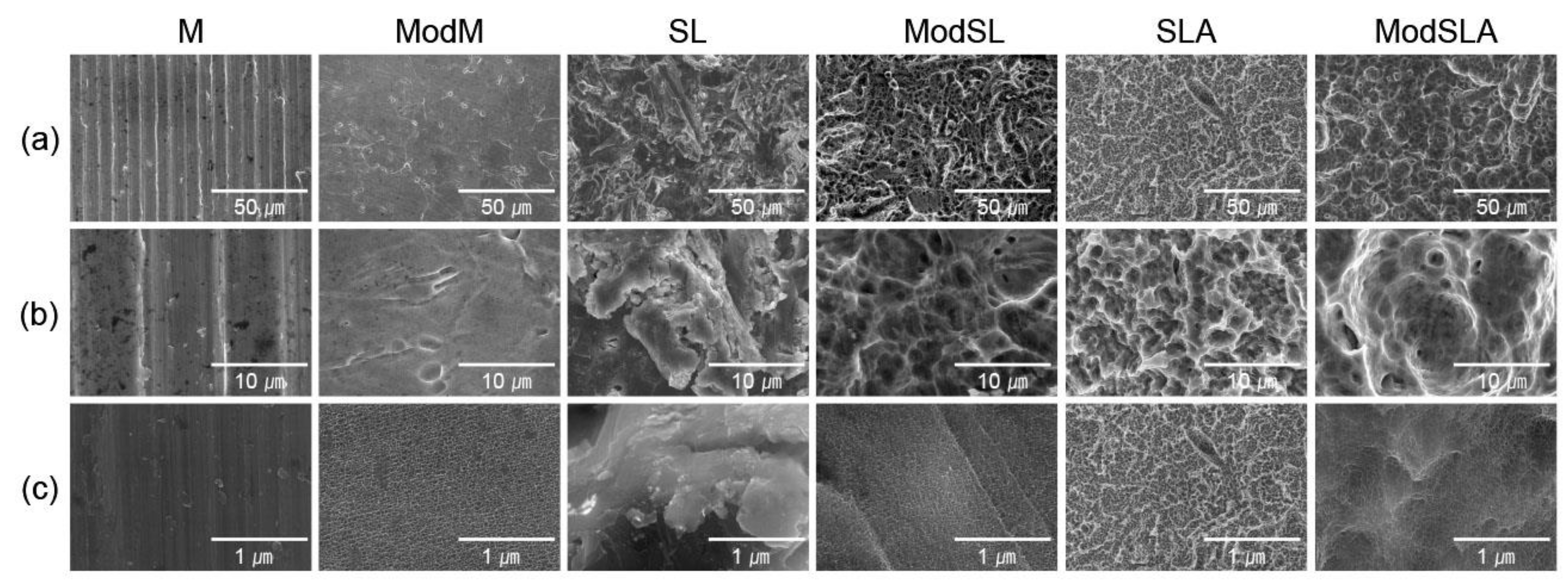
Figure 2.
Three-dimensional profile and quantitative topographical evaluations of Ti surfaces. (A) Three-dimensional scanning images constructed from digital microscope images, (B) results of profile analysis in which Ra (average roughness of profile) and Sa (the center plane average) were evaluated, and (C) water contact angles on the surface of the Ti disks. M: machined surface; ModM: machined surface + eco-friendly solution etching; SL: sandblasted surface; ModSL: sandblasted surface + eco-friendly solution etching; SLA: sandblasted/acid-etched surface; ModSLA: sandblasted/acid-etched surface + eco-friendly solution etching (*p < 0.05).
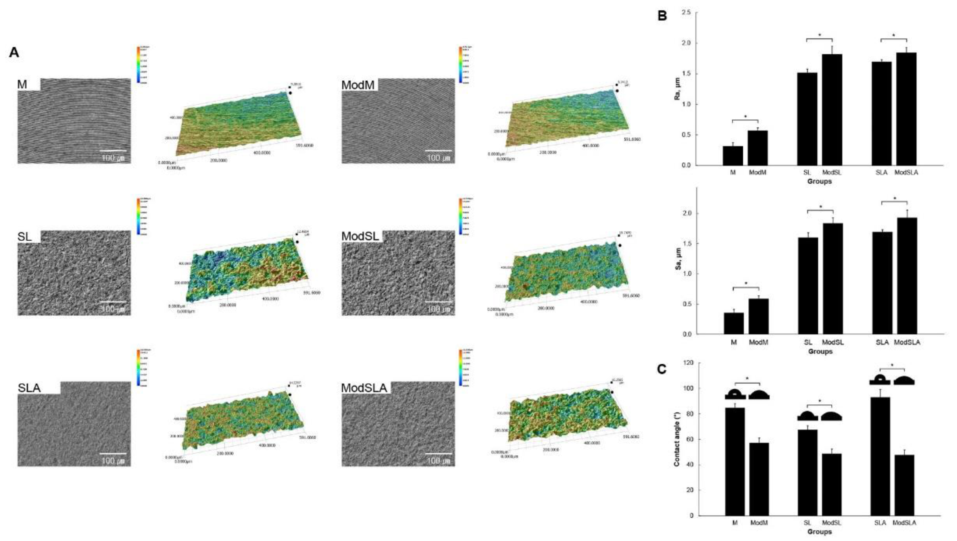
Figure 3.
Typical SEM images showing adhesion of osteoblasts cultured for 1 h on grade 4 Ti surfaces at 2,000× magnification: (a) machined surface, (b) machined surface + eco-friendly solution etching, (c) sandblasted surface, (d) sandblasted surface + eco-friendly solution etching, (e) sandblasted/acid-etched surface, and (f) sandblasted/acid-etched surface + eco-friendly solution etching.
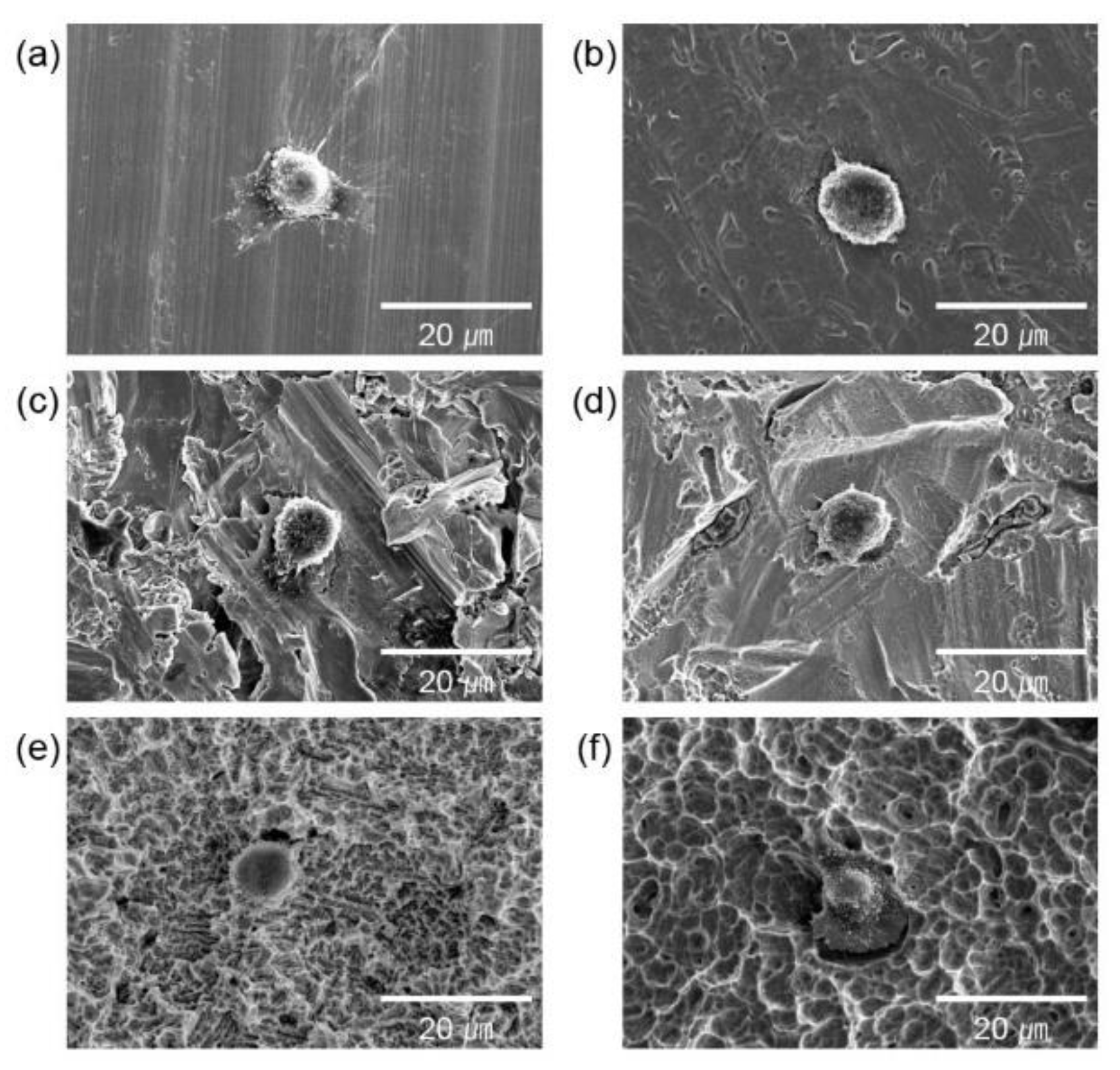
Figure 4.
Typical SEM images showing adhesion of osteoblasts cultured for 24 h on grade 4 Ti surfaces at 2,000× magnification: (a) machined surface, (b) machined surface + eco-friendly solution etching, (c) sandblasted surface, (d) sandblasted surface + eco-friendly solution etching, (e) sandblasted/acid-etched surface, and (f) sandblasted/acid-etched surface + eco-friendly solution etching.
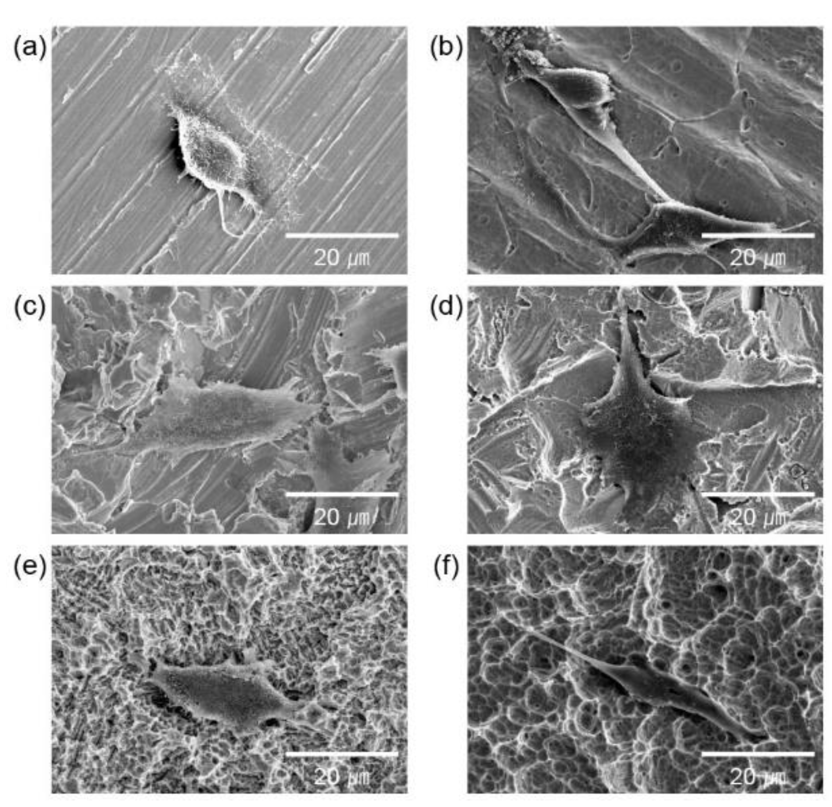
Figure 5.
Fluorescence images of LIVE/DEAD staining of MG-63 cells that were cultured on grade 4 Ti surfaces at 200x magnification: (a) machined surface, (b) machined surface + eco-friendly solution etching, (c) sandblasted surface, (d) sandblasted surface + eco-friendly solution etching, (e) sandblasted/acid-etched surface, and (f) sandblasted/acid-etched surface + eco-friendly solution etching.
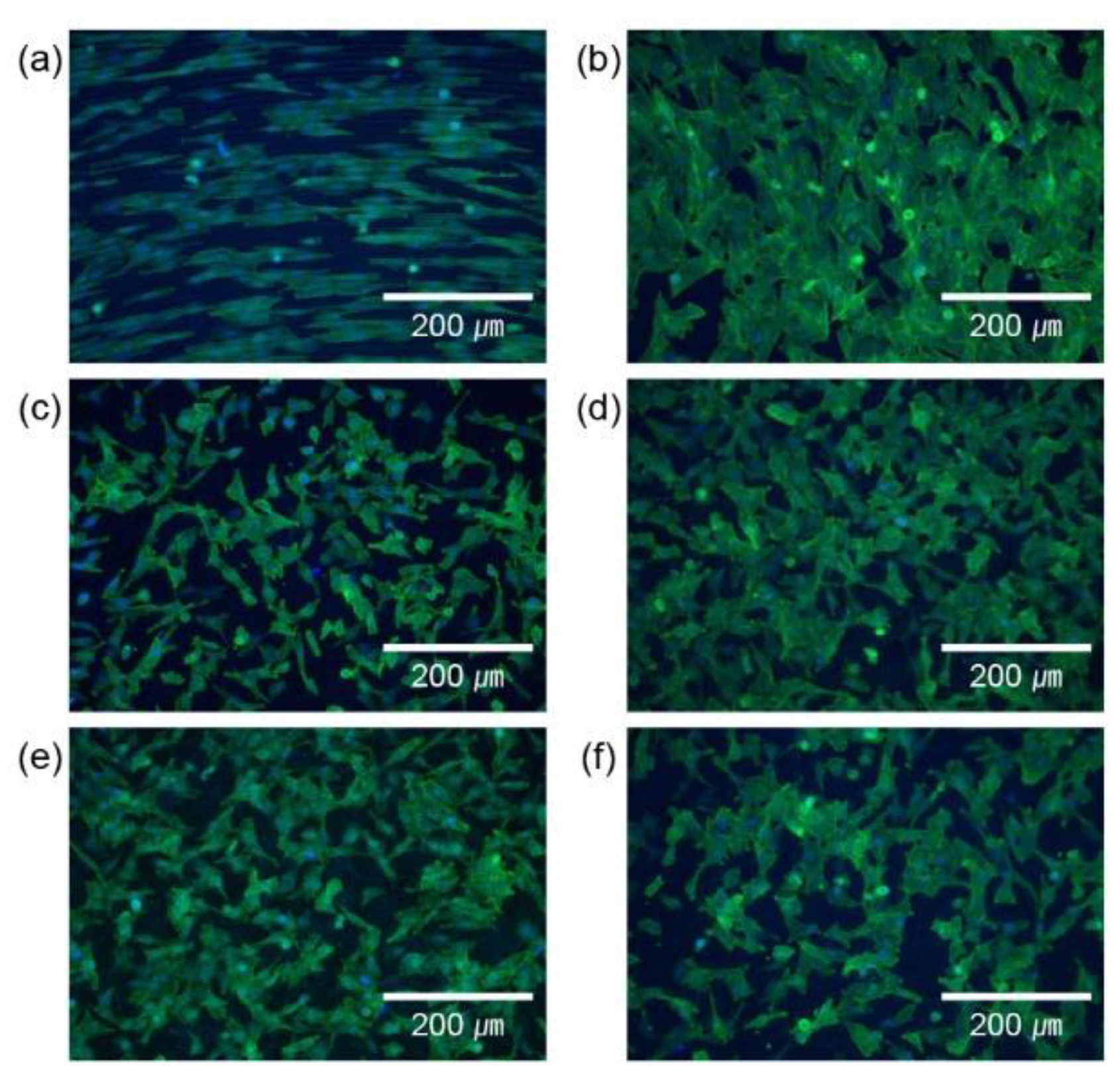
Figure 6.
Survival of osteoblastic cells cultured on the Ti surfaces on day 1.
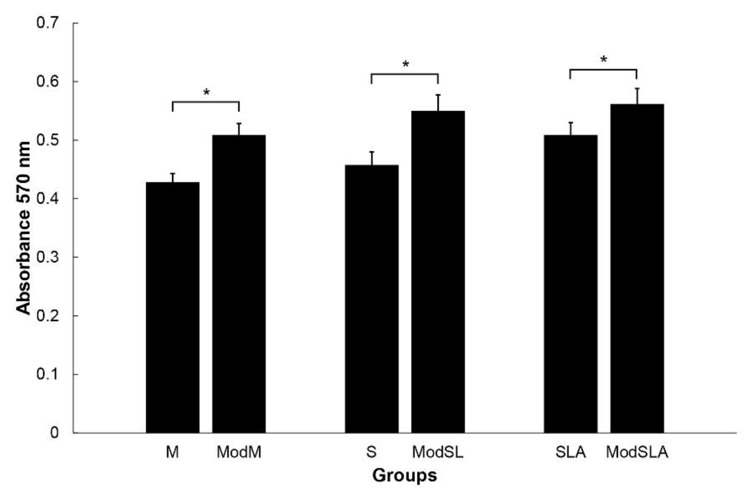
Figure 7.
O 1s spectra (A) and C 1s spectra (B) of all types of Ti surfaces.
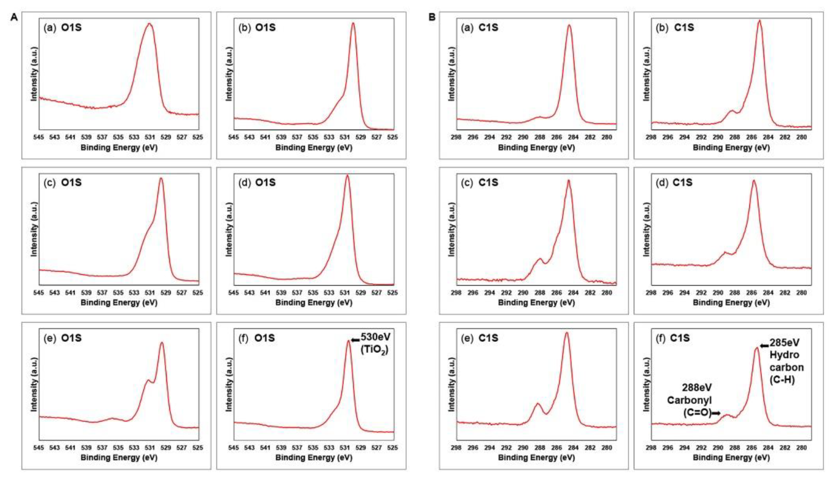
Figure 8.
Ti 2P spectra of all types of Ti surfaces (C).
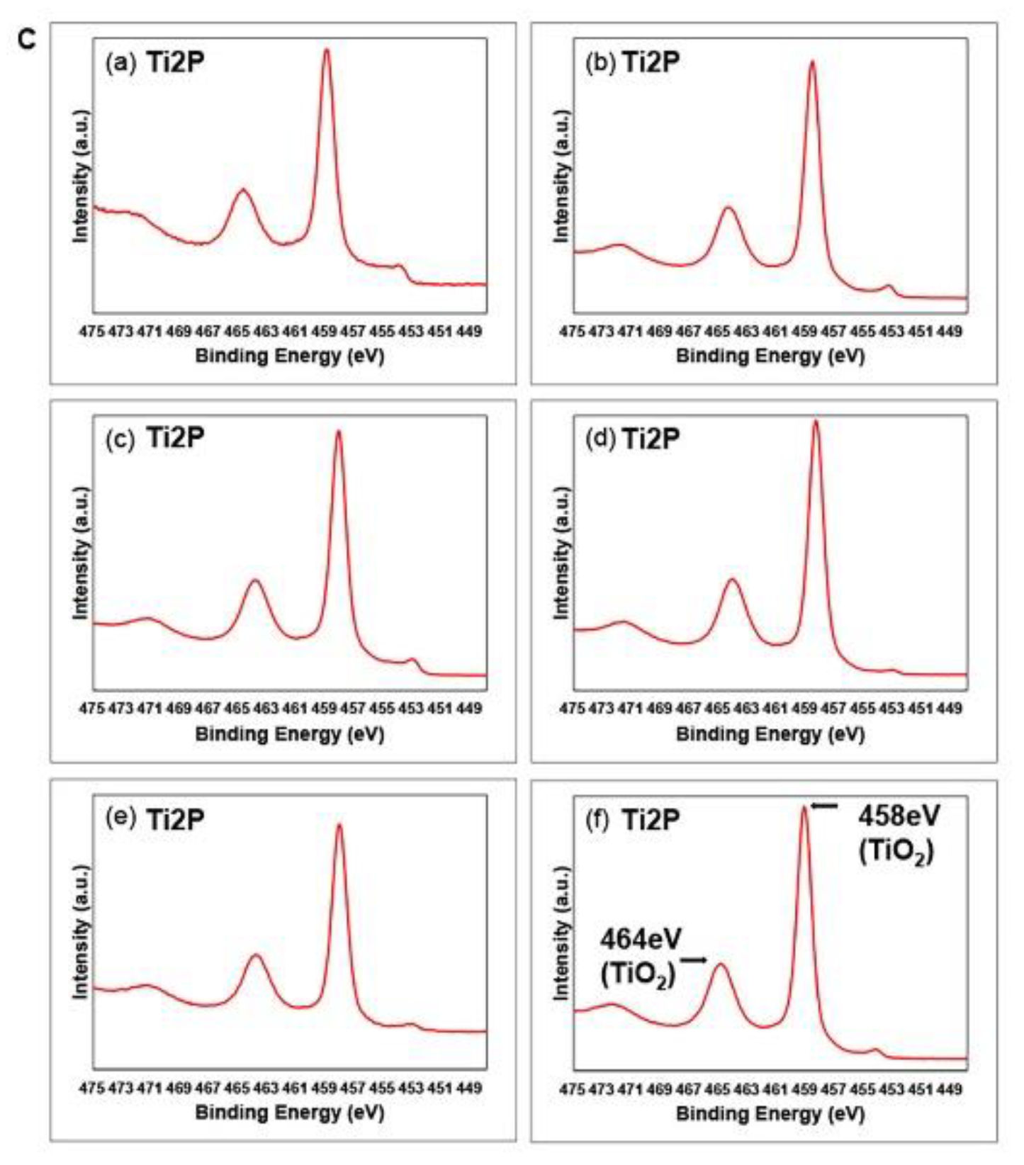

Table 1.
Experimental groups of specimens considered in this study.
| Group (n = 10) | Surface Treatment |
|---|---|
| M | No surface treatment |
| ModM | No surface treatment + eco-friendly solutiona) etching |
| SL | Alumina sandblasted |
| ModSL | Alumina sandblasted + eco-friendly solution etching |
| SLA | Alumina sandblasted + acid-etching |
| ModSLA | Alumina sandblasted + acid-etching + eco-friendly solution etching |
a) 30 wt% H2O2/5 wt% NaHCO3 solution.
Table 2.
Binding energy and atomic concentration (at%) for various surface modification treatments.
| Element | Machined | Sandblasted | SLA | |||||||||
|---|---|---|---|---|---|---|---|---|---|---|---|---|
| M | ModM | SL | ModSL | SLA | ModSLA | |||||||
| at% | BE | at% | BE | at% | BE | at% | BE | at% | BE | at% | BE | |
| M | 5.2 | 459.0 | 19.6 | 458.6 | 16.3 | 458.1 | 17.7 | 458.4 | 14.0 | 458.2 | 21.0 | 459.2 |
| SL | 24.7 | 531.1 | 46.2 | 530.1 | 56.0 | 529.8 | 54.2 | 529.9 | 44.0 | 529.7 | 47.5 | 530.7 |
| SLA | 69.9 | 284.7 | 34.1 | 285.2 | 27.5 | 284.8 | 27.9 | 285.0 | 41.9 | 284.9 | 31.3 | 285.5 |
* BE: Binding Energy.
Disclaimer/Publisher’s Note: The statements, opinions and data contained in all publications are solely those of the individual author(s) and contributor(s) and not of MDPI and/or the editor(s). MDPI and/or the editor(s) disclaim responsibility for any injury to people or property resulting from any ideas, methods, instructions or products referred to in the content. |
© 2023 by the authors. Licensee MDPI, Basel, Switzerland. This article is an open access article distributed under the terms and conditions of the Creative Commons Attribution (CC BY) license (http://creativecommons.org/licenses/by/4.0/).
Copyright: This open access article is published under a Creative Commons CC BY 4.0 license, which permit the free download, distribution, and reuse, provided that the author and preprint are cited in any reuse.


