Submitted:
22 June 2023
Posted:
23 June 2023
You are already at the latest version
Abstract
Introduction: Due to the great development of digital technology, through CAD (Computer - Aided Design) and CAM (Computer - Aided Manufacturing) systems, digital models could be used in orthodontic treatment planning decision-making, as there are numerous studies in the literature that support the validity of measurements of digital models of anterior teeth and the total coefficient of Bolton analysis. The aim of the study was to compare the average length value of the current upper and lower arches with that of a hypothetical nickel-titanium wire and the accuracy of the Bolton index measurement. In the present study, 138 dental casts were analyzed using Ortho3Shape software. Using the Ortho3Shape software, it was possible to measure the real and ideal lower arch lengths and, with regard to Bolton analysis, the values of the anterior and total Bolton coefficients. By comparing the values obtained with those of the study by A. Anand Kumar et al. using CBCT and plaster casts, the reliability of the measurements obtained with such CAD/CAM orthodontic software was evaluated. Considering the validity of digital measurements demonstrated by the studies and systematic reviews in the literature, it can be stated that CAD/CAM digital models can be a viable alternative to plaster models in the coming years, as they can facilitate model preservation and retrieval. For future studies and research, it would be preferable to use intra-oral scanners (IOS) in order to ensure greater accuracy, requiring only one step and ensuring a better outcome for the patient.
Keywords:
digital models
; Bolton index
; orthodontics CAD Software
; extra-oral scanner
1. Introduction
Thanks to the great development of digital technology, through CAD (Computer - Aided Design) and CAM (Computer - Aided Manufacturing) systems, it has been possible to observe an important simplification of diagnosis and an improvement in the planning and execution of orthodontic treatment.
There are three pillars supporting CAD/CAM technology: the first is the acquisition of digital images of patients' dental arches patients' dental arches, the second is the visualisation and manipulation of these images in specific software, and finally the 3D printing files, whether of the devices designed or the models in which the devices will be manufactured [1].
Digital models could be used in decision-making and treatment planning, as there are many studies in the literature that support the validity of the measurements of digital models of the dental arch, the accuracy of the measurements of these models and the measurements of the anterior and total coefficients of the Bolton analysis being confirmed [2,3,4,5].
The Bolton analysis is useful for evaluating this aspect in the search for possible disproportions between the arches. In fact, this analysis is carried out by means of a calculation developed by Wayne Bolton in 1958, from which the Bolton index can be derived, which numerically expresses the ideal ratio between the mesio-distal diameters of the teeth of the upper arch compared to those of the lower arch, of which the mesio-distal diameters, at the contact points, of all the elements from the central incisors to the first molar must first be calculated.
As demonstrated in the literature, the rotational and translational movements of teeth can be analysed and reproduced with great precision using digital configurations and, in more complex orthodontic treatments, a preliminary virtual plan has the potential to allow a significant reduction in errors with a greater probability of predicting the outcome
In addition, digital technologies and artificial intelligence may allow greater opportunities for such planning, as they can be applied in the early stages of the clinical examination to develop a simultaneous virtual plan of all stages of treatment [6,7].
Recently, it has been shown that 3D printing can be a method to digitally design and then produce customisable clear aligners with greater precision as an alternative to conventional orthodontic appliances, while also offering greater fit and effectiveness [8].
The aim of the study, using an extra oral scanner and the Ortho3Shape software, was to compare the mean value of the length of the current upper and lower arches with that of a hypothetical nickel titanium wire and the accuracy of the measurement of the anterior and total coefficient of the Bolton index.
2. Materials and Methods
In the present study, in which all operations were performed by the same operator, data from the 138 selected dental casts were measured using the Ortho3Shape software. The mean value of the current upper and lower arches were compared with hypothetical nickel titanium orthodontic wire.
2.1. Dental casts selection
The inclusion criteria were:
- Caucasian ethnicity.
- Permanent dentition.
- Class I and absence of severe malocclusions.
- Absence of extractions or extensive reconstructions.
- Absence of trauma and maxillo-facial surgery.
- Absence of previous mobile and fixed orthodontic treatment.
- Plaster models correctly scanned in 3D using Ortho3shape software.
The exclusion criteria were:
- Presence of deciduous dental elements.
- Abnormalities of eruption or formation of dental elements.
- Rotated dental elements.
- Agenesis of canine and central incisors.
- Edentulous posterior dental elements.
- Oligontia.
2.2. Procedures
In the present study, all operations were carried out by the same operator, the values taken into consideration (maximum, minimum, mean) from the dental casts selected, were calculated by using the Ortho3Shape software for the different measurements analysed: current length and of a hypothetical nickel-titanium orthodontic wire, the anterior and total Bolton coefficient.
For the analysis of the Bolton index, the lengths of the six anterior teeth of the upper arch and the six anterior teeth of the lower arch were measured (both lengths measured by summing the mesiodistal diameter of each of the dental elements from canine to canine). In order to measure the total coefficient, the lengths of the twelve maxillary and twelve mandibular teeth (both lengths measured by adding up the mesiodistal diameter of each of the dental elements from molar to molar) were measured.
To confirm the accuracy of the measurements of the Bolton indices of the anterior and total coefficients, the mean value of both values, calculated in the present study, were compared with those found in the study by A. Anand Kumar et al. and then both studies are compared with the student t-test [4].
The parameters used were:
- Bolton analysis: this analysis consists of the calculation developed by Wayne Bolton, from which the Bolton index is obtained, which numerically expresses the ideal ratio between the mesio-distal diameters of the teeth of the upper arch in relation to those of the lower arch.
- Anterior Bolton coefficient: ratio between the mesio-distal widths of the 6 mandibular anterior teeth and the mesio-distal widths of the 6 maxillary anterior teeth.
- Total Bolton coefficient: ratio between the mesio-distal widths of the 12 mandibular teeth and the mesio-distal widths of the 12 maxillary teeth, from the contralateral first permanent molar to the contralateral first permanent molar.
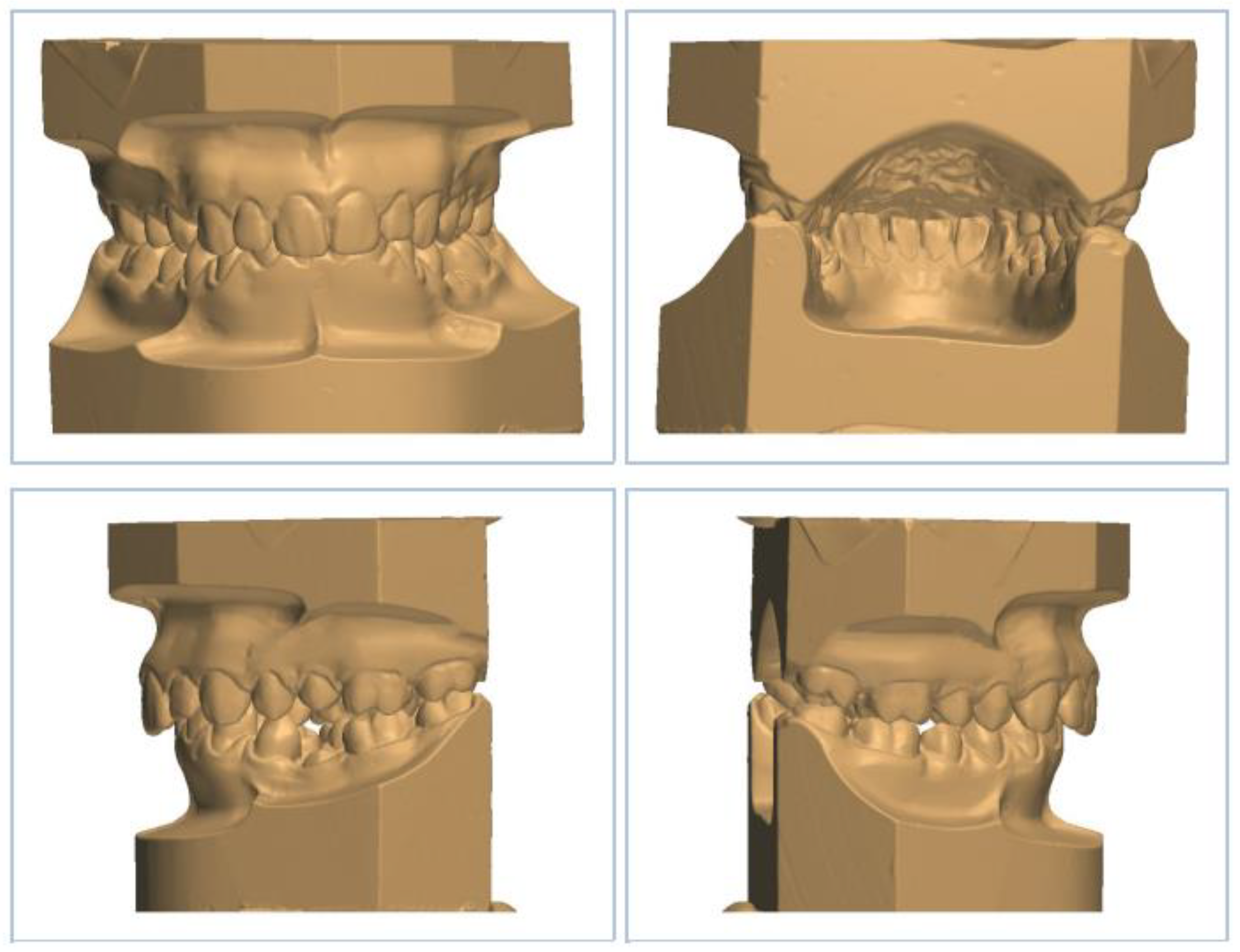
Figure 1.
Digitally reproducedd image of a plaster model of a patient with an occlusion in the maximum intercuspation position, frontal, back, left and right lateral vision can be observed.
Figure 1.
Digitally reproducedd image of a plaster model of a patient with an occlusion in the maximum intercuspation position, frontal, back, left and right lateral vision can be observed.
2.3. Statistical analysis
Each models was evaluated twice, with an interval of 2 weeks, by the same operator. Intraobserver variabilità was caluclated by using Cohen’s Kappa coeffficient. The coefficient obtained ranged between 0.9 and 0.97, that we assessed statistically significant.
The mean value were measured and Student’s T test was adopted, the level of signifacance was set at P < 0.05.
The statistical analysis was performer with the SPSS (Statistical Package for Social Sciences, Chigago, USA) 22.0 program.
The mean value of the anterior and total Bolton coefficients in the present study were calculated and then compared with those in the study by A. Anand Kumar et al. (using CBCT and plaster casts) in order to assess the compatibility of the measurements and values obtained [9].
Subsequently, both studies were compared with the student t-test, in order to compare the mean value of the present study with that of study A. Anand Kumar et al. considering p < 0.05 as the cut-off value for a significant difference [9].
The study by A. Anand Kumar et al. included a sample of 50 adults all over the age of 18, with similar inclusion criteria as in our study:
- Full permanent dentition from first molar to first molar in both upper and lower arch.
- The participant does not have to undergo orthodontic treatment.
- No severe crowding in the dentition [9].
3. Results
The results of the values examined in this study have been listed below in several tables, the average length of the current lower and upper arches and the corresponding hypothetical orthodontic nickel-titanium wires, the anterior and total Bolton coefficient.
Next, the mean value of the anterior and total coefficients of the Bolton analysis in the present study were compared with those calculated in the study by A. Anand Kumar et al. by CBCT and plaster casts, and finally these studies are compared by means of the student t-test [9].
3.1. Digital model arch analysis
Current length and of a hypothetical nickel-titanium orthodontic wire of the upper arch: the mean value calculated in the present study for the current length of the upper arch was 92.13 mm, while the mean value calculated for the hypothetical length of a nickel-titanium orthodontic wire of the upper arch was 103.79 [Table 1].

| Upper Arch Analysis | Hypothetical nickel titanium orthodontic wire upper arch lenght (mm) | Current upper arch length (mm) |
|---|---|---|
| Maximum Value | 124.33 | 108.07 |
| Minimum Value | 80.03 | 66.47 |
| Mean Value | 103.79 | 92.13 |
Consequently, it was possible to conclude that the mean value of the hypothetical length of a nickel-titanium orthodontic wire of the lower arch was 11.66 mm longer than that calculated for the current length of the upper arch.
Current length and of a hypothetical nickel-titanium orthodontic wire of the lower arch: the mean value calculated in the present study for the current length of the upper arch was 88.26 mm, while the mean value calculated for the hypothetical length of a nickel-titanium orthodontic wire of the upper arch was 94.71 mm [Table 2].
| Lower Arch Analysis | Hypothetical nickel titanium orthodontic wire lower arch lenght (mm) | Current lower arch length (mm) |
|---|---|---|
| Maximum Value | 109.91 | 107.63 |
| Minimum Value | 75.32 | 68.47 |
| Mean Value | 94.71 | 88.26 |
Consequently, it was possible to conclude that the mean value of the hypothetical length of a nickel-titanium orthodontic wire of the lower arch was 6.45 mm longer than that calculated for the current length of the lower arch.
3.2. Bolton analysis digital models
| Bolton Analysis: Total Coefficient | Sum 12 maxillary teeth (mm) | Sum 12 mandibular teeth (mm) | Report |
|---|---|---|---|
| Maximum Value | 89.25 | 88.78 | 1.21 |
| Minimum Value | 58.10 | 53.39 | 0.68 |
| Mean Value | 78.99 | 73.77 | 0.94 |
| Bolton Analysis: Anterior Coefficient | Sum 6 maxillary teeth (mm) | Sum 6 mandibular teeth (mm) | Report |
|---|---|---|---|
| Maximum Value | 45.54 | 35.69 | 1.16 |
| Minimum Value | 24.33 | 22.58 | 0.62 |
| Mean Value | 38.16 | 30.10 | 0.79 |
3.3. Comparison analysis Bolton present study and A. Anand Kumar et al. study [4]
| Mean Value | A. Anand Kumar et al. study | Present study | P Value |
|---|---|---|---|
| Anterior coefficient | 0.76 | 0.79 | 0.0028 |
| Total coefficient | 0.91 | 0.94 | 0.0009 |
3.3. Comparison by student t-test of the anterior and total Bolton coefficients of the two studies
- Student t-test: following the test, the value for the anterior coefficient was found to be P = 0.0028 and for the total coefficient was found to be P = 0.0009 both indicating a significant difference.
4. Discussion
As has been done previously in other studies, dental models were digitised to obtain STL format files, the 'Standard Triangulation Language', using CAD/CAM technology, which allow the surface data of the dental arches to be saved on the computer in three-dimensional imaging [10,11,12].
STL files can allow clinicians to quickly obtain diagnostic information, arch width and perimeter, model discrepancies, Bolton discrepancy, overjet and overbite, as well as for the simulation of tooth movements [13].
Thus, the diagnostic configurations obtained through STL files can be used to evaluate better treatment strategies, increase their effectiveness and also allow, through digital planning, a better position of the fixed multibracket appliance on the teeth.
In fixed orthodontic therapy, brackets, bands and buccal tubes are used to transfer force and torque to the teeth, thus inducing tooth movement.
The accurate placement of orthodontic brackets is crucial, as deviations from the correct position of the brackets can lead to undesirable tooth movement.
Deviations from the correct bracket positions can lead to unwanted tooth movement, poor results and prolonged treatment times.
Computer-aided planning and production technology enables virtual planning of bracket positions and the of bracket positions, and bracket transfer splints can therefore be produced economically and easily and also digital processing can minimise positioning errors and increase treatment efficiency.
The accuracy of the final bracket position is defined as the deviation between the planned and actual bracket position [14,15,16].
The stereolithographic format can also be used in 3D printing software to be able to generate the necessary information for the production of 3D printed models, which can enable a more efficient workflow while saving time, a set of errors for linear measurements considered clinically acceptable when replicating plaster models or printing digital models for diagnostic purposes have therefore been defined [17,18].
Again analysing the STL files of the present study, a comparison was made between the average value of the length of the ideal arch and the present arch.
The average value of the length of a hypothetical orthodontic nickel titanium wire of the ideal upper arch is 11.66 mm longer than the average value of the current upper arch length.
The average length value of a hypothetical ideal lower arch nickel titanium orthodontic wire is 6.45 mm longer than the average length value of the current lower arch.
A comparison analysis was then carried out between the present study and that of A. Anand Kumar et al. [9].
Apart from the result of the student's t-test (whose significant difference is due to the different sample sizes of the two studies being compared: 138 dental casts were examined in the present study, whereas 50 patients were examined in the other study), the mean value for the anterior and total coefficient of the Bolton index in the present study were compatible with those calculated in the other study [9].
Thus being able to confirm the reliability of the digitally taken measurements.
Consequently, the measurements that were made in the present retrospective study, both for the ideal and current upper and lower arch lengths, and for the anterior and total Bolton coefficients of the 138 dental casts examined, are useful to be able to establish the standard of such measurements in the Italian adolescent population.
The limitation of the present study is the double step for model reproduction, the first being the reproduction by plaster casts of the dental arches (scanned later) and the second by measuring the digital models using Ortho3Shape software.
5. Conclusions
The present study confirmed what has already been demonstrated in the scientific literature, that digital orthodontics and the reproduction of 3D digital models can contribute to the simplification of diagnosis and treatment planning in Orthodontics.
In the present study, the mean value of the anterior coefficients of the Bolton index was compatible with those of the study by A. Anand Kumar et al. confirming the reliability of digital measurements [9].
In the future, it would be preferable for studies and research to be conducted with the aid of intra-oral scanners (IOS) to ensure greater accuracy, requiring only one step and ensuring a better result for the patient.
6. Full presentation of all results
| Upper Arch Analysis | Current upper arch length (mm) | Hypothetical nichel titanium orthodontic wire lower arch lenght (mm) |
| Patient 1 | 94,70 | 102,36 |
| Patient 2 | 94,50 | 108,85 |
| Patient 3 | 94,44 | 101,37 |
| Patient 4 | 81,62 | 97,00 |
| Patient 5 | 95,72 | 108,36 |
| Patient 6 | 90,33 | 100,29 |
| Patient 7 | 79,86 | 88,12 |
| Patient 8 | 89,67 | 108,06 |
| Patient 9 | 78,74 | 99,55 |
| Patient 10 | 83,76 | 92,20 |
| Patient 11 | 91,00 | 110,58 |
| Patient 12 | 86,58 | 110,04 |
| Patient 13 | 92,59 | 101,63 |
| Patient 14 | 92,37 | 99,90 |
| Patient 15 | 90,30 | 109,69 |
| Patient 16 | 82,01 | 108,37 |
| Patient 17 | 93,87 | 111,27 |
| Patient 18 | 101,64 | 114,35 |
| Patient 19 | 88,62 | 95,76 |
| Patient 20 | 80,47 | 96,34 |
| Patient 21 | 86,98 | 105,62 |
| Patient 22 | 86,23 | 94,75 |
| Patient 23 | 83,54 | 89,70 |
| Patient 24 | 102,56 | 111,30 |
| Patient 25 | 91,37 | 94,42 |
| Patient 26 | 104,08 | 115,41 |
| Patient 27 | 106,23 | 110,76 |
| Patient 28 | 98,82 | 109,05 |
| Patient 29 | 81,83 | 84,01 |
| Patient 30 | 101,04 | 107,49 |
| Patient 31 | 92,41 | 97,13 |
| Patient 32 | 89,72 | 105,47 |
| Patient 33 | 83,72 | 98,82 |
| Patient 34 | 91,70 | 104,02 |
| Patient 35 | 94,56 | 104,93 |
| Patient 36 | 92,10 | 107,76 |
| Patient 37 | 89,59 | 103,34 |
| Patient 38 | 72,29 | 80,03 |
| Patient 39 | 94,31 | 106,65 |
| Patient 40 | 97,73 | 109,58 |
| Patient 41 | 66,47 | 81,03 |
| Patient 42 | 88,89 | 105,33 |
| Patient 43 | 89,50 | 108,16 |
| Patient 44 | 88,84 | 104,82 |
| Patient 45 | 104,13 | 114,82 |
| Patient 46 | 94,84 | 104,13 |
| Patient 47 | 94,55 | 105,17 |
| Patient 48 | 83,18 | 91,92 |
| Patient 49 | 99,83 | 111,57 |
| Patient 50 | 81,87 | 96,40 |
| Patient 51 | 86,87 | 94,73 |
| Patient 52 | 95,57 | 109,20 |
| Patient 53 | 78,22 | 81,32 |
| Patient 54 | 80,31 | 88,51 |
| Patient 55 | 94,72 | 106,17 |
| Patient 56 | 101,66 | 112,43 |
| Patient 57 | 98,68 | 107,85 |
| Patient 58 | 93,59 | 106,49 |
| Patient 59 | 75,14 | 98,14 |
| Patient 60 | 105,13 | 110,52 |
| Patient 61 | 98,93 | 114,18 |
| Patient 62 | 93,49 | 108,29 |
| Patient 63 | 88,48 | 112,58 |
| Patient 64 | 100,83 | 106,87 |
| Patient 65 | 99,57 | 109,59 |
| Patient 66 | 93,37 | 107,73 |
| Patient 67 | 93,27 | 103,72 |
| Patient 68 | 93,37 | 97,27 |
| Patient 69 | 84,75 | 96,49 |
| Patient 70 | 94,21 | 109,93 |
| Patient 71 | 94,76 | 111,29 |
| Patient 72 | 96,07 | 107,55 |
| Patient 73 | 91,42 | 98,41 |
| Patient 74 | 75,93 | 98,57 |
| Patient 75 | 92,49 | 102,35 |
| Patient 76 | 100,02 | 113,14 |
| Patient 77 | 81,98 | 102,29 |
| Patient 78 | 98,58 | 108,87 |
| Patient 79 | 89,80 | 103,67 |
| Patient 80 | 90,66 | 106,21 |
| Patient 81 | 98,43 | 109,06 |
| Patient 82 | 81,87 | 96,40 |
| Patient 83 | 83,59 | 93,65 |
| Patient 84 | 86,54 | 88,03 |
| Patient 85 | 95,62 | 113,91 |
| Patient 86 | 82,61 | 94,96 |
| Patient 87 | 92,62 | 104,29 |
| Patient 88 | 95,11 | 108,58 |
| Patient 89 | 87,93 | 103,75 |
| Patient 90 | 95,92 | 115,54 |
| Patient 91 | 97,56 | 114,93 |
| Patient 92 | 91,05 | 105,26 |
| Patient 93 | 89,65 | 104,38 |
| Patient 94 | 90,31 | 100,07 |
| Patient 95 | 99,80 | 108,69 |
| Patient 96 | 99,25 | 107,74 |
| Patient 97 | 99,65 | 108,51 |
| Patient 98 | 93,23 | 106,62 |
| Patient 99 | 98,74 | 107,73 |
| Patient 100 | 103,20 | 114,91 |
| Patient 101 | 83,50 | 98,43 |
| Patient 102 | 77,34 | 91,35 |
| Patient 103 | 90,42 | 100,16 |
| Patient 104 | 107,28 | 124,33 |
| Patient 105 | 76,72 | 82,74 |
| Patient 106 | 95,59 | 105,94 |
| Patient 107 | 89,20 | 96,43 |
| Patient 108 | 74,33 | 98,75 |
| Patient 109 | 90,46 | 103,82 |
| Patient 110 | 81,16 | 91,02 |
| Patient 111 | 97,46 | 114,01 |
| Patient 112 | 105,58 | 113,23 |
| Patient 113 | 90,71 | 100,67 |
| Patient 114 | 103,88 | 109,00 |
| Patient 115 | 81,19 | 89,56 |
| Patient 116 | 92,63 | 104,66 |
| Patient 117 | 101,73 | 105,60 |
| Patient 118 | 103,52 | 112,50 |
| Patient 119 | 108,07 | 116,11 |
| Patient 120 | 83,94 | 89,74 |
| Patient 121 | 97,23 | 108,51 |
| Patient 122 | 83,76 | 91,38 |
| Patient 123 | 98,95 | 107,48 |
| Patient 124 | 104,91 | 109,86 |
| Patient 125 | 94,60 | 107,47 |
| Patient 126 | 105,49 | 117,68 |
| Patient 127 | 101,20 | 109,80 |
| Patient 128 | 94,43 | 100,00 |
| Patient 129 | 85,01 | 106,50 |
| Patient 130 | 97,13 | 104,90 |
| Patient 131 | 85,04 | 91,41 |
| Patient 132 | 105,15 | 113,90 |
| Patient 133 | 106,77 | 105,89 |
| Patient 134 | 96,75 | 106,45 |
| Patient 135 | 106,53 | 112,55 |
| Patient 136 | 96,22 | 103,69 |
| Patient 137 | 94,04 | 106,94 |
| Patient 138 | 85,78 | 101,97 |
| Maximum value | 108,07 | 124,33 |
| Minimum value | 66,47 | 80,03 |
| Mean value | 92,13 | 103,79 |
| Lower Arch Analysis | Current lower arch length (mm) | Hypothetical nichel titanium orthodontic wire lower arch lenght (mm) |
| Patient 1 | 68,47 | 85,41 |
| Patient 2 | 90,68 | 94,25 |
| Patient 3 | 73,30 | 88,62 |
| Patient 4 | 84,00 | 97,62 |
| Patient 5 | 78,06 | 95,87 |
| Patient 6 | 89,25 | 93,99 |
| Patient 7 | 81,05 | 89,37 |
| Patient 8 | 87,74 | 98,10 |
| Patient 9 | 79,19 | 86,99 |
| Patient 10 | 75,78 | 83,42 |
| Patient 11 | 85,70 | 100,02 |
| Patient 12 | 83,67 | 98,45 |
| Patient 13 | 82,47 | 89,67 |
| Patient 14 | 87,30 | 95,95 |
| Patient 15 | 84,22 | 98,38 |
| Patient 16 | 86,76 | 97,96 |
| Patient 17 | 90,59 | 105,06 |
| Patient 18 | 91,70 | 103,28 |
| Patient 19 | 83,95 | 89,23 |
| Patient 20 | 78,75 | 94,90 |
| Patient 21 | 90,21 | 98,00 |
| Patient 22 | 79,52 | 83,99 |
| Patient 23 | 75,64 | 81,76 |
| Patient 24 | 93,27 | 104,15 |
| Patient 25 | 86,93 | 90,49 |
| Patient 26 | 94,47 | 102,75 |
| Patient 27 | 99,76 | 97,65 |
| Patient 28 | 95,43 | 99,54 |
| Patient 29 | 71,78 | 75,99 |
| Patient 30 | 95,41 | 93,65 |
| Patient 31 | 82,87 | 86,96 |
| Patient 32 | 85,00 | 93,60 |
| Patient 33 | 81,27 | 90,92 |
| Patient 34 | 80,58 | 90,43 |
| Patient 35 | 89,49 | 96,01 |
| Patient 36 | 87,21 | 93,02 |
| Patient 37 | 97,93 | 103,89 |
| Patient 38 | 73,15 | 76,08 |
| Patient 39 | 84,74 | 95,59 |
| Patient 40 | 93,58 | 99,93 |
| Patient 41 | 75,28 | 80,92 |
| Patient 42 | 84,32 | 97,00 |
| Patient 43 | 82,06 | 95,63 |
| Patient 44 | 81,52 | 90,16 |
| Patient 45 | 92,44 | 99,72 |
| Patient 46 | 90,96 | 94,75 |
| Patient 47 | 88,40 | 95,31 |
| Patient 48 | 92,14 | 100,20 |
| Patient 49 | 95,21 | 98,78 |
| Patient 50 | 87,76 | 98,03 |
| Patient 51 | 78,81 | 82,86 |
| Patient 52 | 89,38 | 96,24 |
| Patient 53 | 80,24 | 84,03 |
| Patient 54 | 86,77 | 88,60 |
| Patient 55 | 96,09 | 102,17 |
| Patient 56 | 99,58 | 103,42 |
| Patient 57 | 79,48 | 94,85 |
| Patient 58 | 90,82 | 94,42 |
| Patient 59 | 78,16 | 86,88 |
| Patient 60 | 97,80 | 101,31 |
| Patient 61 | 92,60 | 103,85 |
| Patient 62 | 92,25 | 99,34 |
| Patient 63 | 93,97 | 106,03 |
| Patient 64 | 102,75 | 103,27 |
| Patient 65 | 90,83 | 97,30 |
| Patient 66 | 88,25 | 98,70 |
| Patient 67 | 88,94 | 96,92 |
| Patient 68 | 82,31 | 85,30 |
| Patient 69 | 86,31 | 91,18 |
| Patient 70 | 70,14 | 92,27 |
| Patient 71 | 89,72 | 105,32 |
| Patient 72 | 91,16 | 95,94 |
| Patient 73 | 91,63 | 92,23 |
| Patient 74 | 78,33 | 96,21 |
| Patient 75 | 82,64 | 89,71 |
| Patient 76 | 93,51 | 106,16 |
| Patient 77 | 82,53 | 92,30 |
| Patient 78 | 96,09 | 99,16 |
| Patient 79 | 92,12 | 94,01 |
| Patient 80 | 90,04 | 97,57 |
| Patient 81 | 92,38 | 96,86 |
| Patient 82 | 87,76 | 98,03 |
| Patient 83 | 78,22 | 83,61 |
| Patient 84 | 93,54 | 97,31 |
| Patient 85 | 92,09 | 100,18 |
| Patient 86 | 84,37 | 93,27 |
| Patient 87 | 91,86 | 96,61 |
| Patient 88 | 89,90 | 94,03 |
| Patient 89 | 78,94 | 93,77 |
| Patient 90 | 92,47 | 101,37 |
| Patient 91 | 92,32 | 103,49 |
| Patient 92 | 84,88 | 93,14 |
| Patient 93 | 84,33 | 95,19 |
| Patient 94 | 87,94 | 88,81 |
| Patient 95 | 98,28 | 96,67 |
| Patient 96 | 92,50 | 98,61 |
| Patient 97 | 94,61 | 94,17 |
| Patient 98 | 78,79 | 88,32 |
| Patient 99 | 91,01 | 94,48 |
| Patient 100 | 97,02 | 100,66 |
| Patient 101 | 80,01 | 92,01 |
| Patient 102 | 77,34 | 81,02 |
| Patient 103 | 90,74 | 94,81 |
| Patient 104 | 107,63 | 109,91 |
| Patient 105 | 78,90 | 83,85 |
| Patient 106 | 94,60 | 98,87 |
| Patient 107 | 97,58 | 98,79 |
| Patient 108 | 86,93 | 90,41 |
| Patient 109 | 94,15 | 96,98 |
| Patient 110 | 77,96 | 88,47 |
| Patient 111 | 93,85 | 99,43 |
| Patient 112 | 93,53 | 102,21 |
| Patient 113 | 96,29 | 98,78 |
| Patient 114 | 97,60 | 99,00 |
| Patient 115 | 91,03 | 93,02 |
| Patient 116 | 76,87 | 79,41 |
| Patient 117 | 89,07 | 94,48 |
| Patient 118 | 102,22 | 100,63 |
| Patient 119 | 101,27 | 105,05 |
| Patient 120 | 77,37 | 80,99 |
| Patient 121 | 89,64 | 99,42 |
| Patient 122 | 94,65 | 100,29 |
| Patient 123 | 105,04 | 101,05 |
| Patient 124 | 88,78 | 89,58 |
| Patient 125 | 90,17 | 95,10 |
| Patient 126 | 104,66 | 108,18 |
| Patient 127 | 96,83 | 102,96 |
| Patient 128 | 75,93 | 77,34 |
| Patient 129 | 85,97 | 87,94 |
| Patient 130 | 82,99 | 75,32 |
| Patient 131 | 90,08 | 91,73 |
| Patient 132 | 102,15 | 99,97 |
| Patient 133 | 99,64 | 93,74 |
| Patient 134 | 84,95 | 94,45 |
| Patient 135 | 103,16 | 102,17 |
| Patient 136 | 87,26 | 96,62 |
| Patient 137 | 94,32 | 94,63 |
| Patient 138 | 90,99 | 93,64 |
| Maximum value | 107,63 | 109,91 |
| Minimum value | 68,47 | 75,32 |
| Mean value | 88,26 | 94,71 |
|
Bolton Analysis: Anterior Coefficient |
Sum 6 maxillary teeth (mm) |
Sum 6 mandibular teeth (mm) |
Report |
| Patient 1 | 38,60 | 28,28 | 0,73 |
| Patient 2 | 39,88 | 30,66 | 0,77 |
| Patient 3 | 38,99 | 31,08 | 0,80 |
| Patient 4 | 33,81 | 26,18 | 0,77 |
| Patient 5 | 39,52 | 28,88 | 0,73 |
| Patient 6 | 33,60 | 29,78 | 0,86 |
| Patient 7 | 35,13 | 25,26 | 0,72 |
| Patient 8 | 37,57 | 28,00 | 0,75 |
| Patient 9 | 32,47 | 25,31 | 0,78 |
| Patient 10 | 36,03 | 27,78 | 0,77 |
| Patient 11 | 33,99 | 26,83 | 0,79 |
| Patient 12 | 33,99 | 26,73 | 0,79 |
| Patient 13 | 34,33 | 26,19 | 0,76 |
| Patient 14 | 40,00 | 29,74 | 0,74 |
| Patient 15 | 35,88 | 27,72 | 0,77 |
| Patient 16 | 24,33 | 28,22 | 1,16 |
| Patient 17 | 37,34 | 28,85 | 0,77 |
| Patient 18 | 38,35 | 30,71 | 0,80 |
| Patient 19 | 34,72 | 27,88 | 0,80 |
| Patient 20 | 35,64 | 27,2 | 0,76 |
| Patient 21 | 32,36 | 28,05 | 0,87 |
| Patient 22 | 36,88 | 29,54 | 0,80 |
| Patient 23 | 37,22 | 26,55 | 0,71 |
| Patient 24 | 42,70 | 33,61 | 0,79 |
| Patient 25 | 37,87 | 27,92 | 0,74 |
| Patient 26 | 45,54 | 33,35 | 0,73 |
| Patient 27 | 44,95 | 35,38 | 0,79 |
| Patient 28 | 36,94 | 29,24 | 0,79 |
| Patient 29 | 38,23 | 28,49 | 0,75 |
| Patient 30 | 43,01 | 31,35 | 0,73 |
| Patient 31 | 36,50 | 24,59 | 0,67 |
| Patient 32 | 34,82 | 27,29 | 0,78 |
| Patient 33 | 35,99 | 30,54 | 0,85 |
| Patient 34 | 36,34 | 22,58 | 0,62 |
| Patient 35 | 36,12 | 28,24 | 0,78 |
| Patient 36 | 34,41 | 28,22 | 0,82 |
| Patient 37 | 39,17 | 29,64 | 0,76 |
| Patient 38 | 34,33 | 30,47 | 0,89 |
| Patient 39 | 35,08 | 27,09 | 0,77 |
| Patient 40 | 36,49 | 28,13 | 0,77 |
| Patient 41 | 36,36 | 29,89 | 0,82 |
| Patient 42 | 35,24 | 27,13 | 0,77 |
| Patient 43 | 34,58 | 26,88 | 0,78 |
| Patient 44 | 32,42 | 24,71 | 0,76 |
| Patient 45 | 40,75 | 30,62 | 0,75 |
| Patient 46 | 40,19 | 32,15 | 0,80 |
| Patient 47 | 39,15 | 29,34 | 0,75 |
| Patient 48 | 40,45 | 31,58 | 0,78 |
| Patient 49 | 39,93 | 30,40 | 0,76 |
| Patient 50 | 40,04 | 33,43 | 0,83 |
| Patient 51 | 38,08 | 27,24 | 0,72 |
| Patient 52 | 37,37 | 29,95 | 0,80 |
| Patient 53 | 36,78 | 27,96 | 0,76 |
| Patient 54 | 37,86 | 28,33 | 0,75 |
| Patient 55 | 37,27 | 31,89 | 0,86 |
| Patient 56 | 41,98 | 33,34 | 0,79 |
| Patient 57 | 38,40 | 30,96 | 0,81 |
| Patient 58 | 38,55 | 33,72 | 0,87 |
| Patient 59 | 29,69 | 27,88 | 0,94 |
| Patient 60 | 41,31 | 30,85 | 0,75 |
| Patient 61 | 42,65 | 32,01 | 0,75 |
| Patient 62 | 36,76 | 27,52 | 0,75 |
| Patient 63 | 40,38 | 32,29 | 0,80 |
| Patient 64 | 41,88 | 34,12 | 0,81 |
| Patient 65 | 38,80 | 28,92 | 0,75 |
| Patient 66 | 35,30 | 28,41 | 0,80 |
| Patient 67 | 37,32 | 29,41 | 0,79 |
| Patient 68 | 36,74 | 26,87 | 0,73 |
| Patient 69 | 36,23 | 30,24 | 0,83 |
| Patient 70 | 38,52 | 29,30 | 0,76 |
| Patient 71 | 36,27 | 30,55 | 0,84 |
| Patient 72 | 36,83 | 28,73 | 0,78 |
| Patient 73 | 36,68 | 28,44 | 0,78 |
| Patient 74 | 36,87 | 29,01 | 0,79 |
| Patient 75 | 35,11 | 27,12 | 0,77 |
| Patient 76 | 39,51 | 32,26 | 0,82 |
| Patient 77 | 33,75 | 26,33 | 0,79 |
| Patient 78 | 39,62 | 30,26 | 0,76 |
| Patient 79 | 33,75 | 30,08 | 0,89 |
| Patient 80 | 36,37 | 29,33 | 0,81 |
| Patient 81 | 38,69 | 29,71 | 0,77 |
| Patient 82 | 44,04 | 33,43 | 0,83 |
| Patient 83 | 39,19 | 31,40 | 0,80 |
| Patient 84 | 39,71 | 31,86 | 0,80 |
| Patient 85 | 40,01 | 31,51 | 0,79 |
| Patient 86 | 34,63 | 28,44 | 0,82 |
| Patient 87 | 39,02 | 31,82 | 0,82 |
| Patient 88 | 37,22 | 30,48 | 0,82 |
| Patient 89 | 36,43 | 26,99 | 0,74 |
| Patient 90 | 38,97 | 30,85 | 0,79 |
| Patient 91 | 42,33 | 30,00 | 0,71 |
| Patient 92 | 34,38 | 28,21 | 0,82 |
| Patient 93 | 34,77 | 27,51 | 0,79 |
| Patient 94 | 34,24 | 29,18 | 0,85 |
| Patient 95 | 38,60 | 30,32 | 0,79 |
| Patient 96 | 38,73 | 30,84 | 0,80 |
| Patient 97 | 39,93 | 32,52 | 0,81 |
| Patient 98 | 41,81 | 33,98 | 0,81 |
| Patient 99 | 41,55 | 30,92 | 0,74 |
| Patient 100 | 42,06 | 31,52 | 0,75 |
| Patient 101 | 30,15 | 28,04 | 0,93 |
| Patient 102 | 31,87 | 30,54 | 0,96 |
| Patient 103 | 34,20 | 28,44 | 0,83 |
| Patient 104 | 44,46 | 35,12 | 0,79 |
| Patient 105 | 34,02 | 28,32 | 0,83 |
| Patient 106 | 39,12 | 30,72 | 0,79 |
| Patient 107 | 45,32 | 32,24 | 0,71 |
| Patient 108 | 37,38 | 29,10 | 0,78 |
| Patient 109 | 36,08 | 32,02 | 0,89 |
| Patient 110 | 37,49 | 31,00 | 0,83 |
| Patient 111 | 43,61 | 31,03 | 0,71 |
| Patient 112 | 44,64 | 34,26 | 0,77 |
| Patient 113 | 40,43 | 32,80 | 0,81 |
| Patient 114 | 41,66 | 31,44 | 0,75 |
| Patient 115 | 36,54 | 30,32 | 0,83 |
| Patient 116 | 42,50 | 33,02 | 0,78 |
| Patient 117 | 40,72 | 33,38 | 0,82 |
| Patient 118 | 43,55 | 34,79 | 0,80 |
| Patient 119 | 44,49 | 32,76 | 0,74 |
| Patient 120 | 36,88 | 27,11 | 0,74 |
| Patient 121 | 41,03 | 30,03 | 0,73 |
| Patient 122 | 38,39 | 28,84 | 0,75 |
| Patient 123 | 40,35 | 33,91 | 0,84 |
| Patient 124 | 42,67 | 33,54 | 0,79 |
| Patient 125 | 39,79 | 30,13 | 0,76 |
| Patient 126 | 44,65 | 35,69 | 0,80 |
| Patient 127 | 38,72 | 33,58 | 0,87 |
| Patient 128 | 45,16 | 31,93 | 0,71 |
| Patient 129 | 27,94 | 30,96 | 1,11 |
| Patient 130 | 41,78 | 34,55 | 0,83 |
| Patient 131 | 40,47 | 32,08 | 0,79 |
| Patient 132 | 43,94 | 35,36 | 0,80 |
| Patient 133 | 43,29 | 35,40 | 0,82 |
| Patient 134 | 40,02 | 29,72 | 0,74 |
| Patient 135 | 43,18 | 34,79 | 0,81 |
| Patient 136 | 38,47 | 30,82 | 0,80 |
| Patient 137 | 40,94 | 33,51 | 0,82 |
| Patient 138 | 38,51 | 32,16 | 0,83 |
| Maximum value | 45,54 | 35,69 | 1,16 |
| Minimum value | 24,33 | 22,58 | 0,62 |
| Mean value | 38,16 | 30,12 | 0,79 |
| Bolton Analysis: Total Coefficient | Sum 12 maxillary teeth (mm) |
Sum 12 mandibular teeth (mm) |
Report |
| Patient 1 | 79,71 | 60,18 | 0,75 |
| Patient 2 | 79,46 | 74,27 | 0,93 |
| Patient 3 | 79,56 | 60,33 | 0,76 |
| Patient 4 | 73,23 | 69,17 | 0,94 |
| Patient 5 | 80,14 | 62,02 | 0,77 |
| Patient 6 | 74,55 | 71,79 | 0,96 |
| Patient 7 | 76,63 | 66,54 | 0,90 |
| Patient 8 | 70,15 | 70,37 | 1,00 |
| Patient 9 | 65,69 | 63,29 | 0,96 |
| Patient 10 | 83,76 | 75,78 | 0,90 |
| Patient 11 | 71,76 | 68,57 | 0,96 |
| Patient 12 | 71,51 | 66,77 | 0,93 |
| Patient 13 | 72,83 | 67,41 | 0,93 |
| Patient 14 | 78,07 | 71,93 | 0,92 |
| Patient 15 | 75,28 | 67,96 | 0,90 |
| Patient 16 | 67,79 | 69,77 | 1,03 |
| Patient 17 | 78,43 | 72,83 | 0,93 |
| Patient 18 | 83,91 | 74,73 | 0,89 |
| Patient 19 | 74,07 | 67,44 | 0,91 |
| Patient 20 | 73,92 | 63,64 | 0,86 |
| Patient 21 | 73,58 | 72,44 | 0,98 |
| Patient 22 | 86,23 | 79,52 | 0,92 |
| Patient 23 | 83,54 | 75,64 | 0,91 |
| Patient 24 | 87,13 | 77,42 | 0,89 |
| Patient 25 | 75,66 | 69,59 | 0,92 |
| Patient 26 | 88,02 | 78,2 | 0,89 |
| Patient 27 | 88,40 | 84,63 | 0,96 |
| Patient 28 | 78,85 | 75,50 | 0,96 |
| Patient 29 | 81,83 | 71,78 | 0,88 |
| Patient 30 | 83,93 | 76,22 | 0,91 |
| Patient 31 | 75,25 | 66,12 | 0,88 |
| Patient 32 | 74,96 | 69,05 | 0,92 |
| Patient 33 | 75,92 | 73,60 | 0,97 |
| Patient 34 | 76,33 | 63,90 | 0,84 |
| Patient 35 | 79,47 | 73,84 | 0,93 |
| Patient 36 | 73,46 | 70,17 | 0,96 |
| Patient 37 | 82,88 | 77,39 | 0,93 |
| Patient 38 | 72,29 | 73,15 | 1,01 |
| Patient 39 | 76,82 | 67,92 | 0,88 |
| Patient 40 | 78,20 | 75,53 | 0,97 |
| Patient 41 | 66,47 | 75,28 | 1,13 |
| Patient 42 | 71,67 | 66,22 | 0,92 |
| Patient 43 | 73,97 | 66,47 | 0,90 |
| Patient 44 | 72,44 | 65,45 | 0,90 |
| Patient 45 | 85,10 | 74,81 | 0,88 |
| Patient 46 | 77,97 | 74,75 | 0,96 |
| Patient 47 | 77,33 | 70,72 | 0,91 |
| Patient 48 | 83,18 | 78,16 | 0,94 |
| Patient 49 | 85,19 | 76,66 | 0,90 |
| Patient 50 | 81,87 | 79,99 | 0,98 |
| Patient 51 | 79,64 | 70,23 | 0,88 |
| Patient 52 | 80,13 | 72,62 | 0,91 |
| Patient 53 | 78,22 | 72,10 | 0,92 |
| Patient 54 | 80,31 | 76,87 | 0,96 |
| Patient 55 | 78,15 | 76,65 | 0,98 |
| Patient 56 | 83,80 | 80,25 | 0,96 |
| Patient 57 | 81,18 | 79,48 | 0,98 |
| Patient 58 | 77,03 | 74,29 | 0,96 |
| Patient 59 | 75,14 | 78,16 | 1,04 |
| Patient 60 | 85,68 | 80,50 | 0,94 |
| Patient 61 | 82,58 | 76,18 | 0,92 |
| Patient 62 | 76,94 | 74,10 | 0,96 |
| Patient 63 | 80,99 | 78,76 | 0,97 |
| Patient 64 | 87,42 | 85,77 | 0,98 |
| Patient 65 | 81,57 | 73,55 | 0,90 |
| Patient 66 | 75,95 | 71,09 | 0,94 |
| Patient 67 | 78,47 | 71,89 | 0,92 |
| Patient 68 | 76,55 | 69,20 | 0,90 |
| Patient 69 | 71,97 | 71,12 | 0,99 |
| Patient 70 | 78,18 | 53,39 | 0,68 |
| Patient 71 | 76,22 | 71,99 | 0,94 |
| Patient 72 | 78,24 | 72,60 | 0,93 |
| Patient 73 | 75,69 | 70,67 | 0,93 |
| Patient 74 | 59,30 | 62,59 | 1,06 |
| Patient 75 | 75,49 | 65,94 | 0,87 |
| Patient 76 | 84,03 | 76,50 | 0,91 |
| Patient 77 | 68,61 | 66,22 | 0,97 |
| Patient 78 | 82,27 | 77,23 | 0,94 |
| Patient 79 | 73,10 | 74,18 | 1,01 |
| Patient 80 | 74,58 | 72,24 | 0,97 |
| Patient 81 | 80,43 | 74,68 | 0,93 |
| Patient 82 | 81,87 | 79,99 | 0,98 |
| Patient 83 | 83,59 | 78,22 | 0,94 |
| Patient 84 | 86,54 | 76,28 | 0,88 |
| Patient 85 | 79,63 | 75,18 | 0,94 |
| Patient 86 | 74,92 | 68,35 | 0,91 |
| Patient 87 | 78,50 | 74,70 | 0,95 |
| Patient 88 | 79,18 | 72,66 | 0,92 |
| Patient 89 | 72,44 | 64,74 | 0,89 |
| Patient 90 | 80,33 | 76,36 | 0,95 |
| Patient 91 | 82,71 | 75,55 | 0,91 |
| Patient 92 | 76,64 | 69,24 | 0,90 |
| Patient 93 | 73,39 | 67,12 | 0,91 |
| Patient 94 | 74,48 | 70,11 | 0,94 |
| Patient 95 | 82,51 | 78,94 | 0,96 |
| Patient 96 | 81,43 | 76,89 | 0,94 |
| Patient 97 | 81,95 | 76,02 | 0,93 |
| Patient 98 | 79,82 | 70,85 | 0,89 |
| Patient 99 | 83,42 | 73,96 | 0,89 |
| Patient 100 | 85,51 | 77,54 | 0,91 |
| Patient 101 | 68,02 | 66,64 | 0,98 |
| Patient 102 | 77,34 | 77,34 | 1,00 |
| Patient 103 | 75,97 | 72,11 | 0,95 |
| Patient 104 | 89,25 | 84,89 | 0,95 |
| Patient 105 | 76,72 | 72,10 | 0,94 |
| Patient 106 | 80,43 | 76,22 | 0,95 |
| Patient 107 | 89,20 | 78,27 | 0,88 |
| Patient 108 | 58,10 | 70,39 | 1,21 |
| Patient 109 | 74,51 | 76,06 | 1,02 |
| Patient 110 | 74,26 | 62,71 | 0,84 |
| Patient 111 | 84,33 | 75,43 | 0,89 |
| Patient 112 | 85,75 | 76,45 | 0,89 |
| Patient 113 | 81,74 | 78,90 | 0,97 |
| Patient 114 | 85,86 | 78,61 | 0,92 |
| Patient 115 | 74,33 | 73,96 | 1,00 |
| Patient 116 | 82,26 | 76,87 | 0,93 |
| Patient 117 | 86,05 | 80,28 | 0,93 |
| Patient 118 | 87,69 | 83,21 | 0,95 |
| Patient 119 | 88,77 | 81,23 | 0,92 |
| Patient 120 | 83,94 | 77,37 | 0,92 |
| Patient 121 | 81,57 | 71,91 | 0,88 |
| Patient 122 | 83,76 | 78,54 | 0,94 |
| Patient 123 | 82,46 | 80,93 | 0,98 |
| Patient 124 | 86,55 | 88,78 | 1,03 |
| Patient 125 | 77,75 | 72,97 | 0,94 |
| Patient 126 | 89,17 | 84,58 | 0,95 |
| Patient 127 | 74,84 | 79,81 | 1,07 |
| Patient 128 | 88,77 | 75,93 | 0,86 |
| Patient 129 | 69,74 | 76,45 | 1,10 |
| Patient 130 | 81,50 | 82,99 | 1,02 |
| Patient 131 | 85,04 | 85,37 | 1,00 |
| Patient 132 | 86,49 | 83,02 | 0,96 |
| Patient 133 | 87,06 | 81,67 | 0,94 |
| Patient 134 | 81,12 | 69,22 | 0,85 |
| Patient 135 | 89,25 | 85,09 | 0,95 |
| Patient 136 | 80,35 | 73,51 | 0,91 |
| Patient 137 | 80,16 | 76,82 | 0,96 |
| Patient 138 | 74,83 | 74,35 | 0,99 |
| Maximum value | 89,25 | 88,78 | 1,21 |
| Minimum value | 58,10 | 53,39 | 0,68 |
| Mean value | 78,99 | 73,77 | 0,94 |
References
- Ardila, C.M.; Elorza-Durán, A.; Arrubla-Escobar, D. Efficacy of CAD/CAM Technology in Interventions Implemented in Orthodontics: A Scoping Review of Clinical Trials. BioMed Res. Int. 2022, 2022, 1–7. [Google Scholar] [CrossRef]
- Ferreira, J.B.; O Christovam, I.; Alencar, D.S.; Da Motta, A.F.J.; Mattos, C.T.; Cury-Saramago, A. Accuracy and reproducibility of dental measurements on tomographic digital models: a systematic review and meta-analysis. Dentomaxillofacial Radiol. 2017, 46, 20160455. [Google Scholar] [CrossRef] [PubMed]
- Hoffmann, L.; Sabbagh, H.; Wichelhaus, A.; Kessler, A. Bracket transfer accuracy with two different three-dimensional printed transfer trays vs silicone transfer trays. Angle Orthod. 2022, 92, 364–371. [Google Scholar] [CrossRef]
- Zhou, X.; Zheng, Y.; Zhang, Z.; Zhang, Z.; Wu, L.; Liu, J.; Yang, W.; Wang, J. Customized maxillary incisor position relative to dentoskeletal and soft tissue patterns in Chinese women: A retrospective study. Korean J. Orthod. 2022, 52, 150–160. [Google Scholar] [CrossRef] [PubMed]
- Kustrzycka, D.; Marschang, T.; Mikulewicz, M.; Grzebieluch, W. Comparison of the Accuracy of 3D Images Obtained fromDifferent Types of Scanners: A Systematic Review. J. Heal. Eng. 2020, 2020, 1–7. [Google Scholar] [CrossRef] [PubMed]
- Sabbagh, H.; Heger, S.M.; Stocker, T.; Baumert, U.; Wichelhaus, A.; Hoffmann, L. Accuracy of 3D Tooth Movements in the Fabrication of Manual Setup Models for Aligner Therapy. Materials 2022, 15, 3853. [Google Scholar] [CrossRef] [PubMed]
- Nota, A.; Chegodaeva, A.D.; Ryakhovsky, A.N.; Vykhodtseva, M.A.; Pittari, L.; Tecco, S. One-Stage Virtual Plan of a Complex Orthodontic/Prosthetic Dental Rehabilitation. Int. J. Environ. Res. Public Heal. 2022, 19, 1474. [Google Scholar] [CrossRef] [PubMed]
- Tartaglia, G.M.; Mapelli, A.; Maspero, C.; Santaniello, T.; Serafin, M.; Farronato, M.; Caprioglio, A. Direct 3D Printing of Clear Orthodontic Aligners: Current State and Future Possibilities. Materials 2021, 14, 1799. [Google Scholar] [CrossRef] [PubMed]
- Kumar, A.A.; Phillip, A.; Kumar, S.; Rawat, A.; Priya, S.; Kumaran, V. Digital model as an alternative to plaster model in assessment of space analysis. J. Pharm. Bioallied Sci. 2015, 7, 465–9. [Google Scholar] [CrossRef] [PubMed]
- Moura, W.; Henriques, J.F.C.; Gambardela-Tkacz, C.M.; Cotrin, P.; Garib, D.; Janson, G. Mandibular incisor inclination and gingival recession after treatment with the Jasper Jumper: a 10-year follow-up. Prog. Orthod. 2021, 22, 1–8. [Google Scholar] [CrossRef]
- Cunha, T.d.M.A.d.; Barbosa, I.d.S.; Palma, K.K. Orthodontic digital workflow: devices and clinical applications. Dent. Press J. Orthod. 2021, 26, e21spe6. [Google Scholar] [CrossRef] [PubMed]
- Mai, H.-N.; Lee, D.-H. Accuracy of Mobile Device–Compatible 3D Scanners for Facial Digitization: Systematic Review and Meta-Analysis. J. Med Internet Res. 2020, 22, e22228. [Google Scholar] [CrossRef] [PubMed]
- Weinstein, T.; Marano, G.; Aulakh, R. Five-to-five clear aligner therapy: predictable orthodontic movement for general dentist to achieve minimally invasive dentistry. BMC Oral Heal. 2021, 21, 1–14. [Google Scholar] [CrossRef]
- Pachêco-Pereira, C.; Canto, G.D.L.; Major, P.W.; Flores-Mir, C. Variation of orthodontic treatment decision-making based on dental model type: A systematic review. Angle Orthod. 2014, 85, 501–509. [Google Scholar] [CrossRef] [PubMed]
- Hoffmann, L.; Sabbagh, H.; Wichelhaus, A.; Kessler, A. Bracket transfer accuracy with two different three-dimensional printed transfer trays vs silicone transfer trays. Angle Orthod. 2022, 92, 364–371. [Google Scholar] [CrossRef] [PubMed]
- Faus-Matoses, I.; Barona, C.G.; Barona, C.G.; Barona, C.G.; Zubizarreta-Macho. ; Paredes-Gallardo, V.; Faus-Matoses, V. A Novel Digital Technique for Measuring the Accuracy of an Indirect Bonding Technique Using Fixed Buccal Multibracket Appliances. J. Pers. Med. 2021, 11, 932. [Google Scholar] [CrossRef] [PubMed]
- Jaber, S.T.; Hajeer, M.Y.; Khattab, T.Z.; Mahaini, L. Evaluation of the fused deposition modeling and the digital light processing techniques in terms of dimensional accuracy of printing dental models used for the fabrication of clear aligners. Clin. Exp. Dent. Res. 2020, 7, 591–600. [Google Scholar] [CrossRef] [PubMed]
- Etemad-Shahidi, Y.; Qallandar, O.B.; Evenden, J.; Alifui-Segbaya, F.; Ahmed, K.E. Accuracy of 3-Dimensionally Printed Full-Arch Dental Models: A Systematic Review. J. Clin. Med. 2020, 9, 3357. [Google Scholar] [CrossRef] [PubMed]
Disclaimer/Publisher’s Note: The statements, opinions and data contained in all publications are solely those of the individual author(s) and contributor(s) and not of MDPI and/or the editor(s). MDPI and/or the editor(s) disclaim responsibility for any injury to people or property resulting from any ideas, methods, instructions or products referred to in the content. |
© 2023 by the authors. Licensee MDPI, Basel, Switzerland. This article is an open access article distributed under the terms and conditions of the Creative Commons Attribution (CC BY) license (http://creativecommons.org/licenses/by/4.0/).
Copyright: This open access article is published under a Creative Commons CC BY 4.0 license, which permit the free download, distribution, and reuse, provided that the author and preprint are cited in any reuse.


