
Submitted:
23 May 2023
Posted:
25 May 2023
You are already at the latest version
Abstract
Overuse and misuse of antibiotics can pose the risks of spreading mutant strains that show antimicrobial resistance (AMR), with negative impact on management of bacterial infections and economic implications on healthcare systems. Research and development of natural antibacterial agents could be a priority in the next years to improve number of effective antibacterial molecules and to reduce AMR phenomenon and its development. The present study identified the most effective concentration and contact time of Thymus vulgaris L. essential oil (TEO) to obtain bactericidal effect in vitro against different Gram + and Gram – bacterial strains. Six clinically isolated (wild types) bacterial strains, (Citrobacter freundii, Enterococcus feciorum, Proteus mirabilis, Acinetobacter cioffi, Pasteurella putrefaciens, Klebsiella pneumoniae) and two ATCC (Staphylococcus aureus and Streptococcus mutans) were tested after 1min, 3min and 5min of contact with TEO. Preliminary results on S. aureus after 24h incubation revealed a TEO concentration of 9.28mg/mL (w/v) that completely inhibited bacteria growth keeping cell viability. Total suppression of bacterial growth at all tested contact times was observed for all tested bacterial strains and the results were confirmed after 48h incubation. Bacterial growth suppression was confirmed even with presence of organic component. These preliminary results showed in vitro antimicrobial efficacy of TEO against different Gram+ and Gram- bacterial strains. Future studies are necessary to confirm the reproducibility of these results even on other strains and to define exactly the molecular mechanisms of EOs, in order to consider TEO as a valid alternative of classic antibiotic therapies and subsequently to reduce the occurrence of AMR.
Keywords:
Gram +
; Gram -
; Thymus vulgaris L. essential oil
; contact time
; bacterial growth
1. Introduction
The healing power of plants and their extracts is historically recognized, albeit empirically, as centuries-old medicament. Many of the essential oils (EOs) extracted from plant species are biologically active and have carminative, antispasmodic, antitussive, expectorant, secretomotor, anthelmintic and bactericidal properties [1]. However, with the advent of more effective powerful/potent synthetic antibiotics, the use of plant derivatives as antimicrobials was dramatically reduced. Consequently, antibiotics were largely employed for therapeutic and prophylactic purposes in human medicine, animal husbandry and agriculture, but their indiscriminate and irrational use determined the rapid onset of antimicrobial resistance (AMR). In the recent years, the emergence of AMR to antibiotics led to a trend reversal and the research for new (or old) natural drugs become a priority worldwide. AMR has a global impact on public health with economic implications on healthcare systems and dramatically reducing the effectiveness of antibiotics treatment. AMR is associated with treatments failures of bacterial infections, with prolonged hospitalization periods, and with higher treatments costs and morbidity [2]. It should also be highlighted that AMR led to the spread of multidrug-resistant (MDR). Pseudomonas aeruginosa, Staphylococcus aureus, Salmonella spp., Escherichia coli, Enterococcus spp., are some of the most common and widespread multi-resistant bacteria responsible for community and hospital acquired infections which are widely distributed worldwide. These infections are increasingly reported, confirming the ability of resistant bacteria both to spread easily from the hospital environment in the community and to colonize healthy individuals even in the absence of risk factors that facilitate infections in fragile and elderly patients [3]. These bacteria represent then a serious problem and a critical node for public health also due to the potential interspecies transmission [2,4]. A link has been demonstrated between overuse and/or misuse at sub-therapeutic doses of antibiotics and the development of AMR [2,5,6], increasingly underlining both the need for a proper antibiotic management and for reducing the use of antibiotics, also with the implementation of natural antibacterial agents. In this context, the EOs, which are volatile oils or products of the secondary metabolism of aromatic plants, and the extracts of many plant species represent powerful tool to reduce the use of antibiotics and subsequently the spread of AMR in health-related areas and in the food industry [7,8,9]. Some of these EOs, largely employed in phytotherapy, cosmetics, agricultural pharmaceuticals and food industries are plant extracts with demonstrated biological properties such as anti-inflammatory, sedative, antioxidant, antiviral activities, and employed as substitutes for chemical-based preservatives. In addition, several studies have demonstrated their antibacterial properties against pathogenic bacteria including E. coli, S. enterica, P. aeruginosa, S. aureus, Streptococcus epidermis, Klebsiella pneumoniae, Shigella spp., Proteus vulgaris, and Bacillus cereus, and above all their ability to interfere with bacterial replication [10,11,12,13]. The main characteristics of EOs and their components is the hydrophobicity. This permeability allows the fusion with the lipids of the cell membrane of bacteria and mitochondria and consequently favors the loss to a large extend of molecules and ions. The release of viable intracellular compounds (i.e., proteins, sugar, ATP) inhibits ATP generation and thus activate a series of cascade reactions involving the entire cell and leading to the disruption of cell integrity resulting in bacteria death [4]. To date, antimicrobial activity of EOs is considered quite variable, being conditioned by the chemical composition and/or the concentration of their main components which can act on the vital components of the bacteria (i.e., the lipid bilayer of the cell membrane) or on the main metabolic pathways (i.e., affecting the cell cycle, S phase, inhibiting protein synthesis and DNA replication) [14]. Several species of Gram − bacteria are affected in the mechanisms of regulation of efflux pumps by some components of EOs [15]. The use of EOs alone or in synergistic combinations with commonly used drugs or disinfectants can also allow to reduce the concentrations of both compounds for their synergistic effect with the potential to improve the outcome of therapy and reduce the collateral effects [16,17]. EOs used in combination with antibiotics or as alternative to antibiotics can counteract the emergence of AMR, prolonging the effectiveness of actual antibiotics and/or favoring the reintroduction of drugs that have lost activity against MDR strains [18]. Among EOs, recent studies have demonstrated the high levels of the antibacterial activity of Thymus vulgaris L. essential oil (TEO) [13,19]. Among all the components of TEO, a pivotal role for antimicrobial activity is played by the phenolic components (i.e., carvacrol, thymol, etc.), more effective when used in its entirety compared to single components, confirming the synergistic effect of phenolic molecules of TEO [20]. Although the antimicrobial activity of the phenolic compounds carvacrol and thymol is not fully clarified, some studies have shown that they favor the disruption of outer and inner membrane through the interaction with the membrane proteins, and with some intracellular targets [21,22]. Interestingly, sub-lethal dose of thymol was able to promote the accumulation of misfolded proteins in the outer membrane of Salmonella enterica, and to upregulate gene involved in the synthesis of outer membrane proteins [23]. To date no standardize methods are available for the evaluation of the antimicrobial activity of EOs different from what happens for antibiotics, for which valid, approved, and well-established tests are available [24]. The bactericidal activity of drugs and of EOs is generally assessed with the MIC (Minimum Inhibition Concentration) and the MBC (Minimum Bactericidal Concentration) methods, that represent a relevant metric/assessment value to predict the outcome of a therapy applied to the patient [25,26]. Furthermore, knowing the pharmacodynamic parameters of a molecule makes it possible to optimize their dosage, and to improve the outcome of infections [27]. In this contest, PAE (post antibiotic effect), i.e., suppression of bacterial growth observed after removal of the antimicrobial agent from the culture medium [28], could be used as “new” pharmacodynamic parameter. To our knowledge, a limited number of studies on PAE have been published in relation to the use of Eos [29]. Furthermore, the evaluation of the antimicrobial activity of TEO after a contact time of minutes instead of hours could have greater transnationality in clinical applications and therefore could have important implications such as, for example, the reduction of the dosage and the consequent reduction of therapeutic costs, as well as would allow to have fewer toxic effects. Last but not least, the advantage of counteracting the development of the AMR phenomena should be considered [30,31]. To this purpose, the present study aimed to evaluate the different concentrations of TEO that are able to act bactericidal effect against different bacterial strains, both ATCC and field strains. The innovation of our experimentation is related to the contact time of TEO with the bacterial suspensions. The different Gram + and Gram − bacterial strains were in fact put in contact with several concentrations of TEO for 1 min, 3 min and 5 min in order to identify the most effective concentration capable of having a bactericidal effect after only few minutes of contact.
2. Results
TEO maximum non-cytotoxic concentration was evaluated in vitro with Toxicology Assay Kit and was considered as the TEO concentration at which the viability of treated Madin Darby Bovine Kidney (MDBK) cells decreased by no more than 20% (CC20) respect to the negative control. The CC20 value of TEO was assessed at 1:100 dilution (v/v), corresponding to concentration 9.28mg/mL (w/v) and calculated on mean ± standard deviation (SD) of three experiments. In all the experiments, DMSO did not show any effect on MDBK cells.
The screening for MDR activity of the six clinical samples employed in the present study showed a variable degree of resistance of the different molecules tested. Details of the isolated strains about phenotype determinants of antimicrobial resistance are provided in Table 1.
Preliminary bactericidal results on S. aureus after 24h incubation, revealed that TEO at concentrations of 18,56mg/mL and 9.28mg/mL (w/v) totally inhibited bacteria growth after 5 min of contact at room temperature (RT). A decrease in the antibacterial efficacy was observed when TEO was tested at concentration ranging from 3.712mg/mL to 0.464mg/mL (w/v), corresponding to dilution 1:250 to 1:2000 (v/v) and specifically, S. aureus growth reduction ranged from 101-103CFU/mL when TEO was tested at concentration ranging from 3.172 to 0.928mg/mL (w/v) corresponding to dilution 1:250 to 1:1000 (v/v) (Figure 1).
As reported in Figure 1, no antibacterial efficacy was observed when TEO was tested at 0.464mg/mL (w/v) concentration, corresponding to 1:2000 dilution (v/v). All the results were confirmed when bacterial growth was evaluated after 48h incubation.
Having identified 9.28 mg/mL as an effective antibacterial TEO concentration (w/v) in the absence of cytotoxicity, the bactericidal activity was also evaluated on all bacterial strains considered in the resent study: C. freundi, E. feciorum, P. mirabilis, A. cioffi, P. putrefaciens, K. pneumoniae (field strains) and S. mutans (ATCC 70061) after 5 min, 3 min and 1 min of contact at RT. The same effective concentration, 9.28 mg/mL (w/v), was also employed to evaluate TEO efficacy on S. aureus after 3 min and 1 min of contact at RT. After 24h of incubation, total suppression of bacterial growth was observed at all contact time for all tested bacteria (Figure 2). Results were confirmed when bacterial growth was evaluated after 48h incubation.
Normality of distribution was evaluated by Shapiro-Wilk normality test (W = 0.6004, p-value = 0.0002752). Regarding the correlation between the concentrations of TEO and the number of CFU/mL, a statistically significant inverse correlation between the chosen concentration of TEO and the CFU/mL was observed. Overall, TEO significantly inhibited bacterial growth at the selected concentration of 9.28 mg/mL (w/v) for C. freundi, E. feciorum, P. mirabilis, A. cioffi, P. putrefaciens, K. pneumoniae and S. mutans (ATCC 70061) after 5 min, 3 min and 1 min of contact at RT, (CFU= 0.00 for all tested strains, p < 0.05) (Figure 2).
Bactericidal activity of TEO at the evaluated effective antibacterial concentration (9.28 mg/mL, w/v, corresponding to 1:100 dilution, v/v) was also tested in the presence of organic materials. A suspension of 6% sheep erythrocites was mixted with S. aureus suspension and TEO dilution for 1 min, 3 min and 5 min. After 24h incubation TEO bactericidal activity was also confirmed in the presence of organic material and no bacterial growth was observed.
3. Discussion
The evolution of antimicrobial resistance observed in the last decades is the end result of long-term selective pressure applied to bacteria as a consequence of overuse and misuse of antibiotic prescriptions. The growing development of AMR with the emergence of resistant bacteria implies a correct antibiotic stewardship of the existing therapeutic options, and the progressive need and urgency of developing new antimicrobials molecules. In this perspective, EOs could represent valid alternative antimicrobial agents effective against many microorganisms [32,33]. The antimicrobial activity of EOs compounds derives from their ability to alter the bacterial cell wall and/or the cell membrane, causing the release of lipopolysaccharides [34,35,36]. Furthermore, the targeted cells undergo different structural and metabolic alterations (i.e., changes in ATP balance, modifications of DNA, protein synthesis, intracellular pH and cytoplasmic coagulation, and inhibition of quorum sensing) [35,36]. EOs may also affect cellular growth regulation, nutritional balances and energy conversion in bacterial cells [37]. Properties of EOs can differ depending on the composition and proportion of different components that can exert additive, antagonistic, or synergistic effect [38]. In the present study, the antimicrobial activity of different concentration of TEO was evaluated after 1 min, 3 min and 5 min of contact at RT with suspensions of different bacterial strains. The bacterial growth was then evaluated after 24h and 48h incubation. Preliminary, the potential toxic effect of TEO under the same conditions was evaluated on eukaryotic cells. The CC20 value of TEO at the concentration of 9.28mg/mL(w/v) showed that the above concentration has no effect on cell cultures. Similar results were obtained in previous studies by Oliviera et al. [39] using higher concentrations (25-100mg/mL), and no relevant toxic effects were observed on the cell viability. These results could be related to the antioxidant and protective effect of TEO on cells [39,40], confirming their safety in veterinary and human medicine for disinfection. Having settled in vitro the maximum non-cytotoxic concentration at 1:100 (v/v) TEO dilution, corresponding to 9.28mg/mL (w/v) concentration, our results showed that the non-cytotoxic TEO concentrations completely inhibited microbial growth of all tested bacterial strains (both ATCC and clinical samples) after 1 min of contact, keeping cell viability. Several studies have evaluated the antimicrobial activity of EOs, reporting MIC values from 0.16mg/mL to 10mg/mL (w/v), but most of them evaluated the efficacy after longer contact time (ranging from a minimum of 15 min to 48 h) than those tested in this study [41,42,43,44]. To our knowledge this is the first study that evaluated TEO antimicrobial activity after time contact less than 5 min using very low TEO concentrations. As we tested in our experimentations, a previous study conducted on some bacterial species including S. mutans by de Oliveira et al., [39] demonstrated that TEO was able to reduce the microbial growth after 5 min of exposure. However, the TEO concentration used in this study was 200mg/mL (w/v), a value much higher than that tested in our works, 9.28mg/mL (w/v). However, this data allows us to make some observations. First of all, we can state that when the antimicrobial action of EOs is evaluated in laboratory, it is not simple and it is not always possible to compare results obtained from the application of different laboratory tests and different EOs extracts [38]. Furthermore, and it is the most important aspect, our study demonstrated that TEO has a marked and evident bactericidal action even when used at low concentrations (9.28mg/mL w/v), suggesting its use for future applications in addition in medical devices. In order to improve the use of EOs and/or replace harmful drugs, the effective biological activities of EOs greatly expand the possibilities of their use for practical disinfection applications, reducing costs, potential toxic effects, but above all by expanding the plethora of natural substances that can be used as antibacterial by counteracting the development of AMR.
4. Materials and Methods
4.1. Thymus Vulgaris EO
The pure EOs of Thymus Vulgaris (TEO) was provided by Specchiasol S.r.l. (Bussolengo, VR, Italy) and was stored in a brown glass bottle at the temperature of 0–4°Cfor the entire duration of the experiments. TEO stock concentration of 928 mg/mL (w/v) was estimated by Catella et al., [45]. The chemical composition of TEO was determined by Gas Chromatography/Mass Spectrophotometry (GC/MS) as previously reported [11]. Approximately 25 components were identified representing 98.7% of the total constituents detected [11,19] and among these the main components are thymol (47.01%), o-cymene (19.64%) and γ-terpinene (8.83%).
4.2. TEO cytotoxicity on cell cultures
TEO at stock concentration of 928mg/mL (w/v), was diluted in DMSO and subsequently in Dulbecco Minimal Essential Medium (DMEM) as described above. MDBK cells were cultured in DMEM and TEO cytotoxicity was assessed by XTT assay [46] using the In Vitro Toxicology Assay Kit (Sigma–Aldrich Srl, Milan, Italy) after exposing the cells to the following final TEO dilutions: 1:50, 1:100, 1:250, 1: 500, 1:1000, 1:2000 (v/v), ranging in concentration from 0.464 to 18.560 mg/mL (w/v), for 72h. Cytotoxicity was assessed by measuring the absorbance signal (optical density, OD), at the spectrophotometer. Untreated cells were used as negative control and considered at 0% cytotoxicity and cells treated with equivalent dilutions of DMSO were used as vehicle control. Cytotoxicity data were analyzed with a non-linear curve fitting procedure and the goodness of fit was evaluated by non-linear regression analysis of the dose-response curve. The maximum non-cytotoxic concentration was considered as the TEO concentration at which the viability of treated MDBK cells decreased by no more than 20% (CC20) respect to the negative control [24]. All the experiments were performed in triplicate.
4.3. Bacterial strains
All the experiments were performed using the following bacterial strains: Citrobacter freundii, Enterococcus feciorum, Proteus mirabilis, Acinetobacter cioffi, P. putrefaciens, K. pneumoniae, isolated from samples submitted to the bacteriology laboratory of the Department of Veterinary Medicine, University of Bari, Italy. The six clinical strains were isolated from different samples (skin lesion, blood, milk, eye, mucous membranes, oral and pharyngeal swabs) collected from animals (large ruminants, small ruminants, pets, horses, rabbits and turtles), characterized by morphological studies and identified by means of standard biochemical tests (API 20E and API 20 Staph System, BioMérieux, France). All the strains were stored at -20°C until use in culture medium Tryptic Soy Broth (TSB) (Oxoid, Milan, Italy) with glycerol 20%. Two ATCC strains, S. mutans (ATCC 70061) and S. aureus (ATCC 43300), were also employed (Manassas, VA, USA). Bacterial suspensions for experimental studies were prepared inoculating 200µL of each microorganism in 3mL of TSB and then incubated for 24h at 37°C. For the experiments, 109CFU/mL of each 24h-culture was used, except for S. mutans that after 24h incubation showed a titer of 107CFU/mL.
4.4. Antimicrobial susceptibility
Eleven different antibiotics (Amoxicillin + Clavulanic acid, AMC, 30μg; Ampicillin, AMP, 10μg; Gentamicin, CN, 10μg; Oxytetracycline, OT, 30μg; Ceftriaxone, CRO, 30μg; Enrofloxacyne, ENR, 10µg; Moxyfloxacin, MOX, 5μg; Docycycline, DO, 30μg; Cephalexin, CL, 30μg; Cefotaxime, CTX, 30μg; Co-Trimoxazole, SXT, 25μg) were used to investigate in vitro the antimicrobial activity of the six clinical samples employed in the present study using the disk diffusion method (DDM). The antibiotics were selected on standardized therapeutic protocols available for infection caused by Gram and Gram + and tested according to Clinical & Laboratory Standards Institute (CLSI) guidelines. The European Committee for Antimicrobial Susceptibility Testing (EUCAST) (http://www.eucast.org/clinical_breakpoints/) and/or the indications of CLSI (https://clsi.org/media/2663/m100ed29_sample.pdf) were used for the interpretation of the test after incubation at 37°C for 24h. Based on EUCAST interpretative criteria, the isolate strains were categorized as susceptible (S) or resistant (R) [11]. S. aureus ATCC 43300 was used as a quality control.
4.5. TEO antibacterial activity
The antibacterial activity of TEO was preliminary carried out on S. aureus (ATCC strain 43300). TEO was diluted (1:10) in dimethyl sulfoxide (DMSO; Sigma-Aldrich, St. Louis, Missouri, USA) and subsequently diluted in TSB. The following TEO final dilutions 1:50, 1:100, 1:250, 1:500, 1:1000 and 1:2000 (v/v), corresponding to concentration in w/v from 18.560 to 0.464 mg/mL, were tested with the established S. aureus inoculum in TSB (109CFU/mL) for 5 min at RT. Then, 1mL aliquots of mixture was diluted (ten-fold dilutions starting from 10-1 to 10-9) in TSB and cultured into Plate Count Agar (PCA) plates. Positive control (bacterial suspension without TEO) was contextually diluted and plated as above. All cultured plates were incubated at 37°C for 24h and 48h. All tests were performed in triplicate.
Having identified 1:100 as the highest dilution of TEO completely inhibiting S. aureus growth after 5 min of contact, all the strains, both ATCC and clinical samples, were tested with the aforementioned TEO dilutions to evaluate the antibacterial activity after 1 min, 3 min and 5 min of contact at RT. Bactericidal activity was evaluated after 24h and 48h incubation at 37°C as described above. The tests conducted on S. mutans were carried out by incubating the strain at 37°C in atmosphere with 5% CO2. All the experiments were performed in triplicate.
4.6. Data Analyses
The distribution normality was evaluated by Shapiro-Wilk test. T-Student test for independent samples or One way Analysis of Variance (ANOVA), followed by a Bonferroni test as a post hoc test. A p-value < 0.05 was considered as statistically significant. Statistical analyses were carried out by GraphPad Prism v8.1.2 (Dotmatics, Boston, USA).
4.7. TEO antibacterial activity in the presence of sheep erythrocytes
The antibacterial activity of TEO was evaluated also in the presence of organic sample (i.e., sheep erythrocytes). Briefly, an aliquot of each mixture composed of bacterial suspension and TEO diluted 1:100 (v/v), was tested in the presence of 6% erythrocytes. After 1 min, 3 min and 5 min of contact at RT, 1mL aliquots of each mixture was diluted as described above from 10-1 to 10-9 in TSB, cultured into PCA plates and incubated for 24h and 48h at 37°C. Each test was performed in triplicate.
5. Conclusions
Although these results demonstrated the great effectiveness of TEO after short-time contact, the observed in vitro fast antibacterial activity and biocompatibility of TEO could be a relevant feature for possible therapeutic topical applications on mucosae (oral, nasal, pharyngeal) and skin where rapid antimicrobial effect is required such as mucositis, wounds and dermatitis [47]. Future studies are still needed to confirm the reproducibility of these results using a larger collection of clinical strains, and to define the molecular mechanisms underlying the synergistic activities of EOs with antibiotic molecules or with disinfectants, in order to reduce the occurrence of AMR and contribute to improve public health.
Author Contributions
Conceptualization, A.B. and A.P.; methodology, M.G., D.M., O.A.H. and D.M.; software, F.P. and O.A.H.; validation, M.C.; G.L. and M.T.; formal analysis, P.C. and F.P.; investigation, M.G.; D.M., A.S. and F.P.; resources, A.B.; data curation, P.C., F.P. and A.S.; writing—original draft preparation, M.G.; writing—review and editing, A.P.; visualization, A.B.; supervision, M.T.; A.P.; funding acquisition, A.B. All authors have read and agreed to the published version of the manuscript.
Funding
This research received no external funding.
Informed Consent Statement
Not applicable.
Conflicts of Interest
The authors declare no conflict of interest.
References
- Couladis, M.; Tzakou, O.; Kujundzic, S.; Sokovic, M.; Mimica-Dukic, N. Chemical analysis and antifungal activity of Thymus striatus. Phytother Res 2004, 18, 40–42. [Google Scholar] [CrossRef] [PubMed]
- Buonavoglia, A.; Leone, P.; Solimando, A.G.; Fasano, R.; Malerba, E.; Prete, M.; Corrente, M.; Prati, C.; Vacca, A.; Racanelli, V. Antibiotics or No Antibiotics, That Is the Question: An Update on Efficient and Effective Use of Antibiotics in Dental Practice. Antibiotics (Basel) 2021, 10. [Google Scholar] [CrossRef] [PubMed]
- Buonavoglia, A.; Latronico, F.; Greco, M.F.; D’Abramo, M.; Marinaro, M.; Mangini, F.; Corrente, M. Methicillin-resistant staphylococci carriage in the oral cavity: a study conducted in Bari (Italy). Oral Dis 2010, 16, 465–468. [Google Scholar] [CrossRef] [PubMed]
- Chouhan, S.; Sharma, K.; Guleria, S. Antimicrobial Activity of Some Essential Oils-Present Status and Future Perspectives. Medicines (Basel) 2017, 4. [Google Scholar] [CrossRef]
- Hasan, C.M.; Dutta, D.; Nguyen, A.N.T. Revisiting Antibiotic Resistance: Mechanistic Foundations to Evolutionary Outlook. Antibiotics (Basel) 2021, 11. [Google Scholar] [CrossRef]
- Yang, Y.; Ashworth, A.J.; Willett, C.; Cook, K.; Upadhyay, A.; Owens, P.R.; Ricke, S.C.; DeBruyn, J.M.; Moore, P.A., Jr. Review of Antibiotic Resistance, Ecology, Dissemination, and Mitigation in U.S. Broiler Poultry Systems. Front. Microbiol. 2019, 10, 2639. [Google Scholar] [CrossRef]
- Alarjani, K.M.; Skalicky, M. Antimicrobial resistance profile of Staphylococcus aureus and its in-vitro potential inhibition efficiency. J Infect Public Health 2021, 14, 1796–1801. [Google Scholar] [CrossRef]
- Cowan, M.M. Plant products as antimicrobial agents. Clin Microbiol Rev 1999, 12, 564–582. [Google Scholar] [CrossRef]
- Kowalczyk, A.; Przychodna, M.; Sopata, S.; Bodalska, A.; Fecka, I. Thymol and Thyme Essential Oil-New Insights into Selected Therapeutic Applications. Molecules 2020, 25. [Google Scholar] [CrossRef]
- Dhifi, W.; Bellili, S.; Jazi, S.; Bahloul, N.; Mnif, W. Essential Oils’ Chemical Characterization and Investigation of Some Biological Activities: A Critical Review. Medicines (Basel) 2016, 3. [Google Scholar] [CrossRef]
- Galgano, M.; Capozza, P.; Pellegrini, F.; Cordisco, M.; Sposato, A.; Sblano, S.; Camero, M.; Lanave, G.; Fracchiolla, G.; Corrente, M.; et al. Antimicrobial Activity of Essential Oils Evaluated In Vitro against Escherichia coli and Staphylococcus aureus. Antibiotics (Basel) 2022, 11. [Google Scholar] [CrossRef] [PubMed]
- Naveed, R.; Hussain, I.; Mahmood, M.S.; Akhtar, M. In vitro and in vivo Evaluation of Antimicrobial Activities of Essential Oils Extracted from Some Indigenous Spices. Pakistan Veterinary Journal 2013, 33, 413–417. [Google Scholar]
- Miloš, N.; Jasmina, G.; Isabel, C.F.R.F.; Ricardo, C.C.; Ângela, F.; Tatjana, M.; Dejan, M.; Abdulhamed, G.; Marina, S. Chemical composition, antimicrobial, antioxidant and antitumor activity of Thymus serpyllum L., Thymus algeriensis Boiss. and Reut and Thymus vulgaris L. essential oils. Industrial Crops and Products 2014, 52, 183–190. [Google Scholar] [CrossRef]
- Perrino, E.V.; Valerio, F.; Gannouchi, A.; Trani, A.; Mezzapesa, G. Ecological and Plant Community Implication on Essential Oils Composition in Useful Wild Officinal Species: A Pilot Case Study in Apulia (Italy). Plants (Basel) 2021, 10. [Google Scholar] [CrossRef] [PubMed]
- Devi, K.P.; Nisha, S.A.; Sakthivel, R.; Pandian, S.K. Eugenol (an essential oil of clove) acts as an antibacterial agent against Salmonella typhi by disrupting the cellular membrane. J Ethnopharmacol 2010, 130, 107–115. [Google Scholar] [CrossRef] [PubMed]
- Langeveld, W.T.; Veldhuizen, E.J.; Burt, S.A. Synergy between essential oil components and antibiotics: a review. Crit Rev Microbiol 2014, 40, 76–94. [Google Scholar] [CrossRef]
- Cho, T.J.; Park, S.M.; Yu, H.; Seo, G.H.; Kim, H.W.; Kim, S.A.; Rhee, M.S. Recent Advances in the Application of Antibacterial Complexes Using Essential Oils. Molecules 2020, 25. [Google Scholar] [CrossRef]
- Mariotti, M.; Lombardini, G.; Rizzo, S.; Scarafile, D.; Modesto, M.; Truzzi, E.; Benvenuti, S.; Elmi, A.; Bertocchi, M.; Fiorentini, L.; et al. Potential Applications of Essential Oils for Environmental Sanitization and Antimicrobial Treatment of Intensive Livestock Infections. Microorganisms 2022, 10. [Google Scholar] [CrossRef]
- Galgano, M.; Pellegrini, F.; Fracchiolla, G.; Mrenoshki, D.; Zarea, A.A.K.; Bianco, A.; Del Sambro, L.; Capozzi, L.; Schiavone, A.; Saleh, M.S.; et al. Pilot Study on the Action of Thymus vulgaris Essential Oil in Treating the Most Common Bacterial Contaminants and Salmonella enterica subsp. enterica Serovar Derby in Poultry Litter. Antibiotics (Basel) 2023, 12. [Google Scholar] [CrossRef]
- Dragoljub, L.M.; Marija, V.D.; Tatjana, M.M.-K.; Marija, S.M.; Vojislav, M.Ć. The significance of minor components on the antibacterial activity of essential oil via chemometrics. Lwt 2021, 136, 110305. [Google Scholar] [CrossRef]
- Sikkema, J.; de Bont, J.A.; Poolman, B. Mechanisms of membrane toxicity of hydrocarbons. Microbiol. Rev. 1995, 59, 201–222. [Google Scholar] [CrossRef]
- Juven, B.J.; Kanner, J.; Schved, F.; Weisslowicz, H. Factors that interact with the antibacterial action of thyme essential oil and its active constituents. J Appl Bacteriol 1994, 76, 626–631. [Google Scholar] [CrossRef]
- Di Pasqua, R.; Mamone, G.; Ferranti, P.; Ercolini, D.; Mauriello, G. Changes in the proteome of Salmonella enterica serovar Thompson as stress adaptation to sublethal concentrations of thymol. Proteomics 2010, 10, 1040–1049. [Google Scholar] [CrossRef]
- Yap, P.S.; Yiap, B.C.; Ping, H.C.; Lim, S.H. Essential oils, a new horizon in combating bacterial antibiotic resistance. Open Microbiol J 2014, 8, 6–14. [Google Scholar] [CrossRef]
- Norden, C.W.; Wentzel, H.; Keleti, E. Comparison of techniques for measurement of in vitro antibiotic synergism. J Infect Dis 1979, 140, 629–633. [Google Scholar] [CrossRef]
- Neu, H.C.; Ellner, P.D. The inhibitory quotient. Bull N Y Acad Med 1983, 59, 430–442. [Google Scholar] [PubMed]
- Lee, P.Y.; Chang, W.N.; Lu, C.H.; Lin, M.W.; Cheng, B.C.; Chien, C.C.; Chang, C.J.; Chang, H.W. Clinical features and in vitro antimicrobial susceptibilities of community-acquired Klebsiella pneumoniae meningitis in Taiwan. J Antimicrob Chemother 2003, 51, 957–962. [Google Scholar] [CrossRef] [PubMed]
- Srimani, J.K.; Huang, S.; Lopatkin, A.J.; You, L. Drug detoxification dynamics explain the postantibiotic effect. Mol Syst Biol 2017, 13, 948. [Google Scholar] [CrossRef]
- Huang, D.F.; Xu, J.G.; Liu, J.X.; Zhang, H.; Hu, Q.P. Chemical constituents, antibacterial activity and mechanism of action of the essential oil from Cinnamomum cassia bark against four food-related bacteria. Microbiology 2014, 83, 357–365. [Google Scholar] [CrossRef]
- Spivey, J.M. The postantibiotic effect. Clin Pharm 1992, 11, 865–875. [Google Scholar] [PubMed]
- William, A.C. The Role of Pharmacodynamics in Effective Treatment of Community-Acquired Pathogens *. 2002. [Google Scholar]
- Nazzaro, F.; Fratianni, F.; Cozzolino, R.; Martignetti, A.; Malorni, L.; De Feo, V.; Cruz, A.G.; d’Acierno, A. Antibacterial Activity of Three Extra Virgin Olive Oils of the Campania Region, Southern Italy, Related to Their Polyphenol Content and Composition. Microorganisms 2019, 7. [Google Scholar] [CrossRef] [PubMed]
- Sakkas, H.; Gousia, P.; Economou, V.; Sakkas, V.; Petsios, S.; Papadopoulou, C. In vitro antimicrobial activity of five essential oils on multidrug resistant Gram-negative clinical isolates. J Intercult Ethnopharmacol 2016, 5, 212–218. [Google Scholar] [CrossRef]
- Rasooli, I.; Rezaei, M.B.; Allameh, A. Ultrastructural studies on antimicrobial efficacy of thyme essential oils on Listeria monocytogenes. Int J Infect Dis 2006, 10, 236–241. [Google Scholar] [CrossRef] [PubMed]
- Faleiro, M.L. The mode of antibacterial action of essential oils; 2011; Volume 2, pp. 1143–1156.
- Lopez-Romero, J.C.; Gonzalez-Rios, H.; Borges, A.; Simoes, M. Antibacterial Effects and Mode of Action of Selected Essential Oils Components against Escherichia coli and Staphylococcus aureus. Evid Based Complement Alternat Med 2015, 2015, 795435. [Google Scholar] [CrossRef]
- Swamy, M.K.; Akhtar, M.S.; Sinniah, U.R. Antimicrobial Properties of Plant Essential Oils against Human Pathogens and Their Mode of Action: An Updated Review. Evid Based Complement Alternat Med 2016, 2016, 3012462. [Google Scholar] [CrossRef] [PubMed]
- Pei, R.S.; Zhou, F.; Ji, B.P.; Xu, J. Evaluation of combined antibacterial effects of eugenol, cinnamaldehyde, thymol, and carvacrol against E. coli with an improved method. J Food Sci 2009, 74, M379–M383. [Google Scholar] [CrossRef]
- Oliveira, J.R.; de Jesus Viegas, D.; Martins, A.P.R.; Carvalho, C.A.T.; Soares, C.P.; Camargo, S.E.A.; Jorge, A.O.C.; de Oliveira, L.D. Thymus vulgaris L. extract has antimicrobial and anti-inflammatory effects in the absence of cytotoxicity and genotoxicity. Arch Oral Biol 2017, 82, 271–279. [Google Scholar] [CrossRef]
- Ocana, A.; Reglero, G. Effects of Thyme Extract Oils (from Thymus vulgaris, Thymus zygis, and Thymus hyemalis) on Cytokine Production and Gene Expression of oxLDL-Stimulated THP-1-Macrophages. J Obes 2012, 2012, 104706. [Google Scholar] [CrossRef]
- Iten, F.; Saller, R.; Abel, G.; Reichling, J. Additive antimicrobial effects of the active components of the essential oil of Thymus vulgaris—chemotype carvacrol. Planta Medica 2009, 75, 1055–1055. [Google Scholar] [CrossRef]
- Schott, G.; Liesegang, S.; Gaunitz, F.; Gless, A.; Basche, S.; Hannig, C.; Speer, K. The chemical composition of the pharmacologically active Thymus species, its antibacterial activity against Streptococcus mutans and the antiadherent effects of T. vulgaris on the bacterial colonization of the in situ pellicle. Fitoterapia 2017, 121, 118–128. [Google Scholar] [CrossRef]
- Sakkas, H.; Papadopoulou, C. Antimicrobial Activity of Basil, Oregano, and Thyme Essential Oils. J Microbiol Biotechnol 2017, 27, 429–438. [Google Scholar] [CrossRef] [PubMed]
- Patil, S.M.; Ramu, R.; Shirahatti, P.S.; Shivamallu, C.; Amachawadi, R.G. A systematic review on ethnopharmacology, phytochemistry and pharmacological aspects of Thymus vulgaris Linn. Heliyon 2021, 7, e07054. [Google Scholar] [CrossRef] [PubMed]
- Catella, C.; Camero, M.; Lucente, M.S.; Fracchiolla, G.; Sblano, S.; Tempesta, M.; Martella, V.; Buonavoglia, C.; Lanave, G. Virucidal and antiviral effects of Thymus vulgaris essential oil on feline coronavirus. Res Vet Sci 2021, 137, 44–47. [Google Scholar] [CrossRef] [PubMed]
- Denizot, F.; Lang, R. Rapid colorimetric assay for cell growth and survival. Modifications to the tetrazolium dye procedure giving improved sensitivity and reliability. J Immunol Methods 1986, 89, 271–277. [Google Scholar] [CrossRef]
- Singh, B.R.; Vadhana, P.; Bhardwaj, M.; Or, V.K.; Sinha, D.K.; Singh, S.V. Comparative Antimicrobial Activity of Tea Tree Oil (Melaleuca Oil) and Common Topical Antimicrobials against Bacteria Associated With Wound and Topical Infections. Pharmaceutica Analytica Acta 2016, 7, 1–9. [Google Scholar] [CrossRef]
Figure 1.
Antibacterial activity of TEO at different concentration ranging from 18.560 mg/mL to 0.464 mg/mL (w/v) against S. aureus suspension after 5 min of contact. The starting concentration of S. aureus inoculum was 109CFU/mL in each mixture. Bacterial growth was evaluated after 24h incubation.
Figure 1.
Antibacterial activity of TEO at different concentration ranging from 18.560 mg/mL to 0.464 mg/mL (w/v) against S. aureus suspension after 5 min of contact. The starting concentration of S. aureus inoculum was 109CFU/mL in each mixture. Bacterial growth was evaluated after 24h incubation.
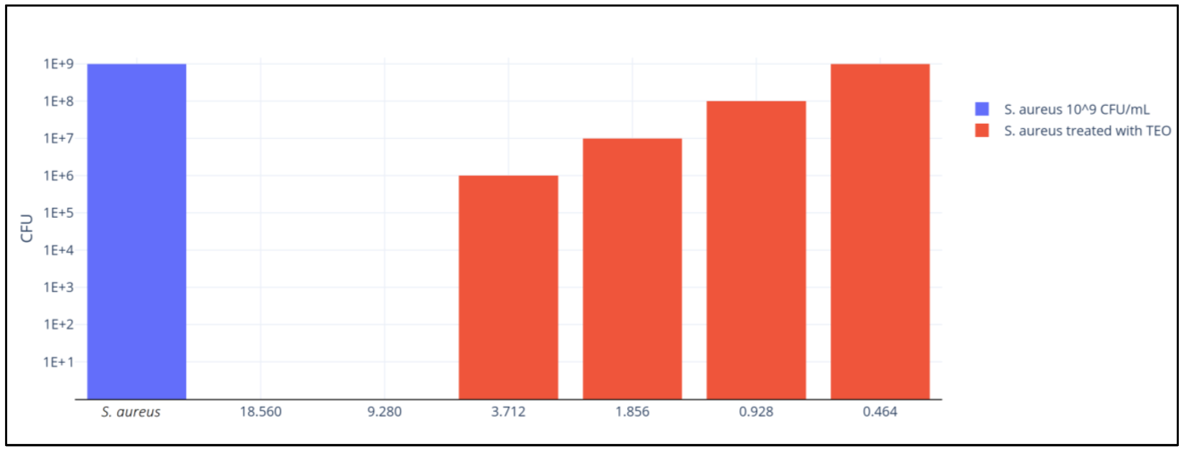
Figure 2.
TEO antibacterial activity evaluated after 1 min, 3 min nd 5 min of contact with different bacterial strains (ATCC and field strains). TEO was employed at 9.28mg/mL concentration (w/v), corresponding to 1:100 diution (v/v) and bacteria inoculum concentration was 109CFU/mL in each mixture. Bacterial growth was evaluated after 24h incubation.
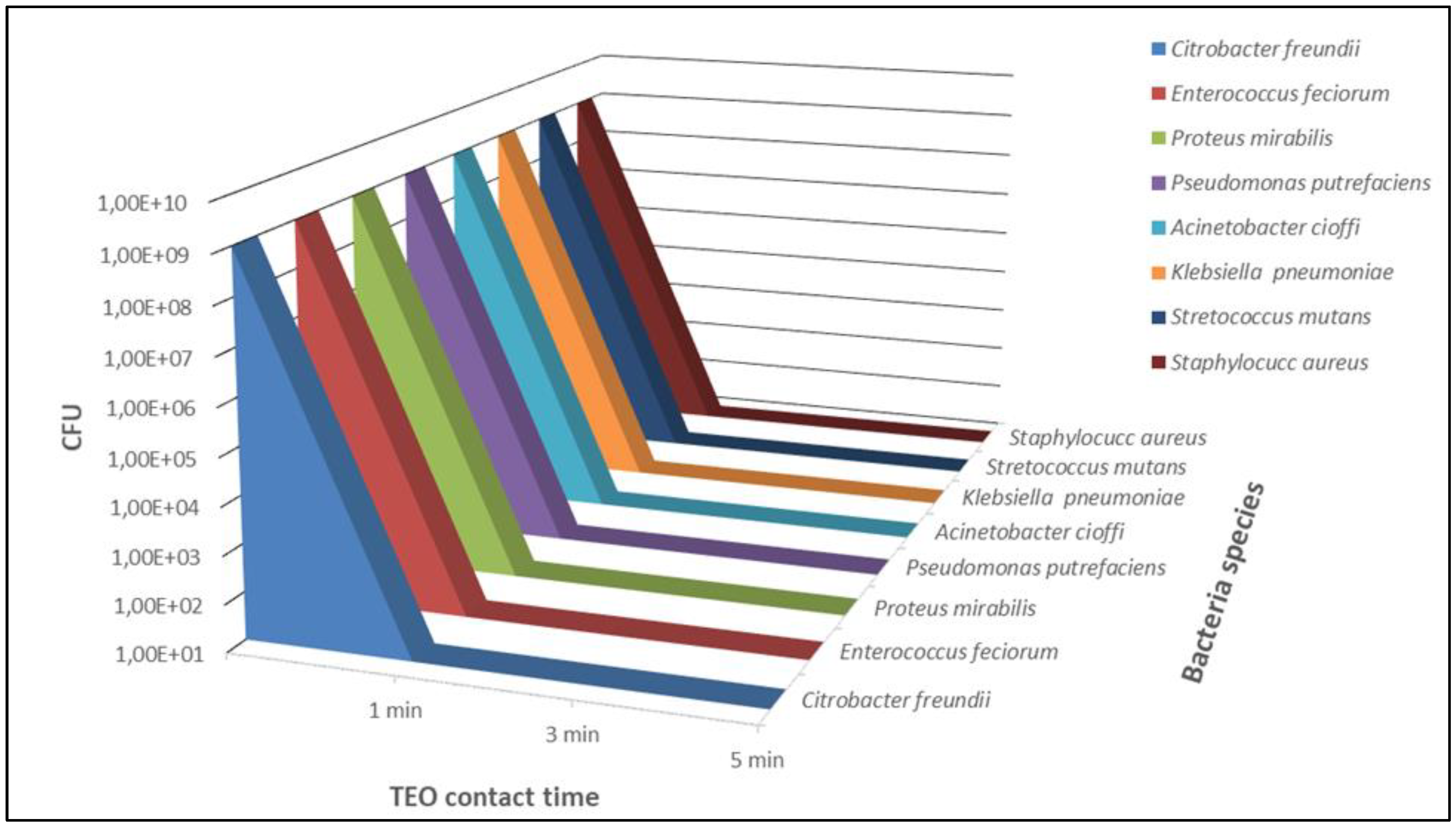

Table 1.
Antimicrobial resistance profiles of the six isolated strains employed in the present study to test TEO activity.
Table 1.
Antimicrobial resistance profiles of the six isolated strains employed in the present study to test TEO activity.
| Antibiotic | Bacterial strains | |||||
|---|---|---|---|---|---|---|
| C. freundii | E. feciorum | P. mirabilis | P. putrefaciens | A. cioffi | K. pneumonie | |
| AMC | S | I | R | R | I | S |
| AMP | I | R | R | R | R | I |
| CN | S | S | I | I | I | I |
| OT | S | R | R | R | R | S |
| CRO | I | S | R | S | R | S |
| ENR | S | S | I | S | I | S |
| MOX | S | S | S | S | S | S |
| DO | S | I | R | R | I | S |
| CL | R | R | R | R | R | S |
| CTX | S | I | R | S | I | S |
| SXT | S | S | R | S | R | S |
Legend: AMC: Amoxicillin + Clavulanic acid; AMP: Ampicillin; CN: Gentamicin; OT: Oxytetracycline; CRO: Ceftriaxone; ENR: Enrofloxacyne; MOX: Moxyfloxacin; DO: Docycycline; CL: Cephalexin; CTX: Cefotaxime; SXT: Co-Trimoxazole. S: sensitive; I: intermediate; R: resistant.
Disclaimer/Publisher’s Note: The statements, opinions and data contained in all publications are solely those of the individual author(s) and contributor(s) and not of MDPI and/or the editor(s). MDPI and/or the editor(s) disclaim responsibility for any injury to people or property resulting from any ideas, methods, instructions or products referred to in the content. |
© 2023 by the authors. Licensee MDPI, Basel, Switzerland. This article is an open access article distributed under the terms and conditions of the Creative Commons Attribution (CC BY) license (http://creativecommons.org/licenses/by/4.0/).
Copyright: This open access article is published under a Creative Commons CC BY 4.0 license, which permit the free download, distribution, and reuse, provided that the author and preprint are cited in any reuse.


