
Submitted:
29 July 2025
Posted:
30 July 2025
You are already at the latest version
Abstract
Background: Antibiotic resistance is one of the most critical challenges in treating bacterial infections today. The widespread and often inappropriate use of antibiotics has accelerated the emergence of resistant bacterial strains, making many conventional therapies ineffective. Without effective strategies to address this growing issue, bacterial pandemics that pose severe threats to global public health could emerge in the future. Objectives: This aim of this study was to investigate in vitro antibacterial activity of Liquidambar orientalis Mill. essential oil against strains of Escherichia coli, Staphylococcus aureus, Enterococcus faecalis, E. faecium, Klebsiella pneumoniae, Acinetobacter baumannii, Listeria monocytogenes, Salmonella Typhimurium, Pseudomonas aeruginosa, Mannheimia haemolytica, Brucella melitensis, and Corynebacterium pseudotuberculosis. Methods: The volatile constituents of L. orientalis Mill. essential oil were analyzed by gas chromatography–mass spectrometry (GC-MS), and its antibacterial activity was assessed using disc diffusion and agar well diffusion methods. The McNemar test was used to statistically evaluate the difference in the detection rates of resistance/sensitivity between disc diffusion test and well diffusion test results. Results: GC-MS analysis identified 21 chemical constituents in L. orientalis Mill. essential oil, with cinnamyl cinnamate (62.56%), (1-methylcyclobutyl) benzene (31.30%), and squalene (2.96%) as the major components. Minor constituents such as α-pinene, camphene, benzaldehyde, and benzyl alcohol were detected at concentrations below 1%. The essential oil demonstrated in vitro antibacterial activity against the tested bacterial strains, with inhibition rates of 58.3% and 62.5% in the disc diffusion and agar well diffusion tests, respectively. Antibacterial activity was slightly higher against Gram-positive bacteria (80.0%) compared to Gram-negative strains (78.57%). The highest activity was observed against S. aureus in the disc diffusion test and against B. melitensis in the agar well diffusion test. No statistically significant difference was observed between the two methods regarding resistant/sensitive detection rates. Conclusions: L. orientalis Mill. essential oil exhibited strong in vitro antibacterial activity against S. aureus, B. melitensis, E. coli, E. faecium, K. pneumoniae, and M. hemolytica, particularly at concentrations of 62.5, 31.25, and 15.62 µg/ml. These results suggest its potential as a broad-spectrum antibacterial agent. Notably, when combined with antibiotics, it may offer therapeutic advantages against B. melitensis, the primary causative agent of human brucellosis. However, further in vivo studies are required to validate these findings and assess its practical applications in treating bacterial infections in both human and veterinary medicine.
Keywords:
L. orientalis Mill. essential oil
; gas chromatography–mass spectrometry (GC-MS)
; antibacterial activity
; disc diffusion test
; agar well diffusion test
Introduction
The genus Liquidambar L., belonging to the family Hamamelidaceae, is distributed over a wide geographical range extending from North America to East Asia. This genus comprises four species of sweetgum: Liquidambar orientalis, L. formosana Hance, L. styraciflua and L. acalycina [1]. In the Mediterranean basin, it is represented by only one species L. orientalis is commonly known as ‘‘oriental sweetgum’’, ‘‘stirace’’ (English), ‘‘orientalischer Amberbaum’’, ‘‘ortenta-lischer Storaxbaum’’ (German), ‘‘styrax’’ (Spanish and Italian) and ‘‘feng’’ (Chinese). In Turkish, it is called ‘günlük’ due to the fragrance of the trees or sığla because of the gum like exudate which develops in response to injury, being more a pathological than physiological response [2].
L. orientalis Mill (Anatolian sweetgum) is grown endemic in some region of Türkiye (Denizli, Fethiye, Dalaman, Marmaris, Aydın and Isparta regions), and various extracts obtained from the plant are used in traditional Turkish medicine [2,3,4].
Many of the medicinal properties of sweetgum come from storax as well as essential oils extracted from the leaves. Storax, also referred to as styrax, is produced by damaging the outer bark of sweetgum trees. Extracts obtained from the roots, trunk, and leaves of the L. orientalis tree through various methods have been reported to contain components such as α-pinene, β-pinene, limonene, sabinene, and terpinen-4-ol, which exhibit antibacterial activity [4,5,6]. Additionally, resin and gum components such as styrene, cinnamic acid, benzyl cinnamate, and cinnamyl alcohol have also been reported to possess antibacterial properties [6,7].
In February 2017, the World Health Organization (WHO) published a document identifying bacteria that should be prioritized in the development of new antibiotics due to their limited treatment options. These include Gram-negative strains of Enterobacteriaceae resistant to carbapenems and cephalosporins, such as Escherichia coli and Klebsiella pneumoniae, as well as Gram-positive strains like methicillin-resistant Staphylococcus aureus (MRSA) and vancomycin-resistant S. aureus (VRSA) [8].
Infectious diseases represent a continuous and increasing threat to human and animal’s health and welfare, and antimicrobial resistance has become more challenging to the treatment of these diseases [9]. Due to the increasing prevalence of life-threatening bacterial, fungal and viral infections and the ability of these pathogens to develop resistance to current treatment strategies, there is a great need to find and develop new compounds to combat them. These molecules must have low toxicity, specific activity and high bioavailability. The most suitable compounds for this task are usually derived from natural sources [10,11].
Various factors, including biological activity evaluations, the type and concentration of active compounds in the plant, genetic characteristics (such as genus, species, and cultivar/genotype), geographical origin, cultivation conditions, climate-related parameters, stage of ripeness, time of harvest, storage practices, and post-harvest handling, also influence the outcomes [12,13].
Although several studies have investigated the antibacterial effects of essential oils from certain species within the genus Liquidambar L. (3,6,7,12), no research to date has examined bacteria that cause serious infections in both humans and animals using two different methods. Therefore, the aim of this study was to evaluate the in vitro antibacterial activity of L. orientalis Mill. essential oil against several clinically important bacterial pathogens affecting both humans and animals.
Materials and Methods
L. orientalis Mill. Oil
L. orientalis Mill. (Anatolian Sweetgum) oil was purchased from a commercial company (Aksuvital Natural Products Food Industry Trade Inc., Party No: 10545, İstanbul-Türkiye). The volatile oil components were analyzed using gas chromatography–mass spectrometry (GC-MS) at the Natural Products Application and Research Center of Isparta Süleyman Demirel University, employing a 1300 Series GC System (Thermo Fisher Scientific Inc., Waltham, MA, USA). For chromatographic separation, a TraceGOLD TG-624SilMS GC column (Thermo Fisher Scientific, Waltham, MA, USA) was employed as the analytical column. Chromatographic evaluations were performed using Xcalibur software for data acquisition and analysis.
Bacterial Strains
A total of 24 bacterial strains, stored at -80°C, were used in this study. The strains were provided by the Department of Microbiology, Faculty of Veterinary Medicine, Balıkesir University (Table 1). The bacteria tested, excluding the reference strains, were isolated from clinical specimens obtained from humans and animals suffering from infections such as cystitis, enteritis, otitis, pneumonia, and soft tissue abscesses.
Control of Bacterial and Fungal Contamination
To detect bacterial contamination, L. orientalis Mill. oil was inoculated onto 7% defibrinated sheep blood agar (1.10886, Merck, Darmstadt, Germany) in two series (one aerobic and one anaerobic), and incubated at 37°C for five days [14]. Additionally, two series of cultures were made on Sabouraud dextrose agar (DM200, Mast Diagnostics, Merseyside, UK), and one series was incubated at 25°C, the other at 37°C for 7 days, to detect fungal contamination [15]. No bacterial or fungal growth was observed after incubation.
Evaluation of Antibacterial Activity
Disc Diffusion Test
The Kirby-Bauer disc diffusion method [16] was employed to evaluate the antibacterial activity of L. orientalis Mill. essential oil, in accordance with the standards of the Clinical and Laboratory Standards Institute [17]. Briefly, L.orientalis Mill. essential oil was dissolved in absolute ethanol (≥99.8% purity, GC; 32221, Sigma-Aldrich, Steinheim, Germany) to prepare solutions at concentrations of 62.5, 31.25, 15.62, 7.8, 3.9, and 1.9 µg/ml (v/v, 1:1). Then, 20 µl of each solution was applied to 6 mm sterile filter paper discs (CT0998B, Oxoid, Basingstoke, UK). Except for B. melitensis, bacterial strains were cultured aerobically in Mueller-Hinton broth (211443, BD, Sparks, MD, USA) at 37°C for 18–24 h. B. melitensis strains were cultured aerobically in tryptic soy broth (1.05459, Merck, Darmstadt, Germany) at 37°C for 24–36 h. The turbidity of bacterial cultures grown in appropriate liquid media was visually adjusted to 0.5 McFarland standard (1.5 × 10⁸ CFU/ml) using sterile saline solution (SSS, pH 7.2). A total of 100 µl of bacterial inoculum was spread onto a 100 mm Petri dish containing Mueller-Hinton agar (MHA; 1.103872, Merck) using a sterile swab. MHA supplemented with 5% defibrinated sheep blood was used for testing B. melitensis strains. Petri dishes were left to stand at room temperature for 30 min before the discs were applied. Then, the soaked discs were placed in the center of the Petri dishes and incubated at 37°C for 18–24 h. B. melitensis strains were incubated under the same conditions for 48 h. A total of 20 µL of absolute ethanol (Sigma-Aldrich) was applied to 6 mm filter paper discs (CT0998B, Oxoid, Basingstoke, UK). The impregnated discs were thoroughly dried at 45°C in an incubator for 18 to 24 h prior to application. These discs were used as negative controls [18], while standard gentamicin (CN, 10 μg, Oxoid, Basingstoke, UK) and streptomycin (S, 10 μg, Oxoid, Basingstoke, UK) discs served as positive reference standards to determine the sensitivity of the tested bacterial pathogens in the disc diffusion test. All experiments were conducted in duplicate, and the diameters of the inhibition zones were measured in millimeters. In the standardization studies conducted in our laboratory using various bacterial strains, it was determined that a zone diameter of 7 mm corresponds approximately to a concentration of 31.25 µg/mL. The diameter of the disc was included in the measured diameter of the inhibition zone, and inhibition zones larger than 7 mm were considered indicative of antibacterial activity.
Agar Well Diffusion Test
The method of Pérez and Anesini [19] was modified for use in the agar well diffusion test in this study. Briefly, the tested bacteria were grown in liquid media as described for the disc diffusion test, and their concentrations were adjusted to McFarland standard No. 0.5 (1.5 × 10⁸ CFU/ml) using SSS. The test medium was prepared by pouring 30 ml of Mueller-Hinton agar (MHA) into a 100 mm diameter Petri dish. After the test medium solidified, wells of 6 mm diameter and 5 mm depth were punched into the agar surface using a sterilized borer. A total of 100 µl of the tested bacterial suspension was spread on the BHA surface using a sterile cotton swab to obtain uniform growth. L. orientalis Mill. essential oil was dissolved in absolute ethanol (Sigma-Aldrich) to prepare solutions with concentrations of 62.5, 31.25, 15.62, 7.8, 3.9, and 1.9 µg/mL (v/v, 1:1), and 50 µL of each solution was dispensed into the respective wells. Negative controls were prepared by applying 10 µL of ethanol onto each plate [20], while gentamicin sulfate (4553100010, Acros Organics, Belgium) and streptomycin sulfate (2311004, Menarini Health and Pharmaceutical Industry Trade Inc., Istanbul, Turkey) were used as positive controls in the test. Petri dishes inoculated with bacteria other than B. melitensis were incubated in a humid jar at 37°C for 24 h. B. melitensis strains were incubated under the same conditions for 48 h. After incubation, the zones of inhibition were measured using a ruler, and the results were reported in millimeters (mm). In standardization studies conducted in our laboratory using various bacterial strains, it was determined that a zone diameter of 6 mm corresponds approximately to a concentration of 31.25 µg/mL. Well diameters larger than 6 mm were considered indicative of antibacterial activity and thus established as the threshold for the antibacterial effect of L. orientalis Mill. essential oil. The diameter of the well was included in the measurement of the inhibition zone diameter. All tests were performed in duplicate, and the average results were reported.
Statistical Analysis
The McNemar test was used to statistically evaluate the difference in the detection rates of resistance/sensitivity between disc diffusion test and well diffusion test results. All statistical analyses were performed using the SPSS 26.0 program. The data were converted into graphs for visual presentation, and artificial intelligence–based tools were used in this process.
Results
GC-MS Analysis of L. orientalis Mill. Essential Oil
The relative amounts (%) of α-pinene, camphene, benzaldehyde, sabinene, p-cymene, dl-limonene, benzyl alcohol, 3-phenyl-1-propanol, cinnamaldehyde, cinnamyl alcohol, ethyl cinnamate, geranyl acetate, torreyol, benzyl cinnamate, 3-methyl-3-phenyl- azetidine, 4-methylcoumarin-7-cinnamate, acetyl tributyl citrate, (1-methylcyclobutyl) benzene, cinnamyl cinnamate, squalene, and other compounds were determined as 0.13, 0.01, 0.02, 0.04, 0.02, 0.03, 0.03, 0.29, 0.05, 0.40, 0.07, 0.05, 0.03, 0.13, 0.14, 0.09, 0.08, 31.3, 62.56, 2.96, and 1.56, respectively (Figure 1, Table 2). The mean concentration of the identified constituents was 4.72%, with a standard deviation of 13.43%, reflecting a high degree of compositional variability driven by the predominance of a few major compounds.
Antibacterial Activity of L. orientalis Mill. Essential Oil
L. orientalis Mill. essential oil exhibited antibacterial activity against the tested bacterial strains at rates of 58.3% and 62.5%, as determined by the disk diffusion and agar well diffusion tests, respectively. Its antibacterial effect was slightly higher against Gram-positive bacteria (80.0%) compared to Gram-negative bacteria (78.57%) (Table 3 and Table 4). The essential oil did not show antibacterial activity (inhibition zone diameter ≤ 7 mm) against 10 bacterial strains at any of the tested dilutions in the disk diffusion test (Table 3). Similarly, it showed no antibacterial activity (inhibition zone diameter ≤ 6 mm) against 9 bacterial strains at all tested dilutions in the agar well diffusion test (Table 4). The highest antibacterial activity was observed against S. aureus with an inhibition zone of 14 mm in the disk diffusion test and against B. melitensis 16 M strain with a 16 mm inhibition zone in the agar well diffusion test (Table 3 and Table 4).
Statistical Analysis
The McNemar test revealed no statistically significant difference in the resistant/sensitive detection rates between disc diffusion test and well diffusion test (exact p=1.000, N=24) (Figure 2).
Discussion
The essential oil of L. orientalis Mill. has been the subject of various studies in the field of traditional, complementary, and alternative medicine across different regions of the world. These investigations have predominantly focused on its antibacterial properties [3,6,11,21,22], wound-healing potential [23,24], antioxidant capacity [12,25], and its effects on peptic ulcer treatment [26,27]. In Türkiye, it has been widely used in folk medicine for centuries due to its well-documented antimicrobial, antioxidant, and antiulcerogenic properties [5].
The main constituents of essential oils – mono- and sesquiterpenes including carbohydrates, phenols, alcohols, ethers, aldehydes and ketones – are responsible for the biological activity of aromatic and medicinal plants as well as for their fragrance [21]. In this study, the relative amounts (%) of the constituents identified in the essential oil of L. orientalis Mill. essential oil were determined as follows: α-pinene (0.13), camphene (0.01), benzaldehyde (0.02), sabinene (0.04), p-cymene (0.02), dl-limonene (0.03), benzyl alcohol (0.03), 3-phenyl-1-propanol (0.29), cinnamaldehyde (0.05), cinnamyl alcohol (0.40), ethyl cinnamate (0.07), geranyl acetate (0.05), torreyol (0.03), benzyl cinnamate (0.13), 3-methyl-3-phenyl-azetidine (0.14), 4-methylcoumarin-7-cinnamate (0.09), acetyl tributyl citrate (0.08), (1-methylcyclobutyl)benzene (31.3), cinnamyl cinnamate (62.56), squalene (2.96), and other compounds (1.56). The mean concentration of the identified constituents was 4.72%, with a standard deviation of 13.43%. This high variability can be attributed to the dominance of a few major compounds, particularly cinnamyl cinnamate and (1-methylcyclobutyl)benzene. These findings are generally consistent with previous studies on L. orientalis. Özbek and Bilek [4] reported high levels of cinnamyl cinnamate and related aromatic esters, highlighting their significance due to their antimicrobial and aromatherapeutic properties. The essential oil composition data presented in this study are in good agreement with the existing literature, reinforcing the chemical diversity and therapeutic potential of L. orientalis. Moreover, the predominance of a few key constituents suggests that these compounds may play a major role in the biological activity of the essential oil.
The antibacterial effectiveness of various plant-derived compounds against a wide range of bacterial strains has been well documented [7,21,22], highlighting the potential of plants and their essential oils as alternative sources of bioactive antibacterial agents. Although several studies have investigated the antibacterial properties of L. orientalis Mill. essential oil [3,6,7], research specifically evaluating its efficacy against bacterial pathogens responsible for severe infections in humans and animals—particularly through simple, cost-effective, and reliable in vitro assays—remains limited. In the present study, the antibacterial activity of L. orientalis essential oil was evaluated against 24 pathogenic bacterial strains, including both reference strains and field isolates from 12 different species known to cause significant infectious diseases. The essential oil exhibited antibacterial activity against 58.3% and 62.5% of the tested strains using the disk diffusion test and agar well diffusion test, respectively. The most potent antibacterial effects were observed against S. aureus (14 mm) and B. melitensis 16M strain (16 mm) using the disk diffusion and agar well diffusion methods, respectively (Table 3 and Table 4). Öztürk et al. [2] reported significant antibacterial activity in L. orientalis extracts, particularly against S. aureus, supporting its traditional use in treating infections. In another study, the antibacterial activity of L. orientalis Mill. essential oil was evaluated against E. coli, S. aureus, and C. albicans. The oil exhibited activity against S. aureus but not against E. coli or C. albicans [19]. Özcan Akyol and Doğanay [28] investigated the effects of styrax liquidus, a resin obtained from L. orientalis, on nosocomial infection agents and reported its efficacy against P. aeruginosa, K. pneumoniae, S. haemolyticus, A. baumannii, and E. faecalis. The observed differences in antibacterial efficacy may be attributed to various factors, including the plant’s genetic makeup, chemotype, cultivation conditions, climatic factors, maturation stage, harvest time, storage conditions, post-harvest handling practices, and the methods applied [19]. The results obtained in this study are consistent with previous findings and further support the broad-spectrum antimicrobial potential of L. orientalis and its essential oil constituents.
Brucellosis is a widespread zoonotic disease caused by various Brucella species. In humans, B. melitensis is the most pathogenic species, followed by B. suis, while B. abortus is generally associated with milder forms of the disease. It mainly affects livestock and wildlife and poses significant public health threats, especially in regions with suboptimal hygiene, food safety, and veterinary care standards. Human contractions occur by consuming contaminated animal products or interacting with infected animals [29,30]. Antibiotics have a major role in the management of brucellosis in humans. However, as with many bacteria, resistance to antibiotics used against Brucella infections has been reported [31,32]. It was stated that [30], although a single antibiotic may be effective, a combination is preferred to prevent the chances of development of resistance and recurrence of disease. In this study, considering that the volatile fatty acids of L. orientalis Mill. were highly effective against B. melitensis in both tests (Table 3 and Table 4), and that the combination of herbal essential oils with antibiotics has been reported to enhance treatment efficacy in human brucellosis [33], we hypothesized that combining L. orientalis Mill. essential oil with antibiotics could positively influence the treatment of infections caused by B. melitensis in humans.
In this study, the antibacterial efficacy of L. orientalis essential oil was found to be slightly higher against Gram-positive bacteria (80.0%) compared to Gram-negative bacteria (78.57%). This observation aligns with the findings of Özcan Akyol and Doğanay [28] and is consistent with previous studies suggesting that Gram-positive bacteria are generally more susceptible to plant-derived essential oils [34]. The enhanced susceptibility of Gram-positive bacteria may be attributed to their cell wall structure, which consists of a thick peptidoglycan layer that is more accessible to hydrophobic compounds such as essential oil constituents. In contrast, Gram-negative bacteria possess an additional outer membrane containing lipopolysaccharides, which can serve as a barrier to the penetration of antibacterial agents [35].
Several methods are commonly employed to assess the antibacterial activity of herbal essential oils, with the disk diffusion and agar well diffusion tests being among the most widely used. In this study, the essential oil demonstrated considerable antibacterial activity, with susceptibility rates of 58.3% and 62.5% observed using the disk diffusion and agar well diffusion methods, respectively. The comparable susceptibility (58.3% vs. 62.5%) and resistance (41.7% vs. 37.5%) rates obtained from both assays indicate that these methods are both appropriate and effective for evaluating the antibacterial potential of plant-derived essential oils. The consistency between the two testing methods supports previous findings by Burt [36], who emphasized the reliability of disk diffusion and agar well diffusion assays when standardized protocols are followed. Furthermore, the observed variation in antibacterial efficacy depending on bacterial strain and assay method is consistent with the review by Hulanková [37], which identified key influencing factors such as diffusion dynamics, inoculum concentration, and compound volatility. These parameters likely contribute to the lack of observed activity against certain bacterial strains at all tested dilutions. In this research, although no statistically significant difference was observed between the two tests in terms of in vitro antibiotic susceptibility (Figure 2), the disc diffusion method was considered more advantageous based on our laboratory experience, due to its simplicity, ease of result interpretation, and cost-effectiveness. These attributes make it a preferred choice for the routine screening of the antibacterial properties of essential oils.
Conclusion
The emergence of antibiotic-resistant bacterial pathogens has rendered most available antibiotics ineffective. Alternative strategies are, therefore, needed to counteract drug-resistant bacterial infections. Our results demonstrate that L. orientalis Mill. essential oil exhibited potent antibacterial activity against E. coli, S. aureus, E. faecium, K. pneumoniae, M. hemolytica, and B. melitensis especially at high concentrations (62.5 and 31.25, and 15.62 µg/ml) in vitro condition. However, in vivo studies are needed to determine the antibacterial activity of L. orientalis Mill. essential oil in the development of alternative treatment strategies for the treatment of infectious diseases caused by these bacteria in human and animals.
Author Contributions
Conceptualization, R.Ş., Z.İ., M.K., and H.A.A.; writing—review and editing, Z.İ., M.G., M.B., Ö.E., and Ş.M.; statistical analysis, Z.İ., and M.G.; methodology, R.Ş., Z.İ., M.B., T.G., and Y.M.; investigation, Ö.K., Ş.M., M.K., and H.A.A.; scientific project administration, R.Ş., Zİ, M.G., M.K., and H.A.A. All authors have read and agreed to the published version of the manuscript.
Funding
This research received no external funding.
Ethical Approval
Ethical approval was not required for this study.
Acknowledgments
The authors would like to express their sincere appreciation to the Department of Microbiology, Faculty of Veterinary Medicine, Balıkesir University, for their assistance in providing both the reference strains and field isolates.”
Conflicts of Interest
The authors declare no conflicts of interest
References
- Hoey, M. T.; Parks, C. R. Isozyme divergence between Eastern Asian, North American, and Turkish species of Liquidambar (Hamamelidaceae). Am. J. Bot. 1991, 78, 938–947. [Google Scholar] [CrossRef]
- Öztürk, M.; Çelik, A.; Güvensen, A.; Hamzaoğlu, E. Ecology of tertiary relict endemic Liquidambar orientalis Mill. forests. For. Ecol. Manag. 2008, 256, 510–518. [Google Scholar] [CrossRef]
- Sağdıç, O.; Özkan, G.; Özcan, M.; Özçelik, S. A study on inhibitory effects of sığla tree (Liquidambar orientalis Mill. var. orientalis) storax against several bacteria. Phytother. Res. 2005, 19, 549–551. [Google Scholar] [CrossRef]
- Özbek, A. G.; Bilek, S. E. Properties and usage of Liquidambar orientalis. Clin. Nutr. Metab. 2018, 1, 1–3. [Google Scholar] [CrossRef]
- Büyükkılıç Altınbaşak, B.; Issa, G.; Zengin Kurt, B. Biological activities and chemical composition of Turkish sweetgum balsam (Styrax liquidus) essential oil. Bezmialem Sci. 2022, 10, 709–715. [Google Scholar] [CrossRef]
- Keskin, D.; Güvensen, N. Investigation of antimicrobial properties and chemical composition of different extracts of sweet gum leaves (Liquidambar orientalis). Int. J. Agric. Environ. Food Sci. 2022, 6, 13–18. [Google Scholar] [CrossRef]
- Sıcak, Y.; Erdoğan Eliuz, E. A. Chemical composition and antimicrobial activity of Anatolian sweetgum (Liquidambar orientalis Mill.) leaf oil. Turk. J, Life Sci. 2018, 3, 277–281. [Google Scholar]
- Bertagnolio, S. , Dobreva, Z., Centner, C. M., Olaru, I. D., Donà, D., Burzo, S., Huttner, B. D., Chaillon, A., Gebreselassie, N., Wi, T., Hasso-Agopsowicz, M. WHO global research priorities for antimicrobial resistance in human health. Lancet Microbe 2024, 5, 100902. [Google Scholar] [CrossRef] [PubMed]
- Salam, M. A.; Al-Amin, M. Y.; Salam, M. T.; Pawar, J. S.; Akhter, N.; Rabaan, A. A.; Alqumber, M. A. Antimicrobial resistance: A growing serious threat for global public health. Healthc 2023, 11. [Google Scholar] [CrossRef]
- Siddique, H.; Pendry, B.; Rashid, M. A.; Rahman, M. M. Medicinal plants used to treat infectious diseases in Bangladesh. J. Herb. Med. 2021, 29, 100484. [Google Scholar] [CrossRef]
- Stan, D.; Enciu, A. M.; et al. Natural compounds with antimicrobial and antiviral effect and nanocarriers. Front. Pharmacol. 2021, 12, 723233. [Google Scholar] [CrossRef]
- Ulusoy, H.; Ceylan, S.; Peker, H. Antioxidant and antimicrobial activity of sweetgum leaf. Polímeros 2021, 31, 1–7. [Google Scholar] [CrossRef]
- Nabti, E.; Meziane, R.; Dob, T. Environmental and genetic factors influencing plant secondary metabolites. Biol. Res. 2020, 53, 45–59. [Google Scholar] [CrossRef]
- Quinn, P. J.; Markey, B. K.; Leonard, F. C.; Fitzpatrick, E. S.; Fanning, S.; Hartigan, P. J. Veterinary Microbiology and Microbial Disease; 2nd ed.; Wiley-Blackwell: West Sussex, UK, 2011.
- Larone, D. H. Larone, D. H. Medically Important Fungi: A Guide to Identification. 5th ed.; ASM Press, Washington, 2011.
- Bauer, A. W.; Kirby, W. M.; Sherris, J. C.; Turck, M. Antibiotic susceptibility testing by a standardized single disk method. Am. J. Clin. Pathol. 1966, 45, 493–496. [Google Scholar] [CrossRef] [PubMed]
- CLSI. Performance standards for antimicrobial disk susceptibility tests, M02, 13th ed. Clin. Lab. Stand. Inst. M02, Wayne, PA.4, 2018.
- Aureli, P.; Costantini, A.; Zolea, S. Antimicrobial activity of some plant essential oils against Listeria monocytogenes. J. Food Prot. 1992, 55, 344–348. [Google Scholar] [CrossRef] [PubMed]
- Pérez, C.; Anesini, C. Antibacterial activity of alimentary plants against Staphylococcus aureus growth. Am. J. Chin. Med. 1994, 22, 169–174. [Google Scholar] [CrossRef]
- Shankar, K. R.; Das, S.; Bujala, P. Phytochemical screening and in vitro antibacterial activity of ethanol and aqueous extracts of Dregea volubilis leaves. Biosci. Biotechnol. Res. Asia 2016, 7, 975–979. [Google Scholar]
- Zhang, B.; Xin, H.; Shang, H.; Zhang, W.; Hei, H. Study on extraction and antibacterial activity of essential oil from Liquidambar formosana. J. Essent. Oil Bear. Plants 2023, 26, 176–189. [Google Scholar] [CrossRef]
- Mancarz, G.F.F.; Lobo, A.C.P.; Baril, M.B.B.; De Assis Franco, F.; Nakashima, T. Antimicrobial and antioxidant activity of the leaves, bark and stems of Liquidambar styraciflua L. (Altingiaceae). Int. J. Curr. Microbiol. App. Sci. 2016, 5, 306–317. [Google Scholar] [CrossRef]
- Parsak, C. K.; Sakman, G.; Çelik, Ü. Wound healing, wound care and complications. Arch. Med. Res. 2007, 16, 145–159. [Google Scholar]
- Kapucuk, F. S.; Dinç, H. Some aromatic plants used in wound treatment in complementary medicine and their usage methods. Exp. Appl. Med. Sci. 2021, 2(4), 257–264. [Google Scholar]
- Saraç, N.; Şen, B. Antioxidant, mutagenic, antimutagenic activities, and phenolic compounds of Liquidambar orientalis Mill. var. orientalis. Ind. Crops Prod. 2014, 53, 60–64. [Google Scholar] [CrossRef]
- Gürbüz, I.; Yeşilada, E.; Demirci, B.; Sezik, E.; Demirci, F.; Başer, K. H. Characterization of volatiles and anti-ulcerogenic effect of Turkish sweetgum balsam (Styrax liquidus). J. Ethnopharmacol. 2013, 148, 332–336. [Google Scholar] [CrossRef]
- Kıyıcı, R.; Akkan, H. A.; Karaayvaz, B. K.; Karaca, M.; Özmen, O.; Kart, A.; Garlı, S. Effects of sweetgum oil on experimental chronic gastritis in rat model. Indian J. Anim. Res. 2024, 58, 1493–1502. [Google Scholar] [CrossRef]
- Özcan Aykol, Ş. M.; Doğanay, D. Antibacterial effect of Liquidambar orientalis Miller resin on nosocomial infection agents. Int. J. Basic Clin. Stud. 2022, 11, 58–66. [Google Scholar]
- Galińska, E. M.; Zagórski, J. Brucellosis in humans – etiology, diagnostics, clinical forms. Ann. Agric. Environ. Med. 2013, 20, 233–238. [Google Scholar]
- Qureshi, K. A.; Parvez, A.; Fahmy, N. A.; Abdel Hady, B. H.; Kumar, S.; Ganguly, A.; Atiya, A.; Elhassan, G.O.; Alfadly, S.O.; Parkkila, S.; Aspatwar, A. Brucellosis: Epidemiology, pathogenesis, diagnosis and treatment–a comprehensive review. Ann. Med. 2023, 55. [Google Scholar] [CrossRef]
- Hayat, Z.; Khan, H.; Ahmad, I.; Habib, H.; Hayat, K. Antibiotics in the management of brucellosis. Gomal J. Med. Sci. 2018, 16, 114–116. [Google Scholar] [CrossRef]
- İlhan, Z.; Solmaz, H.; Ekin, I. H. In vitro antimicrobial susceptibility of Brucella melitensis isolates from sheep in an area endemic for human brucellosis in Turkey. J. Vet. Med. Sci. 2013, 75, 1035–1040. [Google Scholar] [CrossRef]
- Al-Mariri, A.; Safi, M. The antibacterial activity of selected Labiatae (Lamiaceae) essential oils against Brucella melitensis. Iran. J. Med. Sci. 2013, 38, 44–50. [Google Scholar] [PubMed]
- DeCarlo, A.; Zeng, T.; Dosoky, N. S.; Satyal, P.; Setzer, W. N. The essential oil composition and antimicrobial activity of Liquidambar formosana oleoresin. Plants 2020, 9, 822. [Google Scholar] [CrossRef] [PubMed]
- Zhou, G.; Wang, Q.; Wang, Y.; Wen, X.; Peng, H.; Peng, R.; Shi, Q.; Xie, X.; Li, L. Outer membrane porins contribute to antimicrobial resistance in Gram-negative bacteria. Microorganisms 2023, 11, 1690. [Google Scholar] [CrossRef] [PubMed]
- Burt, S. Essential oils: their antibacterial properties and potential applications in foods—a review. Int. J. Food Microbiol. 2004, 94, 223–253. [Google Scholar] [CrossRef]
- Hulankova, R. Methods for determination of antimicrobial activity of essential oils in vitro—A review. Plants 2024, 13, 2784. [Google Scholar] [CrossRef] [PubMed]
Figure 1.
GC-MS chromatogram of L. orientalis Mill. essential oil. The chromatogram displays the major volatile constituents, with cinnamyl cinnamate (62.56%) and (1-methylcyclobutyl) benzene (31.30%) identified as the predominant compounds. Minor components include α-pinene, camphene, and cinnamaldehyde.
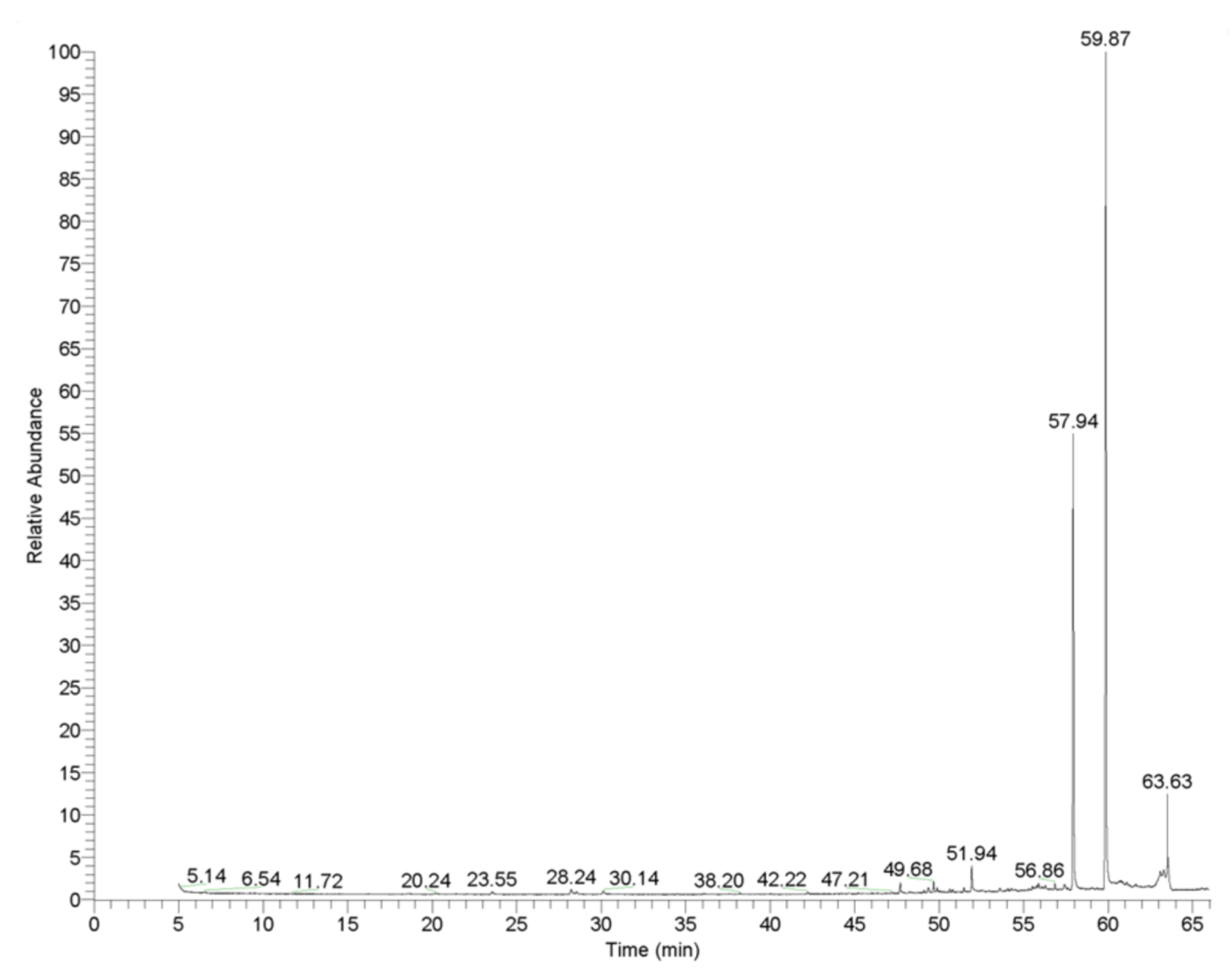
Figure 2.
Statistical comparison between disk diffusion test (A) and well diffusion test (B).
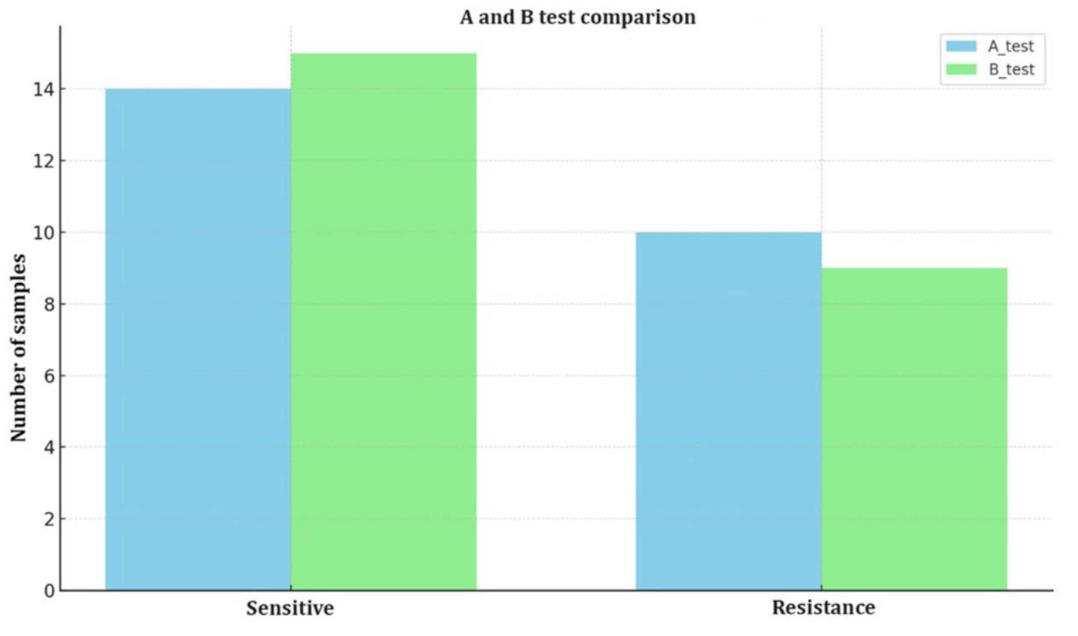

Table 1.
Bacterial strains and their sources.
| Bacteria | Source | |
| 1 | Escherichia coli | ATCC 25932 |
| 2 | Multi-drug resistant Escherichia coli (MDR E. coli) | Animal |
| 3 | Escherichia coli | Human |
| 4 | Escherichia coli | Animal |
| 5 | Staphylococcus aureus subsp. aureus | ATCC 6538 |
| 6 | Methicillin-resistant Staphylococcus aureus (MRSA) | ATCC 43300 |
| 7 | Multi-drug resistant Staphylococcus aureus (MDR S. aureus) | Animal |
| 8 | Staphylococcus aureus | Human |
| 9 | Staphylococcus aureus | Animal |
| 10 | Enterococcus faecalis | ATCC 29212 |
| 11 | Enterococcus faecium | ATCC 6057 |
| 12 | Klebsiella pneumoniae | Human |
| 13 | Klebsiella pneumoniae | Animal |
| 14 | Acinetobacter baumannii | Human |
| 15 | Acinetobacter baumannii | Human |
| 16 | Listeria monocytogenes | ATCC 7644 |
| 17 | Salmonella enterica subsp. enterica serotype Typhimurium | NCTC 12416 |
| 18 | Pseudomonas aeruginosa | Human |
| 19 | Multi-drug resistant Pseudomonas aeruginosa (MDR P. aeruginosa) | Animal |
| 20 | Mannheimia haemolytica | Human |
| 21 | Mannheimia haemolytica | Animal |
| 22 | Brucella melitensis strain 16M (biotype 1) | ATCC 23456 |
| 23 | Brucella melitensis | Animal |
| 24 | Corynebacterium pseudotuberculosis | Animal |
Table 2.
Chemical composition of L. orientalis Mill. essential oil identified by GC-MS analysis, including retention times (min), relative abundances (×106), and percentage amounts of each compound.
Table 2.
Chemical composition of L. orientalis Mill. essential oil identified by GC-MS analysis, including retention times (min), relative abundances (×106), and percentage amounts of each compound.
| Compounds | Retention time (min) | Relative abundance (×106) | Amount (%) | |
| 1 | α-Pinene | 6.54 | 9.40 | 0.13 |
| 2 | Camphene | 7.07 | 0.90 | 0.01 |
| 3 | Benzaldehyde | 8.36 | 1.50 | 0.02 |
| 4 | Sabinene | 9.50 | 2.85 | 0.04 |
| 5 | p-Cymene | 10.14 | 1.70 | 0.02 |
| 6 | dl-Limonene | 11.61 | 1.80 | 0.03 |
| 7 | Benzyl alcohol | 12.54 | 2.10 | 0.03 |
| 8 | 3-Phenyl-1-propanol | 18.59 | 20.50 | 0.29 |
| 9 | Cinnamaldehyde | 28.24 | 3.40 | 0.05 |
| 10 | Cinnamyl alcohol | 28.56 | 28.00 | 0.40 |
| 11 | Ethyl cinnamate | 30.14 | 5.10 | 0.07 |
| 12 | Geraniol acetate | 47.70 | 3.40 | 0.05 |
| 13 | Torreyol | 49.37 | 2.20 | 0.03 |
| 14 | Benzyl cinnamate | 49.68 | 8.90 | 0.13 |
| 15 | 3-Methyl-3-phenyl-azetidin | 51.94 | 10.00 | 0.14 |
| 16 | 4-Methylcoumarine-7-cinnamate | 52.55 | 6.60 | 0.09 |
| 17 | Acetyl tributyl citrate | 56.86 | 5.60 | 0.08 |
| 18 | (1-methylcyclobutyl) benzene | 57.94 | 2201.00 | 31.30 |
| 19 | Cinnamyl cinnamate | 59.87 | 4400.00 | 62.56 |
| 20 | Squalene | 63.63 | 208.00 | 2.96 |
| 21 | Other compounds | - | 110.00 | 1.56 |
| Total | - | - | 100.00 | |
Note: In the GC-MS analysis, the detection limit was 0.01%, the results were expressed in percentage (%), and a minimum peak area threshold of 1% was applied.
Table 3.
Inhibition zone diameters (mm) of L. orientalis Mill. essential oil against tested bacterial strains in the disc diffusion test (zones ≤ 7 mm considered to show no antibacterial activity).
Table 3.
Inhibition zone diameters (mm) of L. orientalis Mill. essential oil against tested bacterial strains in the disc diffusion test (zones ≤ 7 mm considered to show no antibacterial activity).
|
Bacteria and their source |
Concentration of L. orientalis Mill. essential oil (µg/mL) |
Antibiotic discs |
|||||||
| 62.5 | 31.25 | 15.62 | 7.8 | 3.9 | 1.9 | CN | S | ||
| Inhibition zone diameter (mm) | |||||||||
| 1 | E. coli (ATCC 25932) | 8 | 8 | 7 | 7 | 6 | 6 | 20 | 16 |
| 2 | MDR E. coli (Animal) | 6 | 6 | 6 | 6 | 6 | 6 | 10 | 12 |
| 3 | E. coli (Human) | 7 | 7 | 7 | 7 | 6 | 6 | 14 | 12 |
| 4 | E. coli (Animal) | 10 | 8 | 8 | 8 | 8 | 6 | 10 | 12 |
| 5 | S. aureus (ATCC 6538) | 6 | 6 | 6 | 6 | 6 | 6 | 26 | 22 |
| 6 | MRSA (ATCC 43300) | 8 | 8 | 6 | 6 | 6 | 6 | 20 | 12 |
| 7 | MDR S. aureus (Animal) | 7 | 7 | 6 | 6 | 6 | 6 | 10 | 6 |
| 8 | S. aureus (Human) | 6 | 6 | 6 | 6 | 6 | 6 | 18 | 8 |
| 9 | S. aureus (Animal) | 14 | 14 | 10 | 8 | 8 | 6 | 24 | 30 |
| 10 | E. faecalis (ATCC 29212) | 6 | 6 | 6 | 6 | 6 | 6 | 26 | 18 |
| 11 | E. faecalis (ATCC 6057) | 8 | 8 | 8 | 8 | 8 | 6 | 14 | 22 |
| 12 | K. pneumoniae (Animal) | 10 | 8 | 8 | 6 | 6 | 6 | 14 | 10 |
| 13 | K. pneumoniae (Human) | 7 | 7 | 7 | 6 | 6 | 6 | 16 | 8 |
| 14 | A. baumannii (Human) | 10 | 8 | 8 | 8 | 8 | 8 | 24 | 20 |
| 15 | A. baumannii (Human) | 10 | 8 | 8 | 8 | 8 | 8 | 18 | 8 |
| 16 | L. monocytogenes (ATCC 7644) | 6 | 6 | 6 | 6 | 6 | 6 | 14 | 8 |
| 17 | S. typhimurium (NCTC 12416) | 7 | 7 | 6 | 6 | 6 | 6 | 20 | 10 |
| 18 | P. aeruginosa (Human) | 8 | 8 | 8 | 8 | 8 | 8 | 20 | 16 |
| 19 | MDR P. aeruginosa (Animal) | 8 | 8 | 8 | 6 | 6 | 6 | 14 | 20 |
| 20 | M. haemolytica (Animal) | 10 | 8 | 8 | 6 | 6 | 6 | 28 | 30 |
| 21 | M. haemolytica (Animal) | 7 | 6 | 6 | 6 | 6 | 6 | 22 | 24 |
| 22 | B. melitensis (16M) (Animal) | 12 | 12 | 10 | 8 | 8 | 6 | 24 | 26 |
| 23 | B. melitensis (Animal) | 10 | 10 | 8 | 8 | 6 | 6 | 26 | 30 |
| 24 | C. pseudotuberculosis (Animal) | 12 | 10 | 10 | 8 | 8 | 8 | 22 | 18 |
CN: Gentamicin (10 μg, Oxoid, Basingstoke, UK), S: Streptomycin (10 μg, Oxoid, Basingstoke, UK), MDR E. coli: Multi-drug resistant E. coli, MRSA: Methicillin resistant S. aureus, MDR S. aureus: Multi-drug resistant S. aureus, MDR P. aeruginosa: Multi-drug resistant P. aeruginosa.
Table 4.
Inhibition zone diameters (mm) of L. orientalis Mill. essential oil against tested bacterial strains in the agar well diffusion test (zones ≤ 6 mm considered to show no antibacterial activity).
Table 4.
Inhibition zone diameters (mm) of L. orientalis Mill. essential oil against tested bacterial strains in the agar well diffusion test (zones ≤ 6 mm considered to show no antibacterial activity).
|
Bacteria (source) |
Concentration of L. orientalis Mill. essential oil (µg/mL) | Antibiotics | |||||||
| 62.5 | 31.25 | 15.62 | 7.8 | 3.9 | 1.9 | GS | SS | ||
| Inhibition zone diameter (mm) | |||||||||
| 1 | E. coli (ATCC 25932) | 14 | 14 | 12 | 12 | 8 | 8 | 18 | 16 |
| 2 | MDR E. coli (Animal) | 6 | 6 | 6 | 6 | 6 | 6 | 22 | 22 |
| 3 | E. coli (Human) | 6 | 6 | 6 | 6 | 6 | 6 | 18 | 20 |
| 4 | E. coli (Animal) | 12 | 12 | 12 | 10 | 10 | 8 | 28 | 26 |
| 5 | S. aureus (ATCC 6538) | 6 | 6 | 6 | 6 | 6 | 6 | 32 | 26 |
| 6 | MRSA (ATCC 43300) | 12 | 12 | 12 | 10 | 10 | 8 | 20 | 22 |
| 7 | MDR S. aureus (Animal) | 12 | 12 | 12 | 12 | 12 | 10 | 26 | 24 |
| 8 | S. aureus (Human) | 6 | 6 | 6 | 6 | 6 | 6 | 12 | 8 |
| 9 | S. aureus (Animal) | 14 | 10 | 8 | 6 | 6 | 6 | 20 | 18 |
| 10 | E. faecalis (ATCC 29212) | 6 | 6 | 6 | 6 | 6 | 6 | 14 | 8 |
| 11 | E. faecalis (ATCC 6057) | 14 | 12 | 10 | 10 | 8 | 8 | 24 | 26 |
| 12 | K. pneumoniae (Animal) | 14 | 12 | 10 | 10 | 8 | 8 | 16 | 22 |
| 13 | K. pneumoniae (Human) | 6 | 6 | 6 | 6 | 6 | 6 | 14 | 18 |
| 14 | A. baumannii (Human) | 12 | 10 | 10 | 8 | 8 | 8 | 26 | 24 |
| 15 | A. baumannii (Human) | 6 | 6 | 6 | 6 | 6 | 6 | 6 | 12 |
| 16 | L. monocytogenes (ATCC 7644) | 12 | 12 | 12 | 10 | 10 | 8 | 24 | 22 |
| 17 | S. typhimurium (NCTC 12416) | 6 | 6 | 6 | 6 | 6 | 6 | 10 | 8 |
| 18 | P. aeruginosa (Human) | 12 | 12 | 12 | 12 | 10 | 10 | 22 | 14 |
| 19 | MDR P. aeruginosa (Animal) | 10 | 8 | 8 | 8 | 8 | 8 | 26 | 22 |
| 20 | M. haemolytica (Animal) | 14 | 14 | 14 | 12 | 12 | 10 | 16 | 14 |
| 21 | M. haemolytica (Animal) | 6 | 6 | 6 | 6 | 6 | 6 | 18 | 16 |
| 22 | B. melitensis (16M) (Animal) | 16 | 16 | 14 | 12 | 10 | 10 | 24 | 20 |
| 23 | B. melitensis (Animal) | 12 | 10 | 10 | 8 | 8 | 8 | 24 | 18 |
| 24 | C. pseudotuberculosis (Animal) | 12 | 12 | 10 | 10 | 8 | 8 | 22 | 24 |
GS: Gentamicin sulfate (4553100010, Acros Organics, Belgium), SS: Streptomycin sulfate (2311004, Menarini Health and Pharmaceutical Industry Trade Inc. Istanbul, Türkiye), MDR E. coli: Multi-drug resistant E. coli, MRSA: Methicillin resistant S. aureus, MDR S. aureus: Multi-drug resistant S. aureus, MDR P. aeruginosa: Multi-drug resistant P. aeruginosa.
Disclaimer/Publisher’s Note: The statements, opinions and data contained in all publications are solely those of the individual author(s) and contributor(s) and not of MDPI and/or the editor(s). MDPI and/or the editor(s) disclaim responsibility for any injury to people or property resulting from any ideas, methods, instructions or products referred to in the content. |
© 2025 by the authors. Licensee MDPI, Basel, Switzerland. This article is an open access article distributed under the terms and conditions of the Creative Commons Attribution (CC BY) license (http://creativecommons.org/licenses/by/4.0/).
Copyright: This open access article is published under a Creative Commons CC BY 4.0 license, which permit the free download, distribution, and reuse, provided that the author and preprint are cited in any reuse.


