
Submitted:
19 April 2023
Posted:
20 April 2023
You are already at the latest version
Abstract
The liver performs a fundamental role in the regulation of diverse physiological processes. Despite enormous advances in modern medicine, there are no completely effective drugs that stimulate hepatic function, offer complete protection to the organ, or aid in regenerating hepatic cells. Thus, it is necessary to identify alternative plant-origin pharmaceuticals more effective and less toxic for the treatment of hepatic diseases. The aim of this research was to establish the chemical composition of the American cranberry (Oxycoccus macrocarpus) leaves extract and its aminoacids preparations as well as their hepatoprotective activity. 19 phenolic compounds (8 flavonoids (flavones and flavonols), 4 anthocyanins, 2 catechins, and 3 hydroxycinnamic acids) and main amino acids (valine, arginine, glycine, histidine, aspartic acid, and taurine) were identified and quantified in the American cranberry (O. macrocarpus) leaves extract and its 7 aminoacids preparations. The therapeutic and prophylactic consumption of the American cranberry leaves extracts led to a decrease in the intensity of the lipid peroxidation process compared to the control pathology group in the experiment of acute toxic damage to the liver by tetrachloromethane on rats. The most pronounced hepatoprotective activity was established for the American cranberry leaves extracts’ preparations with arginine and valine.
Keywords:
American cranberry (Oxycoccus macrocarpus)
; leaves
; extract
; modification
; amino acid
; hepatoprotective activity.
1. Introduction
The liver performs a fundamental role in the regulation of diverse physiological processes, and its activity is related to different vital functions, such as metabolism, secretion, and storage. Its capacity to detoxify endogenous (waste metabolites) and/or exogenous (toxic compounds) substances of organisms, as well as to synthesize useful agents, has been analyzed since the 1970s by many researchers [1,2,3,4,5]. The liver is also involved in the biochemical processes of growing, providing nutrients, supplying energy, and reproducing [6].
Amongst the gastrointestinal manifestations experienced by COVID-19 patients, those commonly noted are diarrhea, anorexia, nausea, vomiting, and abdominal pain – which can be present even in the absence of respiratory symptoms. Hepatic injury is evident in some patients, the degree of which at times can mirror the severity of the disease; pancreatic injury has been noted as well [7,8].
Although the liver has a high regenerative capacity, endogenous and exogenous stimuli can still result in permanent tissue damage and impairment of liver function [8], and hepatic diseases are a problem worldwide [1,9]. Hepatic diseases can damage the cells, tissues, structure, or liver function. They can be induced by biological factors (bacteria, viruses, and parasites) [10] and autoimmune diseases (immune hepatitis, primary biliary cirrhosis) [11], as well as by the action of different chemicals, such as some drugs, toxic compounds, and unquestionably, excessive consumption of alcohol [1,9]. Despite enormous advances in modern medicine, there are no completely effective drugs that stimulate hepatic function, offer complete protection to the organ, or aid in regenerating hepatic cells [12]. Additionally, some drugs can induce adverse or side effects. Thus, it is necessary to identify alternative plant-origin pharmaceuticals for the treatment of hepatic diseases, with the aim of these agents being more effective and less toxic.
The use of some plants and the consumption of different fruits have played fundamental roles in human health care. Approximately 80% of the world’s population has employed traditional medicine for health care, which is based predominantly on plant materials [5,9]. Thus, in recent years, the consumption of berry extracts has increased as an ingredient in functional foods and dietary supplements, which might or might not be combined with other colored fruits, plants, and herbal extracts. The berry fruits that are habitually consumed in North America include blackberries (Rubus spp.), black raspberries (Rubus occidentalis), red raspberries (Rubus idaeus), strawberries (Fragaria X ananassa), blueberries (Vaccinium corymbosum), and cranberries (Vaccinium macrocarpon) [13]. These fruits have significant antioxidative activity. As oxidative stress and dysfunction of cellular immunity are important indicators in the pathogenesis of hepatic diseases caused by diverse xenobiotics [14]. It was noticed that blueberry fruits effects liver protection and cellular immune function [15].
As mentioned in the overview [1], different types of berries are consumed frequently, and among these, the cranberry was the topic of an experiment with the primary objective of evaluating its antioxidant and hepatoprotective potential against liver mitochondrial damage induced by acute 80.8 g/kg body weight (bw), single injection) and chronic (1.6 g/kg bw, 30 d, biweekly injections) CCl4 intoxication in rats. Both acute and chronic intoxication negatively affected the mitochondrial respiratory parameters in the liver, and the enzymatic activities of succinate dehydrogenase, GPx, and cytoplasmic catalase were significantly inhibited. However, administration of cranberries (7 mg/kg) was effective in diminishing the toxic effects of CCl4, normalizing AlAt and AsAt activity, and bilirubin concentrations. Similarly, it prevented the accumulation of membrane lipid peroxidation products in the rat liver, resulting in apparent preservation of the mitochondrial ultrastructure [16].
The plants, fruits, and compounds described [1] could offer novel alternatives to the limited therapeutic options that exist for the treatment of liver diseases: thus, these foods should be considered in future studies. In general, biologically active substances and extracts from cranberry raw materials provided hepatoprotective activity, the principal mechanisms of action of which are related to their antioxidant potential [17], a characteristic that should motivate and promote the search for effective protective agents, which must be evaluated later in pre-clinical and clinical assays to determine their safety and their chemopreventive capacity.
When cranberry is cultivated on plantations, the bushes are pruned annually, and tons of leaves become waste, while they contain a significant amount of biologically active substances, which properties could be further utilized as sustainably developed products of plant origin. Thus, the American cranberry leaves (Oxycoccus macrocarpus (Ait.) Pursh, Ericaceae) act as by-products and are an interesting material for the development and manufacturing of food supplements with promising preventive and therapeutical properties, possibly with hepatoprotective one.
Previous studies have shown that plants of the Vaccinium genus are promising sources for the creation of hypoglycemic and hypolipidemic agents, because of their significant antioxidant activity, namely extracts from bilberry leaves [18,19], blueberry leaves [20] and bearberry leaves [21,22]. Studies have shown that extracts from the leaves of cranberries are also promising when used to correct insulin-resistant conditions [23,24].
Modification of biologically active molecules by their conjugation with amino acids is a known strategy for modification of their biological properties. For example, the synthetic medicine Valtrex was created by combining acyclovir with the amino acid valine [24], and the medicine L-lysine escinate was created using the modification of the complex of chestnut triterpene saponins (β-escin) with L-lysine [25,26]. Therefore, it has become an interesting question, whether the use of such an approach to modify the total plant extract could result in the improvement of its biological properties. It was previously shown that the modification of motherwort tincture with amino acids has led to the creation of new more active substances with anxiolytic activity [27]. The modification of the blueberry leaves extract with arginine allowed to create a substance with pronounced hypoglycemic and hypolipidemic activities [18]. The modification of the extract of bearberry leaves with phenylalanine allowed to create a substance with a pronounced diuretic and anti-inflammatory effect [21,22] and adding cysteine – a remedy for the management of insulin resistance [28]. All of this indicates the viability of the chosen direction.
Thus, the aim of our research was to establish the chemical composition of the American cranberry (O. macrocarpus) leaves extract and its aminoacids preparations as well as their hepatoprotective activity. To the best of our knowledge, we are the first to combine the active ingredients of cranberry leaves with amino acids and demonstrate their hepatoprotective effects.
2. Results
2.1. HPLC Analysis and Quantification of Major Compounds
The major phenolic compounds of the cranberry leaves extracts were identified by UPLC-MS/MS and HPLC-PDA methods. Characteristic phenolic compounds (cyanidin-3-O-galactoside and 3-O-arabinoside, hyperoside and isoquercitrin, reynoutrin, quercetin-3-O-arabinopyranoside, avicularin, quercitrin, quercetin, kaempferol-3-O-rhamnoside and kaempferol, chlorogenic, neochlorogenic and 4-O-caffeoylquinic acid, p-coumaric acid, (+)-catechin and (–)-epicatechin, procyanidin A1, A2, B1 and B2) were found. Typical MRM chromatograms are presented in Figure 1. Chromatograms and qualitative results of HPLC-PDA method are presented in Table 1, Figure 1, and Figure S2, while typical UPLC-MS/MS chromatograms of phenolic components in the samples are given in Figure S1.
The amino acids were quantified by UPLC-MS/MS method. It was identified that main constituents are amino acids (valine, arginine, glycine, histidine, aspartic acid, and taurine). It should be noted that alanine fragments could not be detected in the MRM chromatograms and this compound was not identified, most likely due to interactions with components of the native extract. Typical MRM chromatograms and results of UPLC-MS/MS are presented in Table 2 and Figure S3.
2.2. Acute Toxicity
In the control pathology group, 2 animals died (1 animal on the 1st day and 1 animal on the 2nd day of the experiment). Thus, mortality in the group of control pathology was 33%. In the other groups, all animals remained alive until the end of the experiment, which indicates the prospects of research in the hepatoprotective activity of the studied extracts.
2.3. Hepatoprotective Activity
The cranberry leaves extracts show hepatoprotective activity in the model of acute tetrachloromethane hepatitis. The fact was confirmed by morphological and biochemical indicators. The results of measuring liver weight coefficients (LWC) for the experimental animals are given in Table 3.
The results of measuring biochemical indicators in blood serum and liver homogenate, such as AlAt, AsAt and TBA-reactants are presented in the Table 4.
3. Discussion
3.1. Phytochemical Research
19 phenolic substances were identified in the American cranberry leaves extracts and their contents were determined by the HPLC method. There were 8 flavonoids (flavones and flavonols), 4 anthocyanins, 2 catechins, and 3 hydroxycinnamic acids. Quercetin glycosides predominate in the extracts. Hyperoside, avicularin, and quercitrin were dominant among the individual substances (Table 1).
The phenolic profile of cranberry leaves and fruits is rather different. There are three classes of flavonoids such as flavonols, anthocyanins, and proanthocyanidins in cranberry fruits [29]. The predominated flavonoids in cranberry fruits are the glycosides of myricetin and quercetin: myricetin-3-galactoside, myricetin-3-arabinofuranoside, quercetin-3-galactoside, quercetin-3-glucoside, quercetin-3-rhamnospyranoside, and quercetin-3-O-(6′′-p-benzoyl)-galactoside [30,31].
Both American cranberry and European cranberry (V. oxycoccos) as a traditionally used crop accumulated a high level of polyphenols. The fruits of European cranberry represent more valuable sources of caffeic acid and quercetin with higher values of total flavonols than American cranberry [31,32,33]. The differences in an accumulation of phenolic compounds in cranberry fruits can be given by various conditions of cultivation, region, weather conditions, harvesting time, and maturity stage [34].
It was noticed that the content of aglycones, such as quercetin and kaempferol increases after the extract E1 modification with amino acids. It can be indicated that the hydrolysis of glycosides is taking place. These can be detected in the extracts E6 and E8 for kaempferol and in the extracts E2, E5, E6, E7, and E8 for quercetin.
The content of glycosides decreases in the extract E1 preparations with amino acids mostly all the time, this occurs due to the addition of amino acids and the possible formation of their conjugates with these substances.
The content of hydroxycinnamic acids in the modified extracts (E2-E8) was significantly lower. It can be explained by creating conjugates with amino acids, which confirms previous research in the highbush blueberry extracts [20].
3.2. Hepatoprotective Activity
The results of the research (Table 3) show that the consumption of hepatotropic poison CCl4 led to a significant increase in LWC, which indicates liver damage. In the experimental groups that took the American cranberry leaves extracts and the referent drug “Silibor”, the changes were insignificant compared to the intact animals. The best corrective effect was shown by the combined extracts (E2 and E3) with arginine and valine, which were more effective than the referent drug “Silibor”. The other studied extracts show activity at the level of the referent drug “Silibor” (E6, E7, and E8) or slightly lower (E1, E2, E5).
In the experiment, it was noticed that in the group of animals with control pathology, the increase in liver weight was 45.17% compared to the intact animals, that indicates liver damage and inflammation. After taking the American cranberry leaves extracts, according to LWC, animals of groups, which received the combined extracts E2 and E3 at a dose of 25 mg/kg (bw), were similar to the intact animals, while the decreases in liver weight were 27.2% and 25.64% respectively compared to the control pathology group. The animals treated with the referent drug “Silibor” have a decrease of 22.5% in liver weight compared to the control pathology group. The consumption of the other studied extracts (E6, E8, E7, E5, E4, E1) caused decreases of 21.92%, 20.74%, 20.16%, 19.37%, 18.40%, 17.80% respectively in liver weight, which is comparative with the values in the animals taking the referent drug “Silibor”.
Thus, the LWC values indicate a positive effect of the American cranberry leaves extracts and the referent drug “Silibor” and a decrease in swelling and normalization of blood circulation in the organ, and therefore a decrease in the intensity of the inflammatory process.
The results of biochemical indicators studies (Table 4) show that a single consumption of tetrachloromethane led to the development of acute toxic liver damage. In the control pathology group, significant intensification of lipid peroxidation processes and exhaustion of the antioxidant protection system were observed, as a result of which the structural and functional integrity of the membranes was violated. The destruction of cell membrane components led to the development of a pronounced cytolytic syndrome, as evidenced by a 3.37 times increase in the AlAt activity in blood serum, and AsAt – by 2.42 times, compared to the parameters of the intact group. The development of acute toxic hepatitis was characterized by an increase in peroxide catabolic transformations, as evidenced by an increase in the content of TBA-reactants in blood serum and liver homogenates of untreated animals by 1.85 times and 2.51 times, respectively, compared to the indicators of the intact animals.
The taking of the studied American cranberry leaves extracts and the referent drug “Silibor” in experimental hepatitis under the conditions of a therapeutic and preventive regimen was accompanied by a noticeable decrease in pathological manifestations and led to a significant decrease in the studied indicators relative to the values in the control pathology group. The referent drug “Silibor” led to a decrease in the activity of the studied enzymes in the blood serum of experimental animals relative to the values in the control pathology group, namely AlAt – by 2.02 times, AsAt – by 1.87 times.
When the American cranberry leaves extract (E1) was given to the animals at a dose of 25 mg/kg (bw), the activity of biochemical indicators in blood serum decreased relative to the values in the control pathology group: AlAt – by 2.0 times, AsAt – by 1.71 times, which was at the level of indicators in the group taking the reference drug “Silibor”.
Adding amino acids to the extract E1 led to increase in their hepatoprotective activity. The best effect was shown by the extracts with amino acids valine (E2) and arginine (E3), which had a high impact on the development of cytolysis syndrome, reducing the activity of AlAt by 3 times and 2.67 times, AsAt by 2.23 times and 2.13 times, respectively, relative to the values in the control pathology group. These extracts were more active than the referent drug “Silibor” and brought the biochemical indicators of blood serum to the level of the intact animals.
The extracts with amino acids histidine (E6) and taurine (E8) showed slightly better activity compared to the group of animals treated with the referent drug “Silibor”. At the same time, the activity of AlAt was 2.42 times and 2.33 times lower, and AsAt was 2.0 times and 2.07 times lower in comparison with the control pathology group.
The studied extracts with aspartic acid (E7) and glycine (E5) have the activity at the level of the referent drug “Silibor”, and the extract with alanine (E4) did not lead to an improvement in the indicators of the antioxidant system in comparison with the group of animals treated with the referent drug “Silibor”.
Simultaneous administration of hepatotropic poison and the American cranberry leaves extracts (E1, E3, E2, E6, E8, E7, E5, and E4) at a dose of 25 mg/kg (bw) resulted in a decrease in the level of TBA-reactants in blood serum by 1.42, 1.76, 1.69, 1.61, 1.57, 1.50, 1.48, and 1.43 times, respectively, and in the liver homogenate – 1.69, 2.23, 2.12, 1.90, 1.98, 1.68, 1.62, and 1.63 times, respectively. The use of the referent drug “Silibor” led to a decrease in the level of TBA-reactants in blood serum and liver homogenate by 1.53 and 1.72 times, respectively.
A significant decrease in the activity of all studied enzymes in blood serum and liver homogenate indicates a positive effect against hepatocytes cytolysis.
The most pronounced hepatoprotective activity was established for the American cranberry leaves extracts’ preparations with arginine and valine, which in a dose of 25 mg/kg (bw) authentically exceeded in activity the referent drug “Silibor” on such indicators as LWC, the activity of AlAt and AsAt enzymes and the level of the product of lipid peroxidation of TBA-reactants and brought them in blood and liver homogenate to the level of the intact animals. The use of American cranberry leaves extract and its preparations with histidine, taurine, aspartic acid, glycine, and alanine did not significantly lead to an increase in hepatoprotective activity. Biochemical indicators of blood serum and liver homogenate were at the same level or slightly lower than the referent drug “Silibor”.
All studied extracts of the American cranberry leaves can be arranged in the following sequence according to the level of hepatoprotective activity: E3 (arginine) → E2 (valine) → E6 (histidine) → E 8 (taurine) → E7 (aspartic acid) → E5 (glycine) → E4 (alanine) → E1.
Thus, the obtained results indicate that in conditions of acute toxic hepatitis caused by tetrachloromethane, the American cranberry leaves extracts have hepatoprotective activity, inhibiting peroxide destructive processes and reducing the development of cytolysis syndrome. The American cranberry leaves extracts perpetrated with arginine and valine have a more pronounced hepatoprotective effect compared to the referent drug “Silibor”.
4. Materials and Methods
4.1. Plant Material
American cranberry leaves were harvested in August 2020 in Kyiv region (Pereyaslav suburbs, 50.10314334026342, 31.46151900698126). The identity of the plant was established by professor Tetiana Gontova, D.Sc. in Pharmacy [24,35]. Voucher specimens were deposited at the Department of Pharmacognosy (National University of Pharmacy, Kharkiv, Ukraine, No. 592-594). The raw material was dried at room temperature in a well-ventilated area for ten days and stored in paper bags The raw material was standardized according to the proposed requirements [24,36,37].
4.2. Preparation of Extracts
Five hundred g of the dried cranberry leaves [24,36], ground to a particle size of 1-2 mm, were placed in an extractor and macerated with 3 L of ethanol:water mixture (1:1, v/v) overnight at room temperature. The extraction was repeated three times with new portions of the solvent (1.0 L). The resulting extracts were combined, settled for 24 h, and filtered through a folding filter. The content of phenolic substances in the liquid extract in terms of gallic acid was 1.04%. Next, the obtained extract was divided into 8 parts. The first one was evaporated to dryness (E1).
To the other parts (500 mL each) amino acids: 10.5 g of valine (E2), 15.71 g of arginine (E3), 8.03 g of alanine (E4), 6.77 g of glycine (E5), 14.00 g of histidine (E6), 11.98 of aspartic acid (E7), and 11.41 g of taurine (E8) were added three times at the equimolar amount to the phenolic compounds (according to determined gallic acid equivalents). The resulting solutions were kept overnight at room temperature and evaporated using a rotary vacuum evaporator to obtain dry residues (E2-E8 accordingly). The extracts were standardized according to the proposed requirements [38].
The extract E1 from the cranberry leaves is a brown loose powder with a specific smell. When it is modified with amino acids, its appearance changes: pink-red shades appear, which indicates the presence of anthocyanins in the extract E1. The extracts E3, E4, E7 and E8 are viscous masses. The extracts E2, E5 and E6 remain powders.
4.3. Chemicals
The following solvents were used in the study: analytical grade acetonitrile, MS grade formic acid, and MS grade acetonitrile from Sigma-Aldrich (Steinheim, Germany), trifluoracetic acid from Merck (Darmstadt, Germany), and ethanol 96.0% from AB Vilniaus degtine (Vilnius, Lithuania). The ultrapure water was purified by Milli–Q® (Millipore, Bedford, MA, USA) water purification system.
Standard substances of neochlorogenic acid (5-O-caffeoylquinic acid), chlorogenic acid (3-O-caffeoylquinic acid), (+)-catechin, cryptochlorogenic acid (4-O-caffeoylquinic acid), (–)-epicatechin, procyanidins A1, A2, B1, and B2, p-coumaric acid, isoquercitrin (quercetin-3-O-glucoside), guaiaverin (quercetin-3-O-arabinopyranoside), avicularin (quercetin-3-O-arabinofuranoside), quercitrin (quercetin-3-O-rhamnoside), afzelin (kaempferol-3-O-rhamnoside), taurine, 17 amino acid mix solution, quercetin, and kaempferol were purchased from Sigma-Aldrich; hyperoside (quercetin-3-O-galactoside), cyanidin-3-O-galactoside, and cyanidin-3-O-arabinoside were obtained from Extrasynthese (Genay, France).
4.4. Analysis and Quantification of Major Compounds
4.4.1. Phenolic Compounds Identification by UPLC-MS/MS
Analysis of phenolic compounds in selected samples was carried out with an Acquity H-class UPLC system (Waters, USA) equipped with a triple quadrupole tandem mass spectrometer (Xevo, Waters, USA) electrospray ionization source (ESI) was used to obtain MS/MS data. YMC Triart C18 (100 x 2.0 mm 1,9 µm) column was used for the separation of phenolic compounds. The column temperature was maintained at 40° C. Gradient elution was performed with a mobile phase consisting of 0.1% formic acid water solution (solvent A) and acetonitrile (solvent B) with the flow rate set to 0.5 ml/min. Linear gradient profile was applied with the following proportions of solvent A: 0 to 1 min – 95%, 5min. – 70%, 7 min. 50%, 7.5 to 8 min. 0%, 8.1 to 10 min. 95%. Negative electrospray ionization was applied for analysis with the following settings: capillary voltage – 2 kV, source temperature –150°C, desolvation temperature –400°C, desolvation gas flow – 700 l/h, cone gas flow – 20 l/h. Identification and peak assignment of phenolic compounds in cranberry extracts were based on a comparison of their retention times and MS/MS spectral data with those of standard compounds.
4.4.2. Phenolic Compounds Analysis by HPLC-PDA
Qualitative and quantitative analysis of phenolic compounds was carried out on HPLC-PDA (Waters e2695 Alliance system, Waters, Milford, MA, USA) system coupled with an ACE Super C18 (250 mm × 4.6 mm, 3 µm) reversed-phase column (ACT, Aberdeen, UK) with column temperature set at 35 °C according to the method reported earlier [39]. The mobile phase delivered at 0.5 mL/min consisted of 0.1% trifluoroacetic acid (eluent A) and acetonitrile (eluent B) with gradient elution: 0 min, 90% A; 0–40 min, 70% A; 40–60 min, 30% A; 60–64 min, 10% A; 64–70 min, 90% A. Injection volume of 10 µL was used. Identification and peak assignment of phenolics compounds in cranberry extracts were based on a comparison of their retention times and absorption spectral data with those of standard compounds. For quantification, linear regression models were obtained using the standard dilution method.
4.4.3. Amino Acids Analysis
Analysis of amino acids was carried out on Acquity H-class (Waters, Milford, MA, USA) UPLC system equipped with Xevo TQD (Waters, Milford, MA, USA) mass spectrometer. One microliter of extracts was injected on a BEH Amide (150 mm × 2.1 mm, 1.7 µm) column (Waters, Milford, MA, USA) with column temperature set at 25 °C. The mobile phase was delivered at 0.4 mL/min. The mobile phase consisted of 0.1% formic acid (eluent A) and acetonitrile (eluent B). Gradient elution was applied with the following settings: 0 min to 1 min., 90% B; 1–6 min, 70% B; 6–9 min, 50% B; 9–10 min, the column was flushed with 50% A; at 10.1 min, the gradient was returned to the initial composition for a total run time of 13 min. Mass spectrometer conditions were set as follows: positive electrospray ionization at 3.5 kV, cone voltage set at 30 V, desolvation gas flow at 800 L/h, gas temperature at 400 °C, and ion source temperature at 120 °C. Identification and peak assignment of amino acids in cranberry extracts were based on a comparison of their retention times and spectral data with those of standard compounds [40]. For quantification, linear regression models were obtained using the standard dilution method.
4.5. Acute Toxicity
The study of acute toxicity is mandatory in the complex of preclinical studies of new medicines. The method of preclinical study of the harmlessness of medicines was used to study the acute toxicity of the American cranberry leaves dry extract [40,41,42].
The research was conducted on white outbred mice of both sexes, which were obtained from the vivarium of Ivano-Frankivsk National Medical University (IFNMU), and have a weight of 18-22 g, which were on a regular diet. In the experiment, groups of 6 animals were used, which were injected with an aqueous solution of the cranberry leaves extracts and a control group. The solutions were consumed intragastrically with the help of a metal probe in increasing doses.
The animals were observed for 14 days. The effect of the extract was evaluated by integral indicators (general condition, changes in body position, skin condition, color of mucous membranes, body temperature) and individual symptoms (diarrhea, drowsiness, tremors, convulsions, etc.).
4.6. Hepatoprotective Activity
The study of the hepatoprotective activity of the cranberry leaves extracts was carried out on the model of acute tetrachloromethane hepatitis [40,41,42]. Research in the hepatoprotective activity of the cranberry leaves extract (E1) and its preparations (E2-E8) with amino acids (valine, arginine, alanine, glycine, histidine, aspartic acid, and taurine) was carried out at the Clinical and biological experimental base of IFNMU in accordance with the National “General Ethical Principles of Animal Experiments” (Ukraine, 2001), which correspond to the provisions of the “European Convention for the Protection of Vertebrate Animals Used for Experimental and Other Scientific Purposes” (Strasbourg, 1986) [43,44,45,46,47].
The research in the hepatoprotective activity of the studied extracts was carried out on 66 white non-linear sexually mature rats grown in the kennel of the Clinical and Biological Experimental Base of IFNMU weighing 160-240 g, which were standardized according to physiological and biochemical indicators and divided into 11 groups of 6 animals each: 1 group – intact animals; 2nd group – control pathology; 3-10 groups – animals which consumed the cranberry leaves extract and its preparation with amino acids at a dose of 25 mg/kg (bw); Group 11 - animals that received the referent drug “Silibor” in a dose of 25 mg/kg (bw) [39]. The laboratory animals were kept in accordance with the current “Sanitary rules regarding the arrangement, equipment, and maintenance of experimental biological clinics (vivariums)” at a temperature of 18-20 °C and relative humidity of 50-55%. They were fed with a full-fledged diet, according to a standard scheme, with free access to water.
To study the hepatoprotective activity, the liver damage was caused in animals of the first to fifth groups by a 50% oil solution of tetrachloromethane in a dose of 0.8 mL per 100 g (bw) for 2 days with an interval of 24 h. The experimental animals consumed aqueous solutions of the studied extracts at a dose of 25 mg/kg (bw). The drug “Silibor” (PhC “Zdorovya”, Kharkiv, Ukraine) was used as a referent medicine. “Silibor” tablets, after removing the shells, were crushed in a mortar and administered intragastrically in the form of a 1% starch suspension. The intact animals consume purified water. The studied extracts and the referent drug were consumed by the animals 1 h and 2 h after taking the hepatotropic poison.
The rats were decapitated on the third day after the first tetrachloromethane taking. The conclusion about the pharmacotherapeutic effectiveness of the studied extract was made on the basis of biochemical and functional indicators of the liver state, which were determined in 24 h after the last taking of tetrachloroethane. Liver weight coefficients were calculated in a percentage, as the ratio of liver and body weights.
The study of biochemical indicators was carried out on the basis of the Center of Bioelementology IFNMU (certificate of technical competence No. 037/19 from June 13, 2019 to June 12, 2024).
The assessment of the intensity of peroxidic destructive transformations in the animals body was determined by the content of TBA-reactants in blood serum and liver homogenate. The effectiveness of the hepatoprotective effect of the extracts was evaluated by changes in the level of alanine aminotransferase (AlAt), aspartate aminotransferase (AsAt) in blood serum, which are hepatospecific markers of cytolysis. The activity of cytolysis enzymes AlAt and AsAt was determined in blood serum using the Reitman-Frenkel method using the standard set of reagents from the company “SIMKO Ltd.” (Dnipro, Ukraine) [39,40,41].
The total level of lipid peroxidation (TLLP) was determined with 2-thiobarbituric acid (TBA) by the spectrophotometric method according to E.N. Korobeinikova method using the biochemical set of the “Filisit-Diagnostika” company (Dnipro, Ukraine).
The hepatoprotective activity of the studied extracts is evidenced by the survival rate of animals, liver weight ratio, and normalization of blood serum biochemical parameters and liver homogenate [40].
4.7. Statistical Analysis
The mean and standard deviation (SD) of samples was calculated according to the monograph “Statistical Analysis of the Results of a Chemical Experiment” of the State Pharmacopoeia of Ukraine [36]. The average sample μ was calculated as the arithmetic mean of all variants (n = 5 of combined samples). At the same time, the spread of options around the average is characterized by the magnitude of the standard deviation s. The uncertainty of this estimate is characterized by the value of the confidence interval, in which the true value μ is given with the given two-way probability P2. Under uncertainty, the confidence interval is understood, usually for the 95% significance level. Limit values of the confidence interval were calculated using Student’s criterion. Quantitative data are presented as the mean ± SD. level of statistical significance was set at not more than p < 0.05 [36,37].
5. Conclusions
The chemical composition and hepatoprotective activity of the cranberry (Oxycoccus macrocarpus (Ait.) Pursh) leaves extract and its 7 amino acids preparations were studied.
19 phenolic substances were identified in the American cranberry leaves extracts and their contents were determined by the HPLC-PDA method. There were 8 flavonoids (flavones and flavonols), 4 anthocyanins, 2 catechins, and 3 hydroxycinnamic acids. Quercetin glycosides predominate in the extracts. Hyperoside, avicularin, and quercitrin were dominant among the individual substances. The main amino acids (valine, arginine, glycine, histidine, aspartic acid, and taurine) were identified and quantified by the UPLC-MS/MS method.
The therapeutic and prophylactic consumption of the American cranberry leaves extracts led to a decrease in the intensity of the lipid peroxidation process compared to the control pathology group in the experiment of acute toxic damage to the liver by tetrachloromethane on rats. The most pronounced hepatoprotective activity was established for the American cranberry leaves extracts’ preparations with arginine and valine.
Supplementary Materials
The following supporting information can be downloaded at the website of this paper posted on Preprints.org, Figure S1: Typical UPLC-MS/MS chromatograms of phenolic components in the samples; Figure S2: Comparative HPLC-PDA profiles at 325 nm of different extracts; Figure S3: Comparative UPLC-MS/MS chromatograms of amino acids (alanine was not identified).
Author Contributions
Conceptualization, A.R, O.K. and V.J.; methodology, O.K., V.J. and L.G.; validation, I.V., V.J. and G.V.; formal analysis, I.V., G.V., V.Z., L.G. and R.H.; investigation, I.V., G.V., V.Z., L.G. and R.H.; resources, A.R, O.K. and V.J.; data curation, O.K., V.J., G.V., V.Z., L.G. and R.H.; writing—original draft preparation, A.R, O.K., I.V., V.J., V.Z. and L.G.; writing—review and editing, A.R, O.K. and V.J.; visualization, I.V., V.Z., L.G. and R.H.; supervision, A.R, O.K. and V.J.; project administration, A.R. and O.K.; funding acquisition, A.R. and O.K. All authors have read and agreed to the published version of the manuscript.
Funding
This work was supported by the Estonian Research Council grant (PRG1903),; CurifyLabs project (VMVFA22189) (Curify) project, and The Estonian Research Council (ETAg) short-term support measure (12.07.2022) for Ukrainian researchers in the Estonian universities’ research and development activities; and the European Union in the MSCA4Ukraine project “Design and development of 3D-printed medicines for bioactive materials of Ukrainian and Estonian medicinal plants origin” [ID number 1232466].
Conflicts of Interest
The authors declare no conflict of interest.
References
- Madrigal-Santillán, E.; Madrigal-Bujaidar, E.; Álvarez-González, I.; Sumaya-Martínez, M.T.; Gutiérrez-Salinas, J.; Bautista, M.; Morales-González, Á.; García-Luna y González-Rubio, M.; Aguilar-Faisal, J.L.; Morales-González, J.A. Review of natural products with hepatoprotective effects. World J. Gastroentero.l 2014, 20, 14787–804. [Google Scholar] [CrossRef]
- Lin, J.H.; Lu, A.Y. Role of pharmacokinetics and metabolism in drug discovery and development. Pharmacol. Rev. 1997, 49, 403–449. [Google Scholar]
- Shanani, S. Evaluation of hepatoprotective efficacy of APCL-A polyherbal formulation in vivo in rats. Indian Drugs 1999, 36, 628–631. [Google Scholar]
- Subramoniam, A.; Pushpangadan, P. Development of phytomedicine for liver diseases. Indian J. Pharmacol. 1999, 31, 166–175. [Google Scholar]
- Adewusi, E.A.; Afolayan, A.J. A review of natural products with hepatoprotective activity. J. Med. Plants Res. 2010, 4, 1318–1334. [Google Scholar]
- Ahsan, M.R.; Islam, K.M.; Bulbul, I.J. Hepatoprotective activity of Methanol Extract of some medicinal plants against carbon tetrachloride-induced hepatotoxicity in rats. Global J. Pharmacol. 2009, 3, 116–122. [Google Scholar]
- Patel, K.P.; Patel, P.A.; Vunnam, R.R.; Hewlett, A.T.; Jain, R.; Jing, R.; Vunnam, S.R. Gastrointestinal, hepatobiliary, and pancreatic manifestations of COVID-19. J. Clin. Virol. 2020, 128, 104386. [Google Scholar] [CrossRef]
- Huang, Y.; Miyamoto, D.; Hidaka, M.; Adachi, T.; Gu, W.L.; Eguchi, S. Regenerative medicine for the hepatobiliary system: A review. J. Hepatobiliary Pancreat. Sci. 2021, 11, 913–930. [Google Scholar] [CrossRef] [PubMed]
- Deshwal, N.; Sharma, A.K.; Sharma, P. Review on hepatoprotective plants. Int. J. Pharm. Sci. Rev. Res. 2011, 7, 15–26. [Google Scholar]
- Casafont-Morencos, F.; Puente, A.; Pons-Romero, F. Infecciones bacterianas y parasitarias del hígado. Medicine 2008, 10, 563–569. [Google Scholar]
- Amengual-Guedan, M.J.; Rodríguez Sánchez, J.L. Autoinmunidad en las enfermedades del hígado (I). Immunologia 2000, 19, 90–102. [Google Scholar]
- Chattopadhyay, R.R. Possible mechanism of hepatoprotective activity of Azadirachta indica leaf extract: part II. J. Ethnopharmacol. 2003, 89, 217–219. [Google Scholar] [CrossRef]
- Madrigal-Santillán, E.; Fragoso-Antonio, S.; Valadez-Vega, C.; Solano-Solano, G.; Pérez, C.Z.; Sánchez-Gutiérrez, M.; IzquierdoVega, J.A.; Gutiérrez-Salinas, J.; Esquivel-Soto, J.; Esquivel-Chirino, C.; Sumaya-Martínez, T.; Fregoso-Aguilar, T.; Mendoza Pérez, J.; Morales-González. J.A. Investigation on the protective effects of cranberry against the DNA damage induced by benzo[a]pyrene. Molecules 2012, 17, 4435–4451. [Google Scholar] [CrossRef]
- Cederbaum, A.I.; Lu, Y.; Wu, D. Role of oxidative stress in alcohol-induced liver injury. Arch. Toxicol. 2009, 83, 519–548. [Google Scholar] [CrossRef]
- Wang, Y.P.; Cheng, M.L.; Zhang, B.F.; Mu, M.; Zhou, M.Y.; Wu, J.; Li, C.X. Effect of blueberry on hepatic and immunological functions in mice. Hepatobiliary Pancreat. Dis. Int. 2010, 9, 164–168. [Google Scholar]
- Cheshchevik, V.T.; Lapshina, E.A.; Dremza, I.K.; Zabrodskaya, S.V.; Reiter, R.J.; Prokopchik, N.I.; Zavodnik, I.B. Rat liver mitochondrial damage under acute or chronic carbon tetrachloride-induced intoxication: protection by melatonin and cranberry flavonoids. Toxicol. Appl. Pharmacol. 2012, 261, 271–279. [Google Scholar] [CrossRef] [PubMed]
- Shanaida, M.; Hudz, N.; Korzeniowska, K.; Wieczorek, P. Antioxidant activity of essential oils obtained from aerial part of some Lamiaceae species. Int. J. Green Pharm. 2018, 12, 200–204. [Google Scholar]
- Koshovyi, O.M.; Zagayko, A.L.; Kolychev, I.O.; Akhmedov, E. Yu.; Komissarenko, A.N. Phytochemical study of the dry extract from bilberry leaves. Azerbaijan Pharmaceutical and Pharmacotherapy Journal 2016, 16, 18–23.11. [Google Scholar]
- Zagayko, A.L.; Kolisnyk, T.Y.; Chumak, O.I.; Ruban, O.A.; Koshovyi, O.M. Evaluation of anti-obesity and lipid-lowering properties of Vaccinium myrtillus leaves powder extract in a hamster model. J. Basic Clinic. Physiol. Pharmacol. 2018, 29, 697–703. [Google Scholar] [CrossRef] [PubMed]
- Koshovyi, O.; Granica, S.; Piwowarski, J.P.; Stremoukhov, O.; Kostenko, Y.; Kravchenko, G. et. al. Highbush Blue-berry (Vaccinium corymbosum L.) Leaves Extract and Its Modified Arginine Preparation for the Management of Metabolic Syndrome – Chemical Analysis and Bioactivity in Rat Model. Nutrients 2021, 13, 2870. [Google Scholar] [CrossRef]
- Chaika, N.; Mazen, M.; Koshovyi, O.; Kravchenko, G.; Goryacha, O.; Kireyev, I. et. al. Research in phytochemical composition and hypoglycemic activity screening of the dry extracts from bearberry leaves. ScienceRise: Pharmaceutical Science 2021, 29, 42–50. [Google Scholar] [CrossRef]
- Chaika, N.; Koshovyi, O.; Raal, A.; Kireyev, I.; Zupanets, A.; Odyntsova, V. Phytochemical profile and pharmacolog-ical activity of the dry extract from Arctostaphylos uva-ursi leaves modified with phenylalanine. ScienceRise: Pharmaceutical Science 2020, 6, 74–84. [Google Scholar] [CrossRef]
- Koshovyi, O.M.; Vlasova, I.K.; Brukhanova, Т. О.; Krasilnikova, О. А.; Kravchenko, G.B.; Zagayko, A.L.; Komisarenko, М. А. (2021). Pat. No. 147975 UA. A method of obtaining a therapeutic and prophylactic agent from the leaves of large-fruited cranberries for the correction of insulin-resistant conditions. No. u 2021 00821, declareted: 02/22/2021, Published: 23.06.2021, Bul. No. 25/2021.
- Vlasova, I.; Gontova, T.; Grytsyk, L.; Zhumashova, G.; Sayakova, G.; Boshkayeva, A.; Shanaida, M.; Koshovyi, О. Determination of standardization parameters of Oxycoccus macrocarpus (Ait.) Pursh and Oxycoccus palustris Pers. Leaves. ScienceRise: Pharmaceutical Science 2022, 3, 48–57. [Google Scholar] [CrossRef]
- Kovalenko, V.N. Compendium 2020—Medicines, MORION: Kiiv, Ukraine, 2020, p. 2700.
- Parfenov, V.A. Use of L-lysine aescinate in central nervous system diseases. Neurol. Neuropsychiatry Psychosom. 2011, 3, 99–104. [Google Scholar] [CrossRef]
- Koshovyi, O.; Raal, A.; Kireyev, I.; Tryshchuk, N.; Ilina, T.; Romanenko, Y.; Kovalenko, S.M.; Bunyatyan, N. Phytochemical and Psychotropic Research of Motherwort (Leonurus cardiaca L.) Modified Dry Extracts. Plants 2021, 10, 230. [Google Scholar] [CrossRef] [PubMed]
- Kravchenko, G.; Krasilnikova, O.; Raal, A.; et al. Arctostaphylos uva-ursi L. leaves extract and its modified cysteine preparation for the management of insulin resistance: chemical analysis and bioactivity. Nat. Prod. Bioprospect. 2022, 12, 30. [Google Scholar] [CrossRef] [PubMed]
- Netto, C.C. Cranberry and its phytochemicals: A review of in vitro anticancer studies. J. Nutr. 2007, 137, 186–193. [Google Scholar] [CrossRef] [PubMed]
- Singh, A.P.; Wilson, T.; Kalk, A.J.; Cheong, J.; Vorsa, N. Isolation of specific cranberry flavonoids for biological activity assessment. Food Chem. 2009, 116, 963–968. [Google Scholar] [CrossRef] [PubMed]
- Jurikova, T.; Skrovankova, S.; Mlcek, J.; Balla, S.; Snopek, L. Bioactive compounds, antioxidant activity, and biological effects of European cranberry (Vaccinium oxycoccos). Molecules 2018, 24, 24. [Google Scholar] [CrossRef] [PubMed]
- Stobnicka, A.; Gniewosz, M. Antimicrobial protection of minced pork meat with the use of swamp cranberry (Vaccinium oxycoccos L.) fruit and pomace extracts. J. Food Sci. Tech. 2018, 55, 62–71. [Google Scholar] [CrossRef]
- Marzullo, L.; Ochkur, O.; Renai, L.; Gotti, R.; Koshovyi, O.; Furlanetto, S.; Orlandini, S.; Del Bubba, M. Quality by Design in optimizing the extraction of (poly)phenolic compounds from Vaccinium myrtillus berries. J. Chromatography A 2022, 1677, 463329. [Google Scholar] [CrossRef] [PubMed]
- Häkkinen, S.H.; Törrönen, A.R. Content of flavonols and selected phenolic acids in strawberries and Vaccinium species: Influence of cultivar, cultivation site and technique. Food Res. Int. 2000, 33, 517–524. [Google Scholar] [CrossRef]
- Dobrochaeva, D.N.; Kotov, M.I.; Prokudin, Y.N.; Barbarich, A.I. Key to Higher Plants of Ukraine. Kyiv: Naukova dumka, 1999.
- European Pharmacopoeia. 10th Ed. Council of Europe, Strasbourg. 2019.
- State Pharmacopoeia of Ukraine, 2nd ed., Ukrainian Scientific Pharmacopoeial Center of Drugs Quality: Kharkiv, Ukraine, 2015. (In Ukrainian).
- Vlasova, І. K.; Koshovyi, O.M. Standardization of dry extracts from large cranberry leaves. Journal of Organic and Pharmaceutical Chemistry 2022, 20, 40–45. [Google Scholar] [CrossRef]
- Vilkickyte, G.; Raudone, L.; Petrikaite, V. Phenolic fractions from Vaccinium vitis-idaea L. and their antioxidant and anticancer activities assessment. Antioxidants 2020, 9, 1261. [Google Scholar] [CrossRef] [PubMed]
- Koshevoi, O.N. Amino-acid and monosaccharide compositions of Salvia officinalis leaves. Chem. Nat. Comp. 2011, 47, 492–493. [Google Scholar] [CrossRef]
- Stefanov, O.V. Preclinical Studies of Drugs, Avitsenna: Kyiv, Ukraine, 2001 (in Ukrainian).
- Kovalenko, V.M. Preclinical research of medicinal products in Ukraine. Pharmacol. Med. Toxicol. 2009, 5, 56–61. [Google Scholar]
- Huzio, N.; Grytsyk, A.; Raal, A.; Grytsyk, L.; Koshovyi, O. Phytochemical and Pharmacological Research in Agrimonia eupatoria L.Herb Extract with Anti-Inflammatory and Hepatoprotective Properties. Plants 2022, 11, 2371. [Google Scholar] [CrossRef]
- Shanaida, M.; Hudz, N.; Jasicka-Misiak, I.; Wieczorek, P.P. Polyphenols and pharmacological screening of a Monarda fistulosa L. dry extract based on a hydrodistilled residue by-product. Front. Pharmacol. 2021, 12, 1–10. [Google Scholar] [CrossRef]
- The Law of Ukraine "On the Protection of Animals from Cruel Treatment" dated 12/15/2009.
- The Order of the Ministry of Health of Ukraine, No. 944 dated 14.12.2009 "On approval of the Procedure for preclinical study of medicinal products and examination of materials of preclinical study of medicinal products".
- Commission of the European Communities: Council Directive of 18 December 1986 on the Lows, regulating the Application of Principles of Good Laboratory Practice and the Verification of Their Applications for Tests on Chemical Substances (87/18/EEC)/ The Rules Governing Medicinal Products in the European Community. 1991. V. 1. P. 145 – 146.
Figure 1.
HPLC-PDA profiles at 325 nm, showing phenolics separation in extracts of cranberry leaves. Peak assignments: 1—neochlorogenic acid, 2—chlorogenic acid, 3—(+)-catechin, 4—4-O-caffeoylquinic acid, 5—cyanidin-3-O-galactoside, 6—(–)-epicatechin, 7—cyanidin-3-O-arabinoside, 8—proacyanidin A1, 9—p-coumaric acid, 10—hyperoside, 11—isoquercitrin, 12—procyanidin A2, 13—reynoutrin, 14—quercetrin-3-O-arabinopyranoside, 15—avicularin, 16—quercitrin, 17—kaempferol-3-O-rhamnoside, 18—quercetin, 19—kaempferol.
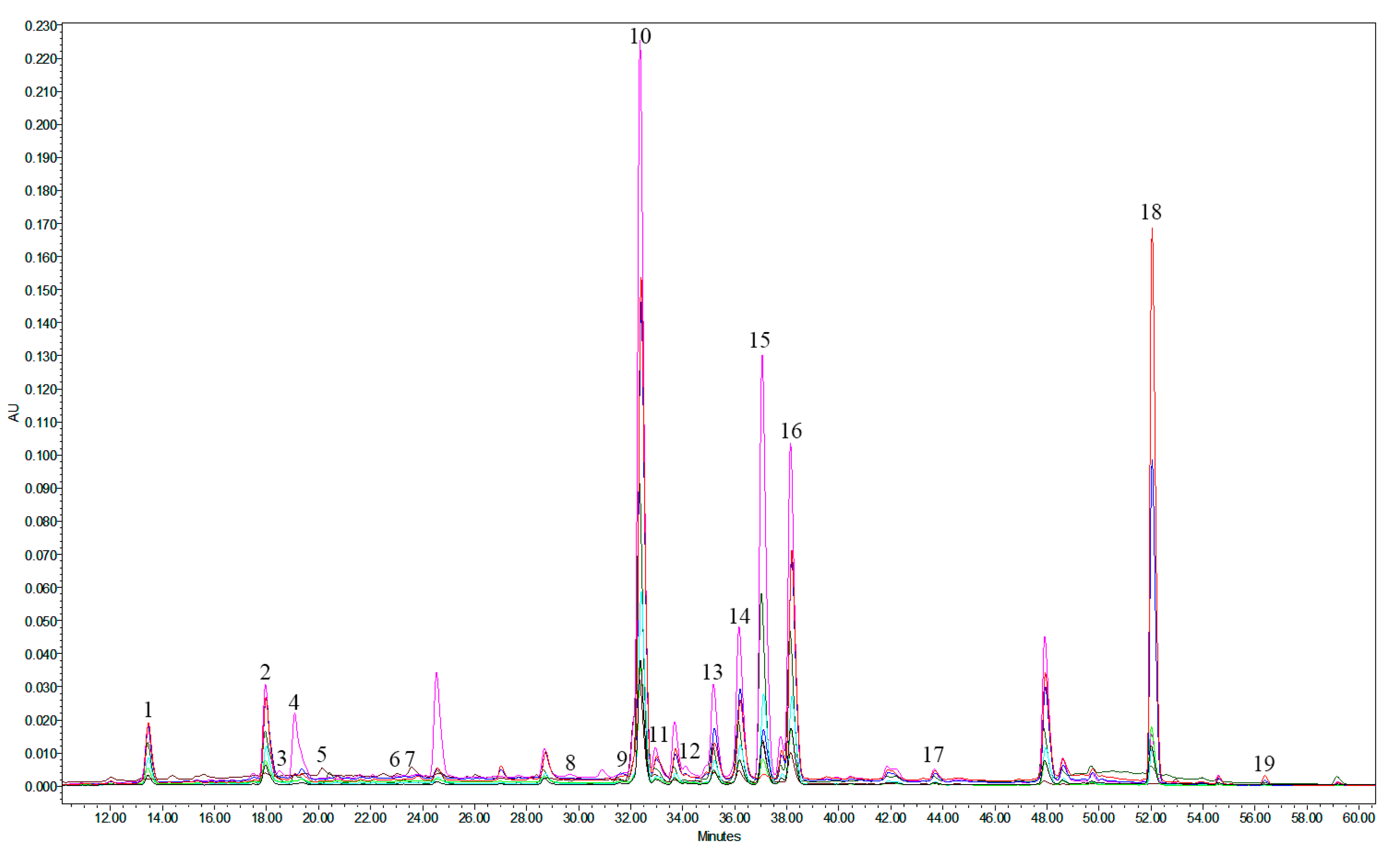

Table 1.
Composition of phenolic substances in the American cranberry leaves extracts determined by HPLC-PDA method.
Table 1.
Composition of phenolic substances in the American cranberry leaves extracts determined by HPLC-PDA method.
| Phenolic compounds | Content in the extract, mg/g | |||||||
|---|---|---|---|---|---|---|---|---|
| E1 | E2 | E3 | E4 | E5 | E6 | E7 | E8 | |
| Cyanidin-3-O-galactoside | ND | NQ | ND | ND | NQ | NQ | 0.054 ± 0.009 | 0.105 ± 0.006 |
| Cyanidin-3-O-arabinoside | ND | NQ | ND | ND | NQ | NQ | 0.129 ± 0.011 | 0.210 ± 0.004 |
| Hyperoside | 16.125 ± 0.307 | 6.848 ± 0.067 | 0.908 ± 0.006 | 8.044 ± 0.104 | 10.705 ± 0.109 | 6.407 ± 0.241 | 5.279 ± 0.020 | 5.604 ± 0.013 |
| Isoquercitrin | 1.167 ± 0.035 | 0.399 ± 0.004 | 0.171 ± 0.005 | 0.480 ± 0.015 | 0.667 ± 0.002 | 0.397 ± 0.011 | 0.339 ± 0.004 | 0.404 ± 0.009 |
| Reynoutrin | 3.924 ± 0.064 | 1.586 ± 0.015 | 0.188 ± 0.005 | 2.007 ± 0.009 | 2.542 ± 0.021 | 1.499 ± 0.061 | 1.084 ± 0.003 | 0.760 ± 0.014 |
| Quercetin-3-O-arabinopyranoside | 5.821 ± 0.058 | 2.414 ± 0.023 | 0.243 ± 0.011 | 2.903 ± 0.056 | 3.911 ± 0.046 | 2.306 ± 0.054 | 1.801 ± 0.005 | 1.548 ± 0.015 |
| Avicularin | 12.860 ± 0.407 | 3.084 ± 0.027 | 0.595 ± 0.008 | 5.930 ± 0.119 | 6.446 ± 0.054 | 1.712 ± 0.069 | 0.707 ± 0.014 | 0.067 ± 0.003 |
| Quercitrin | 12.333 ± 0.361 | 4.703 ± 0.039 | 0.477 ± 0.015 | 5.569 ± 0.113 | 7.541 ± 0.087 | 4.655 ± 0.102 | 3.777 ± 0.006 | 3.858 ± 0.036 |
| Quercetin | 0.952 ± 0.067 | 2.823 ± 0.046 | 0.103 ± 0.008 | 0.717 ± 0.010 | 2.612 ± 0.067 | 4.153 ± 0.107 | 4.096 ± 0.011 | 6.992 ± 0.042 |
| Kaempferol-3-O-rhamnoside | 0.351 ± 0.013 | 0.110 ± 0.002 | 0.004 ± 0.001 | 0.175 ± 0.002 | 0.196 ± 0.005 | 0.111 ± 0.009 | 0.117 ± 0.002 | 0.119 ± 0.001 |
| Kaempferol | 0.050 ± 0.002 | 0.054 ± 0.001 | 0.009 ± 0.001 | 0.014 ± 0.001 | 0.052 ± 0.002 | 0.068 ± 0.003 | 0.049 ± 0.001 | 0.082 ± 0.001 |
| Chlorogenic acid | 2.356 ± 0.035 | 1.117 ± 0.015 | 0.133 ± 0.003 | 0.685 ± 0.012 | 1.599 ± 0.046 | 1.222 ± 0.026 | 0.657 ± 0.010 | 0.672 ± 0.002 |
| Neochlorogenic acid | 2.497 ± 0.108 | 0.720 ± 0.017 | 0.047 ± 0.001 | 0.687 ± 0.006 | 1.733 ± 0.064 | 1.093 ± 0.017 | 0.687 ± 0.010 | 0.735 ± 0.021 |
| 4-O-caffeoylquinic acid | 1.100 ± 0.045 | 0.834 ± 0.019 | 0.175 ± 0.001 | 0.649 ± 0.031 | 0.913 ± 0.008 | 0.863 ± 0.025 | 0.208 ± 0.002 | 0.235 ± 0.005 |
| p-Coumaric acid | 0.136 ± 0.010 | 0.070 ± 0.001 | 0.011 ± 0.001 | 0.017 ± 0.001 | 0.093 ± 0.006 | 0.071 ± 0.003 | 0.030 ± 0.002 | 0.016 ± 0.001 |
| (+)-Catechin | 0.911 ± 0.042 | 0.415 ± 0.007 | 0.509 ± 0.002 | 0.666 ± 0.016 | 0.758 ± 0.006 | 0.484 ± 0.082 | 0.391 ± 0.023 | 0.383 ± 0.004 |
| (–)-Epicatechin | 0.247 ± 0.003 | 1.407 ± 0.055 | 0.190 ± 0.017 | 2.147 ± 0.097 | 2.166 ± 0.059 | 1.540 ± 0.060 | 0.886 ± 0.012 | 0.444 ± 0.009 |
| Procyanidin A1 | 1.313 ± 0.026 | 1.007 ± 0.069 | 0.139 ± 0.007 | 1.026 ± 0.067 | 1.398 ± 0.093 | 1.209 ± 0.036 | 0.447 ± 0.016 | 0.297 ± 0.019 |
| Procyanidin A2 | 2.053 ± 0.032 | 1.689 ± 0.025 | 0.069 ± 0.003 | 2.823 ± 0.006 | 4.047 ± 0.074 | 2.708 ± 0.049 | 1.303 ± 0.016 | 0.902 ± 0.007 |
| Total amount (mg/g) | 64.196 | 29.280 | 3.971 | 34.539 | 47.379 | 30.498 | 22.041 | 23.433 |
ND—not detected, NQ—not quantified (amount below LOQ).
Table 2.
Composition of Amino Acids in the Extracts, Determined by UPLC-MS-MS method.
| Sample | Compound | Content in the extract, mg/g |
|---|---|---|
| E2 | Valine | 665.842 ± 109.839 |
| E3 | Arginine | 384.767 ± 122.482 |
| E4 | Alanine | ND |
| E5 | Glycine | 673.744 ± 97.677 |
| E6 | Histidine | 550.647 ± 90.841 |
| E7 | Aspartic acid | 100.918 ± 15.911 |
| E8 | Taurine | 904.408 ± 89.096 |
ND—not detected.
Table 3.
LWC of the experimental animals.
| Group # | A group of experimental animals | m animal, | m liver | LWC |
|---|---|---|---|---|
| 1 | Intact animals | 174.17 ± 11.70 | 6.13 ± 0.44 | 3.52 ± 0.14 |
| 2 | Control pathology (CCl4) | 235.00 ± 15.90 | 11.99 ± 0.56 | 5.11 ± 0.49 |
| 3 | E1 | 210.00 ± 9.38 | 8.89 ± 0.68 | 4.20 ± 0.16 |
| 4 | E2 | 233.33 ± 8.57 | 8.87 ± 0.59 | 3.80 ± 0.20 |
| 5 | E3 | 218.33 ± 12.27 | 8.12 ± 0.67 | 3.72 ± 0.19 |
| 6 | E4 | 221.67 ± 12.27 | 9.25 ± 0.95 | 4.17 ± 0.27 |
| 7 | E5 | 191.67 ± 7.90 | 7.89 ± 0.55 | 4.12 ± 0.15 |
| 8 | E6 | 210.00 ± 9.63 | 8.42 ± 0.54 | 3.99 ± 0.17 |
| 9 | E7 | 225.00 ± 11.00 | 9.22 ± 1.05 | 4.08 ± 0.28 |
| 10 | E8 | 226.67 ± 12.71 | 9.20 ± 0.75 | 4.05 ± 0.12 |
| 11 | Silibor | 216.67 ± 6.35 | 8.58 ± 0.46 | 3.96 ±0.14 |
Table 4.
The effect of the American cranberry leaves extracts on the course of acute toxic hepatitis caused by tetrachloromethane (M ± m).
Table 4.
The effect of the American cranberry leaves extracts on the course of acute toxic hepatitis caused by tetrachloromethane (M ± m).
| # | A group of animals | Biochemical indicators | |||
|---|---|---|---|---|---|
| Blood serum | Liver homogenate | ||||
| AlAt, μmol/h.ml | AsAt, μmol/h.ml | TBA-reactants nmol/ml | TBA-reactants nmol/ml | ||
| 1 | Intact animals | 0.76 ± 0.05 | 0.96 ± 0.06 | 3.23 ± 0.12 | 2.45 ± 0.09 |
| 2 | Control pathology (CCl4) | 2.56 ± 0.11* | 2.32 ± 0.11* | 5.96 ± 0.28* | 6.15 ± 0.26* |
| 3 | E1 | 1.28 ± 0.04*/** | 1.36 ± 0.06*/**/# | 4.21 ± 0.20*/**/# | 3.64 ± 0.13*/** |
| 4 | E2 | 0.96 ± 0.04*/**/# | 1.09 ± 0.05*/**/# | 3.52 ± 0.16*/**/# | 2.90 ± 0.14*/**/# |
| 5 | E3 | 0.85 ± 0.06*/**/# | 1.04 ± 0.052*/**/# | 3.38 ± 0.12*/**/# | 2.76 ± 0.15*/**/# |
| 6 | E4 | 1.31 ± 0.05*/** | 1.34 ± 0.05*/**/# | 4.15 ± 0.21*/**/# | 3.78 ± 0.17*/**/# |
| 7 | E5 | 1.26 ± 0.06*/** | 1.30 ± 0.05*/**/# | 4.01 ± 0.22*/** | 3.80 ± 0.17*/**/# |
| 8 | E6 | 1.06 ± 0.06*/**/# | 1.16 ± 0.06*/**/# | 3.71 ± 0.16*/**/# | 3.24 ± 0.17*/**/# |
| 9 | E7 | 1.21 ± 0.05*/** | 1.28 ± 0.07*/** | 3.97 ± 0.26*/** | 3.65 ± 0.17*/** |
| 10 | E8 | 1.10 ± 0.04*/**/# | 1.12 ± 0.05*/**/# | 3.80 ± 0.17*/**/# | 3.10 ± 0.17*/**/# |
| 11 | Silibor | 1.27 ± 0.06*/** | 1.24 ± 0.05*/** | 3.90 ± 0.16*/** | 3.57 ± 0.16*/** |
Notes. * - the reliability of the deviation in relation to the data of the intact animals group (р ≤ 0.05). ** - the reliability of the deviation in relation to the data of the control pathology group (p ≤ 0.05). # - the reliability of the deviation in relation to the data of the comparison drug "Silibor" group (р ≤ 0.05).
Disclaimer/Publisher’s Note: The statements, opinions and data contained in all publications are solely those of the individual author(s) and contributor(s) and not of MDPI and/or the editor(s). MDPI and/or the editor(s) disclaim responsibility for any injury to people or property resulting from any ideas, methods, instructions or products referred to in the content. |
© 2023 by the authors. Licensee MDPI, Basel, Switzerland. This article is an open access article distributed under the terms and conditions of the Creative Commons Attribution (CC BY) license (http://creativecommons.org/licenses/by/4.0/).
Copyright: This open access article is published under a Creative Commons CC BY 4.0 license, which permit the free download, distribution, and reuse, provided that the author and preprint are cited in any reuse.


