
Submitted:
02 March 2023
Posted:
03 March 2023
You are already at the latest version
Abstract
Wound healing is a process through which skin maintains itself. Once a wound occurs, the inflammatory and proliferative stages are instigated in reaction to injury. It is established that wound restorative comprises four stages including haemostasis, inflammation, proliferation, and remodeling. The amelioration of wound healing is very challenging as tumors can develop at the site of chronic injury. There are numerous plants, plant extracts and plant based natural products were widely used by tribal communities from ancient times for the treatment of cuts, burns, scars, burns and wounds. The therapeutic potential of these plants is recognized due to the presence of phytomolecules such as phenolic compounds, flavonoids, triterpenoids, saponins, tannins, alkaloids and glycosides. The plant used for the treatments of wound healing includes Achillea millefolium, Andrographis paniculata, Boswellia sacra, Calendula officinalis, Crocus sativus, Curcuma longa, Ehretia laevis, Ehretia microphylla, Glycyrrhiza glabra, Malva sylvestris, Rosmarinus officinalis and Salvia officinalis. This assemblage comprises the structures of phytomolecules isolated from the different extracts of these plants, mechanistic insights and important key findings responsible for wound healing. The mechanistic insights involved in wound healing are similar to cytotoxic, anti-inflammatory and antioxidant agents such as ROS generation, DNA fragmentation and western blotting. This review article is an effort to bridge the gaps in the prevailing literature and thus offers gigantic scope for researchers and academicians betrothed in validation of the customary claims and development of safer and efficient and worldwide recognized natural potential candidates as drugs for healing of wounds, burns and cuts.
Keywords:
natural products
; wound healing
; phytomolecules
; cytotocxic
; curcumin
1. Introduction
Wound healing progression is a systematic structure of overlying or overlapping, interacting processes generally categorized into four different phases such as i) Coagulation involves vasoconstriction and platelet aggregations, ii) Inflammation comprises killing of microorganisms and elimination of debris, iii) Migration/proliferation/re-epithelialization/granulation includes fibroblast proliferation, collagen synthesis, angiogenesis and development of granulating tissue and iv) Maturation/remodeling comprises of fibroblast apoptosis and collagen remodeling [1,2].
It was assumed that when a wound is tenacious due to physical, chemical or biological damage it may lead to growth and repair genes activation in surrounding tissues and thus causing cancer [3,4,5,6,7]. It has been hypothesized that oncogenes and pro-oncogenes were expressed not only in cancer but also in wound healing revealed that these genes are involved in normal, growth and repair processes [8,9,10,11,12]. In 1863 postulated by Virchow that prior injuries and a chronic irritation are a prerequisite for tumorigenesis [13,14]. Similarly, Dvorak proposed that tumors or cancers are wounds that do not heal such as gastric ulcer may be responsible for gastric cancer [15,16]. In one of the studies it was reported that tumor or cancer had been induced by Rous sarcoma virus only growing at the site of the virus injection [17]. Similarly, inflammation of lungs by smoking may lead to lung cancer [18], inflammation of colon, pancreas [19] and liver may also be responsible for the cancer of these tissues [20].
The repairing of tissues at the site of wound in desired time to elicit cessation of wound healing is one of the keystones in cancer therapy. The cancer mitigation consists of three steps including elimination of causes responsible for firm wounds, repairing of cells at the site of wound or cancer and distribution of substrates and a mass of growth and repair factors required for healing of wound at the site of tumor [21]. When an ulcer or wound is healed or cured, cancer cells will finally finish through apoptosis or differentiation. Strategies to abolish tumor cells without healing main wounds will permit for subsequent reappearance of cancer [22,23].
Natural products play an important role in the mitigation of an ample range of diseases such as cancer, microbial infections, wound healing, inflammation, ulcer, antioxidant, HIV, respiratory and liver disorders [24,25,26,27]. The therapeutic effect of herbal remedies against such diseases is due to the presence of phytomolecules including alkaloids, flavonoids, glycosides, tannins, phenolic molecules and triterpenoids [28,29,30]. Numerous tribal communities have been using herbal products from ancient times for the cure of cuts, ulcers, burns, wounds, skin diseases and venereal diseases. To validate the traditional use of natural plants and their products, numerous researchers were working continuously to establish the role of plants and their phytomolecules in the amelioration of wounds because of their less side effects, economical, ease of availability are the chief benefits of the natural products [31].
In Ayurveda more than one thousand plants were documented for the management of more than 1200 diseases [32]. Moreover these plants were widely used by tribal communities for the cure of numerous illnesses. Ayurveda wound has been described as “Varna” which means wound or ulcer [33]. Maharshi Sushruta explained in meticulous manner about the healing of wounds. Vranashotha is similar to the inflammation which is the primary step in the pathogenesis of wounds. Several types of wounds which were originated due to the defects in human functions such as Pitta (hormone and enzymes), Kapha (fluids of body) and Vata (nerve impulses), to trauma, like Bhinna (perforated wound), Picchita (contusion), Chinna (cut wound), Kshata (lacerated wound), Ghrista (abrasion wound) and Viddha (puncture wound). These elements have resemblance with the modern system of medicine that includes Vasoconstriction followed by changes in blood flow, cellular and inflammatory response. In the last phase there is tissue damage which causes wound or vrana [33]. Wound in modern and Ayurvedic system of medicine has been presented in (Figure 1).
Plants exhibiting antioxidant [34], anti-inflammatory and antimicrobial properties are accelerating in fighting infection; wound healing and cancer treatment [35,36]. The plants rich in phenolic compounds, flavonoids were reported for their significant anti-proliferative and wound healing properties [37,38,39]. The action of such phytomolecules because of their free radical scavenging property, antioxidant and astringent actions [40].
Recently, numerous groups working on natural products and established the wound healing potential of medicinal plants reported in the review literature. This review article highlights the natural plants, their phytomolecules along with their mechanistic insights, structures of the active molecules and important key findings in the treatment of wound healing in relationship with cancer treatment. Thus this review will be of great interest for natural chemist and medicinal researchers working on biological activities of phytomolecules in wound healing.
2. Material and Methods
Literature documented on wound healing, natural plants and phytomolecules in the treatment of wound healing have been searched using electronic databases like Pubmed, Clarivate analytics, Scopus, Science Direct, Springer and exhaustive library search. The structure of the phytomolecules were prepared using chemdraw ultra 8.0 version. To present a unique and innovative contribution to the scientific field we have considered only peer-reviewed research papers with significant results as evidence and authentic references were considered. In this assemblage, plants, phytomolecules, and effects on wound healing and cancer have been explored. Pubchem and ChemSpider databases have been used to check the IUPAC names of the isolated phytoconstituents.
3. Medicinal Plants and Their Phytomolecules
Numerous herbal plants have been widely used in folklore medicines for the treatment of wounds, cuts, scars, warts, burns and skin infections. Plant-derived extracts and their isolated phytomolecules backing the tissue regeneration documented by various mechanisms, and thus help together to recover the entire healing process [41,42]. Presently, the effectiveness of several herbs is well recognized with their mechanisms [43]. Consequently, natural products and their pure phytomolecules are developing sources of different remedial compounds for the mitigation of various diseases, amongst which is healing of wounds [44,45]. The herbs and their phytomolecules presented here were selected since they are widely used in the amelioration of wound healing.
3.1. Aloe vera
Aloe vera leaves have been widely used from ancient times for the treatment of wound healing, burns, cuts and scars. Aloe vera gel is also used nowadays in numerous cosmetic preparations to prevent the wrinkles and healing of wounds [46,47]. It contains vitamins like vitamin A, E and C. The leaves of plant comprise flavonoids and polysaccharides such as aloesin (1), emodin (2), aloin (3), acemannan (4) and glucomannans (5) respectively [48,49]. Several researchers reported that the aloesin has the tendency to modulate the inflammatory response by stimulating the keratinocyte and fibroblast proliferation and hence promoting cell migration. Other studies also established that the aloesin at the same dose of 50 mg/kg exhibit its wound healing and cytotoxic and mutagenic potential in peripheral blood [50,51,52,53,54]. To clarify the mechanistic studies of A. vera that it exhibits wound healing potential by stimulating fibroblast proliferation, collagen deposition, and impeding overproduction, angiogenesis and also accumulation of proteins of matrix. Similarly, in an another approach acemannan shows its effects in wound healing via cell proliferation, stimulation of vascular endothelial growth factor (VEGF) and causes cell cycle arrest from G1 to S phaes and augmenting proliferation markers like cyclin D1 in concentration dependent manner [55,56,57,58]. The structures of compounds (1-5) were presented in (Figure 2) along with important key findings.
3.2. Achillea millefolium
Achillea millefolium commonly known as yarrow plant belongs to Asteraceae family and is traditionally used in the mitigation of cuts, abrasions, wounds and ulcers [59,60,61,62]. It has been established that the plant contains volatile oils. Plant also contains phytomolecules such as apigenin (6), chlorogenic acid (7), luteolin (8) and rutin (9) [63,64]. Numerous in vivo and in vitro studies established the wound healing potential of plant through diverse mechanisms. The plant was also documented for its antioxidant and anti-inflammatory properties attributed mainly due to the presence of flavonoids [65,67]. Dorjsembe et al. reported the wound healing property of methanolic extract of aerial parts of the plant via increasing epithelialization. The mechanism of action of the plant is associated with increased levels of Akt expressions and β-catenin [68]. Structure of the compound (6-9) are depicted in (Figure 3).
3.3. Andrographis paniculata
Andrographis paniculata is well recognized as “king of bitter” widely accepted in India, China and Asian countries for the relief of fever, wound healing, itching, allergy and also used by tribal communities for the cure of snake bite [69,70,71,72]. Numerous researchers reported antioxidant, wound healing, anticancer, antimicrobial, anti-inflammatory potential of the plant [73]. Al-Bayaty et al. reported the wound healing effect Andrographis paniculata in rats and significantly improved after application of 10% aqueous leaf extract of the plant [74]. The mechanism of action of Andrographis paniculata in animals exhibited reduced inflammation, increased angiogenesis, reduced scarring and also enhancing number of collagen fibres in treated wounds of animals [74]. The action of the plant is attributed mainly due to the presence of androgrpholide. Andrographolide is a γ-lactone, diterpenoid and carbobicyclic compound that has been clinically screened and to exhibit effects in autoimmune disorders [74,75]. Structure of andrographolide (10) depicted in (Figure 4).
3.4. Boswellia sacra
Boswellia sacra is a resinous plant widely available in India, Middle East and Africa for the mitigation of anti-inflammatory and wound healing [76,77]. It has been established that the boswellic acid extracted from the plant induces differentiation and apoptosis of fibrosarcoma and melanoma and cells. The dry extract of the plant has been screened for wound healing and anticancer potential [78,79]. All the boswellic acids have been reported for their anticancer potential by various researchers using in vitro studies to induce apoptosis which is documented by assays such as DAPI staining, ROS generation and western blotting exhibited the expression levels and cleavage of PARP in cancer cells. To elucidate the mechanistic action of the plant via direct action on neovascularization along with accelerating the collagen extracellular matrix, amplifying the growth of granulation tissue, re-epithelialization and thus subsidizing to reduce scarring and improved skin tissue repairs. Structure of main compounds (11-14) have been presented in (Figure 5).
3.5. Calendula officinalis
Calendula officinalis is widely used from the 13th century in Europe for the mitigation of wound healing, cosmetic and in personal care products [80,81]. It is a most common garden plant of India, Europe, China and the United States of America. It is also used in the form of tincture, cream, liquid extracts in various skin and hair care products [82,83,84,85]. C. officinalis have been documented for its ample range of pharmacological actions which is attributed due to the presence of flavonoids like quercetin and rutin along with carotenoids, quinones, coumarins and calendic acid [85,86]. Several in vitro studies were performed to establish the mechanism for wound healing potential of the plant. It was found that hydroalcoholic extract of plants has the ability to escalate the proliferation and migration of fibroblasts via activation of Akt and PIK3 dependent pathways [87,88]. The structures of compounds (15- 18) were depicted in (Figure 6) along with their important key findings. The in vivo study reported by Dinda et al. reveals that the aqueous extract of plant augmented the contraction of wounds in BALB/c mice via promoting the tissue growth factor in α- smooth muscle actin and connective tissue [89].
3.6. Crocus sativus
Crocus sativus (Saffron) belongs to the family Iridaceae, it is widely used in spices in Asian countries like India and China [90]. Plant has been widely accepted by tribal communities and documented in Ayurveda, Unani and Chinese systems of medicine [91,92]. Saffron exhibited ample range of pharmacological action which is mainly attributed because of the presence of bioactive molecules, carotenoids like crocin, corcetin, safranal and picrocrocin along with phenolic compounds, flavonoids and terpenoids [93]. Several researchers established a broad spectrum of activities including antioxidants [94,95,96,97], anticancer, wound healing and anti-inflammatory [98,99]. Crocin (19) inhibited squalene peroxidation and thus preventing the release of inflammatory mediators, curbing the expression of glycosylation-related and NF-kB-related genes. The 20% saffron extract exhibited the wound healing effect in animals. Structures of compounds (19-22) obtained from C. sativus were presented in (Figure 7) along with important key findings.
3.7. Curcuma longa
Curcumin (Haldi) consists of dried rhizomes of Curcuma longa belonging to the Zingiberacee family [29]. It is commonly known as turmeric and widely used as spices in our daily life. It mainly contains polyphenolic compounds known as curcuminoids [30]. It is documented in folklore medicine and Ayurveda as a potential anti-inflammatory agent in the mitigation of various inflammatory conditions [100,101,102]. The plant is also recognized for its ample range of pharmacological activities like wound healing [103], antioxidant [104], anticancer [29,30], anti-aging [105], anti-HIV [26,27] and free radical scavenging activities [106]. Radical-scavenging ability of curcumin (23) has been documented on wound healing. In vitro studies indicate that curcumin augments collagen deposition, granulation tissue formation, fibroblast migration and re-epithelialization. Compound (23) also improves contraction of wound through the remodeling stage by amplifying the production of TGF-β and thus causes fibroblast proliferation. In vitro studies established that curcumin recovers wound healing contraction by fibroblast proliferation and migration along with inhibiting the production of TNF-α and activity of NF-kB in inflammatory stages [107,108]. Nowadays, researchers have more concern on the bioavailability of curcumin because of its poor solubility and rapid metabolism [109]. In order to increase the solublity of curcumin various derivative have been prepared. Moreover, curcumin has been reported for its anticancer and wound healing potential in forms of nanoparticles, hydrogels, nanofibers and numerous novel combinations in the cure of chronic wounds [30,110]. Structure of curcumin and its structure activity relationship depicted in (Figure 8 and Figure 9) along with wound healing potential of curcuminoids (24-25) present in turmeric presented in (Figure 10).
3.8. Ehretia laevis
Ehretia laevis Roxb. belongs to family Boraginaceae, has been broadly used as folk remedy for the mitigation of different varieties of ailments of gastrointestinal tract, infectious disorders, respiratory and reproductive system. Literature surveys reveal that E. laevis has been widely used by several tribal communities of Asian countries for the cure of many disorders. Several qualitative and quantitative phytochemical investigations on E. laevis revealed the presence of phytomolecules such as phenolic acids, pentacyclic triterpenoids, flavonoids, steroids depicted in (Figure 11a) and (Figure 11b) along with vitamins and minerals, carbohydrates and amino acids. Fresh plant parts, crude extracts, and isolated phytomolecules have been documented to exhibit wide spectrum of therapeutic potential such as antidiarrheal, antiarthritic, antioxidant, anti-inflammatory, antidysenteric, antidiabetic, antiulcer and wound activities [31].
A tribal community of Wardha district of Maharashtra employed E. laevis for the relief of wound healing [111]. Similarly, folklore physicians of the Garasia community of Rajasthan also commended paste prepared from leaves of plant for the early healing of wounds and cuts [112]. Topical application of paste prepared from the leaves of plant exhibted wound healing potential reported by Thakre et al. Investigators applied paste in thirty-four patients and patients were scrutinized on the basis of parameters such as age group, sex, chronic, fresh, infected and non-infected. A definite amount of paste has been smeared for an interval of one week. To elucidate the mechanism of action of E. laevis amplifies the proliferation and quickens the wound healing potential by reducing the superoxide anion and nitric oxide production. The results found that wounds were healed fully from a minimum of one week to nine weeks and three days in all the subjects excluding one [113].
Recently, a case report has been published for the topical application of E. leavis in the management of anal fissure (Parikartika) [114,115]. It was invented that after all the obligatory procedures, the efficacy of E. laevis had been assessed on the basis of parameters like itching, bleeding, pain and healing. Patients were found to be healthy with no signs of bleeding, itching, and pain after three weeks of topical application on fissure of rectum [116]. A wide antimicrobial spectrum of leaves and barks can be a rationale for its wound healing activity.
3.9. Ehretia microphylla
Ehretia microphylla Lamk (Boraginaceae) also recognized as Scorpion bush [117]. It is commonly known as Wild Tea and Tsaang Gubat by several tribal communities of Asia [118]. E. microphylla has been reported in Siddha system of medicine of Materia Medica [119]. It is extensively found in the subtropical areas of south-eastern and southern Asia, Hainan, Guangdong and Taiwan regions of South China. [120].
Numerous reports accepted on E. microphylla established as a potential plant for the cure of eczema, scabies and pruritus [121]. Plant has also been used in the mitigation of inflammation, asthma, jaundice, skin diseases, cancer and wound healing numerous tribes of Australia, Africa and Asia [122]. Sharma et al. reported the anticancer potential of the plant against a panel of human cancer cell lines and found that the chloroform extract of the plant exhibits significant effect against MCF-7 breast cancer cell lines. The activity of the plant was attributed due to the presence of phytomolecules such as triterpenoids, saponins, flavonoids and phenolic compounds which were revealed in phytochemical screening of the plant. Structures of promising compounds of Ehretia microphylla were depicted in Figure 11a,b. The available literature also reveals that phenolic compounds and triterpenoids are the most bioactive compounds which are responsible for wound healing and anticancer potential of conventionally used phytomolecules such as α-amyrins and β-amyrins ursolic acid belong to the class of triterpenoid however, gallic acid are phenolic compounds exhibited their wound healing and anticancer potential against numerous human cancer cells [123]. It was established that the topical application of E. microphylla extract augmented wound healing with substantial differences in the re-epithelialization in treated animals. The plant augments re- epithelialization, collagen deposition, granulation tissue formation and fibroblast migration [122].
3.10. Glycyrrhiza glabra
Glycyrrhiza glabra (Fabaceae) commonly known as mulethi. The plant has also been known as licorice, as it is the main active constituent of the plant. Plant has been widely used for its ample range of therapeutic activities such as antioxidant, anti-inflammatory, antibacterial and antiulcer [124,125,126]. As an antiulcer, it provides a protective coating over the gastric ulcers [127]. The plant also has demulcent action which is beneficial in the healing of wounds. The extract of glycyrrhiza still has been used by tribal communities for the relief of cough and bronchitis. The main phytomolecules of Glycyrrhiza glabra includes triterpenoid saponins, glycyrrhizinic acid, liquiritin, isoliquitin, chalcone, flavonoids and isoflavonoids [124]. Several studies demonstrate that the plant has been extensively used for the mitigation of oral and gastric ulcers [128,129,130,131,132,133]. The 3% aqueous extract of the plant had been reported to treat wounds and cuts due to its anti-inflammatory and antioxidant properties which are attributed to reduce the free radicals at wound areas [134]. In an another study Siriwattanasatorn et al. established that the ethanol extract of plant enhance the proliferation and accelerates the wound healing by decreasing the nitric oxide and superoxide anion production with the IC50 values of 46.35 ± 0.43 µg/mL and 28.62 ± 1.91 µg/mL respectively [135]. Structures of compounds (40-42) obtained from the plant were depicted in (Figure 12).
3.11. Malva sylvestris
Malva sylvestris has been employed as medicinal herb since ancient times for the treatment of skin care, acne as an emollient and antiseptic [136,137,138]. Plant has also been used for its anti-inflammatory and antimicrobial potential for the mitigation of cut, burns and wound healings [139,140,141]. Sylvestris flower extract contains flavones, flavonols, malvidin, malvin, malvaline, delphinidin, genistein; myricetin, anthocyanin, which are responsible for their pharmacological and biological activities [142,143]. Afshar et al. established the wound healing potential of plants using in vivo mouse wound models. It was found that 1% of extract increased the collagen formation [144]. Moreover, 5% and 10% creams prepared from the extract showed significant effects in wound healing. In another approach 15 % w/w herbal extracts of plants were reducing the acute and chronic inflammation during wound dressing and also enhanced the collagen synthesis [144,145,146]. Structures of compounds (43-51) have been presented in (Figure 13) along with important key findings of mechanistic insights.
3.12. Rosmarinus officinalis
Plant belongs to the family Lamiaceae. It is a well known plant known as rosemary. Numerous research documents on this plant established that the plant contains various secondary metabolites which was recognized by high performance liquid chromatography, gas chromatography and LC-MS techniques. The secondary metabolites includes phenolic compounds, flavonoids, ursolic acid, hesperidin, luteolin-3ʹ-O-D-glucuronide, diosmin, genkwanin, hispidulin and eriocitrin were found in extracts prepared from various parts of plants [147,148]. Rosemary has a wide spectrum of activity including antioxidant, anti-ageing, dermatological problems like damage of skin by UV radiations, skin cancer. Apart from the therapeutic uses, the plant has also been widely accepted by the cosmetic industry as well. Structures of the compounds isolated from the plant were depicted in (Figure 14). Rosemary oil has a great deal of potential in wound healing. Topical formulation made up of chitosan in conjunction with rosemary oil applied on rat excision wound model revealed a significant effect. Rosemary oil 10 % along with tea oil exhibited the synergistic effect in wound healing. The oil exhibited the potential wound healing improvements, granulation tissue production along with angiogenesis. The oil revealed complete activation of hair follicles and re-epithelialization [149,150].
3.13. Salvia officinalis
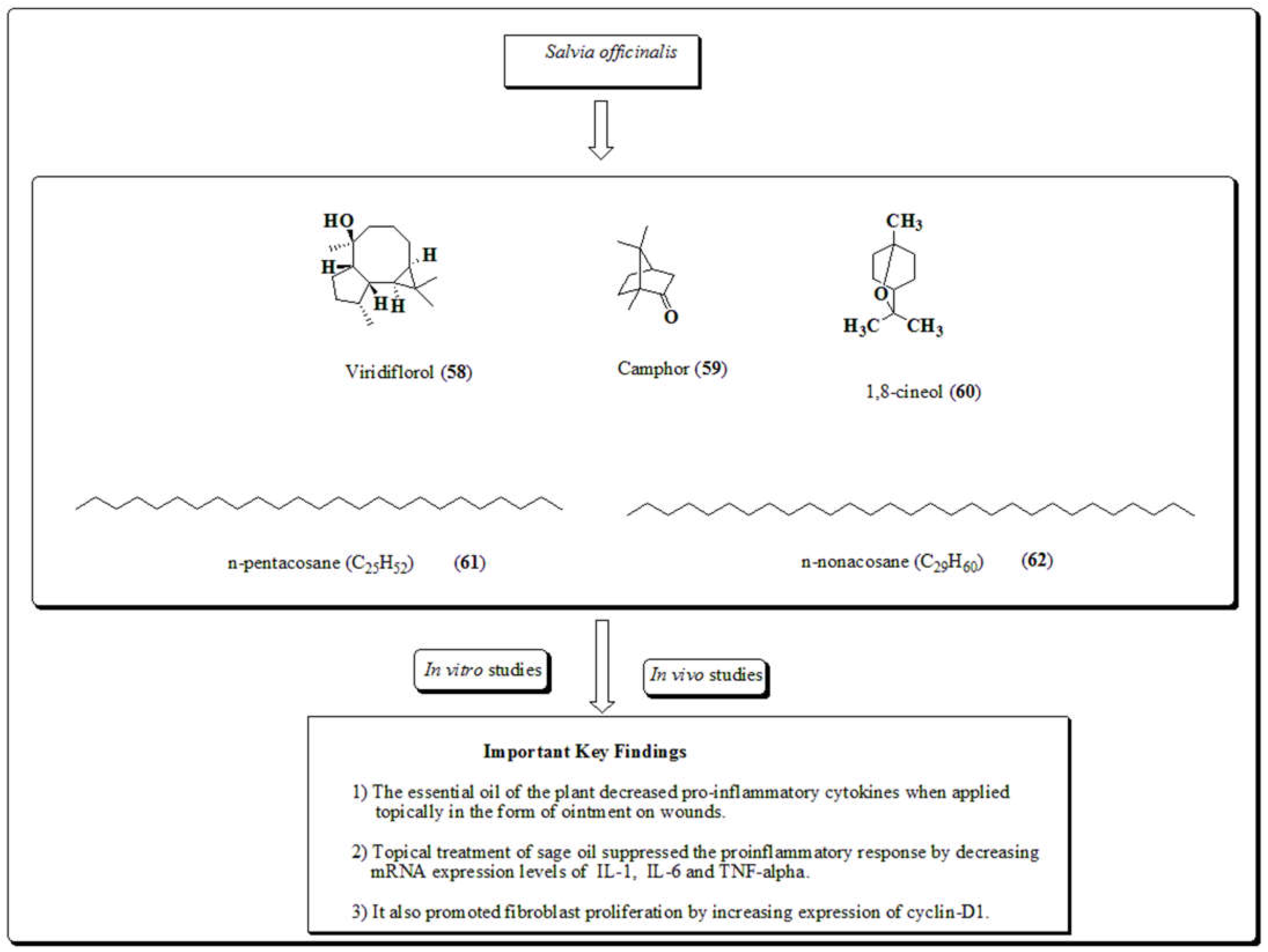
Plant has been commonly known as garden sage or simply sage. It is a green subshrub with greyish leaves, wood like stem and bluish purple flowers. It belongs to the family Lamiaceae. It is widely accepted traditionally by various tribal communities against diverse illnesses. The name saliva originated from the Latin word “Salvere” which means “feel healthy and well” [151]. Numerous studies established that the plant contains essential oils, terpenoids, viridiflorol, camphor, 1,8-cineole, n-pentacosane and n- nonacosane [152]. Oil of the plant is used for its ample range of activities including wound healing, antioxidant [151], antibacterial and anti-inflammatory actions [151,152,153,154]. In vivo study demonstrated that the essential oil of plants exhibited noteworthy effects in wound healing by granulation tissue production and angiogenesis. The plant oil has the ability to decrease mRNA expression levels of IL-1, IL-6, TNF-alpha, and stimulated fibroblast proliferation by enhancing expression of cyclin-D1. Results of a rat excision model revealed that 10% of oil exhibited rapid wound healing. It was also found that when essential oil of plants used along with tea oil showed better results than the previous. The effects were attributed due to the presence of monoterpene which are responsible for wound healing and antioxidant properties [155,156]. Structures of compounds (58-62) of plant along with important key findings were depicted in (Figure 15).
3.14. Miscellaneous
Apart from the above listed plants there are also numerous plants such as Plantago major [157], Ficus racemosa [158], Holarrhena antidysentrica [159], Colebrookea oppositifolia [160], Piper betel [161,162], Rheum emodi [163], Moringa oleifera [164] and their phytomolecules played a vital role in the healing of wounds. All these plants and their extracts have been widely used since ancient times by several tribal communities for the mitigation of wound healing, gastric ulcers, diabetic ulcers, cuts, antioxidant, antibacterial, antifungal and anti-inflammatory properties. The therapeutic potential of these plants was attributed due to the presence of phytomolecules including phenolic compounds, flavonoids, terpenes, steroidal terpenoids and polysaccharides. A topical gel prepared from aloe vera and 10 % Plantago major were used in the treatment of diabetic foot ulcers. A hydroalcoholic extract prepared from P. major was employed one time on the wound for two weeks to reduce the size of the wound. The outcomes of P. major were revealed that the plant and its molecule might be good candidates for forthcoming studies on wound healing [165].
5. Conclusions
In view of the potential of phytomolecules in numerous biological activities like anticancer, antioxidant, anti-inflammatory, antimicrobial, antifungal, anti-HIV, skin disorders and wound healing. This assemblage presents the medicinal plants and structures of their phytomolecules include alkaloids, phenolic molecules, saponins, triterpenoids, flavonoids, glycosides and tannins along with their biological activities special focused on anti-proliferative and wound healing potential and important key findings. Plants presented in this articles such as Aloe vera, Achillea millefolium, Andrographis paniculata, Boswellia sacra, Calendula officinalis, Crocus sativus, Curcuma longa, Ehretia laevis, Ehretia microphylla, Glycyrrhiza glabra, Malva sylvestris, Rosmarinus officinalis and Salvia officinalis. In vitro studies of cucuma longs revealed that curcumin improves wound healing contraction by fibroblast proliferation and migration by inhibiting the production of TNF-α and activity of NF-kB in inflammatory stages. It has been described for its anticancer and wound healing potential in forms of nanoparticles, hydrogels, nanofibers and numerous novel combinations in the cure of chronic wounds. The natural plants, their extracts, and phytomolcules present in medicinal plants were established as significant elements in homeostasis, re-epithelialization, regeneration by enhancing collagen production and fibroblasts proliferation. Flavonoids, phenolic compounds, triterpenoids and carotenoids hinder oxidative stress and stimulate antioxidant activity in the mitigation of cancer and wound repairs. In current era numerous practices validated the traditional use of these plants, extracts, phytomolecules and their formulation are moving toward the progress of pioneering wound care remedies in conjunction with herbal healing agents in the form of modern products like nanoparticles, nanostructures, nano-fibers and nano-formulations.
Author Contributions
Conceptualization, F.N.-K. and S.K.; methodology, R.S.; D.K. and P.S. software, data curation, P.S.; writing—original draft preparation, P.S., D.K., K.D. R.S. and S.K., writing—review and editing, F.N.-K. and S.K.; visualization, R.S.; K.D.. and S.K.; supervision, F.N.-K., S.K. and P.S.; project administration, F.N.-K. and P.S.; funding acquisition, F.N.-K. All authors have read and agreed to the published version of the manuscript.
Funding
F.N.-K. acknowledges a Calestous Juma Science Leadership Fellowship by the Bill & Melinda Gates Foundation.
Institutional Review Board Statement
Not applicable.
Data Availability Statement
All the figures data have been presented in the final version of manuscript.
Acknowledgments
Authors are thankful to the Vice-Chancellor of Punjabi University Patiala, India for his encouragement. Authors are also grateful to Head, Department of Pharmaceutical Sciences and Drug Research, Punjabi University Patiala, Punjab, India for providing the necessary facilities and his constant moral support.
Conflicts of Interest
The authors declare no conflict of interest.
References
- Janis, J.E.; Harrison, B. Wound Healing: Part I. Basic Science. Plast. Reconstr. Surg. 2016, 138, 199e–207e. [Google Scholar] [CrossRef]
- Broughton, G.; Janis, J.E.; Attinger, C.E. The Basic Science of Wound Healing. Plast. Reconstr. Surg. 2006, 117, 12S–34S. [Google Scholar] [CrossRef]
- Xiaolong, M.; Neil, H.R. Cancer is a functional repair tissue. Med. Hypotheses. 2006, 66, 486–490. [Google Scholar]
- Martins-Green, M.; Boudreau, N.; Bissell, M.J. Inflammation is responsible for the development of wound-induced tumors in chickens infected with Rous sarcoma virus. . 1994, 54, 4334–41. [Google Scholar]
- Liu, S.; Umezu-Goto, M.; Murph, M.; Lu, Y.; Liu, W.; Zhang, F.; Yu, S.; Stephens, L.C.; Cui, X.; Murrow, G.; et al. Expression of Autotaxin and Lysophosphatidic Acid Receptors Increases Mammary Tumorigenesis, Invasion, and Metastases. Cancer Cell 2009, 15, 539–550. [Google Scholar] [CrossRef]
- Liu, S.; Murph, M.; Panupinthu, N.; Mills, G.B. ATX-LPA receptor axis in inflammation and cancer. Cell Cycle 2009, 8, 3695–3701. [Google Scholar] [CrossRef]
- Okada, Y. Expression of fos family and jun family proto-oncogenes during corneal epithelial wound healing. Curr. Eye Res. 1996, 15, 824–832. [Google Scholar] [CrossRef]
- Anderson, M.W. Role of proto-oncogene activation in carcinogenesis. Environ. Health Perspect. 1992, 98, 13–24. [Google Scholar] [CrossRef]
- Meden, H. Elevated serum levels of a c-erbB-2 oncogene product in ovarian cancer patients and in pregnancy. J. Cancer Res. Clin. Oncol. 1994, 120, 378–381. [Google Scholar] [CrossRef]
- Abitbol, M.; Delezoide, A.-L.; Pelet, A.; Amiel, J.; Pachnis, V.; Munnich, A.; Lyonnet, S.; Vekemans, M.; Attié-Bitach, T.; Gérard, M.; et al. Expression of theRET proto-oncogene in human Embryos. Am. J. Med Genet. 1998, 80, 481–486. [Google Scholar] [CrossRef]
- Quenby, S.; Gazvani, M.; Brazeau, C.; Neilson, J.; Lewis-Jones, D.; Vince, G. Oncogenes and tumour suppressor genes in first trimester human fetal gonadal development. Mol. Hum. Reprod. 1999, 5, 737–741. [Google Scholar] [CrossRef]
- Stiles, C.D. The biological role of oncogenes--insights from platelet-derived growth factor: Rhoads Memorial Award lecture. . 1985, 45, 5215–5218. [Google Scholar]
- Virchow, R.; Virchow, R. Aetiologie der neoplastischen Geschwulste/Pathogenie der neoplastischen Geschwulste. Verlag von, Hirschwald, Berlin, Germany, 1863.
- Matthias, S.; Werner, S. Cancer as an overhealing wound: an old hypothesis revisited, Nature reviews, Mol. Cell Biol. 2008, 9, 628–638. [Google Scholar]
- Haddow, A. Molecular repair, wound healing, and carcinogenesis: tumor production a possible over healing? Adv. Cancer Res. 1972, 16, 181–234. [Google Scholar]
- Dvorak, H.F. Tumors: wounds that do not heal. Similarities between tumor stroma generation and wound healing. N. Engl. J. Med. 1986, 315, 1650–1659. [Google Scholar] [CrossRef]
- Dolberg, D.S.; Hollingsworth, R.; Hertle, M.; Bissell, M.J. Wounding and Its Role in RSV-Mediated Tumor Formation. Science 1985, 230, 676–678. [Google Scholar] [CrossRef]
- Emerich, S. Induction of preneoplastic lung lesions in guinea pigs by cigarette smoke inhalation and their exacerbation by high dietary levels of vitamins C and E. Carcinogenesis, 2005, 26, 605–612. [Google Scholar]
- Platz, E.A.; De Marzo, A.M. Epidemiology of Inflammation and Prostate Cancer. J. Urol. 2004, 171, S36–40. [Google Scholar] [CrossRef]
- Eming, S.A.; Krieg, T.; Davidson, J.M. Inflammation in Wound Repair: Molecular and Cellular Mechanisms. J. Investig. Dermatol. 2007, 127, 514–525. [Google Scholar] [CrossRef]
- Dahiya, R.; Lee, C.; Haughney, P.C.; Chui, R.; Ho, R.; Deng, G. Differential gene expression of transforming growth factors alpha and beta, epidermal growth factor, keratinocyte growth factor and their receptors in fetal and adult human prostatic tissues and cancer cell lines. Urology 1996, 48, 963–970. [Google Scholar] [CrossRef]
- Martinez-Jaramillo, G. In vitro proliferation and expansion of hematopoietic progenitors present in mobilized peripheral blood from normal subjects and cancer patients. Stem Cells Develop. 2004, 13, 382–389. [Google Scholar] [CrossRef] [PubMed]
- Ouahes, N.; Phillips, T.J.; Park, H.-Y. Expression of c-fos and c-Ha-ras Proto-oncogenes is Induced in Human Chronic Wounds. Dermatol. Surg. 1998, 24, 1354–1358. [Google Scholar] [CrossRef]
- Kumar, D.; Sharma, P.; Singh, H.; Nepali, K.; Gupta, G.K.; Jain, S.K.; Ntie-Kang, F. The value of pyrans as anticancer scaffolds in medicinal chemistry. RSC Adv. 2017, 7, 36977–36999. [Google Scholar] [CrossRef]
- Kumar, D.; Jain, S.K. A Comprehensive Review of N-Heterocycles as Cytotoxic Agents. Curr. Med. Chem. 2016, 23, 4338–4394. [Google Scholar] [CrossRef]
- Kaur, R.; Sharma, P.; Gupta, G.K.; Ntie-Kang, F.; Kumar, D. Structure-Activity-Relationship and Mechanistic Insights for Anti-HIV Natural Products. Molecules 2020, 25, 2070. [Google Scholar] [CrossRef]
- Kumar, D.; Sharma, P.; Shabu; Kaur, R. ; Lobe, M.M.M.; Gupta, G.K.; Ntie-Kang, F. In search of therapeutic candidates for HIV/AIDS: rational approaches, design strategies, structure–activity relationship and mechanistic insights. RSC Adv. 2021, 11, 17936–17964. [Google Scholar] [CrossRef]
- Sharma, P.; Kumar, D.; Shri, R.; Kumar, S. Mechanistic Insights and Docking Studies of Phytomolecules as Potential Candidates in the Management of Cancer. Curr. Pharm. Des. 2022, 28, 2704–2724. [Google Scholar] [CrossRef]
- Singla, R.K.; Sharma, P.; Dubey, A.K.; Gundamaraju, R.; Kumar, D.; Kumar, S.; Madaan, R.; Shri, R.; Tsagkaris, C.; Parisi, S.; et al. Natural Product-Based Studies for the Management of Castration-Resistant Prostate Cancer: Computational to Clinical Studies. Front. Pharmacol. 2021, 12, 732266. [Google Scholar] [CrossRef] [PubMed]
- Singla, R.K.; Sharma, P.; Kumar, D.; Gautam, R.K.; Goyal, R.; Tsagkaris, C.; Dubey, A.K.; Bansal, H.; Sharma, R.; Shen, B. The role of nanomaterials in enhancing natural product translational potential and modulating endoplasmic reticulum stress in the treatment of ovarian cancer. Front. Pharmacol. 2022, 13, 987088. [Google Scholar] [CrossRef]
- Sharma, P.; Shri, R.; Ntie-Kang, F.; Kumar, S. Phytochemical and Ethnopharmacological Perspectives of Ehretia laevis. Molecules 2021, 26, 3489. [Google Scholar] [CrossRef]
- Kumar, B.; Vijayakumar, M.; Govindarajan, R.; Pushpangadan, P. Ethnopharmacological approaches to wound healing—Exploring medicinal plants of India. J. Ethnopharmacol. 2007, 114, 103–113. [Google Scholar] [CrossRef]
- Biswas, T.K.; Mukherjee, B. Plant Medicines of Indian Origin for Wound Healing Activity: A Review. Int. J. Low. Extremity Wounds 2003, 2, 25–39. [Google Scholar] [CrossRef]
- Matsuda, H.; Morikawa, T.; Ando, S.; Toguchida, I.; Yoshikawa, M. Structural Requirements of Flavonoids for Nitric Oxide Production Inhibitory Activity and Mechanism of Action. Bioorganic Med. Chem. 2003, 11, 1995–2000. [Google Scholar] [CrossRef]
- Claude, A.C.; Jean, C.L.; Patric, T.; Christelle, P.; Gerard, H.; Albert, J.C.; Jean, L.D. Chalcones: Structural requirements for antioxidant, estrogenic and antiproliferative activities. Anticancer Res. 2001, 21, 3949–3956. [Google Scholar]
- Kumar, D.; Nepali, K.; Bedi, P.; Kumar, S.; Malik, F.; Jain, S. 4,6-diaryl Pyrimidones as Constrained Chalcone Analogues: Design, Synthesis and Evaluation as Antiproliferative Agents. Anti-Cancer Agents Med. Chem. 2015, 15, 793–803. [Google Scholar] [CrossRef]
- Middleton, E.J.V.R. Effect of plant flavonoids on immune and inflammatory cell function. Adv. Exp. Med. Biol. 1998, 439, 175–182. [Google Scholar]
- Kumar, D.; Singh, O.; Nepali, K.; Bedi, P.M.S.; Qayum, A.; Singh, S.; Jain, S.K. Naphthoflavones as Anti-proliferative Agents: Design, Synthesis and Biological Evaluation. Anticancer Agents Med. Chem. 2016, 16, 881–890. [Google Scholar] [CrossRef]
- Kumar, D.; Malik, F.; Bedi, P.M.S.; Subheet, J. 2,4-diarylpyrano[3,2-c]chromen-5(4H)-ones as coumarin-chalcone conjugates : Design, synthesis and biological evaluation as apoptosis inducing agents. Chem. Pharm. Bull. 2016, 64, 399–409. [Google Scholar] [CrossRef]
- Vitale, S.; Colanero, S.; Placidi, M.; Di Emidio, G.; Tatone, C.; Amicarelli, F.; D’alessandro, A.M. Phytochemistry and Biological Activity of Medicinal Plants in Wound Healing: An Overview of Current Research. Molecules 2022, 27, 3566. [Google Scholar] [CrossRef]
- Mehta, P.; Shah, R.; Lohidasan, S.; Mahadik, K. Pharmacokinetic profile of phytoconstituent(s) isolated from medicinal plants—A comprehensive review. J. Tradit. Complement. Med. 2015, 5, 207–227. [Google Scholar] [CrossRef]
- Maver, T.; Maver, U.; Kleinschek, K.S.; Smrke, D.M.; Kreft, S. A review of herbal medicines in wound healing. Int. J. Dermatol. 2015, 54, 740–751. [Google Scholar] [CrossRef]
- Rathi, B.S.; Bodhankar, S.; Baheti, A. Evaluation of aqueous leaves extract of Moringa oleifera Linn for wound healing in albino rats. . 2006, 44, 898–901. [Google Scholar]
- Nayak, B.; Pereira, L.M.P. Catharanthus roseus flower extract has wound-healing activity in Sprague Dawley rats. BMC Complement. Altern. Med. 2006, 6, 41–41. [Google Scholar] [CrossRef]
- Ovais, M.; Khalil, A.T.; Islam, N.U.; Ahmad, I.; Ayaz, M.; Saravanan, M.; Shinwari, Z.K.; Mukherjee, S. Role of plant phytochemicals and microbial enzymes in biosynthesis of metallic nanoparticles. Appl. Microbiol. Biotechnol. 2018, 102, 6799–6814. [Google Scholar] [CrossRef]
- Long, V. Aloe Vera in Dermatology—The Plant of Immortality. JAMA Dermatol. 2016, 152, 1364–1364. [Google Scholar] [CrossRef]
- Zeng, W.M.; Parus, A.; Barnes, C.W.; Hiro, M.E.; Robson, M.C.; Payne, W.G. Aloe vera—Mechanisms of Action, Uses, and Potential Uses in Plastic Surgery and Wound Healing. Surg. Sci. 2020, 11, 312–328. [Google Scholar] [CrossRef]
- Sánchez-Machado, D.I.; López-Cervantes, J.; Sendón, R.; Sanches-Silva, A. Aloe vera : Ancient knowledge with new frontiers. Trends Food Sci. Technol. 2017, 61, 94–102. [Google Scholar] [CrossRef]
- Salehi, B.; Albayrak, S.; Antolak, H.; Kr˛egiel, D.; Pawlikowska, E.; Sharifi-Rad, M.; Uprety, Y.; Tsouh Fokou, P.V.; Yousef, Z.; Amiruddin Zakaria, Z. Aloe Genus Plants: From Farm to Food Applications and Phytopharmacotherapy. Int. J. Mol. Sci. 2018, 19, 2843–2852. [Google Scholar] [CrossRef]
- Hormozi, M.; Assaei, R.; Boroujeni, M.B. The Effect of Aloe vera on the Expression of Wound Healing Factors (TGF_1 and BFGF) in Mouse Embryonic Fibroblast Cell: In Vitro Study. Biomed. Pharmacother. 2017, 88, 610–616. [Google Scholar] [CrossRef] [PubMed]
- Wahedi, H.M.; Jeong, M.; Chae, J.K.; Gil Do, S.; Yoon, H.; Kim, S.Y. Aloesin from Aloe vera accelerates skin wound healing by modulating MAPK/Rho and Smad signaling pathways in vitro and in vivo. Phytomedicine 2017, 28, 19–26. [Google Scholar] [CrossRef] [PubMed]
- Yamao, M.; Naoki, H.; Kunida, K.; Aoki, K.; Matsuda, M.; Ishii, S. Distinct predictive performance of Rac1 and Cdc42 in cell migration. Sci. Rep. 2015, 5, 17527. [Google Scholar] [CrossRef] [PubMed]
- De Oliveira, A.C.L.; Tabrez, S.; Shakil, S.; Khan, M.I.; Asghar, M.N.; Matias, B.D.; da Silva Batista, J.M.A.; Rosal, M.M.; de Lima, M.M.D.F.; Gomes, S.R.F. Mutagenic, Antioxidant andWound Healing Properties of Aloe vera. J. Ethnopharmacol. 2018, 227, 191–197. [Google Scholar]
- Oliveira, R.N.; Mancini, M.C.; De Oliveira, F.C.S.; Passos, T.M.; Quilty, B.; Da Silva Moreira Thiré, R.M.; McGuinness, G.B. FTIR analysis and quantification of phenols and flavonoids of five commercially available plants extracts used in wound healing. Matéria 2016, 21, 767–779. [Google Scholar] [CrossRef]
- Teplicki, E.; Ma, Q.; Castillo, D.E.; Zarei, M.; Hustad, A.P.; Chen, J.; Li, J. The Effects of Aloe vera on Wound Healing in Cell Proliferation, Migration, and Viability. Wounds Compend. Clin. Res. Pract. 2018, 30, 263–268. [Google Scholar]
- Rossiter, H.; Barresi, C.; Pammer, J.; Rendl, M.; Haigh, J.; Wagner, E.F.; Tschachler, E. Loss of Vascular Endothelial Growth Factor A Activity in Murine Epidermal Keratinocytes Delays Wound Healing and Inhibits Tumor Formation. Cancer Res 2004, 64, 3508–3516. [Google Scholar] [CrossRef]
- Chantarawaratit, P.; Sangvanich, P.; Banlunara, W.; Soontornvipart, K.; Thunyakitpisal, P. Acemannan sponges stimulate alveolar bone, cementum and periodontal ligament regeneration in a canine class II furcation defect model. J. Periodontal Res. 2013, 49, 164–178. [Google Scholar] [CrossRef]
- Xing, W.; Guo, W.; Zou, C.-H.; Fu, T.-T.; Li, X.-Y.; Zhu, M.; Qi, J.-H.; Song, J.; Dong, C.-H.; Li, Z.; et al. Acemannan accelerates cell proliferation and skin wound healing through AKT/mTOR signaling pathway. J. Dermatol. Sci. 2015, 79, 101–109. [Google Scholar] [CrossRef]
- Cavalcanti, A.M.; Baggio, C.H.; Freitas, C.S.; Rieck, L.; de Sousa, R.S.; Da Silva-Santos, J.E.; Mesia-Vela, S.; Marques, M.C.A. Safety and antiulcer efficacy studies of Achillea millefolium L. after chronic treatment in Wistar rats. J. Ethnopharmacol. 2006, 107, 277–284. [Google Scholar] [CrossRef] [PubMed]
- Benedek, B.; Kopp, B. Achillea millefolium L. s.l. revisited: Recent findings confirm the traditional use. Wien. Med. Wochenschr. 2007, 157, 312–314. [Google Scholar] [CrossRef]
- Chavez-Silva, F.; Ceron-Romero, L.; Arias-Duran, L.; Navarrete-Vazquez, G.; Almanza-Perez, J.; Roman-Ramos, R.; Ramirez-Ávila, G.; Perea-Arango, I.; Villalobos-Molina, R.; Estrada-Soto, S. Antidiabetic Effect of Achillea millefollium through Multitarget Interactions: Glucosidases Inhibition, Insulin Sensitization and Insulin Secretagogue Activities. J. Ethnopharmacol. 2018, 212, 1–7. [Google Scholar] [CrossRef]
- Ali, S.I.; Gopalakrishnan, B.; Venkatesalu, V. Pharmacognosy, Phytochemistry and Pharmacological Properties of Achillea millefolium L. : A Review. Phytother. Res. 2017, 31, 1140–1161. [Google Scholar] [CrossRef]
- Nemeth, E. Essential Oil Composition of Species in the GenusAchillea. J. Essent. Oil Res. 2005, 17, 501–512. [Google Scholar] [CrossRef]
- Nemeth, E.; Bernath, J. Biological Activities of Yarrow Species (Achillea spp.). Curr. Pharm. Des. 2008, 14, 3151–3167. [Google Scholar] [CrossRef] [PubMed]
- Bakkali, F.; Averbeck, S.; Averbeck, D.; Idaomar, M. Biological Effects of Essential Oils-A Review. Food Chem. Toxicol. 2008, 46, 446–475. [Google Scholar] [CrossRef] [PubMed]
- Vitalini, S.; Beretta, G.; Iriti, M.; Orsenigo, S.; Basilico, N.; Dall’Acqua, S.; Iorizzi, M.; Fico, G. Phenolic compounds from Achillea millefolium L. and their bioactivity. Acta Biochim. Pol. 2011, 58, 203–209. [Google Scholar] [CrossRef] [PubMed]
- Tadic, V.; Arsic, I.; Zvezdanovic, J.; Zugic, A.; Cvetkovic, D.; Pavkov, S. The Estimation of the Traditionally Used Yarrow (Achillea millefolium L. Asteraceae) Oil Extracts with Anti-Inflammatory Potential in Topical Application. J. Ethnopharmacol. 2017, 199, 138–148. [Google Scholar]
- Dorjsembe, B.; Lee, H.J.; Kim, M.; Dulamjav, B.; Jigjid, T.; Nho, C.W. Achillea asiatica extract and its active compounds induce cutaneous wound healing. J. Ethnopharmacol. 2017, 206, 306–314. [Google Scholar] [CrossRef] [PubMed]
- Akbar, S. Andrographis paniculata: a review of pharmacological activities and clinical effects. Alternative Medicine Review 2011, 16, 66–77. [Google Scholar]
- Kabir, M.H.; Hasan, N.; Rahman, M.; Rahman, A.; Alam Khan, J.; Hoque, N.T.; Bhuiyan, R.Q.; Mou, S.M.; Jahan, R.; Rahmatullah, M. A survey of medicinal plants used by the Deb barma clan of the Tripura tribe of Moulvibazar district, Bangladesh. J. Ethnobiol. Ethnomedicine 2014, 10, 19–19. [Google Scholar] [CrossRef]
- Kumar, R.A.; Sridevi, K.; Kumar, N.V.; Nanduri, S.; Rajagopal, S. Anticancer and immune stimulatory compounds from Andrographis paniculata. J. Ethnopharmacol. 2004, 92, 291–295. [Google Scholar] [CrossRef]
- Chen, L.X.; He, H.; Xia, G.Y.; Zhou, K.L.; Qiu, F. A new flavonoid from the aerial parts of Andrographis paniculata. Nat. Prod. Res. 2014, 28, 138–143. [Google Scholar] [CrossRef] [PubMed]
- Adedapo, A.A.; Adeoye, B.O.; Sofidiya, M.O.; Oyagbemi, A.A. Antioxidant, antinociceptive and anti-inflammatory properties of the aqueous and ethanolic leaf extracts of Andrographis paniculata in some laboratory animals. J. Basic Clin. Physiol. Pharmacol. 2014, 26, 327–334. [Google Scholar] [CrossRef] [PubMed]
- Al-Bayaty, F.H.; Abdulla, M.A.; Hassan, M.I.A.; Ali, H.M. Effect of Andrographis paniculata leaf extract on wound healing in rats. Nat. Prod. Res. 2012, 26, 423–429. [Google Scholar] [CrossRef] [PubMed]
- Shamsizadeh, A.; Roohbakhsh, A.; Ayoobi, F.; Moghaddamahmadi, A. The role of natural products in the prevention and treatment of multiple sclerosis, in Nutrition and Lifestyle in Neurological Autoimmune Diseases, Watson, R.R.; Killgore, W.D.S.; Eds., Academic Press, Cambridge, MA, USA, 2017, pp. 249–260.
- Shedoeva, A.; David, L.; Upton, Z.; Chen, F. Wound Healing and the Use of Medicinal Plants. Evid.-Based Complement. Altern. Med. 2019, 1-30.
- Ammon, H.P.T. Boswellic Acids in Chronic Inflammatory Diseases. Planta Medica 2006, 72, 1100–1116. [Google Scholar] [CrossRef] [PubMed]
- Glaser, T.; Winter, S.; Groscurth, P.; Safayhi, H.; Sailer, E.-R.; Ammon, H.P.T.; Schabet, M.; Weller, M. Boswellic acids and malignant glioma: induction of apoptosis but no modulation of drug sensitivity. Br. J. Cancer 1999, 80, 756–765. [Google Scholar] [CrossRef] [PubMed]
- Liu, J.-J.; Nilsson. ; Oredsson, S.; Badmaev, V.; Zhao, W.-Z.; Duan, R.-D. Boswellic acids trigger apoptosis via a pathway dependent on caspase-8 activation but independent on Fas/Fas ligand interaction in colon cancer HT-29 cells. Carcinog. 2002, 23, 2087–2093. [Google Scholar] [CrossRef]
- Nicolaus, C.; Junghanns, S.; Hartmann, A.; Murillo, R.; Ganzera, M.; Merfort, I. In vitro studies to evaluate the wound healing properties of Calendula officinalis extracts. J. Ethnopharmacol. 2017, 196, 94–103. [Google Scholar] [CrossRef] [PubMed]
- Leach, M.J. Calendula officinalis and Wound Healing: A Systematic Review. . 2008, 20, 236–243. [Google Scholar]
- Muley, B.; Khadabadi, S.; Banarase, N. Phytochemical Constituents and Pharmacological Activities of Calendula officinalis Linn (Asteraceae): A Review. Trop. J. Pharm. Res. 2009, 8, 455–465. [Google Scholar] [CrossRef]
- Arora, D.; Rani, A.; Sharma, A. A review on phytochemistry and ethnopharmacological aspects of genus Calendula. Pharmacogn. Rev. 2013, 7, 179–187. [Google Scholar] [CrossRef]
- Shafeie, N.; Naini, A.T.; Jahromi, H.K. Comparison of Different Concentrations of Calendula officinalis Gel on CutaneousWound Healing. Biomed. Pharmacol. J. 2015, 8, 979–992. [Google Scholar] [CrossRef]
- Givol, O.; Kornhaber, R.; Visentin, D.; Cleary, M.; Haik, J.; Harats, M. A systematic review of Calendula officinalis extract for wound healing. Wound Repair Regen. 2019, 27, 548–561. [Google Scholar] [CrossRef] [PubMed]
- Oliveira, R.N.; Mancini, M.C.; De Oliveira, F.C.S.; Passos, T.M.; Quilty, B.; Da Silva Moreira Thiré, R.M.; McGuinness, G.B. FTIR analysis and quantification of phenols and flavonoids of five commercially available plants extracts used in wound healing. Matéria 2016, 21, 767–779. [Google Scholar] [CrossRef]
- Fronza, M.; Heinzmann, B.; Hamburger, M.; Laufer, S.; Merfort, I. Determination of the wound healing effect of Calendula extracts using the scratch assay with 3T3 fibroblasts. J. Ethnopharmacol. 2009, 126, 463–467. [Google Scholar] [CrossRef] [PubMed]
- Dinda, M.; Dasgupta, U.; Singh, N.; Bhattacharyya, D.; Karmakar, P. PI3K-Mediated Proliferation of Fibroblasts by Calendula officinalis Tincture: Implication in Wound Healing. Phytotherapy Res. 2015, 29, 607–616. [Google Scholar] [CrossRef]
- Dinda, M.; Mazumdar, S.; Das, S. Water fraction of Calendula officinalis hydroethanol extract stimulates in vitro and in vivo proliferation of dermal fibroblasts in wound healing. Phytotherap. Res. 2016, 30, 1696–1707. [Google Scholar] [CrossRef]
- Kumar, R.; Singh, V.; Devi, K.; Sharma, M.; Singh, M.K.; Ahuja, P.S. State of Art of Saffron (Crocus sativus L. ) Agronomy: A Comprehensive Review. Food Rev. Int. 2008, 25, 44–85. [Google Scholar]
- Christodoulou, E.; Kadoglou, N.P.; Kostomitsopoulos, N.; Valsami, G. Saffron: a natural product with potential pharmaceutical applications. J. Pharm. Pharmacol. 2015, 67, 1634–1649. [Google Scholar] [CrossRef] [PubMed]
- Abu-Izneid, T.; Rauf, A.; Khalil, A.A.; Olatunde, A.; Khalid, A.; Alhumaydhi, F.A.; Aljohani, A.S.M.; Uddin, S.; Heydari, M.; Khayrullin, M.; et al. Nutritional and health beneficial properties of saffron (Crocus sativusL): a comprehensive review. Crit. Rev. Food Sci. Nutr. 2020, 62, 2683–2706. [Google Scholar] [CrossRef]
- Alonso, G.L.; Zalacain, A.; Carmona, M. Saffron. In Handbook of Herbs and Spices, Elsevier: Amsterdam, The Netherlands, 2012; pp. 469–498.
- Del-Angel, D.; Martínez, N.; Cruz, M.; Urrutia, E.; Riverón-Negrete, L.; Abdullaev, F. SAFFRON EXTRACT AMELIORATES OXIDATIVE DAMAGE AND MITOCHONDRIAL DYSFUNCTION IN THE RAT BRAIN. Acta Hortic. 2007, 359–366. [Google Scholar] [CrossRef]
- Cerdá-Bernad, D.; Valero-Cases, E.; Pastor, J.-J.; Frutos, M.J. Saffron bioactives crocin, crocetin and safranal: effect on oxidative stress and mechanisms of action. Crit. Rev. Food Sci. Nutr. 2020, 62, 3232–3249. [Google Scholar] [CrossRef]
- Nanda, S.; Madan, K. The role of Safranal and saffron stigma extracts in oxidative stress, diseases and photoaging: A systematic review. Heliyon 2021, 7, e06117. [Google Scholar] [CrossRef]
- Li, S.; Liu, X.; Lei, J.; Yang, J.; Tian, P.; Gao, Y. Crocin Protects Podocytes Against Oxidative Stress and Inflammation Induced by High Glucose Through Inhibition of NF-KB. Cell Physiol. Biochem. 2017, 42, 1481–1492. [Google Scholar] [CrossRef] [PubMed]
- Festuccia, C.; Mancini, A.; Gravina, G.L.; Scarsella, L.; Llorens, S.; Alonso, G.L.; Tatone, C.; Di Cesare, E.; Jannini, E.A.; Lenzi, A.; et al. Antitumor Effects of Saffron-Derived Carotenoids in Prostate Cancer Cell Models. BioMed Res. Int. 2014, 2014, 45–56. [Google Scholar] [CrossRef] [PubMed]
- Colapietro, A.; Mancini, A.; D'Alessandro, A.M.; Festuccia, C. Crocetin and Crocin from Saffron in Cancer Chemotherapy and Chemoprevention. Anti-Cancer Agents Med. Chem. 2019, 19, 38–47. [Google Scholar] [CrossRef]
- Hassan, A. Curcuma Longa, Turmeric: A Monograph. Aust. J. Med. Herbal. 2006, 18, 66–76. [Google Scholar]
- Memarzia, A.; Khazdair, M.R.; Behrouz, S.; Gholamnezhad, Z.; Jafarnezhad, M.; Saadat, S.; Boskabady, M.H. Experimental and Clinical Reports on Anti-Inflammatory, Antioxidant, and Immunomodulatory Effects of Curcuma Longa and Curcumin, an Updated and Comprehensive Review. BioFactors Oxf. Engl. 2021, 47, 311–350. [Google Scholar] [CrossRef]
- Anamika, B. Extraction of Curcumin. J. Env. Sci Toxicol Food Technol 2012, 1, 1–16. [Google Scholar]
- Akbik, D.; Ghadiri, M.; Chrzanowski, W.; Rohanizadeh, R. Curcumin as a wound healing agent. Life Sci. 2014, 116, 1–7. [Google Scholar] [CrossRef]
- Ak, T.; Gülçin, I. Antioxidant and radical scavenging properties of curcumin. Chem. Interactions 2008, 174, 27–37. [Google Scholar] [CrossRef]
- Thangapazham, R.L.; Sharad, S.; Maheshwari, R.K. Skin regenerative potentials of curcumin. BioFactors 2013, 39, 141–149. [Google Scholar] [CrossRef] [PubMed]
- Barzegar, A.; Moosavi-Movahedi, A.A. Intracellular ROS Protection Efficiency and Free Radical-Scavenging Activity of Curcumin. PLoS ONE 2011, 6, e26012. [Google Scholar] [CrossRef]
- Panchatcharam, M.; Miriyala, S.; Gayathri, V.S.; Suguna, L. Curcumin improves wound healing by modulating collagen and decreasing reactive oxygen species. Mol. Cell. Biochem. 2006, 290, 87–96. [Google Scholar] [CrossRef] [PubMed]
- Yen, Y.H.; Pu, C.M.; Liu, CW.; Chen, Y.C.; Chen, Y.C.; Liang, C.J.; Hsieh, J.H.; Huang, H.F.; Chen, Y.L. Curcumin accelerates cutaneous wound healing via multiple biological actions: The involvement of TNF-α, MMP-9, α-SMA, and collagen, Int Wound J. 2018, 15, 605–617.
- Mohanty, C.; Das, M.; Sahoo, S.K. Emerging role of nanocarriers to increase the solubility and bioavailability of curcumin. Expert Opin. Drug Deliv. 2012, 9, 1347–1364. [Google Scholar] [CrossRef] [PubMed]
- Pandey, V.K.; Ajmal, G.; Upadhyay, S.N.; Mishra, P.K. Nano-fibrous scaffold with curcumin for anti-scar wound healing. Int. J. Pharm. 2020, 589, 119858. [Google Scholar] [CrossRef] [PubMed]
- Rao, M.; Kumar, P.; Das, B. Comparative study of efficacy of Jatyadi Ghrita Pichu and Yasthimadhu Ghrita Pichu in the management of parikartika (fissure-in-ano). Int. J. Ayurveda Pharma Res. 2016, 4, 1–9. [Google Scholar]
- Meena, K.L.; Yadav, B.L. Some ethnomedicinal plants used by the Garasia tribe of Siroh, Rajasthan. Indian J. Trad. Knowl. 2011, 10, 354–357. [Google Scholar]
- Samantaray, S.; Bishwal, R.; Singhai, S. Clinical efficacy of jatyadi taila in parikartika (fissure-in-ano). World J. Pharm. Med. Res. 2017, 3, 250–254. [Google Scholar]
- Tichkule, S.V.; Khandare, K.B.; Shrivastav, P.P. Proficiency of Khanduchakka Ghrit in the management of Parikartika: A case report. J. Indian Syst. Med. 2019, 4, 47–50. [Google Scholar] [CrossRef]
- Tafere, Y.; Chanie, S.; Dessie, T.; Gedamu, H. Assessment of prevalence of dental caries and the associated factors among patients attending dental clinic in Debre Tabor general hospital: a hospital-based cross-sectional study. BMC Oral Heal. 2018, 18, 119–125. [Google Scholar] [CrossRef]
- Deshpande, R.; Walimbe, H.; Jadhav, M.; Deshpande, N.; Devare, S. Comparative evaluation of antimicrobial activity of various extracts of ‘Morinda pubescens’ in different concentration on human salivary microflora. Int. J. Pharm. Pharm. Sci. 2013, 5, 910–912. [Google Scholar]
- Cagampan, D.; Lacuata, K.; Ples, M.; Ii, R. Effect of Ehretia microphylla on the blood cholesterol and weight of ICR mice (Mus musculus). Natl. J. Physiol. Pharm. Pharmacol. 2018, 8, 983–987. [Google Scholar] [CrossRef]
- Demetrio, L.V.J.; Jeannie, I.A.; Juliana, J.M.P.; Esperanza, C.C.; Windell, L.R. Antibacterial activities of ethanol extracts of Philippine medicinal plants against multidrug-resistant bacteria. Asian Pac. J. Trop. Biomed. 2015, 5, 532–540. [Google Scholar]
- Murugesa, M.K.S. Gunapadam. Part-I; Tamil Nadu Siddha Medical Board: Chennai, India, 1956; pp. 274–280. [Google Scholar]
- Roeder, E.; Wiedenfeld, H. Plants containing pyrrolizidine alkaloids used in the traditional Indian medicine--including ayurveda. . 2013, 68, 83–92. [Google Scholar] [PubMed]
- Vista, F.E.S.; Dalmacio, L.M.M.; Corales, L.G.M.; Salem, G.M.; Galula, J.U.; Chao, D.Y. Antiviral Effect of Crude Aqueous Extracts from Ten Philippine Medicinal Plants against Zika virus. Acta Med. Phillipin. 2022, 54, 195–202. [Google Scholar] [CrossRef]
- Safe, S.H.; Prather, P.L.; Brents, L.K.; Chadalapaka, G.; Jutooru, I. Unifying Mechanisms of Action of the Anticancer Activities of Triterpenoids and Synthetic Analogs. Anti-Cancer Agents Med. Chem. 2012, 12, 1211–1220. [Google Scholar] [CrossRef] [PubMed]
- Sharma, P.; Shri, R.; Kumar, S. Phytochemical and In Vitro Cytotoxic Screening of Chloroform Extract of Ehretia microphylla Lamk. Stresses 2022, 2, 384–394. [Google Scholar] [CrossRef]
- Komes, D.; Belščak-Cvitanović, A.; Jurić, S.; Bušić, A.; Vojvodić, A.; Durgo, K. Consumer acceptability of liquorice root (Glycyrrhiza glabraL.) as an alternative sweetener and correlation with its bioactive content and biological activity. Int. J. Food Sci. Nutr. 2015, 67, 53–66. [Google Scholar] [CrossRef]
- Jeon, J.S.; Kim, H.T.; Kim, M.G.; Oh, M.S.; Hong, S.R.; Yoon, M.H.; Shin, H.C.; Shim, J.H.; Afifi, N.A.; Hacımuftuo glu, A.; et al. Simultaneous Detection of Glabridin, (-), α-Bisabolol, and Ascorbyl Tetraisopalmitate in Whitening Cosmetic Creams Using HPLC-PAD. Chromatographia 2016, 79, 851–860. [Google Scholar] [CrossRef]
- Pastorino, G.; Cornara, L.; Soares, S.; Rodrigues, F.; Oliveira, M.B.P.P. Liquorice (Glycyrrhiza glabra): A phytochemical and pharmacological review. Phytother. Res. 2018, 32, 2323–2339. [Google Scholar] [CrossRef]
- Memariani, Z.; Hajimahmoodi, M.; Minaee, B.; Khodagholi, F.; Yans, A.; Rahimi, R.; Amin, G.; Moghaddam, G.; Toliyat, T.; Sharifzadeh, M. Protective Effect of a Polyherbal Traditional Formula Consisting of Rosa damascena Mill. , Glycyrrhiza glabra L. and Nardostachys jatamansi DC., Against Ethanol-Induced Gastric Ulcer. Iran. J. Pharm. Res. 2017, 16, 694–707. [Google Scholar]
- Al-Refai, A.S.; Najeeb, V.D. Antibacterial effect and healing potential of topically applied licorice root extract on experimentally induced oral wounds in rabbits. Saudi J. Oral Sci. 2015, 2, 10. [Google Scholar] [CrossRef]
- Chen, X.; Fang, D.; Li, L.; Chen, L.; Li, Q.; Gong, F.; Fang, M. Glycyrrhizin ameliorates experimental colitis through attenuating interleukin-17-producing T cell responses via regulating antigen-presenting cells. Immunol. Res. 2017, 65, 666–680. [Google Scholar] [CrossRef]
- Shah, S.L.; Wahid, F.; Khan, N.; Farooq, U.; Shah, A.J.; Tareen, S.; Ahmad, F.; Khan, T. Inhibitory Effects of Glycyrrhiza glabra and Its Major Constituent Glycyrrhizin on Inflammation-Associated Corneal Neovascularization. Evid. Based Complement. Alternat. Med. 2018, 2018, 1–8. [Google Scholar] [CrossRef]
- Oloumi, M.M.; Derakhshanfar, A.; Nikpour, A. Healing Potential of Liquorice Root Extract on Dermal Wounds in Rats. J. Vet. Res. 2007, 62, 147–154. [Google Scholar]
- Li, B.; Wang, J.H.-C. Fibroblasts and myofibroblasts in wound healing: Force generation and measurement. J. Tissue Viability 2011, 20, 108–120. [Google Scholar] [CrossRef]
- Geethalakshmi, R.; Sakravarthi, C.; Kritika, T.; Kirubakaran, M.A.; Sarada, D.V.L. Evaluation of Antioxidant and Wound Healing Potentials ofSphaeranthus amaranthoidesBurm.f. BioMed Res. Int. 2013, 2013, 1–7. [Google Scholar] [CrossRef]
- Zangeneh, A.; Pooyanmehr, M.; Zangeneh, M.M.; Moradi, R.; Rasad, R.; Kazemi, N. Therapeutic effects of Glycyrrhiza glabra aqueous extract ointment on cutaneous wound healing in Sprague Dawley male rats. Comp. Clin. Pathol. 2019, 28, 1507–1514. [Google Scholar] [CrossRef]
- Siriwattanasatorn, M.; Itharat, A.; Thongdeeying, P.; Ooraikul, B. In VitroWound Healing Activities of Three Most Commonly Used Thai Medicinal Plants and Their Three Markers. Evid.-Based Complement. Altern. Med. 2020, 2020. [Google Scholar] [CrossRef] [PubMed]
- Barros, L.; Carvalho, A.M.; Ferreira, I.C. Leaves, flowers, immature fruits and leafy flowered stems of Malva sylvestris: A comparative study of the nutraceutical potential and composition. Food Chem. Toxicol. 2010, 48, 1466–1472. [Google Scholar] [CrossRef] [PubMed]
- Quave, C.L.; Plano, L.R.; Pantuso, T.; Bennett, B.C. Effects of extracts from Italian medicinal plants on planktonic growth, biofilm formation and adherence of methicillin-resistant Staphylococcus aureus. J. Ethnopharmacol. 2008, 118, 418–428. [Google Scholar] [CrossRef]
- DellaGreca, M.; Cutillo, F.; Abrosca, B.D.; Fiorentino, A.; Pacifico, S.; Zarrelli, A. Antioxidant and Radical Scavenging Properties of Malva Sylvestris. Nat. Prod. Commun. 2009, 4, 893–896. [Google Scholar] [CrossRef]
- Leporatti, M.L.; Ghedira, K. Comparative analysis of medicinal plants used in traditional medicine in Italy and Tunisia. J. Ethnobiol. Ethnomedicine 2009, 5, 31–40. [Google Scholar] [CrossRef]
- Pirbalouti, A.G.; Azizi, S.; Koohpayeh, A.; Hamedi, B. Wound healing activity of Malva sylvestris and Punica granatum in alloxan-induced diabetic rats. . 2010, 67, 511–516. [Google Scholar]
- Pirbalouti, A.G.; Koohpyeh, A. Wound Healing Activity of Extracts of Malva sylvestris and Stachys lavandulifolia. Int. J. Biol. 2010, 3, 174–180. [Google Scholar] [CrossRef]
- Gasparetto, J.C.; Martins, C.A.F.; Hayashi, S.S.; Otuky, M.F.; Pontarolo, R. Ethnobotanical and Scientific Aspects of Malva sylvestris L. : A Millennial Herbal Medicine. J. Pharm. Pharmacol. 2012, 64, 172–189. [Google Scholar]
- Mousavi, S.M.; Hashemi, S.A.; Behbudi, G.; Mazraedoost, S.; Omidifar, N.; Gholami, A.; Chiang, W.-H.; Babapoor, A.; Rumjit, N.P. A Review on Health Benefits of Malva sylvestris L. Nutritional Compounds for Metabolites, Antioxidants, and Anti-Inflammatory, Anticancer, and Antimicrobial Applications. Evidence-Based Complement. Altern. Med. 2021, 2021, e5548404. [Google Scholar] [CrossRef]
- Afshar, M.; Ravarian, B.; Zardast, M.; Moallem, S.A.; Fard, M.H.; Valavi, M. Evaluation of cutaneous wound healing activity of Malva sylvestris aqueous extract in BALB/c mice. . 2015, 18, 616–625. [Google Scholar]
- Nasiri, E.; Hosseinimehr, S.J.; Azadbakht, M.; Akbari, J.; Enayati-Fard, R.; Azizi, S. Effect of Malva sylvestris cream on burn injury and wounds in rats. Avicenna J. phytomed. 2016, 5, 341–354. [Google Scholar]
- Almasian, A.; Najafi, F.; Eftekhari, M.; Ardekani, M.R.S.; Sharifzadeh, M.; Khanavi, M. Polyurethane/carboxymethylcellulose nanofibers containing Malva sylvestris extract for healing diabetic wounds: Preparation, characterization, in vitro and in vivo studies. Mater. Sci. Eng. C 2020, 114, 111039. [Google Scholar] [CrossRef]
- Mena, P.; Cirlini, M.; Tassotti, M.; Herrlinger, K.A.; Dall’Asta, C.; Del Rio, D. Phytochemical Profiling of Flavonoids, Phenolic Acids, Terpenoids, and Volatile Fraction of a Rosemary (Rosmarinus Officinalis L. ) Extract. Molecules 2016, 21, 1576. [Google Scholar] [CrossRef] [PubMed]
- del Baño, M.J.; Lorente, J.; Castillo, J.; Benavente-García, O.; Marín, M.P.; Del Río, J.A.; Ortuño, A.; Ibarra, I. Flavonoid Distribution during the Development of Leaves, Flowers, Stems, and Roots ofRosmarinus officinalis. Postulation of a Biosynthetic Pathway. J. Agric. Food Chem. 2004, 52, 4987–4992. [Google Scholar] [CrossRef] [PubMed]
- Umasankar, K.; Nambikkairaj, B.; Backyavathy, D.M. Effect of Topical Treatment of Rosmarinus Officinalis Essential Oil on Wound Healing in Streptozotocin Induced Diabetic Rats. Nat. Environ. Pollut. Technol. 2012, 11, 5–11. [Google Scholar]
- Labib, R.M.; Ayoub, I.M.; Michel, H.E.; Mehanny, M.; Kamil, V.; Hany, M.; Magdy, M.; Moataz, A.; Maged, B.; Mohamed, A. Appraisal on the wound healing potential of Melaleuca alternifolia and Rosmarinus officinalis L. essential oil-loaded chitosan topical preparations. PLOS ONE 2019, 14, e0219561. [Google Scholar] [CrossRef] [PubMed]
- Wang, M.; Li, J.; Rangarajan, M.; Shao, Y.; LaVoie, E.J.; Huang, T.-C.; Ho, C.-T. Antioxidative Phenolic Compounds from Sage (Salvia officinalis). J. Agric. Food Chem. 1998, 46, 4869–4873. [Google Scholar] [CrossRef]
- Venskutonis, P.R. Effect of drying on the volatile constituents of thyme (Thymus vulgaris L.) and sage (Salvia officinalis L.). Food Chem. 1997, 59, 219–227. [Google Scholar] [CrossRef]
- Kamatou, G.P.P.; Makunga, N.P.; Ramogola, W.P.; Viljoen, A.M. South African Salvia species: A review of biological activities and phytochemistry. J. Ethnopharmacol. 2008, 119, 664–672. [Google Scholar] [CrossRef] [PubMed]
- Topçu, G. Bioactive Triterpenoids from Salvia Species. J. Nat. Prod. 2006, 69, 482–487. [Google Scholar] [CrossRef] [PubMed]
- Raal, A.; Orav, A.; Arak, E. Composition of the Essential Oil of Salvia officinalis L. from Various European Countries. Nat. Prod. Res. 2007, 21, 406–411. [Google Scholar] [CrossRef]
- Karimzadeh, S.; Farahpour, M.R. Topical Application of Salvia officinalis Hydro ethanolic Leaf Extract Improves Wound Healing Process. Indian J. Exp. Biol. 2017, 55, 98–106. [Google Scholar]
- Zubair, M.; Widén, C.; Renvert, S.; Rumpunen, K. Water and ethanol extracts of Plantago major leaves show anti-inflammatory activity on oral epithelial cells. J. Tradit. Complement. Med. 2018, 9, 169–171. [Google Scholar] [CrossRef] [PubMed]
- Sen, S.K.; Behara, L.M. Ethnomedicinal plants used against skin diseases in Bargat district of Orissa. Ethnobotan. 2003, 15, 90–96. [Google Scholar]
- Patil, K.S.; Mandewgade, S.D. Wound healing activity of leaves of Lawsonia alba. J. Nat. Remed. 2003, 3, 129–133. [Google Scholar]
- Kumar, D.; Singla, R.K.; Sharma, R.; Sharma, P.; Kumar, L.; Kaur, N.; Dhawan, R.K.; Sharma, S.; Dua, K. Phytochemistry and Polypharmacological Potential of Colebrookea oppositifolia Smith. Curr. Top. Med. Chem. 2023, 23, 334–348. [Google Scholar] [CrossRef] [PubMed]
- Sarma, S.K.; Bhattacharya, B.K.; Devi, B. Traditional use of herbal medicine by Modahi tribe of Nalabari district of Assam. Ethnobotan. 2002, 14, 103–111. [Google Scholar]
- Thomas, J.; Britto, A. Ethnobotanical study in Wyanad District of Kerala. J. Econom. Taxonom. Bot. 2003, 27, 915–924. [Google Scholar]
- Nautiyal, S.; Maikhuri, R.K.; Rao, K.S.; Saxena, K.G. Medicinal Plant Resources in Nanda Devi Biosphere Reserve in the Central Himalayas. J. Herbs, Spices Med. Plants 2001, 8, 47–64. [Google Scholar] [CrossRef]
- Udupa, S.L.; Udupa, A.L.; Kulkarni, D.R. Studies on anti-inflammatory and wound healing properties of Moringa oleifera and Aegle marmelos. Fitoterapia 1994, 65, 119–123. [Google Scholar]
- Ghanadian, M.; Soltani, R.; Homayouni, A.; Khorvash, F.; Jouabadi, S.M.; Abdollahzadeh, M. The Effect of Plantago major Hydroalcoholic Extract on the Healing of Diabetic Foot and Pressure Ulcers: A Randomized Open-Label Controlled Clinical Trial. Int. J. Low. Extremity Wounds 2022. [Google Scholar] [CrossRef]
Figure 1.
Representation of wounds in the modern and Ayurvedic system of medicine.
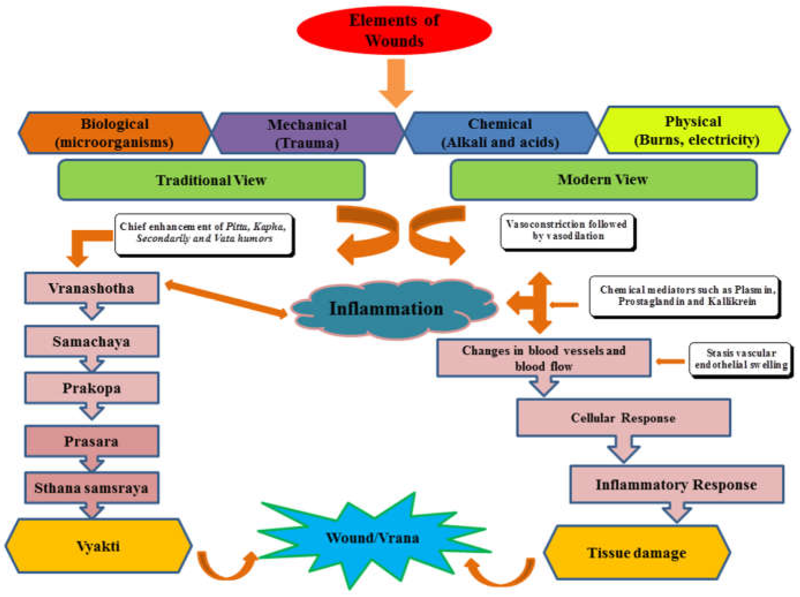
Figure 2.
Structure of compounds (1-5) along with their important key findings.
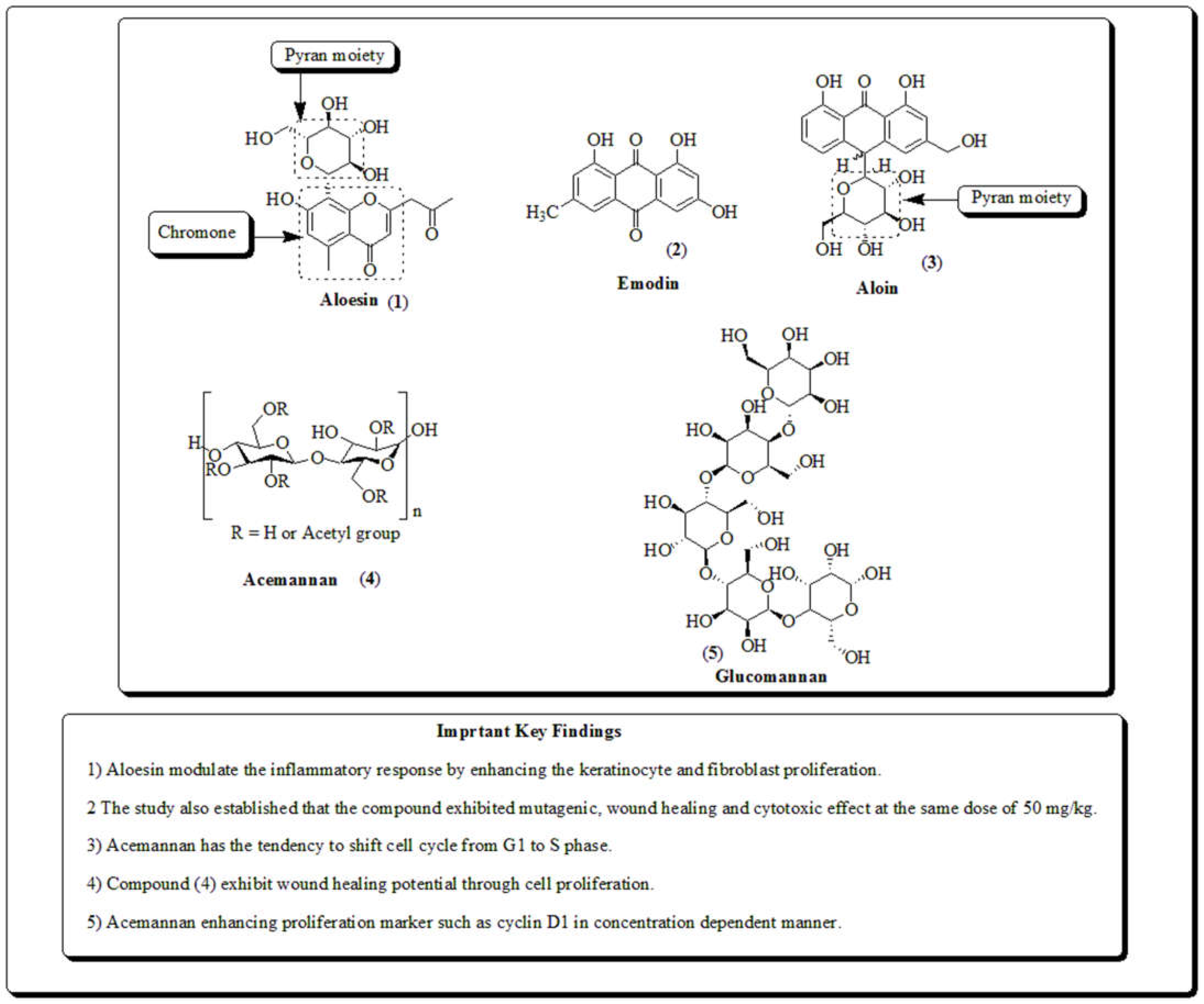
Figure 3.
Structure of compounds (6-9) along with their important key findings.
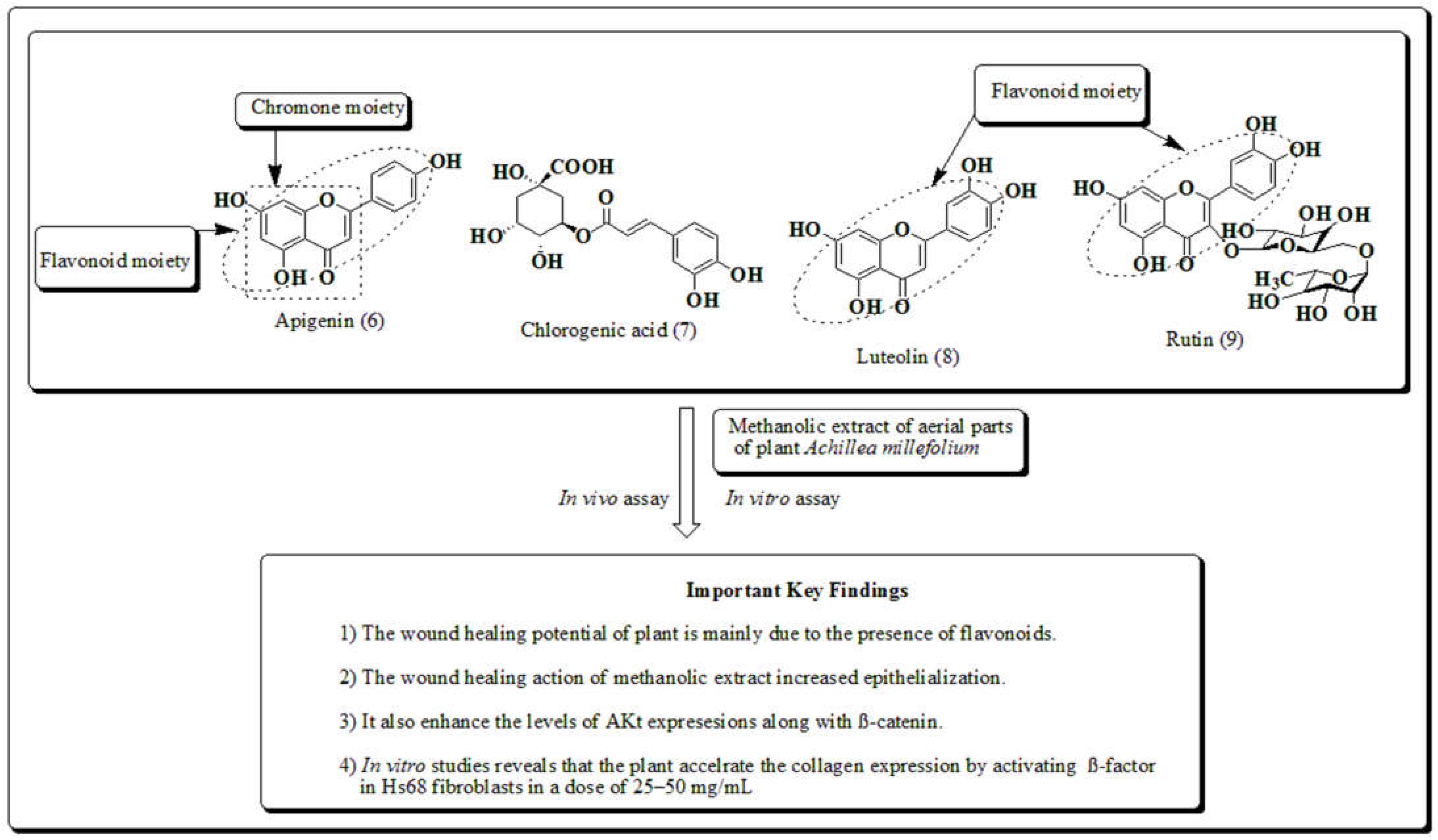
Figure 4.
Structure of andrographolide along with important key findings.
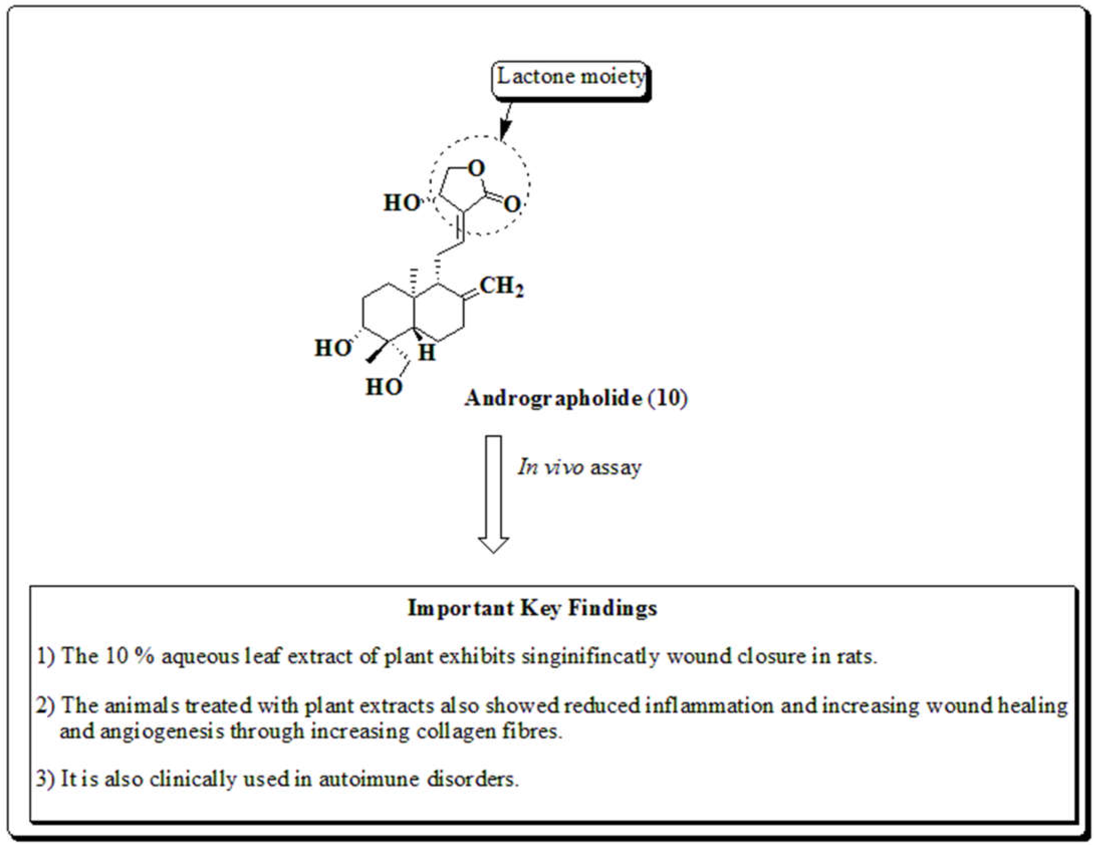
Figure 5.
Structure of compounds (11-14) along with their important key findings.
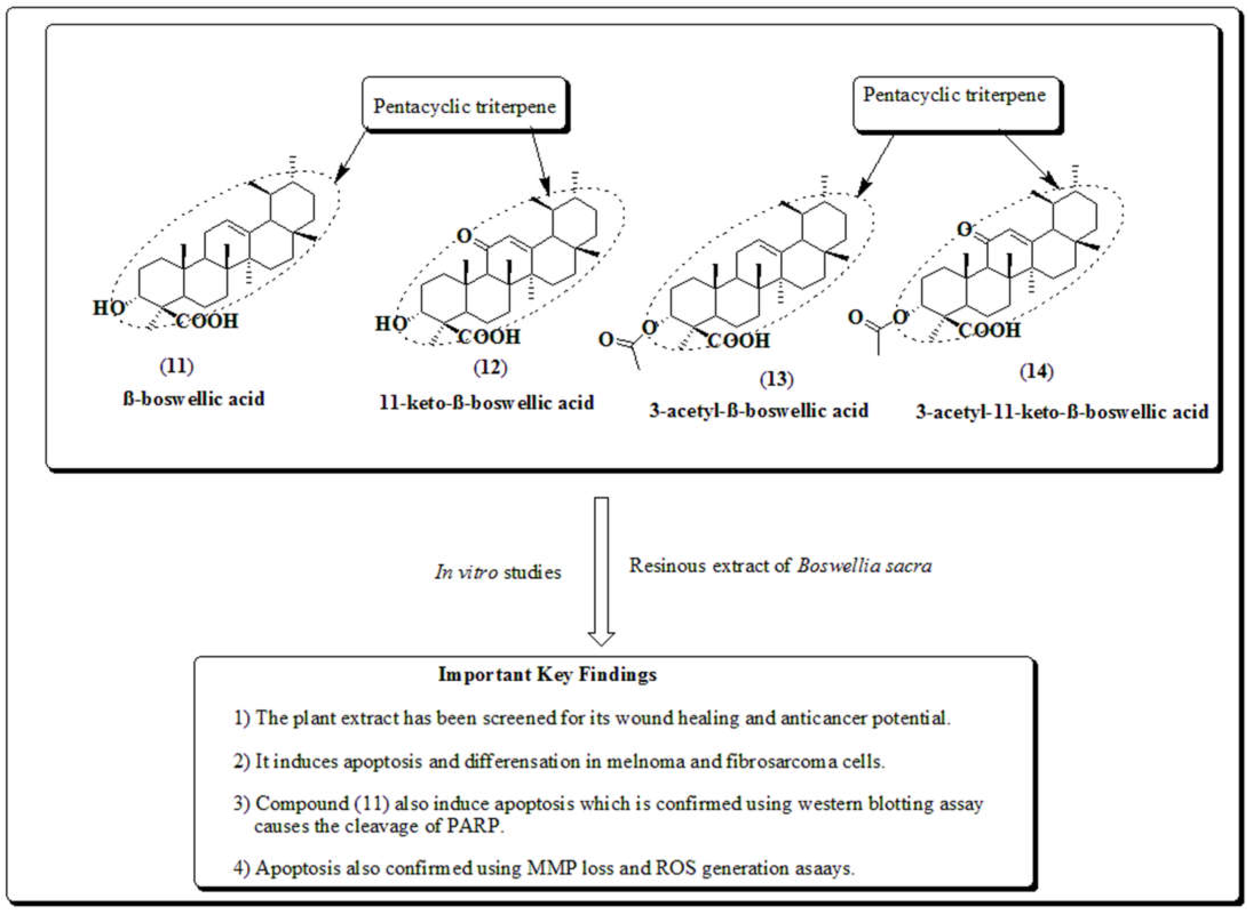
Figure 6.
Structure of compounds (15-18) along with their their important key findings.
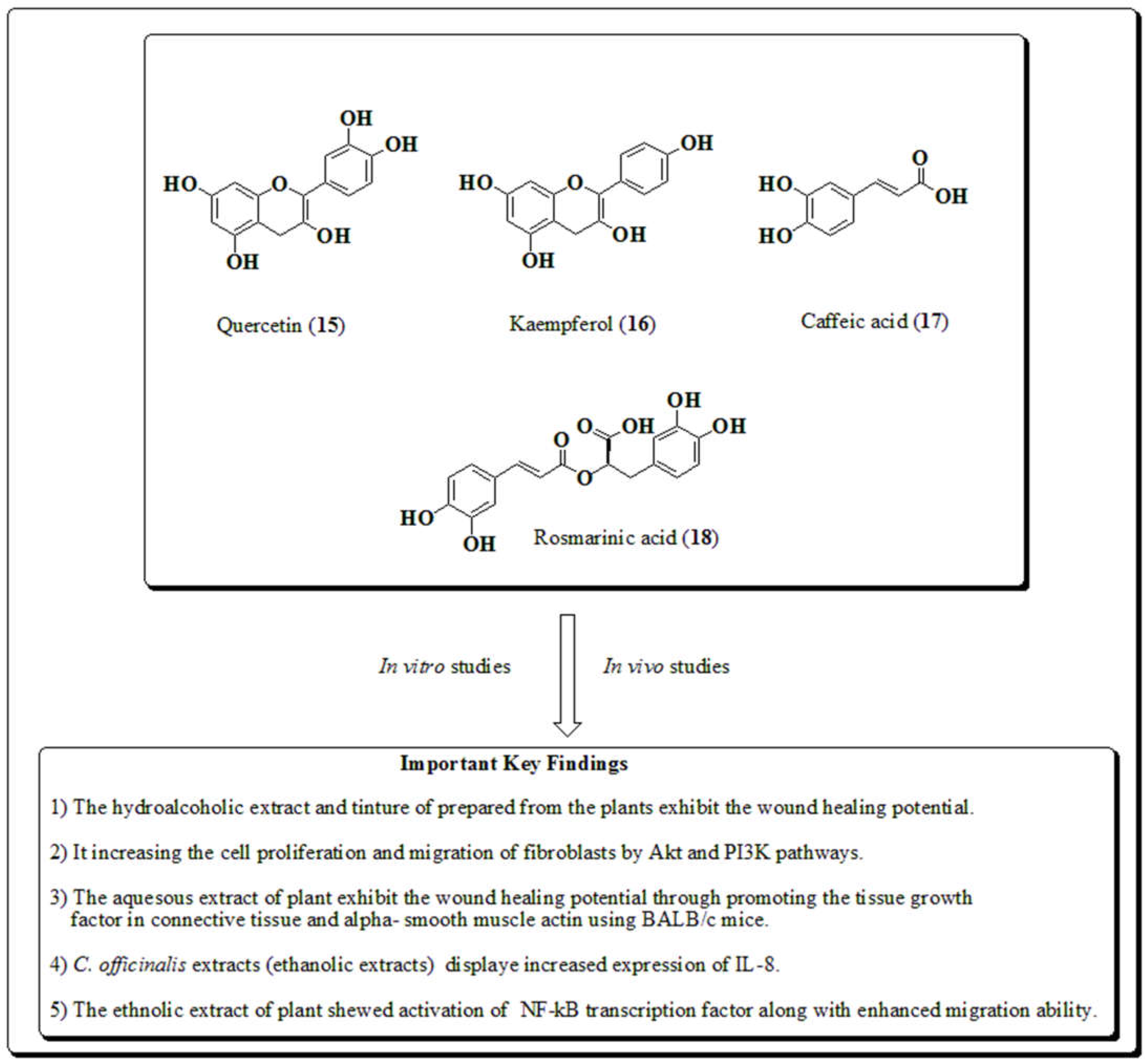
Figure 7.
Structures of compounds (19-22) along with important key findings.
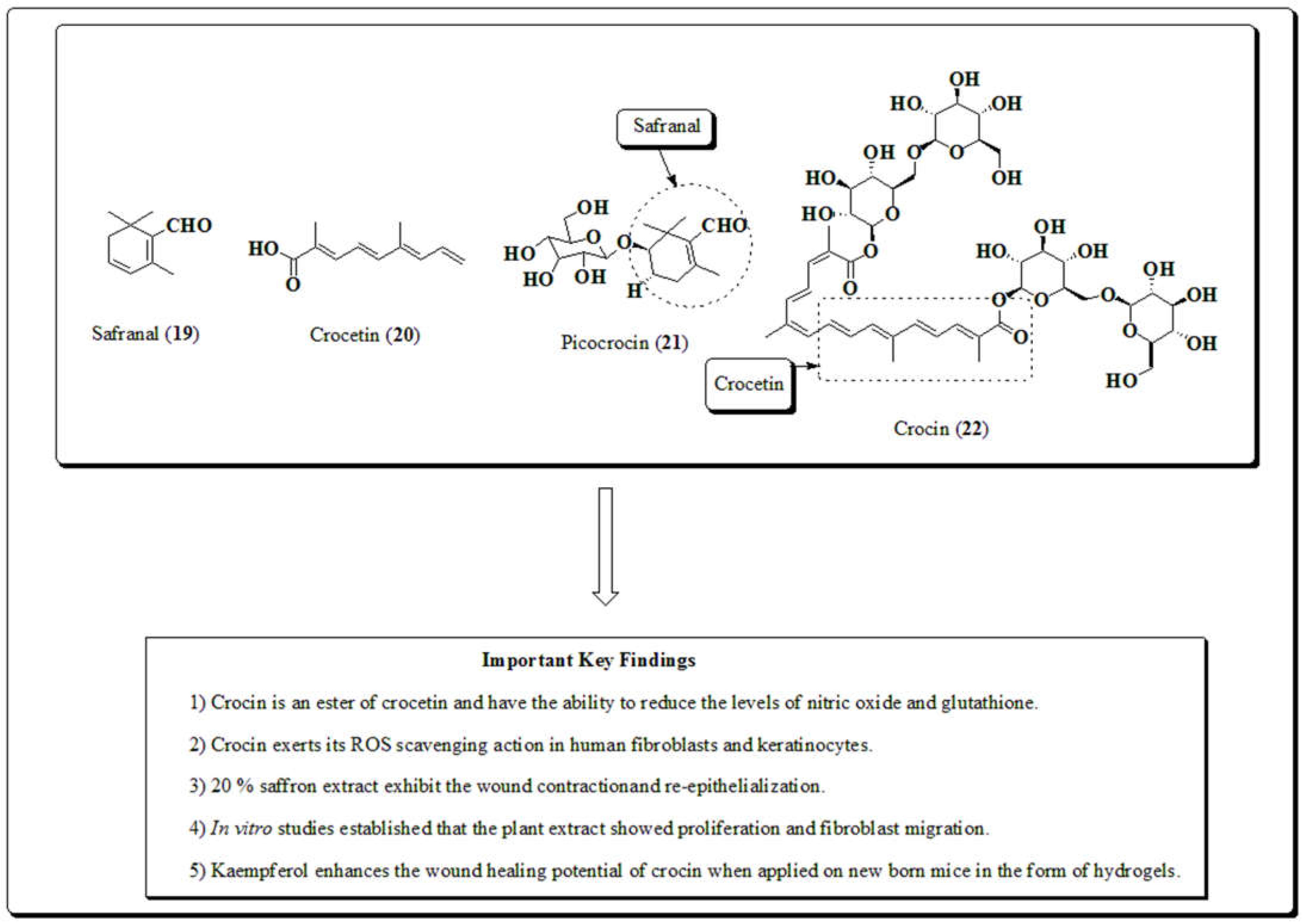
Figure 8.
Structure of curcumin and important key findings.
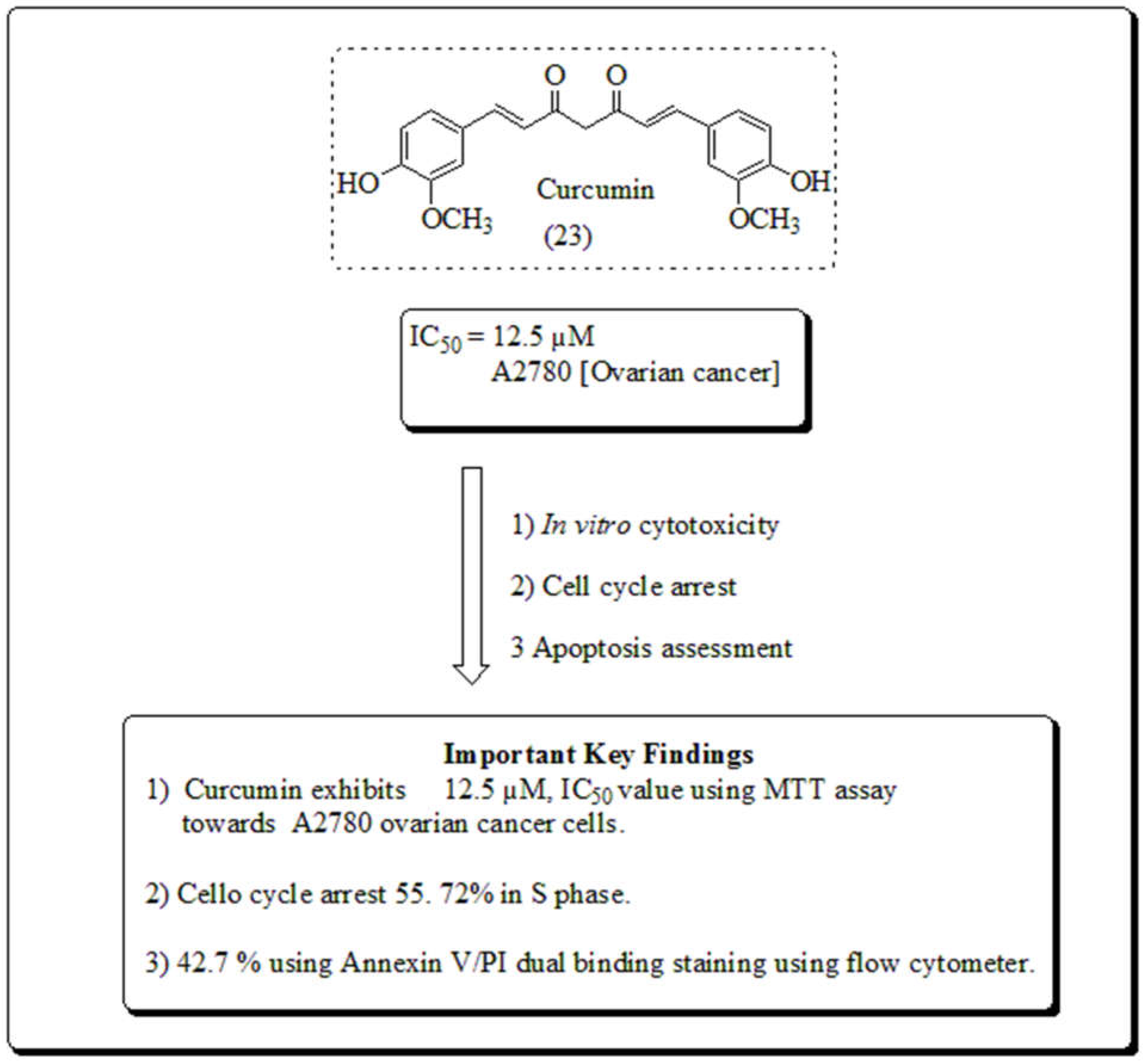
Figure 9.
Structure of curcumin and its structure activity relationships.
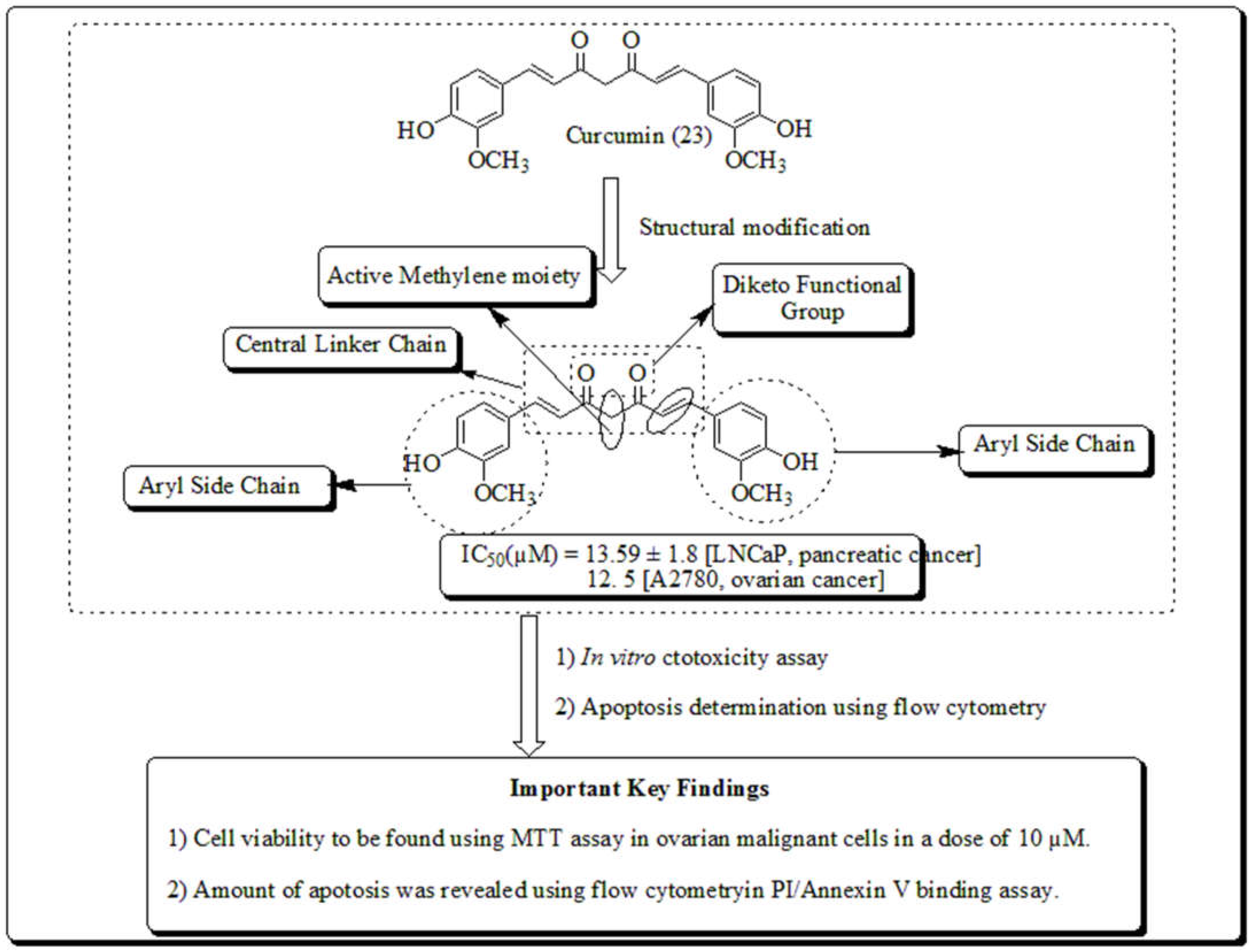
Figure 10.
Structure of curcuminoids of turmeric.
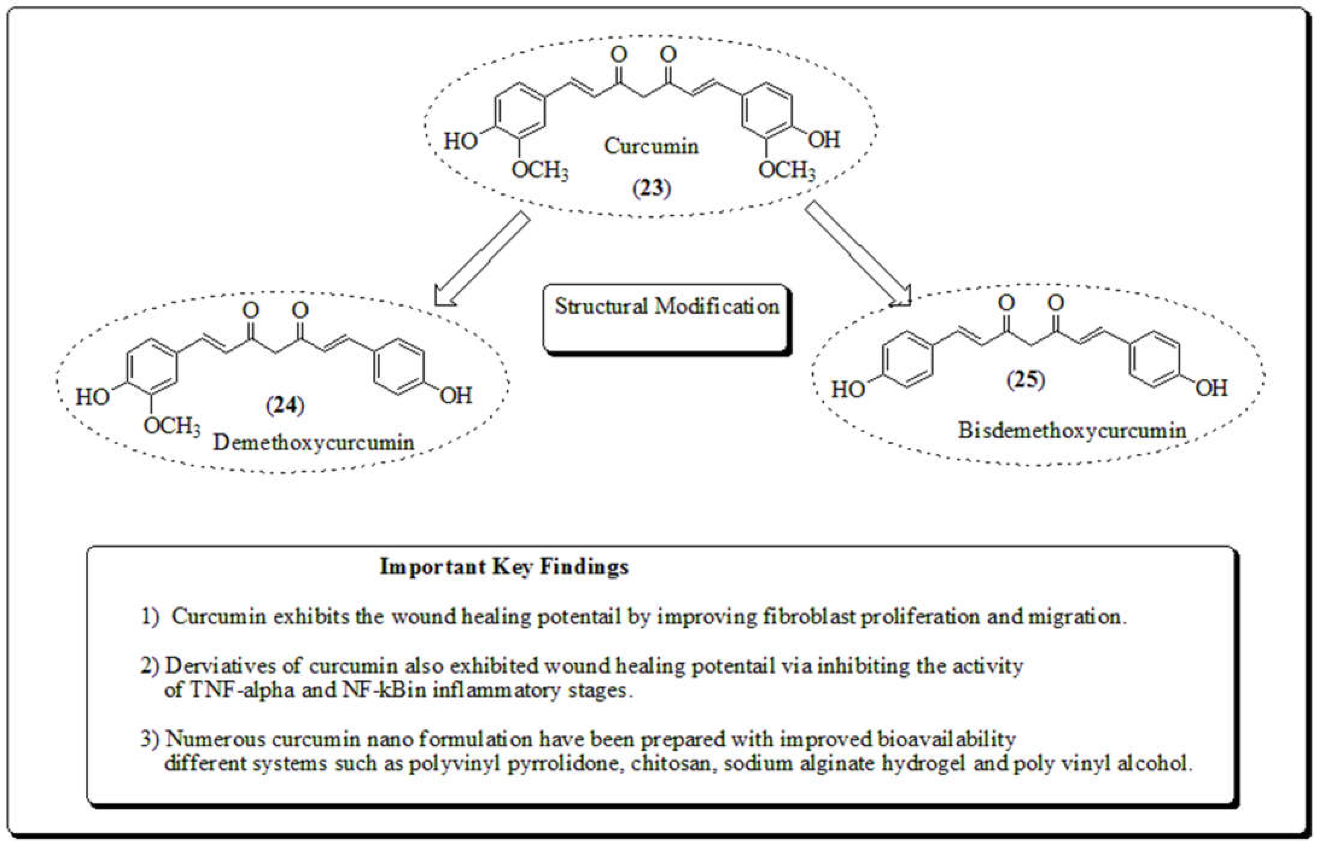
Figure 11.
(a). Structure of phytomolecules (26-35) obtained from Ehretia laevis and Ehretia microphylla. (b). Structure of promising phytomolecules (36-39) of Ehretia laevis and Ehretia microphylla.
Figure 11.
(a). Structure of phytomolecules (26-35) obtained from Ehretia laevis and Ehretia microphylla. (b). Structure of promising phytomolecules (36-39) of Ehretia laevis and Ehretia microphylla.
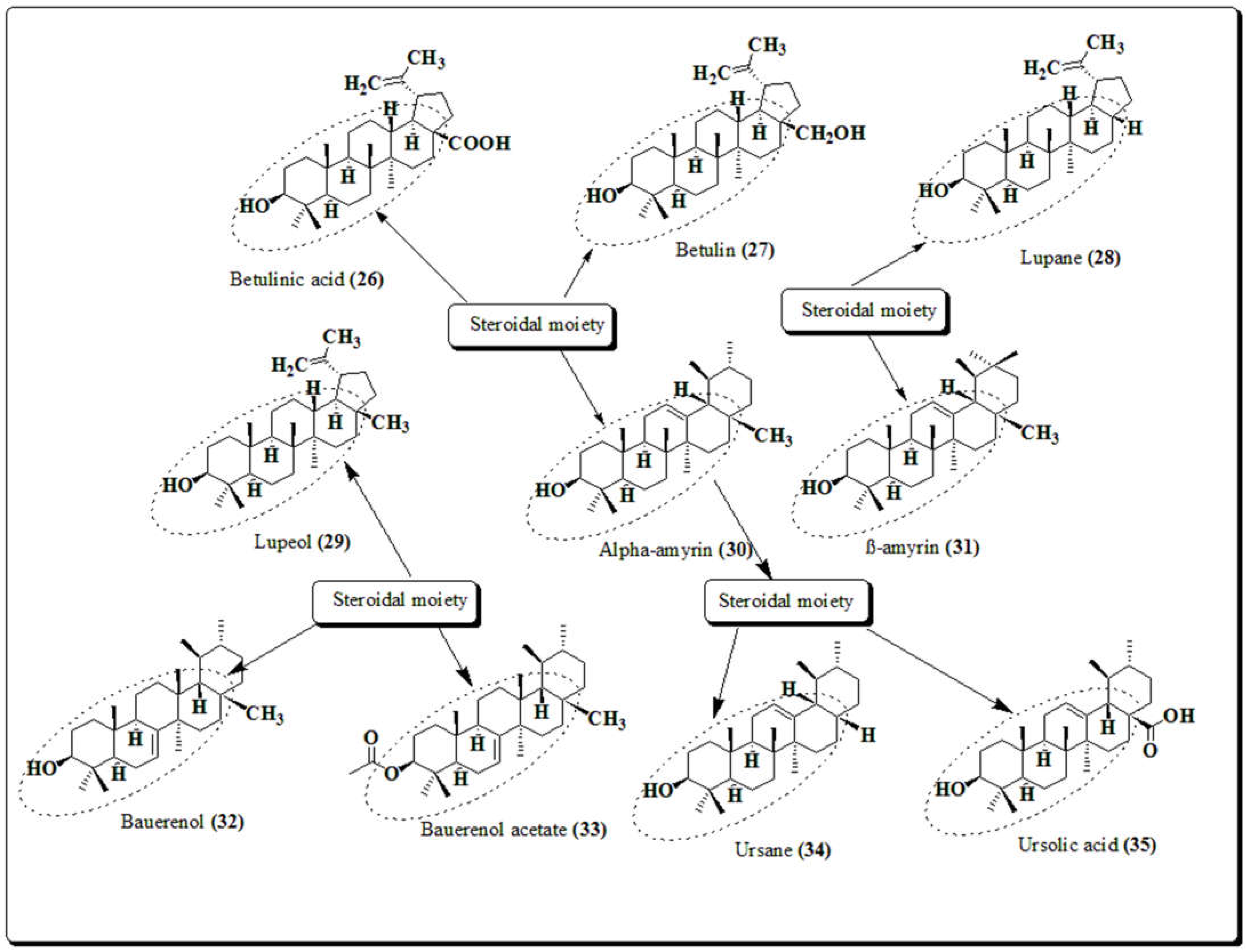
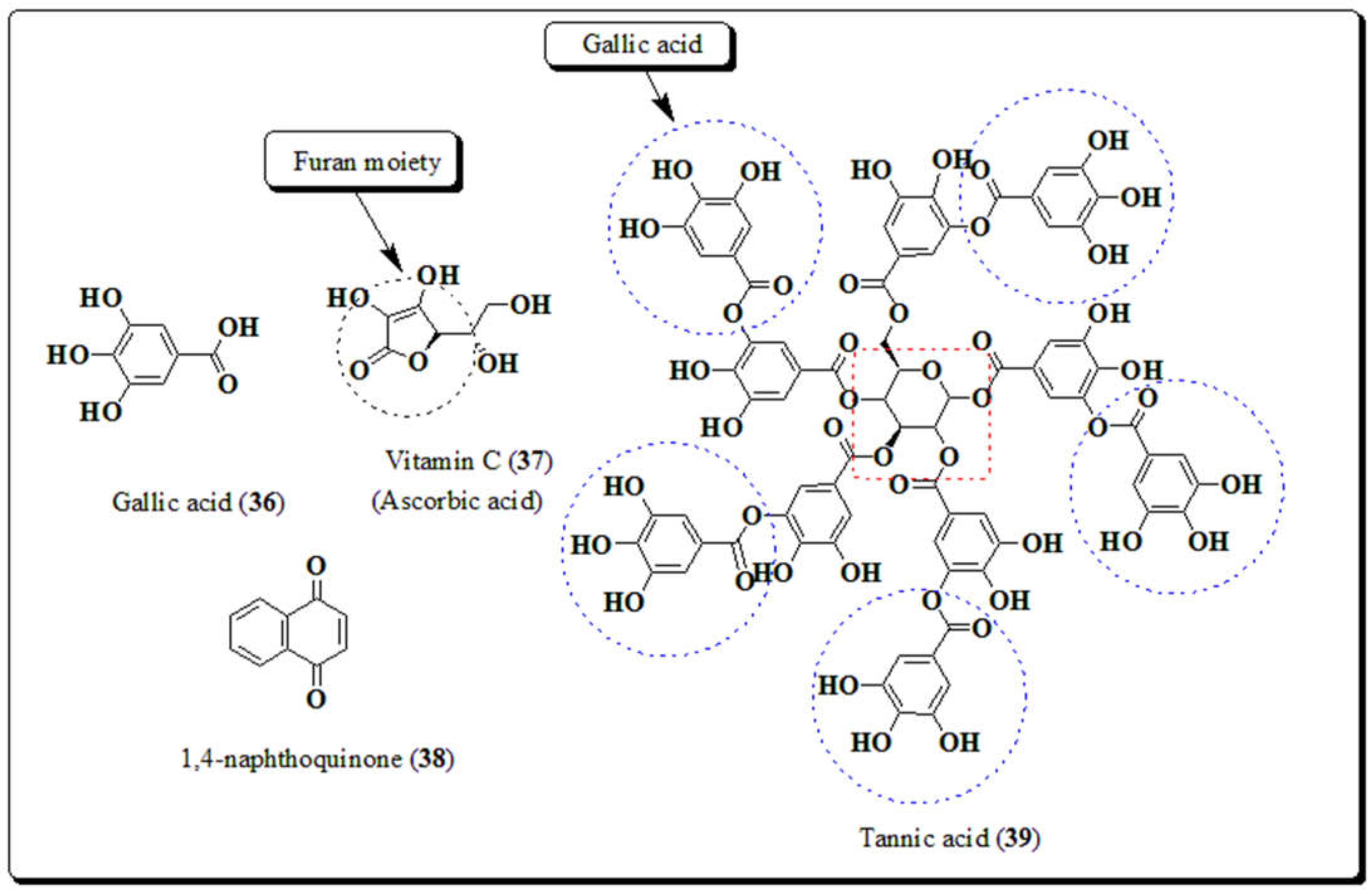
Figure 12.
Structures of compounds (40-42) along with important key findings.
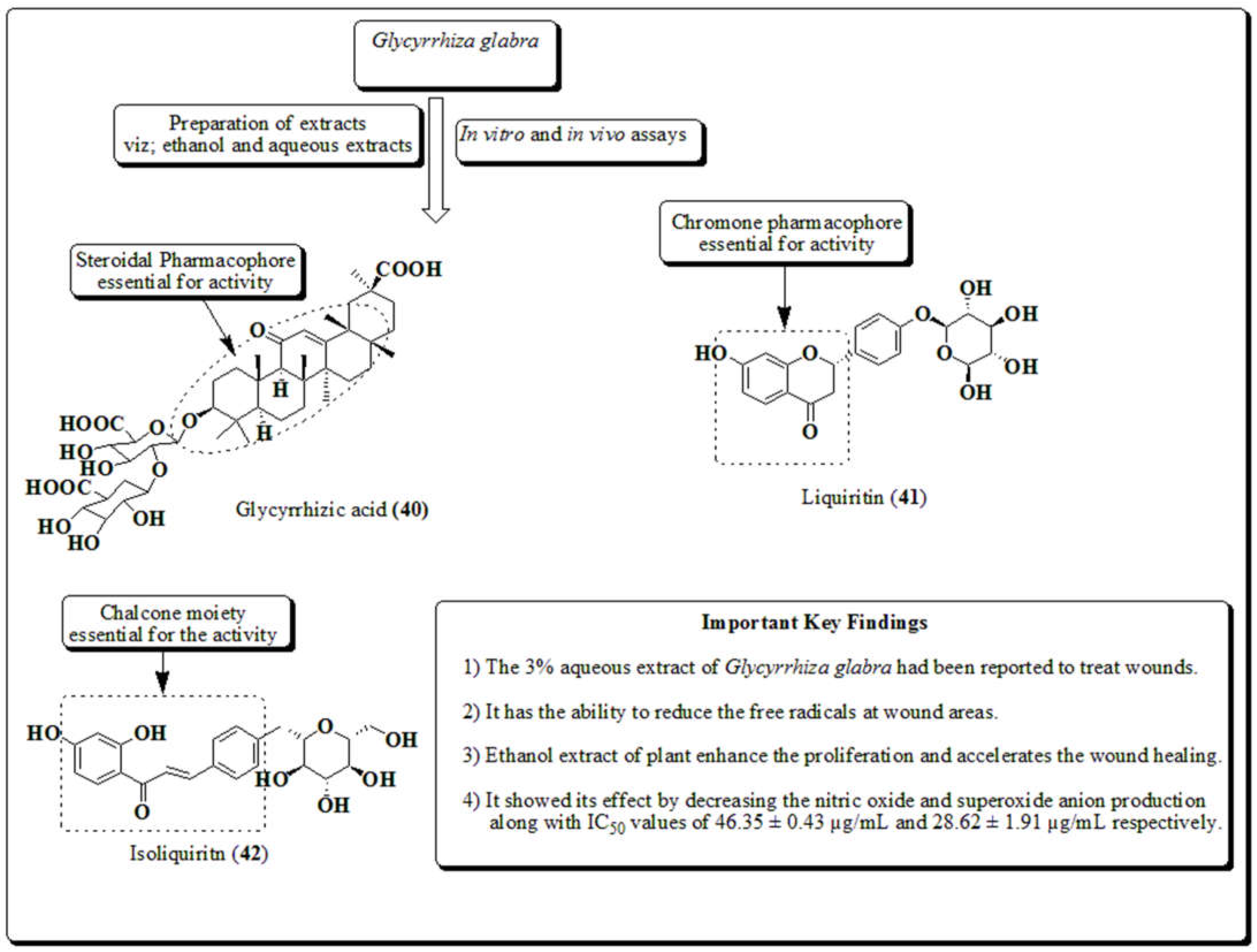
Figure 13.
Structures of compounds (43-51) obtained from Malva sylvestris along with important key findings.
Figure 13.
Structures of compounds (43-51) obtained from Malva sylvestris along with important key findings.
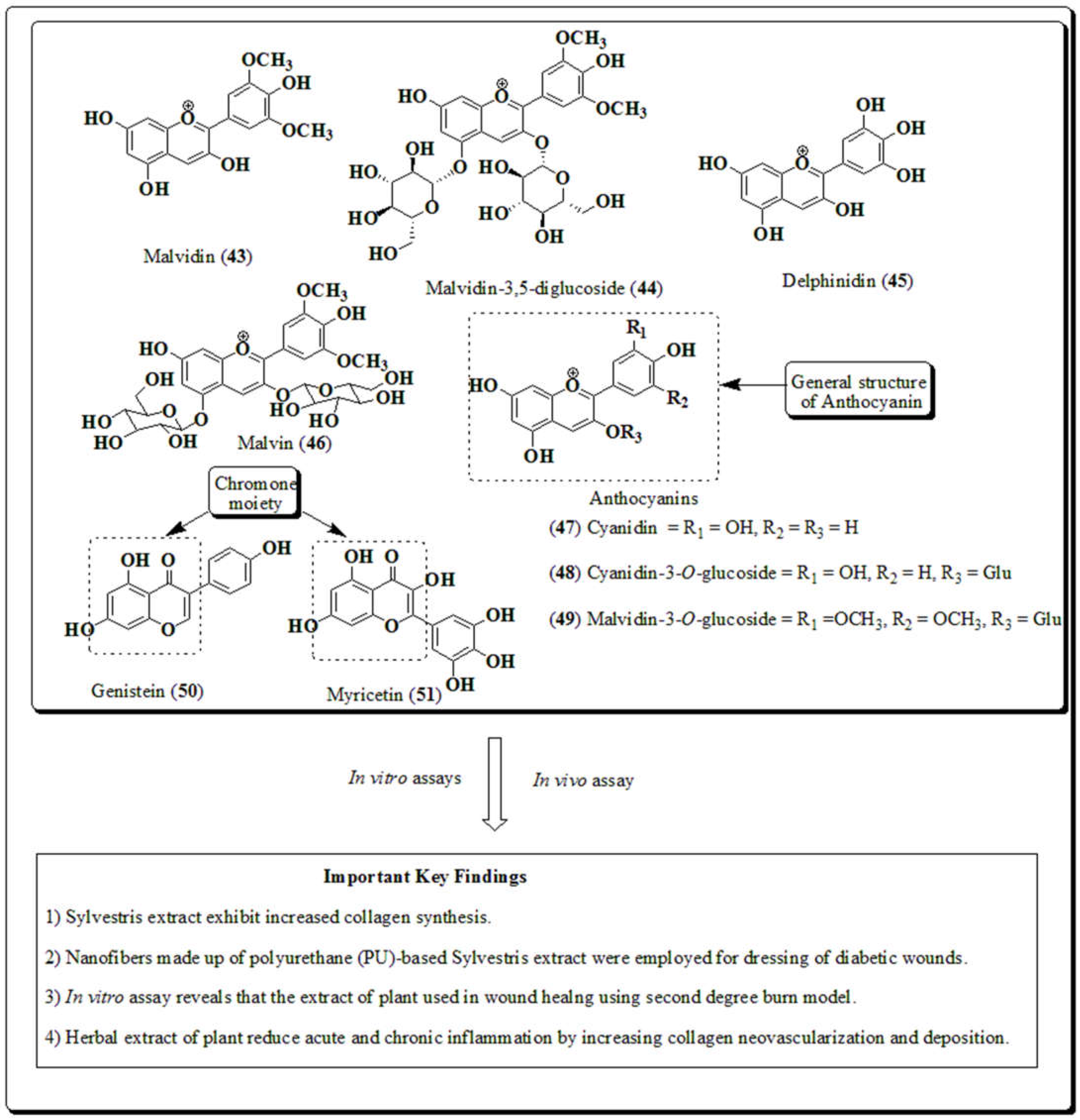
Figure 14.
Structures of compound of plant (52-57) along with important key findings.
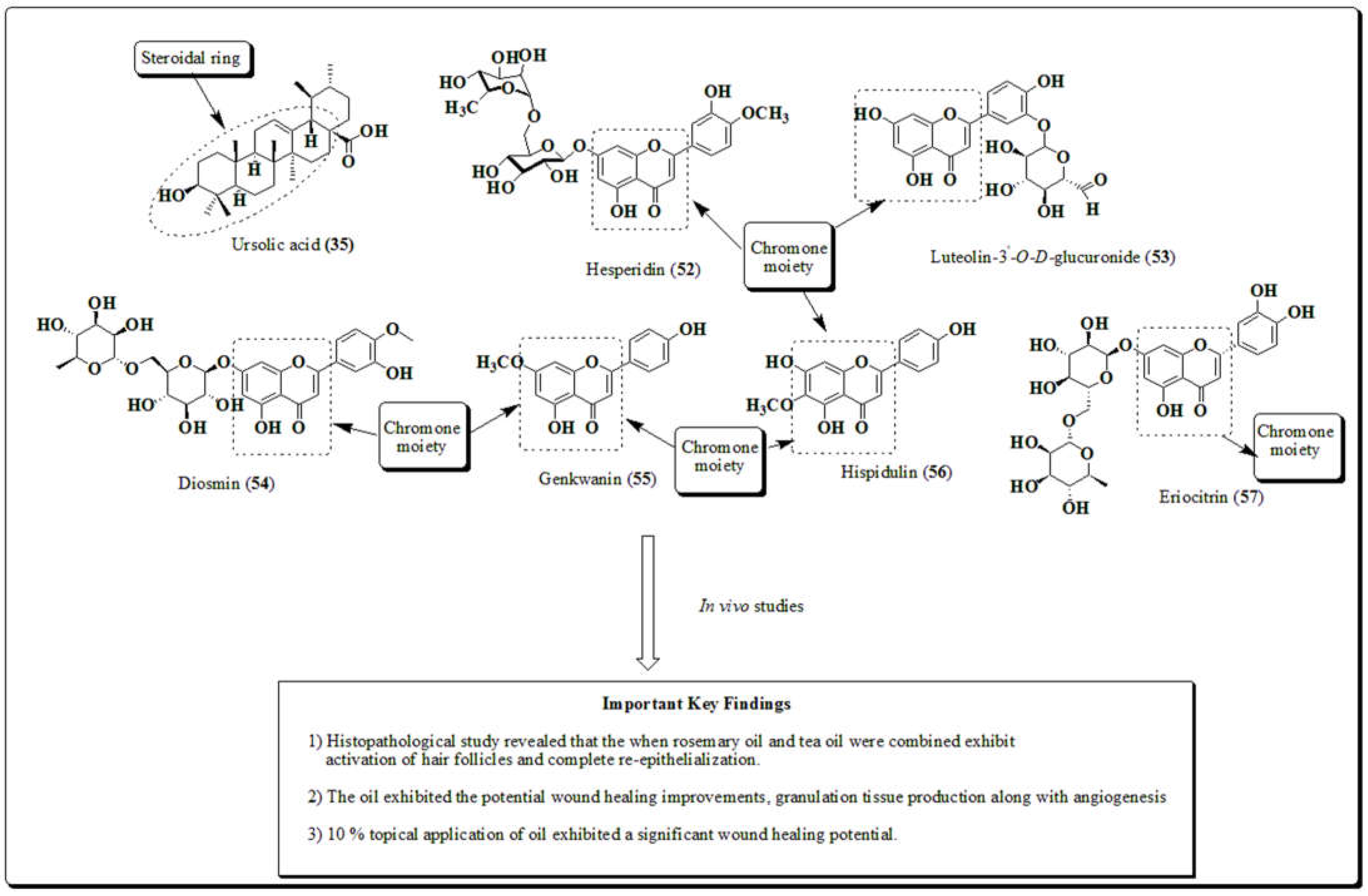
Figure 15.
Structures of compounds (58-62-60) along with important key findings .
Disclaimer/Publisher’s Note: The statements, opinions and data contained in all publications are solely those of the individual author(s) and contributor(s) and not of MDPI and/or the editor(s). MDPI and/or the editor(s) disclaim responsibility for any injury to people or property resulting from any ideas, methods, instructions or products referred to in the content. |
© 2023 by the authors. Licensee MDPI, Basel, Switzerland. This article is an open access article distributed under the terms and conditions of the Creative Commons Attribution (CC BY) license (http://creativecommons.org/licenses/by/4.0/).
Copyright: This open access article is published under a Creative Commons CC BY 4.0 license, which permit the free download, distribution, and reuse, provided that the author and preprint are cited in any reuse.


