
Submitted:
18 January 2023
Posted:
19 January 2023
You are already at the latest version
Abstract
A series of LaF3:Ce3+ phosphors for the application in photodynamic therapy is synthesized using a one-stage solvothermal synthesis. The conditions providing the maximum intensity of UV- and X-ray-excited luminescence, lowest size and highest colloidal stability of the phosphor nanoparticles are found to respectively include cerium content 5% mol., use of ethanol as the reaction medium for the solvothermal synthesis and addition of polyvinylpyrrolidone as a stabilizer in an optimized amount.
Keywords:
X-ray luminescence
; solvothermal synthesis
; LaF3:Ce
; Zeta-potential
; PEG
; PVP
; PEI
1. Introduction
Fluorides have many advantages as fluorescent raw materials due to their low phonon energy and high ionicity [1]. Compared to oxide luminescent materials, rare earth fluorides have optical clarity, low vibrational energy, minimal dopant ion excited state quenching, and are also less prone to staining due to the formation of hole centers under the action of an ionizing emitter [2]. Fluoride nanocrystals doped with rare earth ions are of great interest due to their potential applications in lighting and displays [3,4,5,6,7], boost converters [8,9], biological fluorescent labels [10,11], transparent glass [12], scintillators [13], optical amplifiers [14], solar cell amplification [15] and photodynamic therapy [16,17]. Recently, some researchers have devoted themselves to the development of LaF3 nanocrystals doped with rare earth ions due to their high photochemical stability, low toxicity and biocompatibility. In addition, their luminescent properties, including sharp absorption and emission lines and long lifetimes, are almost independent of particle size and can be tuned through doping with various lanthanide ions [18]. Ce3+ is a strong emitter with a nanosecond luminescence lifetime that is shorter than rare earth elements with 4f configurations due to the allowed 5d-4f optical transition, which is independent of crystal field state mixing due to its dipole nature. Although the ion–lattice interaction for the 5d configuration is higher than for the 4f configuration, nonradiative decay with multiphonon emission is impossible due to the large distance between the 5d band and the nearest 4f level [19,20,21]. LaF3, which has a large band gap (according to various sources: from 6.04 eV [22] to 10.1 eV [23]), is an ideal host for studying Ce3+ fluorescence for scintillators, since the 4f and 5d levels of cerium are located in the gap receiving grid. In addition, interactions between optically active Ce3+ ions can decrease when these ions are replaced by La3+ ions, which have very similar physical and chemical properties but are not optically active [24].
Agglomeration of LaF3 nanocrystals doped with lanthanide ions is very common; firstly, because of the reduced open surface in nanocrystals which reduces surface energy, and secondly, as a rule, nanocrystals that do not have a special coating have a surface charge closer to neutral, which leads to aggregation and flocculation of particles due to the action of van der Waals forces of attraction on them. This can lead to physical instability [25,26]. To increase biocompatibility, as well as to prevent agglomeration, the particles are coated with various stabilizers. The most widely used and studied stabilizer is polyethylene glycol (PEG). The process of applying a PEG coating to biomaterials to impart a latent characteristic is commonly referred to as PEGylation. It is now well known that PEGylation has many attractive properties; for example, it has been shown to increase the half-life of a drug in the body, prolonging its potency and thus reducing dosing frequency. The ability of PEG to prolong carrier circulation time is due mainly to its physical properties, which, in turn, can reduce or prevent protein absorption. It has been approved by the FDA for use in a variety of dosage forms. PEGylation remains the reference process for the development of biologically relevant systems with in vivo characteristics. However, the disadvantages of this stabilizer have also become known: a long elimination time, lack of biodegradability, formation of undesirable by-products, and mechanical degradation [27]. Alternatively, polyethyleneimine and polyvinylpyrrolidone have also been successfully used as stabilizers.
This study is related to the improvement of the parameters of the process used for the synthesis of LaF3 nanocrystals by the solvothermal method. The influence of the reaction medium and stabilizers on the structural, morphological and surface properties were studied using appropriate mathematical and analytical methods. After that, Ce3+ ions of the optimal concentration were doped into the optimal host nanomaterial, and the optical characteristics for medical applications were studied.
2. Materials and Methods
A series of fluoride phosphors with the composition LaF3:Ce3+ 4...6%mol. was synthesized. A typical synthesis [28] was carried out in ethylene glycol or ethanol. Water solution of cerium and lanthanum chlorides (8 mmol) was well mixed and added to ethylene glycol. After adding stabilizer polyethylene glycol Mw = 20000 (PEG-20000), the mixture was vigorously stirred on a magnetic stirrer at room temperature. Ammonium fluoride NH4F (12 ml of 110 mmol solution) was dissolved in ethylene glycol or water, and added to the above solution, and again stirred vigorously at room temperature for 30 minutes. Finally, the mixture was transferred to a Teflon liner, placed in a sealed stainless steel autoclave, where it was subjected to a solvothermal treatment at 200°C for 4 hours, and cooled naturally to room temperature. The precipitate was washed in ethanol and deionized water several times, and the final products were dried at 40°C for 6 hours in a normal atmosphere. To determine the most suitable stabilizer that will prevent particle coalescence and ensure biocompatibility, we are implementing a one-stage hydrothermal synthesis of LaF3:Ce3+ nanocrystals with various organic surfactants, in particular, polyethylene glycol (PEG-200, PEG-2000, PEG-20.000), polyethyleneimine (PEI Mw 60,000-80,000), polyvinylpyrrolidone (PVP Mw 1,300,000) and investigating the luminescent and surface properties of nanoparticles. All samples have a cerium concentration of 5%mol.
The structural characteristics of the final products were investigated by powder X-ray diffraction (XRD) using Cu-Ka radiation (l = 0.15405 nm) on a Rigaku-RINT2200 diffractometer. The morphology and size of the obtained samples were observed using field emission scanning electron microscope (FE-SEM, JSM-7001F, JEOL). The emission spectra of ultraviolet and visible photoluminescence were recorded using a laboratory setup - a spectrofluorometer with two monochromators (model of an ultramonochromatic light source) equipped with a xenon lamp as an exciter. The obtained spectra were analyzed by deconvolution into Gaussian bands. The X-ray luminescence spectra were measured on a laboratory setup with a copper anode at a voltage of 80 kV and a current of 62 mA. The decay time was measured with a Hamamatsu compact fluorescence lifetime spectrometer 11367 (TR 50 ns, frequency 10000 MH). FTIR spectrum is recorded between 4000 and 400 cm-1. Dried samples were measured with FT/IR-6300, JASCO. Surface charge and size distribution of the samples were measured with Zetasizer Ultra and analyzed with ZS Xplorer software by Malvern Pnalytical. All measurements were carried out at room temperature.
3. Results and Discussion
To determine the optimal concentration of cerium, a series of syntheses of samples with a concentration of 4...6 mol% was carried out. Figure 1 shows the XRD patterns of the obtained samples. The structure of the obtained LaF3:Ce3+ nanoparticles is in good agreement with the hexagonal structure of bulk LaF3 (card 72-1435). The diffraction peaks for all samples are broadened, which indicates the nanocrystalline nature of the samples. The average crystallite sizes were estimated from the Scherrer equation [29],
where k = 1, λ = 0,154184 nm represents the wavelength of Cu K radiation, Θ is the angle of the X-ray diffraction peak, and ß represents the corrected half width of the diffraction peak. Taking the eight main peaks (002, 110, 111, 112, 300, 113, 220, 221, 223), the average crystallite sizes of LaF3:4%Ce3+, LaF3:5%Ce3+ and LaF3:5%Ce3+ are about 25.1, 31.9 and 33.1 nm, respectively. Figure 1-b shows the peak (111) at 2Θ = 27.6°, it can be seen that with an increase in the concentration of cerium, the peak will shift towards lower degrees, which indicates an increase in the amount of the cerium fluoride phase.
Dhkl = kλ/ßcosΘ
Figure 2 shows the Photo and X-ray luminescence spectra of LaF3:Ce phosphors with different activator concentrations. Photoluminescence was measured at room temperature with an excitation wave of 255 nm (Figure 2(a)).
Broadband radiation in the region from 270 to 400 nm and peaking at 300 nm can be attributed to the 5d–4f transition of Ce3+. With an increase in the doping concentration of Ce3+, the radiation intensity gradually increases, reaching a maximum at a concentration of 5%mol., and then decreases. The decrease in the radiation intensity is related to the concentration effect of quenching at a higher doping concentration of Ce3+. A similar behavior is also observed upon X-ray excitation (Figure2(b)).
It is known that the luminescence spectra of trivalent lanthanide ions in crystals are mainly due to two types of electronic transitions: the 4f–4f transition and the 5d–4f transition. The excited electronic configuration of Ce3+ has the form 5d1 and it is not shielded from the surroundings. The 5d electron interacts strongly with neighboring anion ligands in compounds, which leads to broadband emission. The 4f orbital is shielded from the environment by the filled 5s2 and 5p6 orbitals. Therefore, the effect of the main lattice on optical transitions within the 4fn configuration is small [30].
It is important to use the correct technique to synthesize nanoparticles for a medical application because each nanoparticle must have the appropriate morphology for the desired application. It is known that toxicity increases from spherical to acicular particles [31]. Therefore, it is very important to synthesize nanosized particles, and also, if possible, to produce their ideal shape. We thought that solvothermal methods would control the morphology to an appropriate degree. When conducting a solvothermal reaction, many factors must be taken into account, such as the concentrations of the reactants, the solubility of the reactants, the reaction temperature, the reaction time, the choice of solvent, and the pressure, all of which can be varied. In our previous papers, we have already discussed the optimal parameters for the synthesis of yttrium fluoride [32]. In this article, we paid special attention to the solvent used as the reaction medium. In this case, we used ethanol and ethylene glycol (EG).
The solvothermal reaction was carried out at 200°C for 4 h using PEG-20000 as a stabilizer and with cerium concentration 5%mol. As a result of synthesis using these reaction media, pure hexagonal lanthanum fluoride was obtained (Figure 3). Thus, it can be concluded that the synthesis medium does not affect the formation of the phase. This statement was also confirmed to us in the work [32].
Figure 4 and Figure 5 show SEM images and size distribution histograms calculated from the acquired images, respectively.
SEM images (Figure4) display that the obtained particles are oval in shape and do not exceed nanosize. These samples are also histograms in Figure 5, which were read from the acquired images using the software. It should be noted that the synthesis medium has practically no effect on the particle morphology, however, a narrower particle size distribution was obtained as a result of synthesis in an ethanol medium. Also, in this medium, as calculations show, it is possible to obtain an average particle size of about 30 nm, while in an ethylene glycol (EG) medium the average size is about 45 nm. Thus, ethanol was chosen as the reaction medium for the solvothermal synthesis of lanthanum fluoride.
Reducing the particle size increases the number of surface atoms and the surface energy of the resulting particles, so these nanoparticles must be stabilized with suitable compounds [33]. The stabilization of nanoparticles is carried out before their actual use in any technological applications by modifying their surface with appropriate stabilizers. This surface modification with stabilizers (also called capping ligands) is critical to achieving sufficient repulsive forces between particles that prevent particle aggregation and help obtain a stable suspension of particles. A special polymer coating allows for higher biocompatibility, prevents agglomeration, and also makes it possible to obtain a stable colloidal nanosuspension. We synthesized a series of samples with various stabilizers: polyethylene glycol (PEG) with Mw 200 and 2000, polyvinylpyrrolidone (PVP) and polyethyleneimine (PEI). At present, the control of the morphology and size of fluoride nanoparticles during their preparation is an important factor that needs to be addressed for their use in medicine.
The functional groups of the powders were Identified using FT-IR as shown in Figure 6. A broad absorption band around 3500-3400 cm-1 refers to stretching vibrations (-OH). In the spectrum of PEG200 and PEG2000, characteristic bands of certain functional groups were found, such as a band at 2874.2 cm–1 belonging to the –CH group [34] and a band at 1103.9 cm–1 belonging to the –C–O–C group [35]. These two characteristic bands and some other bands near 1103.9 cm-1 also appear in the PEG- LaF3:Ce spectrum, indicating successful conjugation of PEG to the LaF3:Ce surface. The spectrum of PEI shows weak peaks in the 1170–1050 cm−1 range that correspond to C-C, C-O-C and C-O (in alcohols) stretching vibrations (associated with carbo-hydrate ring), and C-N stretching. The second sharp peak is observed in the wavenumber range 1632–1622 cm−1; this large band may be assigned to C=O asymmetric stretching in (COO−), (-N-H) bending vibration (for primary and secondary amines) and/or open-chain imino groups (-C=N-). In addition, stretching vibrations of -CH bonds are appearing in the region 2980–2850 cm−1 confirming the expected presence of linear aliphatic chains. The spectrum of PVP has characteristic peaks C=O (1660 cm-1) and C-N (1290 cm-1), which confirms the functionalization of PVP lanthanum fluoride nanoparticles through intermolecular hydrogen bonding.
Particles with zeta potential of ±30mV have an optimal surface charge, which is acceptable for biocompatibility [36]. Zeta potential values are typically in the range of +100 to −100 mV. The high value of the zeta potential of nanocrystals indicates a good physical stability of nanosuspensions due to the electrostatic repulsion of individual particles. A zeta potential value other than -30 mV to +30 mV is generally considered to have sufficient repulsive force to achieve better physical colloidal stability. On the other hand, a low zeta potential can lead to aggregation and flocculation of particles due to attractive Van der Waals forces on them, which can lead to physical instability [37].
As can be seen from Figure 7, PEG-coated particles have an extremely positive charge, which can be as high as 70 mV. In the case of PEI, the particles have both positive and negative charges, which directly leads to a high probability of agglomeration. PVP has positively charged particles, most of which have a charge of about 40 mV, which is close to the optimum particle charge to achieve colloidal stability. Therefore, PVP was chosen as a suitable stabilizer.
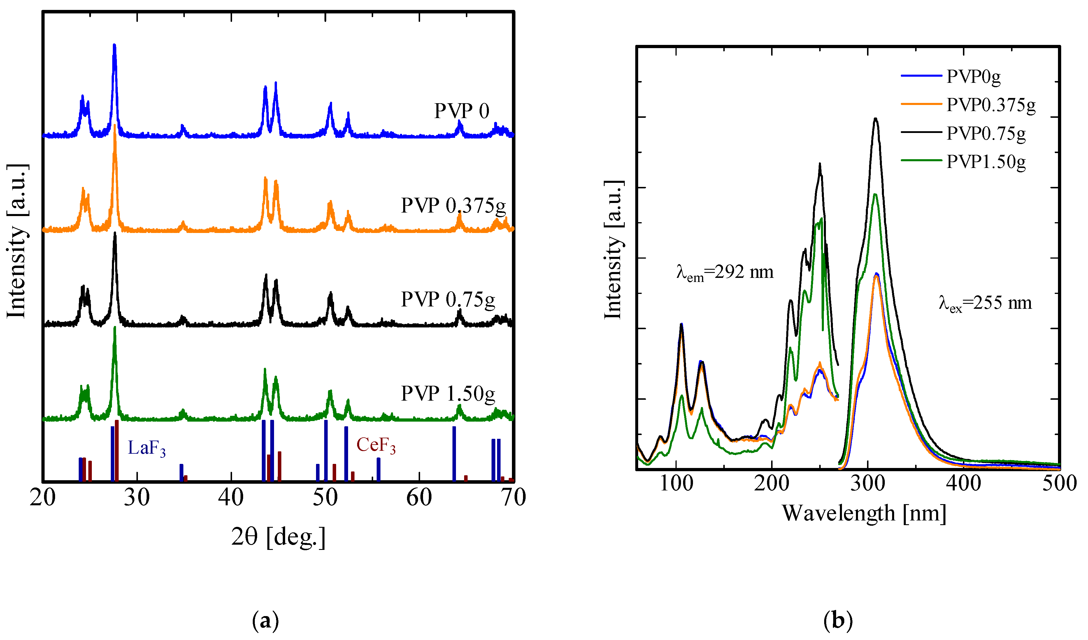
After determining the optimal surfactant, an experiment was conducted to determine the optimal amount of PVP. Four samples were synthesized with different PVP amount: 0 g, 0.375 g, 0.75 g and 1.5 g. Figure 8(a) shows the diffraction patterns of the obtained samples. As noted earlier, the presence of surfactants does not affect the phase composition. Therefore, it is possible to observe hexagonal lanthanum and cerium fluoride without impurity phases.
Figure 8(b) shows the excitation and luminescence spectra of the obtained samples. Peaks located in the region of 90-150 nm are related to excitation of the phosphor host – lanthanum fluoride. As can be seen from the excitation spectra, the main peaks, in contrast to the emission spectra, are bifurcated. We have a hypothesis to explain this phenomenon – that the particles of these samples are nanosized. Similar results have already been observed in [38]. The emission spectra of the obtained samples demonstrate the same pattern as for the samples obtained earlier. The sample with a PVP content of 0.75 g has the highest intensity.
4. Conclusions
LaF3 nanoparticles doped with cerium (Ce3+) at a concentration of 4...6 mol.% were synthesized by the solvothermal method in various media: ethanol and ethylene glycol. Ethanol was chosen as the optimal reaction medium for the synthesis, and the sample with a cerium concentration of 5 mol % exhibits the highest intensity upon UV and X-ray excitation. In this synthesis procedure, polyethylene glycol, polyvinylpyrrolidone, and polyethyleneimine were used as stabilizers that bind to the surface of the particle, limiting the growth of the nanoparticles. The average particle size of the resulting nanoparticles is 30 nm, and these particles can be dispersed in water to form a colloidal solution. Due to their low toxicity, water-soluble luminescent LaF3:Ce nanoparticles can be used in future biological experiments.
Author Contributions
Conceptualization, Dorokhina A.M., Kominami H. and Sychov M.M.; methodology, Kominami H., Bakhmetyev V.V. and Dorokhina A.M.; investigation, Dorokhina A.M., Ishihara R., Kominami H..; resources, Kominami H.., Aoki T., Morii H.; writing—original draft preparation, Dorokhina A.M.; writing—review and editing, Kominami H. All authors have read and agreed to the published version of the manuscript.
Acknowledgments
This work was partly supported by a MEXT scholarship.
Conflicts of Interest
The authors declare no conflict of interest.
References
- Yan, R.X.; Li, Y.D. Down/Up Conversion in Ln3+-Doped YF3 Nanocrystals. Adv. Funct. Mater. 2005, 15, 763. [Google Scholar] [CrossRef]
- Tan, M.C.; Al-Baroudi, L.; Riman, R.E. Surfactant effects on efficiency enhancement of infrared-to-visible upconversion emissions of NaYF4:Yb-Er. Appl. Mater. Interfaces 2011, 3, 3910–5. [Google Scholar] [CrossRef]
- Wang, Z.L.; Chan, H.L.W.; Li, H.L.; Hao, J.H. Highly efficient low-voltage cathodoluminescence of LaF3:Ln3+ (Ln=Eu3+,Ce3+,Tb3+) spherical particles. Appl. Phys. Lett. 2008, 94, 141106. [Google Scholar] [CrossRef]
- Jia, P.Y.; Lin, J.; Yu, M. Sol–gel deposition and luminescence properties of LiYF4:Tb3+ thin films. Journal of Luminescence 2007, 122–123, 134–136. [Google Scholar] [CrossRef]
- Tuyet, D.T.; Quan, V.T.H.; Bondzior, B.; Dereń, P.J.; Velpula, R.T.; Nguyen, H.P.T.; Tuyen, L.A.; Hung, N.Q.; Nguyen, H.-D. Deep red fluoride dots-in-nanoparticles for high color quality micro white light-emitting diodes. Opt. Express 2020, 28, 26189–26199. [Google Scholar] [CrossRef]
- Kemnitz, E.; Mahn, S.; Krahl, T. Nano metal fluorides: small particles with great properties. ChemTexts 2020, 6, 19. [Google Scholar] [CrossRef]
- Quan, Z.; Yang, D.; Yang, P.; Zhang, X.; Lian, H.; Liu, X.; Lin, J. Uniform Colloidal Alkaline Earth Metal Fluoride Nanocrystals: Nonhydrolytic Synthesis and Luminescence Properties. Inorg. Chem. 2008, 47, 20–9509. [Google Scholar] [CrossRef]
- Heer, S.K.; Kцmpe, *!!! REPLACE !!!*; H.-U., Gьdel; Haase, M. Highly Efficient Multicolour Upconversion Emission in Transparent Colloids of Lanthanide-Doped NaYF4 Nanocrystals. Adv. Mater. 2004, 16, 2102. [Google Scholar] [CrossRef]
- Schietinger, S.; Menezes, L.S.; Lauritzen, B.; Benson, O. Observation of Size Dependence in Multicolor Upconversion in Single Yb3+, Er3+ Codoped NaYF4 Nanocrystals. Nano Lett. 2009, 9, 2477–2481. [Google Scholar] [CrossRef]
- Yi, G.S.; Lu, H.C.; Zhao, S.Y.; Ge, Y.; Yang, W.J.; Chen, D.P.; Guo, L.H. Synthesis, Characterization, and Biological Application of Size-Controlled Nanocrystalline NaYF4:Yb,Er Infrared-to-Visible Up-Conversion Phosphors. Nano Lett. 2004, 4, 2191–2196. [Google Scholar] [CrossRef]
- Nam, S.H.; Bae, Y.M.; Park, Y.I.; Kim, J.H.; Kim, H.M.; Choi, J.S.; Lee, K.T.; Hyeon, T.; Suh, Y.D. Long-term real-time tracking of lanthanide ion doped upconverting nanoparticles in living cells. Angew Chem Int Ed Engl. 2011, 50, 6093–7. [Google Scholar] [CrossRef]
- Zhou, S.F.; Jiang, N.; Miura, K.; Tanabe, S.; Shimizu, M.; Sakakura, M.; Shimotsuma, Y.; Nishi, M.; Qiu, J.R.; Hirao, K. Simultaneous Tailoring of Phase Evolution and Dopant Distribution in the Glassy Phase for Controllable Luminescence. J. Am. Chem. Soc. 2010, 132, 17945–17952. [Google Scholar] [CrossRef]
- Wang, Z.L.; Quan, Z.W.; Jia, P.Y.; Lin, C.K.; Luo, Y.; Chen, Y.; Fang, J.; Zhou, W.; O’Connor, C.J.; Lin, J. Cerium (III) Fluoride Thin Films by XPS. Chem. Mater. 2006, 18, 2030. [Google Scholar] [CrossRef]
- Sivakumar, S.; van Veggel, F.C.J.M.; Raudsepp, M. Bright White Light through Up-Conversion of a Single NIR Source from Sol−Gel-Derived Thin Film Made with Ln3+-Doped LaF3 Nanoparticles. J. Am. Chem. Soc 2005, 127, 12464–12465. [Google Scholar] [CrossRef] [PubMed]
- Liu, S.-M.; Chen, W.; W, Z.-G. Luminescence Nanocrystals for Solar Cell Enhancement. Journal of Nanoscience and Nanotechnology 2010, 10, 1418–1429. [Google Scholar] [CrossRef]
- Liu, Y.F.; Chen, W.; Wang, S.P.; Joly, A.G. Investigation of water-soluble x-ray luminescence nanoparticles for photodynamic activation. Appl. Phys. Lett. 2008, 92, 043901. [Google Scholar] [CrossRef]
- Chen, W. Nanoparticle Self-Lighting Photodynamic Therapy for Cancer Treatment. Journal of Biomedical Nanotechnology 2008, 4, 369–376. [Google Scholar] [CrossRef]
- Wang, F.; Han, Y.; Lim, C.S.; Lu, Y.; Wang, J.; Xu, J.; Chen, H.; Zhang, C.; Hong, M.; Liu, X. Simultaneous phase and size control of upconversion nanocrystals through lanthanide doping. Nature. 2010, 463, 1061–5. [Google Scholar] [CrossRef] [PubMed]
- Moses, W.W.; Derenzo, S.E.; Weber, M.J.; Ray-Chaudhuri, A.K.; Cerrina, F. Scintillation mechanisms in cerium fluoride. Journal of Luminescence 1994, 59, 89–100. [Google Scholar] [CrossRef]
- Yao, M.; Joly, A.G.; Chen, W. Formation and Luminescence Phenomena of LaF3:Ce3+ Nanoparticles and Lanthanide−Organic Compounds in Dimethyl Sulfoxide. J.Phys. Chem. C 2010 2010, 114, 2–826. [Google Scholar] [CrossRef]
- Birowosuto, M.D.; Dorenbos, P.; van Eijk, C.W.E.; Krmer, K.W.; Gudel, H.U. Scintillation properties and anomalous Ce3+ emission of Cs2NaREBr6:Ce3+ (RE = La,Y,Lu). J. Phys. : Condens. Matter 2006, 18, 6133. [Google Scholar] [CrossRef]
- Petousis, I.; Mrdjenovich, D.; Ballouz, E. High-throughput screening of inorganic compounds for the discovery of novel dielectric and optical materials. Sci Data 2017, 4, 160134. [Google Scholar] [CrossRef]
- Tabatabaee, F.; Sabbagh Alvani, A.A.; Sameie, H. Ce3+-doped LaF3 nanoparticles: Wet-chemical synthesis and photo-physical characteristics “optical properties of LaF3:Ce nanomaterials. Met. Mater. Int. 2014, 20, 169–176. [Google Scholar] [CrossRef]
- Canning, A.; Chaudhry, A.; Boutchko, R.; Jensen, N.G. First-principles study of luminescence in Ce-doped inorganic scintillators. Phys. Rev. 2011, 83, 125115. [Google Scholar] [CrossRef]
- Freitas, C.; Müller, R.H. Spray-drying of solid lipid nanoparticles (SLNTM). European Journal of Pharmaceutics and Biopharmaceutics 1998, 46, 145–151. [Google Scholar] [CrossRef]
- Shah, S.; McRae, A.F.; Marioni, R.E.; Harris, S.E.; Gibson, J.; Henders, A.K.; Redmond, P.; Cox, S.R.; Pattie, A.; Corley, J.; Murphy, L.; Martin, N.G.; Montgomery, G.W.; Starr, J.M.; Wray, N.R.; Deary, I.J.; Visscher, P.M. Genetic and environmental exposures constrain epigenetic drift over the human life course. Genome Res. 2014, 24, 1725–33. [Google Scholar] [CrossRef]
- Zhang, F.; Liu, M.-R.; Wan, H.-T. Discussion about Several Potential Drawbacks of PEGylated Therapeutic Proteins. Biological and Pharmaceutical Bulletin, 2014, 37, 335–339. [Google Scholar] [CrossRef] [PubMed]
- Sudheendra, L.; Das, G.K.; Li, C.; Stark, D.; Cena, J.; Cherry, S.; Kennedy, I.M. NaGdF4:Eu3+ Nanoparticles for Enhanced X-ray Excited Optical Imaging. Chem. Mater. 2014, 26, 1881–1888. [Google Scholar] [CrossRef] [PubMed]
- Vinila, V.S.; Isac, J. Design, Fabrication, and Characterization of Multifunctional Nanomaterials; Elsevier, 2022. [Google Scholar]
- Guo, H.; Zhang, T.; Qia, Y.M.; Zhao, L.H.; Li, Z.Q. Ionic Liquid-Based Approach to Monodisperse Luminescent LaF3:Ce,Tb Nanodiskettes: Synthesis, Structural and Photoluminescent Properties. Journal of Nanoscience and Nanotechnology 2010, 10, 1913–1919. [Google Scholar] [CrossRef]
- Nakashima, K.; Ueno, S.; Wada, S. Solvothermal synthesis of KNbO3 nanocubes using various organic solvents. Journal of the Ceramic Society of Japan 2014, 122, 547–551. [Google Scholar] [CrossRef]
- Dorokhina, A.M. Study of the properties of Ce3+-doped fluoride nanophosphors: phase composition, morphology, luminescence. J. Phys.: Conf. Ser. 2021, 2056, 012048. [Google Scholar] [CrossRef]
- Chaudhuri, R.G.; Paria, S. Core/Shell Nanoparticles: Classes, Properties, Synthesis Mechanisms, Characterization, and Applications. Chem. Rev. 2012, 112, 2373–2433. [Google Scholar] [CrossRef] [PubMed]
- Mu, H.; Wang, X.; Lin, P.H.; Yao, Q.; Chen, C. Chlorotyrosine promotes human aortic smooth muscle cell migration through increasing superoxide anion production and ERK1/2 activation. Atherosclerosis. 2008, 201, 67–75. [Google Scholar] [CrossRef] [PubMed]
- Petersen, H.; Fechner, P.M.; Martin, A.L. , et al. Polyethylenimine-graftpoly(ethylene glycol) copolymers: influence of copolymer block structure on DNA complexation and biological activities as gene delivery system. Bioconjug Chem. 2002, 13, 845–854. [Google Scholar] [CrossRef]
- Samimi, S.; Maghsoudnia, N.; Eftekhari, R.B.; Dorkoosh, F. Chapter 3 - Lipid-Based Nanoparticles for Drug Delivery Systems, Editor(s): Shyam S. Mohapatra, Shivendu Ranjan, Nandita Dasgupta, Raghvendra Kumar Mishra, Sabu Thomas, In Micro and Nano Technologies, Characterization and Biology of Nanomaterials for Drug Delivery; Elsevier; pp. 47–76.
- Joseph, E.; Singhvi, G. Chapter 4 - Multifunctional nanocrystals for cancer therapy: a potential nanocarrier, Editor(s): Alexandru Mihai Grumezescu, Nanomaterials for Drug Delivery and Therapy; William Andrew Publishing, 2019; pp. 91–116. [Google Scholar]
- Chen, Y.-P.; Luo, D.-L.; Xu, Q.-Y.; Yang, S.-L.; Tang, T.; Wang, X.-Y. 无机材料学报Structure and Fluorescence Quenching of Li2O-MgO-Al2O3-SiO2 Glasses Doped with Trivalent Cerium. 2014, 29, 967–971. [Google Scholar]
Figure 1.
XRD patterns of LaF3:Ce: (a) – with different Ce3+ concentration; (b) – XRD peak (111) at 27.6°.
Figure 1.
XRD patterns of LaF3:Ce: (a) – with different Ce3+ concentration; (b) – XRD peak (111) at 27.6°.
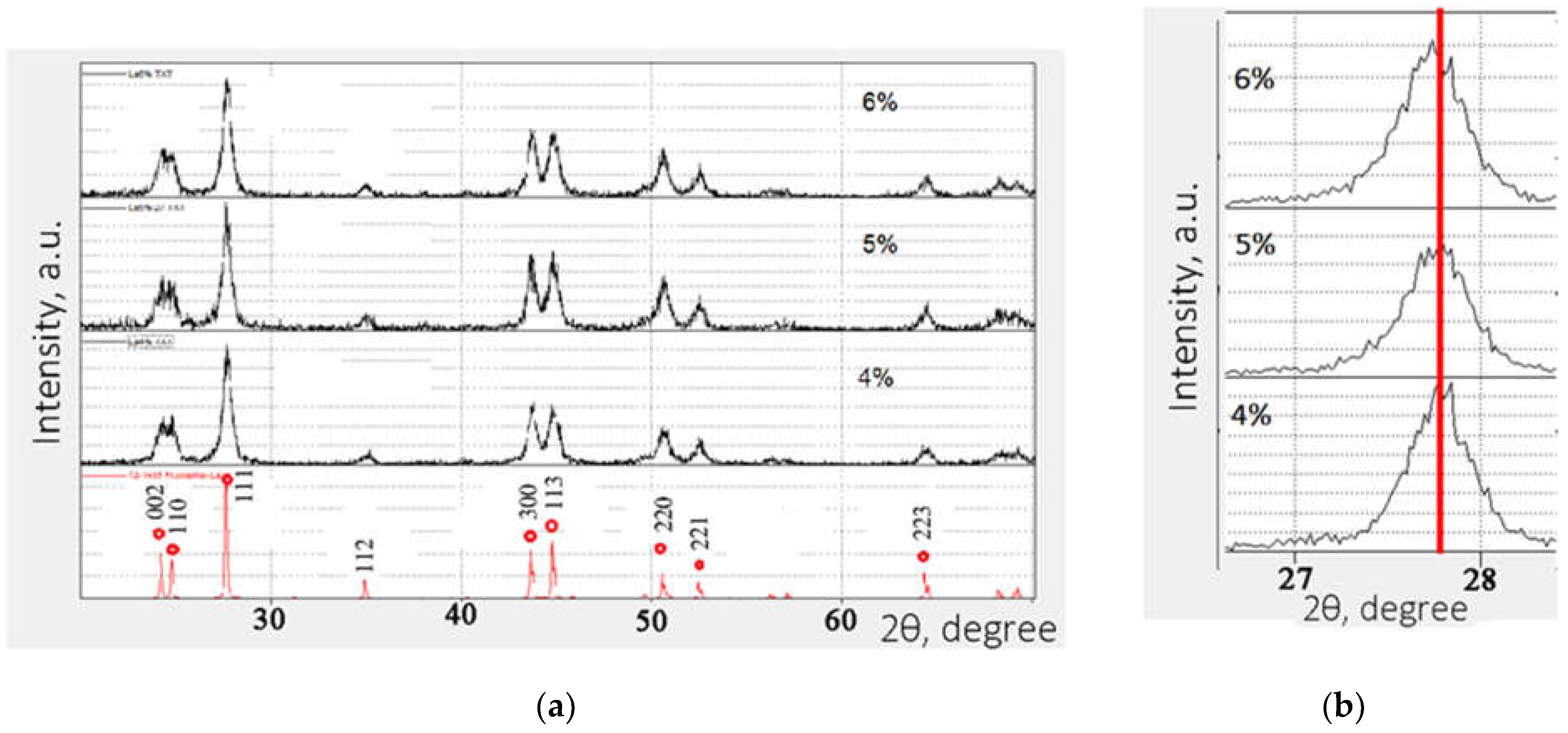
Figure 2.
Photo-luminescent (a) and X-Ray luminescent spectra of LaF3:Ce with different Ce3+ concentration.
Figure 2.
Photo-luminescent (a) and X-Ray luminescent spectra of LaF3:Ce with different Ce3+ concentration.
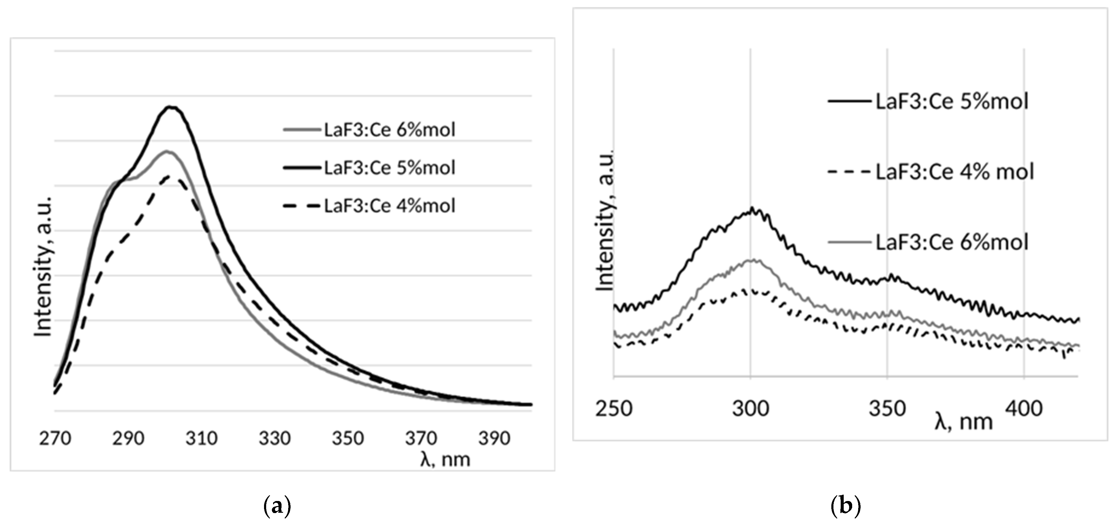
Figure 3.
XRD patterns of LaF3:Ce synthesized in various media.
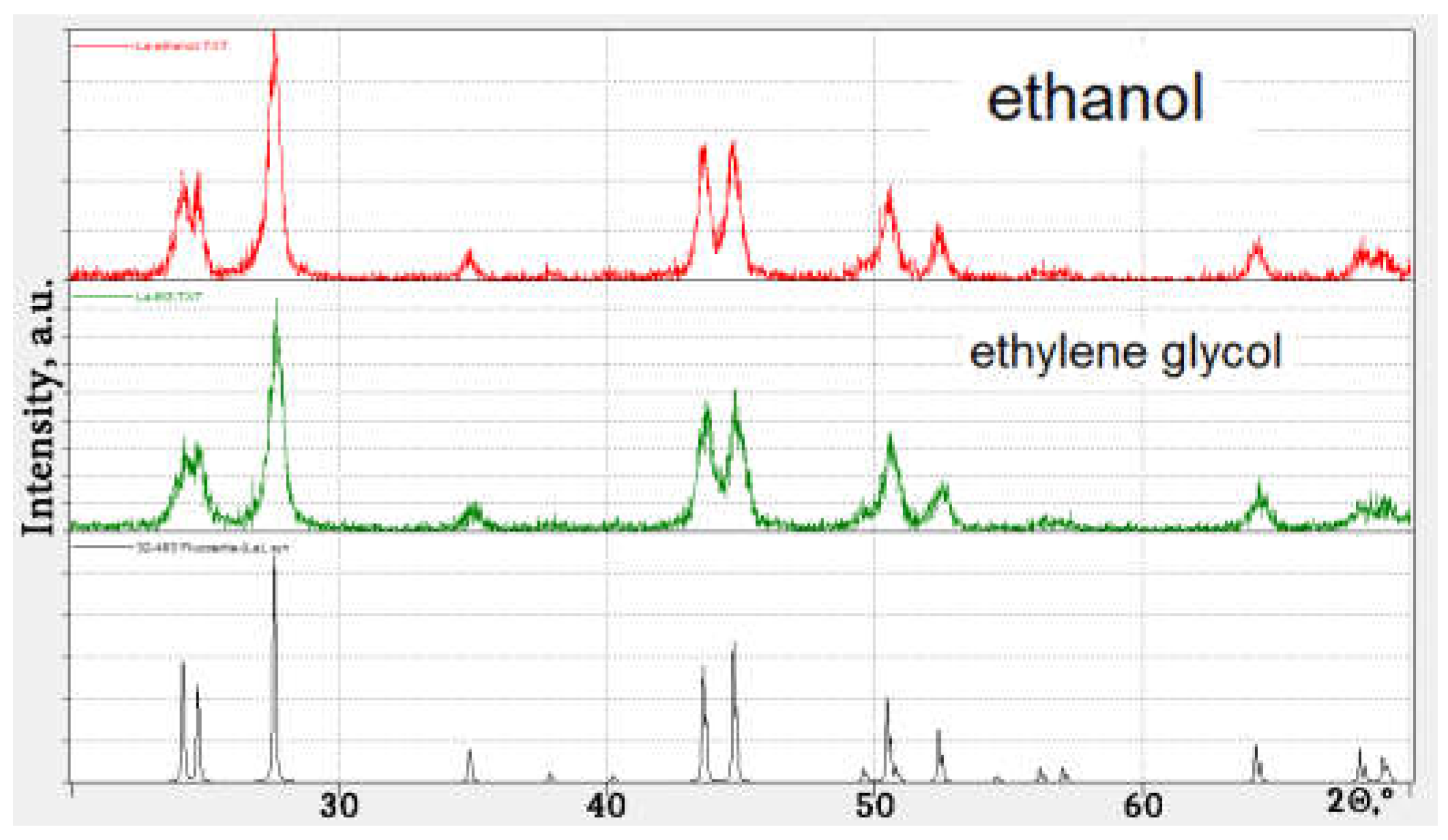
Figure 4.
SEM images of LaF3:Ce 5%mol. Synthesized in Ethanol (a) and EG (b).
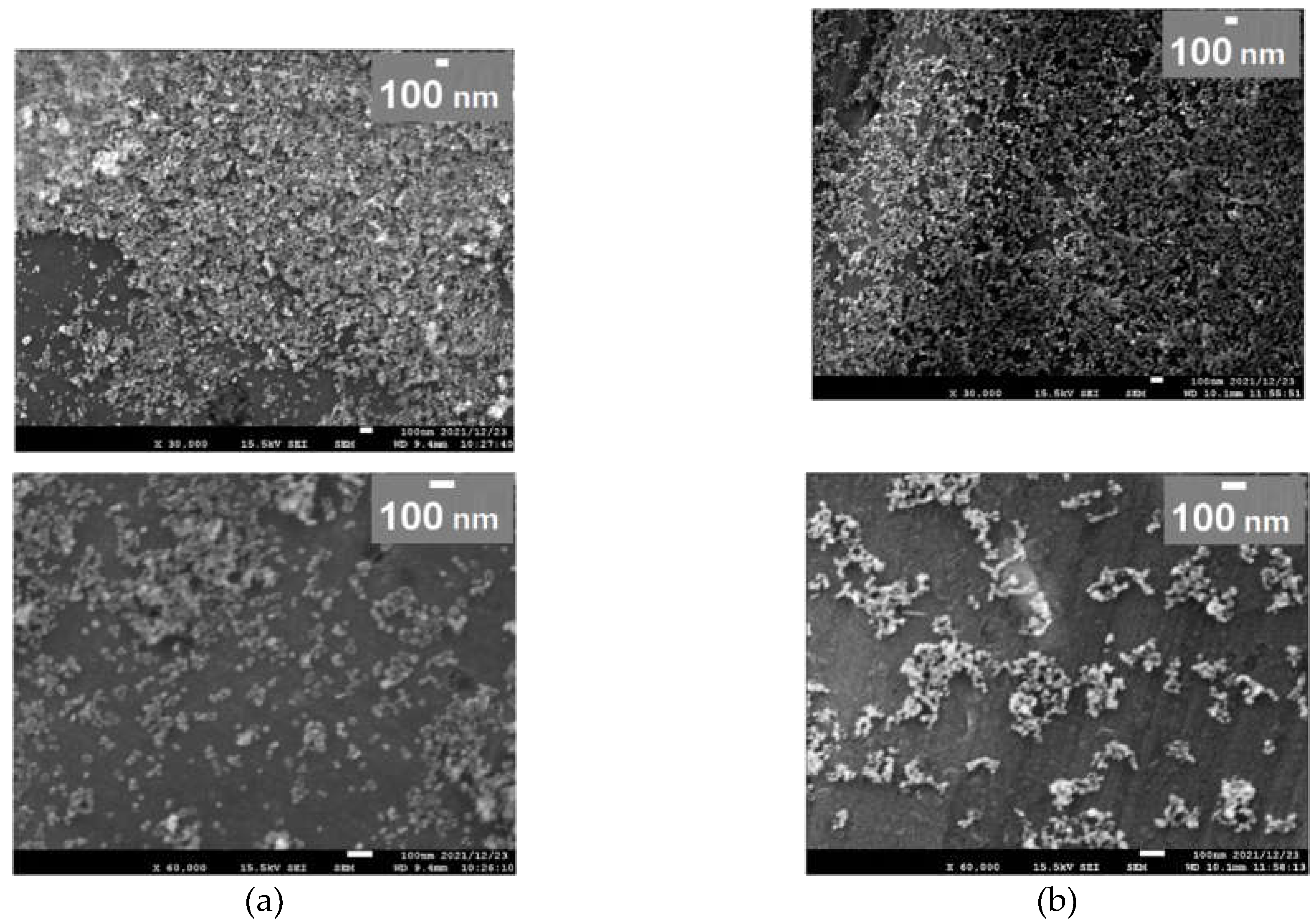
Figure 5.
Size distribution of LaF3:Ce 5%mol. Synthesized in Ethanol (a) and EG (b).
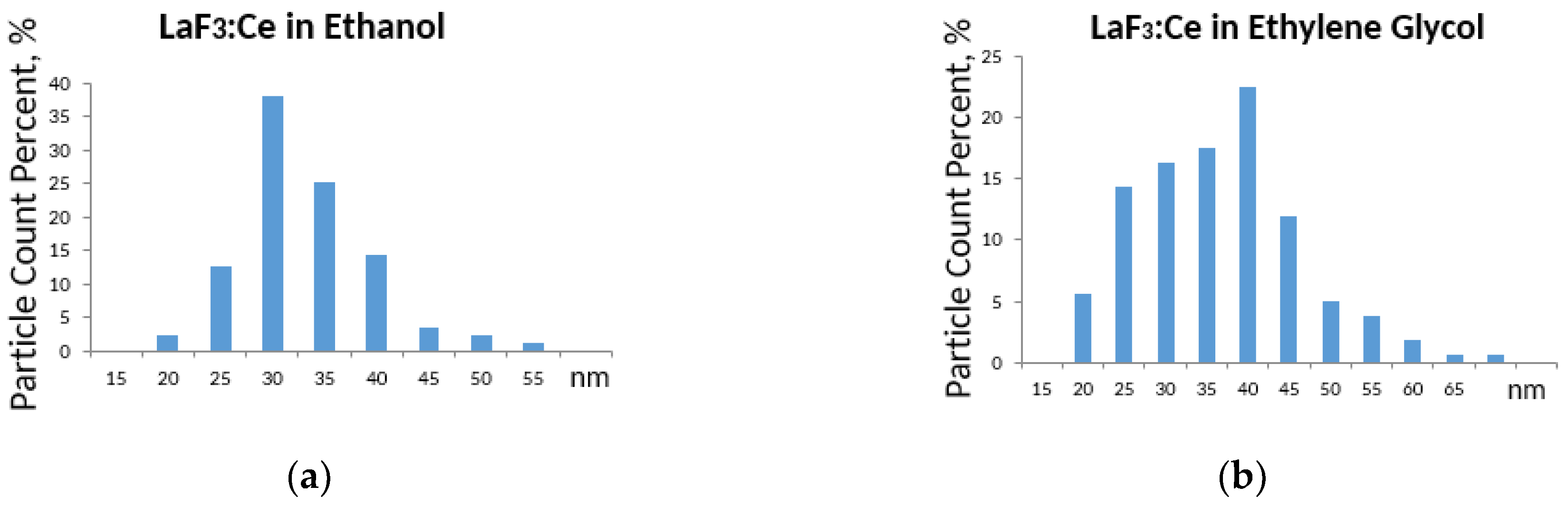
Figure 6.
FT-IR spectra of LaF3:Ce with different stabilizers.
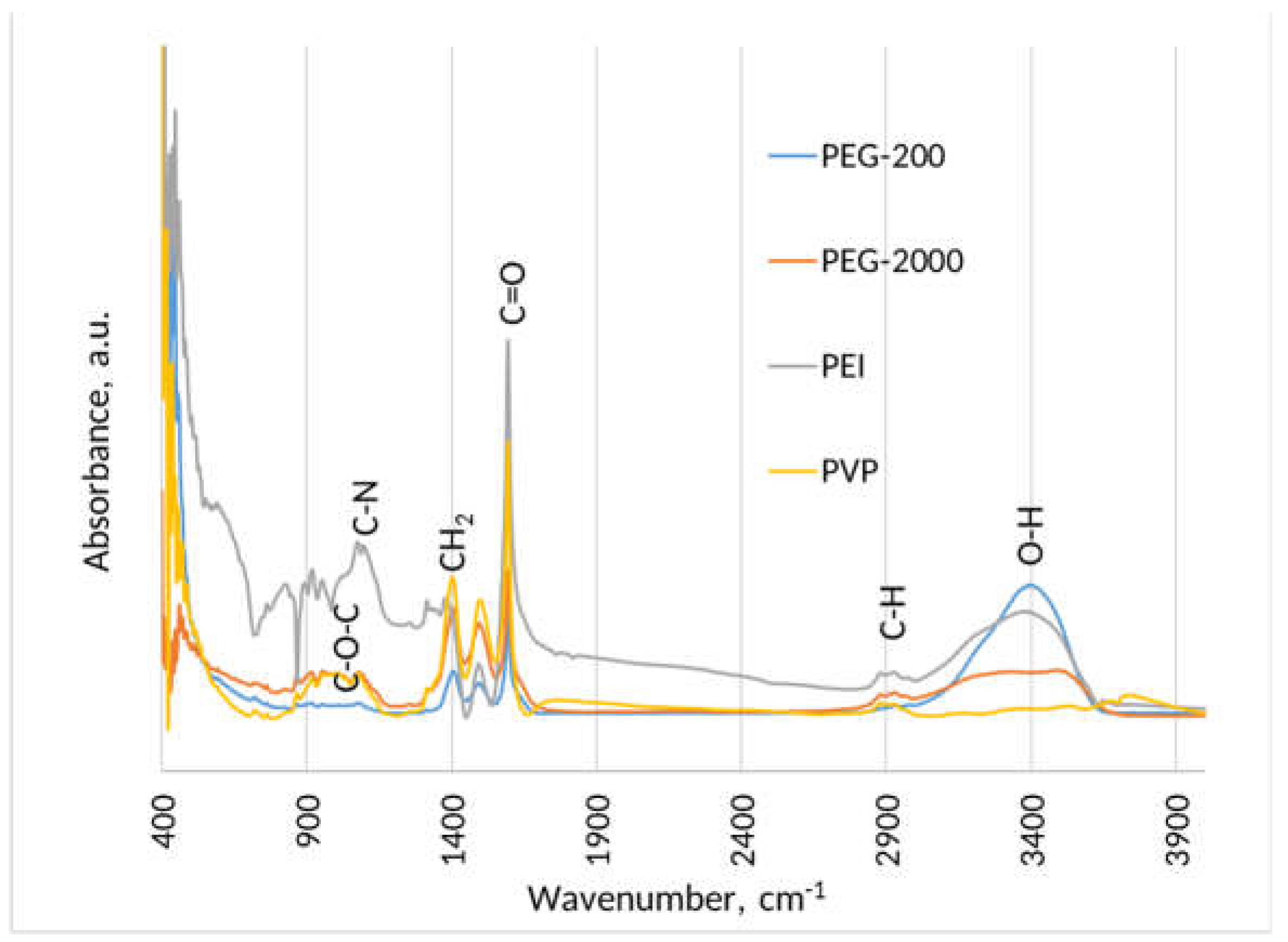
Figure 7.
Zeta-potential of LaF3:Ce 5%mol with different stabilizers.
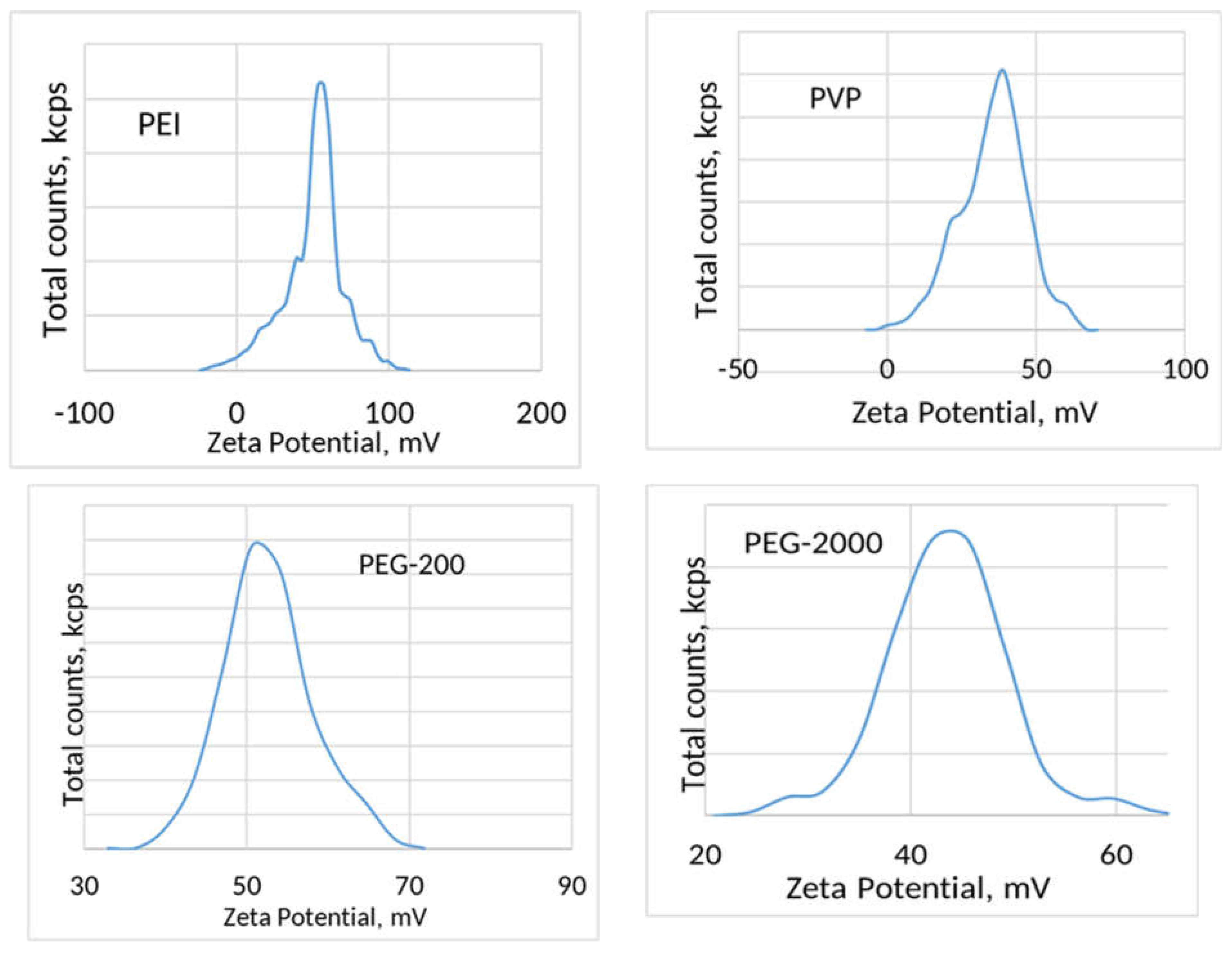
Figure 8.
XRD patterns (a) and photoluminescent spectra of LaF3:Ce with different amount of PVP.
Disclaimer/Publisher’s Note: The statements, opinions and data contained in all publications are solely those of the individual author(s) and contributor(s) and not of MDPI and/or the editor(s). MDPI and/or the editor(s) disclaim responsibility for any injury to people or property resulting from any ideas, methods, instructions or products referred to in the content. |
© 2023 by the authors. Licensee MDPI, Basel, Switzerland. This article is an open access article distributed under the terms and conditions of the Creative Commons Attribution (CC BY) license (http://creativecommons.org/licenses/by/4.0/).
Copyright: This open access article is published under a Creative Commons CC BY 4.0 license, which permit the free download, distribution, and reuse, provided that the author and preprint are cited in any reuse.


