
Submitted:
20 September 2021
Posted:
21 September 2021
You are already at the latest version
Abstract
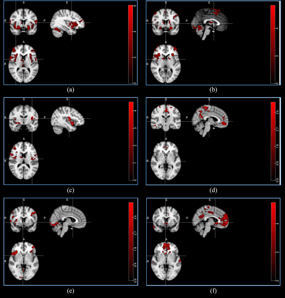
Olfactory system is a vital sensory system in mammals, giving them the ability to connect with their environment. Anosmia, or the complete loss of olfaction ability, which could be caused by injuries, is an interesting topic for inspectors with the aim of diagnosing patients. Sniffing test is currently utilized to examine if an individual is suffering from anosmia; however, functional Magnetic Resonance Imaging (fMRI) provides unique information about the structure and function of the different areas of the human brain, and therefore this noninvasive method could be used as a tool to locate the olfactory-related regions of the brain. In this study, by recruiting 31 healthy and anosmic individuals, we investigated the neural BOLD responses in the olfactory cortices following two odor stimuli, rose and eucalyptus, by using a 3T MR scanner. Comparing the two groups, we observed a network of brain areas being more active in the normal individuals when smelling the odors. In addition, a number of brain areas also showed an activation decline during the odor stimuli, which is hypothesized as a resource allocation deactivation. This study illustrated alterations in the brain activity between the normal individuals and anosmic patients when smelling odors, and could potentially help for a better anosmia diagnosis in the future.
Keywords:
Olfaction
; functional MRI
; Anosmia
Copyright: This open access article is published under a Creative Commons CC BY 4.0 license, which permit the free download, distribution, and reuse, provided that the author and preprint are cited in any reuse.


