
Submitted:
10 October 2019
Posted:
12 October 2019
You are already at the latest version
Abstract
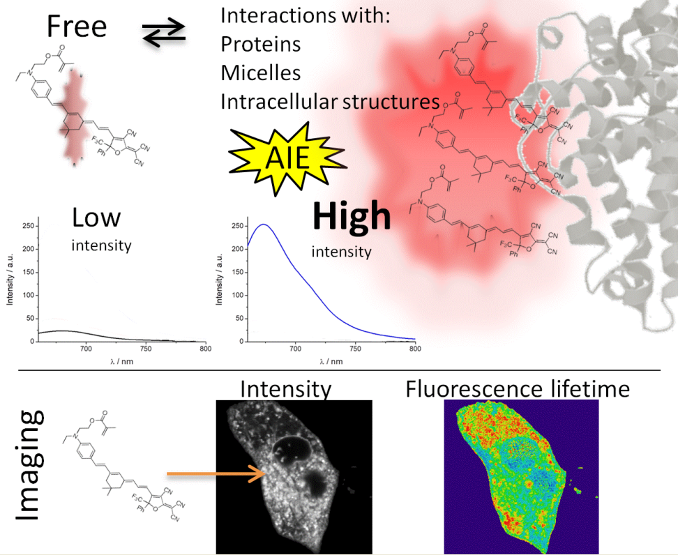
Biological samples are a complex and heterogeneous matrix where different macromolecules with different physicochemical parameters cohabit in reduced spaces. The introduction of fluorophores into these samples, such as in the interior of cells, can produce changes in the fluorescence emission properties of these dyes caused by the specific physicochemical properties of cells. This effect can be especially intense with solvatofluorochromic dyes, where changes in the polarity environment surrounding the dye can drastically change the fluorescence emission. In this article, we studied the photophysical behavior of a new dye and confirmed the aggregation-induced emission (AIE) phenomenon with different approaches, such as by using different solvent proportions, increasing the viscosity, forming micelles and adding bovine serum albumin (BSA), through analysis of the absorption and steady-state and time-resolved fluorescence. Our results show the preferences of the dye for nonpolar media, exhibiting AIE under specific conditions through immobilization. Additionally, this approach offers the possibility of easily determining the critical micelle concentration (CMC). Finally, we studied the rate of spontaneous incorporation of the dye into cells by fluorescence lifetime imaging and observed the intracellular pattern produced by AIE. Interestingly, different intracellular compartments present strong differences in fluorescence intensity and fluorescence lifetime. We used this difference to isolate different intracellular regions to selectively study these regions. Interestingly, the fluorescence lifetime shows a strong difference in different intracellular compartments, facilitating selective isolation for a detailed study of specific organelles.
Keywords:
aggregated enhanced emission
; photophysics
; bioimaging
Copyright: This open access article is published under a Creative Commons CC BY 4.0 license, which permit the free download, distribution, and reuse, provided that the author and preprint are cited in any reuse.


