
Submitted:
02 June 2018
Posted:
05 June 2018
You are already at the latest version
Abstract
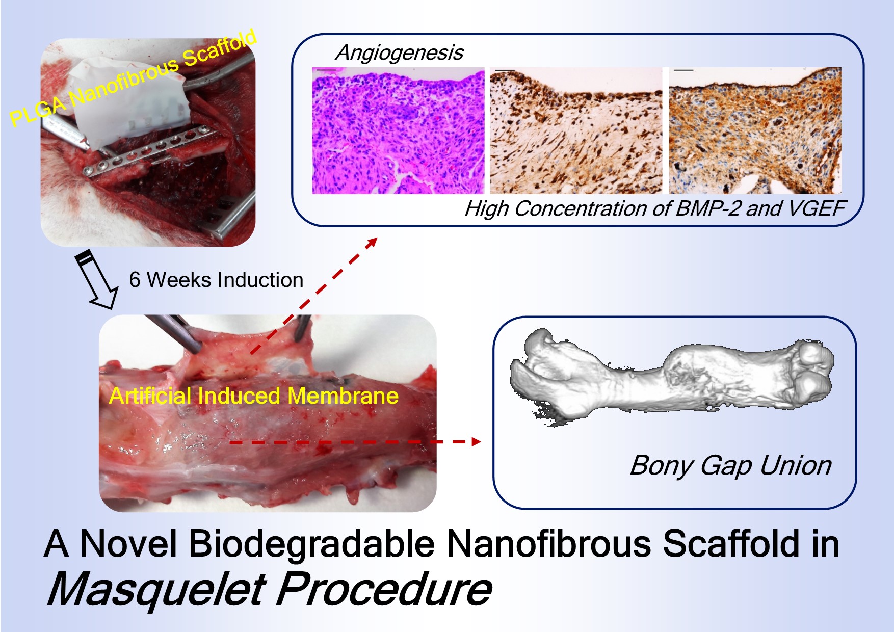
Masquelet induced-membrane technique for the treatment of segmental bone defects includes a two-stage surgical procedure, and polymethylmethacrylate (PMMA) plays a major role in the treatment. However, the PMMA spacer must be surgically removed. Here, we investigated the potential of poly (lactic-co-glycolic acid) (PLGA) nanofibers, a biodegradable material to replace PMMA spacer, allowing the bioactive membrane to be induced, and the spacer to degrade without the additional surgery on a rabbit femoral segmental bone defect model. PLGA nanofibers were shown to degrade completely six weeks after implantation in the investigated animals, and a thick membrane was found to circumferentially fold around the segmental bone defects. Results from image studies demonstrated that, in the group without bone graft, all studied femurs exhibited either nonunion or considerable malunion. In contrast, the femurs in the bone graft group had a high union rate without considerable deformities. Histological examinations suggested that the membranous tissue in this group was rich in small blood vessels and the expression of BMP2 and VEGF increased. Our results demonstrate that the biodegradable PLGA nanofibers may be useful for replacing the PMMA spacer as the bioactive-membrane inducer, facilitating the process of healing and removing the need for repeated surgeries.
Keywords:
biodegradable nanofibers
; PLGA
; masquelet technique
Copyright: This open access article is published under a Creative Commons CC BY 4.0 license, which permit the free download, distribution, and reuse, provided that the author and preprint are cited in any reuse.


